Editorial

Metallodrugs: Mechanisms of Action, Molecular Targets and Biological Activity

by
Giarita Ferraro
* and
Antonello Merlino
*
Department of Chemical Sciences, University of Naples Federico II, Complesso Universitario di Monte Sant’Angelo, Via Cinthia 21, I-80126 Naples, Italy
*
Authors to whom correspondence should be addressed.
Int. J. Mol. Sci. 2022, 23(7), 3504; https://doi.org/10.3390/ijms23073504
Submission received: 3 March 2022 / Accepted: 21 March 2022 / Published: 23 March 2022
The research interest in the field of inorganic medicinal chemistry had a large increase after the serendipitous discovery of the cytotoxic activity of cisplatin by Rosenberg at the end of 1960s [1]. Since then, cisplatin has entered clinical practice and become one of the most common treatments for solid tumors. Unfortunately, the use of cisplatin and its derivatives is associated with undesired side effects, such as general toxicity and intrinsic and acquired drug-resistance [2]. For these reasons, alternative metallodrugs based on non-Pt metals have been synthetized, characterized and tested for biological activity in recent years [3,4,5]. Although, initially, DNA seemed the exclusive target for metallodrugs [6], successive studies have revealed that various metabolites, peptides and proteins have a central role in the recognition, transport and mechanism of action of these compounds [7,8,9]. Thus, a deeper understanding of the molecular bases of the interaction of metallodrugs with these molecules is needed.
The aim of this Special Issue was to collect computational and experimental data on the biological activity of well-established and novel metallodrugs, in addition to information on the interactions of metals and metal-based drugs with metabolites, nucleic acids, peptides and proteins. Herein, the 11 original articles published in this Special Issue are summarized and discussed in the frame of recent advances in the field.
Four manuscripts submitted for this Special Issue report new data on the cytotoxic and potential antitumor actions of Pt(II), Fe(III), Cu(II), Zn(II) and Ir(III) compounds [10,11,12,13].
Annunziata et al. describe the synthesis, characterization and cytotoxic activity of square-planar cationic Pt(II) complexes containing glucoconjugated triazole ligands [10]. The results obtained with the newly synthetized compounds were compared to those found using parent five-coordinate (5C) complexes bearing the same triazole ligands [14]. The square-planar species were more stable and less cytotoxic than the corresponding 5C compounds, but they exhibited a certain selectivity. These results suggest that the stability of Pt compounds is important for preserving their performance as cytotoxic agents, and support the hypothesis that coordinative saturation can be a point in favor of their biological action [15].
In the paper by Marchetti and coworkers, the in vitro anticancer activity of piano stool mononuclear and binuclear Ir(III) complexes based on the pentamethylcyclopentadienyl ligand (Cp*) was evaluated [11]. Various compounds were synthetized, characterized and assessed for their cytotoxicity against six human and rodent cancer cell lines. The results of the study indicated that the 2-phenylpyridyl (PhPy) mononuclear derivatives [Ir(η5-C5Me4H)(kN,kCPhPy)Cl] and [Ir(η5-C5Me4(4-C6H4F))(kN,kCPhPy)Cl] were the most active molecules among those under investigation. They showed good selectivity, were inactive towards healthy cells, and had a triple effect on cancer cells. Their cytotoxic activity was reached through proliferation inhibition, apoptosis activation and senescence induction. This latter effect is significant to note, since it is rarely observed for organoiridium complexes.
Verreault and coworkers investigated the cytotoxic activity of six iron-based ferrocifens against 15 glioblastoma patient-derived cell lines. The authors found that the studied molecules showed an IC50 value ranging from nanomolar to micromolar [12]. The results provide new information on the mechanism of action of ferrocifens, and indicate that differences in the chemical structures of these molecules significantly alter their behavior against brain cancer.
Klein and coworkers extensively characterized three complexes of Fe(III), Cu(II) and Zn(II) bearing the redox active, non-innocent ligand opo (opo = 9-oxido-phenalenone) by 1H nuclear magnetic resonance, electron paramagnetic resonance, UV-vis absorption spectroscopy and electrochemical analysis [13]. They assessed their stability in different organic solvents and tested their cytotoxic activity. They found that the compounds showed antiproliferative activity against colon and breast cancer cell lines in the micromolar range [13].
The work by Szefler et al. presents a computational study on the affinity of carboplatin to B vitamins, aiming to establish if the vitamins—as components of beet and carrot juice—could possibly lead to a reduction in the efficacy of this drug. Their data indicated that carboplatin can bind vitamins B3 and B6, and their computational prediction was also verified by experimental data [16].
Hemphill and coworkers investigated the potential cellular and molecular targets of a trithiolato-bridged arene ruthenium complex conjugated to 9-(2-hydroxyethyl)-adenine [17], which inhibits the protozoan parasites Toxoplasma gondii and Trypanosoma brucei. Transmission electron microscopy (TEM) images revealed structural alterations of parasite mitochondria after a few hours’ exposure to the drug. Furthermore, Ru complex molecular targets were analyzed by differential affinity chromatography coupled to shotgun–mass spectrometry, and a mitochondrial ATP-synthase subunit was identified as the main Ru complex target. Altogether, these results demonstrated that the trithiolato-bridged arene ruthenium complex can interfere with key steps of cellular metabolism.
The use of dinuclear trithiolato-bridged arene ruthenium complexes as drugs in vivo is limited by their scarce solubility in water. An attempt to overcome this limitation and increase their selectivity for cancer cells was carried out by encapsulating the trithiolato-bridged arene ruthenium complex Diruthenium-1 within a horse spleen apo-ferritin (hsAFt) nanocage [18]. hsAFt was also used to encapsulate Au, Pt, Ru and heterobimetallic compounds [19].
The same approach was also used to encapsulate arsenoplatin-1 (AP-1) within hsAFt [20]. AP-1 is a dual-action anticancer metallodrug with a promising pharmacological profile that features the simultaneous presence of a cisplatin-like center and an arsenite center [21,22]. The adduct formed upon the encapsulation of AP-1 within hsAFt was structurally characterized (Figure 1A): AP-1 binds the side chain of His49 upon the release of the chloride ligand (Figure 1B). The adduct was less toxic than the free drug, but more selective, since the concentration needed to kill cancer cells is half that needed to kill immortalized cells [20].
Structural data have been also collected on adducts formed upon the reaction of a model protein hen egg white lysozyme with the paddlewheel dirhodium tetraacetate complex (Rh2(act)4, act = acetate) under different experimental conditions [23]. This compound exhibited in vivo anticancer activity against Ehrlich ascites, L1210 tumors, sarcoma 180 and P388 leukemia [24,25,26]. The structures revealed that Rh2(act)4 degrades under the investigated conditions, at a variance with that observed upon the reaction of the same compounds with other proteins [27]. Dimeric Rh-Rh units and monomeric fragments mainly bind the protein close to His, Asp and Lys side chains. The work, which describes rare examples of structures of Rh/protein adducts [28], demonstrates that Ru2(act)4 reacts with lysozyme differently from the analogous Ru2(act)4 complex [29] and from RhCl3 [30].
Moreover, the interaction of metal ions and metal complexes with peptides is also distinctly important. Three papers in the Special Issue report studies based on metals or metal compounds and peptide recognition processes.
D. Marasco and coworkers studied the possible use of three Pt(II) compounds (bearing 1, 10-phenathroline, 2, 2′:6′, 2″-terpyridine (terpy) and 2, 2′-bipyridine) as inhibitors of the aggregation of the β-amyloid (Aβ) peptide Aβ21–40 [31]. The misfolding of Aβ, its aggregation, the formation of fibrils and deposition in the brain all play major roles in the development of Alzheimer’s disease [32]. The investigated compounds inhibited Aβ21–40 aggregation and modulated the peptide disaggregation via the formation of adducts where the metal center loses chloride ligands. The compound with terpy induced the formation of soluble monomeric forms of Aβ21–40 and reduced the toxicity of the peptide in human SH-SY5Y neuroblastoma cells. These results pave the way for the future application of metal-based drugs in the treatment of neurodegenerative diseases.
La Mendola and coworkers studied the interaction between copper and different peptides [33,34], including: a peptide derived from the N-terminal domain of Angiogenin (Ang) [33]; a ribonuclease essential for angiogenesis stimulation [35]; and peptides belonging to the N-terminal domain of nerve growth (NGF) [34], a member of the neurotrophin family that is essential for neuron survival [36]. They demonstrated that sequence 1–17 of Ang is involved in copper uptake and that the acetylation of the terminal amino group of the peptide decreases the intracellular metal level [33]. Since several diseases are characterized by an upregulated Ang expression together with an altered copper metabolism, these alterations can be used as potential targets for the design and development of specific therapies [37,38].
Furthermore, the stability constants of copper complex species formed with the dimeric forms of the sequence 1–15 of NGF have been studied [34]. At a physiological pH, NGF peptides bind copper ion with a higher affinity than N-terminal peptides of Aβ, and a lower affinity than that observed for peptides derived from human copper transporter 1 (hCtr1), the protein responsible for copper uptake in neurons. This interaction suggests NGF as a possible regulator of copper homeostasis in the synaptic space, which has a key role in the process of memory formation.
Altogether, these new studies show once again the importance of metals in regulating the activity and function of peptides, and the importance of studying metal compounds endowed with unique biological properties. The data suggest that the investigations on the design and development of new metal-based drugs will continue, since new therapeutic applications of metal complexes can be explored. We hope that, in the near future, we can gain a better understanding of these mechanisms of action, as well as of the targeting and activation strategies of these compounds; a deeper knowledge of these aspects will lead to future generations of metallodrugs with reduced side effects and a wider spectrum of activity.

Funding

This research received no external funding.

Conflicts of Interest

The authors declare no conflict of interest.

References

  1. Rosenberg, B. Chapter 2—Cisplatin: Its history and possible mechanisms of action. In Cisplatin: Current Status and New Developments; Prestayko, A.W., Crooke, S.T., Carter, S.K., Eds.; Elsevier: Amsterdam, The Netherlands, 1980; pp. 9–20. [Google Scholar]
  2. Florea, A.-M.; Büsselberg, D. Cisplatin as an anti-tumor drug: Cellular mechanisms of activity, drug resistance and induced side effects. Cancers 2011, 3, 1351–1371. [Google Scholar] [CrossRef] [PubMed]
  3. Komeda, S.; Casini, A. Next-generation anticancer metallodrugs. Curr. Top. Med. Chem. 2012, 12, 219–235. [Google Scholar] [CrossRef] [PubMed]
  4. Yeo, C.I.; Ooi, K.K.; Tiekink, E.R.T. Gold-based medicine: A paradigm shift in anti-cancer therapy? Molecules 2018, 23, 1410. [Google Scholar] [CrossRef] [PubMed] [Green Version]
  5. Thota, S.; Rodrigues, D.A.; Crans, D.C.; Barreiro, E.J. Ru(II) compounds: Next-generation anticancer metallotherapeutics? J. Med. Chem. 2018, 61, 5805–5821. [Google Scholar] [CrossRef]
  6. Jamieson, E.R.; Lippard, S.J. Structure, recognition, and processing of cisplatin−DNA adducts. Chem. Rev. 1999, 99, 2467–2498. [Google Scholar] [CrossRef]
  7. Xiong, X.; Liu, L.-Y.; Mao, Z.-W.; Zou, T. Approaches towards understanding the mechanism-of-action of metallodrugs. Coord. Chem. Rev. 2022, 453, 214311. [Google Scholar] [CrossRef]
  8. Merlino, A. Recent advances in protein metalation:structural studies. Chem. Commun. 2021, 57, 1295–1307. [Google Scholar] [CrossRef]
  9. Marloye, M.; Berger, G.; Gelbcke, M.; Dufrasne, F. A survey of the mechanisms of action of anticancer transition metal complexes. Future Med. Chem. 2016, 18, 2263–2286. [Google Scholar] [CrossRef]
  10. Annunziata, A.; Liberti, D.; Bedini, E.; Cucciolito, M.E.; Loreto, D.; Monti, D.M.; Merlino, A.; Ruffo, F. Square-planar vs. trigonal bipyramidal geometry in Pt(II) complexes containing triazole-based glucose ligands as potential anticancer agents. Int. J. Mol. Sci. 2021, 22, 8704. [Google Scholar] [CrossRef]
  11. De Palo, A.; Draca, D.; Murrali, M.G.; Zacchini, S.; Pampaloni, G.; Mijatovic, S.; Maksimovic-Ivanic, D.; Marchetti, F. A comparative analysis of the in vitro anticancer activity of iridium(III) {η5-C5Me4R} complexes with variable R groups. Int. J. Mol. Sci. 2021, 22, 7422. [Google Scholar] [CrossRef]
  12. Vessières, A.; Quissac, E.; Lemaire, N.; Alentorn, A.; Domeracka, P.; Pigeon, P.; Sanson, M.; Idbaih, A.; Verreault, M. Heterogeneity of response to iron-based metallodrugs in glioblastoma is associated with differences in chemical structures and driven by fas expression dynamics and transcriptomic subtypes. Int. J. Mol. Sci. 2021, 22, 10404. [Google Scholar] [CrossRef] [PubMed]
  13. Butsch, K.; Haseloer, A.; Schmitz, S.; Ott, I.; Schur, J.; Klein, A. FeIII, CuII and ZnII complexes of the rigid 9-oxido-phenalenone ligand—Spectroscopy, electrochemistry, and cytotoxic properties. Int. J. Mol. Sci. 2021, 22, 3976. [Google Scholar] [CrossRef] [PubMed]
  14. Annunziata, A.; Cucciolito, M.E.; Esposito, R.; Imbimbo, P.; Petruk, G.; Ferraro, G.; Pinto, V.; Tuzi, A.; Monti, D.M.; Merlino, A.; et al. A highly efficient and selective antitumor agent based on a glucoconjugated carbene platinum(II) complex. Dalton Trans. 2019, 48, 7794–7800. [Google Scholar] [CrossRef] [PubMed]
  15. Annunziata, A.; Cucciolito, M.E.; Esposito, R.; Ferraro, G.; Monti, D.M.; Merlino, A.; Ruffo, F. Five-coordinate Pt(II) compounds as potential anticancer agents. Eur. J. Inorg. Chem. 2020, 11, 918–929. [Google Scholar] [CrossRef]
  16. Szefler, B.; Czeleń, P.; Krawczyk, P. The affinity of carboplatin to b-vitamins and nucleobases. Int. J. Mol. Sci. 2021, 22, 3634. [Google Scholar] [CrossRef]
  17. Anghel, N.; Müller, J.; Serricchio, M.; Jelk, J.; Bütikofer, P.; Boubaker, G.; Imhof, D.; Ramseier, J.; Desiatkina, O.; Păunescu, E.; et al. Cellular and molecular targets of nucleotide-tagged trithiolato-bridged arene ruthenium complexes in the protozoan parasites toxoplasma gondii and trypanosoma brucei. Int. J. Mol. Sci. 2021, 22, 10787. [Google Scholar] [CrossRef]
  18. Petruk, G.; Monti, D.M.; Ferraro, G.; Pica, A.; D’Elia, L.; Pane, F.; Amoresano, A.; Furrer, J.; Kowalski, K.; Merlino, A. Encapsulation of the dinuclear trithiolato-bridged arene ruthenium complex diruthenium-1 in an apoferritin nanocage: Structure and cytotoxicity. Chem. Med. Chem. 2019, 14, 594–602. [Google Scholar] [CrossRef]
  19. Monti, D.M.; Ferraro, G.; Merlino, A. Ferritin-based anticancer metallodrug delivery: Crystallographic, analytical and cytotoxicity studies. Nanomed. Nanotechnol. Biol. Med. 2019, 20, 101997. [Google Scholar] [CrossRef]
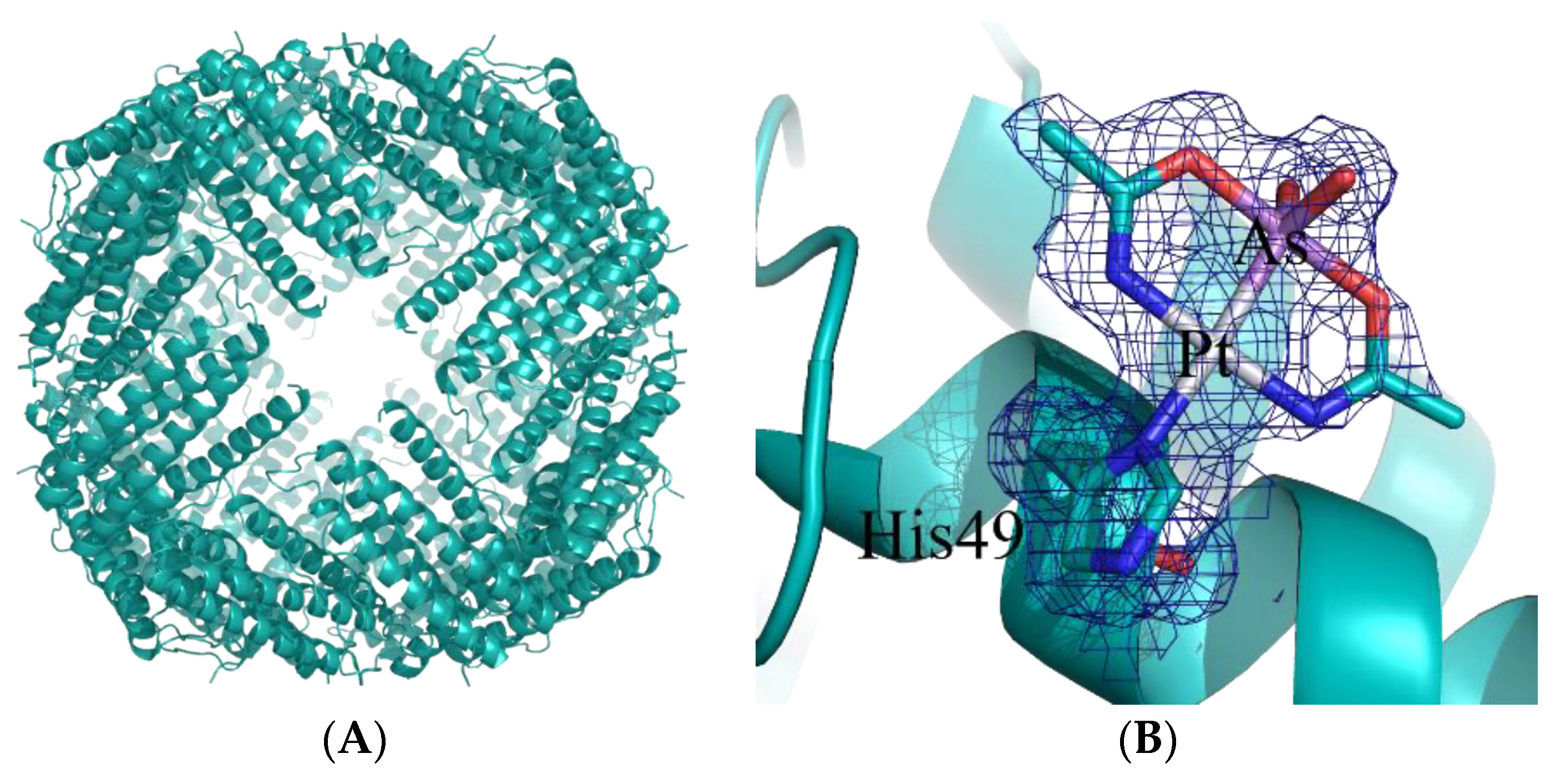
  20. Ferraro, G.; Pratesi, A.; Cirri, D.; Imbimbo, P.; Monti, D.M.; Messori, L.; Merlino, A. Arsenoplatin-ferritin nanocage: Structure and cytotoxicity. Int. J. Mol. Sci. 2021, 22, 1874. [Google Scholar] [CrossRef]
  21. Miodragović, Đ.U.; Quentzel, J.A.; Kurutz, J.W.; Stern, C.L.; Ahn, R.W.; Kandela, I.; Mazar, A.; O’Halloran, T.V. Robust structure and reactivity of aqueous arsenous acid–platinum (II) anticancer complexes. Angew. Chem. Int. Ed. 2013, 52, 10749–10752. [Google Scholar] [CrossRef] [Green Version]
  22. Miodragović, Đ.; Merlino, A.; Swindell, E.P.; Bogachkov, A.; Ahn, R.W.; Abuhadba, S.; Ferraro, G.; Marzo, T.; Mazar, A.P.; Messori, L.; et al. Arsenoplatin-1 is a dual pharmacophore anticancer agent. J. Am. Chem. Soc. 2019, 141, 6453–6457. [Google Scholar] [CrossRef] [PubMed]
  23. Loreto, D.; Ferraro, G.; Merlino, A. Unusual structural features in the adduct of dirhodium tetraacetate with lysozyme. Int. J. Mol. Sci. 2021, 22, 1496. [Google Scholar] [CrossRef] [PubMed]
  24. Chang, I.; Woo, W.S. Effects of Rh2(O2CC2H5)4L2 on the replication of ehrlich tumor cells in vivo. Korean Biochem. J. 1976, 9, 175–180. [Google Scholar]
  25. Bear, J.L. Rhodium compounds for antitumor use. In Proceedings of 9th International Conference of the International Precious Metals Institute, New York, NY, USA; Zysk, E.D., Bonucci, J.A., Eds.; International Precious Metals Institute: Allentown, PA, USA, 1986; pp. 337–344. [Google Scholar]
  26. Erck, A.; Rainen, L.; Whileyman, J.; Chang, I.M.; Kimball, A.P.; Bear, J.L. Studies of rhodium(II) carboxylates as potential antitumor agents. Proc. Soc. Exp. Biol. Med. 1974, 145, 1278–1283. [Google Scholar] [CrossRef] [PubMed]
  27. Ferraro, G.G.; Pratesi, A.; Messori, L.; Merlino, A. Protein interactions of dirhodium tetraacetate:a structural study. Dalton Trans. 2020, 49, 2412–2416. [Google Scholar] [CrossRef]
  28. Loreto, D.; Merlino, A. The interaction of rhodium compounds with proteins: A structural overview. Coord. Chem. Rev. 2021, 442, 213999. [Google Scholar] [CrossRef]
  29. Messori, L.; Marzo, T.; Fernandes Sanches, R.N.; Rehman, H.-U.; de Oliveira Silva, D.; Merlino, A. Unusual structural features in the lysozyme derivative of tetrakis(acetato)chlorido diruthenium(II,III) complex. Angew. Chem. Int. Ed. 2014, 53, 6172–6175. [Google Scholar] [CrossRef] [PubMed]
  30. Ueno, T.; Abe, S.; Koshiyama, T.; Ohki, T.; Hikage, T.; Watanabe, Y. Elucidation of metal-ion accumulation induced by hydrogen bonds on protein surfaces by using porous lysozyme crystals containing RhIII ions as the model surfaces. Chem. Eur. J. 2010, 16, 2730–2740. [Google Scholar] [CrossRef]
  31. Manna, S.L.; Florio, D.; Iacobucci, I.; Napolitano, F.; Benedictis, I.D.; Malfitano, A.M.; Monti, M.; Ravera, M.; Gabano, E.; Marasco, D. A comparative study of the effects of platinum (II) complexes on β-amyloid aggregation: Potential neurodrug applications. Int. J. Mol. Sci. 2021, 22, 3015. [Google Scholar] [CrossRef]
  32. Shoghi-Jadid, K.; Small, G.W.; Agdeppa, E.D.; Kepe, V.; Ercoli, L.M.; Siddarth, P.; Read, S.; Satyamurthy, N.; Petric, A.; Huang, S.-C. Localization of neurofibrillary tangles and beta-amyloid plaques in the brains of living patients with alzheimer disease. Am. J. Geriatr. Psychiatry 2002, 10, 24–35. [Google Scholar] [CrossRef]
  33. Tabbì, G.; Cucci, L.M.; Pinzino, C.; Munzone, A.; Marzo, T.; Pizzanelli, S.; Satriano, C.; Magrì, A.; La Mendola, D. Peptides Derived from angiogenin regulate cellular copper uptake. Int. J. Mol. Sci. 2021, 22, 9530. [Google Scholar] [CrossRef] [PubMed]
  34. Magrì, A.; La Mendola, D.; Rizzarelli, E. Nerve growth factor peptides bind copper(II) with high affinity: A thermodynamic approach to unveil overlooked neurotrophin roles. Int. J. Mol. Sci. 2021, 22, 5085. [Google Scholar] [CrossRef] [PubMed]
  35. Sheng, J.; Xu, Z. Three decades of research on angiogenin: A review and perspective. Acta Biochim. Biophys. Sin. 2016, 48, 399–410. [Google Scholar] [CrossRef] [PubMed] [Green Version]
  36. Sofroniew, M.V.; Howe, C.L.; Mobley, W.C. Nerve growth factor signaling, neuroprotection, and neural repair. Annu. Rev. Neurosci. 2001, 24, 1217–1281. [Google Scholar] [CrossRef]
  37. Marzo, T.; La Mendola, D. The effects on angiogenesis of relevant inorganic chemotherapeutics. Curr. Top. Med. Chem. 2021, 21, 73–86. [Google Scholar] [CrossRef]
  38. Cucci, L.M.; Satriano, C.; Marzo, T.; La Mendola, D. Angiogenin and copper crossing in wound healing. Int. J. Mol. Sci. 2021, 22, 10704. [Google Scholar] [CrossRef]
Figure 1. Cartoon representation of the overall structure of the hsAFt nanocage (A) and details of AP-1 binding site (B). An AP-1 moiety binds to the ND1 atom of His49. In panel (B), the 2Fo-Fc electron density map is colored in blue and contoured at 0.5σ.
Ijms 23 03504 g001
Publisher’s Note: MDPI stays neutral with regard to jurisdictional claims in published maps and institutional affiliations.

