
Use of Photodynamic Therapy Associated with Antimicrobial Peptides for Bacterial Control: A Systematic Review and Meta-Analysis

 , and
, and
Abstract
:1. Introduction
2. Results
2.1. Search Results
2.2. Synthesis of Results
2.3. Risk of Bias Assessments for In Vitro Studies
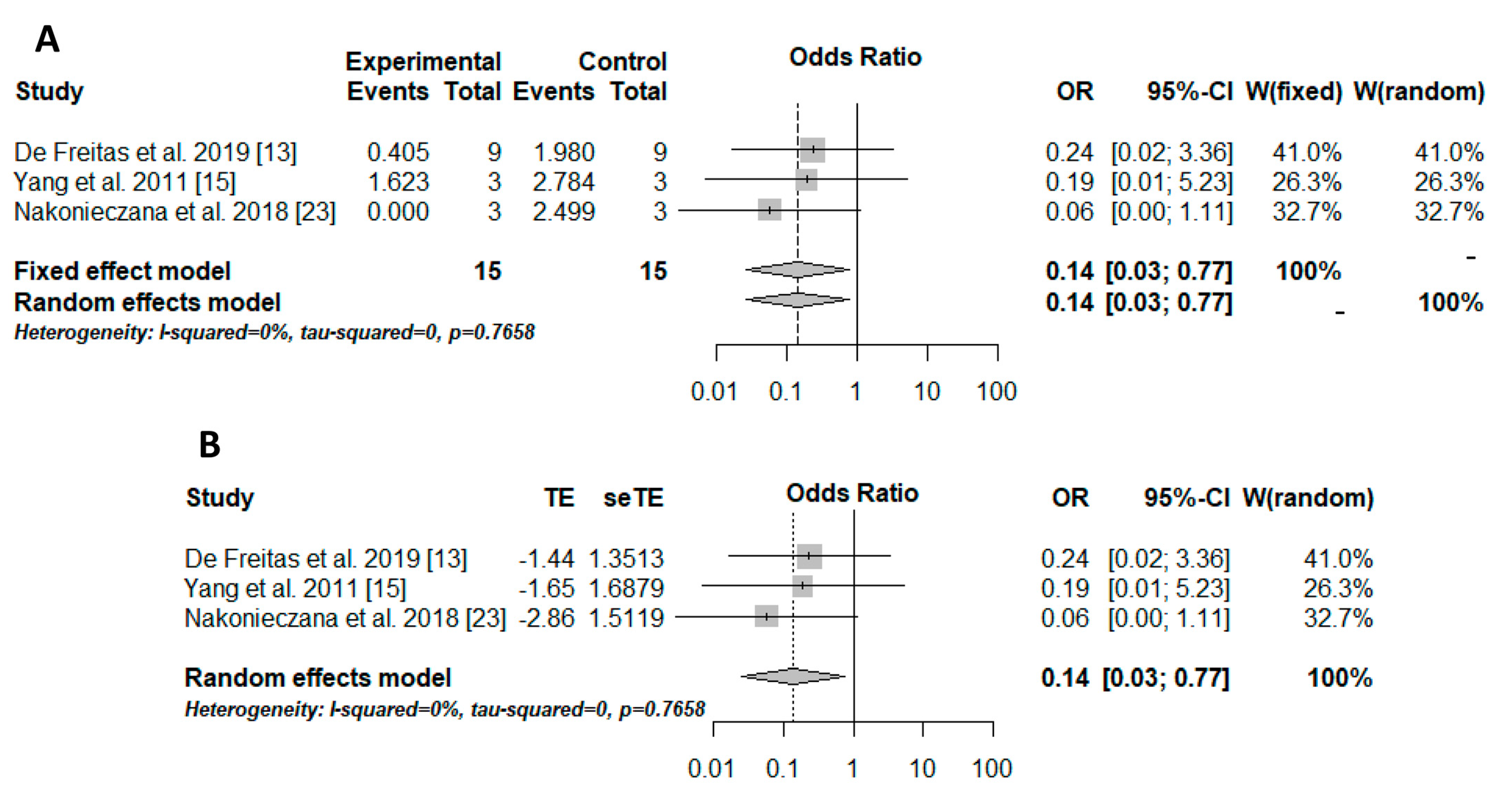
2.4. Meta-Analysis
3. Discussion
4. Materials and Methods
4.1. Protocol and Registration
4.2. Data Extraction and Research Question
4.3. Eligibility Criteria
4.4. Search Strategy
4.5. Meta-Analysis and Quantitative Approaches
5. Conclusions
Author Contributions
Funding
Conflicts of Interest
References
- Christaki, E.; Marcou, M.; Tofarides, A. Antimicrobial Resistance in Bacteria: Mechanisms, Evolution, and Persistence. J. Mol. Evol. 2020, 88, 26–40. [Google Scholar] [CrossRef] [PubMed]
- Watkins, R.R.; Bonomo, R.A. Overview: Global and Local Impact of Antibiotic Resistance. Infect. Dis. Clin. N. Am. 2016, 30, 313–322. [Google Scholar] [CrossRef] [PubMed]
- Garcia, B.A.; Panariello, B.H.D.; de Freitas-Pontes, K.M.; Duarte, S. Candida biofilm matrix as a resistance mechanism against photodynamic therapy. Photodiagnosis Photodyn. Ther. 2021, 36, 102525. [Google Scholar] [CrossRef]
- Dias, L.M.; Klein, M.I.; Jordão, C.C.; Carmello, J.C.; Bellini, A.; Pavarina, A.C. Successive applications of Antimicrobial Photodynamic Therapy effects the susceptibility of Candida albicans grown in medium with or without fluconazole. Photodiagnosis Photodyn. Ther. 2020, 32, 102018. [Google Scholar] [CrossRef]
- Janeth Rimachi Hidalgo, K.; Carmello, J.C.; Carolina Jordão, C.; Aboud Barbugli, P.; de Sousa Costa, C.A.; Mima, E.; Pavarina, A.C. Antimicrobial Photodynamic Therapy in Combination with Nystatin in the Treatment of Experimental Oral Candidiasis In-duced by Candida albicans Resistant to Fluconazole. Pharmaceuticals 2019, 12, 140. [Google Scholar] [CrossRef] [Green Version]
- Gilaberte, Y.; Rezusta, A.; Juarranz, A.; Hamblin, M.R. Editorial: Antimicrobial Photodynamic Therapy: A New Paradigm in the Fight against Infections. Front. Med. 2021, 8, 788888. [Google Scholar] [CrossRef] [PubMed]
- Dai, T.; Huang, Y.-Y.; Hamblin, M.R. Photodynamic therapy for localized infections—State of the art. Photodiagnosis Photodyn. Ther. 2009, 6, 170–188. [Google Scholar] [CrossRef] [PubMed] [Green Version]
- Giuliani, F.; Martinelli, M.; Cocchi, A.; Arbia, D.; Fantetti, L.; Roncucci, G. In Vitro Resistance Selection Studies of RLP068/Cl, a New Zn(II) Phthalocyanine Suitable for Antimicrobial Photodynamic Therapy. Antimicrob. Agents Chemother. 2010, 54, 637–642. [Google Scholar] [CrossRef] [PubMed] [Green Version]
- Panariello, B.; Klein, M.I.; Alves, F.; Pavarina, A.C. DNase increases the efficacy of antimicrobial photodynamic therapy on Candida albicans biofilms. Photodiagnosis Photodyn. Ther. 2019, 27, 124–131. [Google Scholar] [CrossRef]
- Aida, K.L.; Kreling, P.F.; Caiaffa, K.S.; Calixto, G.M.F.; Chorilli, M.; Spolidorio, D.M.; Santos-Filho, N.A.; Cilli, E.M.; Duque, C. Antimicrobial peptide-loaded liquid crystalline precursor bioadhesive system for the prevention of dental caries. Int. J. Nanomed. 2018, 13, 3081–3091. [Google Scholar] [CrossRef] [Green Version]
- Caiaffa, K.S.; Dos Santos, V.R.; Abuna, G.F.; Santos-Filho, N.A.; Cilli, E.M.; Sakai, V.T.; Cintra, L.; Duque, C. Cytocom-patibility and Synergy of EGCG and Cationic Peptides Against Bacteria Related to Endodontic Infections, in Planktonic and Biofilm Conditions. Probiotics Antimicrob. Proteins 2021, 13, 1808–1819. [Google Scholar] [CrossRef]
- De Freitas, L.M.; Lorenzon, E.; Santos-Filho, N.; Zago, L.H.D.P.; Uliana, M.P.; de Oliveira, K.T.; Cilli, E.M.; Fontana, C.R. Antimicrobial Photodynamic therapy enhanced by the peptide aurein 1.2. Sci. Rep. 2018, 8, 4212. [Google Scholar] [CrossRef] [PubMed] [Green Version]
- De Freitas, L.M.; Lorenzón, E.N.; Cilli, E.M.; de Oliveira, K.T.; Fontana, C.R.; Mang, T.S. Photodynamic and pep-tide-based strategy to inhibit Gram-positive bacterial biofilm formation. Biofouling 2019, 35, 742–757. [Google Scholar] [CrossRef]
- Bourré, L.; Giuntini, F.; Eggleston, I.M.; Mosse, C.A.; MacRobert, A.J.; Wilson, M. Effective photoinactivation of Gram-positive and Gram-negative bacterial strains using an HIV-1 Tat peptide–porphyrin conjugate. Photochem. Photobiol. Sci. 2010, 9, 1613–1620. [Google Scholar] [CrossRef] [PubMed]
- Yang, K.; Gitter, B.; Rüger, R.; Wieland, G.D.; Chen, M.; Liu, X.; Albrecht, V.; Fahr, A. Antimicrobial peptide-modified lipo-somes for bacteria targeted delivery of temoporfin in photodynamic antimicrobial chemotherapy. Photochem. Photobiol. Sci. 2011, 10, 1593–1601. [Google Scholar] [CrossRef] [PubMed]
- Liu, F.; Soh Yan Ni, A.; Lim, Y.; Mohanram, H.; Bhattacharjya, S.; Xing, B. Lipopolysaccharide neutralizing pep-tide-porphyrin conjugates for effective photoinactivation and intracellular imaging of gram-negative bacteria strains. Bioconjugate Chem. 2012, 23, 1639–1647. [Google Scholar] [CrossRef] [PubMed]
- Dosselli, R.; Tampieri, C.; Ruiz-González, R.; De Munari, S.; Ragàs, X.; Sánchez-García, D.; Agut, M.; Nonell, S.; Reddi, E.; Gobbo, M. Synthesis, Characterization, and Photoinduced Antibacterial Activity of Porphyrin-Type Photosensitizers Conjugated to the Antimicrobial Peptide Apidaecin 1b. J. Med. Chem. 2013, 56, 1052–1063. [Google Scholar] [CrossRef]
- Johnson, G.A.; Muthukrishnan, N.; Pellois, J.-P. Photoinactivation of Gram Positive and Gram Negative Bacteria with the Antimicrobial Peptide (KLAKLAK)2 Conjugated to the Hydrophilic Photosensitizer Eosin Y. Bioconjugate Chem. 2013, 24, 114–123. [Google Scholar] [CrossRef]
- Dosselli, R.; Ruiz-González, R.; Moret, F.; Agnolon, V.; Compagnin, C.; Mognato, M.; Sella, V.; Agut, M.; Nonell, S.; Gobbo, M.; et al. Synthesis, Spectroscopic, and Photophysical Characterization and Photosensitizing Activity toward Prokaryotic and Eukaryotic Cells of Porphyrin-Magainin and -Buforin Conjugates. J. Med. Chem. 2014, 57, 1403–1415. [Google Scholar] [CrossRef]
- Johnson, G.A.; Ellis, E.A.; Kim, H.; Muthukrishnan, N.; Snavely, T.; Pellois, J.-P. Photoinduced Membrane Damage of E. coli and S. aureus by the Photosensitizer-Antimicrobial Peptide Conjugate Eosin-(KLAKLAK)2. PLoS ONE 2014, 9, e91220. [Google Scholar] [CrossRef] [Green Version]
- Le Guern, F.; Sol, V.; Ouk, C.; Arnoux, P.; Frochot, C.; Ouk, T.-S. Enhanced Photobactericidal and Targeting Properties of a Cationic Porphyrin following the Attachment of Polymyxin B. Bioconjugate Chem. 2017, 28, 2493–2506. [Google Scholar] [CrossRef]
- Le Guern, F.; Ouk, T.-S.; Ouk, C.; Vanderesse, R.; Champavier, Y.; Pinault, E.; Sol, V. Lysine Analogue of Polymyxin B as a Significant Opportunity for Photodynamic Antimicrobial Chemotherapy. ACS Med. Chem. Lett. 2018, 9, 11–16. [Google Scholar] [CrossRef] [PubMed]
- Nakonieczna, J.; Wolnikowska, K.; Ogonowska, P.; Neubauer, D.; Bernat, A.; Kamysz, W. Rose Bengal-Mediated Photoin-activation of Multidrug Resistant Pseudomonas aeruginosa Is Enhanced in the Presence of Antimicrobial Peptides. Front. MiCrobiol. 2018, 9, 1949. [Google Scholar] [CrossRef] [PubMed]
- Gao, Y.; Wang, J.; Hu, D.; Deng, Y.; Chen, T.; Jin, Q.; Ji, J. Bacteria-Targeted Supramolecular Photosensitizer Delivery Vehicles for Photodynamic Ablation Against Biofilms. Macromol. Rapid Commun. 2019, 40, e1800763. [Google Scholar] [CrossRef] [PubMed]
- Feese, E.; Gracz, H.S.; Boyle, P.D.; Ghiladi, R.A. Towards microbe-targeted photosensitizers: Synthesis, characterization and in vitro photodynamic inactivation of the tuberculosis model pathogen M. smegmatis by porphyrin-peptide conjugates. J. Porphyr. Phthalocyanines 2019, 23, 1414–1439. [Google Scholar] [CrossRef]
- Zhang, A.N.; Wu, W.; Zhang, C.; Wang, Q.Y.; Zhuang, Z.N.; Cheng, H.; Zhang, X.Z. A versatile bacterial mem-brane-binding chimeric peptide with enhanced photodynamic antimicrobial activity. J. Mater. Chem. B 2019, 7, 1087–1095. [Google Scholar] [CrossRef]
- Chu, J.C.H.; Chin, M.L.; Wong, C.T.T.; Hui, M.; Lo, P.; Ng, D.K.P. One-Pot Synthesis of a Cyclic Antimicrobial Peptide-Conjugated Phthalocyanine for Synergistic Chemo-Photodynamic Killing of Multidrug-Resistant Bacteria. Adv. Ther. 2021, 4, 202000204. [Google Scholar] [CrossRef]
- Gao, Q.; Huang, D.; Deng, Y.; Yu, W.; Jin, Q.; Ji, J.; Fu, G. Chlorin e6 (Ce6)-loaded supramolecular polypeptide micelles with enhanced photodynamic therapy effect against Pseudomonas aeruginosa. Chem. Eng. J. 2021, 417, 129334. [Google Scholar] [CrossRef]
- Judzewitsch, P.R.; Corrigan, N.; Wong, E.H.H.; Boyer, C.A.J.M. Photo-Enhanced Antimicrobial Activity of Polymers Containing an Embedded Photosensitiser. Angew. Chem. Int. Ed. 2021, 60, 24248–24256. [Google Scholar] [CrossRef]
- Qiu, L.; Wang, C.; Lei, X.; Du, X.; Guo, Q.; Zhou, S.; Cui, P.; Hong, T.; Jiang, P.; Wang, J.; et al. Gelatinase-responsive release of an antibacterial photodynamic peptide against Staphylococcus aureus. Biomater. Sci. 2021, 9, 3433–3444. [Google Scholar] [CrossRef]
- Qiu, L.; Wang, C.; Lan, M.; Guo, Q.; Du, X.; Zhou, S.; Cui, P.; Hong, T.; Jiang, P.; Wang, J.; et al. Antibacterial Photodynamic Gold Nanoparticles for Skin Infection. ACS Appl. Bio Mater. 2021, 4, 3124–3132. [Google Scholar] [CrossRef] [PubMed]
- Mishra, B.; Epand, R.F.; Epand, R.; Wang, G. Structural location determines functional roles of the basic amino acids of KR-12, the smallest antimicrobial peptide from human cathelicidin LL-37. RSC Adv. 2013, 3, 19560–19571. [Google Scholar] [CrossRef] [PubMed] [Green Version]
- Reinhardt, A.; Neundorf, I. Design and Application of Antimicrobial Peptide Conjugates. Int. J. Mol. Sci. 2016, 17, 701. [Google Scholar] [CrossRef] [PubMed] [Green Version]
- Nayab, S.; Aslam, M.A.; Rahman, S.U.; Sindhu, Z.U.D.; Sajid, S.; Zafar, N.; Razaq, M.; Kanwar, R.; Amanullah. A Review of Antimicrobial Peptides: Its Function, Mode of Action and Therapeutic Potential. Int. J. Pept. Res. Ther. 2022, 28, 46. [Google Scholar] [CrossRef]
- Boparai, J.K.; Sharma, P.K. Mini Review on Antimicrobial Peptides, Sources, Mechanism and Recent Applications. Protein Pept. Lett. 2020, 27, 4–16. [Google Scholar] [CrossRef]
- Hancock, R.; Alford, M.A.; Haney, E.F. Antibiofilm activity of host defence peptides: Complexity provides opportunities. Nat. Reviews. Microbiol. 2021, 19, 786–797. [Google Scholar] [CrossRef]
- Huan, Y.; Kong, Q.; Mou, H.; Yi, H. Antimicrobial Peptides: Classification, Design, Application and Research Progress in Multiple Fields. Front. Microbiol. 2020, 11, 582779. [Google Scholar] [CrossRef]
- Haney, E.F.; Mansour, S.C.; Hancock, R.E.W. Antimicrobial Peptides: An Introduction; Methods in Molecular Biology; Humana Press: New York, NY, USA, 2017; Volume 1548. [Google Scholar] [CrossRef]
- Santos-Filho, N.A.; Righetto, G.M.; Pereira, M.R.; Piccoli, J.P.; Almeida, L.M.; Leal, T.C.; Cilli, E.M. Effect of C-terminal and N-terminal dimerization and alanine scanning on antibacterial activity of the analogs of the peptide p-BthTX-I. Pept. Sci. 2021, e24243. [Google Scholar] [CrossRef]
- Li, J.; Fernández-Millán, P.; Boix, E. Synergism between Host Defence Peptides and Antibiotics against Bacterial Infections. Curr. Top. Med. Chem. 2020, 20, 1238–1263. [Google Scholar] [CrossRef]
- Andersson, D.I.; Hughes, D. Selection and Transmission of Antibiotic-Resistant Bacteria. Microbiol. Spectr. 2017, 5, 101128. [Google Scholar] [CrossRef]
- Kmeid, J.G.; Youssef, M.M.; Kanafani, Z.A.; Kanj, S.S. Combination therapy for Gram-negative bacteria: What is the evidence? Expert Rev. Anti-Infect. Ther. 2013, 11, 1355–1362. [Google Scholar] [CrossRef] [PubMed]
- Kumar, A.; Safdar, N.; Kethireddy, S.; Chateau, D. A survival benefit of combination antibiotic therapy for serious infections associated with sepsis and septic shock is contingent only on the risk of death: A meta-analytic/meta-regression study. Crit. Care Med. 2010, 38, 1651–1664. [Google Scholar] [CrossRef] [PubMed]
- Carneiro, V.A.; De Oliveira, S.T.; Silva, R.L.; Duarte, H.D.S.; Silva, M.L.; Matos, M.N.C.; Cavalcante, R.M.B.; Figueira, C.S.; Lorenzón, E.N.; Cilli, E.M.; et al. Antimicrobial and Antibiofilm Activity of Lys-[Trp6]hy-a1 Combined with Ciprofloxacin against Gram-Negative Bacteria. Protein Pept. Lett. 2020, 27, 1124–1131. [Google Scholar] [CrossRef] [PubMed]
- Jang, J.; Hur, H.-G.; Sadowsky, M.; Byappanahalli, M.; Yan, T.; Ishii, S. Environmental Escherichia coli: Ecology and public health implications—A review. J. Appl. Microbiol. 2017, 123, 570–581. [Google Scholar] [CrossRef] [PubMed] [Green Version]
- Lister, J.L.; Horswill, A.R. Staphylococcus aureus biofilms: Recent developments in biofilm dispersal. Front. Cell. Infect. Microbiol. 2014, 4, 178. [Google Scholar] [CrossRef] [Green Version]
- Maiti, P.; Chatterjee, S.; Dey, R.; Kundu, A. Biofilms on indwelling urologic devices: Microbes and antimicrobial management prospect. Ann. Med Health Sci. Res. 2014, 4, 100–104. [Google Scholar] [CrossRef] [Green Version]
- Rabin, N.; Zheng, Y.; Opoku-Temeng, C.; Du, Y.; Bonsu, E.; Sintim, H.O. Biofilm formation mechanisms and targets for developing antibiofilm agents. Future Med. Chem. 2015, 7, 493–512. [Google Scholar] [CrossRef]
- Parsek, M.R.; Singh, P.K. Bacterial Biofilms: An Emerging Link to Disease Pathogenesis. Annu. Rev. Microbiol. 2003, 57, 677–701. [Google Scholar] [CrossRef]
- Kiedrowski, M.; Horswill, A.R. New approaches for treating staphylococcal biofilm infections. Ann. N. Y. Acad. Sci. 2011, 1241, 104–121. [Google Scholar] [CrossRef]
- Archer, N.K.; Mazaitis, M.J.; Costerton, J.W.; Leid, J.G.; Powers, M.E.; Shirtliff, M.E. Staphylococcus aureus biofilms: Properties, regulation, and roles in human disease. Virulence 2011, 2, 445–459. [Google Scholar] [CrossRef] [Green Version]
- Solano, C.; Echeverz, M.; Lasa, I. Biofilm dispersion and quorum sensing. Curr. Opin. Microbiol. 2014, 18, 96–104. [Google Scholar] [CrossRef] [PubMed] [Green Version]
- Crusca, E., Jr.; Rezende, A.A.; Marchetto, R.; Mendes-Giannini, M.J.; Fontes, W.; Castro, M.S.; Cilli, E.M. Influence of N-terminus modifications on the biological activity, membrane interaction, and secondary structure of the antimicrobial peptide hylin-a1. Pept. Sci. 2011, 96, 41–48. [Google Scholar] [CrossRef] [PubMed]
- Demidova, T.N.; Hamblin, M.R. Effect of cell-photosensitizer binding and cell density on microbial photoinactivation. Antimicrob. Agents Chemother. 2005, 49, 2329–2335. [Google Scholar] [CrossRef] [PubMed] [Green Version]
- Banfi, S.; Caruso, E.; Caprioli, S.; Mazzagatti, L.; Canti, G.; Ravizza, R.; Gariboldi, M.; Monti, E. Photodynamic effects of porphyrin and chlorin photosensitizers in human colon adenocarcinoma cells. Bioorganic Med. Chem. 2004, 12, 4853–4860. [Google Scholar] [CrossRef]
- Ferreira, J.; Menezes, P.F.C.; Kurachi, C.; Bagnato, V.S.; Sibata, C.; Allison, R.R. Photostability of different chlorine photo-sensitizers. Laser Phys. Lett. 2008, 5, 156–161. [Google Scholar] [CrossRef]
- Kwiatkowski, S.; Knap, B.; Przystupski, D.; Saczko, J.; Kędzierska, E.; Knap-Czop, K.; Kotlińska, J.; Michel, O.; Kotowski, K.; Kulbacka, J. Photodynamic therapy—Mechanisms, photosensitizers and combinations. Biomed. Pharmacother. 2018, 106, 1098–1107. [Google Scholar] [CrossRef]
- Bock, G.R.; Harnett, S. Photosensitizing Compounds: Their Chemistry, Biology and Clinical Use; John Wiley & Sons: Hoboken, NJ, USA, 2008; Volume 146. [Google Scholar]
- Saltaji, H.; Armijo-Olivo, S.; Cummings, G.G.; Amin, M.; Da Costa, B.R.; Flores-Mir, C. Influence of blinding on treatment effect size estimate in randomized controlled trials of oral health interventions. BMC Med Res. Methodol. 2018, 18, 42. [Google Scholar] [CrossRef] [Green Version]
- Greene, T. Randomized controlled trials 5: Determining the sample size and power for clinical trials and cohort studies. Methods Mol. Biol. 2015, 1281, 225–247. [Google Scholar]
- Vetter, T.R. Fundamentals of Research Data and Variables: The Devil Is in the Details. Anesth. Analg. 2017, 125, 1375–1380. [Google Scholar] [CrossRef]
- Shamseer, L.; Moher, D.; Clarke, M.; Ghersi, D.; Liberati, A.; Petticrew, M.; Shekelle, P.; Stewart, L.A.; The PRISMA-P Group. Preferred reporting items for systematic review and meta-analysis protocols (PRISMA-P) 2015: Elaboration and explanation. BMJ 2015, 349, g7647. [Google Scholar] [CrossRef] [Green Version]
- Ouzzani, M.; Hammady, H.; Fedorowicz, Z.; Elmagarmid, A. Rayyan—A web and mobile app for systematic reviews. Syst. Rev. 2016, 5, 210. [Google Scholar] [CrossRef] [PubMed] [Green Version]


| Study (Year) | Study Design | Peptide | Irradiation Time | Wavelength | Photosensitizer | Microorganism | Culture Type | Sample Size | Outcomes |
|---|---|---|---|---|---|---|---|---|---|
| Bourré et al. 2010 [14] | In vitro | Tat | 30, 43, 60, and 120 s | 410 nm | Tetracks (phenol) and porphyrin | Escherichia coli Staphylococcus aureus Pseudomonas aeruginosa Streptococcus pyogenes | Suspension | ND | Reduction in the concentration of 1 uM from 3 to 6 log10 CFU/mL. The greatest effect was in the first 30 s. |
| Yang et al. 2011 [15] | In vitro | WLBU2 | 100 s | 652 nm | Temoporfin + WLBU2 | S. aureus (methicillin resistant) P. aeruginosa | Suspension | 3 | Reduction by 100% for S. aureus (aPDT only and aPDT + peptide) and reduction by 2 log10 CFU/mL for P. aeruginosa (aPDT + peptide). |
| Liu et al. 2012 [16] | In vitro | WI13WF (YVLWKRKRKFCFI-amide) | 2, 5, and 10 min | 400 to 900 nm | Protoporphyrin IX | E. coli Salmonella enteric Klebsiella pneumoniae | Suspension | ND | Peptide and PS conjugate 99% lethal. |
| Dosseli et al. 2013 [17] | In vitro | Apidaecin | ND | 600–750 nm 390–460 nm | Porphyrin | E. coli S. aureus | Suspension | ND | Reduction by 100% for E. coli. |
| Johnson et al. 2013 [18] | In vitro | (KLAKLAK)2 | 30 min | 525 nm | (KLAKLAK)2 + Eosin Y | Acinetobacter baumannii P. aeruginosa E. coli S. aureus Staphylococcus epidermidis | Suspension | ND | Reduction by 99% for all microorganisms. |
| Dosseli et al. 2014 [19] | In vitro | Magainin Buforin | ND | 390–460 nm | Porphyrin | E. coli S. aureus (methicillin resistant) | Suspension | ND | Reduction by 100% for all microorganisms. |
| Johnson et al. 2014 [20] | In vitro | (KLAKLAK)2 | 2 min 5 min 30 min | 525 nm | (KLAKLAK)2 + Eosin Y | E. coli S. aureus | Suspension | 3 | Reduction by 50% for all microorganisms (2 min of irradiation). Reduction by 90% (5 min of irradiation). Reduction by 99.99% (30 min of irradiation). |
| Le guern et al. 2017 [21] | In vitro | Polymyxin B | 20 h | 420 nm | Porphyrin | S. aureus E. coli P. aeruginosa | Suspension | ND | Antibactericidal activity of the PS and peptide association on 3 strains. |
| De Freitas et al. 2018 [12] | In vitro | Aurein 1.2 (AU) | ND | 660 nm | Methylene blue Chlorin e6 | S. aureus A. baumannii E. coli Enterococcus faecium | Suspension | 9 | S. aureus reduction
A. baumannii reduction
E. coli reduction
E. faecium reduction
|
| Le guern et al. 2018 [22] | In vitro | Polymyxin B modified by lysine | 20 h | 420 nm | Porphyrin | S. aureus E. coli P. aeruginosa | Suspension | ND | Reduced antibacterial activity of polymyxin modified by lysine. |
| Nakonieczana et al. 2018 [23] | In vitro | CAMEL Pexiganan | 668 s 1335 s 2668 s | 514 nm | Rose-bengal (RB) | P. aeruginosa | Suspension | 3 | Reduction by 2.06 log10 CFU/mL for RB + CAM. Reduction by 6.00 log10 CFU/mL for RB + PEX. |
| Gao et al. 2019 [24] | In vitro | Magainin I | 2 min 4 min 8 min | 660 nm | Magainin I + Chlorin e6 | P. aeruginosa S. aureus (methicillin resistant) | Biofilm | ND | P. aeruginosa 2 min (0.385 log10 CFU/mL reduction) 4 min (1.645 log10 CFU/mL reduction) 8 min (6.724 log10 CFU/mL reduction) S. aureus 2 min (0.922 log10 CFU/mL reduction) 4 min (3.796 log10 CFU/mL reduction) 8 min (6.586 log10 CFU/mL reduction) |
| De Freitas et al. 2019 [13] | In vitro | AU (GLFDIIKKIAESF-NH2) (AU)2K[(GLFDIIKKIAESF)2-k] | ND | 664 nm | Methylene blue Chlorin e6 | Enterococcus faecalis S. aureus E. faecium | Biofilm | 9 | Reducing the early biofilm stage
|
| Feese et al. 2019 [25 | In vitro | Alkyne 1-Zn TMPYP | 5, 15, and 30 min | 400 to 700 nm | Porphyrin | Mycobacterium smegmatis | Suspension | Inactivation of 4 Log10 CFU/mL when associated with porphyrin and 1-Zn. | |
| Zhang et al. 2019 [26] | In vitro | (KLAKLAK)2(KLA) | 5 min (in vivo) 10 min (in vitro) | 660 nm | PpIX PPK = PpIX + (KLAKLAK)2(KLA) | S. aureus E. coli | Suspension | ND | Inhibition rate S. aureus = 100% for both PS E. coli = 100% (PPK)/50% (PpIX) |
| Chu et al. 2021 [27] | In vitro | Bacitracin | 5 and 30 min | 610 nm | Phthalocyanine | E. coli S. aureus | Suspension | 9 | High phototoxicity of the Peptide with PS. The group without light 99% reduced. |
| Gao et al. 2021 [28] | In vitro/in vivo | PEGylated polypeptide | 5 min | 660 nm | PEGylated polypeptide + Chlorin e6 | P. aeruginosa | Biofilm | ND | Total eradication of P. aeruginosa biofilms. |
| Judzewitsch et al. 2021 [29] | In vitro | ZnTTP-AC | 30 min | Green-light irradiation | ZnTTP-AC | S. aureus P. aeruginosa | Suspension | 3 | 4.5 log10 CFU/mL reduction for S. aureus. Total reduction for P. aeruginosa. |
| Qiu et al. 2021 [a] [30] | In vitro/in vivo | GKRWWKWWR-RPLGVRG | 5 min | 660 nm | GKRWWKWWR-RPLGVRG + Chlorin e6 | S. aureus E. coli | Suspension | 3 | Total reduction for S. aureus 90% reduction for E. coli |
| Qiu et al. 2021 [b] [31] | In vitro/in vivo | GKRWWKWWRR | 10 min 20 min 30 min | 660 nm | GKRWWKWWRR + Chlorin e6 + AuNPs | S. aureus E. coli | Biofilm | 3 | S. aureus 10 min (~50% viability) 20 min (~20% viability) 30 min (~2.5% viability) E. coli 10 min (~60% viability) 20 min (~42.5% viability) 30 min (~10% viability) |
| Studies/Questions | Was the Dose or Exposure Level Administered Adequately Randomized? | Was the Allocation to Study Groups Adequately Concealed? | Were the Experimental Conditions Identical Across Study Groups? | Were Research Personnel Blind to the Study Group During the Study? | Were the Outcome Data Complete without Attrition or Exclusion from the Analysis? | Is the Exposure Characterization Reliable? | Is the Outcome Assessment (Including Blinding of Assessors) Reliable? | Were There No Other Potential Threats to Internal Validity? |
|---|---|---|---|---|---|---|---|---|
| Bourré et al. 2010 [14] | ++ | ++ | ++ | -- | ++ | ++ | -- | -- |
| Yang et al. 2011 [15] | ++ | ++ | ++ | -- | ++ | ++ | -- | -- |
| Liu et al. 2012 [16] | ++ | ++ | ++ | -- | ++ | ++ | -- | -- |
| Dosseli et al. 2013 [17] | ++ | ++ | ++ | -- | -- | ++ | -- | -- |
| Johnson et al. 2013 [18] | ++ | ++ | ++ | -- | ++ | ++ | -- | -- |
| Dosseli et al. 2014 [19] | ++ | ++ | ++ | -- | ++ | ++ | -- | -- |
| Johnson et al. 2014 [20] | ++ | ++ | ++ | -- | ++ | ++ | -- | -- |
| Le Guern et al. 2017 [21] | ++ | ++ | ++ | -- | ++ | ++ | -- | -- |
| De Freitas et al. 2018 [12] | ++ | ++ | ++ | -- | ++ | ++ | -- | -- |
| Le Guern et al. 2018 [22] | ++ | ++ | ++ | -- | ++ | ++ | -- | -- |
| Nakonieczana et al. 2018 [23] | ++ | ++ | ++ | -- | ++ | ++ | -- | -- |
| Gao et al. 2019 [24] | ++ | ++ | ++ | -- | ++ | ++ | -- | -- |
| De Freitas et al. 2019 [13] | ++ | ++ | ++ | -- | ++ | ++ | -- | -- |
| Fesse et al. 2019 [25] | ++ | ++ | ++ | -- | ++ | ++ | -- | -- |
| Zhang et al. 2019 [26] | ++ | ++ | ++ | -- | ++ | ++ | -- | -- |
| Chu et al. 2021 [27] | ++ | ++ | ++ | -- | ++ | ++ | -- | -- |
| Gao et al. 2021 [28] | ++ | ++ | ++ | -- | ++ | ++ | -- | -- |
| Judzewitsch et al. 2021 [29] | ++ | ++ | ++ | -- | ++ | ++ | -- | -- |
| Qiu et al. 2021a [30] | ++ | ++ | ++ | -- | ++ | ++ | -- | -- |
| Qiu et al. 2021b [31] | ++ | ++ | ++ | -- | ++ | ++ | -- | -- |
Publisher’s Note: MDPI stays neutral with regard to jurisdictional claims in published maps and institutional affiliations. |
© 2022 by the authors. Licensee MDPI, Basel, Switzerland. This article is an open access article distributed under the terms and conditions of the Creative Commons Attribution (CC BY) license (https://creativecommons.org/licenses/by/4.0/).
Share and Cite
Dias, L.M.; Ferrisse, T.M.; Medeiros, K.S.; Cilli, E.M.; Pavarina, A.C. Use of Photodynamic Therapy Associated with Antimicrobial Peptides for Bacterial Control: A Systematic Review and Meta-Analysis. Int. J. Mol. Sci. 2022, 23, 3226. https://doi.org/10.3390/ijms23063226
Dias LM, Ferrisse TM, Medeiros KS, Cilli EM, Pavarina AC. Use of Photodynamic Therapy Associated with Antimicrobial Peptides for Bacterial Control: A Systematic Review and Meta-Analysis. International Journal of Molecular Sciences. 2022; 23(6):3226. https://doi.org/10.3390/ijms23063226
Chicago/Turabian StyleDias, Luana Mendonça, Túlio Morandin Ferrisse, Karine Sousa Medeiros, Eduardo Maffud Cilli, and Ana Claudia Pavarina. 2022. "Use of Photodynamic Therapy Associated with Antimicrobial Peptides for Bacterial Control: A Systematic Review and Meta-Analysis" International Journal of Molecular Sciences 23, no. 6: 3226. https://doi.org/10.3390/ijms23063226
APA StyleDias, L. M., Ferrisse, T. M., Medeiros, K. S., Cilli, E. M., & Pavarina, A. C. (2022). Use of Photodynamic Therapy Associated with Antimicrobial Peptides for Bacterial Control: A Systematic Review and Meta-Analysis. International Journal of Molecular Sciences, 23(6), 3226. https://doi.org/10.3390/ijms23063226