Review

Glucose Metabolism and Aging of Hematopoietic Stem and Progenitor Cells

1
Department of Medicine V, University of Heidelberg, Im Neuenheimer Feld 410, 69120 Heidelberg, Germany
2
Molecular Medicine Partnership Unit Heidelberg, European Molecular Biology Laboratory (EMBL) & Heidelberg University, 69120 Heidelberg, Germany
3
Genomics Core Facility, European Molecular Biology Laboratory (EMBL), Meyerhofstr. 1, 69117 Heidelberg, Germany
4
Physical Chemistry of Biosystems, Inst, Heidelberg University, Im Neuenheimer Feld 253, 69120 Heidelberg, Germany
5
European Molecular Biology Laboratory, Meyerhofstr. 1, 69117 Heidelberg, Germany
6
Department for Cell Physiology and Metabolism, Centre Medical Universitaire, University of Geneva, Rue Michel-Servet 1, 1211 Geneva, Switzerland
*
Author to whom correspondence should be addressed.
Int. J. Mol. Sci. 2022, 23(6), 3028; https://doi.org/10.3390/ijms23063028
Submission received: 19 January 2022 / Revised: 26 February 2022 / Accepted: 2 March 2022 / Published: 11 March 2022
(This article belongs to the Special Issue Hematopoietic Stem Cells, Metabolic Regulations, and Aging)

Abstract

:
Comprehensive proteomics studies of human hematopoietic stem and progenitor cells (HSPC) have revealed that aging of the HSPC compartment is characterized by elevated glycolysis. This is in addition to deregulations found in murine transcriptomics studies, such as an increased differentiation bias towards the myeloid lineage, alterations in DNA repair, and a decrease in lymphoid development. The increase in glycolytic enzyme activity is caused by the expansion of a more glycolytic HSPC subset. We therefore developed a method to isolate HSPC into three distinct categories according to their glucose uptake (GU) levels, namely the GUhigh, GUinter and GUlow subsets. Single-cell transcriptomics studies showed that the GUhigh subset is highly enriched for HSPC with a differentiation bias towards myeloid lineages. Gene set enrichment analysis (GSEA) demonstrated that the gene sets for cell cycle arrest, senescence-associated secretory phenotype, and the anti-apoptosis and P53 pathways are significantly upregulated in the GUhigh population. With this series of studies, we have produced a comprehensive proteomics and single-cell transcriptomics atlas of molecular changes in human HSPC upon aging. Although many of the molecular deregulations are similar to those found in mice, there are significant differences. The most unique finding is the association of elevated central carbon metabolism with senescence. Due to the lack of specific markers, the isolation and collection of senescent cells have yet to be developed, especially for human HSPC. The GUhigh subset from the human HSPC compartment possesses all the transcriptome characteristics of senescence. This property may be exploited to accurately enrich, visualize, and trace senescence development in human bone marrow.

1. Introduction

The regenerative power of a living organism is reflected by the potential of its somatic stem cells to replace damaged tissues, especially in a biological system that is characterized by a high cell turnover such as the hematopoietic system [1,2]. A living organism is hence as old as its somatic stem cells [3].
In murine models, aging of the hematopoietic system is reflected by a decrease in regenerative potential and in the competence of the adaptive immune system [4,5,6,7,8,9,10]. The hematopoietic stem and progenitor cells (HSPC) undergo quantitative and functional changes with age, resulting in a diminished engraftment potential [4,5,6,7,8,9]. Decline of the immune system is associated with the propensity to develop cancers. This degeneration is a sum product of interaction between HSPC and the cellular determinants in the bone marrow niche. The latter has been shown to significantly contribute to the decline in HSPC function over time and include mesenchymal stromal cells (MSC), endothelial cells of the vascular system, skeletal stem cells, as well as macrophages/monocytes and lymphocytes [10,11,12,13,14,15,16,17]. However, transplantation of aged HSPC into young mice was not able to improve regenerative function [4,7,18], suggesting the significant role of intrinsic factors in the aging HSPC.
Studies on HSPC in murine aging models have advanced our knowledge of the mechanisms of the aging process. Present evidence indicates that aging involves a gradual, heterogeneous deficiency in the structure and function of a subpopulation of HSPC that have become senescent [19]. With the advances made in multi-omics analysis, HSPC from young mice have been compared with aged mice and the differential gene expression profiles of aged HSPC characterized [20]. Transcriptomics studies have provided a blueprint of the underlying molecular mechanisms and indicated that genes associated with DNA damage and repair, myeloid lineage specification, as well as with myeloid malignancies were upregulated in old HSPC [7,19].
While most of the current knowledge on mechanisms of aging has been gained from animal models [21], similar studies on human HSPC have been scarce [22,23]. In mice, elimination of aged HSPC has been shown to induce a revival of normal HSPC proliferation and differentiation, leading to a rejuvenation of the hematopoietic compartment [24,25]. It is, however, not clear if the knowledge gained in murine models of aging can be extrapolated to human systems.
In this review, we will focus on the role of intrinsic factors that contribute to HSPC aging, under consideration of the recent knowledge gained from comprehensive proteomics and single-cell transcriptomics studies. Following the cue provided by multi-omics studies, the center of attention will be on the metabolic deregulation associated with aging HSPC and their potential relevance for “senolysis” therapy strategies.

2. Aging and Senescence

Whereas aging is defined as the progressive changes in an organism that lead to a decline of biological functions needed for survival, senescence is defined as an irreversible form of long-term cell cycle arrest induced by intracellular or extracellular damage in individual cells [19,26,27,28,29,30]. Senescence is a consequence of injuries such as genomic instability and telomere attrition [19,31,32]. The purpose of senescence is to limit the proliferation of damaged cells and to protect the organism from malignant cell transformation. Senescence also plays an essential and physiological role during normal development such as embryogenesis and is needed for tissue homeostasis [33]. However, accumulation of senescent cells in the stem cell compartment leads to detrimental effects, with an overall decline in regenerative potential [19,29,34,35].
Multi-omics studies of HSPC in murine models for aging have indicated that a small number of senescent cells within the HSPC compartment are responsible for all the consequences of aging. Within the physiological aging process, factors that initiate cellular senescence include telomere attrition, oxidative stress, DNA damage and metabolic dysfunction [19,29,36].

3. Lessons Learned from Murine Models of Aging

Studies on aging of the hematopoietic system in mice have demonstrated that the number of primitive HSPC, defined as Thy-1loSca-1hiLin Mac-1CD4c-kit+cells [4] or “cobblestone area forming cells” [5] in the bone marrow increased by two- to ten-fold in aged animals [4,5,7,8,9]. However, these HSPC exhibited functional defects such as diminished regenerative potential in serial transplantation assays. Transplantation of aged HSPC into young mice was not able to improve regenerative function [4,7], suggesting the significance of intrinsic factors in the aging HSPC. This decline in engraftment potential with age is supported by clinical studies demonstrating that HSPC harvested from elderly donors are associated with a decreased transplantation success rate [37]. The increase in phenotypically primitive HSPC may be a compensatory mechanism to overcome their loss in function.
Transcriptomics studies comparing aged and young murine HSPC have indicated lineage skewing of HSPC towards myeloid differentiation at the cost of lymphoid development [21,38], delays in cell cycling and irreversible growth arrest [39,40,41], elevated inflammation and stress responses [42,43], replicative stress [44], and chromatin remodelling and DNA hypomethylation [42,45,46,47,48] in aged HSPC. Correlating transcriptomics with cellular function in murine HSPC at a single-cell level, Grover et al. showed that an increase in platelet priming was the predominant age-dependent change [38] and this might contribute to the age-associated decrease in lymphopoiesis.
To determine whether a consistent aging signature could be derived from published and unpublished transcriptomics studies, Svendsen et al. have reviewed 16 datasets comparing the transcriptomes of old versus young murine HSPC [20]. They detected an overall increase in transcriptional activation in aged HSPC, and established that the majority of the deregulated genes upon aging are membrane associated. Some of the deregulated genes are directly involved in the functional decline. With this meta-analysis, they have provided a reference atlas of the transcriptomics landscape for aging of murine HSPC. This group of authors have also demonstrated that the aged HSPC pool is heterogeneous, with the presence of “young-like” HSPC in aged bone marrow [20].
Applying immunophenotypic analysis and gene expression profiling of human primitive HSPC from different age groups, Pang et al. demonstrated that HSPC increased in frequency with age, were less quiescent, and exhibited myeloid-biased differentiation potential [22]. Moreover, genes associated with cell cycle, myeloid lineage specification and myeloid malignancies were upregulated in elderly human HSPC, confirming some of the observations found in mice.

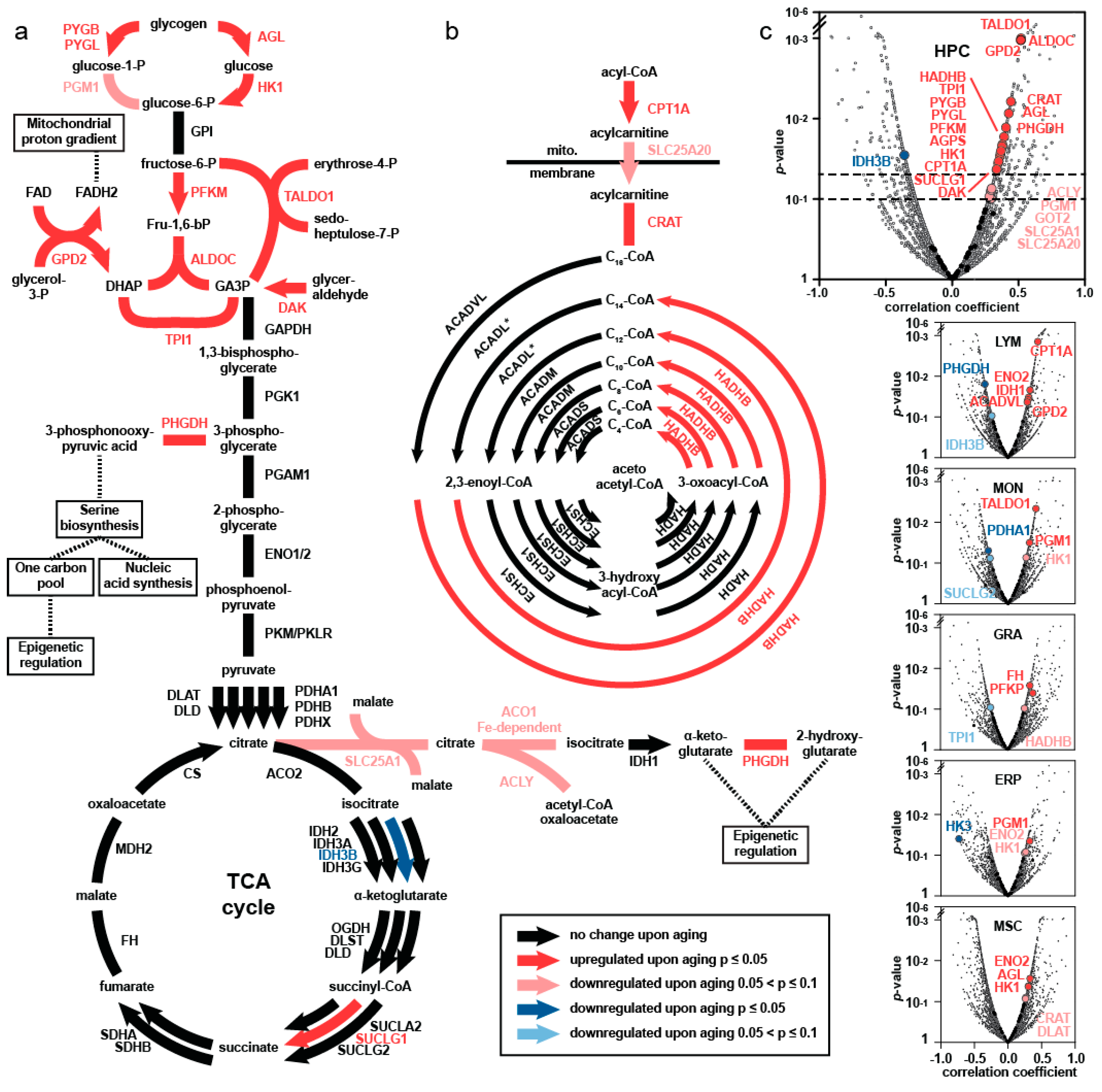
4. Comprehensive Proteomics Approach—Major Findings

Using an unbiased, comprehensive proteomic approach, our group has analyzed the molecular features of aging of HSPC as well as five other cell types that constitute the niche in human bone marrow. We have presented an atlas of the age-associated alterations in the proteomics landscapes of human HSPC, as well as subpopulations comprising the bone marrow niche derived from 59 human subjects (age range 20–60 years), i.e., across the time span of 40 years [23]. The results of proteomics studies are summarized in Figure 1 (modified from [23]).
Among the significant changes in abundance of proteins with age in the HSPC, we found a significant age-associated increase in enzymes catalyzing the upper part of glycolysis: hexokinase 1 (HK1), phosphofructokinase M (PFKM), as well as the glycolytic enzymes aldolase C (ALDOC) and triosephosphate isomerase 1 (TPI1). The principal findings are demonstrated in Figure 2 (from [23]). The enzymes involved in glycogen metabolism, glycogen phosphorylases brain and liver form (PYGB, PYGL), and glycogen debranching enzyme (AGL) were significantly more abundant in old HSPC. Compatible with these changes, an increase in abundance of trans-aldolase 1 (TALDO1), glycerol-3-phosphate dehydrogenase (GPD2), and dihydroxy-acetone kinase (DAK) provides further evidence for elevated activity in the upper part of the central carbon metabolism in aging HSPC (see Figure 2). The second phase of central carbon metabolism, characterized by the production of ATP, NADH and pyruvate and proteins involved in the Krebs cycle, remained unaffected. This upregulation of the upper part of the glycolytic pathway is unique for the HSPC compartment and is not found in the other five cell types of the niche, including MSC, as summarized in Figure 2.
Other significant changes found with the proteomics study include an increase in synthesis of prostaglandins and thromboxanes, in arachidonic acid metabolism, in metabolism of nitric oxide and in proteins that are associated with myeloid development in older HSPC (Figure 1). There was a remarkable decrease in abundance for proteins responsible for lymphoid development, for stabilization of DNA replication and for methylation processes. The enhanced metabolic and anabolic activity of aged HSPC, especially of central carbon metabolism is, however, the most novel and unique finding using a proteomics approach.

5. Elevated Central Carbon Metabolism in HSPC upon Aging

To validate our proteomics results, we have also assessed and compared the enzymatic activities of representative glycolytic enzymes hexokinase 1 (HK 1), phosphofructokinase M, aldolase C (ALDO C) and triosephosphate isomerase 1 (TPI 1) in the CD34+ cells derived from old versus young human subjects. In addition, activity of adenylate kinase (ADK), an enzyme regulating glucose-driven energy metabolism was also examined. Our data demonstrated that the activities of ADK, TPI and HK were significantly increased in the elderly cohort [49], validating the results from the proteomics studies.
Glucose metabolism has been shown to play a pivotal role in governing stem cell fate in terms of proliferation, differentiation, or dormancy on the one hand [50], and to influence chromatin structure and transcription on the other [51]. Our proteomics and functional studies have provided evidence for a prominent shift in metabolism of human HSPC during the aging process [23,49]. The upregulation of the preparatory phase of glycolysis, of fatty acid oxidation, and of acetyl-CoA metabolism, with redirection of glycolytic carbons to pathways that branch out of the Krebs cycle for anabolic processes, i.e., for nucleotides, lipids, amino acids synthesis, was reminiscent of the Warburg effect [51,52,53].
The question then arises whether this increase in glycolytic enzyme levels is caused by an increase in these enzymes on a per-cell basis upon aging, or by the expansion of one of the HSPC subsets that have become more glycolytic than the others.
We have assessed the changes in glycogen content in HSPC from young (19–31 years in this study) and old (59–71 years in this study) human subjects via a semi-quantitative PAS analysis [49]. The results are summarized in Figure 3 and Table 1. For older human subjects, the average glycogen content was 3.5-fold higher and was highly significant (one-sided t-test, p = 1.2 × 10⁻⁶) (Figure 3a). For young human subjects, the glycogen content was relatively homogeneous and most CD34+ cells showed intermediate and low glucose uptake. There were scarcely any CD34+ cells with the GUhigh phenotype in young subjects.
Table 1 shows a list of the fractions of GUhigh, GUinter, and GUlow CD34+ cells from young (<35 years, n = 3) versus older (>50 years; n = 7) human subjects. Whereas there is no difference in the percentages of GUinter and GUlow fractions between young and older subjects, the difference in GUhigh is significant (p = 0.02, one-sided t-test).
Single-cell RNA sequencing (scRNA-seq) analysis of aged CD34+ cells in comparison to young CD34+ has provided further evidence that the re-wiring of central carbon metabolism was found only in the myeloid-primed subpopulation of HSPC and not in the lymphoid-primed cells, nor in the CD34+ subset that showed no lineage commitment [23,49].
Our results have therefore provided evidence that the upregulation of glycolytic enzymes is caused by the relative expansion of a subset of CD34+ cells that have become more glycolytic [49].

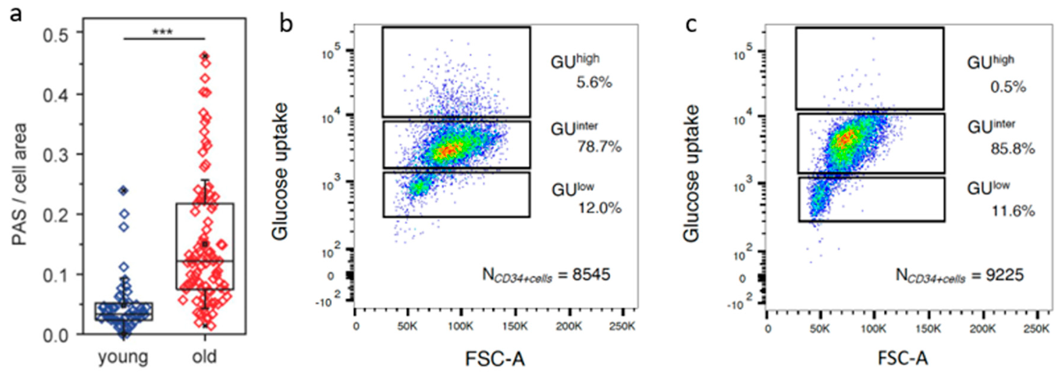
6. Separation of HSPC According to Carbon Metabolic Levels

Following the queue that a small population of HSPC with elevated central carbon metabolism might be responsible for the aging HSPC phenotype in humans, we developed a method to isolate subsets of HSPC according to their levels of carbon metabolism, using glucose uptake (GU) as a surrogate marker [49].
Given the knowledge gained from the semi-quantitative PAS reactivity and functional enzymatic assays, the variations in glycolytic metabolism could be exploited to isolate the HSPC according to their respective GU capacity. From the bone marrow of older human subjects, we were consistently able to separate the HSPC into GUhigh, GUinter and GUlow subpopulations. The same is true for adults younger than 35 years, with the exception that the GUhigh subset was hardly detectable. After separation of CD34+ cells according to glucose metabolic levels, the GUhigh, GUinter and GUlow subsets were harvested for further characterization using single-cell RNA sequencing (scRNA-seq) technology.

7. Single-Cell Transcriptome Studies of HSPC with Different Levels of Carbon Metabolism

We have performed scRNA-Seq studies on the HSPC (CD34+ cells) derived from 10 healthy human subjects (old adults n = 6; age range: 62–74 years, young adults n = 4; age range: 21–33 years) with special focus on the comparisons between GUhigh versus GUlow and GUinter from each individual [54]. For the comparisons of HSPC with different levels of glucose metabolism, 196 cells from each of the categories GUhigh, GUinter and GUlow isolated from each individual human subject tested were sequenced and the transcriptome profiles compared. As a reference, 196 CD34+ cells from individual old subjects were compared with 196 CD34+ cells derived from individual young human subjects [49,54].

7.1. DNA Damage, Telomere Attrition, and Cell Cycle Arrest

Damage of the telomeres represents one of the major injuries that induce senescence upon physiological aging [31]. The telomere region is protected by a multi-protein complex shelterin, with POT1 and TERF2 as major components [19,55]. TERF2 and POT1 act independently to repress either the ATM or ATR kinase signaling pathway required for efficient non-homologous end-joining of dysfunctional telomeres [55,56]. A deficiency in TERF2 or POT1 induces genome instability by shortening telomeres and causes DNA damage, which in turn activates a kinase cascade involving first ATM and ATR and then CHEK1 and CHEK2, resulting in sustained p53 activation [57,58]. The latter then leads to a permanent arrest of the cell cycle by persistent induction of the cyclin-dependent kinase inhibitor CDKN1A (P21CIP1), resulting in hypo-phosphorylated RB and ultimately in cell cycle exit [58,59].
We have compared the differential gene expression profiles of these genes in the GUhigh versus GUlow subset. The results are summarized in Table 2. The deregulation pattern, as delineated above, supports the notion that the GUhigh subset exhibits the characteristics of senescent cells.
As a reference, the expression profiles of these genes in the CD34+ cells derived from older (>60 years) versus young adults (<33 years) were compared and depicted in Table 2. While the correlation to the expression profiles of genes involved in cell cycle arrest was highly significant for GUhigh versus GUlow, the corresponding comparison between old versus young adult CD34+ cells showed only a trend in some and statistical significance in a few of the interrogated genes (Table 2).
In murine models of aging, the INK4/ARF locus seems to play an important role within the context of sustained p53 activation [19]. Both p16INK4a and ARF are encoded by the CDKN2A gene, and p15INK4b encoded by the CDKN2B gene. ARF inhibits the ubiquitin ligase MDM2, subsequently contributing to increased levels of P53 [60]. We could not detect any significant difference in comparing the expression levels of CDKN2A or CDKN2B between GUhigh versus GUlow subsets, nor between old versus young CD34+ cells. This is in agreement with reports from the literature that senescence in murine cells is more dependent on p19Arf than that in human cells [61,62].

7.2. Senescence-Associated Secretory Phenotype (SASP)

The production of a combination of inflammatory factors called senescence-associated secretory phenotype (SASP) is another characteristic of senescent cells [29]. Whereas the combination of secreted factors depends on the specific cell type and the inducer, some of the key effectors of the SASP seem to be shared by senescence cells of diverse origins. NOTCH1, CCAAT/enhancer-binding protein beta (CEBPB) and nuclear factor kappa B (NFKB) are some of the major shared regulators [63,64]. Cytokines such as IL1A, IL1B, and IL6 are also involved. The differential expressions of some of these genes in GUhigh versus GUlow subsets were interrogated and the results summarized in Table 2.
The differential gene expression analysis revealed that the GUhigh showed a consistently higher expression of genes involved in SASP: NOTCH1, CEBPA, CEBPB, NFKB1, NFKB2, IL1B, and IL1R2. The results are compatible with the notion that the GUhigh subset comprises cells of the SASP phenotype. As a reference, the expression profiles of these genes in the CD34+ cells derived from older (>60 years) in comparison to young adults (<35 years) are also listed in Table 2.

7.3. TP53, Apoptosis, and Pro-Survival Pathways

Another major feature of senescent cells is their survival despite activated DNA damage responses, increased levels of SASP inflammatory cytokines, and cell cycle arrest—all factors that might have induced apoptosis. Senescent cells are, however, equipped with anti-apoptotic and pro-survival mechanisms [65,66,67]. Using transcript array analysis, Zhu et al. demonstrated upregulation of negative regulators of apoptosis and pro-survival gene sets in senescence versus non-senescent cells [68]. Using a pre-adipocyte radiation-induced senescence model, increased expression of pro-survival networks around EFNB-1, EFNB-3, PI3KCD, p21 (CDKN1A), PAI-1 (SERPINE1), PAI-2 (SERPINB2), BCL-xL, and MCL-1 has been shown [68]. Among these genes, the roles of upregulations of BCL2, BCL-xL (BCL2L1), and MCL1 as prominent anti-apoptotic factors in murine HSPC have been extensively examined [25,62,68].
Our scRNA-seq studies showed that the GUhigh subset of CD34+ cells expressed significant upregulation of these anti-apoptotic and pro-survival genes (see Table 2) [54].

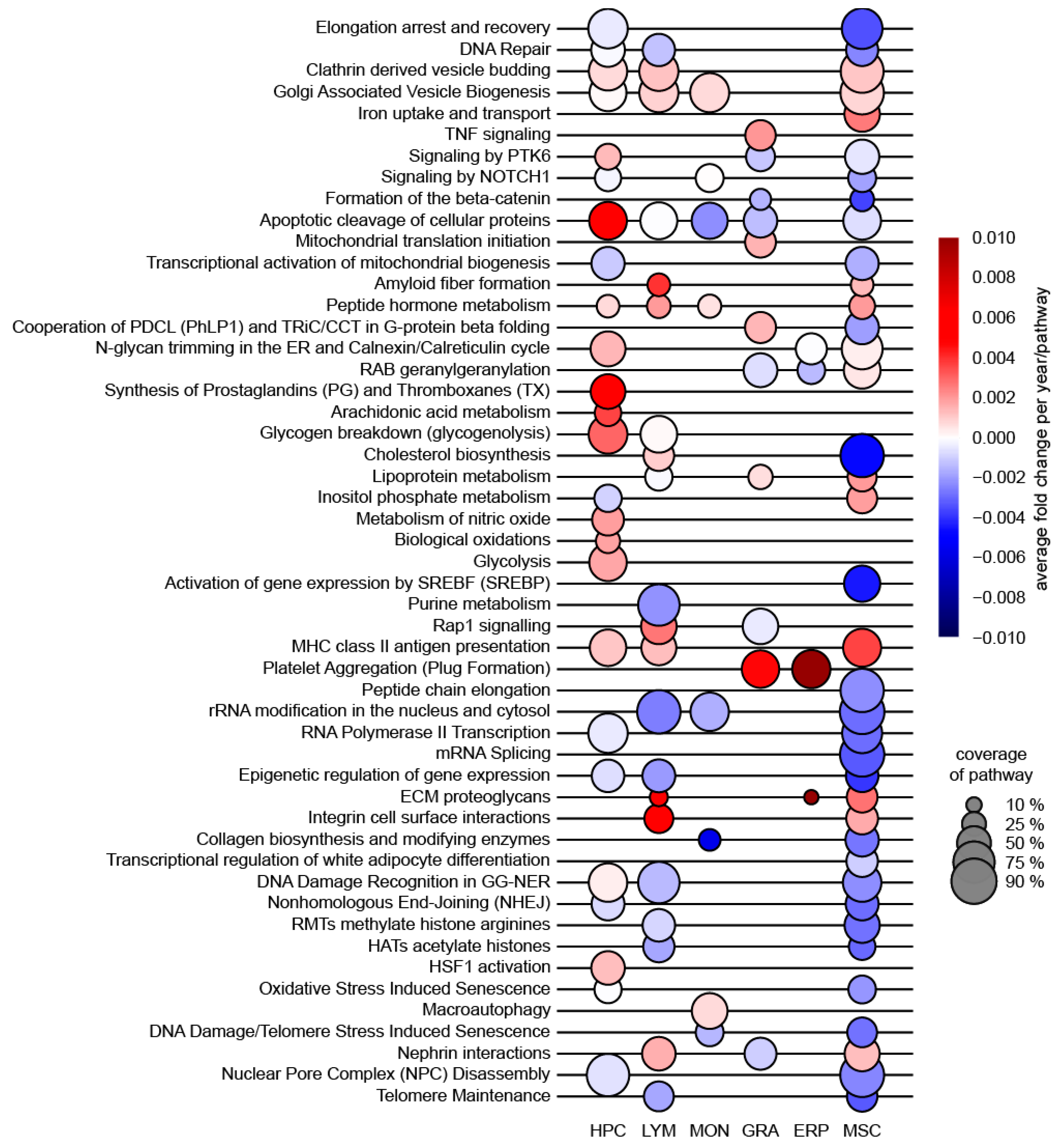
8. Gene Set Enrichment Analysis (GSEA)

We applied gene set enrichment analysis (GSEA; version 4.2.1, Broad Institute, Inc., Massachusetts Institute of Technology, and Regents of the University of California) to identify pathways and processes that were coordinately up- or downregulated with senescence [69]. The “Hallmarks” gene sets and “ENSEMBL Gene identifiers MSigDB.v7.4chip” were applied to determine the differential expression profiles in biological processes associated with GUhigh versus GUinter and/or GUlow subpopulations [70].
In the GUhigh phenotype, 40 of 50 gene sets interrogated by “Hallmarks” are upregulated, 30 of which significantly enriched at a nominal p value of <1%, and 35 gene sets are significantly enriched at p = <5%. In Table 3, we have summarized the four most prominently elevated pathways associated with senescence [54].
Gene sets that were significantly upregulated include (1) G2M checkpoint; (2) MTORC1 signaling; (3) inflammatory response; (4) apoptosis (Table 3). Gene sets significantly downregulated include interferon gamma and interferon alpha response [54].
The upregulation of the pathway G2M checkpoint in the GUhigh subset is compatible with the cycle arrest that is characteristic for senescent cells. Inflammatory response is closely linked to the phenotype SASP. Mammalian target of rapamycin (MTOR) activity has been shown to be increased in HSPC from old mice compared to those from young mice and suppression of MTOR by rapamycin prolonged lifespan in mice [71,72]. Finally, upregulation of apoptosis/pro-survival as well as P53 pathways in aged murine HSPC has been reported to be characteristic [41]. The simultaneous upregulation of these Hallmark gene sets in the GUhigh subset has therefore provided further evidence for the senescence phenotype in this population.
We have performed the same GSEA algorithm comparing the CD34+ cells derived from old versus young adults. While upregulation of all the gene sets involved in the aforementioned biological processes was highly significant in comparing the GUhigh versus GUlow subset, only the upregulation of the “P53 Pathway” in the aged CD34+ cells reached statistical significance at p = <5% [49,54]. The proportion of senescent cells in the whole CD34+ population is probably too small for an appropriate comparison. This observation again provides evidence that the GUhigh subset in the HSPC compartment is highly enriched for senescent cells.

9. GSEA Using “Aging Signature Gene Sets” as Reference

Svendsen et al. surveyed 16 published and unpublished transcriptome datasets from comparisons of aged and young murine HSPC [20]. They were able to identify a list of 220 genes that were consistently deregulated in aging murine HSPC. Their meta-analysis and re-analysis of the published data suggested that this gene set collection of aging signature (AS) remained stable and robust. They concluded that this AS can be used as a reference for differentially expressed genes upon HSPC aging.
Using this gene set as a reference, we have performed additional GSEA to compare the differential expression profiles of GUhigh versus GUlow, as well as GUhigh versus GUinter+low. The results are summarized in Table 3. The GUhigh subset showed high scores, whereas the GUlow subset low scores for the gene set “aging signature” [20]. This is also true when the GUlow subpopulation was compared with GUinter (data not shown).
This additional comparison using the “aging signature” has again confirmed that the GUhigh subpopulation is highly enriched for senescence in the human HSPC compartment. Another remarkable finding is also that the expression profile of the GUlow subset has maintained that of primitive and young adult HSPC and did not change with age in all these studies. This is compatible with the reports from Svendsen et al. that a subset of murine HSPC have maintained a “young-like” phenotype.

10. The Causative Role of Senescent Cells in Aging and Their Elimination

Senescent cells have been shown to play a causative role in age-related pathologies in mice [71,72,73,74]. The abundance of senescence cells among the total number of somatic stem cell compartments may be relatively low, i.e., up to a maximum of 15% in some tissues. Their impact is, however, probably magnified by the secretion of pro-inflammatory cytokines that constitute the SASP [64,73,74]. The SASP in turn accounts for many of the consequences of senescent cell accumulation, leading to dysfunction of the stem cell pool as well as to diseases of the respective tissue. Elimination of or a reduction in senescent cells from the stem cell pool may eventually ameliorate functional decline of HSPC.
In animal models, genetic ablation of senescent cells (p16Ink4a-expressing cells) by activating a drug-inducible pro-apoptotic gene has been shown to attenuate the onset of age-related pathologies of progeroid mice [24]. In a subsequent study by the same group, p16Ink4a-expressing cells were consistently eliminated by the use of a transgene INK-ATTAC that induced apoptosis in the respective cells upon administration of a specific drug [25]. The authors concluded that therapeutic removal was able to extend healthy lifespan in mice. Since then, a number of studies have provided further evidence that reducing the burden of senescent cells by as little as 30% could ameliorate age-related disabilities and chronic diseases [26,27,75].
Senescent cells in mice have been shown to be well equipped with anti-apoptotic and pro-survival mechanisms [24,62,68,76]. Anti-apoptotic proteins such as BCL2, BCL-XL, and BCL-W are upregulated and seem to be essential for the survival of senescent HSPC in animal models [24,25,76]. This has inspired the development of inhibitors of anti-apoptotic factors as “senolytic” agents to eliminate senescent cells [25,68,76,77]. Inhibitors of anti-apoptotic factors, e.g., ABT263 and ABT737, are able to selectively deplete senescent HSPC and clearance of the latter has led to rejuvenation not only of HSPC, but also to improved cardiovascular function, reduced osteoporosis and extended health span in naturally aging mice [76,78].
Our finding of significantly elevated glucose metabolism in the senescent population of human HSPC compartment indicates that this subset is dependent on elevated central carbon metabolism for survival, in analogy to the increased expression of anti-apoptotic factors. Modulation of the glycolytic pathways may therefore represent another therapeutic principle for senolysis treatment.

11. Conclusions

Comprehensive and unbiased proteomics studies of the HSPC have provided evidence that aging of the HSPC compartment is characterized by elevated glycolysis. Whereas other significant changes found with this proteomics approach, such as an increase in differentiation bias towards the myeloid lineage, in DNA repair, in cellular metabolism, and a decrease in proteins responsible for lymphoid development and for stabilization of DNA replication have been described in murine transcriptomics studies, the enhanced central carbon metabolism activity in aged human HSPC is novel [23].
Subsequent studies have demonstrated that the increase in glycolytic enzyme level is caused by the expansion of a HSPC subset that has become more glycolytic than the others and not on a per cell basis. Provided with this knowledge, we developed a method to isolate the HSPC according to their glucose metabolic levels in three distinct categories: the GUhigh, GUinter and GUlow subsets. The GUhigh subset is coupled with differentiation bias towards myeloid lineages. GSEA of the transcriptomes of the GUhigh versus GUlow subset, or GUhigh versus GUinter subsets, have demonstrated that the gene sets for cell cycle arrest, MTORC1 signaling, inflammatory response, and anti-apoptosis pathways are significantly upregulated in the GUhigh population. Applying the transcriptomic “aging signature” gene set proposed by [20], the GUhigh subset achieves high scores for this “aging signature”.
Most of the current knowledge about the properties of senescent cells in the HSPC compartment is based on experiments in cultured cells and in murine models of aging. There are no specific markers or marker constellations that could enable us to identify and enrich senescent cells for mechanistic studies. With the separation of HSPC according to glucose metabolic levels, we have shown that we are able to enrich the senescent population in the GUhigh subset in the human HSPC compartment of the bone marrow. As the viability and functional integrity of this subset are well preserved, these cells may serve as starting material for further mechanistic characterization of senescent HSPC.
In analogy to the Warburg effect in cancer cells, our data have provided strong evidence for the dependency of senescent HSPC on elevated central carbon metabolism as well as on MTORC1 pathway for survival. During development and aging of HSPC, drastic metabolic shifts to meet the demand of hematopoiesis during transition occurs [79]. The glycolytic and MTORC1 pathways integrate inputs from nutrient and growth signals to regulate general cellular processes such as protein and lipid synthesis, autophagy, and metabolism [80]. In this respect, MTOR has been shown to regulate the senescence-associated secretory phenotype (SASP) and senescent growth arrest [80,81]. Based on upstream signaling of MTORC1, a relationship between carbohydrate consumption and MTORC1 activity has been demonstrated, specifically through the insulin growth factor pathway [82]. Multiple studies have demonstrated that caloric restriction can retard the aging decline [83]. Hence, caloric restriction, or agents that modulate the glycolytic pathway such as Metformin may modulate glycolysis and MTORC1 pathways and eliminate the senescent population within the HSPC compartment.
Senescent cells and cancer cells share many common properties. Agents that block the apoptotic pathways that cancer cells are dependent on have been shown to be effective as senolytic drugs in aged mice. ABT-263 and ABT-737 are examples that target the B cell lymphoma 2 (BCL-2) protein family members. These drugs are, however, associated with toxic side effects such as neutropenia and thrombocytopenia. Targeting the central carbon metabolism or the closely related MTORC1 signaling pathway may offer better alternatives for developing senolysis strategies.
In summary, our series of studies are unique in the following aspects. First of all, almost all present-day knowledge on aging of HSPC and most proof-of-principle investigations for elimination of senescent cells have been gained from studies in mice. Without any doubt, great advances have been achieved, but the knowledge must be validated in the human system. Our comprehensive transcriptome and proteome datasets have contributed to bridge this gap. As delineated in this review, many of the principal mechanisms of senescence in mice can be confirmed in human system and yet there are differences. Another significant aspect is the discovery of the close association of elevated central carbon metabolism with senescence. Thus far, isolation and collection of senescent cells have been extremely difficult as specific markers or marker constellations for their identification have yet to be developed. This is specially the case in human HSPC. The enrichment of HSPC, with all the characteristics of senescence by glucose metabolism, in conjunction with single-cell high-throughput technology, may represent an important stepping stone towards accurate visualization, collection and tracking of senescence in human bone marrow.

Funding

This research was funded by Deutsche Forschungsgemeinschaft (DFG) (HO914/10 to A.D.H., CRC873 to A.D.H and M.T., EU-FP7. (Grant 306240 “SyStemAge” to A.C.G and A.D.H.) A.D.H. is thankful for the support from Thiele-Stiftung, M.T. for the support from Nakatani Foundation, and S.R. for the support from German Science Foundation (RA3166/1-1) and German Federal Ministry of Education and Health.

Institutional Review Board Statement

The studies mentioned in this review have been approved by the Ethics Committee for Human Subjects at the University of Heidelberg and the details are deliberated in References [23,49].

Informed Consent Statement

Written and signed informed consent was obtained from each human subject. The details are deliberated in References [23,49].

Data Availability Statement

The mass spectrometry proteomics data have been deposited at the ProteomeXchange Consortium via the PRIDE partner repository with the dataset identifier PXD007048. Raw data for both the single-cell RNA-seq and bulk RNA-seq experiments have been deposited in the Gene Expression Omnibus (GEO), database under accession code GSE115353, as described in detail in References [23,49].

Acknowledgments

We would like to acknowledge the extraordinary support by the staff of the Genomics Core Facility of European Molecular Biology Laboratory.

Conflicts of Interest

The authors declare no conflict of interest.

References

  1. Iscove, N.N.; Nawa, K. Hematopoietic stem cells expand during serial transplantation in vivo without apparent exhaustion. Curr. Biol. 1997, 7, 805–808. [Google Scholar] [CrossRef] [Green Version]
  2. Schlessinger, D.; Van Zant, G. Does functional depletion of stem cells drive aging? Mech. Ageing Dev. 2001, 122, 1537–1553. [Google Scholar] [CrossRef]
  3. Ho, A.D.; Wagner, W.; Mahlknecht, U. Stem cells and ageing. EMBO Rep. 2005, 6, 535–538. [Google Scholar] [CrossRef] [PubMed]
  4. Morrison, S.J.; Wandycz, A.M.; Akashi, K.; Globerson, A.; Weissman, I.L. The aging of hematopoietic stem cells. Nat. Med. 1996, 2, 1011–1016. [Google Scholar] [CrossRef] [PubMed]
  5. De Haan, G.; Nijhof, W.; Van Zant, G. Mouse strain-dependent changes in frequency and proliferation of hematopoietic stem cells during aging: Correlation between lifespan and cycling activity. Blood 1997, 89, 1543–1550. [Google Scholar] [CrossRef] [Green Version]
  6. Offner, F.; Kerre, T.; de Smedt, M.; Plum, J. Bone marrow CD34+cells generate fewer T cells in vitro with increasing age and following chemotherapy. Br. J. Haematol. 1999, 104, 801–808. [Google Scholar] [CrossRef]
  7. Sudo, K.; Ema, H.; Morita, Y.; Nakauchi, H. Age-associated characteristics of murine hematopoietic stem cells. J. Exp. Med. 2000, 192, 1273–1280. [Google Scholar] [CrossRef] [Green Version]
  8. Liang, Y.; Van Zant, G.; Szilvassy, S.J. Effects of aging on the homing and engraftment of murine hematopoietic stem and progenitor cells. Blood 2005, 106, 1479–1487. [Google Scholar] [CrossRef] [Green Version]
  9. Dykstra, B.; Olthof, S.; Schreuder, J.; Ritsema, M.; de Haan, G. Clonal analysis reveals multiple functional defects of aged murine hematopoietic stem cells. J. Exp. Med. 2011, 208, 2691–2703. [Google Scholar] [CrossRef] [Green Version]
  10. Geiger, H.; de Haan, G.; Florian, M.C. The ageing haematopoietic stem cell compartment. Nat. Rev. Immunol. 2013, 13, 376–389. [Google Scholar] [CrossRef]
  11. Matteini, F.; Mulaw, M.A.; Florian, M.C. Aging of the hematopoietic stem cell niche: New tools to answer an old question. Front. Immunol. 2021, 12, 738204. [Google Scholar] [CrossRef] [PubMed]
  12. Gnani, D.; Crippa, S.; Della Volpe, L.; Rossella, V.; Conti, A.; Lettera, E.; Rivis, S.; Ometti, M.; Fraschini, G.; Bernardo, M.E.; et al. An early-senescence state in aged mesenchymal stromal cells contributes to hematopoietic stem and progenitor cell clonogenic impairment through the activation of a pro-inflammatory program. Aging Cell 2019, 18, e12933. [Google Scholar] [CrossRef] [PubMed] [Green Version]
  13. Oda, A.; Tezuka, T.; Ueno, Y.; Hosoda, S.; Amemiya, Y.; Notsu, C.; Kasahara, T.; Nishiyama, C.; Goitsuka, R. Niche-induced extramedullary hematopoiesis in the spleen is regulated by the transcription factor Tlx1. Sci. Rep. 2018, 8, 8308. [Google Scholar] [CrossRef] [PubMed] [Green Version]
  14. Gottschling, S.; Saffrich, R.; Seckinger, A.; Krause, U.; Horsch, K.; Miesala, K.; Ho, A.D. Human mesenchymal stromal cells regulate initial self-renewing divisions of hematopoietic progenitor cells by a beta1-integrin-dependent mechanism. Stem Cells 2007, 25, 798–806. [Google Scholar] [CrossRef] [PubMed]
  15. Wagner, W.; Horn, P.; Bork, S.; Ho, A.D. Aging of hematopoietic stem cells is regulated by the stem cell niche. Exp. Gerontol. 2008, 43, 974–980. [Google Scholar] [CrossRef] [PubMed] [Green Version]
  16. Bork, S.; Pfister, S.; Witt, H.; Horn, P.; Korn, B.; Ho, A.D.; Wagner, W. DNA methylation pattern changes upon long-term culture and aging of human mesenchymal stromal cells. Aging Cell. 2010, 9, 54–63. [Google Scholar] [CrossRef] [Green Version]
  17. Ambrosi, T.H.; Marecic, O.; McArdle, A.; Sinha, R.; Gulati, G.S.; Tong, X.; Wang, Y.; Steininger, H.M.; Hoover, M.Y.; Koepke, L.S.; et al. Aged skeletal stem cells generate an inflammatory degenerative niche. Nature 2021, 597, 256–262. [Google Scholar] [CrossRef]
  18. Ambrosi, T.H.; Chan, C. Skeletal Stem Cells as the Developmental Origin of Cellular Niches for Hematopoietic Stem and Progenitor Cells. Curr. Top. Microbiol. Immunol. 2021, 434, 1–31. [Google Scholar]
  19. McHugh, D.; Gil, J. Senescence and aging: Causes, consequences, and therapeutic avenues. J. Cell Biol. 2018, 217, 65–77. [Google Scholar] [CrossRef]
  20. Svendsen, A.F.; Yang, D.; Kim, K.; Lazare, S.; Skinder, N.; Zwart, E.; Mura-Meszaros, A.; Ausema, A.; von Eyss, B.; de Haan, G.; et al. A comprehensive transcriptome signature of murine hematopoietic stem cell aging. Blood 2021, 138, 439–451. [Google Scholar] [CrossRef]
  21. Rossi, D.J.; Bryder, D.; Zahn, J.M.; Ahlenius, H.; Sonu, R.; Wagers, A.J.; Weissman, I.L. Cell intrinsic alterations underlie hematopoietic stem cell aging. Proc. Natl. Acad. Sci. USA 2005, 102, 9194–9199. [Google Scholar] [CrossRef] [PubMed] [Green Version]
  22. Pang, W.W.; Price, E.A.; Schoo, D.; Beerman, I.; Maloney, W.J.; Rossi, D.J.; Schrier, S.L.; Weissmann, I.L. Human bone marrow hematopoietic stem cells are increased in frequency and myeloid-biased with age. Proc. Natl. Acad. Sci. USA 2011, 108, 20012–20017. [Google Scholar] [CrossRef] [PubMed] [Green Version]
  23. Hennrich, M.L.; Romanov, N.; Horn, P.; Jaeger, S.; Eckstein, V.; Steeples, V.; Ye, F.; Ding, X.; Poisa-Beiro, L.; Lai, M.C.; et al. Cell-specific proteome analyses of human bone marrow reveal molecular features of age-dependent functional decline. Nat. Comm. 2018, 9, 4004. [Google Scholar] [CrossRef] [PubMed]
  24. Baker, D.J.; Wijshake, T.; Tchkonia, T.; LeBrasseur, N.K.; Childs, B.G.; van de Sluis, B.; Kirkland, J.L.; van Deursen, J.M. Clearance of p16Ink4a–positive senescent cells delay ageing-associated disorders. Nature 2011, 479, 232–236. [Google Scholar] [CrossRef] [PubMed]
  25. Baker, D.J.; Childs, B.G.; Durik, M.; Wijers, M.E.; Sieben, C.J.; Zhong, J.; Saltness, R.A.; Jeganathan, K.B.; Verzosa, G.C.; Pezeshki, A.; et al. Naturally occurring p16(Ink4a)-positive cells shorten healthy lifespan. Nature 2016, 530, 184–189. [Google Scholar] [CrossRef] [PubMed] [Green Version]
  26. Kirkland, J.L. Translating advances from the basic biology of aging into clinical application. Exp. Gerontol. 2013, 48, 1–5. [Google Scholar] [CrossRef] [Green Version]
  27. Tchkonia, T.; Zhu, Y.; van Deursen, J.; Campisi, J.; Kirkland, J.L. Cellular senescence and the senescent secretory phenotype: Therapeutic opportunities. J. Clin. Investig. 2013, 123, 966–972. [Google Scholar] [CrossRef] [Green Version]
  28. Deursen, J.M. The role of senescent cells in aging. Nature 2014, 509, 439–446. [Google Scholar] [CrossRef] [Green Version]
  29. Gorgoulis, V.; Adams, P.D.; Alimonti, A.; Bennett, D.C.; Bischof, O.; Bishop, C.; Campisi, J.; Collado, M.; Evangelou, K.; Ferbeyre, G.; et al. Cellular senescence: Defining a path forward. Cell 2019, 179, 814–827. [Google Scholar] [CrossRef]
  30. Roger, L.; Tomas, F.; Gire, V. Mechanisms and regulation of cellular senescence. Inter. J. Mol. Sci. 2021, 22, 13173. [Google Scholar] [CrossRef]
  31. Vaziri, H.; Dragowska, W.; Allsopp, R.C.; Thomas, T.E.; Harley, C.B.; Lansdorp, P.M. Evidence for a mitotic clock in human hematopoietic stem cells: Loss of telomeric DNA with age. Proc. Natl. Acad. Sci. USA 1994, 91, 9857–9860. [Google Scholar] [CrossRef] [PubMed] [Green Version]
  32. Rossi, D.J.; Bryder, D.; Seita, J.; Nussenzweig, A.; Hoeijmakers, J.; Weissman, I.L. Deficiencies in DNA damage repair limit the function of haematopoietic stem cells with age. Nature 2007, 447, 725–729. [Google Scholar] [CrossRef] [PubMed]
  33. Muñoz-Espín, D.; Cañamero, M.; Maraver, A.; Gómez-López, G.; Contreras, J.; Murillo-Cuesta, S.; Rodríguez-Baeza, A.; Varela-Nieto, I.; Ruberte, J.; Collado, M.; et al. Programmed cell senescence during mammalian embryonic development. Cell 2013, 155, 1104–1118. [Google Scholar] [CrossRef] [PubMed] [Green Version]
  34. Casella, G.; Munk, R.; Kim, K.M.; Piao, Y.; De, S.; Abdelmohsen, K.; Gorospe, M. Transcriptome signature of cellular senescence. Nucleic Acid Res. 2019, 47, 7294–7305. [Google Scholar] [CrossRef] [Green Version]
  35. Campisi, J.; Kapathi, P.; Lithgow, G.J.; Melow, S.; Newman, J.C.; Verdin, E. From discoveries in ageing research to therapeutics for healthy ageing. Nature 2019, 571, 183–192. [Google Scholar] [CrossRef] [Green Version]
  36. López-Otín, C.; Blasco, M.A.; Partridge, L.; Serrano, M.; Kroemer, G. The hallmarks of aging. Cell 2013, 153, 1194–1217. [Google Scholar] [CrossRef] [Green Version]
  37. Kollman, C.; Howe, C.W.S.; Anasetti, C.; Antin, J.H.; Davies, S.M.; Filipovich, A.H.; Hegland, J.; Kamani, N.; Kernan, N.A.; King, R.; et al. Donor characteristics as risk factors in recipients after transplantation of bone marrow from unrelated donors: The effect of donor age. Blood 2001, 98, 2043–2051. [Google Scholar] [CrossRef]
  38. Grover, A.; Sanjuan-Pla, A.; Thongjuea, S.; Carrelha, J.; Giustacchini, A.; Gambardella, A.; Macaulay, I.; Mancini, E.; Luis, T.C.; Mead, A.; et al. Single-cell RNA sequencing reveals molecular and functional platelet bias of aged haematopoietic stem cells. Nat. Commun. 2016, 7, 11075. [Google Scholar] [CrossRef]
  39. Noda, S.; Ichikawa, H.; Miyoshi, H. Hematopoietic stem cell aging is associated with functional decline and delayed cell cycle progression. Biochem. Biophys. Res. Commun. 2009, 383, 210–215. [Google Scholar] [CrossRef]
  40. Kowalczyk, M.S.; Tirosh, I.; Heckl, D.; Rao, T.N.; Dixit, A.; Haas, B.J.; Schneider, R.K.; Wagers, A.J.; Ebert, B.L.; Regev, A. Single-cell RNA-seq reveals changes in cell cycle and differentiation programs upon aging of hematopoietic stem cells. Genome Res. 2015, 25, 1860–1872. [Google Scholar] [CrossRef] [Green Version]
  41. Kirschner, K.; Chandra, T.; Kiselev, V.; Flores-Santa Cruz, D.; Macaulay, I.C.; Park, H.J.; Li, J.; Kent, D.G.; Kumar, R.; Pask, D.C.; et al. Proliferation drives aging-related functional decline in a subpopulation of the hematopoietic stem cell compartment. Cell Rep. 2017, 19, 1503–1511. [Google Scholar] [CrossRef] [Green Version]
  42. Chambers, S.M.; Shaw, C.A.; Gatza, C.; Fisk, C.J.; Donehower, L.A.; Goodell, M.A. Aging hematopoietic stem cells decline in function and exhibit epigenetic dysregulation. PLoS Biol. 2007, 5, e201. [Google Scholar] [CrossRef] [PubMed]
  43. Mann, M.; Mehta, A.; de Boer, C.G.; Kowalczyk, M.S.; Lee, K.; Farouq, D.; Regev, A.; Baltimore, D. Heterogeneous responses of hematopoietic stem cells to inflammatory stimuli are altered with age. Cell Rep. 2018, 25, 2992–3005. [Google Scholar] [CrossRef] [PubMed] [Green Version]
  44. Flach, J.; Bakker, S.T.; Mohrin, M.; Conroy, P.C.; Pietras, E.M.; Reynaud, D.; Alvarez, S.; Diolaiti, M.E.; Ugarte, F.; Forsberg, E.C.; et al. Replication stress is a potent driver of functional decline in ageing haematopoietic stem cells. Nature 2014, 512, 198–202. [Google Scholar] [CrossRef] [PubMed] [Green Version]
  45. Beerman, I.; Bock, C.; Garrison, B.S.; Smith, Z.D.; Gu, H.; Meissner, A.; Rossi, D.J. Proliferation-dependent alterations of the DNA methylation landscape underlie hematopoietic stem cell aging. Cell Stem Cell 2013, 12, 413–425. [Google Scholar] [CrossRef] [PubMed] [Green Version]
  46. Beerman, I.; Rossi, D.J. Epigenetic control of stem cell potential during homeostasis, aging, and disease. Cell Stem Cell 2015, 16, 613–625. [Google Scholar] [CrossRef] [Green Version]
  47. Wahlestedt, M.; Norddahl, G.L.; Sten, G.; Ugale, A.; Frisk, M.M.; Mattsson, R.; Deierborg, T.; Sigvardsson, M.; Bryder, D. An epigenetic component of hematopoietic stem cell aging amenable to reprogramming into a young state. Blood 2013, 121, 4257–4264. [Google Scholar] [CrossRef] [Green Version]
  48. Sun, D.; Luo, M.; Jeong, M.; Rodriguez, B.; Xia, Z.; Hannah, R.; Wang, H.; Le, T.; Faull, K.F.; Chen, R.; et al. Epigenomic Profiling of Young and Aged HSCs Reveals Concerted Changes during Aging that Reinforce Self-Renewal. Cell Stem Cell 2014, 14, 673–688. [Google Scholar] [CrossRef] [Green Version]
  49. Poisa-Beiro, L.; Landry, T.J.; Sauer, S.; Yamamoto, A.; Eckstein, V.; Romanov, N.; Raffel, S.; Hoffmann, G.F.; Bork, P.; Benes, V.; et al. Glycogen accumulation, central carbon metabolism, and aging of hematopoietic stem and progenitor cells. Sci. Rep. 2020, 10, 11597. [Google Scholar] [CrossRef]
  50. Shyh-Chang, N.; Daley, G.Q.; Cantley, L.C. Stem cell metabolism in tissue development and aging. Development 2013, 140, 2535–2547. [Google Scholar] [CrossRef] [Green Version]
  51. Heiden, M.G.V.; Cantley, L.C.; Thompson, C.B. Understanding the Warburg effect: The metabolic requirements of cell proliferation. Science 2009, 324, 1029–1033. [Google Scholar] [CrossRef] [PubMed] [Green Version]
  52. Warburg, O.H. Über den Stoffwechsel der Carcinomzelle. Wien. Klein. Wochenschr. 1925, 4, 534–536. [Google Scholar] [CrossRef]
  53. Smith, B.; Schafer, X.L.; Ambeskovic, A.; Spencer, C.M.; Land, H.; Munger, J. Addiction to coupling of the Warburg effect with glutamine catabolism in cancer cells. Cell Rep. 2016, 17, 821–836. [Google Scholar] [CrossRef] [PubMed] [Green Version]
  54. Poisa-Beiro, L.; Landry, J.; Rafffel, S.; Eckstein, V.; Gavin, A.C.; Tanaka, M.; Benes, V.; Ho, A.D. Elevated central carbon metabolism–a hallmark for senescent cells in aging human hematopoietic stem cell compartment. Blood 2021, 138, 1088. [Google Scholar] [CrossRef]
  55. Denchi, E.L.; de Lange, T. Protection of telomeres through independent control of ATM and ATR by TRF2 and POT1. Nature 2007, 448, 1068–1071. [Google Scholar] [CrossRef]
  56. Salama, R.; Sadaie, M.; Hoare, M.; Narita, M. Cellular senescence and its effector programs. Genes Dev. 2014, 28, 99–114. [Google Scholar] [CrossRef] [Green Version]
  57. Kruiswijk, F.; Labuschagne, C.F.; Vousden, K.H. p53 in survival, death and metabolic health: A lifeguard with a licence to kill. Nat. Rev. Mol. Cell Biol. 2015, 16, 393–405. [Google Scholar] [CrossRef]
  58. d’Adda di Fagagna, F. Living on a break: Cellular senescence as a DNA damage response. Nat. Rev. Cancer 2008, 8, 512–522. [Google Scholar] [CrossRef]
  59. Fumagalli, M.; Rossiello, F.; Clerici, M.; Barozzi, S.; Cittaro, D.; Kaplunov, J.M.; Bucci, G.; Dobreva, M.; Matti, V.; Beausejour, C.M.; et al. Telomeric DNA damage is irreparable and causes persistent DNA damage response activation. Nat. Cell Biol. 2012, 14, 355–365. [Google Scholar] [CrossRef] [Green Version]
  60. Baar, M.P.; Brandt, R.M.; Putavet, D.A.; Klein, J.D.; Derks, K.W.; Bourgeois, B.R.; Stryeck, S.; Rijksen, Y.; Willigenburg, H.V.; Feijtel, D.A.; et al. Targeted Apoptosis of Senescent Cells Restores Tissue Homeostasis in Response to Chemotoxicity and Aging. Cell 2017, 169, 132–147. [Google Scholar] [CrossRef] [Green Version]
  61. Ben-Porath, I.; Weinberg, R.A. The signals and pathways activating cellular senescence. Int. J. Biochem. Cell Biol. 2005, 37, 961–976. [Google Scholar] [CrossRef] [PubMed]
  62. Deursen, J.M. Senolytic therapies for healthy longevity. Science 2019, 364, 636–637. [Google Scholar] [CrossRef] [PubMed]
  63. Acosta, J.C.; O’Loghlen, A.; Banito, A.; Guijarro, M.V.; Augert, A.; Raguz, S.; Fumagalli, M.; Da Costa, M.; Brown, C.; Popov, N.; et al. Chemokine Signaling via the CXCR2 Receptor Reinforces Senescence. Cell 2008, 133, 1006–1018. [Google Scholar] [CrossRef] [PubMed] [Green Version]
  64. Kuilman, T.; Michaloglou, C.; Vredeveld, L.C.; Douma, S.; van Doorn, R.; Desmet, C.J.; Aarden, L.A.; Mooi, W.J.; Peeper, D.S. Oncogene-induced senescence relayed by an interleukin-dependent inflammatory network. Cell 2008, 133, 958–961. [Google Scholar] [CrossRef] [Green Version]
  65. Ito, Y.; Hoare, M.; Narita, M. Spatial and temporal control of senescence. Trends Cell Biol. 2017, 27, 820–832. [Google Scholar] [CrossRef] [Green Version]
  66. Ghosal, G.; Chen, J. DNA damage tolerance: A double-edged sword guarding the genome. Transl. Cancer Res. 2013, 2, 107–129. [Google Scholar]
  67. Zhu, Y.; Tchkonia, T.; Pirtskhalava, T.; Kirkland, J.L. The Achilles‘heel of senescent cells: From transcriptome to senolytic drugs. Aging Cell 2015, 14, 644–658. [Google Scholar] [CrossRef]
  68. Zhu, Y.; Tchkonia, T.; Fuhrmann-Stroissnigg, H.; Kirkland, J.L. Identification of a novel senolytic agent, navitoclax, targeting the Bcl-2 family of anti-apoptotic factors. Aging Cell 2016, 15, 428–435. [Google Scholar] [CrossRef]
  69. Subramanian, A.; Tamayo, P.; Mootha, V.K.; Mukherjee, S.; Ebert, B.L.; Gillette, M.A.; Paulovich, A.; Pomeroy, S.L.; Golub, T.R.; Lander, E.S.; et al. Gene set enrichment analysis: A knowledge-based approach for interpreting genome-wide expression profiles. Proc. Natl. Acad. Sci. USA 2005, 102, 15545–15550. [Google Scholar] [CrossRef] [Green Version]
  70. Liberzon, A.; Birger, C.; Thorvaldsdóttir, H.; Ghandi, M.; Mesirov, J.P.; Tamayo, P. The molecular signatures database (MSigDB) hallmark gene set collection. Cell Syst. 2015, 1, 417–425. [Google Scholar] [CrossRef] [Green Version]
  71. Chen, C.; Liu, Y.; Liu, Y.; Zheng, P. mTOR regulation and therapeutic rejuvenation of aging hematopoietic stem cells. Sci. Signal 2009, 2, ra75. [Google Scholar] [CrossRef] [PubMed] [Green Version]
  72. Harrison, D.E.; Strong, R.; Sharp, Z.D.; Nelson, J.F.; Astle, C.M.; Flurkey, K.; Nadon, N.L.; Wilkinson, J.E.; Frenkel, K.; Carter, C.S.; et al. Rapamycin fed late in life extends lifespan in genetically heterogenious mice. Nature 2009, 460, 392–395. [Google Scholar] [CrossRef] [PubMed] [Green Version]
  73. Herbig, U.; Ferreira, M.; Condel, L.; Carey, D.; Sedivy, J.M. Cellular senescence in aging primates. Science 2006, 311, 1257. [Google Scholar] [CrossRef] [Green Version]
  74. Coppé, J.P.; Kauser, K.; Campisi, J.; Beauséjour, C.M. Secretion ofvascular endothelial growth factor by primary human fibroblasts atsenescence. J. Biol. Chem. 2006, 281, 29568–29574. [Google Scholar] [CrossRef] [Green Version]
  75. Kirkland, J.L.; Tchkonia, T. Clinical strategies and animal models for developing senolytic agents. Exp. Gerontol. 2014, 14, 291–295. [Google Scholar] [CrossRef] [Green Version]
  76. Chang, J.; Wang, J.; Shao, L.; Laberge, R.M.; Zhuo, D. Clearance of senescent cells by ABT263 rejuvenates aged hematopoietic stem cells in mice. Nat. Med. 2016, 22, 78–83. [Google Scholar] [CrossRef] [PubMed] [Green Version]
  77. Budamagunta, V.; Foster, T.C.; Zhou, D. Cellular senescence in lymphoid organs and immunosenescence. Aging 2021, 13, 19920–19941. [Google Scholar] [CrossRef] [PubMed]
  78. Yosef, R.; Pilpel, N.; Tokarsky-Amiel, R.; Biran, A.; Odavya, Y.; Cohen, S.; Vadai, E.; Dassa, L.; Shahar, E.; Condiotti, R.; et al. Directed elimination of senescent cells by inhibition of BCL-W and BCL-XL. Nat. Comm. 2016, 10, 11190. [Google Scholar] [CrossRef]
  79. Nakamura-Ishizu, A.; Ito, K.; Suda, T. Hematopoietic stem cell metabolism during development and aging. Dev. Cell 2020, 54, 239–255. [Google Scholar] [CrossRef]
  80. Saxton, R.A.; Sabatini, D.M. mTOR signaling in growth, metabolism and disease. Cell 2017, 168, 960–976. [Google Scholar] [CrossRef] [Green Version]
  81. Herranz, N.; Gallage, S.; Mellone, M.; Wuestefeld, T.; Klotz, S.; Hanley, C.J.; Raguz, S.; Acosta, J.C.; Innes, A.J.; Banito, A.; et al. mTOR regulates MAP KAPK2 translation to control the senescence-associated secretory phenotype. Nat. Cell Biol. 2015, 17, 1205–1217. [Google Scholar] [CrossRef] [PubMed] [Green Version]
  82. Laberge, R.-M.; Sun, Y.; Orjalo, A.V.; Patil, C.K.; Freund, A.; Zhou, L.; Curran, S.C.; Davalos, A.R.; Wilson-Edell, K.A.; Liu, S.; et al. MTOR regulates the pro-tumorigenic senescence-associated secretory phenotype by promoting IL1A translation. Nat. Cell Biol. 2015, 17, 1049–1061. [Google Scholar] [CrossRef] [PubMed]
  83. Mitchell, S.J.; Madrigal-Matute, J.; Scheibye-Knudsen, M.; Fang, E.; Aon, M.; González-Reyes, J.A.; Cortassa, S.; Kaushik, S.; Gonzalez-Freire, M.; Patel, B. Effects of Sex, Strain, and Energy Intake on Hallmarks of Aging in Mice. Cell Metab. 2016, 23, 1093–1112. [Google Scholar] [CrossRef] [PubMed] [Green Version]
Figure 1. Prominent proteome changes upon aging in six individual cell types in human bone marrow (modified from Hennrich et al. [23]). Selected pathways that were significantly altered, i.e., either up- or downregulated with age are depicted. The size of each circle represents the percentage coverage of a pathway with proteins assessed by LF quantification in our dataset. The color indicates the direction of change, with red being upregulated and blue being downregulated with age.
Ijms 23 03028 g001
Figure 2. Changes in central carbon metabolism of HSPC upon aging (modified from Hennrich et al. [23]). (a) Glucose metabolism pathway and the tricarboxylic acid (TCA) cycle are depicted. The respective enzymes are shown in capital letters and the color encodes for changes upon aging. Glycogen is broken down and the products are fed into glycolysis. Glucose-6 phosphate is metabolized to dihydroxyacetone-phosphate (DHAP) and glyceraldehyde-3-phosphate (GA3P). The enzymes GPD2 and DAK that feed the pool of DHAP and GA3P increase upon aging as well as TALDO1. (b) A specific set of enzymes involved in the mitochondrial beta-oxidation of fatty acids increase in abundance with age. (c) Volcano plots of all proteins quantified in the respective cell populations. The elevation of enzymes involved in the central carbon metabolism is found only in the HSPC.
Ijms 23 03028 g002
Figure 3. Glycogen accumulation and glucose uptake capacity of the CD34+ HSPC (modified from Poisa et al. [49]). (a) Results of the semi-quantitative assessment of intensity of PAS reaction as surrogate marker for glycogen in CD34+ cells from young versus old human subjects. The average glycogen content was significantly higher in old subjects than young subjects < 35 years (one-sided t-test, p = 1.2 × 10⁻⁶, *** p < 0.001). (b,c) Glucose uptake capacity of the total CD34+ cells was assessed by Cayman’s Glucose Uptake Assay Kit. Incubation for 30 min with 1.75 μg/mL 2-NBDG yielded a dose-dependent uptake of glucose into the CD34+ cells. The latter could then be separated according to their respective levels of glucose uptake by a FAC-Sorter into three distinct subpopulations according to the glucose uptake (GU) capabilities: GUlow, GUinter, and GUhigh. (b) An example of separation of CD34+ cells from a human subject > 50 years according to GU capacity. (c) An example of separation of CD34+ cells from a young subject (<35 years). In young subjects only, GUinter and GUlow, but scarcely any GUhigh cells could be detected.
Ijms 23 03028 g003
Table 1. Fraction of GUhigh, GUinter, and GUlow cells from young (≤35 years) versus older (>50 years) healthy human subjects. ns: not significant. (Slightly modified from Poisa-Beiro et al. [49].
CD34+ SubsetsYoung
(n = 3)
in %
Old
(n = 7)
in %
p
GUhigh1.7 ± 1.55.4 ± 3.50.02
GUinter66.5 ± 36.966.4 ± 22.5ns
GUlow31.8 ± 36.728.2 ± 21.7ns
Table 2. Differential expressions of genes involved in biological processes. (a) Cell cycle arrest; (b) senescence-associated secretory phenotype; (c) apoptosis and pro-survival.
Gene SymbolsGUhigh vs. GUlow
p Values
Old vs. Young
p Values
(a) Cell cycle arrest
TERF2***0.073
POT10.082*
CHEK1***0.633
CHEK2***0.717
TP53**
CDKN1A (P21 CIP1)***0.498
CDKN2A (P15 INK4A)0.2440.103
RB1 ***0.193
(b) Senescence associated secretory phenotype
NOTCH1***0.530
CEBPA*****
CEBPB***0.281
NFKB1*0.451
NFKB2*0.330
IL1B****
IL1R10.0810.101
IL1R2**na
(c) Apoptosis and pro-survival
BCL2***0.0899
BCL2L1***0.681
BCL3***0.18
MCL1****
Genes that are downregulated are depicted in blue, and those that are upregulated in red. Significance codes: *** p < 0.001, ** p < 0.01, and * p <0.05.
Table 3. Gene set enrichment analysis (GSEA) of the GUhigh versus GUlow subsets.
GS TITLE (HALLMARK ANALYSIS)SIZEESNESNOM
p-val
FDR
q-val
RANK AT MAXLEADING
EDGE
aG2M CHECKPOINT1980.592.390.0000.0002893tags = 49%,
list = 13%,
signal = 56%
bMTORC1 SIGNALING1980.431.920.0000.0034682tags = 45%,
list = 22%,
signal = 57%
cINFLAMMA-TORY RESPONSE1720.401.750.0080.0105872tags = 47%,
list = 27%,
signal = 64%
dAPOPTOSIS1520.341.550.0140.0304055tags = 30%,
ist = 19%,
signal = 37%
eSVENDSEN et al. (2021)1760.462.030.0000.0004453tags = 49%,
list = 13%,
signal = 56%
In the GUhigh phenotype, of all 50 gene sets interrogated by “Hallmarks”, 30 gene sets are significantly enriched at a nominal p value of <1%, and 35 gene sets are significantly enriched at p = <5%. In Table 3, we have summarized the four most prominently elevated pathways associated with senescence (a) G2M checkpoint; (b) mTORC1 signaling; (c) inflammatory response; (d) apoptosis; (e) using gene set “aging signature” proposed by Svendsen et al. 2021. ES = enrichment score; NES = normalized enrichment score; NOM p-val = nominal p value; FDR = false discovery rate.
Publisher’s Note: MDPI stays neutral with regard to jurisdictional claims in published maps and institutional affiliations.

