
In-Vitro Catalytic and Antibacterial Potential of Green Synthesized CuO Nanoparticles against Prevalent Multiple Drug Resistant Bovine Mastitogen Staphylococcus aureus


Abstract
:1. Introduction
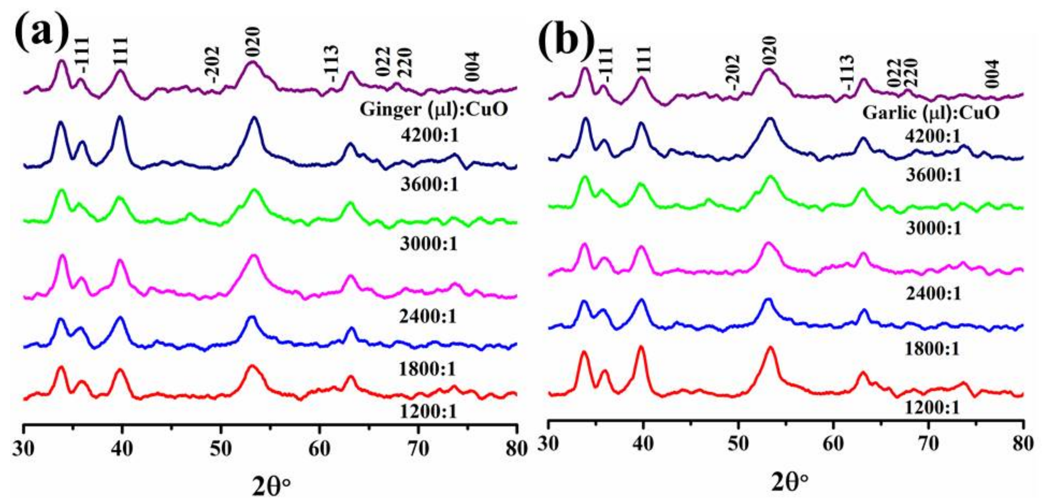
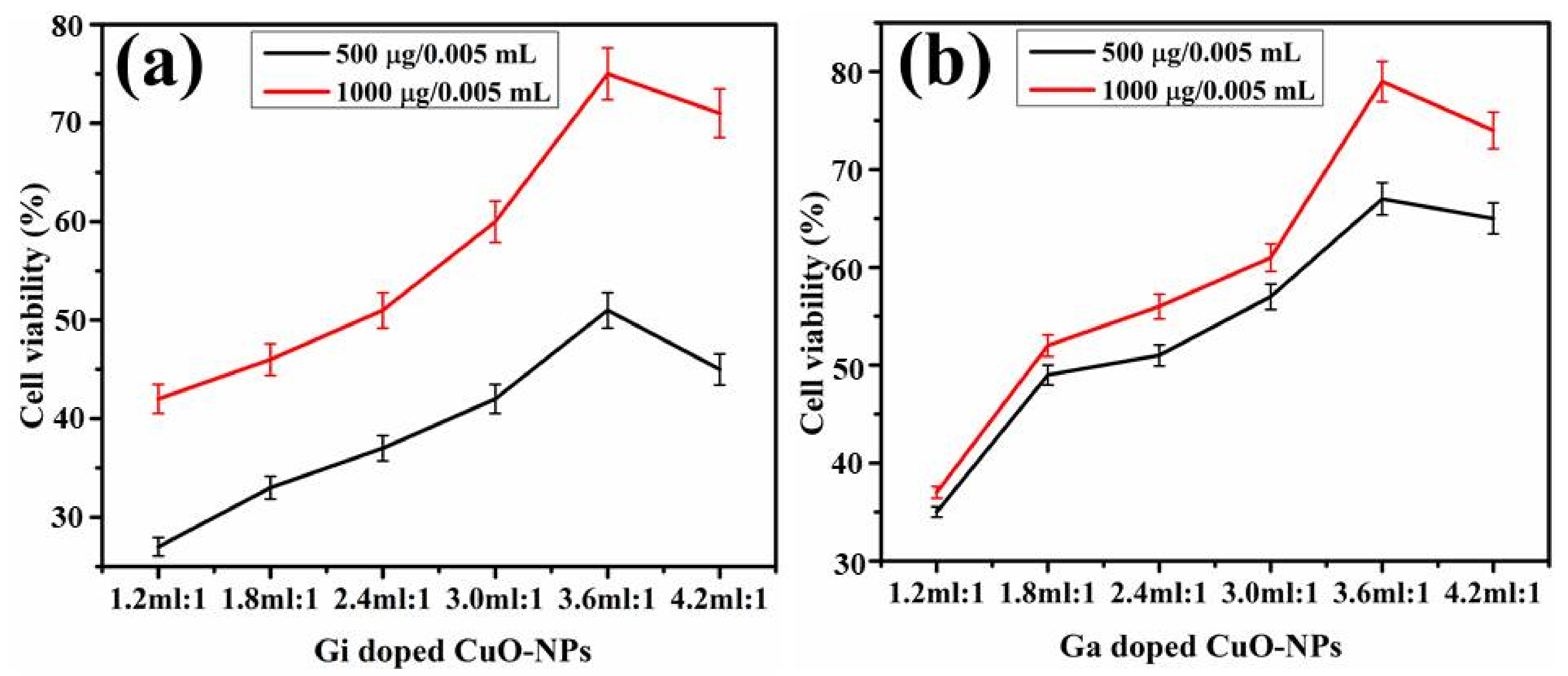
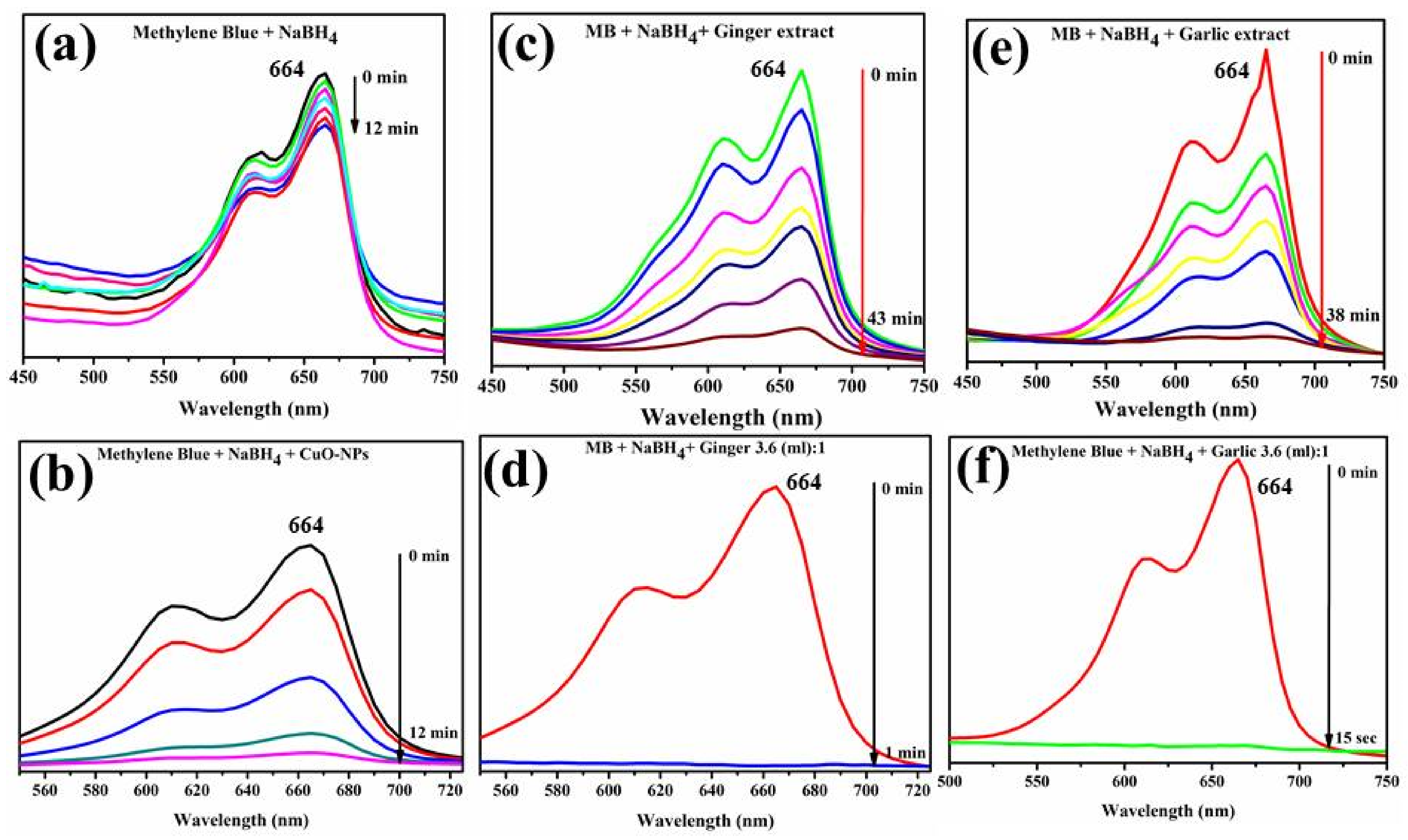
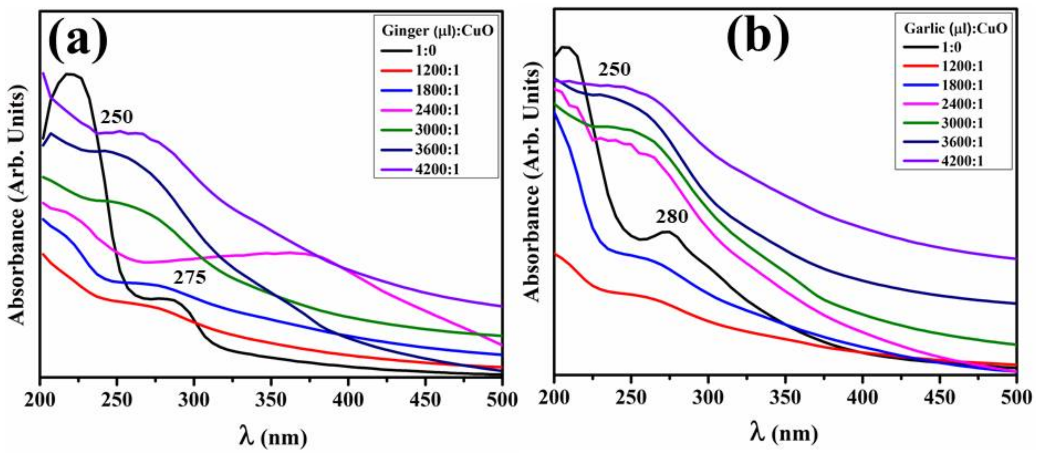
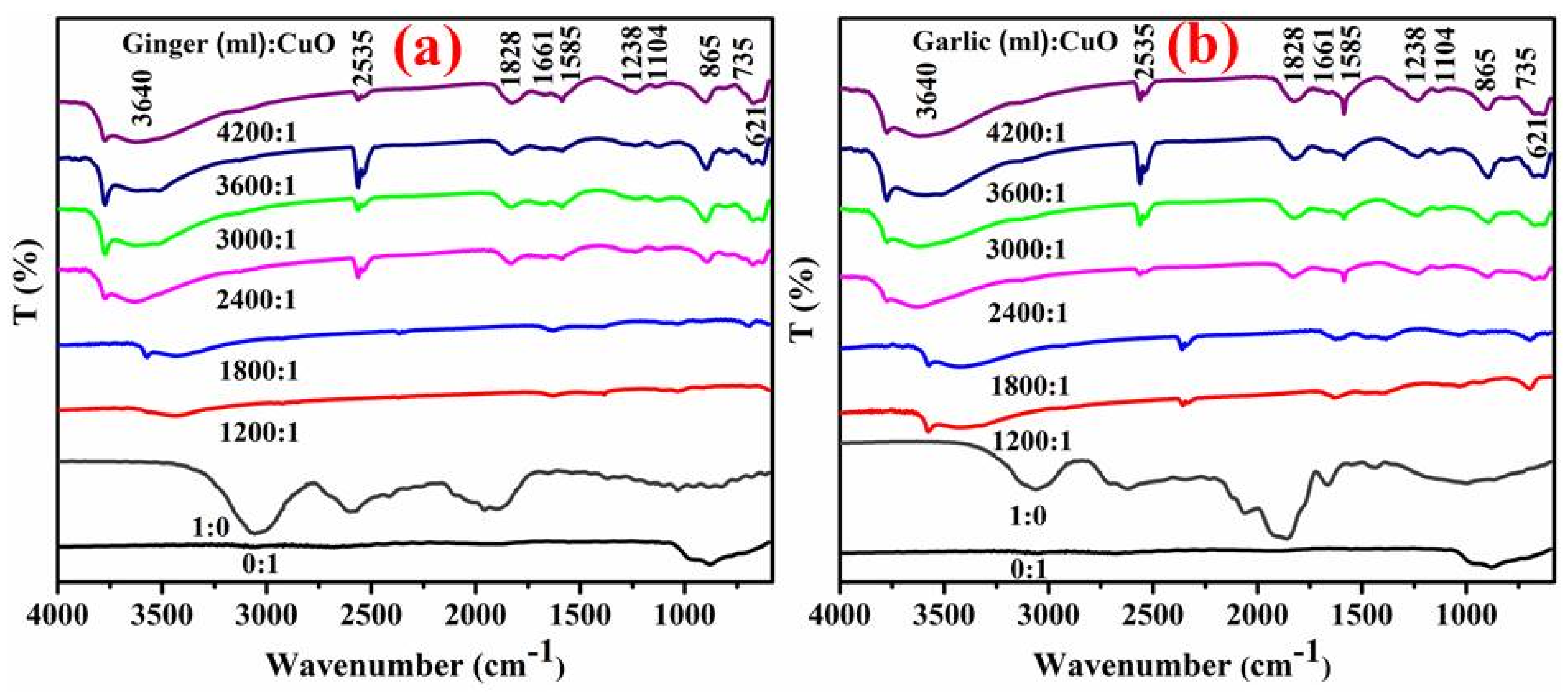
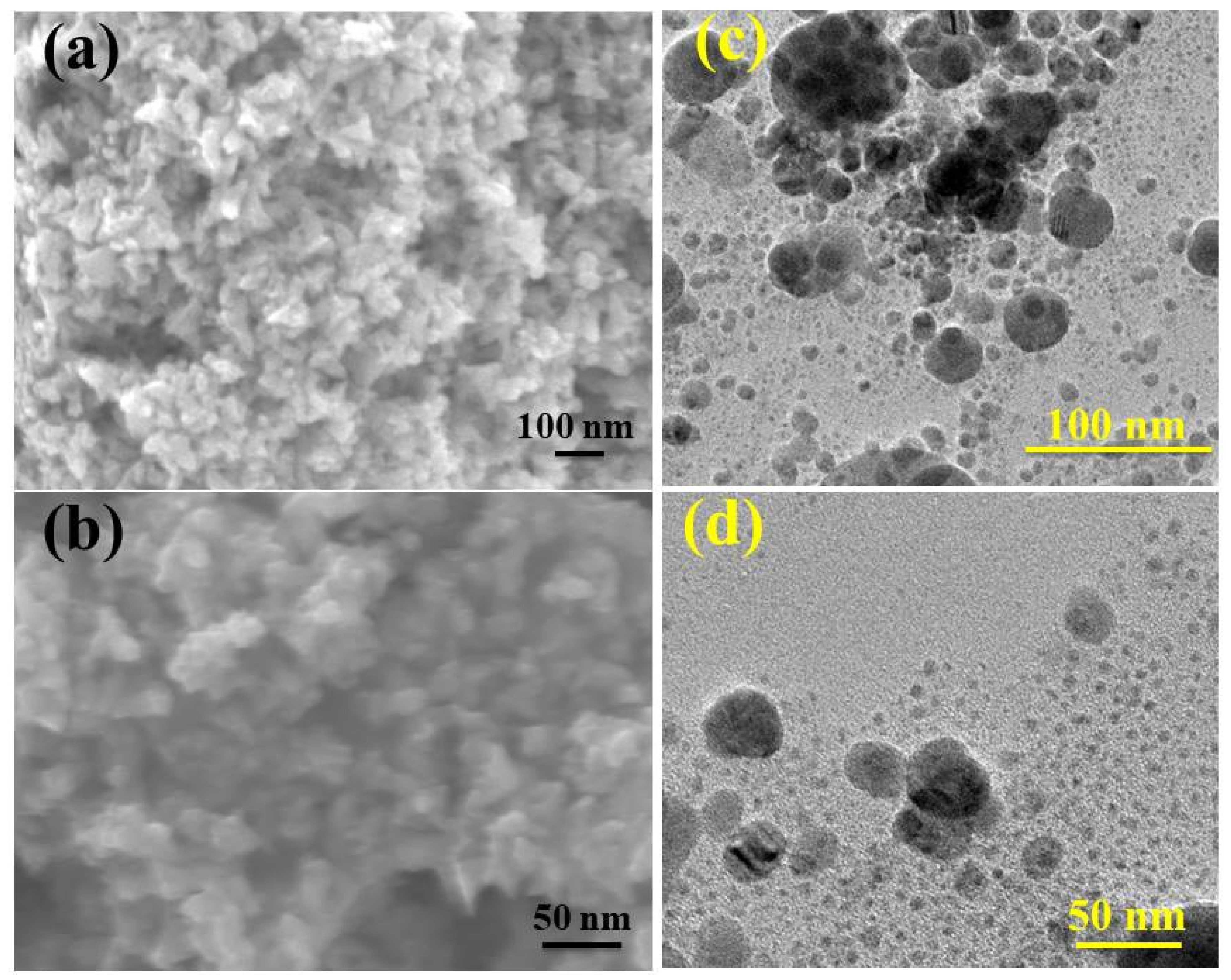
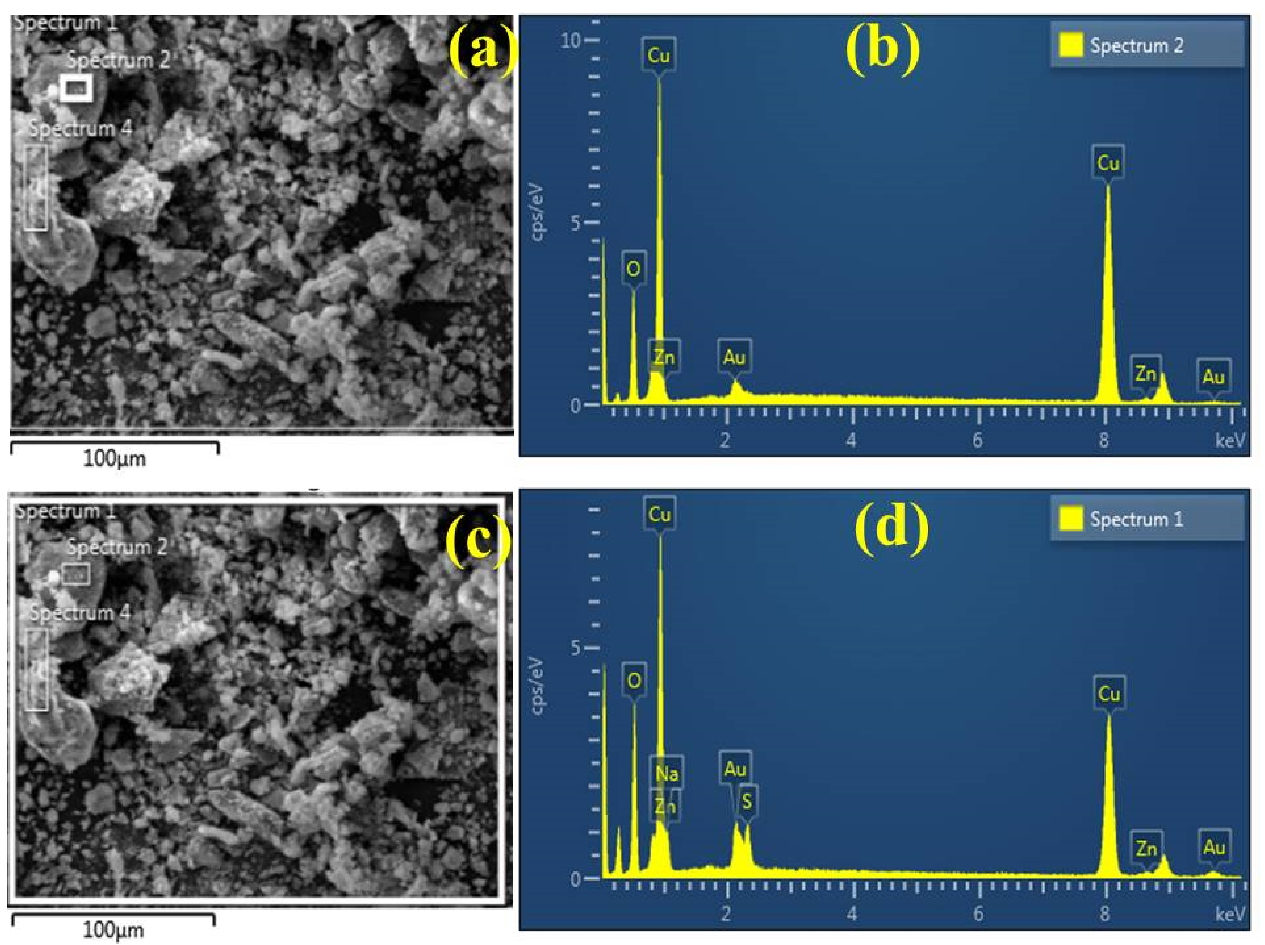
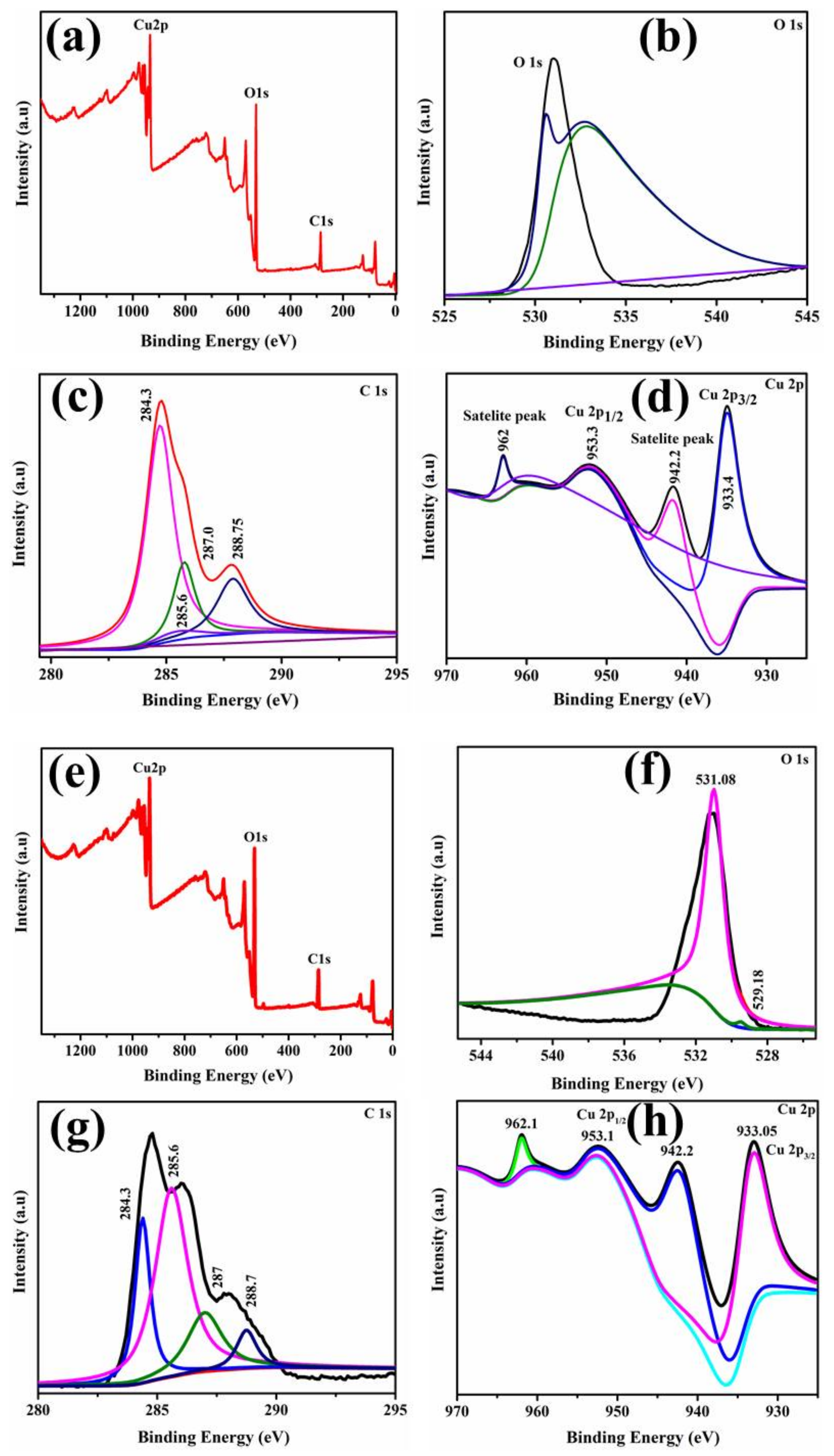
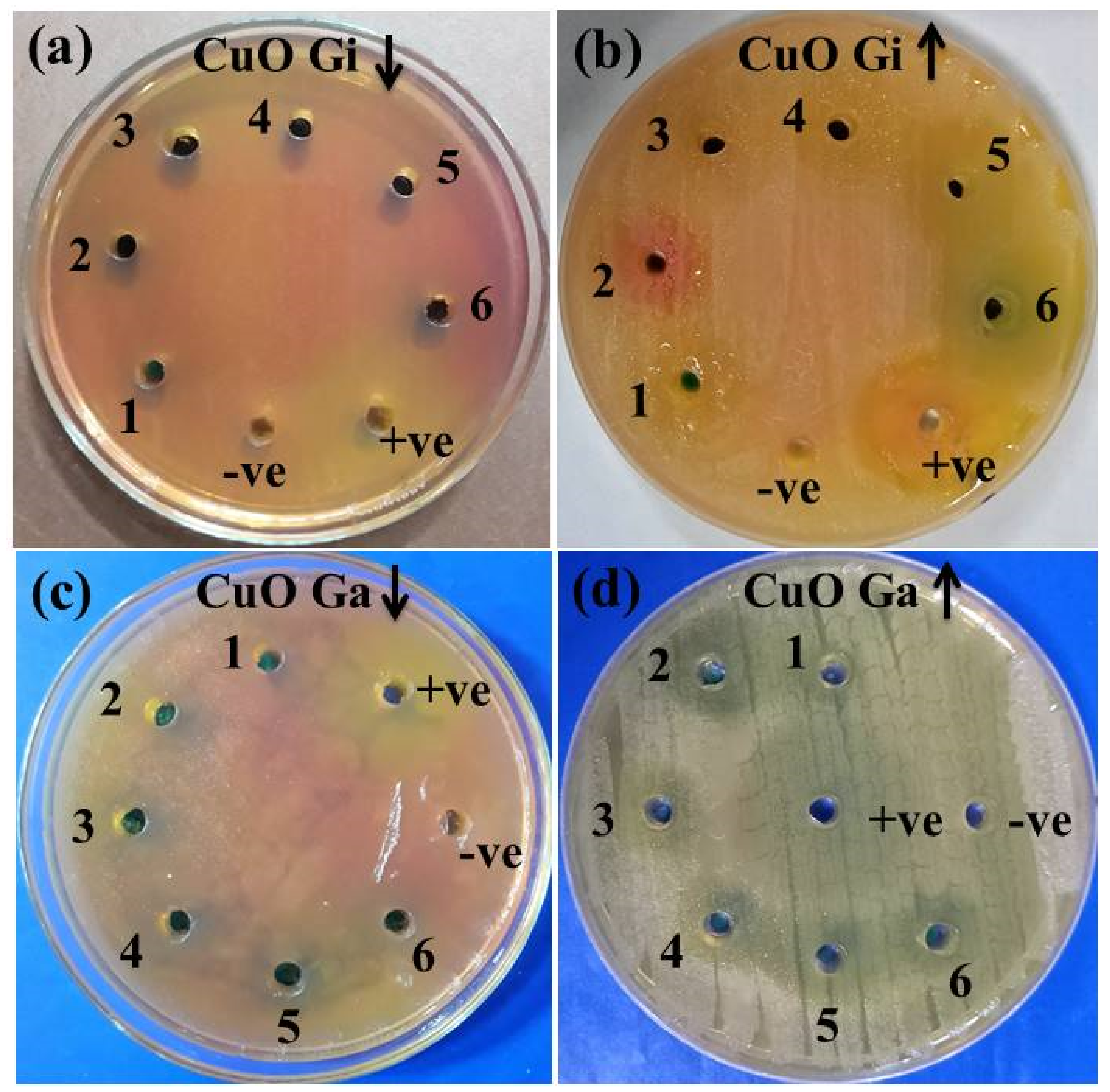
2. Results
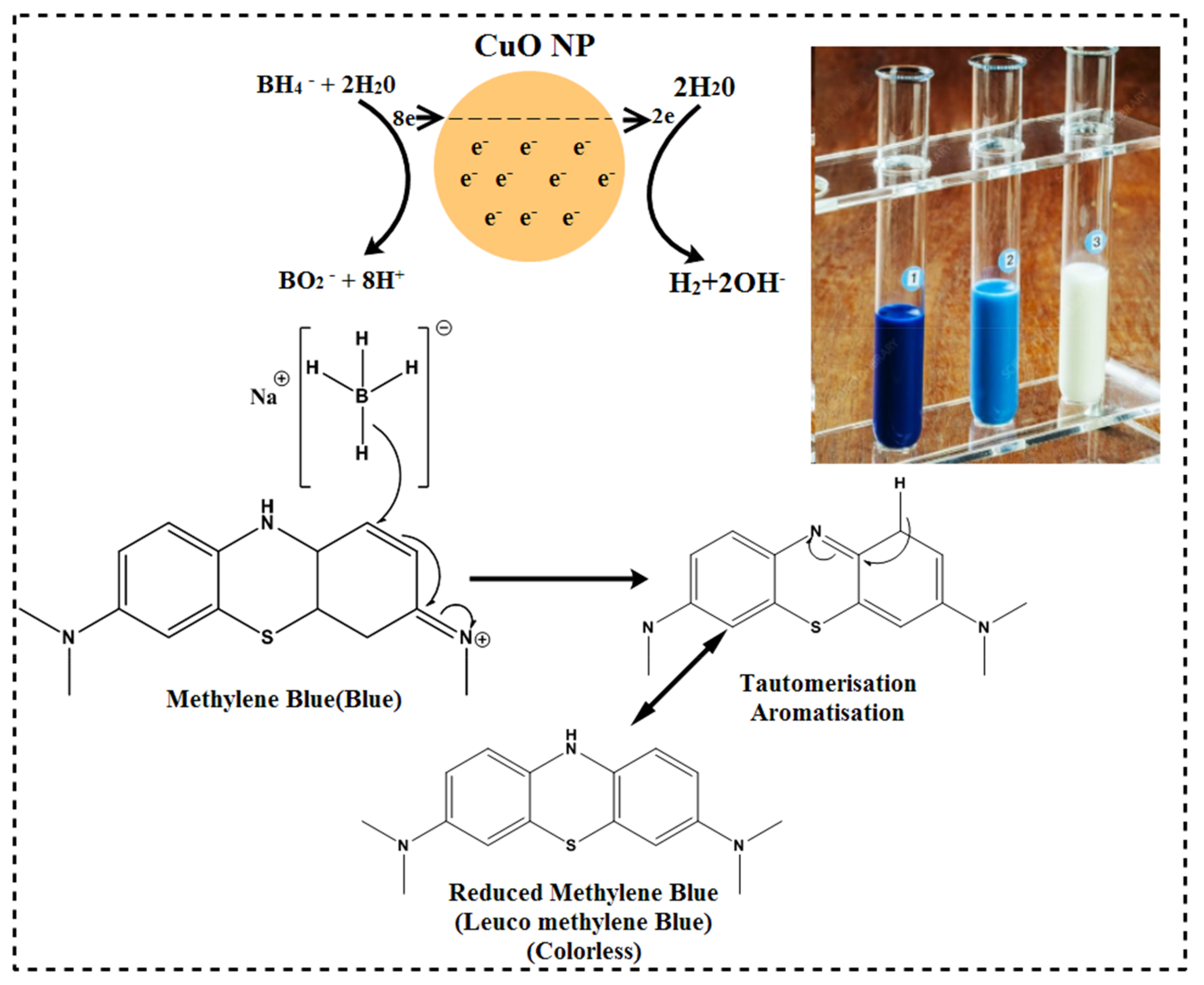
Catalytic Activity
3. Discussion
4. Materials and Methods
4.1. Materials
4.2. Aqueous Extracts Preparation
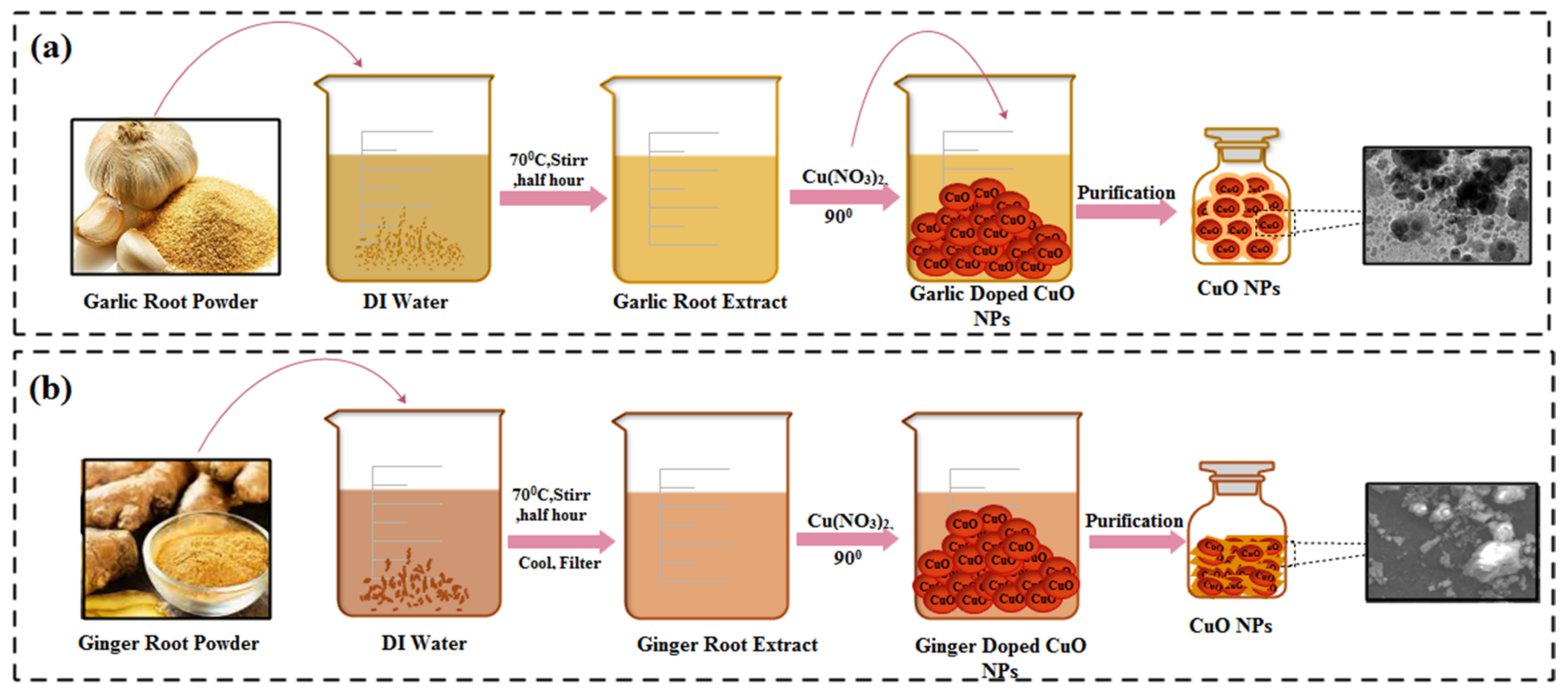
4.3. Green Fabrication of CuO NPs
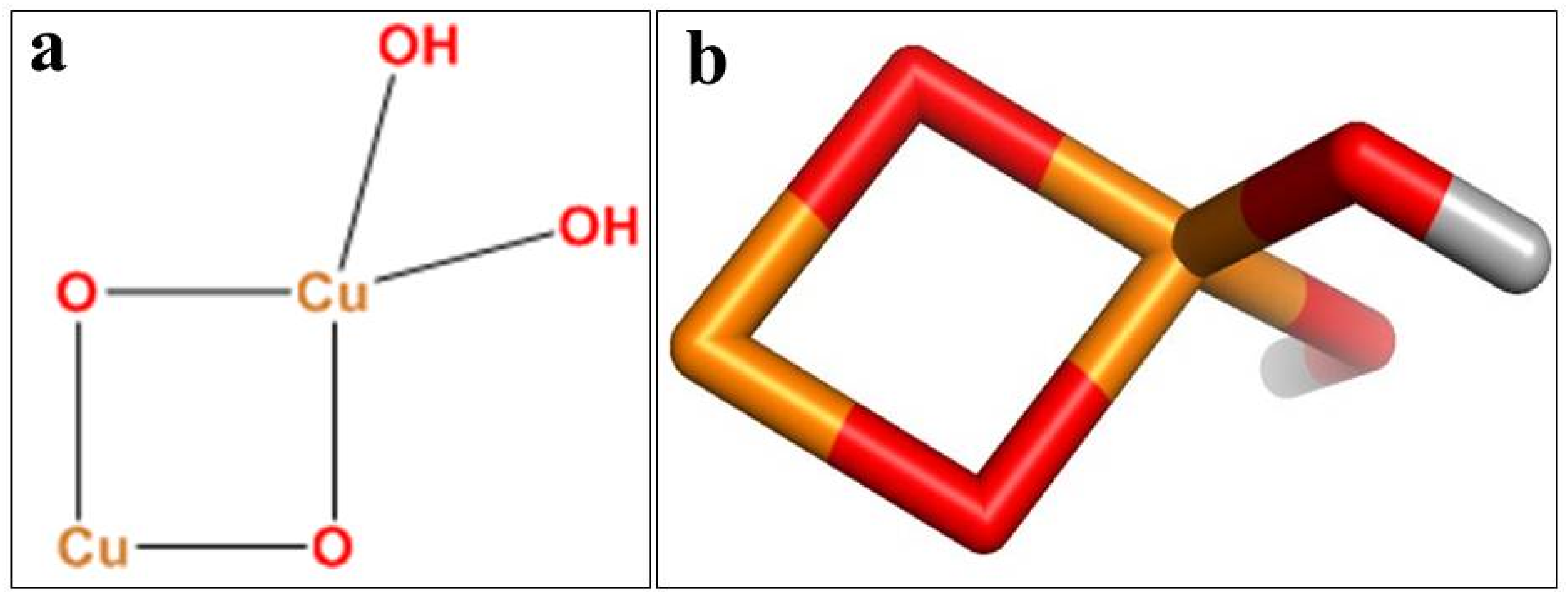
Characterization of Synthesized CuO NPs
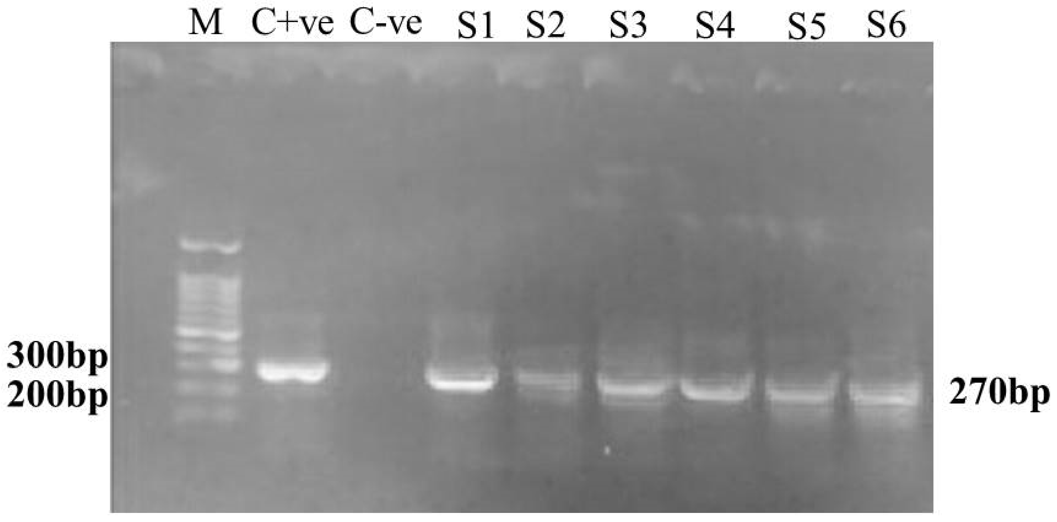
4.4. Separation and Identification of MDR S. aureus
4.4.1. Collection of Samples
4.4.2. Isolation of MDR S. aureus
4.4.3. Quantification of DNA with a NanoDrop Spectrophotometer
4.4.4. Primers Used and Polymerase Chain Reaction Amplification
4.5. Antimicrobial Activity
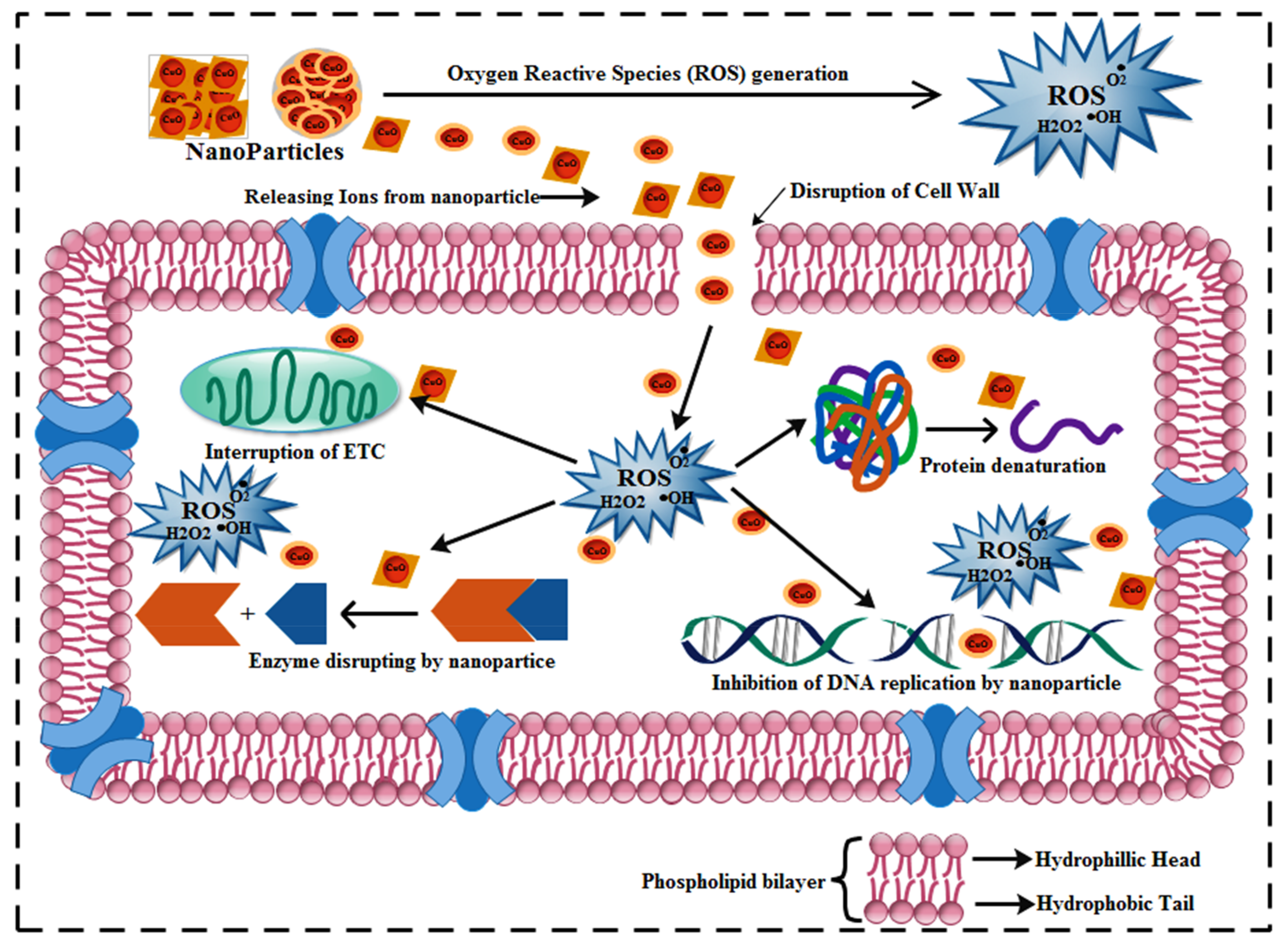
4.6. Effect of Doped CuO NPs on Bacterial Growth and Viability
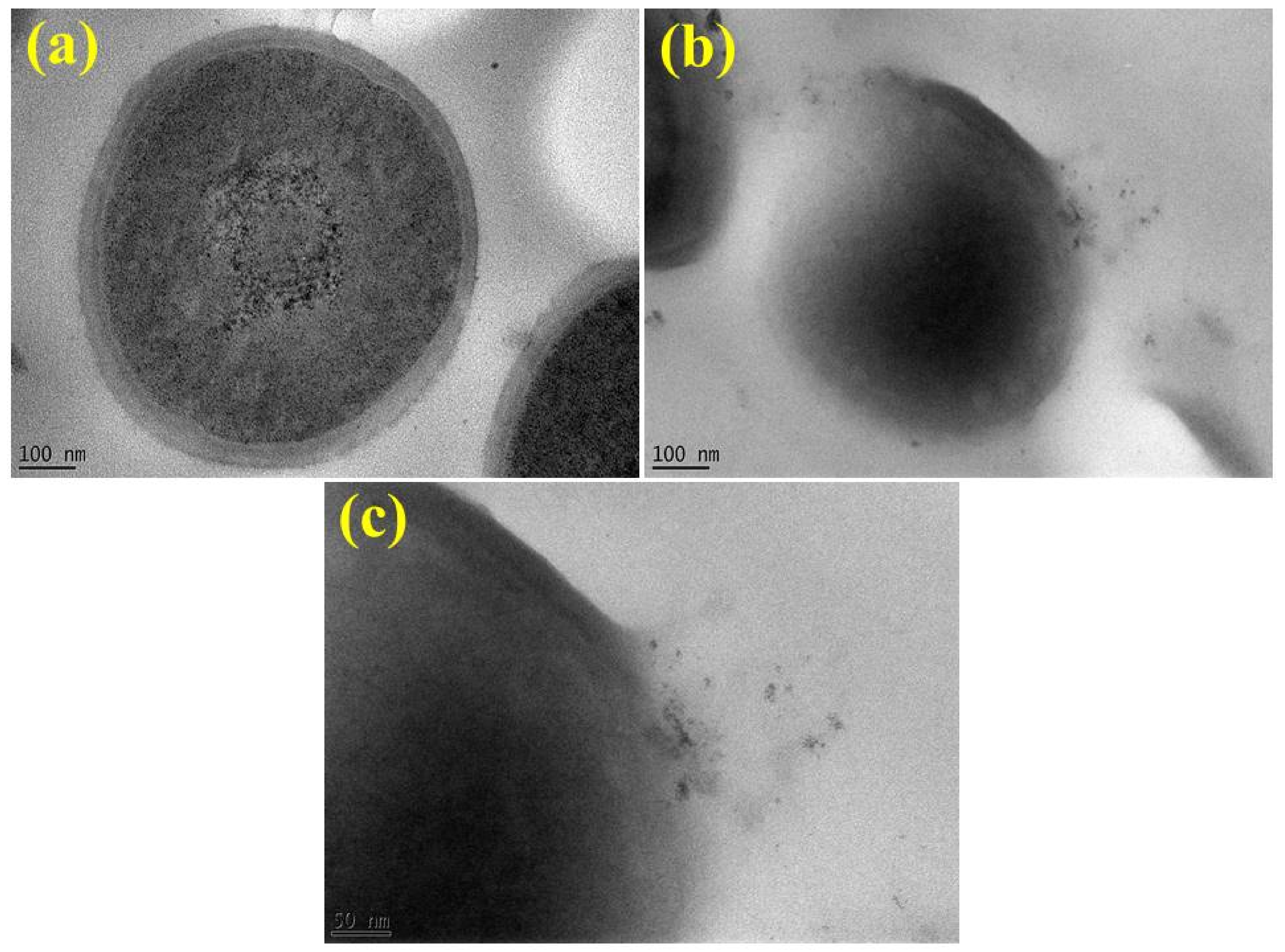
4.6.1. Pathogen and NPs Interaction with TEM Imaging
4.6.2. Statistical Analysis
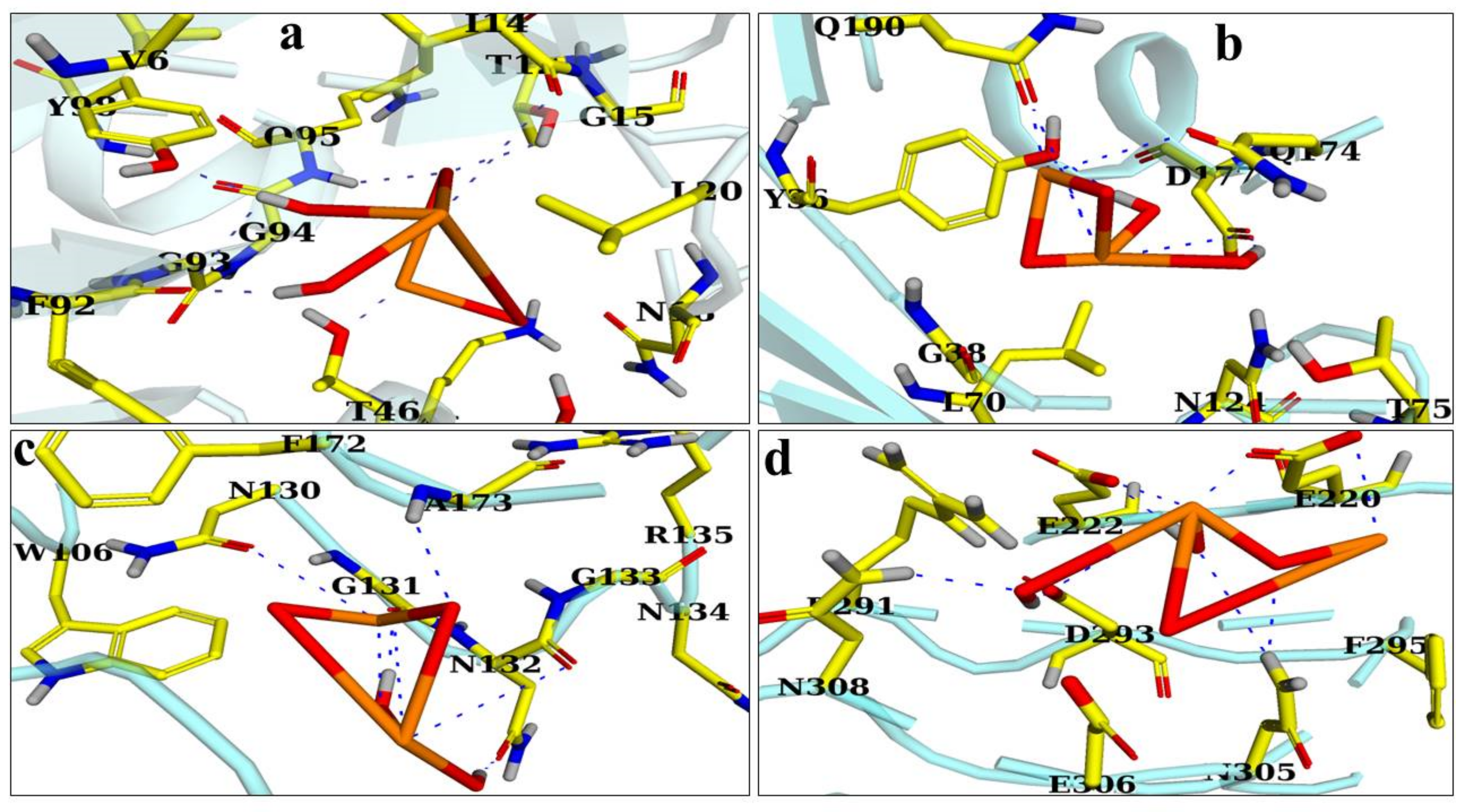
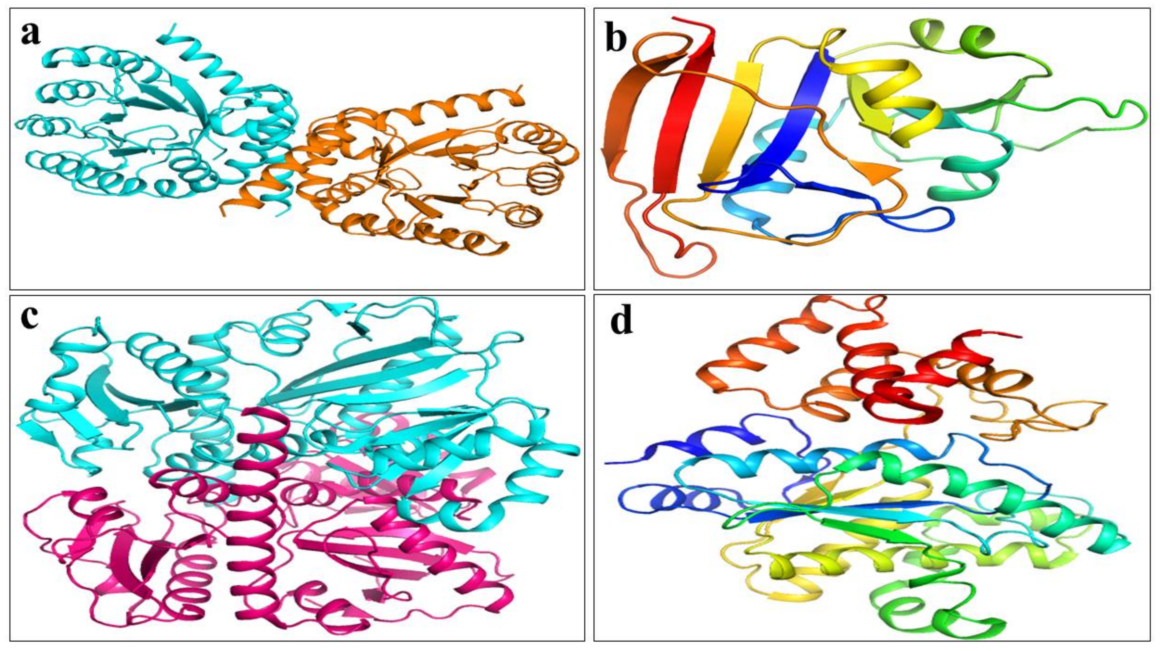
4.7. Molecular Docking Analysis
4.8. Catalytic Activity
5. Conclusions
Supplementary Materials
Author Contributions
Funding
Institutional Review Board Statement
Informed Consent Statement
Data Availability Statement
Acknowledgments
Conflicts of Interest
References
- Dodd, F. Mastitis—progress on control. J. Dairy Sci. 1983, 66, 1773–1780. [Google Scholar] [CrossRef]
- Rinaldi, M.; Li, R.W.; Capuco, A.V. Mastitis associated transcriptomic disruptions in cattle. Vet. Immunol. Immunopathol. 2010, 138, 267–279. [Google Scholar] [CrossRef] [PubMed]
- Hussain, R.; Javed, M.T.; Khan, A.; Mahmood, F.; Kausar, R. Mastitis and associated histo-pathological consequences in the context of udder morphology. Int. J. Agric. Biol. 2012, 14, 947–952. [Google Scholar]
- Hussain, R.; Khan, A.; Javed, M.T.; Ali, F. Morphometric and pathological studies on mammary gland of slaughtered Nili-Ravi buffaloes. Pak. J. Agri. Sci. 2013, 50, 123–130. [Google Scholar]
- Oliver, S.; Gonzalez, R.; Hogan, J.; Jayarao, B.; Owens, W. Microbiological Procedures for the Diagnosis of Bovine Udder Infection and Determination of Milk Quality; The National Mastitis Council. Inc.: Verona, WI, USA, 2004. [Google Scholar]
- Hawkey, P. The growing burden of antimicrobial resistance. J. Antimicrob. Chemother. 2008, 62, i1–i9. [Google Scholar] [CrossRef]
- Komolafe, O. Antibiotic resistance in bacteria-an emerging public health problem. Malawi Med. J. 2003, 15, 63–67. [Google Scholar] [CrossRef] [Green Version]
- Lowy, F.D. Staphylococcus aureus infections. N. Engl. J. Med. 1998, 339, 520–532. [Google Scholar] [CrossRef]
- Bradley, A.; Leach, K.; Breen, J.; Green, L.; Green, M. Survey of the incidence and aetiology of mastitis on dairy farms in England and Wales. Vet. Record. 2007, 160, 253–258. [Google Scholar] [CrossRef] [Green Version]
- Barkema, H.; Schukken, Y.; Zadoks, R. Invited review: The role of cow, pathogen, and treatment regimen in the therapeutic success of bovine Staphylococcus aureus mastitis. J. Dairy Sci. 2006, 89, 1877–1895. [Google Scholar] [CrossRef] [Green Version]
- Paterson, G.K.; Harrison, E.M.; Holmes, M.A. The emergence of mecC methicillin-resistant Staphylococcus aureus. Trends Microbiol. 2014, 22, 42–47. [Google Scholar] [CrossRef] [Green Version]
- Barkema, H.W.; Green, M.; Bradley, A.J.; Zadoks, R. Invited review: The role of contagious disease in udder health. J. Dairy Sci. 2009, 92, 4717–4729. [Google Scholar] [CrossRef] [PubMed]
- Brady, R.A.; Graeme, A.; Leid, J.G.; Prior, M.L.; Costerton, J.W.; Shirtliff, M.E. Resolution of Staphylococcus aureus biofilm infection using vaccination and antibiotic treatment. Infect. Immun. 2011, 79, 1797–1803. [Google Scholar] [CrossRef] [PubMed] [Green Version]
- Kenar, B.; Kuyucuoğlu, Y.; Şeker, E. Antibiotic susceptibility of coagulase-negative staphylococci isolated from bovine subclinical mastitis in Turkey. Pak. Vet. J. 2012, 32, 390–393. [Google Scholar]
- Declercq, P.; Petre, D.; Gordts, B.; Voss, A. Complicated community-acquired soft tissue infection by MRSA from porcine origin. Infection 2008, 36, 590–592. [Google Scholar] [CrossRef]
- Witte, W.; Strommenger, B.; Stanek, C.; Cuny, C. Methicillin-resistant Staphylococcus aureus ST398 in humans and animals, Central Europe. Emerg. Infect. Dis. 2007, 13, 255. [Google Scholar] [CrossRef]
- Nunan, C.; Young, R. MRSA in Farm Animals and Meat, a New Threat to Human Health; Soil Association: Bristol, UK, 2007. [Google Scholar]
- Elhaig, M.M.; Selim, A.; Mahmoud, M.M.; El-Gayar, E.K. Molecular confirmation of Trypanosoma evansi and Babesia bigemina in cattle from lower Egypt. Pak. Vet. J. 2016, 36, 409–414. [Google Scholar]
- Qayyum, A.; Khan, J.A.; Hussain, R.; Avais, M.; Ahmad, N.; Khan, M.S. Investigation of milk and blood serum biochemical profile as an indicator of sub-clinical mastitis in Cholistani cattle. Pak. Vet. J. 2016, 36, 275–279. [Google Scholar]
- Yilmaz, R.; Cangul, I.; Onat, K.; Akkoc, A.; Ozyigit, M.; Akdesir, E. Histopathological, immunohistochemical and bacteriological characterization of Mycoplasma bovis pneumonia in cattle. Pak. Vet. J. 2016, 36, 316–321. [Google Scholar]
- Adesiyun, A.A.; Webb, L.; Romain, H.T. Prevalence and characteristics of Staphylococcus aureus strains isolated from bulk and composite milk and cattle handlers. J. Food Prot. 1998, 61, 629–632. [Google Scholar] [CrossRef]
- Jahan, M.; Rahman, M.; Parvej, M.S.; Chowdhury, S.M.Z.H.; Haque, M.E.; Talukder, M.A.K.; Ahmed, S. Isolation and characterization of Staphylococcus aureus from raw cow milk in Bangladesh. J. Adv. Vet. Anim. Res. 2015, 2, 49–55. [Google Scholar] [CrossRef]
- Zecconi, A.; Piccinini, R. Staphilococcus aureus: A problem for Italian dairy herds. Int. Dairy Fed. 1998, 22, 25–26. [Google Scholar]
- Alonzo, F., III; Benson, M.A.; Chen, J.; Novick, R.P.; Shopsin, B.; Torres, V.J. Staphylococcus aureus leucocidin ED contributes to systemic infection by targeting neutrophils and promoting bacterial growth in vivo. Mol. Microbiol. 2012, 83, 423–435. [Google Scholar] [CrossRef] [Green Version]
- Kreger, A.S.; Kim, K.S.; Zaboretzky, F.; Bernheimer, A.W. Purification and properties of staphylococcal delta hemolysin. Infect. Immun. 1971, 3, 449–465. [Google Scholar] [CrossRef] [PubMed] [Green Version]
- Malachowa, N.; Kobayashi, S.D.; Braughton, K.R.; Whitney, A.R.; Parnell, M.J.; Gardner, D.J.; DeLeo, F.R. Staphylococcus aureus leukotoxin GH promotes inflammation. J. Infect. Dis. 2012, 206, 1185–1193. [Google Scholar] [CrossRef] [PubMed]
- Ventura, C.L.; Malachowa, N.; Hammer, C.H.; Nardone, G.A.; Robinson, M.A.; Kobayashi, S.D.; DeLeo, F.R. Identification of a novel Staphylococcus aureus two-component leukotoxin using cell surface proteomics. PLoS ONE 2010, 5, e11634. [Google Scholar] [CrossRef] [PubMed]
- Wang, R.; Braughton, K.R.; Kretschmer, D.; Bach, T.H.L.; Queck, S.Y.; Li, M.; Kennedy, A.D.; Dorward, D.W.; Klebanoff, S.J.; Peschel, A. Identification of novel cytolytic peptides as key virulence determinants for community-associated MRSA. Nat. Med. 2007, 13, 1510–1514. [Google Scholar] [CrossRef]
- Kobayashi, S.D.; Malachowa, N.; DeLeo, F.R. Pathogenesis of Staphylococcus aureus abscesses. Am. J. Pathol. 2015, 185, 1518–1527. [Google Scholar] [CrossRef] [Green Version]
- Raj, A.; Lawrence, R.S.; Jalees, M.; Lawrence, K. Anti-bacterial activity of zinc oxide nanoparticles prepared from Brassica oleraceae leaves extract. Int. J. Adv. Res. 2015, 3, 322–328. [Google Scholar]
- Van den Bogaard, A.E.; Stobberingh, E.E. Epidemiology of resistance to antibiotics: Links between animals and humans. Int. J. Antimicrob. Agents 2000, 14, 327–335. [Google Scholar] [CrossRef]
- Allahverdiyev, A.M.; Abamor, E.S.; Bagirova, M.; Rafailovich, M. Antimicrobial effects of TiO2 and Ag2O nanoparticles against drug-resistant bacteria and leishmania parasites. Future Microbiol. 2011, 6, 933–940. [Google Scholar] [CrossRef]
- Padnya, P.; Gorbachuk, V.; Stoikov, I. The Role of Calix [n] arenes and Pillar [n] arenes in the Design of Silver Nanoparticles: Self-Assembly and Application. Int. J. Mol. Sci. 2020, 21, 1425. [Google Scholar] [CrossRef] [PubMed] [Green Version]
- Khatami, M.; Alijani, H.Q.; Heli, H.; Sharifi, I. Rectangular shaped zinc oxide nanoparticles: Green synthesis by Stevia and its biomedical efficiency. Ceram. Int. 2018, 44, 15596–15602. [Google Scholar] [CrossRef]
- Drummer, S.; Madzimbamuto, T.; Chowdhury, M. Green Synthesis of Transition-Metal Nanoparticles and Their Oxides: A Review. Materials 2021, 14, 2700. [Google Scholar] [CrossRef] [PubMed]
- Asghari, F.; Jahanshiri, Z.; Imani, M.; Shams-Ghahfarokhi, M.; Razzaghi-Abyaneh, M. Antifungal nanomaterials: Synthesis, properties, and applications. In Nanobiomaterials in Antimicrobial Therapy; Elsevier: Amsterdam, The Netherlands, 2016; pp. 343–383. [Google Scholar]
- Liu, H.; Zheng, S.; Xiong, H.; Alwahibi, M.S.; Niu, X. Biosynthesis of copperoxide nanoparticles using Abies spectabilis plant extract and analyzing its antinociceptive and anti-inflammatory potency in various mice models. Arab. J. Chem. 2020, 13, 6995–7006. [Google Scholar] [CrossRef]
- Ijaz, F.; Shahid, S.; Khan, S.A.; Ahmad, W.; Zaman, S. Green synthesis of copper oxide nanoparticles using Abutilon indicum leaf extract: Antimicrobial, antioxidant and photocatalytic dye degradation activitie. Trop. J. Pharm. Res. 2017, 16, 743–753. [Google Scholar] [CrossRef] [Green Version]
- Dey, A.; Manna, S.; Chattopadhyay, S.; Mondal, D.; Chattopadhyay, D.; Raj, A.; Das, S.; Bag, B.G.; Roy, S. Azadirachta indica leaves mediated green synthesized copper oxide nanoparticles induce apoptosis through activation of TNF-α and caspases signaling pathway against cancer cells. J. Saudi Chem. Soc. 2019, 23, 222–238. [Google Scholar] [CrossRef]
- Aminuzzaman, M.; Kei, L.; Liang, W. Green and Sustainable Technology. AIP Conf. Proc. 2017, 1828, 020016. [Google Scholar]
- Shanan, Z.J.; Hadi, S.M.; Shanshool, S.K. Structural analysis of chemical and green synthesis of CuO nanoparticles and their effect on biofilm formation. Baghdad Sci. J. 2018, 15, 211–216. [Google Scholar]
- Keabadile, O.P.; Aremu, A.O.; Elugoke, S.E.; Fayemi, O.E. Green and Traditional Synthesis of Copper Oxide Nanoparticles—Comparative Study. Nanomaterials 2020, 10, 2502. [Google Scholar] [CrossRef]
- Chouke, P.; Potbhare, A.; Dadure, K.; Mungole, A.; Meshram, N.; Chaudhary, R.; Rai, A.; Chaudhary, R. An antibacterial activity of Bauhinia racemosa assisted ZnO nanoparticles during lunar eclipse and docking assay. Mater. Today Proc. 2020, 29, 815–821. [Google Scholar] [CrossRef]
- Chouke, P.B.; Potbhare, A.K.; Bhusari, G.S.; Somkuwar, S.; Shaik, D.P.; Mishra, R.K.; Chaudhary, R.G. Green fabrication of Zinc oxide nanospheres by Aspidopterys Cordata for effective antioxidant and antibacterial activity. Adv. Mater Lett. 2019, 10, 355–360. [Google Scholar] [CrossRef]
- Król, A.; Pomastowski, P.; Rafińska, K.; Railean-Plugaru, V.; Buszewski, B. Zinc oxide nanoparticles: Synthesis, antiseptic activity and toxicity mechanism. Adv. Colloid Interface Sci. 2017, 249, 37–52. [Google Scholar] [CrossRef] [PubMed]
- Jamdagni, P.; Khatri, P.; Rana, J. Green synthesis of zinc oxide nanoparticles using flower extract of Nyctanthes arbor-tristis and their antifungal activity. J. King Saud. Univ. Sci. 2018, 30, 168–175. [Google Scholar] [CrossRef] [Green Version]
- Vinayagam, R.; Selvaraj, R.; Arivalagan, P.; Varadavenkatesan, T. Synthesis, characterization and photocatalytic dye degradation capability of Calliandra haematocephala-mediated zinc oxide nanoflowers. J. Photochem. Photobiol. B Biol. 2020, 203, 111760. [Google Scholar] [CrossRef] [PubMed]
- Mary, A.A.; Ansari, A.T.; Subramanian, R. Sugarcane juice mediated synthesis of copper oxide nanoparticles, characterization and their antibacterial activity. J. King Saud Univ. Sci. 2019, 31, 1103–1114. [Google Scholar] [CrossRef]
- Kargar, M.; Ghashang, M.; Mohammad Shafiee, M. An Efficient Green Synthesis of Copper Oxide NanoCrystalline. Int. J. Bio-Inorg. Hybrid Nanomater. 2015, 4, 87–92. [Google Scholar]
- Cuong, H.N.; Pansambal, S.; Ghotekar, S.; Oza, R.; Hai, N.T.T.; Viet, N.M.; Nguyen, V.H. New frontiers in the plant extract mediated biosynthesis of copper oxide (CuO) nanoparticles and their potential applications: A review. Environ. Res. 2022, 203, 111858. [Google Scholar] [CrossRef]
- Maham, M.; Sajadi, S.M.; Kharimkhani, M.M.; Nasrollahzadeh, M. Biosynthesis of the CuO nanoparticles using Euphorbia Chamaesyce leaf extract and investigation of their catalytic activity for the reduction of 4-nitrophenol. IET Nanobiotechnol. 2017, 11, 766–772. [Google Scholar] [CrossRef]
- Bordbar, M.; Sharifi-Zarchi, Z.; Khodadadi, B. Green synthesis of copper oxide nanoparticles/clinoptilolite using Rheum palmatum L. root extract: High catalytic activity for reduction of 4-nitro phenol, rhodamine B., and methylene blue. J. Sol-Gel Sci. Technol. 2017, 81, 724–733. [Google Scholar] [CrossRef]
- Nasrollahzadeh, M.; Maham, M.; Sajadi, S.M. Green synthesis of CuO nanoparticles by aqueous extract of Gundelia tournefortii and evaluation of their catalytic activity for the synthesis of N-monosubstituted ureas and reduction of 4-nitrophenol. J. Colloid Interface Sci. 2015, 455, 245–253. [Google Scholar] [CrossRef]
- Patel, V.K.; Bhattacharya, S. Solid state green synthesis and catalytic activity of CuO nanorods in thermal decomposition of potassium periodate. Mater. Res. Express 2017, 4, 095012. [Google Scholar] [CrossRef] [Green Version]
- Nasrollahzadeh, M.; Sajadi, S.M.; Rostami-Vartooni, A.; Hussin, S.M. Green synthesis of CuO nanoparticles using aqueous extract of Thymus vulgaris L. leaves and their catalytic performance for N-arylation of indoles and amines. J. Colloid Interface Sci. 2016, 466, 113–119. [Google Scholar] [CrossRef] [PubMed]
- Patil, A.S.; Patil, M.D.; Lohar, G.M.; Jadhav, S.T.; Fulari, V.J. Supercapacitive properties of CuO thin films using modified SILAR method. Ionics 2017, 23, 1259–1266. [Google Scholar] [CrossRef]
- Pansambal, S.; Deshmukh, K.; Savale, A.; Ghotekar, S.; Pardeshi, O.; Jain, G.; Aher, Y.; Pore, D. Phytosynthesis and biological activities of fluorescent CuO nanoparticles using Acanthospermum hispidum L. extract. J. Nanostructures 2017, 7, 165–174. [Google Scholar] [CrossRef]
- Baig, N.; Saleh, T.A. Superhydrophobic Polypropylene Functionalized with Nanoparticles for Efficient Fast Static and Dynamic Separation of Spilled Oil from Water. Glob. Chall. 2019, 3, 1800115. [Google Scholar] [CrossRef] [Green Version]
- Ghosh, S.; Kundu, S.; Naskar, M.K. Mesoporous CuO nanostructures for low-temperature CO oxidation. Bull. Mater. Sci. 2021, 44, 1–8. [Google Scholar] [CrossRef]
- Hamza, M.F.; Wei, Y.; Mira, H.; Adel, A.H.; Guibal, E. Synthesis and adsorption characteristics of grafted hydrazinyl amine magnetite-chitosan for Ni (II) and Pb (II) recovery. Chem. Eng. J. 2019, 362, 310–324. [Google Scholar] [CrossRef] [Green Version]
- Jurado-López, B.; Vieira, R.S.; Rabelo, R.B.; Beppu, M.M.; Casado, J.; Rodríguez-Castellón, E. Formation of complexes between functionalized chitosan membranes and copper: A study by angle resolved XPS. Mater. Chem. Phys. 2017, 185, 152–161. [Google Scholar] [CrossRef]
- Ortega-Liebana, M.; Chung, N.; Limpens, R.; Gomez, L.; Hueso, J.; Santamaria, J.; Gregorkiewicz, T. Uniform luminescent carbon nanodots prepared by rapid pyrolysis of organic precursors confined within nanoporous templating structures. Carbon 2017, 117, 437–446. [Google Scholar] [CrossRef]
- Poulston, S.; Parlett, P.; Stone, P.; Bowker, M. Surface oxidation and reduction of CuO and Cu2O studied using XPS and XAES. Surf. Interface Anal. 1996, 24, 811–820. [Google Scholar] [CrossRef]
- Brakstad, O.G.; Aasbakk, K.; Maeland, J.A. Detection of Staphylococcus aureus by polymerase chain reaction amplification of the nuc gene. J. Clin. Microbiol. 1992, 30, 1654–1660. [Google Scholar] [CrossRef] [PubMed] [Green Version]
- Jesudoss, S.K.; Vijaya, J.J.; Kennedy, L.J.; Rajan, P.I.; Al-Lohedan, A.H.; Ramalingam, R.J.; Kaviyarasu, K.; Bououdina, M. Studies on the efficient dual performance of Mn1–xNixFe2O4 spinel nanoparticles in photodegradation and antibacterial activity. J. Photochem. Photobio. B Biol. 2016, 165, 121–132. [Google Scholar] [CrossRef]
- Navale, G.R.; Rout, C.S.; Gohil, K.N.; Dharne, M.S.; Late, D.J.; Shinde, S.S. Oxidative and membrane stress-mediated antibacterial activity of ws 2 and rgo-ws 2 nanosheets. Rsc Adv. 2015, 5, 74726–74733. [Google Scholar] [CrossRef]
- Ahmed, B.; Hashmi, A.; Khan, M.S.; Musarrat, J. ROS mediated destruction of cell membrane, growth and biofilms of human bacterial pathogens by stable metallic AgNPs functionalized from bell pepper extract and quercetin. Adv. Powder Technol. 2018, 29, 1601–1616. [Google Scholar] [CrossRef]
- Ahmed, B.; Solanki, B.; Zaidi, A.; Khan, M.S.; Musarrat, J. Bacterial toxicity of biomimetic green zinc oxide nanoantibiotic: Insights into ZnONP uptake and nanocolloid–bacteria interface. Toxicol. Res. 2019, 8, 246–261. [Google Scholar] [CrossRef] [PubMed] [Green Version]
- Ali, K.; Ahmed, B.; Ansari, S.M.; Saquib, Q.; Al-Khedhairy, A.A.; Dwivedi, S.; Alshaeri, M.; Khan, M.S.; Musarrat, J. Comparative in situ ROS mediated killing of bacteria with bulk analogue, Eucalyptus leaf extract (ELE)-capped and bare surface copper oxide nanoparticles. Mater. Sci. Eng. C 2019, 100, 747–758. [Google Scholar] [CrossRef] [PubMed]
- Ali, K.; Ahmed, B.; Khan, M.S.; Musarrat, J. Differential surface contact killing of pristine and low EPS Pseudomonas aeruginosa with Aloe vera capped hematite (α-Fe2O3) nanoparticles. J. Photochem. Photobiol. B Biol. 2018, 188, 146–158. [Google Scholar] [CrossRef] [PubMed]
- Haider, A.; Ijaz, M.; Imran, M.; Naz, M.; Majeed, H.; Khan, J.; Ali, M.; Ikram, M. Enhanced bactericidal action and dye degradation of spicy roots’ extract-incorporated fine-tuned metal oxide nanoparticles. Appl. Nanosci. 2020, 10, 1–10. [Google Scholar] [CrossRef]
- Haroon, M.; Zaidi, A.; Ahmed, B.; Rizvi, A.; Khan, M.S.; Musarrat, J. Effective inhibition of phytopathogenic microbes by eco-friendly leaf extract mediated silver nanoparticles (AgNPs). Indian J. Microbiol. 2019, 59, 273–287. [Google Scholar] [CrossRef]
- Haider, A.; Ijaz, M.; Ali, S.; Haider, J.; Imran, M.; Majeed, H.; Shahzadi, I.; Ali, M.M.; Khan, J.A.; Ikram, M. Green synthesized phytochemically (Zingiber officinale and Allium sativum) reduced nickel oxide nanoparticles confirmed bactericidal and catalytic potential. Nanoscale Res. Lett. 2020, 15, 1–11. [Google Scholar] [CrossRef]
- Raja, P.I.; Vijaya, J.J.; Jesudoss, S.; Kaviyarasu, K.; Kennedy, L.J.; Jothiramalingam, R.; Al-Lohedan, H.A.; Vaali-Mohammed M-A. Green-fuel-mediated synthesis of self-assembled NiO nano-sticks for dual applications—Photocatalytic activity on Rose Bengal dye and antimicrobial action on bacterial strains. Mater. Res. Express 2017, 4, 085030. [Google Scholar] [CrossRef]
- Jayawardena, H.S.N.; Jayawardana, K.W.; Chen, X.; Yan, M. Maltoheptaose promotes nanoparticle internalization by Escherichia coli. Chem. Commun. 2013, 49, 3034–3036. [Google Scholar] [CrossRef] [PubMed] [Green Version]
- Fatimah, I.; Aftrid, Z.H.V.I. Characteristics and antibacterial activity of green synthesized silver nanoparticles using red spinach (Amaranthus Tricolor, L.) leaf extract. Green Chem. Lett. Rev. 2019, 12, 25–30. [Google Scholar] [CrossRef] [Green Version]
- David, S.A.; Rajadurai, S.I.; Kumar, S.V. Biosynthesis of copper oxide nanoparticles using Momordica charantia leaf extract and their characterization. Int. J. Adv. Res. Sci. Eng. 2017, 6, 313–320. [Google Scholar]
- Ren, G.; Hu, D.; Cheng, E.W.; Vargas-Reus, M.A.; Reip, P.; Allaker, R.P. Characterisation of copper oxide nanoparticles for antimicrobial applications. Int. J. Antimicrob. Agents 2009, 33, 587–590. [Google Scholar] [CrossRef] [PubMed]
- Nabila, M.I.; Kannabiran, K. Biosynthesis, characterization and antibacterial activity of copper oxide nanoparticles (CuO NPs) from actinomycetes. Biocatal. Agric. Biotechnol. 2018, 15, 56–62. [Google Scholar] [CrossRef]
- Bigdeli, F.; Morsali, A.; Retailleau, P. Syntheses and characterization of different zinc (II) oxide nano-structures from direct thermal decomposition of 1D coordination polymers. Polyhedron 2010, 29, 801–806. [Google Scholar] [CrossRef]
- Ganesan, S.; Babu, I.G.; Mahendran, D.; Arulselvi, P.I.; Elangovan, N.; Geetha, N.; Venkatachalam, P. Green engineering of titanium dioxide nanoparticles using Ageratina altissima (L.) King & HE Robines. medicinal plant aqueous leaf extracts for enhanced photocatalytic activity. Ann. Phytomed 2016, 5, 69–75. [Google Scholar]
- Jafarirad, S.; Mehrabim, M.; Divband, B.; Kosari-Nasab, M. Biofabrication of zinc oxide nanoparticles using fruit extract of Rosa canina and their toxic potential against bacteria: A mechanistic approach. Mater. Sci. Eng. C 2016, 59, 296–302. [Google Scholar] [CrossRef]
- Jayaprakash, J.; Srinivasan, N.; Chandrasekaran, P.; Girija, E. Synthesis and characterization of cluster of grapes like pure and Zinc-doped CuO nanoparticles by sol–gel method. Spectrochim. Acta Part A Mol. Biomol. Spectrosc. 2015, 136, 1803–1806. [Google Scholar] [CrossRef]
- Velsankar, K.; Kumar, A.R.M.; Preethi, R.; Muthulakshmi, V.; Sudhahar, S. Green synthesis of CuO nanoparticles via Allium sativum extract and its characterizations on antimicrobial, antioxidant, antilarvicidal activities. J. Environ. Chem. Eng. 2020, 8, 104123. [Google Scholar] [CrossRef]
- Shankar, S.S.; Rai, A.; Ahmad, A.; Sastry, M. Rapid synthesis of Au, Ag, and bimetallic Au core–Ag shell nanoparticles using Neem (Azadirachta indica) leaf broth. J. Colloid Interface Sci. 2004, 275, 496–502. [Google Scholar] [CrossRef] [PubMed]
- Rasul Suleria, H.A.; Sadiq Butt, M.; Muhammad Anjum, F.; Saeed, F.; Batool, R.; Nisar Ahmad, A. Aqueous garlic extract and its phytochemical profile; special reference to antioxidant status. Int. J. Food Sci. Nutr. 2012, 63, 431–439. [Google Scholar] [CrossRef] [PubMed]
- Salam, H.A.; Sivaraj, R.; Venckatesh, R. Green synthesis and characterization of zinc oxide nanoparticles from Ocimum basilicum L. var. purpurascens Benth-Lamiaceae leaf extract. Mater. Lett. 2014, 131, 16–18. [Google Scholar] [CrossRef]
- Selvarajan, E.; Mohanasrinivasan, V. Biosynthesis and characterization of ZnO nanoparticles using Lactobacillus plantarum VITES07. Mater. Lett. 2013, 112, 180–182. [Google Scholar] [CrossRef]
- Yang, Y.; Matsubara, S.; Nogami, M.; Shi, J. Controlling the aggregation behavior of gold nanoparticles. Mater. Sci. Eng. B 2007, 140, 172–176. [Google Scholar] [CrossRef]
- Yang, J.A.; Lohse, S.E.; Murphy, C.J. Tuning cellular response to nanoparticles via surface chemistry and aggregation. Small 2014, 10, 1642–1651. [Google Scholar] [CrossRef]
- Hood, M.A.; Mari, M.; Muñoz-Espí, R. Synthetic strategies in the preparation of polymer/inorganic hybrid nanoparticles. Materials 2014, 7, 4057–4087. [Google Scholar] [CrossRef] [PubMed]
- Larmagnac, A.; Eggenberger, S.; Janossy, H.; Vörös, J. Stretchable electronics based on Ag-PDMS composites. Sci. Rep. 2014, 4, 7254. [Google Scholar] [CrossRef] [PubMed] [Green Version]
- Mallakpour, S.; Zeraatpisheh, F. Preparation and morphology distinguishing of novel ZnO ultrafine particle filled nanocomposites contain new poly (amide-imide) via ultrasonic process. J. Polym. Res. 2012, 19, 1–10. [Google Scholar] [CrossRef]
- Gudding, R.O.A.R. Differentiation of staphylococci on the basis of nuclease properties. J. Clin. Microbiol. 1983, 18, 1098–1101. [Google Scholar] [CrossRef] [PubMed] [Green Version]
- Victor, R.; Lachica, F.; Jang, S.S.; Hoeprich, P.D. Thermonuclease seroinhibition test for distinguishing Staphylococcus aureus from other coagulase-positive staphylococci. J. Clin. Microbiol. 1979, 9, 141–143. [Google Scholar] [CrossRef] [PubMed]
- Brakstad, O.G.; Maeland, J.A. Generation and characterization of monoclonal antibodies against Staphylococcus.aureus. Acta Pathol. Microbiol. Immunol. Scand. 1989, 97, 166–174. [Google Scholar] [CrossRef]
- Liebl, W.; Rosenstein, R.; Götz, F.; Schleifer, K.H. Use of staphylococcal nuclease gene as DNA probe for Staphylococcus aureus. FEMS Microbiol. Lett. 1987, 44, 179–184. [Google Scholar] [CrossRef]
- Shortle, D. A genetic system for analysis of staphylococcal nuclease. Gene 1983, 22, 181–189. [Google Scholar] [CrossRef]
- Xiao, Z.P.; Ma, T.W.; Liao, M.L.; Feng, Y.T.; Peng, X.C.; Li, J.L.; Li, Z.P.; Wu, Y.; Luo, Q.; Deng, Y. Tyrosyl-tRNA synthetase inhibitors as antibacterial agents: Synthesis, molecular docking and structure–activity relationship analysis of 3-aryl-4-arylaminofuran-2 (5H)-ones. Eur. J. Med. Chem. 2011, 46, 4904–4914. [Google Scholar] [CrossRef]
- Hawser, S.; Lociuro, S.; Islam, K. Dihydrofolate reductase inhibitors as antibacterial agents. Biochem. Pharmacol. 2006, 71, 941–948. [Google Scholar] [CrossRef]
- Hitchings, G.H.; Burchall, J.J. Inhibition of folate biosynthesis and function as a basis for chemotherapy. Adv. Enzymol. Relat. Areas Mol. Biol. 1965, 27, 417–468. [Google Scholar]
- Indana, M.K.; Gangapuram, B.R.; Dadigala, R.; Bandi, R.; Guttena, V. A novel green synthesis and characterization of silver nanoparticles using gum tragacanth and evaluation of their potential catalytic reduction activities with methylene blue and Congo red dyes. J. Anal. Sci. Technol. 2016, 7, 19. [Google Scholar] [CrossRef] [Green Version]
- Ganapuram, B.R.; Alle, M.; Dadigala, R.; Dasari, A.; Maragoni, V.; Guttena, V. Catalytic reduction of methylene blue and Congo red dyes using green synthesized gold nanoparticles capped by salmalia malabarica gum. Int. Nano Lett. 2015, 5, 215–222. [Google Scholar] [CrossRef] [Green Version]
- Saha, J.; Begum, A.; Mukherjee, A.; Kumar, S. A novel green synthesis of silver nanoparticles and their catalytic action in reduction of Methylene Blue dye. Sustain. Environ. Res. 2017, 27, 245–250. [Google Scholar] [CrossRef]
- Sutradhar, P.; Saha, M.; Maiti, D. Microwave synthesis of copper oxide nanoparticles using tea leaf and coffee powder extracts and its antibacterial activity. J. Nanostructure Chem. 2014, 4, 86. [Google Scholar] [CrossRef] [Green Version]
- Veisi, H.; Karmakar, B.; Tamoradi, T.; Hemmati, S.; Hekmati, M.; Hamelian, M. Biosynthesis of CuO nanoparticles using aqueous extract of herbal tea (Stachys Lavandulifolia) flowers and evaluation of its catalytic activity. Sci. Rep. 2021, 11, 1983. [Google Scholar] [CrossRef] [PubMed]
- Iwalokun, B.; Ogunledun, A.; Ogbolu, D.; Bamiro, S.; Jimi-Omojola, J. In vitro antimicrobial properties of aqueous garlic extract against multidrug-resistant bacteria and Candida species from Nigeria. J. Med. Food 2004, 7, 327–333. [Google Scholar] [CrossRef] [PubMed]
- Cherian, T.; Ali, K.; Saquib, Q.; Faisal, M.; Wahab, R.; Musarrat, J. Cymbopogon citratus functionalized green synthesis of CuO-nanoparticles: Novel prospects as antibacterial and antibiofilm agents. Biomolecules 2020, 10, 169. [Google Scholar] [CrossRef] [Green Version]
- Pisano, M.B.; Kumar, A.; Medda, R.; Gatto, G.; Pal, R.; Fais, A.; Era, B.; Cosentino, S.; Uriarte, E.; Santana, L. Antibacterial activity and molecular docking studies of a selected series of hydroxy-3-arylcoumarins. Molecules 2019, 24, 2815. [Google Scholar] [CrossRef] [PubMed] [Green Version]
- Oefner, C.; Parisi, S.; Schulz, H.; Lociuro, S.; Dale, G.E. Inhibitory properties and X-ray crystallographic study of the binding of AR-101, AR-102 and iclaprim in ternary complexes with NADPH and dihydrofolate reductase from Staphylococcus aureus. Acta Crystallogr. Sect. D Biol. Crystallogr. 2009, 65, 751–757. [Google Scholar] [CrossRef] [PubMed]
- Liu, S.; Chang, J.S.; Herberg, J.T.; Horng, M.M.; Tomich, P.K.; Lin, A.H.; Marotti, K.R. Allosteric inhibition of Staphylococcus aureusd-alanine: D-alanine ligase revealed by crystallographic studies. Proc. Natl. Acad. Sci. USA 2006, 103, 15178–15183. [Google Scholar] [CrossRef] [Green Version]
- Rehman, K.; Chohan, T.A.; Waheed, I.; Gilani, Z.; Akash, M.S.H. Taxifolin prevents postprandial hyperglycemia by regulating the activity of α-amylase: Evidence from an in vivo and in silico studies. J. Cell. Biochem. 2019, 120, 425–438. [Google Scholar] [CrossRef] [Green Version]
- Jain, A.N. Surflex: Fully automatic flexible molecular docking using a molecular similarity-based search engine. J. Med. Chem. 2003, 46, 499–511. [Google Scholar] [CrossRef]
- Clark, M.; Cramer, R.D., III; Van Opdenbosch, N. Validation of the general purpose Tripos 5.2 force field. J. Comput. Chem. 1989, 10, 982–1012. [Google Scholar] [CrossRef]
- Jain, A.N. Scoring noncovalent protein-ligand interactions: A continuous differentiable function tuned to compute binding affinities. J. Comput. Aided Mol. Des. 1996, 10, 427–440. [Google Scholar] [CrossRef] [PubMed]
- Welch, W.; Ruppert, J.; Jain, A.N. Hammerhead: Fast, fully automated docking of flexible ligands to protein binding sites. Chem. Biol. 1996, 3, 449–462. [Google Scholar] [CrossRef] [Green Version]


| Sr. No | Absorption Peaks (cm−1) | 1 Functional Moieties/Bond | Absorption Peaks (cm−1) | 2 Functional Moieties |
|---|---|---|---|---|
| 1 | 701 | S-O stretching band | 584 | Cu and O bond |
| 2 | 1040 | C-N stretch of aliphatic amines | 621 | C–H bend in alkynes |
| 3 | 1362 | aromatic amines | 735 | metal-oxygen bonds |
| 4 | 1633 | C=O | 865 | CH functional groups |
| 5 | 3582 | hydroxyl bond | 1104 | C–OH |
| 1238 | aliphatic nitro compound | |||
| 1585 | aromatic ring C-C stretch | |||
| 1661 | C=O carbonyl group | |||
| 1828 | C=C stretching of aromatic ring | |||
| 2535 | CO2 stretching vibrations | |||
| 3640 | O-H | |||
| Sr. No | Absorption Peaks | 1 Functional Bond | Absorption Peaks | 2 Functional Bond |
|---|---|---|---|---|
| 1 | 701 | S-O stretching band | 584 | Cu and O bond |
| 2 | 916 | primary/secondary amines | 621 | C–H bend in alkynes |
| 3 | 1048 | O-C=O | 735 | metal-oxygen bonds |
| 4 | 1362 | aromatic amines | 865 | CH functional groups |
| 5 | 1635 | moisture content | 1104 | C–OH |
| 6 | 2363 | –C=NH+ in charged amines | 1238 | aliphatic nitro compound |
| 7 | 3582 | hydroxyl-moiety | 1585 | C-C stretch |
| 1661 | C=O | |||
| 1828 | C=C | |||
| 2535 | CO2 stretching vibrations | |||
| 3640 | O-H | |||
| Microorganism | Sample | 1 Inhibition Zone (mm) | 2 Inhibition Zone (mm) | ||
|---|---|---|---|---|---|
| 500 μg/50 μL | 1000 μg/50 μL | 500 μg/50 μL | 1000 μg/50 μL | ||
| MDR S. aureus | (1.2 mL:1) 1 | 2.05 | 3.15 | 1.1 | 1.25 |
| (1.8 mL:1) 2 | 2.45 | 3.45 | 2.15 | 2.4 | |
| (2.4 mL:1) 3 | 2.8 | 3.8 | 2.3 | 2.7 | |
| (3.0 mL:1) 4 | 3.15 | 4.5 | 2.8 | 3.05 | |
| (3.6 mL:1) 5 | 3.8 | 5.65 | 3.55 | 4.45 | |
| (4.2 mL:1) 6 | 3.35 | 5.35 | 3.4 | 4.05 | |
| Ciprofloxacin | 7.5 | 7.5 | 7.5 | 7.5 | |
| DIW | 0 | 0 | 0 | 0 | |
| Proteins | Docking Complex | CScore a | Crash Score b | Polar Score c | G Score d | PMF Score e | D score f | Chem Score g | Amino Acid Interaction |
|---|---|---|---|---|---|---|---|---|---|
| DHFR | CuO | 5.18 | −0.04 | 4.63 | −42.4 | 0.257 | −285.8 | −3.7 | I14, T12, Q95, F92, Y98, T46, T121 |
| TyrRS | CuO | 6.77 | −0.11 | 6.41 | −37.05 | 6.409 | −338.9 | −3.7 | Q190, Y36, D177, Q174 |
| ddlB | CuO | 6.08 | −0.23 | 5.59 | −90.86 | 14.03 | −217.0 | −2.09 | N308, D293, E222, E220, N305 |
| DHPS | CuO | 4.08 | −0.06 | 4.78 | −32.55 | 2.892 | −160.6 | −0.36 | A173, G131, N130, N132 |
Publisher’s Note: MDPI stays neutral with regard to jurisdictional claims in published maps and institutional affiliations. |
© 2022 by the authors. Licensee MDPI, Basel, Switzerland. This article is an open access article distributed under the terms and conditions of the Creative Commons Attribution (CC BY) license (https://creativecommons.org/licenses/by/4.0/).
Share and Cite
Ul-Hamid, A.; Dafalla, H.; Hakeem, A.S.; Haider, A.; Ikram, M. In-Vitro Catalytic and Antibacterial Potential of Green Synthesized CuO Nanoparticles against Prevalent Multiple Drug Resistant Bovine Mastitogen Staphylococcus aureus. Int. J. Mol. Sci. 2022, 23, 2335. https://doi.org/10.3390/ijms23042335
Ul-Hamid A, Dafalla H, Hakeem AS, Haider A, Ikram M. In-Vitro Catalytic and Antibacterial Potential of Green Synthesized CuO Nanoparticles against Prevalent Multiple Drug Resistant Bovine Mastitogen Staphylococcus aureus. International Journal of Molecular Sciences. 2022; 23(4):2335. https://doi.org/10.3390/ijms23042335
Chicago/Turabian StyleUl-Hamid, Anwar, Hatim Dafalla, Abbas Saeed Hakeem, Ali Haider, and Muhammad Ikram. 2022. "In-Vitro Catalytic and Antibacterial Potential of Green Synthesized CuO Nanoparticles against Prevalent Multiple Drug Resistant Bovine Mastitogen Staphylococcus aureus" International Journal of Molecular Sciences 23, no. 4: 2335. https://doi.org/10.3390/ijms23042335
APA StyleUl-Hamid, A., Dafalla, H., Hakeem, A. S., Haider, A., & Ikram, M. (2022). In-Vitro Catalytic and Antibacterial Potential of Green Synthesized CuO Nanoparticles against Prevalent Multiple Drug Resistant Bovine Mastitogen Staphylococcus aureus. International Journal of Molecular Sciences, 23(4), 2335. https://doi.org/10.3390/ijms23042335