Article

A Polymorphism of Bactericidal/Permeability-Increasing Protein Affects Its Neutralization Efficiency towards Lipopolysaccharide

Institute of Clinical Microbiology and Hygiene, University Hospital Regensburg, Franz-Josef-Strauß-Allee 11, 93053 Regensburg, Germany
*
Author to whom correspondence should be addressed.
Int. J. Mol. Sci. 2022, 23(3), 1324; https://doi.org/10.3390/ijms23031324
Submission received: 30 December 2021 / Revised: 19 January 2022 / Accepted: 20 January 2022 / Published: 25 January 2022
(This article belongs to the Special Issue Lipopolysaccharides)

Abstract

:
Gram-negative sepsis driven by lipopolysaccharide (LPS) has detrimental outcomes, especially in neonates. The neutrophil-derived bactericidal/permeability-increasing protein (BPI) potently neutralizes LPS. Interestingly, polymorphism of the BPI gene at position 645 (rs4358188) corresponds to a favorable survival rate of these patients in the presence of at least one allele 645 A as opposed to 645 G. When we exploited the existing X-ray crystal structure, the corresponding amino acid at position 216 was revealed as surface exposed and proximal to the lipid-binding pocket in the N-terminal domain of BPI. Our further analysis predicted a shift in surface electrostatics by a positively charged lysine (BPI216K) exchanging a negatively charged glutamic acid (BPI216E). To investigate differences in interaction with LPS, we expressed both BPI variants recombinantly. The amino acid exchange neither affected affinity towards LPS nor altered bactericidal activity. However, when stimulating human peripheral blood mononuclear cells, BPI216K exhibited a superior LPS-neutralizing capacity (IC50 12.0 ± 2.5 pM) as compared to BPI216E (IC50 152.9 ± 113.4 pM, p = 0.0081) in respect to IL-6 secretion. In conclusion, we provide a functional correlate to a favorable outcome of sepsis in the presence of BPI216K.

1. Introduction

Sepsis is a life-threatening disorder with a high global burden. In 2017, approximately 19.7% of recorded global deaths were related to sepsis [1]. Since almost half of the reported cases occurred in children younger than five years, pediatric sepsis is of special concern [1]. In particular, the outcome of neonatal sepsis is detrimental with an estimated 17.6% of patients dying [2]. Sepsis is characterized by a dysregulated and overwhelming host immune response towards invading pathogens and their pathogen-associated molecular patterns (PAMPs). In Gram-negative infection, lipopolysaccharide (LPS) is a highly immunostimulatory PAMP known for driving sepsis [3,4].
Bactericidal/permeability-increasing protein (BPI) is a neutrophil-derived cationic protein with bactericidal activity towards Gram-negative bacteria [5,6,7] and is known for its potent anti-inflammatory, LPS-neutralizing activity [8]. Neutralization of LPS has been attributed to the highly cationic N-terminal tip of BPI [9]. However, a contribution of two lipid-binding pockets found in both the N-terminal and C-terminal barrel of the protein has been considered [10,11]. Potent inhibition of LPS-induced TNF secretion by BPI has been shown in vitro when added to human whole blood [12]. Moreover, BPI also attenuated the LPS-induced release of pro-inflammatory interleukin (IL)-6 and TNF in vivo in rats and humans [13,14]. Albeit the death rate was high even before supplementation could be started, the administration of an N-terminal fragment of BPI significantly decreased the amputation rate and tended to decrease the mortality of children with severe meningococcal sepsis [15]. Importantly, clinical studies have indicated an impact of the single nucleotide polymorphism (SNP) rs4358188 within the BPI gene in the context of sepsis. Thereby, an exchange of guanine (G) to adenine (A) at nucleotide 645 leads to the substitution of glutamic acid (BPI216E) with lysine (BPI216K) at amino acid position 216. Premature neonates heterozygous for both BPI variants had a significantly lower risk for developing sepsis compared to those homozygous for BPI216E [16]. Congruently, in an independent cohort of neonatal sepsis, nine out of 14 non-survivors (64.3%) were homozygous for BPI216E opposed to 76 of the 315 surviving patients (24.1%), also indicating a higher frequency of BPI216E in sepsis with lethal outcomes (calculated according to data published in [17]).
This study, exploiting an existing X-ray crystal structure of BPI, revealed that amino acid 216 is proximal to the lipophilic binding pocket at the N-terminal domain implicating a potential effect on the binding to the negatively charged core of LPS. Thus, we compared the functional properties of the two BPI variants with special emphasis on bactericidal, LPS-binding, and LPS-neutralizing properties. We provide new evidence that an exchange of amino acids with an opposing charge proximal to the lipid-binding pocket enhances LPS neutralization by BPI, thus, explaining favorable outcomes in sepsis. This finding has an impact on the risk stratification of sepsis patients and supports the substitution of BPI especially in patients homozygous for BPI216E.

2. Results

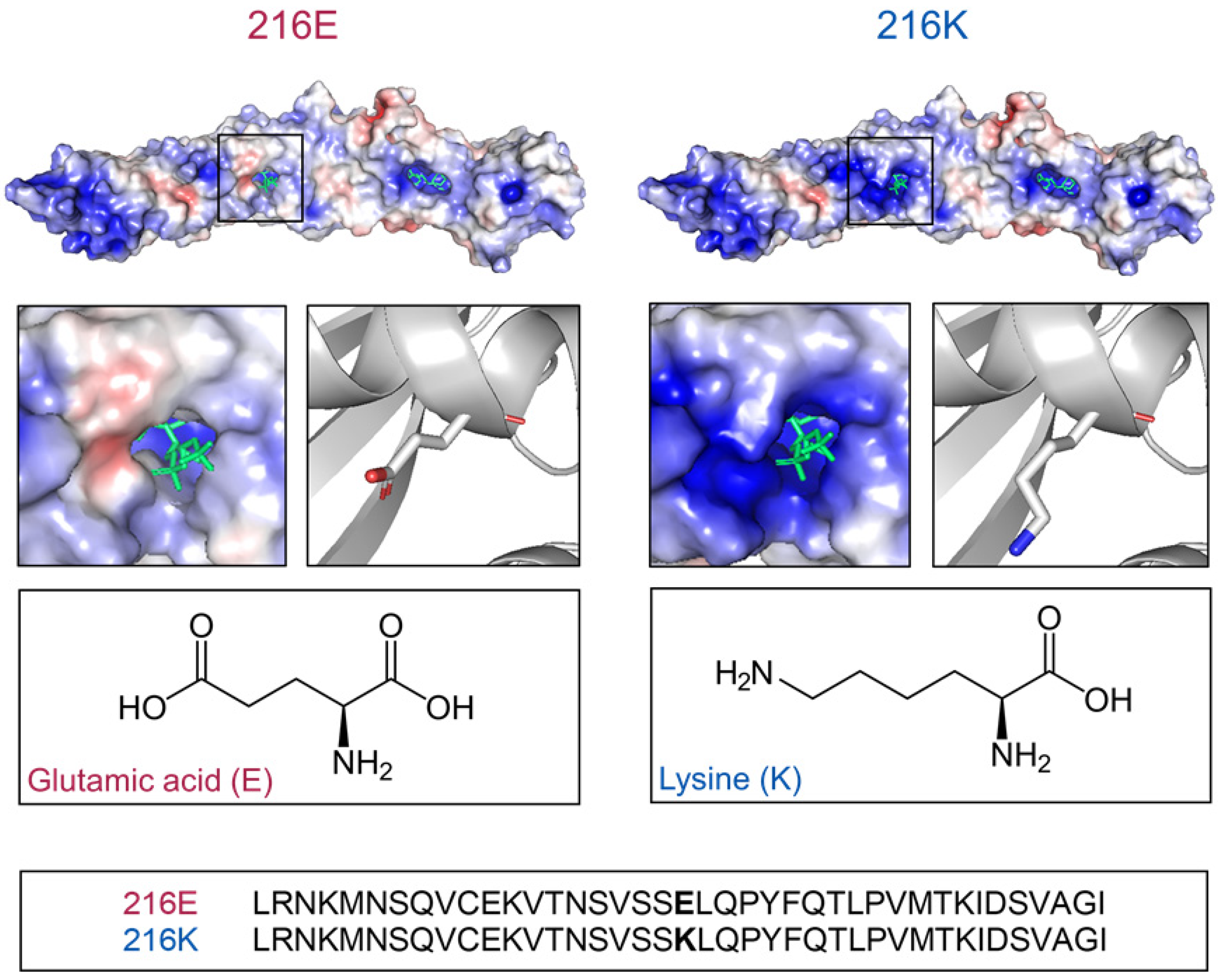
2.1. Models of BPI216E and BPI216K Reveal Different Charge Distributions Proximal to the Lipid Binding Pocket

The interaction of BPI with the acyl chains of LPS has been proposed to be mediated by the apolar binding pockets found in the C-terminal and N-terminal domains of the protein [10,11]. Interestingly, the SNP rs4358188 localizes proximal to the entrance of the N-terminal lipid-binding pocket where it results in a substantial electrostatic change. While BPI216E with a glutamic acid at position 216 displays a partially negative charge, rs4358188 determines an exchange to lysine in BPI216K with a consecutive positive charge (Figure 1). Thus, the SNP at codon position 216 leads to a BPI variant with a distinct change in the charge of surface patches surrounding the N-terminal apolar binding pocket.

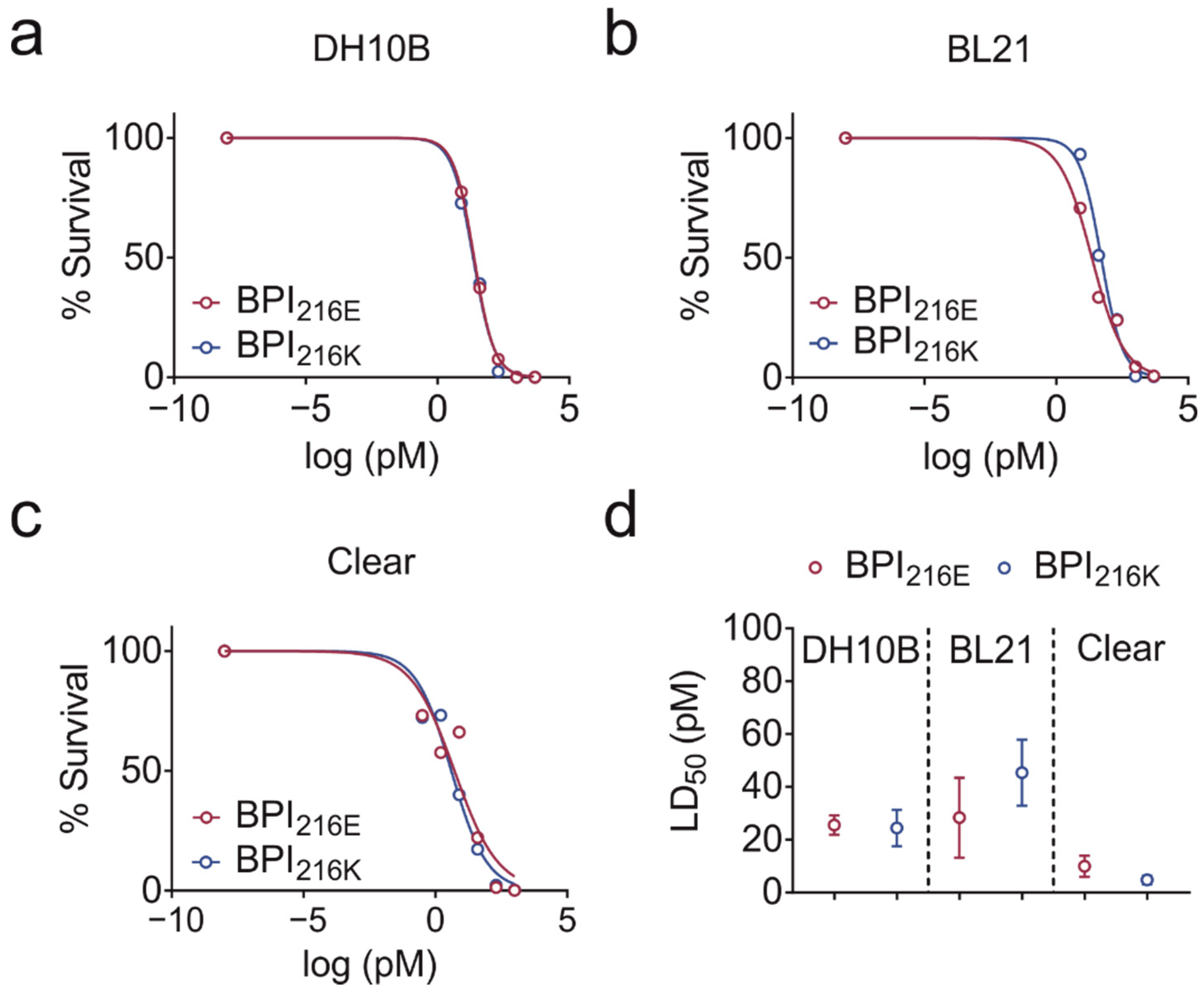
2.2. BPI216E and BPI216K Do Not Differ in Bactericidal Activity

Both a 25 kDa N-terminal and a 30 kDa C-terminal fragment of BPI are capable of LPS neutralization, whereas only the N-terminal fragment was described to harbor antibacterial activity [12]. To analyze if the differences in electrostatics at the N-terminal lipid-binding pocket of the BPI variants influence the bactericidal and LPS neutralizing activities of BPI, we performed bacterial killing and LPS binding assays.
BPI216E and BPI216K were equally potent at inhibiting the growth of E. coli DH10B and BL21 in a concentration-dependent manner (Figure 2a,b) as indicated by the mean lethal dose (LD50) of BPI216E and BPI216K towards E. coli DH10B (25.6 pM ± 3.7 and 24.4 pM ± 6.9) and E. coli BL21 (28.4 pM ± 15.1 and 45.4 pM ± 12.5; Figure 2d). We also tested the LD50 of the two variants towards Clear Coli® BL21, which expresses a genetically modified LPS variant consisting of lipid IVA, i.e., a lipid A precursor lacking the 2′ and 3′ acyl chain and associated oligosaccharides [18]. In this context, previous data demonstrated that BPI binding to the lipid A portion of LPS depends on the number of lipid A acyl chains and that long oligosaccharide chains of membrane-anchored LPS sterically hinder BPI from binding to the bacterial membrane [19,20,21]. In accordance, the LD50 was lower towards Clear Coli® BL21 than towards strains DH10B and BL21 but independent of the BPI variant with an LD50 of 10.0 pM ± 4.0 for BPI216E and 4.8 pM ± 1.7 for BPI216K (Figure 2c,d). Summarizing, no difference in bactericidal activity towards different E. coli strains was observed for the BPI variants.

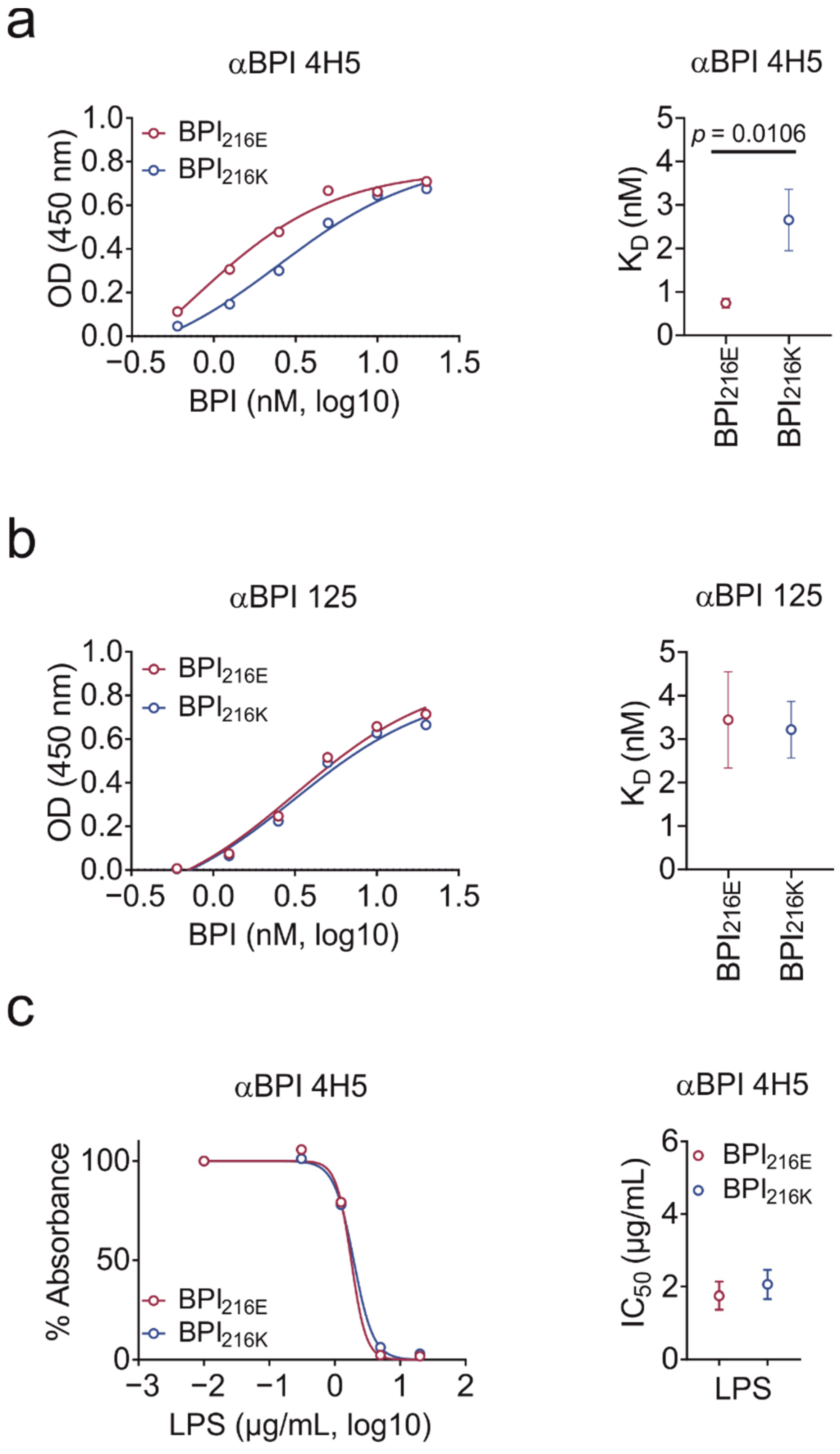
2.3. BPI216K Does Not Exhibit Higher Affinity to LPS as Compared to BPI216E

Next, we compared the binding capacity of BPI216E and BPI216K towards solid-phase LPS. LPS binding affinity was significantly different for BPI216E and BPI216K when detecting BPI binding with αBPI clone 4H5 (Figure 3a). Unexpectedly, the calculated KD of BPI216K towards negatively charged LPS was higher than for BPI216E despite disadvantageous charge distribution. The binding site of clone 4H5 to BPI is not known. However, the sequence and/or possible consecutive conformational differences between BPI216E and BPI216K could result in different binding affinities of antibody 4H5 towards the variants. Therefore, we repeated the test with a second, newly generated αBPI antibody (αBPI clone 125, Figure 3b). Importantly, the difference in BPI216E and BPI216K towards LPS binding disappeared. To retest clone 4H5, we performed an inhibition assay by pre-incubating BPI with liquid-phase LPS and set absorbance measured for binding of BPI to the LPS-coated plate without pre-incubation to 100%. In accordance with the results for clone 125, liquid-phase LPS inhibited binding of BPI216E and BPI216K to solid-phase LPS in a comparable extent (Figure 3c). Summarizing, despite having opposing charges proximal to the lipid-binding pocket, BPI216K does not exhibit superior binding towards the negatively charged LPS as compared to BPI216E.

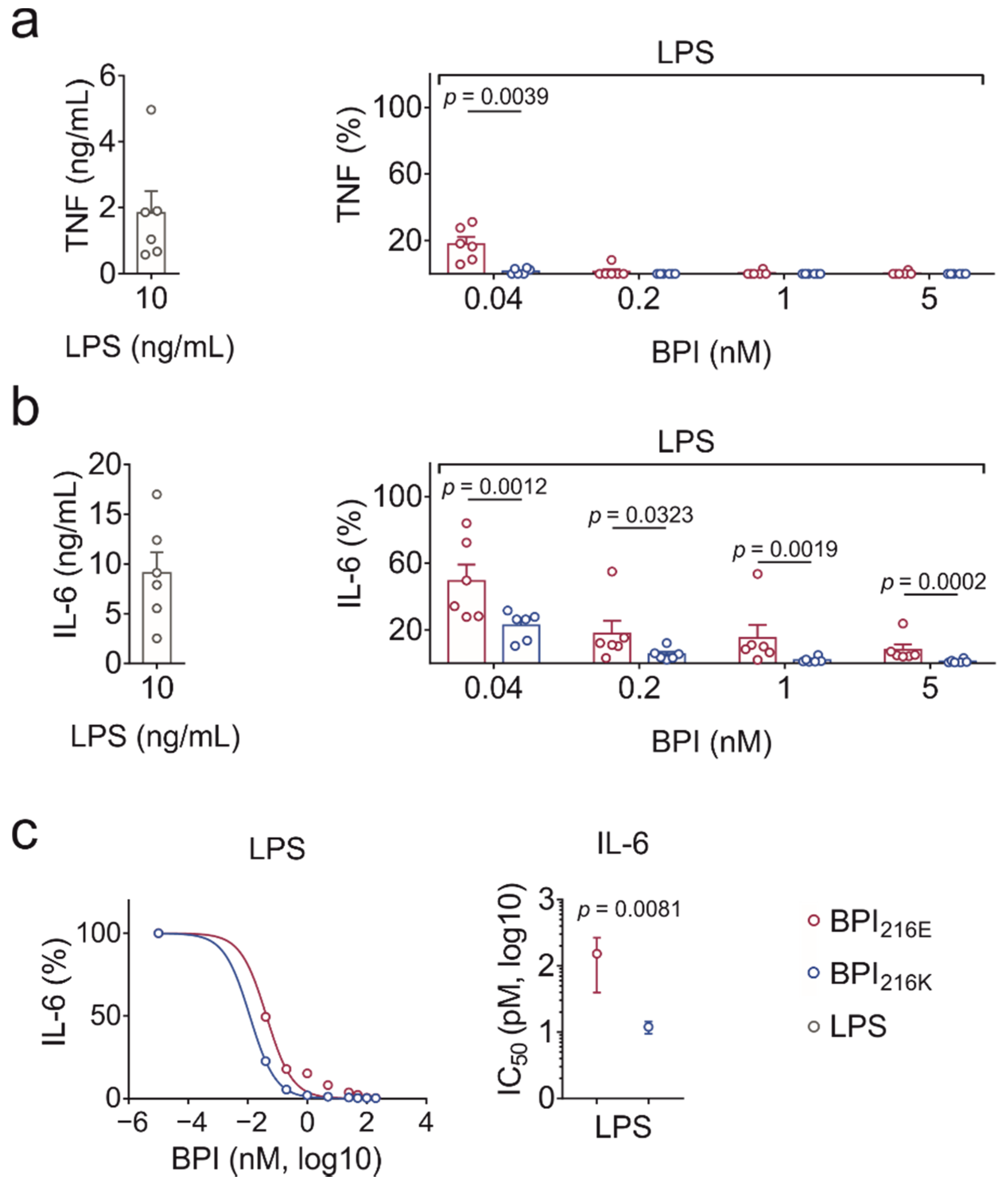
2.4. BPI216K Exceeds BPI216E at Inhibition of LPS-Induced Cytokine Secretion

To evaluate the LPS neutralization capacity, human peripheral blood mononuclear cells (PBMCs) were incubated with either BPI variant and increasing concentrations of LPS for 24 h before cytokine response was determined in the cell culture supernatants. Both BPI variants were more potent in neutralizing TNF than IL-6 (Figure 4a,b). For both cytokines, BPI216E was significantly less potent than BPI216K in LPS neutralization (Figure 4a,b). Since the results became too variable at BPI concentrations lower than those indicated, the IC50 could only be determined for IL-6. Hereby, extrapolation revealed an IC50 of 152.9 ± 113.4 pM for BPI216E and significantly lower values of 12.0 ± 2.5 pM for BPI216K (Figure 4c). Therefore, on average, BPI216K exhibited a more than ten-fold higher capacity to neutralize LPS in PBMC culture than BPI216E.

3. Discussion

The reduced LPS-neutralization capacities of BPI216E compared to BPI216K shown in this study provide an explanation for an increased susceptibility to Gram-negative sepsis and mortality in neonates in association with BPI216E [16,17]. Because of the differently charged amino acids located proximal to the lipid-binding site in the N-terminal domain of BPI, we initially expected differences in binding of BPI216E and BPI216K to LPS. However, no conclusive difference in respect to the binding of LPS by either BPI variant was detected, indicating a minor contribution of amino acid 216. Moreover, both variants displayed equal bactericidal activity independent of oligosaccharide chains or the number of acyl chains in lipid A of the tested strains. Since BPI216E neutralizes LPS less efficiently than BPI216K, BPI216E presumably enables a more sensitive perception of bacterial invasion. In this context, the SNP at position 216 K is associated with a predisposition to bacterial infection in hematopoietic stem cell transplant (HSCT) patients [22]. Additionally, BPI216E was linked to an increased risk of graft versus host disease (GvHD) after HSCT [23], possibly attributed to the key role of LPS in the pathophysiology of GvHD [24,25].
Although the elevation of sepsis frequency and association with increased mortality was seen in pediatric patients with variant BPI216E [16,17], no difference was found in an adult cohort study [26]. Neonatal neutrophilic granulocytes showed reduced levels [27,28] and extrusion [29] of BPI in neutrophilic granulocytes compared to adults. Lower basal levels of BPI may lead to a more pronounced phenotype of the respective BPI variants, possibly explaining divergent effects seen for children and adults. However, low patient numbers may limit the validity of the studies in neonates, and an analysis of larger cohorts is needed. LPS is recognized by toll-like receptor (TLR) 4 [30]. The combination of rs4358188 with SNPs in genes related to TLR-related pathways, namely IL1RL and ITGB2, seems to significantly increase the risk for developing specific IgE directed against a food allergen [31], thereby attributing a higher risk in the presence of BPI216K. Fittingly, LPS can ameliorate ongoing allergic inflammation in dependence of TLR4 in murine models [32].
A previous study performed in whole blood stimulated with LPS at 1 ng/mL showed an IC50 for TNF at a BPI concentration of 4 nM [12]. Our use of PBMCs in serum-free conditions revealed LPS neutralization by BPI even at a picomolar range, emphasizing the enormous potential of BPI to neutralize LPS. A limitation of our study is that we did not provide a mechanistic insight concerning the difference in neutralization potency between the variants. This must be addressed in further studies. Although KD towards LPS was comparable, distinct association and dissociation rates might possibly explain our findings. The charge of amino acid 216 might also contribute to conformational changes in BPI which were previously found for the N-terminal part of BPI upon binding to LPS membranes [33]. In addition, the described maximum binding capacity of 40 LPS molecules per BPI might be affected [34]. Since BPI polymorphism possibly influences a variety of diseases including sepsis, GvHD, and allergy, in vivo models would be of interest. Compared to humans, constitutive BPI expression in mice is impaired [35] and murine BPI was shown to exhibit lower bactericidal and LPS-neutralization capacities than human BPI [36]. However, BPI-deficient mice were recently generated [37,38] and should be suited as basic tools for the comparison of therapeutically applied BPI216E and BPI216K in different disease conditions.
In conclusion, the decreased LPS-neutralizing capacity of BPI216E as compared to BPI216K provides an explanation for the association of BPI216E with the risk of unfavorable outcomes in sepsis [16,17]. Lethality in this patient group during the course of Gram-negative sepsis is high [2] and the application of a recombinant N-terminal domain of BPI showed partial clinical success in children with meningococcal infection [14,15]. Given the rise in multidrug-resistant Gram-negative bacteria, the administration of BPI during Gram-negative sepsis, especially in patients with a BPI216E phenotype and/or BPI deficiency of various causes [39], should be re-evaluated in clinical studies.

4. Materials and Methods

4.1. Generation of Recombinant Human BPI

The generation of recombinant human BPI variants was performed as described [40] with slight modifications. In brief, a pCR3 vector (Invivogen, Toulouse, France) construct comprising an N-terminal HA signal peptide, amino acids 32 to 487 of either BPI216E or BPI216K, and a C-terminal FLAG-Tag was transfected in Expi293F™ cells using the ExpiFectamine™ 293 Transfection Kit (Thermo Fisher Scientific, Waltham, MA, USA). The expressed protein was purified by cation exchange chromatography via a HiTrap™ SP HP column (Cytiva, Marlborough, MA, USA). Fractions containing the protein of interest were pooled and purified by size exclusion chromatography using a HighLoad 16/600 Superdex 75 pg column (Cytiva, Marlborough, MA, USA), concentrated via ultrafiltration (Amicon Ultra-15, Merck Millipore, Darmstadt, Germany), and dialyzed against PBS. Concentration was determined by DC-Protein Assays (Bio-Rad Laboratories, Feldkirchen, Germany).

4.2. Generation and Purification of BPI Antibodies

Mice were immunized with recombinant BPI (amino acids 32 to 487) by Davids Biotechnology (Regensburg, Germany) to generate hybridoma clones. These clones were screened by ELISA for the production of antibodies directed against human BPI. The positive IgG clone 125 was selected for expansion and the antibody was purified via a HiTrap Protein G HP antibody purification column (Cytiva, Marlborough, MA, USA).

4.3. Solid-Phase BPI Binding Assay

The setup for the BPI binding assay was adopted from Bülow et al., 2018 [40]. To summarize, streptavidin-coated 96-well plates (NuncTM ImmobilizerTM Streptavidin F96 clear, Thermo Fisher Scientific, Waltham, MA, USA) were coated with biotinylated LPS derived from E. coli O111:B4 (2 μg/mL; Invivogen, Toulouse, France) in PBS and agitated overnight at 23 °C and 350 rpm. Plates were then washed with NaCl HEPES buffer (150 mM NaCl and 50 mM HEPES) containing 0.01% casein (Applied Biosystems, Waltham, MA, USA). After blocking with 1% BSA at 37 °C and three washing steps, plates were incubated with BPI at concentrations as indicated. For inhibition experiments, 20 nM BPI was pre-incubated with E. coli O111:B4-derived LPS (Invivogen, Toulouse, France) in NaCl HEPES buffer containing 0.1% BSA for 30 min using LoBind tubes (Eppendorf, Hamburg, Germany). After washing, bound BPI was detected by murine anti-human BPI monoclonal antibody 4H5 (Hycult Biotech, Uden, The Netherlands) or antibody 125 followed by detection via an HRP-conjugated rabbit anti-mouse IgG (Dianova, Hamburg, Germany). TMB (BD Biosciences, Heidelberg, Germany) was used as a substrate of the peroxidase. The reaction was stopped after seven minutes with 1 N HCl (Carl ROTH, Karlsruhe, Germany). Absorbance was then measured at 450 nm in a microplate reader (Model 550, Bio-Rad Laboratories, Feldkirchen, Germany).

4.4. Dose Response Experiments for Bactericidal Activity

E. coli strains DH10B (Invitrogen, Carlsbad, CA, USA), BL21 (Lucigen, Middleton, WI, USA), and Clear Coli® BL21 (Lucigen, Middleton, WI, USA) were cultivated on Columbia blood agar plates (Thermo Fisher Scientific, Waltham, MA, USA). Single colonies were transferred into lysogeny broth medium (Carl Roth, Karlsruhe, Germany) and incubated overnight at 37 °C at 220 rpm. Subsequently, the optical density (OD) of the bacteria was adjusted to 0.1. Inoculated broths were further incubated until an OD of 0.4 was reached. Bacteria were then pelleted and diluted in PBS with 0.01% Tween 80 (Merck Millipore, Darmstadt, Germany) to a final concentration of 1 × 10⁴ bacteria per mL. Bacterial suspensions were then incubated with decreasing concentrations of BPI for 1 h at 37 °C and an aliquot, containing 5 × 10² bacteria, was immediately plated on blood agar plates. Plates were incubated overnight at 37 °C and colonies were quantified the next day.

4.5. Isolation of Human Peripheral Blood Mononuclear Cells

For the isolation of PMBCs, blood was drawn from healthy male volunteers and collected in heparinized blood collectors (Li-Heparin-Gel-Monovette, Sarstedt, Nümbrecht, Germany). Blood was then diluted in RPMI-1640 (Sigma-Aldrich, Taufkirchen, Germany) and centrifuged in LeucosepTM tubes containing FICOLL® PAQUE PLUS (Oxford Immunotec, Abingdon, UK) at 1000× g for 10 min. PBMCs were then isolated from the interphase and washed two times. Subsequently, the pellet was resuspended in AIM V® Medium (Thermo Fisher Scientific, Waltham, MA, USA) and 5 × 10⁴ cells were seeded into a 96-well plate at a final volume of 100 µL. PBMCs were allowed to rest for 4 h prior to combined stimulation with BPI and LPS.

4.6. Quantification of Cytokine Levels

To quantify cytokine levels in the supernatants of stimulated PBMCs, the Luminex® 100 system was used (Austin, TX, USA). Antibodies to detect human IL-6 (human IL-6 ELISA set) and TNF (human TNF ELISA set) were purchased from BD Bioscience (Heidelberg, Germany). Measured concentrations were calculated by using LiquiChip Analyzer Software (Qiagen, Hilden, Germany).

4.7. Structure Modeling, Graphical Depictions, and Statistics

The PDB structure for human BPI216E (10.2210/pdb1EWF/pdb [41]) was mutated in PyMOL (PyMOL Molecular Graphics System, Version 2.3.2 Schrödinger, LLC., New York, NY, USA) to BPI216K. Rendering of three-dimensional structures was performed in PyMOL. Electrostatic surface potentials for BPI216E and BPI216K were calculated with the APBS plugin [42] for PyMOL. Graphical depictions and statistical analyses were performed using GraphPad Prism, version 7 for Windows (GraphPad Software, San Diego, CA, USA). For comparison of BPI216E and BPI216K, using a ratio paired t-test, okines values beyond the linear range of the standard curve werication of the test. To enablection of LD50 and IC50 curves on a logarithmic scale, concentrations with values of zero were represented as values at least 30-fold beyond the lowest applied concentration of the respective substance.

Author Contributions

Conceptualization, S.B.; methodology, K.U.E., J.M.H., K.T.M., L.Z., M.W. and M.T.; visualization, K.U.E., J.M.H. and L.Z.; validation, L.Z., M.W. and M.T.; formal analysis, K.U.E., J.M.H., K.T.M. and S.B.; investigation, S.B. and A.G.; resources, A.G.; data curation, K.U.E., J.M.H., K.T.M. and L.Z.; writing—original draft preparation, K.U.E., J.M.H. and K.T.M.; writing—review and editing, S.B.; supervision, S.B.; project administration, S.B. and A.G.; funding acquisition, A.G. All authors have read and agreed to the published version of the manuscript.

Funding

This work was supported by the Deutsche Forschungsgemeinschaft [DFG, project number 324392634-TRR 221(B13)].

Institutional Review Board Statement

This study was carried out in accordance with the recommendations of the Declaration of Helsinki. All donors of PBMCs gave written informed consent in accordance with the Declaration of Helsinki. The protocol for the isolation of PBMCs was approved by the local ethics committee (Ethikkommision an der Universität Regensburg, approval number 16-302_1-101).

Informed Consent Statement

Informed consent was obtained from all subjects involved in the study.

Data Availability Statement

Data are contained within the article.

Acknowledgments

We gratefully acknowledge experimental assistance of Maria Drießlein, University Hospital Regensburg, Germany.

Conflicts of Interest

The authors declare no conflict of interest.

References

  1. Rudd, K.E.; Johnson, S.C.; Agesa, K.M.; Shackelford, K.A.; Tsoi, D.; Kievlan, D.R.; Colombara, D.V.; Ikuta, K.S.; Kissoon, N.; Finfer, S.; et al. Global, regional, and national sepsis incidence and mortality, 1990–2017: Analysis for the Global Burden of Disease Study. Lancet 2020, 395, 200–211. [Google Scholar] [CrossRef] [Green Version]
  2. Fleischmann, C.; Reichert, F.; Cassini, A.; Horner, R.; Harder, T.; Markwart, R.; Tröndle, M.; Savova, Y.; Kissoon, N.; Schlattmann, P.; et al. Global incidence and mortality of neonatal sepsis: A systematic review and meta-analysis. Arch. Dis. Child. 2021, 106, 745–752. [Google Scholar] [CrossRef] [PubMed]
  3. Opal, S.M.; Scannon, P.J.; Vincent, J.L.; White, M.; Carroll, S.F.; Palardy, J.E.; Parejo, N.A.; Pribble, J.P.; Lemke, J.H. Relationship between plasma levels of lipopolysaccharide (LPS) and LPS-binding protein in patients with severe sepsis and septic shock. J. Infect. Dis. 1999, 180, 1584–1589. [Google Scholar] [CrossRef] [PubMed] [Green Version]
  4. Henneke, P.; Dramsi, S.; Mancuso, G.; Chraibi, K.; Pellegrini, E.; Theilacker, C.; Hübner, J.; Santos-Sierra, S.; Teti, G.; Golenbock, D.T.; et al. Lipoproteins are critical TLR2 activating toxins in group B streptococcal sepsis. J. Immunol. 2008, 180, 6149–6158. [Google Scholar] [CrossRef]
  5. Weiss, J.; Olsson, I. Cellular and subcellular localization of the bactericidal/permeability-increasing protein of neutrophils. Blood 1987, 69, 652–659. [Google Scholar] [CrossRef] [Green Version]
  6. Skopelja, S.; Hamilton, B.J.; Jones, J.D.; Yang, M.-L.; Mamula, M.; Ashare, A.; Gifford, A.H.; Rigby, W.F. The role for neutrophil extracellular traps in cystic fibrosis autoimmunity. JCI Insight 2016, 1, e88912. [Google Scholar] [CrossRef] [Green Version]
  7. Brinkmann, V.; Reichard, U.; Goosmann, C.; Fauler, B.; Uhlemann, Y.; Weiss, D.S.; Weinrauch, Y.; Zychlinsky, A. Neutrophil extracellular traps kill bacteria. Science 2004, 303, 1532–1535. [Google Scholar] [CrossRef]
  8. Weiss, J.; Elsbach, P.; Shu, C.; Castillo, J.; Grinna, L.; Horwitz, A.; Theofan, G. Human bactericidal/permeability-increasing protein and a recombinant NH2-terminal fragment cause killing of serum-resistant gram-negative bacteria in whole blood and inhibit tumor necrosis factor release induced by the bacteria. J. Clin. Investig. 1992, 90, 1122–1130. [Google Scholar] [CrossRef] [Green Version]
  9. Beamer, L.J.; Carroll, S.F.; Eisenberg, D. The BPI/LBP family of proteins: A structural analysis of conserved regions. Protein Sci. 1998, 7, 906–914. [Google Scholar] [CrossRef] [Green Version]
  10. Beamer, L.J.; Carroll, S.F.; Eisenberg, D. The three-dimensional structure of human bactericidal/permeability-increasing protein. Biochem. Pharmacol. 1999, 57, 225–229. [Google Scholar] [CrossRef]
  11. Beamer, L.J.; Carroll, S.F.; Eisenberg, D. Crystal structure of human BPI and two bound phospholipids at 2.4 angstrom resolution. Science 1997, 276, 1861–1864. [Google Scholar] [CrossRef] [PubMed]
  12. Ooi, C.E.; Weiss, J.; Doerfler, M.E.; Elsbach, P. Endotoxin-neutralizing properties of the 25 kD N-terminal fragment and a newly isolated 30 kD C-terminal fragment of the 55–60 kD bactericidal/permeability-increasing protein of human neutrophils. J. Exp. Med. 1991, 174, 649–655. [Google Scholar] [CrossRef] [PubMed] [Green Version]
  13. Jin, H.; Yang, R.; Marsters, S.; Ashkenazi, A.; Bunting, S.; Marra, M.N.; Scott, R.W.; Baker, J.B. Protection against endotoxic shock by bactericidal/permeability-increasing protein in rats. J. Clin. Investig. 1995, 95, 1947–1952. [Google Scholar] [CrossRef] [PubMed] [Green Version]
  14. Von der Möhlen, M.A.; Kimmings, A.N.; Wedel, N.I.; Mevissen, M.L.; Jansen, J.; Friedmann, N.; Lorenz, T.J.; Nelson, B.J.; White, M.L.; Bauer, R. Inhibition of endotoxin-induced cytokine release and neutrophil activation in humans by use of recombinant bactericidal/permeability-increasing protein. J. Infect. Dis. 1995, 172, 144–151. [Google Scholar] [CrossRef]
  15. Levin, M.; Quint, P.A.; Goldstein, B.; Barton, P.; Bradley, J.S.; Shemie, S.D.; Yeh, T.; Kim, S.S.; Cafaro, D.P.; Scannon, P.J.; et al. Recombinant bactericidal/permeability-increasing protein (rBPI21) as adjunctive treatment for children with severe meningococcal sepsis: A randomised trial. rBPI21 Meningococcal Sepsis Study Group. Lancet 2000, 356, 961–967. [Google Scholar] [CrossRef]
  16. Esposito, S.; Zampiero, A.; Pugni, L.; Tabano, S.; Pelucchi, C.; Ghirardi, B.; Terranova, L.; Miozzo, M.; Mosca, F.; Principi, N. Genetic polymorphisms and sepsis in premature neonates. PLoS ONE 2014, 9, e101248. [Google Scholar] [CrossRef] [Green Version]
  17. Michalek, J.; Svetlikova, P.; Fedora, M.; Klimovic, M.; Klapacova, L.; Bartosova, D.; Elbl, L.; Hrstkova, H.; Hubacek, J.A. Bactericidal permeability increasing protein gene variants in children with sepsis. Intensiv. Care Med. 2007, 33, 2158–2164. [Google Scholar] [CrossRef]
  18. Mamat, U.; Wilke, K.; Bramhill, D.; Schromm, A.B.; Lindner, B.; Kohl, T.A.; Corchero, J.L.; Villaverde, A.; Schaffer, L.; Head, S.R.; et al. Detoxifying Escherichia coli for endotoxin-free production of recombinant proteins. Microb. Cell Fact. 2015, 14, 57. [Google Scholar] [CrossRef] [Green Version]
  19. Gazzano-Santoro, H.; Parent, J.B.; Conlon, P.J.; Kasler, H.G.; Tsai, C.M.; Lill-Elghanian, D.A.; Hollingsworth, R.I. Characterization of the structural elements in lipid A required for binding of a recombinant fragment of bactericidal/permeability-increasing protein rBPI23. Infect. Immun. 1995, 63, 2201–2205. [Google Scholar] [CrossRef] [Green Version]
  20. Weiss, J.; Beckerdite-Quagliata, S.; Elsbach, P. Resistance of gram-negative bacteria to purified bactericidal leukocyte proteins: Relation to binding and bacterial lipopolysaccharide structure. J. Clin. Investig. 1980, 65, 619–628. [Google Scholar] [CrossRef]
  21. Capodici, C.; Chen, S.; Sidorczyk, Z.; Elsbach, P.; Weiss, J. Effect of lipopolysaccharide (LPS) chain length on interactions of bactericidal/permeability-increasing protein and its bioactive 23-kilodalton NH2-terminal fragment with isolated LPS and intact Proteus mirabilis and Escherichia coli. Infect. Immun. 1994, 62, 259–265. [Google Scholar] [CrossRef] [PubMed] [Green Version]
  22. Guinan, E.C.; Palmer, C.D.; Mancuso, C.J.; Brennan, L.; Stoler-Barak, L.; Kalish, L.A.; Suter, E.E.; Gallington, L.C.; Huhtelin, D.P.; Mansilla, M.; et al. Identification of single nucleotide polymorphisms in hematopoietic cell transplant patients affecting early recognition of, and response to, endotoxin. Innate Immun. 2014, 20, 697–711. [Google Scholar] [CrossRef] [PubMed] [Green Version]
  23. Wermke, M.; Maiwald, S.; Schmelz, R.; Thiede, C.; Schetelig, J.; Ehninger, G.; Bornhäuser, M.; Wassmuth, R. Genetic variations of interleukin-23R (1143AG) and BPI (A645G), but not of NOD2, are associated with acute graft-versus-host disease after allogeneic transplantation. Biol. Blood Marrow Transplant. 2010, 16, 1718–1727. [Google Scholar] [CrossRef] [PubMed] [Green Version]
  24. Hill, G.R.; Crawford, J.M.; Cooke, K.R.; Brinson, Y.S.; Pan, L.; Ferrara, J.L. Total Body Irradiation and Acute Graft-Versus-Host Disease: The Role of Gastrointestinal Damage and Inflammatory Cytokines. Blood 1997, 90, 3204–3213. [Google Scholar] [CrossRef] [PubMed]
  25. Hill, G.R.; Koyama, M. Cytokines and costimulation in acute graft-versus-host disease. Blood 2020, 136, 418–428. [Google Scholar] [CrossRef] [PubMed]
  26. Hubacek, J.A.; Stüber, F.; Fröhlich, D.; Book, M.; Wetegrove, S.; Ritter, M.; Rothe, G.; Schmitz, G. Gene variants of the bactericidal/permeability increasing protein and lipopolysaccharide binding protein in sepsis patients: Gender-specific genetic predisposition to sepsis. Crit. Care Med. 2001, 29, 557–561. [Google Scholar] [CrossRef]
  27. Qing, G.; Howlett, S.; Bortolussi, R. Lipopolysaccharide binding proteins on polymorphonuclear leukocytes: Comparison of adult and neonatal cells. Infect. Immun. 1996, 64, 4638–4642. [Google Scholar] [CrossRef] [Green Version]
  28. Levy, O.; Martin, S.; Eichenwald, E.; Ganz, T.; Valore, E.; Carroll, S.F.; Lee, K.; Goldmann, D.; Thorne, G.M. Impaired innate immunity in the newborn: Newborn neutrophils are deficient in bactericidal/permeability-increasing protein. Pediatrics 1999, 104, 1327–1333. [Google Scholar] [CrossRef]
  29. Nupponen, I.; Turunen, R.; Nevalainen, T.; Peuravuori, H.; Pohjavuori, M.; Repo, H.; Andersson, S. Extracellular release of bactericidal/permeability-increasing protein in newborn infants. Pediatr. Res. 2002, 51, 670–674. [Google Scholar] [CrossRef]
  30. Rosadini, C.V.; Kagan, J.C. Early innate immune responses to bacterial LPS. Curr. Opin. Immunol. 2017, 44, 14–19. [Google Scholar] [CrossRef] [Green Version]
  31. Reijmerink, N.E.; Bottema, R.W.B.; Kerkhof, M.; Gerritsen, J.; Stelma, F.F.; Thijs, C.; van Schayck, C.P.; Smit, H.A.; Brunekreef, B.; Koppelman, G.H.; et al. TLR-related pathway analysis: Novel gene-gene interactions in the development of asthma and atopy. Allergy 2010, 65, 199–207. [Google Scholar] [CrossRef] [PubMed]
  32. Hollingsworth, J.W.; Whitehead, G.S.; Lin, K.L.; Nakano, H.; Gunn, M.D.; Schwartz, D.A.; Cook, D.N. TLR4 signaling attenuates ongoing allergic inflammation. J. Immunol. 2006, 176, 5856–5862. [Google Scholar] [CrossRef] [PubMed] [Green Version]
  33. Domingues, M.M.; Lopes, S.C.D.N.; Santos, N.C.; Quintas, A.; Castanho, M.A.R.B. Fold-unfold transitions in the selectivity and mechanism of action of the N-terminal fragment of the bactericidal/permeability-increasing protein (rBPI(21)). Biophys. J. 2009, 96, 987–996. [Google Scholar] [CrossRef] [PubMed] [Green Version]
  34. Tobias, P.S.; Soldau, K.; Iovine, N.M.; Elsbach, P.; Weiss, J. Lipopolysaccharide (LPS)-binding proteins BPI and LBP form different types of complexes with LPS. J. Biol. Chem. 1997, 272, 18682–18685. [Google Scholar] [CrossRef] [Green Version]
  35. Lennartsson, A.; Pieters, K.; Vidovic, K.; Gullberg, U. A murine antibacterial ortholog to human bactericidal/permeability-increasing protein (BPI) is expressed in testis, epididymis, and bone marrow. J. Leukoc. Biol. 2005, 77, 369–377. [Google Scholar] [CrossRef] [Green Version]
  36. Wittmann, I.; Schönefeld, M.; Aichele, D.; Groer, G.; Gessner, A.; Schnare, M. Murine bactericidal/permeability-increasing protein inhibits the endotoxic activity of lipopolysaccharide and gram-negative bacteria. J. Immunol. 2008, 180, 7546–7552. [Google Scholar] [CrossRef] [Green Version]
  37. Theprungsirikul, J.; Skopelja-Gardner, S.; Burns, A.S.; Wierzbicki, R.M.; Rigby, W.F.C. Bactericidal/Permeability-Increasing Protein Preeminently Mediates Clearance of Pseudomonas aeruginosa In Vivo via CD18-Dependent Phagocytosis. Front. Immunol. 2021, 12, 659523. [Google Scholar] [CrossRef]
  38. Kong, Q.; Lv, Z.; Kang, Y.; An, Y.; Liu, Z.; Zhang, J. Bactericidal Permeability Increasing Protein Deficiency Aggravates Acute Colitis in Mice by Increasing the Serum Levels of Lipopolysaccharide. Front. Immunol. 2020, 11, 614169. [Google Scholar] [CrossRef]
  39. Palmer, C.D.; Guinan, E.C.; Levy, O. Deficient expression of bactericidal/permeability-increasing protein in immunocompromised hosts: Translational potential of replacement therapy. Biochem. Soc. Trans. 2011, 39, 994–999. [Google Scholar] [CrossRef] [Green Version]
  40. Bülow, S.; Zeller, L.; Werner, M.; Toelge, M.; Holzinger, J.; Entzian, C.; Schubert, T.; Waldow, F.; Gisch, N.; Hammerschmidt, S.; et al. Bactericidal/Permeability-Increasing Protein Is an Enhancer of Bacterial Lipoprotein Recognition. Front. Immunol. 2018, 9, 2768. [Google Scholar] [CrossRef] [Green Version]
  41. Kleiger, G.; Beamer, L.J.; Grothe, R.; Mallick, P.; Eisenberg, D. The 1.7 A crystal structure of BPI: A study of how two dissimilar amino acid sequences can adopt the same fold. J. Mol. Biol. 2000, 299, 1019–1034. [Google Scholar] [CrossRef] [PubMed] [Green Version]
  42. Baker, N.A.; Sept, D.; Joseph, S.; Holst, M.J.; McCammon, J.A. Electrostatics of nanosystems: Application to microtubules and the ribosome. Proc. Natl. Acad. Sci. USA 2001, 98, 10037–10041. [Google Scholar] [CrossRef] [PubMed] [Green Version]
Figure 1. Comparison of the surface charge surrounding the N-terminal apolar lipid-binding pockets of BPI216E and BPI216K. Three-dimensional modeling and electrostatic potential of BPI216E and BPI216K with red areas representing negative and blue areas representing positive charges. Bound phosphatidylcholine is shown in green. Detailed view of the N-terminal binding pocket and zoomed-in ribbon diagram indicating the glutamic acid and lysine residues of BPI216E and BPI216K, respectively. Amino acid sequences for BPI216E and BPI216K from position 196 to 236 are shown.
Ijms 23 01324 g001
Figure 2. Bactericidal activity of BPI216E and BPI216K towards E. coli. (ac) Dose-response experiments with E. coli strains DH10B (a), BL21 (b), and Clear Coli® BL21 (c) incubated with increasing concentrations of BPI216E and BPI216K. (d) LD50 is depicted for each tested E. coli strain. Colony numbers of untreated bacteria were set as a reference to 100% (ac). Data are shown as the means (ac) or means ± SEM (d) of three biological replicates. Student’s ratio paired t-test revealed no significant differences.
Ijms 23 01324 g002
Figure 3. Equal binding of BPI variants to LPS. (a,b) Binding of BPI to solid-phase LPS as detected with αBPI antibody clone 4H5 (a) or αBPI antibody 125 newly generated in mice (b). KD values are provided. (c) BPI216E and BPI216K were pre-incubated with increasing concentrations of liquid-phase LPS before adding the mixture to LPS-coated plates. IC50 values are depicted. Absorbance measured for binding of BPI to the LPS-coated plate without pre-incubation with liquid-phase LPS was set as a reference to 100% (c). Data are shown as the means (left) or means ± SEM (right) of three biological replicates. Statistical testing was performed using the Student’s ratio paired t-test. Significance is indicated by the p value.
Ijms 23 01324 g003
Figure 4. Comparison of the LPS neutralizing capacity of BPI216E and BPI216K. (a,b) Levels of TNF and IL-6 in supernatants of human PBMCs after 24 h of stimulation with LPS (10 ng/mL) ± BPI. (c) Dose–response curve and IC50 of BPI216E and BPI216K as shown for IL-6 secretion. Cytokine secretion for LPS alone was set as a reference to 100% (a,b, right panel, as well as c, left panel). Data are shown as the means (c, left panel) or means ± SEM (a,b, both panels, and c, right panel) of six biological replicates. Statistical testing was performed using the Student’s ratio paired t-test. Significance is indicated by p values.
Ijms 23 01324 g004
Publisher’s Note: MDPI stays neutral with regard to jurisdictional claims in published maps and institutional affiliations.

