Article

A General Signal Pathway to Regulate Multiple Detoxification Genes Drives the Evolution of Helicoverpa armigera Adaptation to Xenobiotics

College of Plant Protection, China Agricultural University, Beijing 100193, China
*
Author to whom correspondence should be addressed.
Int. J. Mol. Sci. 2022, 23(24), 16126; https://doi.org/10.3390/ijms232416126
Submission received: 2 November 2022 / Revised: 4 December 2022 / Accepted: 13 December 2022 / Published: 17 December 2022
(This article belongs to the Section Molecular Biology)

Abstract

:
The study of insect adaptation to the defensive metabolites of host plants and various kinds of insecticides in order to acquire resistance is a hot topic in the pest-control field, but the mechanism is still unclear. In our study, we found that a general signal pathway exists in H. armigera which can regulate multiple P450s, GSTs and UGTs genes to help insects decrease their susceptibility to xenobiotics. Knockdown of HaNrf2 and HaAhR expression could significantly increase the toxicity of xenobiotics to H. armigera, and simultaneously decrease the gene expression of P450s, GSTs and UGTs which are related to the xenobiotic metabolism and synthesis of insect hormone pathways. Then, we used EMSA and dual luciferase assay to verify that a crosstalk exists between AhR and Nrf2 to regulate multiple P450s, GSTs and UGTs genes to mediate H. armigera susceptibility to plant allelochemicals and insecticides. The detoxification genes’ expression network which can be regulated by Nrf2 and AhR is still unknown, and there were also no reports about the crosstalk between AhR and Nrf2 that exist in insects and can regulate multiple detoxification genes’ expression. Our results provide a new general signaling pathway to reveal the adaptive mechanism of insects to xenobiotics and provides further insight into designing effective pest-management strategies to avoid the overuse of insecticides.

1. Introduction

The study of insect adaptation to defensive metabolites in host plants and varying kinds of insecticides in order to acquire resistance is a popular focus in the pest-control field, however, the mechanism of insect adaptation to these xenobiotics is still unclear [1,2]. Cotton bollworm (Helicoverpa armigera), as a herbivorous insect, has a wide range of host plant species, and can be harmful to cotton, tomato, maize, tobacco, as well as other crops. The evolutionary pressure of varied plants’ allelochemicals and insecticides have caused herbivorous insects to evolve corresponding countermeasures [1,2].
Cotton, being one of the primary host plants of H. armigera, has evolved efficient defense strategies against insects involving the synthesis of the toxic gossypol. Some insects cannot feed on cotton due to the toxic nature of gossypol, but H. armigera has apparently been successful in adapting to this toxin during a long evolutionary process. Solanaceae plants such as tomato use the allelochemical 2-tridecanone to defend against insects, but the cotton bollworm has successfully evolved and adapted to toxic 2-tridecanone. As there has been a widespread usage of various insecticides to control cotton bollworm, the insect has also developed a resistance to insecticides. The above problems lead to the failure of pest control. Increased detoxification enzyme activity is one of the most common strategies used by insects to resist the deleterious effects of xenobiotics [1,2]. Insect detoxification of insecticides and plant allelochemicals can be divided into three phases: phase I (oxidation, hydrolysis, reduction), phase II (conjugation), and phase III (excretion) [1,2]. In phase I, insecticides and/or plant allelochemicals are transformed into more hydrophilic metabolites by enzymes such as cytochrome P450 monooxygenases (P450s). P450s act to detoxify xenobiotics by introducing reactive or polar groups into the chemical structure [3]. In phase II, enzymes such as glutathione S transferases (GSTs) and UDP-glucuronosyltransferases (UGTs) conjugate the resulting metabolites of phase I to increase their hydrophilicity [4]. In phase III, after further conjugation, cleavage, and/or oxidation, the phase II-generated products are transported out of the cells by ATP-binding cassette (ABC) and other transmembrane transporters, where further degradation occurs [5,6]. Interestingly, many studies have found that multi over-expression of P450, GST, and UGT genes work together to mediate the plants allelochemicals tolerance and insecticide resistance in H. armigera [7].
Transcription factors play important roles in regulating detoxification genes involved in the xenobiotic metabolism [8]. However, the transcriptional regulation mechanism of these detoxification genes is still unclear, due to the vast number of detoxification enzyme genes present. In Drosophila, the nuclear receptor hormone DHR96 (receptor-like in 96) was found to regulate the expression of P450 genes associated with DDT resistance [9]. In Bemisia tabaci, a recent study reported that a P450 gene mediated resistance to imidacloprid by a mitogen-activated protein kinases (MAPKs) pathway [10]. Notably, recent studies in multiple insect species reported that insects have co-opted a xenobiotic stress response pathway that involves reactive oxygen species and the transcription factors Nrf2 (nuclear factor erythroid-2 related factor-2) or AhR (aryl hydrocarbon receptor) to up-regulate the expression of some detoxification gene members involved in insecticides resistance [11,12,13].
Nrf2 transcription factors are conserved in invertebrates, vertebrates, and metazoans [14]. Known as Cnc isoform C (CncC) in Drosophila, and skinhead family member 1 (SKN-1) in metazoans, Nrf2s are homologous proteins that play important roles in regulating cellular defenses against oxidative stressors or xenobiotics [6]. Normally, Nrf2 is retained in the cytoplasm by the actin-binding protein Keap1 (kelch-like ECH-associated protein 1) [15]. However, Keap1 acts as a stress sensor that can release Nrf2 into the nucleus under oxidative stress, after which Nrf2 binds to its heterodimer bZIP protein partner Maf (muscle aponeurosis fibromatosis). The heterodimer binds to xenobiotic response elements of target detoxification genes to regulate their expression [15]. AhR is a ligand-activated transcription factor that plays important roles in metabolic detoxification adaptation and normal physiology in mammals [16]. Metabolic detoxification adaptation is based on the coordinated regulation of some xenobiotic-metabolizing enzymes [3].
Despite some excellent studies on the single detoxification gene member induced by host plant allelochemicals or insecticides being regulated by Nrf2 or AhR, the network regarding the detoxification genes’ expression which can be regulated by Nrf2 and AhR is still unknown, and there were also no reports about the crosstalk between AhR and Nrf2 that exist in insects and can regulate the detoxification genes’ expression. The study of insect adaptation to defensive metabolites of host plants and various kinds of insecticides in order to acquire resistance is a hot topic in the pest-control field, but the mechanism is still unclear.
In this study, we found numerous P450s, GSTs, and UGTs gene promoter sequences which possess at least one Nrf2 binding site or AhR binding site in H. armigera. Some P450, GST, and UGT gene promoters simultaneously contain a very closely or overlapping Nrf2 and AhR binding sites. We then used RNA interference (RNAi) to investigate the function of HaNrf2 and HaAhR in H. armigera susceptibility to the plant’s allelochemical (gossypol and 2-tridecanone) and insecticides. Employing RNA sequencing, quantitative polymerase chain reaction (qPCR), and Nrf2 and AhR binding site prediction, EMSA and dual luciferase assay, we determined that the expression of most of the detoxification genes’ expression is regulated by a general crosstalk between HaNrf2 and HaAhR. There were also no reports on the existence of the crosstalk between AhR and Nrf2 in insects or the proof that they both can regulate P450s, GSTs, and UGTs genes which were related to the xenobiotic metabolism or synthesis of insect hormone pathways. As is well known, a lot of plants allelochemicals or insecticides will not only have the phenotype to kill insects directly, but also show an ability to decrease the growth rate of the insects. However, the molecular mechanism of this phenotype is still unknown [1,17,18]. Our results suggest that a crosstalk exists between AhR and Nrf2 pathways in H. armigera to mediate the susceptibility to plant allelochemicals and insecticides. Our results provide a general signal pathway for revealing the adaptive mechanism of insects to xenobiotics, and explain why many xenobiotics have both the phenotype of toxic action and affect the decreasing growth rates in insects. The results of this study improve our understanding of detoxification genes regulation in H. armigera and provide more insight into designing effective pest-management strategies to avoid the overuse of insecticides. The transcription factors HaNrf2 and HaAhR could serve as potential targets of dsRNA spraying for pest management.

2. Results

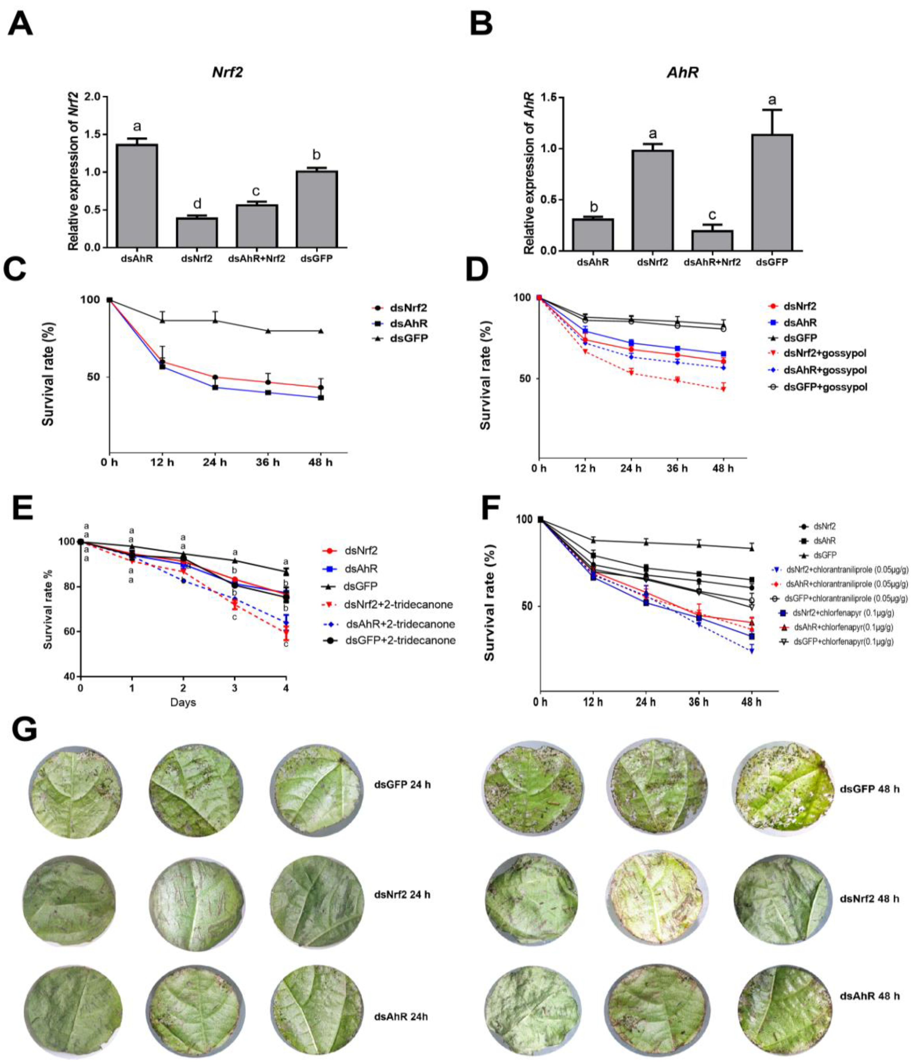
2.1. Effects of RNAi HaNrf2 and HaAhR Genes on Xenobiotics Susceptibility of H. armigera

In this study, we found that 1999 bp fragments upstream of the translation start sites (ATG) of all P450, GST and UGT genes, most of them were related by xenobiotic metabolism or synthesis of insect hormone pathways in H. armigera, and had at least one Nrf2 binding site or AhR binding site (Table S2).
HaNrf2 and HaAhR are essential genes for insect survival, so we investigated the effects of HaNrf2 or HaAhR genes on the susceptibility of H. armigera to gossypol, 2-tridecanone or insecticides by RNAi knockdown method. In Lepidoptera, knockdown of the gene expression by RNAi has sometimes proven difficult to achieve, but gene silencing is easily achieved by feeding high concentrations dsRNA [19].
Full-length sequences of the H. armigera HaNrf2 (KU355787.1) and HaAhR (XM_021331544.1) genes were cloned and sequenced. From these, dsRNAs were synthesized and fed to H. armigera larvae along with the plant allelochemical gossypol, 2-tridecanone or insecticides for 24 h. Afterwards, mortality was monitored and recorded. HaNrf2 or HaAhR dsRNA-treated larvae showed significant reductions in HaNrf2 or HaAhR gene expression compared to larvae treated with GFP dsRNA (Figure 1A,B). Compared to the control, HaNrf2 gene expression was suppressed by 62% 24 h after feeding larvae an artificial diet containing 15 μg/g (w:w) of HaNrf2 dsRNA, but the expression of HaAhR in dsNrf2-treated larvae was higher than in larvae treated with dsGFP control (Figure 1A). The transcription levels of HaAhR were suppressed (inhibition ratio over 70%) at 24 h after feeding with HaAhR dsRNA (15 μg/g) (Figure 1B). The larvae treated both with HaNrf2 and HaAhR dsRNA generated the lowest HaAhR gene expression, but not the lowest HaNrf2 gene expression in all treatments (Figure 1A,B). Reduced expression of HaNrf2 or HaAhR genes was predicted to significantly reduce the tolerance of H. armigera fed on cotton leaves (Figure 1C), or artificial diet mixed with gossypol, 2-tridecanone, chlorantraniliprole and chlorfenapyr (Figure 1D–F). Moreover, the HaNrf2 and HaAhR dsRNA coated cotton leaves enhanced resistance against H. armigera (Figure 1G).

2.2. RNA Sequencing and Analysis

To determine transcriptome changes of HaNrf2- and HaAhR-regulated genes in H. armigera, RNA sequencing of nine libraries prepared from dsGFP-, dsNrf2-, and dsAhR-treated larvae was performed. In order to select the xenobiotics-induced genes, RNA sequencing libraries prepared from gossypol, chlorantraniliprole-treated larvae and acetone-treated larvae was performed (Table S3). The transcriptome data of 2-tridecanone-induced larvae were referred to in a previous study (Zhang et al., 2015). A total of 2484 genes were up-regulated and 1991 genes were down-regulated in HaNrf2 Knockdown H. armigera larvae, while 1187 genes were up-regulated and 870 genes were down-regulated in HaAhR knockdown H. armigera larvae (Figure S1).
To explore the potential function of down-regulated genes, GO annotation enrichment and KEGG pathway analysis were conducted. GO analysis results showed that most of the down-regulated genes associated with RNAi knockdown of HaNrf2 or HaAhR were classified into ‘cellular component’, ‘biological process’, and ‘molecular function’ processes (Figure S2). Furthermore, the results of KEGG pathway analysis of HaNrf2 knockdown larvae showed that 437 out of 1991 down-regulated genes were mapped to KEGG orthologous terms in the KEGG pathway database, which were classified into ‘Metabolism of xenobiotics by cytochrome P450’ (16 genes), ‘drug metabolism-cytochrome P450’ (16 genes), and ‘drug metabolism-other enzymes pathways’ (23 genes) categories (Table S4). The results of KEGG pathway analysis of HaAhR knockdown larvae showed that 143 out of 870 down-regulated genes mapped to KEGG orthologous terms in the KEGG pathway database, which were classified into ‘Metabolism of xenobiotics by cytochrome P450’ (14 genes), ‘drug metabolism-cytochrome P450’ (13 genes) and ‘drug metabolism-other enzymes pathways’ (16 genes) categories (Table S5).

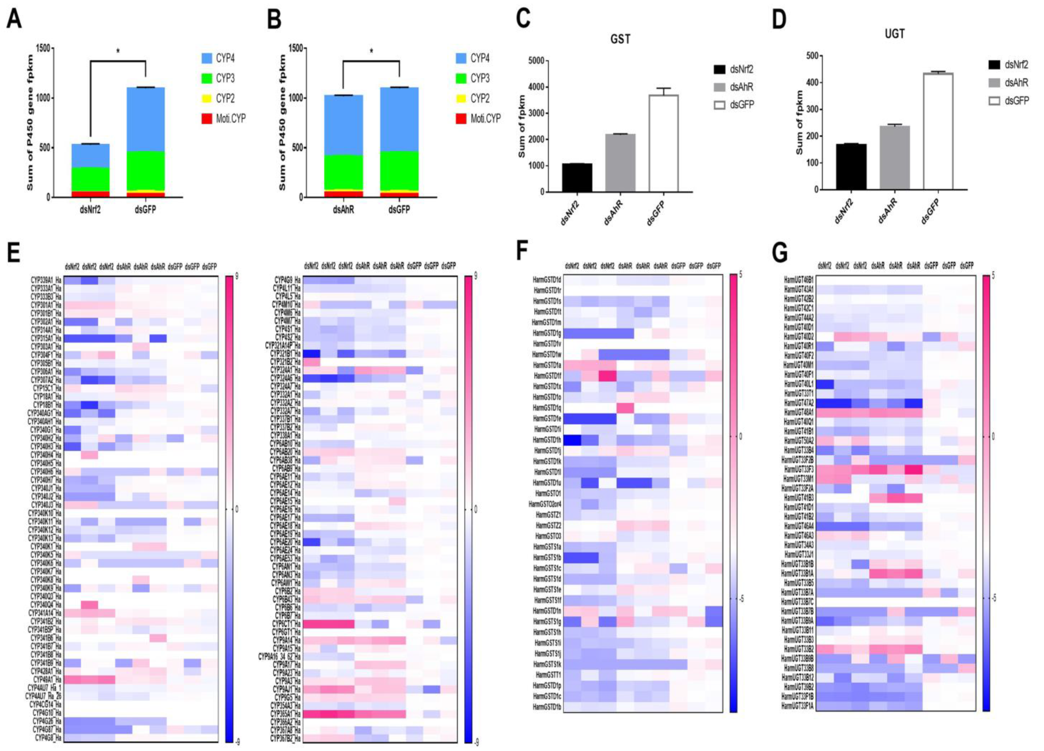
2.3. Analysis of P450s, UGTs and GSTs Genes Were Down-Regulated by RNAi HaNrf2 and HaAhR

The DEGs were selected based on DEGSeq2 analysis using |log2 (fold change)| ≥ 1 and p < 0.05 as cut-offs between the dsGFP and dsNrf2 or dsAhR groups, and this criteria was used to determine whether HaNrf2 and HaAhR regulate the expression of detoxification genes in H. armigera. Gene expression analysis of all three-phase enzymes (P450s, GSTs, UGTs) is shown in Figure 2. Transcriptome sequencing was used to detect gene expression after RNAi knockdown of HaNrf2 or HaAhR. RNAi knockdown of HaNrf2 decreased the sum FPKM of P450 genes up to 48% (Figure 2A) and RNAi knockdown of HaAhR decreased the sum FPKM of P450 genes up to 90% (Figure 2A,B); most of the CYP3 and CYP4 P450 genes were significantly down-regulated by RNAi knockdown of HaNrf2 or HaAhR. In all, 23 of 113 P450 genes were down-regulated in the HaAhR knockdown treatment, and 39 of 113 P450 genes were down-regulated in the HaNrf2 knockdown treatment (Figure 2E). The same phenomenon was observed for the GST and UGT gene families; RNAi knockdown of HaNrf2 genes decreased the sum FPKM of GST and UGT genes up to 19% and 38%, respectively; RNAi knockdown of HaAhR decreased the expression of GSTs and UGTs by up to 59% and 54%, respectively (Figure 2C,D). Most GST- and UGT-associated genes were down-regulated by RNAi knockdown of HaNrf2 or HaAhR; 25 of 41 GST genes were significantly down-regulated by knockdown of HaNrf2 and 12 of 41 GST genes were significantly down-regulated by of knockdown HaAhR (Figure 2F). A total of 24 of 46 UGT genes were down-regulated by knockdown of HaNrf2 and 22 of 46 UGT genes were down-regulated by knockdown of HaAhR (Figure 2G).
All the FPKM of detoxification enzyme genes P450, GST, and UGT in each of the libraries is shown in Table S3. According to the genes with p < 0.05 and |log2 (fold change)| ≥ 1 were considered to be significantly differently expressed. Most of P450, GST, and UGT genes related to the insecticide metabolism, which were down-regulated by knockdown of HaNrf2 or HaAhR (Figure 2). Interestingly, they also possess a lot of genes related to the synthesis of insect hormone pathways, just as CYP18A1, CYP307A2, CYP306A1, CYP302A1, CYP315A1 and CYP314A1 are also down-regulated by both knockdown of HaNrf2 or HaAhR (Figure 2).

2.4. Transcription Factor Binding Sites Prediction and RT-qPCR Validation of Down-Regulated Genes

We used RT-qPCR to validate the down-regulation of P450s, GSTs and UGTs genes associated with RNAi knockdown of HaNrf2 or HaAhR genes, twelve P450 genes (CYP18B1, CYP4L5, CYP4L11, CYP4S2, CYP6AN1, CYP6AE14, CYP6AE20, CYP6B2, CYP6B6, CYP6B7, CYP4M7, and CYP321B1) were selected, including 9 P450 genes which are significantly induced by gossypol, 9 P450 genes which are up-regulated by 2-tridecanone and 7 P450 genes which are up-regulated by chlorantraniliprole, simultaneously down-regulated by RNAi HaNrf2 and HaAhR (Figure 3). The RT-qPCR expression results for these P450 genes were consistent with the transcriptome data; all were down-regulated by knockdown of both HaNrf2 or HaAhR (Figure 3), CYP18B1, CYP4L5, CYP4L11, CYP4S2, CYP6AN1, CYP6AE14, CYP6AE20, CYP6B6, CYP6B7, CYP4M7, and CYP321B1 were decreased up to 12%, 30%, 64%, 32%, 20%, 23%, 21%, 30%, 12%, 29% and 8%, respectively, in HaNrf2 knockdown H. armigera; and CYP18B1, CYP4L5, CYP4L11, CYP4S2, CYP6AN1, CYP6AE14, CYP6AE20, CYP6B2, CYP6B6, CYP6B7, CYP4M7, and CYP321B1 were decreased up to 27%, 33%, 68%, 42%, 42%, 35%, 70%, 43%, 27%, 52%, 46% and 12%, respectively, in HaAhR knockdown H. armigera (Figure 3D). Interestingly, simultaneously knocking down the expression of HaNrf2 and HaAhR will cause the lowest expression of each selected-P450 genes, except CYP321B1 and CYP6B2 (Figure 3D). To further clarify the regulatory relationship between HaNrf2, HaAhR, and P450 genes, 1999 bp fragments upstream of the translation start sites (ATG) of CYP18B1, CYP4L5, CYP4L11, CYP4S2, CYP6AN1, CYP6AE14, CYP6AE20, CYP6B2, CYP6B6, CYP6B7, CYP4M7 and CYP321B1 genes were selected as potential promoter regions for transcription factor binding site prediction; sequences are shown in Table S6. The prediction results showed that the Nrf2 binding site was found in all selected P450 gene promoters, with each promoter sequence having at least one Nrf2 binding site, and some containing up to nine Nrf2 binding sites in one promoter sequence as was predicted for CYP6AN1 (Figure 3A). The AhR binding site was found in CYP18B1, CYP4L5, CYP6AN1, CYP6AE14, CYP6AE20, CYP6B6, CYP6B7 and CYP321B1 promoters; only CYP4L11, CYP4S2, CYP6B2 and CYP4M7 promoters lacked the AhR binding site (Figure 3A).
For GST and UGT genes, we validated the effects of HaNrf2 and HaAhR knockdown on the expression of seven GST genes (GSTD1d, GSTD1s, GSTD1p, GSTD1c, GSTD1k, GSTD1l, and GSTS1f) and six UGT genes (UGT40L1, UGT40M1, UGT41B1, UGT39B2, UGT44A2 and UGT43A1) (Figure 3F,H). RT-qPCR analysis determined that expression for all of these genes was consistent with the transcriptome data (Figure 3F,H). The expression of GSTD1s, GSTD1l, GSTS1f, UGT40L1, UGT40M1, UGT41B1, UGT39B2, UGT44A2 and UGT43A1 genes was increased by gossypol treatment (Figure 3G,I); GSTD1c, GSTD1k, GSTD1l, GSTS1f, UGT40F1 and UGT40M1 are up-regulated by 2-tridecanone treatment (Figure 3G,I); GSTD1c, GSTD1l, GSTS1f, UGT40L1, UGT40M1, UGT41B1, UGT39B2, UGT44A2 and UGT43A1 are induced by chlorantraniliprole treatment (Figure 3G,I). Interestingly, simultaneously knocking down the expression of HaNrf2 and HaAhR will cause the lowest expression of each selected GST genes or UGT genes (Figure 3F,H). As was the case for the P450 genes, 1999 bp fragments upstream of the translation start sites (ATG) of the seven GST genes (GSTD1d, GSTD1s, GSTD1p, GSTD1c, GSTD1k, GSTD1l and GSTS1f) and six UGT genes (UGT40L1, UGT40M1, UGT41B1, UGT39B2, UGT44A2 and UGT43A1) were selected as potential promoter regions for transcription factor binding site prediction; sequences are shown in Table S6. For GST genes, the promoter sequences were predicted to have at least one Nrf2 binding site, and some contained up to five Nrf2 binding sites in a single promoter sequence as was predicted for GSTD1d (Figure 3B,C). The AhR binding site was found in GSTD1s, GSTD1p, GSTD1c, GSTD1k, GSTD1l and GSTS1f gene promoters, only GSTD1d was lacking the AhR binding site (Figure 3B). All the six UGT genes promoter sequences were predicted to both have Nrf2 and AhR binding sites (Figure 3C).

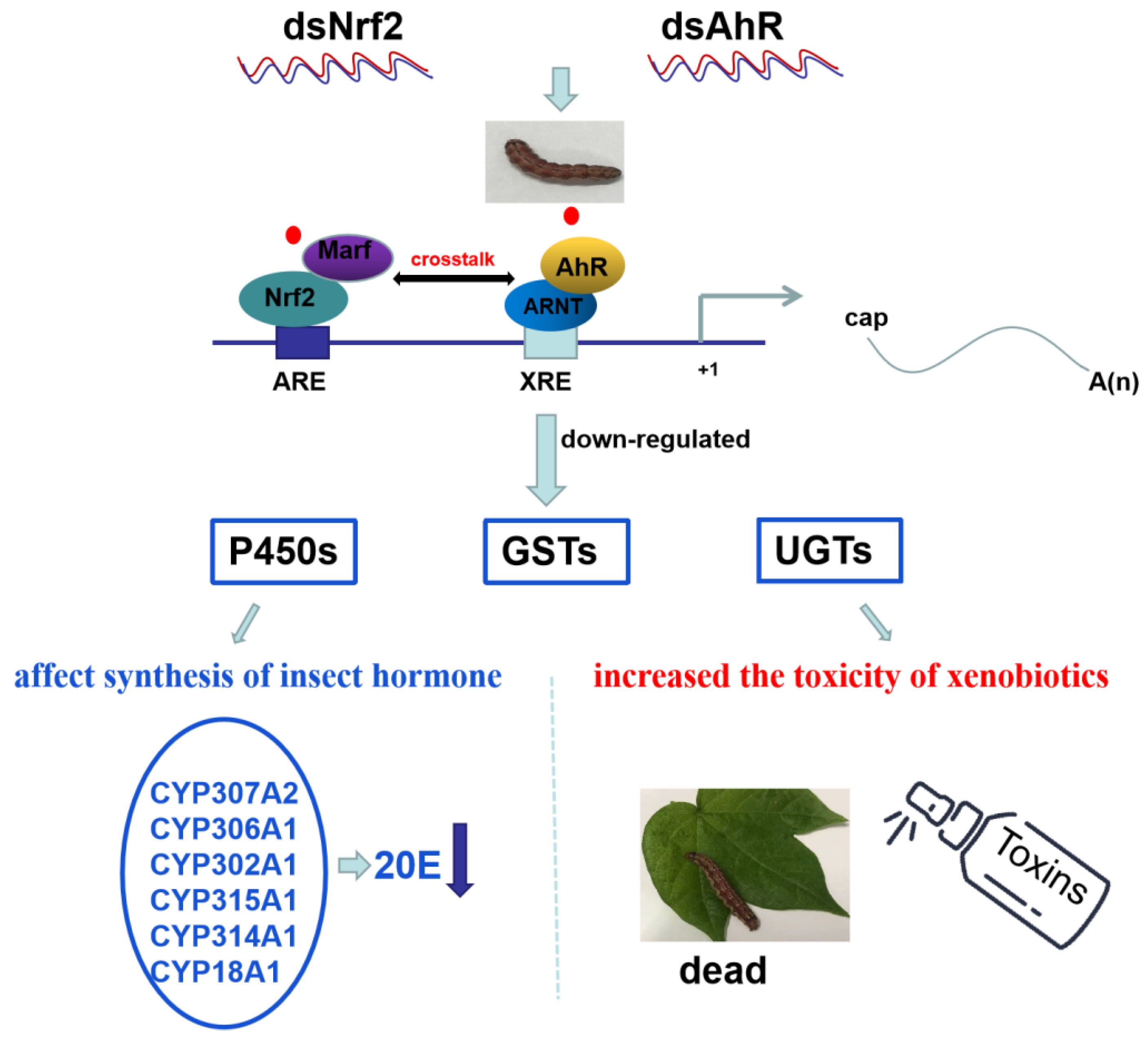
2.5. RNAi HaNrf2 and HaAhR Effects on the Synthesis of Insect Hormone

Studies have revealed that CYP18A1, CYP307A2, CYP306A1, CYP302A1, CYP315A1 and CYP314A1 genes are essential for each of the steps in 20E biosynthesis [20,21,22]. CYP18A1 belongs to the CYP2 clan and takes part in 20E inactivation, converting 20E to 20, 26-dihydroxyecdysone [23]. The expression of CYP18A1, CYP307A2, CYP306A1 CYP302A1, CYP315A1 and CYP314A1 in H. armigera are all down-regulated by knockdown HaNrf2 (Figure 4B), while these genes’ expressions are also down-regulated by knockdown HaAhR, except CYP315A1 (Figure 4B). These genes promoter sequences contain at least one HaNrf2 or HaAhR binding sites (Figure 4A).

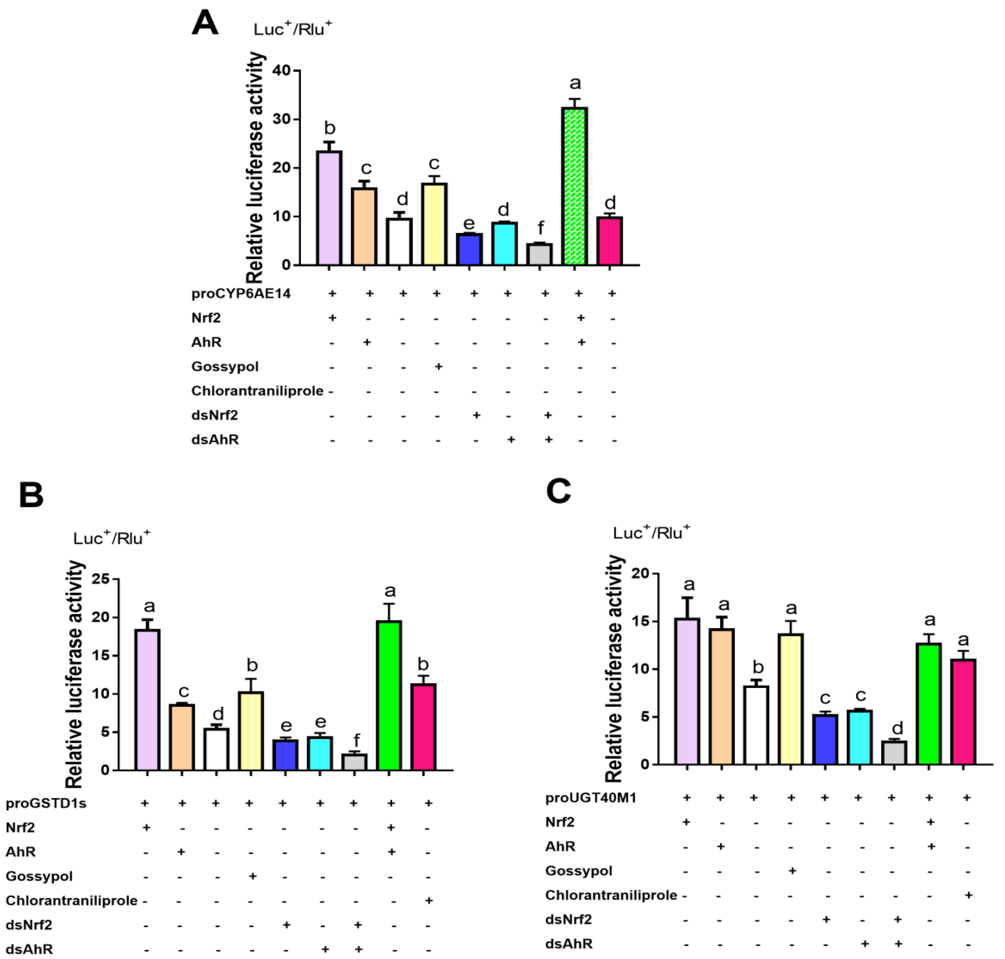
2.6. Detoxification Genes Regulated by HaNrf2 and HaAhR in H. armigera

2-tridecanone-induced P450s genes (CYP6B2, CYP6B6 and CYP6B7) are chosen to confirm the regulation relationship between HaNrf2 and HaAhR. The promoter activity of CYP6B2, CYP6B6 and CYP6B7 promoter induced by 2-tridecanone and the deletion of XRE/AhR or ARE/Nrf2 elements effects on 2-tridecanone-induced promoter activity are shown in Figure 5. In order to roughly define the regions of CYP6B2, CYP6B6 and CYP6B7 harboring putative cis elements, a set of CYP6B2, CYP6B6 and CYP6B7 promoter 5′ progressive deletion constructs were, respectively, co-transfected with the control plasmid of phRL-TK into H. armigera fat body cells. Compared to the construct Full-6B2, the progressive 5′ deletions to cut-1386, cut-928, cut-610 and cut-160 were constructed. The 5′ deletion to 610 and 160 significantly decreased the promoter activity (Figure 5A). Compared to the Full-6B2 construct, all the progressive 5′ deletion constructs can be induced by 2-tridecanone, except cut-160 are completely blocked (Figure 5A). Compared to the construct Full-6B6, the progressive 5′ deletions to cut-971, cut-681, cut-310 and cut-180 were constructed. The 5′ deletion to 180 significantly reduced promoter activity, whereas the 5′ deletion to 310 did not significantly decrease the basal promoter activity (Figure 5B). A similar trend was observed for 2-tridecanone-induced promoter activity. Compared to the Full-6B6 construct, all the progressive 5′ deletion constructs decreased the 2-tridecanone-induced activity, except the constructs to 681 and 180 (Figure 5B). For the construct Full-6B7, the progressive 5′ deletions to cut-780, cut-400 and cut-200 were constructed. The 5′ deletion to 200 significantly decreased the promoter activity (Figure 5C). Compared to the Full-6B7 construct, all the progressive 5′ deletion constructs can be induced by 2-tridecanone, except cut-200 are completely blocked (Figure 5C).
In order to eliminate the interaction effects between ARE/Nrf and XRE/AhR elements completely, we generated a set of internal deletion constructs that deleted, separately, one of the two transcription factor binding sites or deleted both of the sites from the short construct of CYP6B2, CYP6B6 and CYP6B7 promoters (Figure 5D–F). Both the deleted ARE/Nrf2 element and XRE/AhR element significantly reduced the 2-tridecanone-induced activity and the basal activity of the short construct (Figure 5D–F). Taken together, these findings reveal that the XRE/AhR and ARE/Nrf2 elements are necessary for both the basal and the 2-tridecanone-induced expression of CYP6B2, CYP6B6 and CYP6B7.
A dual luciferase assay was used to detect the promoter activity of gossypol and chlorantraniliprole-induced CYP6AE14, GSTD1s, and UGT40M1 genes which also transiently transfected with dsNrf2, dsAhR, HaAhR or HaNrf2 (Figure 6). Compared to the control construct, the activity of the CYP6AE14 promoter was enhanced 2.46- and 1.65-fold, respectively, by transfection with HaNrf2 or HaAhR (Figure 6A); but the activity of the CYP6AE14 promoter was decreased by 34% and 10%, respectively, by transfection with dsNrf2 or dsAhR (Figure 6A). When co-transfection with dsNrf2 and dsAhR, the promoter activity of CYP6AE14 was decreased by 55%; but CYP6AE14 promoter activity was increased 3.40-fold after co-transfection with HaNrf2 and HaAhR (Figure 6A). The promoter activity of GSTD1s was significantly enhanced 3.39- and 1.7-fold, respectively, by transfection with HaNrf2 or HaAhR (Figure 6B); and the promoter activity of UGT40M1s were significantly enhanced 1.86-and 1.73-fold, respectively, by transfection with HaNrf2 or HaAhR (Figure 6C). The activity of the GSTD1s promoter was decreased 34% and 10%, respectively, by transfection with dsNrf2 or dsAhR (Figure 6B), and the promoter activity of the UGT40M1 was decreased 34% and 10%, respectively, by transfection with dsNrf2 or dsAhR (Figure 6C). When co-transfection with dsNrf2 and dsAhR, the promoter activity of GSTD1s and UGT40M1 was decreased by 63% and 71%, respectively; but GSTD1s and UGT40M1 promoter activity was increased 3.60-fold and 1.54-fold after co-transfection with HaNrf2 and HaAhR (Figure 6B,C). The promoter activity of CYP6AE14, GSTD1s, and UGT40M1s was significantly induced by gossypol and chlorantraniliprole treatment (Figure 6). These results confirm that both the HaNrf2 and HaAhR signal pathways play a significant role in the activation of CYP6AE14, GSTD1s, and UGT40M1 genes’ expression.
To determine whether the essential elements XRE/AhR and ARE/Nrf2 are recognized and specifically bound by proteins from nuclear extracts prepared from un-induced (ethanol-treated) and/or 2-tridecanone-induced H. armigera fat body cells, we conducted sEMSA (Figure 7A). We used biotin-labeled double-stranded oligos containing the XRE/AhR element or the ARE/Nrf2 element in CYP6B6 promoters as the probe. As the competitors, we used unlabeled XRE/AhR or ARE/Nrf2 (i.e., cold probe) and an unlabeled unrelated nonspecific sequence. In the absence of competitors, both the un-induced (Lane 4, Lane 9 in Figure 7A) and the 2-tridecanone-induced (Lane 2–3, 5 and Lane 7–8, 10 in Figure 7A) nuclear extracts formed two sequence specific DNA-protein complexes with the ARE/Nrf2 (Lane 2–5 in Figure 7A) and XRE/AhR (Lane 7–10 in Figure 7A) biotin probes. These complexes were completely out-competed by an excess of the XRE/AhR and ARE/Nrf2 cold competitor probes (Lane 3 and 8 in Figure 7A). The DNA-protein complexes with 2-tridecanone-induced nuclear extracts and HaNrf2 specific antibody displayed a stronger signal than those with un-induced nuclear extracts and HaNrf2-specific antibody; the DNA-protein complexes with 2-tridecanone-induced nuclear extracts and HaAhR-specific antibody displayed a stronger signal than those with un-induced nuclear extracts and HaAhR-specific antibody (Figure 7A).
After H. armigera fat body cells were transfected with the pAcV5His-Red-HaNrf2 construct for 24 h, the subcellular localization of HaNrf2 was observed. We found that HaNrf2 was mainly located in the cytoplasm with the red florescence (Figure 7B). These fat body cells were then treated with 2-tridecanone in a final concentration of 125 μM for 4 h. We observed that the subcellular localization of Nrf2 was mainly in the nucleus following 2-tridecanone treatment (Figure 7B), while the control cells treated with ethanol still had signals indicating that Nrf2 was localized to the cytoplasm (Figure 7B).
The 1999 bp fragments upstream of the translation start sites (ATG) of HaAhR gene were selected as potential promoter regions for transcription factor binding site prediction; the prediction results showed that the Nrf2 and AhR binding sites were found in HaAhR gene promoters (Figure 8C). When co-transfection with HaNrf2, the promoter activity of HaAhR was enhanced 1.4-fold; but the HaAhR promoter activity was decreased about 60% after co-transfection with dsNrf2 (Figure 8C). The EMSA method was used to verify the transcription factor HaNrf2 and HaAhR binding sites, a single-target shift band can be clearly detected in each treatment (Figure 8A,B).
Based on these results, a schematic illustration of the proposed model of how the crosstalk between HaNrf2 and HaAhR regulate the P450s, GSTs and UGTs and mediate the susceptibility of xenobiotics (gossypol, 2-tridecanone and chlorantraniliprole) in H. armigera is presented in Figure 9.

3. Discussion

Analysis of insect genome sequences has revealed that expansion of the P450, GST, and UGT gene families is related to plant allelochemicals and insecticides resistance [24,25]. Xenobiotic transcription factors play important roles in the regulation of detoxification genes involved in xenobiotic metabolism [8]. It was also reported that Nrf2 and AhR transcription factors are involved in the regulation of some three-phase enzyme gene members responsible for insecticide resistance [13,26,27].
Several studies in insects have shown that some three-phase enzymes genes, such as some members of P450s, GSTs, UGTs, and ABC transporter genes regulated by Nrf2, play important roles in insecticide resistance [11,26,28,29,30,31]. For example, in Tribolium castaneum, Nrf2 regulates genes involved in insecticide detoxification such as CYP4G7, CYP4G14, GST1, ABCAUB, ABCAA1, ABCAA1L, and ABCA9B [26]. Hu et al. (2020) showed that Nrf2 regulates the expression of CYP321A16 and CYP332A1, which mediate chlorpyrifos resistance in Spodoptera exigua [11]. Moreover, in Spodoptera litura, Chen et al. (2018), Lu et al. (2020) also found that Nrf2 regulates GSTe1 and CYP6AB12 expression in response to insecticide [28,29]. Shi et al. (2021) reported that transcription factor Nrf2 regulates the expression of some detoxification genes that mediate indoxacarb resistance in Spodoptera litura [30]. In Bombyx mori, transcription levels of CYP302A1 and CYP306A1 were significantly decreased in cells after treatment with BmNrf2 dsRNA, whereas their transcription levels were significantly increased (2.15- and 1.31-fold, respectively) after treatment with the Nrf2 agonist curcumin [31]. In a study in Drosophila melanogaster, CYP6DA1 contained Nrf2 binding sites in the 5′-promoter core region, and changes in these binding sites were associated with altered promoter activity of CYP6DA1 as well as resistance to DDT [32]. In Helicoverpa zea, induction of the phase I gene CYP321A1 by flavone compounds has been shown to be mediated by Nrf2 [33].
In insects, an XRE element of AhR has been functionally characterized from the promoters of CYP6B1 and CYP6B4 genes; this element is associated with the xanthotoxin cascade [34]. CYP6B4 and CYP6B1 promoters contain putative XRE-AhR elements identical to the aryl hydrocarbon response elements present in mammalian phase I detoxification genes. Transfection of CYP6B4 and CYP6B1 promoters containing XRE-AhR elements were induced significantly by benzo(α)pyrene, an aryl hydrocarbon, as well as by xanthotoxin, an allelochemical encountered in host plants [35]. Mutational analysis of the CYP6B1 promoter indicated that the XRE-AhR element is necessary for both constitutive and xanthotoxin-inducible induction of the CYP6B1 promoter [36]. The aryl hydrocarbon receptor (AhR) regulates CYP6DA2 in Aphis gossypii, conferring gossypol and spirotetramat tolerance [37]. AhR also controls the over expression of P450 genes in D. melanogaster that are resistant to insecticides [6].
Despite some excellent studies on the single detoxification gene member induced by host plant allelochemicals or insecticides regulated by Nrf2 or AhR, the network of the detoxifications genes which can be regulated by Nrf2 and AhR is still unknown in insects. There had also been no reports about the crosstalk between AhR and Nrf2 which can regulate the expression of detoxification genes in insects. The study of insect adaptation to the defensive metabolites of host plants and various kinds of insecticides in order to acquire resistance is a hot topic in the field of controlling pests, however, the mechanism of insect adaptation to these xenobiotics is still unclear. Our results found that Nrf2 and AhR signaling pathways in H. armigara are not only regulated in the form of one detoxification gene member to one kind of xenobiotic, but are also regulated by multiple detoxification genes (including genes related to the synthesis of insect hormone and xenobiotics resistance.) It should be a general signaling pathway for revealing the adaptive mechanism of insects to xenobiotics.
This study comprehensively elucidated the effect of HaNrf2 and HaAhR knockdown on the detoxification expression in H. armigera. We found that knocking down the expression of HaNrf2 or HaAhR by RNAi can significantly increase the sensitivity of H. armigera to gossypol, 2-tridecanone, chlorantraniliprole and chlorfenapyr (Figure 1). To investigate the function of HaNrf2 and HaAhR in regulating the sensitivity of plant allelochemicals and insecticides in H. armigara, full-length sequences of the H. armigera Nrf2 and AhR genes were cloned and used for designing dsRNAs specific to each gene. dsHaNrf2 and dsHaAhR treatment enhanced cotton leaf defenses against H. armigera feeding, and the reduced expression of HaNrf2 and HaAhR both significantly increase the gossypol, 2-tridecanone, chlorantraniliprole and chlorfenapyr susceptibility of H. armigera larvae fed on dsRNA-coated cotton leaves or an artificial diet mixed with xenobiotics (Figure 1). Some studies also reported that silencing of detoxification enzyme genes associated with plant allelochemical tolerance or insecticide resistance in insects can result in their increased susceptibility to plant allelochemicals or insecticides [5,19,30]. In this study, our results revealed that HaNrf2 and HaAhR are both key regulatory factors involved in mediating H. armigera susceptibility to xenobiotics.
To comprehensively elucidate the genes regulated by HaNrf2 and HaAhR in H. armigera, we performed RNA sequencing on H. armigera larvae that were treated with HaNrf2, HaAhR, or GFP dsRNA. Differential gene expression analysis showed that 2484 genes were up-regulated and 1991 genes were down-regulated by RNAi knockdown of HaNrf2, and 1187 genes were up-regulated and 870 genes were down-regulated by RNAi knockdown of HaAhR (Figure S1). Based on the expected pattern of positive regulation between HaNrf2 or HaAhR and their target genes, we mainly focused on the down-regulated genes which included a lot of three-phase detoxification enzyme genes. Interestingly, we found HaNrf2 knockdown decreased the sum FPKM of P450 genes by up to 48% (Figure 2A), and HaAhR knockdown decreased the sum FPKM of P450 genes by up to 90% (Figure 2B). In all, 23 of 113 P450 genes in H. armigera were down-regulated by HaAhR knockdown, and 39 of 113 P450 genes were down-regulated by HaNrf2 knockdown (Figure 2E). The same phenomenon was observed for the GST and UGT gene families; knockdown of HaNrf2 or HaAhR decreased the sum FPKM of GST and UGT genes (Figure 2F,G).
Previous studies have found that the expression of P450 genes, UGT genes, and GST genes can be significantly induced by gossypol, 2-tridecanone, chlorantraniliprole or chlorfenapyr treatments [17,18]. P450s are important components of Phase I detoxification enzymes. Our previous study showed that multiple over-expressed detoxification genes play important roles in H. armigera 2-tridecanone tolerance [17], with 2-tridecanone inducing the highest cluster genes (CYP6B2, CYP6B6 and CYP6B7) also being regulated by HaNrf2 and HaAhR (Figure 5). The promoter activity of CYP6B2, CYP6B6 and CYP6B7 promoter induced by 2-tridecanone and the deletion of XRE/AhR or ARE/Nrf2 elements’ effects on 2-tridecanone-induced promoter activity are shown in Figure 5. In order to roughly define the regions of CYP6B2, CYP6B6 and CYP6B7 harboring putative cis elements, a set of CYP6B2, CYP6B6 and CYP6B7 promoter 5′ progressive and ARE/Nrf and XRE/AhR elements deletion constructs were, respectively, co-transfected with the control plasmid of phRL-TK into H. armigera fat body cells. We found that the essential cis elements required for both basal and 2-tridecanone-induced expression of CYP6B2, CYP6B6 and CYP6B7 are located in the region containing proximal AhR and Nrf2 transcription factor binding sites (Figure 5). Then, we used confucus to observe that the subcellular localization of HaNrf2 was mainly localized in the nucleus following 2-tridecanone treatment (Figure 7B), while the control cells still had no signals indicating that HaNrf2 was transferred from cytoplasm to nucleus, which implies that 2-tridecanone requires more HaNrf2 to actively enter in nucleus (Figure 7B). The above results mean that 2-tridecanone-induced CYP6B2, CYP6B6 and CYP6B7 genes’ overexpression is regulated by both HaNrf and HaAhR pathways.
CYP6AE14 was an especially promising candidate as one P450 gene involved in cotton gossypol detoxification by H. armigera [38]. When CYP6AE14 was silenced in H. armigera via feeding of dsRNA or transgenic cotton transformed with a construct expressing dsRNA, larval growth was greatly reduced in the presence of gossypol [38]. CYP6AE14 genes are induced by gossypol treatment in H. armigera; it also can by down-regulated by RNAi HaNrf2 or HaAhR (Figure 2). Accordingly, we suggest that treatment with dsHaNrf2- and dsHaAhR-enhanced cotton leaf defenses against H. armigera and knocking down HaNrf2 and HaAhR can affect the expression of multiple detoxification enzyme genes associated with gossypol tolerance. To this end, we used a dual luciferase assay to detect the promoter activity of CYP6AE14, GSTD1s, and UGT40M1 genes in H. armigera larvae transfected with HaAhR or HaNrf2. Compared to the control construct, the activities of the CYP6AE14, GSTD1s, and UGT40M1 promoters were significantly enhanced by co-transfection with HaNrf2 or HaAhR (Figure 6). These results confirm that both Nrf2 and AhR transcription factors play a significant role in the activation of downstream expression in genes belonging to the P450, GST, and UGT gene families. The promoter activity of CYP6AE14, GSTD1s, and UGT40M1s was significantly induced by gossypol and chlorantraniliprole treatment (Figure 6). All above results confirm that both HaNrf2 and HaAhR signaling pathways play a significant role in the activation of CYP6AE14, GSTD1s, and UGT40M1 genes’ expression treated by gossypol or chlorantraniliprole. H. armigera P450s in the CYP6 subfamily were down-regulated by RNAi HaNrf2 or HaAhR (Figure 2), which were also shown to be involved in the tolerance of xanthotoxin, gossypol acetate, 2-tridecanone, nicotine, and coumarin [6,39], and P450-associated increases in detoxification were also shown to be the primary cause of high insecticide resistance in H. armigera [40].
CYP18A1, CYP307A2, CYP306A1, CYP302A1, CYP315A1 and CYP314A1 genes are related to the synthesis of insect hormones 20E. 20E is a polyhydroxylated steroid hormone that controls molting and thereby affects the growth of arthropods. Studies using insects have revealed that the Halloween P450 genes (CYP18A1, CYP307A2, CYP306A1, CYP302A1, CYP315A1 and CYP314A1) are essential for each of the steps in 20E biosynthesis [20,21,22]. CYP18A1 belongs to the CYP2 clan and takes part in 20E inactivation, converting 20E to 20, 26-dihydroxyecdysone [23]. The expression of CYP18A1, CYP307A2, CYP306A1, CYP302A1, CYP315A1 and CYP314A1 in H. armigera are all down-regulated by knockdown HaNrf2 (Figure 4B), while these genes’ expressions are also down-regulated by knockdown HaAhR, except CYP315A1 (Figure 4B). These genes’ promoter sequences at least contain one HaNrf2 or HaAhR binding site (Figure 4A). Previous studies found that a lot of plant allelochemicals or insecticides, similar to gossypol, 2-tridecanone, chlorantraniliprole will not only decrease the growth rate of insects, but can also decrease the susceptibility to these xenobiotics [1,17,18]. Our results provide a general signaling pathway for revealing the adaptive mechanism of insects to xenobiotic, and explain why many xenobiotic will not only decrease the growth rate of insects, but also can decrease their capability to metabolize xenobiotics. Our results prove that a pathway may exist in HaNrf2 and HaAhR to regulate the P450 genes which are related to insect hormones synthesis.
The EMSA method was used to verify transcription factor HaNrf2 and HaAhR binding sites in HaAhR promoter sequence, showing a single-target shift band can be clearly detected in each treatment (Figure 8A,B). These results confirm that there is crosstalk between HaNrf2 and HaAhR transcription factors that plays a significant role in the activation of downstream detoxification genes’ expression. In mammals, AhR has been reported to affect the Nrf2 expression [41,42]. There may be direct crosstalk between the Nrf2 and the AhR pathways for the induction of phase II genes (NQO1, GSTA2 and UGT1A6) in mammals [41]. Some researchers also demonstrated that Nrf2 gene transcription is directly modulated by AhR activation in mammals [42]. However, there have been no reports about the crosstalk between Nrf2 and AhR signaling pathways in insects. Our research reported that expression of AhR and Nrf2 are partially dependent on each other, implying that Nrf2 modulates both transcription of AhR and its downstream targets in insects. Our results confirm that there is crosstalk between the HaNrf2 and HaAhR signaling pathways to regulate the expression of detoxification genes in H. armigera.
The RNAi method has been widely recognized as a novel and safe strategy in pest management due to its high sequence-dependent specificity and simplicity to degrade. Therefore, HaNrf2 and HaAhR could serve as potential targets of dsRNA spraying for pest management.

4. Materials and Methods

4.1. Insects

The cotton bollworm population used in this study was collected from Handan in Hebei Province, China in 1998, and reared on an artificial diet in an air-conditioned room maintained at 26 ± 1 °C, 70–80% relative humidity, and under a photoperiod of 16:8 h (L:D). The artificial diet was supplied as described in our previous study [19]. Adults were held under the same conditions and supplied with a 10% sugar solution.

4.2. RNA Isolation, Reverse Transcription, and Gene Cloning

Total RNA was isolated from H. armigera larvae using TRIzol® reagent (Invitrogen, Carlsbad, CA, USA). One µg of RNA sample was used as the template for synthesis of first-strand cDNA, using the cDNA synthesis kit (Takara, Japan). Specific primers of HaNrf2 (KU355787.1) and HaAhR (XM_021331544.1) were designed and synthesized to amplify the complete open reading frame region (Table S1).

4.3. HaNrf2 and HaAhR RNAi

Based on HaNrf2 and HaAhR sequences, we used online prediction software E-RNAi (http://www.dkfz.de/signaling/e-rnai3/) (accessed on 1 January 2021) [43] to predict putative RNAi sites and designed specific primers using DNAMAN 6.0 software. RNAi method is according to our previous study [19], HaNrf2, HaAhR, and green fluorescence protein (GFP) genes fragments for in vitro transcription reactions were amplified by PCR using primers that contain the T7 promoter sequence at the 5′ end (Table S1). The products were used as templates to synthesize dsRNAs using the MEGAscript T7 transcription kit (Invitrogen, USA). The dsRNA product sizes (412 bp for dsHaNrf2; 438 bp for dsHaAhR; 288 bp for dsHaGFP) were analyzed by 1% agarose gel electrophoresis and quantified using a spectrophotometer. The 2nd instar larvae, after being starved for 12 h, were offered an artificial diet containing HaAhR or HaNrf2 dsRNA (15 μg/g, w/w) for 24 h; GFP dsRNA (15 μg/g, w/w) was used as a negative control. Thirty larvae were used for each treatment, and three replications were performed. The dsRNA-mediated depletion of HaNrf2 or HaAhR transcripts was analyzed by RT-qPCR using the primers listed in Table S1. Each sample was analyzed in triplicate and normalized to the internal control, EF-1α [19]. Statistical analyses were performed using GraphPad Prism 5.0 software (GraphPad Prism, San Diego, USA), and a p value < 0.05 was considered statistically significant.

4.4. Xenobiotics Treatment following HaNrf2 or HaAhR Knockdown

To determine the effects of HaNrf2 and HaAhR knockdown on the efficacy of the plant allelochemicals and insecticides against H. armigera, gossypol was dissolved in acetone to concentration of 0.02 mg/g, (w:w) then mixed with 1 g of artificial diet and 15 μg/g (w:w) of dsNrf2, dsAhR, or dsGFP; 2-tridecanone was dissolved in ethanol to concentration of 0.05 mg/g (w:w) then mixed with 1 g of artificial diet and 15 μg/g (w:w) of dsNrf2, dsAhR, or dsGFP; chlorantraniliprole was dissolved in N, N-dimethylformamide to concentration of 0.05 μg/g (w:w) then mixed with 1 g of artificial diet and 15 μg/g (w:w) of dsNrf2, dsAhR, or dsGFP; chlorfenapyr was dissolved in acetone to concentration of 0.1 μg/g (w:w) then mixed with 1 g of artificial diet and 15 μg/g (w:w) of dsNrf2, dsAhR, or dsGFP; The solvents were used as the negative control; dsGFP combined with different pesticides are used as the negative control. Fifty 1nd instar larvae were used for each treatment, and the experiments were repeated in triplicate. Cotton leaf discs (20 mm diameter) were cut using a stainless-steel cork-borer, placed in cell culture plates filled with 1 mL of 2% agar to maintain leaf humidity, and coated with 1 μg of dsRNA per leaf. Ten 1nd instar larvae were used for each treatment, and the experiments were repeated in triplicate. Mortality of larvae reared at 25 ± 2 °C, 65% ± 5% RH, and under a 14:10 h (L:D) photoperiod was recorded after each collection time point. Larvae were considered dead if they failed to make a coordinated movement when prodded with a brush. Mean and standard errors were obtained from at least three independent replications. Statistical analyses were performed using GraphPad Prism 5.0 software, and a p value < 0.05 was considered statistically significant.

4.5. RNA Sequencing

Total RNA isolated from dsNrf2-, dsAhR- and dsGFP-treated 2nd instar H. armigera larvae for 24 were used to prepare RNA sequencing libraries using the NEBNext® UltraTM RNA Library Prep Kit for Illumina® (NEB, Beverly, MA, USA) following the manufacturer’s recommendations. Thirty larvae were used for each treatment, and the experiments were repeated in triplicate. The library preparations were sequenced on an Illumina Hiseq 2500 platform at the Novogene Bioinformatics Institute (Beijing, China). Then, 2 mg/g gossypol or 2μg/g chlorantraniliprole-treated six instar H. armigera larvae for 24 h and acetone-alone-treated larvae were used to prepare RNA sequencing. The transcriptome data of 2-tridecanone-induced larvae were referred to in our previous study [17].
Gene expression levels were estimated by the number of Fragments Per Kilobase of transcript sequence per Million base pairs sequenced (FPKM) [44]. Differentially expressed gene analysis was performed using the DESeq2 R package 1.16.1 [45]. Genes with p < 0.05 and |log2 (fold change)| ≥ 1 were considered differentially expressed. Gene Ontology (GO) enrichment analysis (p < 0.05) was used to identify functional modules of down-regulated genes [46]. To further characterize the down-regulated genes, we performed pathway analysis using the Kyoto Encyclopedia of Genes and Genomes (KEGG) pathway database (http://www.genome.jp/kegg/) (accessed on 1 January 2021).

4.6. Quantitative PCR Analysis of Down-Regulated Genes and Prediction of Transcription Factor Binding Sites

Reverse transcription-quantitative PCR (RT-qPCR) was performed to verify the results of the RNA-seq data. Nine detoxification genes from the down-regulated group were selected for expression analysis by RT-qPCR, and EF-1a was used as the reference gene [19]. The primer sequences used in the RT-qPCR analyses are given in Table S1. The relative gene expression levels were calculated using the 2−∆∆CT method [47]. The RT-qPCR data was expressed as the mean ± SD of three independent biological replicates. Promoter sequence information of the down-regulated detoxification genes was acquired from the H. armigera genome [48]. Transcription factor binding sites in the promoters were predicted using JASPAR [49].

4.7. Transient Transfection and Dual Luciferase Assay

The homologous H. armigera fat body cell line was generously provided by Dr. Huan Zhang (Zoology, CAS, China). The cell line was routinely maintained with sf900-III insect serum-free medium (Thermo Fisher Scientific, USA) supplemented with 10% heat-inactivated fetal bovine serum (Gibco, USA) at 37 °C. The full-length coding regions of HaNrf2 and HaAhR were constructed into pAcV5His vector [13]. The H. armigera fat body cells were seeded into 96-well plates (1 × 10⁴ cells/ well) then transiently co-transfected with different gene promoters fused to pGL4 luciferase reporter constructs (1 mg/well) combined with pAcV5His-Red-HaNrf2 (0.5 mg/well), pAcV5His-GFP-HaAhR (0.5 mg/well), dsNrf2 (4 μg/well) or dsAhR (4 μg/well). The internal renilla luciferase plasmid phRL-TK (Promega; 0.2 mg/well) was used as the control reporter plasmid. Cellfectin-II reagent (Invitrogen; 1 μL per well) was used to transfect all of the plasmids into fat body cells. After 24 h, the cells were harvested and lysed, and lysates were used to measure renilla and firefly luciferase activities with a Dual-Luciferase Reporter Assay System (Promega, Wisconsin, USA) on a GloMax® 96 Microplate Luminometer (Promega, Madison, WI, USA). The relative firefly luciferase activity, normalized against the renilla luciferase activity, reported for each construct was used to calculate the mean and standard error of four independent transfections of one representative sample. Statistical analyses were performed using GraphPad Prism version 5.0 software (GraphPad Prism, San Diego, CA, USA), and p values < 0.05 were considered statistically significant.

4.8. Super Electrophoretic Mobility Shift Assays (sEMSA)

H. armigera fat body cells seeded into wells of 6-well plates (1 × 10⁶ cells/well) were treated with 2-tridecanone at a final concentration of 125 μM or an equal volume of ethanol (control). After 24 h, the cells were harvested and used to prepare control and 2-tridecanone-induced H. armigera fat body cell nuclear extracts according to Lahiri and Ge (2000) [50]. Biotin end-labeled double-stranded oligonucleotides (Biotin Nrf2: 5′biotin-taacaacTCATGCTTGCAGcttttcta-3′biotin; the fragment targeted the region between −152 to −162 of the CYP6B6 promoter; biotin AhR: 5′biotin-tcacCATGACACGTGCAAacgc-3′biotin the fragment targeted the region in the CYP6B6 promote) containing the essential Nrf2 or AhR binding site (the capital letters in the sequence) were used as the probe for sEMSA. DNA-nuclear extract binding reactions were carried out in a 20 µL volume containing 0.17 µg of H. armigera fat body cell nuclear extracts, 2 µL of 10× binding buffer (10 mM Tris, pH 7.5, 50 mM KCl, 5 mM MgCl2, 1 mM dithiothreitol, 0.05% Nonidet P-40, and 2.5% glycerol), 1 µL of poly (dI-dC) (1 µg/µL), 80 fmol of the biotin-labeled probe, and 0.5 µL of the Nrf2 or AhR polyclonal antibody. We used pET28a vector to express the fusion proteins of Nrf2 and AhR, and then used these fusion proteins to prepare polyclonal antibodies against Nrf2 and AhR in male New Zealand rabbits. Prepared and purified polyclonal anti-Nrf2 and anti-AhR antibodies were stored at −70 °C. The reaction mixture was preincubated for 10 min at 25 °C, after which the probe was added. Incubation continued for another 20 min at 25 °C. Competition reactions were performed in the same conditions, except for the addition of a 200-fold molar excess of unlabeled competitor oligonucleotides (Nrf2: 5′-taacaacTCATGCTTGCAGcttttcta-3′, the fragment was between −152 to −162 of CYP6B6 promoter; AhR: 5′-tcacCATGACACGTGCAAacgc-3′, the fragment was located in CYP6B6 promoter, each fragment contained 4 or 5 flanking nucleotides at both ends.
Biotin end-labeled double-stranded oligonucleotides (Biotin Nrf2: 5′biotin-aaATGCTtatt-3′; Biotin AhR: 5′biotin-gaacGCGTGgcc-3′) containing the essential Nrf2 or AhR binding site in HaAhR promoter sequences (the capital letters in the sequence) were also used as the probe for sEMSA. The reaction mixture including the fusion protein HaNrf2 or HaAhR was preincubated for 10 min at 25 °C, after which the probe was added. Incubation continued for another 20 min at 25 °C. Competition reactions were performed in the same conditions, except for the addition of a 200-fold molar excess of unlabeled competitor oligonucleotides (Biotin Nrf2: 5′biotin-ggATGCTccgt-3′; Biotin AhR: 5′biotin-tcatGCGTGtgc-3′, each fragment contained 4 or 5 flanking nucleotides at both ends. All the reaction mixtures were electrophoresed through a non-denaturing polyacrylamide gradient gel (Bio-Rad, Hercules, CA, USA) for 2.5 h in 0.5 TBE buffer at 80 volts at 4 °C, and then electro-transferred onto a Hybond-N nylon membrane (Amersham Biosciences, Uppsala, Sweden) for 30 min at 220 mA at 4 °C. The retarded DNA-nuclear protein complexes on the membrane were fixed by UV cross-linking and visualized with a LightShift Chemiluminescent Electrophoretic Mobility Shift Assay Kit (Thermo Fisher Scientific, Waltham, USA). The intensities of the bands formed were assessed via X-ray.

4.9. Laser Confocal Light Microscopy

H. armigera fat body cells were transfected with the construct of pAcV5His-Red-Nrf2 (0.5 mg/well) for 24 h. The supplemented medium was discarded and the new supplemented medium (which contained 10% of fetal bovine serum) were mixed with 2-tridecanone (final concentration 125 μM) or control, added into the wells, and incubated for 6 h. After incubation, H. armigera fat body cells were fixed in 4% neutral buffered paraformaldehyde (NBP) for 15 min and then treated with 0.5% Triton X-100 at room temperature for 10 min. The cells were stain by DAPI for 10 min. After washing 5 times with PBS, visualization and localization of Nrf2 in the fat body cells were analyzed by laser scanning confocal microscopy Fv1000 (Olympus, Japan), and processed with Photoshop CS, version 8 (Adobe Systems, San Jose, CA, USA).

4.10. Statistical Analyses

The test data were expressed as “mean ± standard error (SE)”. One-way ANOVA was performed on the test data using GraphPad Prism version 5.0 software (GraphPad Prism, San Diego, USA), and the expression data before and after treatment were analyzed for significance using Student’s t-test.

5. Conclusions

Taken together, we found most P450s, GSTs, and UGsT gene promoters have at least one Nrf2 binding site or AhR binding site, and some P450, GST, and UGT genes’ promoters even simultaneously contain a very closely or overlapping Nrf2 binding site and AhR binding site. Using RNAi, RNA sequencing, RT-qPCR analysis, transcription factor binding site prediction, EMSA, and dual luciferase reporter assays, we demonstrated that there is crosstalk between HaNrf2 and HaAhR to regulate the expression of multiple detoxification genes in H. armigera. Although much is known about each role of the Nrf2 or AhR signal pathway in the regulation of some insects’ detoxification gene members, the network regarding the detoxification genes which can be regulated by Nrf2 and AhR is still unknown, and there were also no reports about the crosstalk between AhR and Nrf2 that exist in insects and can regulate the detoxification genes’ expression. In this study, we found many P450s, GSTs, and UGTs gene members can down-regulate by HaNrf2 and HaAhR knockdown, which may imply that the effects of HaNrf2 and HaAhR knockdown are not limited in response to one kind of xenobiotic, and may also affect the susceptibility to other plant allelochemicals or insecticides in H. armigera. Our results provide a new general signaling pathway for revealing the adaptive mechanism of insects to xenobiotics, and explain why many xenobiotics have the phenotype of toxic action and affect the growth rate in insects. Elucidating the mechanisms underlying detoxification enzyme genes’ regulation mediated by AhR and Nrf2 transcription factors will illuminate how detoxification genes’ expressions are coordinately up-regulated in insects that have acquired the tolerance ability to defend against plant allelochemicals or insecticides. The results of this study improve our understanding of detoxification genes’ regulation in H. armigera and provide more insight into designing effective pest-management strategies to reduce the overuse of insecticides. The transcription factors HaNrf2 and HaAhR could serve as potential targets for pest management.

Supplementary Materials

The following supporting information can be downloaded at: https://www.mdpi.com/article/10.3390/ijms232416126/s1.

Author Contributions

Conceptualization, L.Z. and X.G.; methodology, L.Z. and S.L.; software, L.Z.; validation, L.Z., S.L., M.L. and M.G.; formal analysis, L.Z.; investigation, L.Z.; resources, X.G.; data curation, L.Z.; writing—original draft preparation, L.Z.; writing—review and editing, X.G.; visualization, L.Z.; supervision, X.G.; funding acquisition, L.Z. and X.G. All authors have read and agreed to the published version of the manuscript.

Funding

This research was supported by the National Natural Science Foundation of China (31601655 and 32272581).

Institutional Review Board Statement

Not applicable.

Informed Consent Statement

Not applicable.

Data Availability Statement

Not applicable.

Conflicts of Interest

The authors declare no conflict of interest.

References

  1. Heckel, D.G. Insect detoxification and sequestration strategies. In Annu. Plant Rev.; John Wiley & Sons, Ltd.: Hoboken, NJ, USA, 2014; pp. 77–114. [Google Scholar]
  2. Schuler, M.A. P450s in plant-insect interactions. Biochem. Biophys. Acta 2011, 1814, 36–45. [Google Scholar] [CrossRef] [PubMed]
  3. Nauen, R.; Bass, C.; Feyereisen, R.; Vontas, J. The role of cytochrome P450s in insect toxicology and resistance. Annu. Rev. Entomol. 2022, 67, 105–124. [Google Scholar] [CrossRef] [PubMed]
  4. Koirala, S.; Moural, T.; Zhu, F. Functional and structural diversity of insect Glutathione S-transferases in xenobiotic adaptation. Int. J. Biol. Sci. 2022, 18, 5713–5723. [Google Scholar] [CrossRef] [PubMed]
  5. Jin, M.H.; Liao, C.Y.; Chakrabarty, S.; Zheng, W.G.; Wu, K.M.; Xiao, Y.T. Transcriptional response of ATPbinding cassette (ABC) transporters to insecticides in the cotton bollworm, Helicoverpa armigera. Pestic. Biochem. Physiol. 2019, 154, 46–59. [Google Scholar] [CrossRef] [PubMed]
  6. Misra, J.R.; Lam, G.; Thummel, C.S. Constitutive activation of the Nrf2/Keap1 pathway in insecticide-resistant strains of Drosophila. Insect Biochem. Mol. 2013, 43, 1116–1124. [Google Scholar] [CrossRef] [Green Version]
  7. Cheng, T.; Wu, J.; Wu, Y.; Chilukuri, R.V.; Huang, L.; Yamamoto, K.; Feng, L.; Li, W.; Chen, Z.; Guo, H.; et al. Genomic adaptation to polyphagy and insecticides in a major East Asian noctuid pest. Nat. Ecol. Evol. 2017, 1, 1747. [Google Scholar] [CrossRef] [Green Version]
  8. Zhang, D.D.; Chapman, E. The role of natural products in revealing NRF2 function. Nat. Prod. Rep. 2022, 37, 797–826. [Google Scholar] [CrossRef]
  9. Afschar, S.; Toivonen, J.M.; Hoffmann, J.M.; Tain, L.S.; Wieser, D.; Finlayson, A.J.; Driege, Y.; Alic, N.; Emran, S.; Stinn, J.; et al. Nuclear hormone receptor DHR96 mediates the resistance to xenobiotics but not the increased lifespan of insulin-mutant Drosophila. Proc. Natl. Acad. Sci. USA 2016, 113, 1321–1326. [Google Scholar] [CrossRef] [Green Version]
  10. Yang, X.; Deng, S.; Wei, X.; Yang, J.; Zhao, Q.; Yin, C.; Du, T.; Guo, Z.; Xia, J.; Yang, Z.; et al. MAPK-directed activation of the whitefly transcription factor CREB leads to P450-mediated imidacloprid resistance. Proc. Natl. Acad. Sci. USA 2020, 117, 10246–10253. [Google Scholar] [CrossRef]
  11. Hu, B.; Ren, M.; Fan, J.; Huang, S.; Wang, X.; Elzaki, M.E.A.; Bass, C.; Palli, S.R.; Su, J.Y. Xenobiotic transcription factors CncC and maf regulate expression of CYP321A16 and CYP332A1 that mediate chlorpyrifos resistance in Spodoptera exigua. J. Hazard. Mater. 2020, 398, 122971. [Google Scholar]
  12. Palli, S.R. CncC/Maf-mediated xenobiotic response pathway in insects. Arch. Insect Biochem. 2020, 104, e21674. [Google Scholar] [CrossRef] [PubMed]
  13. Zhang, L.; Lv, S.L.; Liu, Y.; Yang, L.W.; Liang, P.; Gao, X.W. Cellular redox-related transcription factor Nrf2 mediation of HaTrf response to host plant allelochemical 2-Tridecanone in Helicoverp armigera. J. Agric. Food Chem. 2020, 68, 6919–6926. [Google Scholar] [CrossRef] [PubMed]
  14. Sykiotis, G.P.; Bohmann, D. Stress-activated Cap’n’collar transcription factors in aging and human disease. Sci. Signal. 2010, 3, 3. [Google Scholar] [CrossRef] [PubMed] [Green Version]
  15. Baird, L.; Dinkova-Kostova, A.T. The cytoprotective role of the Keap1-Nrf2 pathway. Arch. Toxicol. 2011, 85, 241–272. [Google Scholar] [CrossRef] [PubMed]
  16. Denison, M.S.; Nagy, S.R. Activation of aryl hydrocarbon receptor by structurally diverse exogenous and endogenous chemicals. Annu. Rev. Pharmacol. 2003, 43, 309–334. [Google Scholar] [CrossRef]
  17. Zhang, L.; Lu, Y.; Xiang, M.; Shang, Q.; Gao, X. The retardant effect of 2-Tridecanone, mediated by cytochrome P450, on the development of cotton bollworm, Helicoverpa armigera. BMC Genom. 2016, 17, 954. [Google Scholar] [CrossRef] [Green Version]
  18. Celorio-Mancera, M.P.; Ahn, S.J.; Heiko, V.; David, G.H. Transcriptional responses underlying the hormetic and detrimental effects of the plant secondary metabolite gossypol on the generalist herbivore Helicoverpa armigera. BMC Genom. 2011, 2, 575. [Google Scholar] [CrossRef] [Green Version]
  19. Zhang, L.; Lu, Y.; Xiang, M.; Shang, Q.; Gao, X. A transferrin gene associated with development and 2-tridecanone tolerance in Helicoverpa armigera. Insect Mol. Biol. 2015, 24, 155–166. [Google Scholar] [CrossRef] [Green Version]
  20. Petryk, A.; Warren, J.T.; Marqués, G.; Jarcho, M.P.; Gilbert, L.I.; Kahler, J.; Parvy, J.-P.; Li, Y.; Dauphin-Villemant, C.; O’Connor, M.B. Shade is the Drosophila P450 enzyme that mediates the hydroxylation of ecdysone to the steroid insect molting hormone 20-hydroxyecdysone. Proc. Natl. Acad. Sci. USA 2003, 100, 13773–13778. [Google Scholar] [CrossRef] [Green Version]
  21. Gilbert, L.I.; Rybczynski, R.; Warren, J.T. Control and biochemical nature of the ecdysteroidogenic pathway. Annu. Rev. Entomol. 2002, 47, 883–916. [Google Scholar] [CrossRef]
  22. Kim, F.R.; Michael, B.O.C.; Lawrence, I.G. Molecular evolution of the insect Halloween family of cytochrome P450s: Phylogeny, gene organization and functional conservation. Insect Biochem. Mol. Biol. 2007, 37, 741–753. [Google Scholar]
  23. Kim, F.R.; Naoki, Y.; Michael, B.O.C. Steroid hormone inactivation is required during the juvenile-adult transition in Drosophila. Dev. Cell 2011, 19, 895–902. [Google Scholar]
  24. Jensen, N.B.; Zagrobelny, M.; Hjernø, K.; Olsen, C.E.; Houghton-Larsen, J.; Borch, J.; Møller, B.L.; Bak, S. Convergent evolution in biosynthesis of cyanogenic defence compounds in plants and insects. Nat. Commun. 2011, 2, 273. [Google Scholar] [CrossRef]
  25. Dermauw, W.; Van Leeuwen, T.; Feyereisen, R. Diversity and evolution of the P450 family in arthropods. Insect Biochem. Mol. 2020, 127, 103490. [Google Scholar] [CrossRef] [PubMed]
  26. Kalsi, M.; Palli, S.R. Cap n collar transcription factor regulates multiple genes coding for proteins involved in insecticide detoxification in the red flour beetle, Tribolium castaneum. Insect Biochem. Mol. 2017, 90, 43–52. [Google Scholar] [CrossRef] [PubMed]
  27. Gaddelapati, S.C.; Kalsi, M.; Roy, A.; Palli, S.R. Cap ‘n’ collar C regulates genes responsible for imidacloprid resistance in the Colorado potato beetle, Leptinotarsa decemlineata. Insect Biochem. Mol. 2018, 99, 54–62. [Google Scholar] [CrossRef]
  28. Chen, S.; Lu, M.; Zhang, N.; Zou, X.; Mo, M.; Zheng, S. Nuclear factor erythroid-derived 2-related factor 2 activates glutathione S-transferase expression in the midgut of Spodoptera litura (Lepidoptera: Noctuidae) in response to phytochemicals and insecticides. Insect Mol. Biol. 2018, 27, 522–532. [Google Scholar] [CrossRef]
  29. Lu, K.; Cheng, Y.; Li, W.; Li, Y.; Zeng, R.; Song, Y. Activation of CncC pathway by ROS burst regulates cytochrome P450 CYP6AB12 responsible for λ-cyhalothrin tolerance in Spodoptera litura. J. Hazard. Mater. 2020, 387, 121698. [Google Scholar] [CrossRef]
  30. Shi, L.; Shi, Y.; Liu, M.F.; Zhang, Y.; Liao, X.L. Transcription factor CncC potentially regulates the expression of multiple detoxification genes that mediate indoxacarb resistance in Spodoptera litura. Insect Sci. 2021, 28, 1426–1438. [Google Scholar] [CrossRef]
  31. Li, J.; Mao, T.; Wang, H.; Lu, Z.; Qu, J.; Fang, Y.; Chen, J.; Li, M.; Cheng, X.; Hu, J.; et al. The CncC/keap1 pathway is activated in high temperature-induced metamorphosis and mediates the expression of Cyp450 genes in silkworm, Bombyx mori. Biochem. Biophys. Res. Commun. 2019, 514, 1045–1050. [Google Scholar] [CrossRef]
  32. Wan, H.; Liu, Y.; Li, M.; Zhu, S.; Li, X.; Pittendrigh, B.R.; Qiu, X. Nrf2/Maf-binding-site-containing functional Cyp6a2 allele is associated with DDT resistance in Drosophila melanogaster. Pest Manag. Sci. 2014, 70, 1048–1058. [Google Scholar] [CrossRef] [PubMed]
  33. Noda, S.; Harada, N.; Hida, A.; Fujii-Kuriyama, Y.; Motohashi, H.; Yamamoto, M. Gene expression of detoxifying enzymes in AhR and Nrf2 compound null mutant mouse. Biochem. Biophys. Res. Commun. 2003, 303, 105–111. [Google Scholar] [CrossRef] [PubMed]
  34. Brown, R.P.; McDonnell, C.M.; Berenbaum, M.R.; Schuler, M.A. Regulation of an insect cytochrome P450 monooxygenase gene (CYP6B1) by aryl hydrocarbon and xanthotoxin response cascades. Gene 2005, 26, 39–52. [Google Scholar] [CrossRef] [PubMed]
  35. McDonnell, C.M.; Brown, R.P.; Berenbaum, M.R.; Schuler, M.A. Conserved regulatory elements in the promoters of two allelochemical-inducible cytochrome P450 genes differentially regulate transcription. Insect Biochem. Mol. 2004, 34, 1129–1139. [Google Scholar] [CrossRef] [PubMed]
  36. Brown, R.P.; Berenbaum, M.R.; Schuler, M.A. Transcription of a lepidopteran cytochrome P450 promoter is modulated by multiple elements in its 5′-UTR and repressed by 20-hydroxyecdysone. Insect Mol. Biol. 2004, 13, 337–347. [Google Scholar] [CrossRef] [PubMed]
  37. Peng, T.; Chen, X.; Pan, Y.; Zheng, Z.; Wei, X.; Xi, J.; Zhang, J.; Gao, X.; Shang, Q. Transcription factor aryl hydrocarbon receptor/aryl hydrocarbon receptor nuclear translocator is involved in regulation of the xenobiotic tolerance-related cytochrome P450 CYP6DA2 in Aphis gossypii Glover. Insect Mol. Biol. 2017, 26, 485–495. [Google Scholar] [CrossRef] [PubMed]
  38. Mao, Y.-B.; Cai, W.-J.; Wang, J.-W.; Hong, G.-J.; Tao, X.-Y.; Wang, L.-J.; Huang, Y.-P.; Chen, X.-Y. Silencing a cotton bollworm P450 monooxygenase gene by plant-mediated RNAi impairs larval tolerance of gossypol. Nat. Biotechnol. 2007, 25, 1307–1313. [Google Scholar] [CrossRef]
  39. Wang, H.; Shi, Y.; Wang, L.; Liu, S.; Wu, S.; Yang, Y.; Feyereisen, R.; Wu, Y. CYP6AE gene cluster knockout in Helicoverpa armigera reveals role in detoxification of phytochemicals and insecticides. Nat. Commun. 2018, 9, 4820. [Google Scholar] [CrossRef] [Green Version]
  40. Tao, X.; Xue, X.; Huang, Y.; Chen, X.; Mao, Y. Gossypol-enhanced P450 gene pool contributes to cotton bollworm tolerance to a pyrethroid insecticide. Mol. Ecol. 2012, 21, 4371–4385. [Google Scholar] [CrossRef]
  41. Anwar-Mohamed, A.; Elshenawy, O.H.; Soshilov, A.A.; Denison, M.S.; Chris, L.X.; Klotz, L.O.; El-Kadi, A.O. Methylated pentavalent arsenic metabolites are bifunctional inducers, as they induce cytochrome P450 1A1 and NAD(P)H:quinone oxidoreductase through AhR- and Nrf2-dependent mechanisms. Free. Radic. Biol. Med. 2014, 67, 171–187. [Google Scholar] [CrossRef]
  42. Miao, W.M.; Hu, L.G.; Scrivens, P.J.; Batist, G. Transcriptional regulation of NF-E2 p45-related factor (NRF2) expression by the aryl hydrocarbon receptor-xenobiotic response element signaling pathway direct crosstalk between phase I and II drug-metabolizing enzymes. J. Biol. Chem. 2005, 280, 20340–20348. [Google Scholar] [CrossRef] [PubMed] [Green Version]
  43. Horn, T.; Boutros, M. E-RNAi: A web application for the multi-species design of RNAi reagents-2010 update. Nucleic Acids Res. 2010, 38, 332–339. [Google Scholar] [CrossRef] [PubMed]
  44. Mortazavi, A.; Williams, B.A.; Mccue, K.; Schaeffer, L.; Wold, B. Mapping and quantifying mammalian transcriptomes by RNA-Seq. Nat. Methods 2008, 5, 621–628. [Google Scholar] [CrossRef] [PubMed]
  45. Love, M.I.; Huber, W.; Anders, S. Moderated estimation of fold change and dispersion for RNA-seq data with DESeq2. Genome Biol. 2014, 15, 550. [Google Scholar] [CrossRef] [Green Version]
  46. Young, M.D.; Wakefield, M.J.; Smyth, G.K.; Oshlack, A. Gene ontology analysis for RNA-seq: Accounting for selection bias. Genome Biol. 2010, 11, R14. [Google Scholar] [CrossRef] [Green Version]
  47. Livak, K.J.; Schmittgen, T.D. Analysis of relative gene expression data using real-time quantitative PCR and the 2−∆∆CT method. Methods 2001, 25, 402–408. [Google Scholar] [CrossRef]
  48. Pearce, S.L.; Clarke, D.F.; East, P.D.; Elfekih, S.; Gordon, K.H.J.; Jermiin, L.S.; McGaughran, A.; Oakeshott, J.G.; Papanikolaou, A.; Perera, O.P.; et al. Genomic innovations, transcriptional plasticity and gene loss underlying the evolution and divergence of two highly polyphagous and invasive Helicoverpa pest Species. BMC Biol. 2017, 15, 63. [Google Scholar] [CrossRef] [Green Version]
  49. Mathelier, A.; Fornes, O.; Arenillas, D.J.; Chen, C.-Y.; Denay, G.; Lee, J.; Shi, W.; Shyr, C.; Tan, G.; Worsley-Hunt, R.; et al. JASPAR 2016: A major expansion and update of the open-access database of transcription factor binding profiles. Nucleic Acids Res. 2016, 44, 110–115. [Google Scholar] [CrossRef] [Green Version]
  50. Lahiri, D.K.; Ge, Y.W. Electrophoretic mobility shift assay for the detection of specific DNA-protein complex in nuclear extracts from the cultured cells and frozen autopsy human brain tissue. Brain Res. Protoc. 2000, 5, 257–265. [Google Scholar] [CrossRef]
Figure 1. The effect on the susceptibility of xenobiotics by RNAi HaNrf2 and HaAhR in H. armigera. (A) RT-qPCR analysis of HaNrf2 gene expression after RNAi knockdown; (B) RT-qPCR analysis of HaAhR gene expression after RNAi knockdown; (C) mortality of H. armigera fed on cotton leaves coated with dsNrf2 or dsAhR; (D) mortality after HaNrf2 and HaAhR knockdown in H. armigera fed an artificial diet containing 0.02 mg/g gossypol; (E) mortality after HaNrf2 and HaAhR knockdown in H. armigera fed an artificial diet containing 0.05 mg/g 2-tridecanone; (F) mortality after HaNrf2 and HaAhR knockdown in H. armigera fed an artificial diet containing 0.05 μg/g chlorantraniliprole or 0.1 μg/g chlorfenapyr; (G) resistance of dsNrf2 and dsAhR coated cotton leaves against H. armigera. Means and SEs from three biological replicates are shown. Different letters on error bars show significant differences (p < 0.05).
Ijms 23 16126 g001
Figure 2. Down-regulation of P450 (A,B,E), GST (C,F) and UGT (D,G) detoxification genes putatively regulated by HaNrf2 and HaAhR. Means and SEs from three biological replicates are shown. Asterisks on error bars show significant differences (p < 0.05).
Ijms 23 16126 g002
Figure 3. Transcription factor binding sites prediction (AC) and RT-qPCR validation (DI) of down-regulated P450, GST and UGT genes associated with HaNrf2 and HaAhR knockdown and xenobiotics (gossypol, 2-tridecanone and chlorantraniliprole) treatment. Analysis of the P450, GST and UGT genes’ promoter regions were performed to identify possible regulatory elements. Antioxidant response element (ARE) for Nrf2 (ARE/Nrf2: TMANNRTGAYNNNGCRWWW; DMATGCTKWGT; WCAGHAW), and xenobiotic response element (XRE) from the aryl hydrocarbon receptor (XRE/AhR: TNGCGTG; NCACGCNNN; GAKKTGCGTGASAAGAG; MRGSYTCTTCTCACGCAACTCC; KYKKCTCACGCWRYW; DCACGCAASKCHSAAW; CACGTGCAA). Element position “+1” is defined relative to the start codon ATG. Asterisks on error bars show significant differences (p < 0.05).
Ijms 23 16126 g003
Figure 4. RNAi HaNrf2 and HaAhR affects the biosynthesis and metabolism of insect hormones. (A) Analysis of genes’ promoter regions was performed to identify possible regulatory elements. Antioxidant response element (ARE) for Nrf2 (ARE/Nrf2: TMANNRTGAYNNNGCRWWW; DMATGCTKWGT; WCAGHAW), and xenobiotic response element (XRE) from the aryl hydrocarbon receptor (XRE/AhR: TNGCGTG; NCACGCNNN; GAKKTGCGTGASAAGAG; MRGSYTCTTCTCACGCAACTCC; KYKKCTCACGCWRYW; DCACGCAASKCHSAAW; CACGTGCAA). Element position “+1” is defined relative to the start codon ATG. (B) RNAi HaNrf2 and HaAhR effects on the genes’ expression in synthesis pathway of insect hormone. Genes both down-regulated following treatment with dsHaNrf2 or dsHaAhR are shown with solid blue lines. Genes only down-regulated following treatment with dsHaNrf2 are shown with solid green lines.
Ijms 23 16126 g004
Figure 5. The promoter activity of CYP6B2 (A), CYP6B6 (B) and CYP6B7 (C) induced by 2-tridecanone and the deletion of XRE/AhR or ARE/Nrf2 elements affects 2-tridecanone-induced CYP6B2 (D), CYP6B6 (E) and CYP6B7 (F) promoter activity. Element position “+1” is defined relative to the start codon ATG. Different letters on error bars show significant differences (p < 0.05).
Ijms 23 16126 g005
Figure 6. Different xenobiotics (gossypol and chlorantraniliprole) induced the promoter activity of CYP6AE14 (A), GSTD1s (B) and UGT40M1 (C) genes, which also transiently transfected with dsNrf2, dsAhR, HaAhR or HaNrf2. Different letters on error bars show significant differences (p < 0.05).
Ijms 23 16126 g006
Figure 7. (A) sEMSA analysis of the expression of HaNrf2 and HaAhR in nuclear cells after being treated by 2-tridecanone. Nuclear extracts (NE) were prepared from un-induced (un-induced NE) or 2-tridecanone-induced (induced NE) (125 μM) homologous H. armigera fat body cells. Lane 1, only Nrf2 probe, no nuclear extracts; Lane 2, Nrf2 probe and Nrf2 antibody with nuclear extracts from induced cells, in competition with unrelated nonspecific sequence competitors (200-fold greater than that of the labeled probe); Lane 3, Nrf2 probe and Nrf2 antibody with nuclear extracts from induced cells, in competition with unlabeled probe (concentration 200-fold greater than that of the labeled probe); Lane 4, Nrf2 probe and Nrf2 antibody with nuclear extracts from un-induced cells; Lane 5, Nrf2 probe and Nrf2 antibody with nuclear extracts from 2-tridecanone-induced cells; Lane 6, only AhR probe, no nuclear extracts; Lane 7, AhR probe and AhR antibody with nuclear extracts from 2-tridecanone-induced cells, in competition with unrelated nonspecific sequence competitors (200-fold greater than that of the labeled probe); Lane 8, AhR probe and AhR antibody with nuclear extracts from induced cells, in competition with unlabeled probe (concentration 200-fold greater than that of the labeled probe); Lane 9, AhR probe and AhR antibody with nuclear extracts from un-induced cells; Lane 10, AhR probe and AhR antibody with nuclear extracts from induced cells. (B) 2-Tridecanone induced HaNrf2 transfer into the nucleus. DAPI: DAPI staining the nucleus region; Red: using recombinant pAC-v5-Red-Nrf2 plasmid to show the localization of Nrf2; Merger: subcellular localization of Nrf2; 2-tridecanone: the subcellular localization of Nrf2 when the fat body cells were treated with 125 μM 2-tridecanone, Ethanol: the subcellular localization of Nrf2 when the fat body cells were treated with ethanol. Scale bar in 2-tridecanone-1, Ethanol-1: 100 μm; Scale bar in Ethanol-2, Ethanol-3, 2-Tridecanone-2, 2-Tridecanone-3: 20 μm.
Ijms 23 16126 g007
Figure 8. The crosstalk between HaNrf2 and HaAhR regulates the P450s, UGTs and GSTs genes in H. armigera. (A) EMSA method to verify the HaNrf2 binding sits in the promoter of HaAhR; (B) EMSA method to verify the HaAhR binding sits in the promoter of HaAhR; (C) overexpressed HaNrf2 and HaAhR effects on the promoter activity of HaAhR. Element position “+1” is defined relative to the start codon ATG. Different letters on error bars show significant differences (p < 0.05).
Ijms 23 16126 g008
Figure 9. A schematic illustration of the proposed model of how the crosstalk between HaNrf2 and HaAhR regulate the P450s, GSTs and UGTs genes and mediate the susceptibility of xenobiotics in H. armigera. Xenobiotics are represented by a red dot.
Ijms 23 16126 g009
Publisher’s Note: MDPI stays neutral with regard to jurisdictional claims in published maps and institutional affiliations.

