Article

Regulation of Apoptosis and Oxidative Stress by Oral Boswellia Serrata Gum Resin Extract in a Rat Model of Endometriosis

by
Ramona D’Amico
1,†,
Daniela Impellizzeri
1,†,
Marika Cordaro
2,
Rosalba Siracusa
1,
Livia Interdonato
1,
Rosalia Crupi
3,
Enrico Gugliandolo
3,
Francesco Macrì
3,
Davide Di Paola
1,
Alessio Filippo Peritore
1,
Roberta Fusco
1,*,
Salvatore Cuzzocrea
1,‡ and
Rosanna Di Paola
3,‡
1
Department of Chemical, Biological, Pharmaceutical and Environmental Sciences, University of Messina, Viale Ferdinando Stagno D’Alcontres, n 31, 98166 Messina, Italy
2
Department of Biomedical, Dental and Morphological and Functional Imaging, University of Messina, Via Consolare Valeria, 98125 Messina, Italy
3
Department of Veterinary Sciences, University of Messina, Viale Annunzita, 98168 Messina, Italy
*
Author to whom correspondence should be addressed.
These authors contributed equally to this work.
Int. J. Mol. Sci. 2022, 23(23), 15348; https://doi.org/10.3390/ijms232315348
Submission received: 5 October 2022 / Revised: 29 November 2022 / Accepted: 30 November 2022 / Published: 5 December 2022
(This article belongs to the Special Issue Molecular Research on Endometriosis)

Abstract

:
Endometriosis (EMS) is a gynecological disease characterized by inflammation, oxidative stress, and apoptosis dysregulation. This study aims to evaluate the effect of Boswellia serrata gum resin extract (BS) on the endometriotic lesions in a rat model of endometriosis. We divided female rats into three groups, including Sham, EMS, EMS + BS. In the EMS and EMS + BS groups, pathology was induced and after 7 days by the abdominal high-frequency ultrasound (hfUS) analysis the presence of the endometriotic lesions was confirmed. Subsequently, the EMS + BS group was administered with BS (100 mg/Kg) daily for another 7 days. At the end of the experiment, the hfUS analysis was repeated and the animals were sacrificed to evaluate the size and histoarchitecture of the endometriotic implants. Pelvic ultrasound showed increased size of the endometriotic lesions in the Endo group, while BS administration reduced the lesion size. The macroscopic analysis confirmed the reduced area and volume of the endometriotic lesions of the EMS + BS group. The histological analysis showed reduced characteristic of ectopic stroma and glands in the animals treated with BS. Western blot analyses were conducted to evaluate the nuclear factor erythroid 2-related factor 2 (Nrf2) pathway. BS increases the expression of Nfr2 in the nucleus and the expression of its downstream antioxidant proteins NQO-1 and HO-1. Moreover, it reduced lipid peroxidation and increased glutathione (GSH) levels, and glutathione peroxidase (GPx) and superoxide dismutase (SOD) activities. BS administration also restored the impaired apoptotic pathway in the lesions by reducing Bcl-2 expression and increasing Bax and cleaved caspase 9 levels. The BS apoptotic effect was also confirmed by the cleavage of PARP, another specific marker of apoptosis, and by the TUNEL assay. Our results show that BS administration resulted in an effective and coordinated suppression of Endo owing to its antioxidant and antiapoptotic activities.

1. Introduction

Endometriosis (EMS) is a common pathology characterized by the growth of the endometriotic tissues outside of the uterine cavity [1]. The main symptoms of the pathology are infertility, chronic pelvic pain, menstrual irregularity, and dyspareunia. All these features result in a reduced quality of life of patients [2]. How the pathology starts and develops is still under investigation. The most accepted theory is the Sampson’s hypothesis in which the retrograde menstruation phenomenon has a key role. According to Sampson, during menstruation, fragments of endometriotic tissue migrate outside the cavity and reach the peritoneum. Here the tissue adheres to the walls of the peritoneal cavity and develops in endometriotic lesions. Histologically these endometriotic lesions are characterized by an external tissue that encapsulates the epithelial cells and the stroma. From the molecular point of view the ectopic endometrium is characterized by dysregulated homeostasis of the anti-inflammatory, anti-oxidative, and antiapoptotic pathways. Retrograde menstruation permits transport of pro-oxidant mediators, such as heme, apoptotic endometriotic cells, and iron, which are well-known activators of oxidative stress, into the peritoneum of women with EMS [3].
Reactive oxygen species (ROS) are responsible for the promotion of the growth of endometriotic stromal cells inducing hyperproliferation and reducing apoptosis [4]. Redox-sensitive nuclear factor erythroid-derived 2-like 2 (Nrf2) is responsible for the control of the transcription of endogenous antioxidant enzymes and protects against oxidative injury which is generated by inflammation and oxidative stress, thereby promoting the development of EMS [5]. It has been demonstrated that Nrf2-mediated modulation of cell death has a key role in this pathology. Furthermore, endometriotic lesions are characterized by impaired apoptotic pathway [6]. Apoptosis has a key role in maintaining tissue homeostasis by eliminating dysfunctional and excess cells. Bcl-2 family proteins are the main mediators involved in the pathway. In particular, Bcl-2 and Bax proteins participate in the process promoting and preventing apoptosis [7]. The Bax protein promotes a cascade effect releasing cytochrome c from mitochondria and inducing cell death. On the contrary, Bcl-2 blocks Bax activity and inhibits the activation of apoptotic machinery [8]. Several agents are used for the treatment and management of EMS.
In recent years, botanical products and medicinal herbs have become popular for gynecological disorders [9,10,11,12] including EMS [13,14]. Evidence of the efficacy of natural substances in EMS-associated symptoms has been described in the literature [15].
Boswellia serrata belongs to the family Burseraceae and has been widely used for management of inflammatory diseases [16,17]. It includes several compounds, chemically characterized by pentacyclic triterpenoid structures [18], in the gum resin that is responsible for pharmacological effects [19,20]. In particular, the Boswellia serrata gum resin extract (BS) showed important anti-oxidant and anti-inflammatory activities in many in vivo experimental models including myocardial I/R injury [21], bowel disease [22], and pulmonary fibrosis [23]. Indeed BS showed neuroprotective activities on H2O2-induced injury in vitro [24] and cerebral ischemic damage in mice by activation of the Nrf2 pathway [25]. BS also displayed efficacy on several tumor cell lines [26,27,28,29,30] showing anticancer activity by inducing apoptosis and preventing cell proliferation.
Starting from these findings, we aimed to evaluate the effect of oral BD administration in a rat model of EMS, in particular, investigating its effects on oxidative stress-induced impaired apoptosis during the pathology.

2. Results

2.1. Monitoring of Endometriotic Lesions Development

Endometriotic lesions were detected seven days from the induction in the inner surface of the peritoneal cavity in both groups (Figure 1A,B) and no difference were detected in lesions number (Figure 1C) and diameter (Figure 1D). Rats were divided into two groups, untreated (EMS) and orally administered BS (EMS + BS) from the seventh day until the fourteenth day. Fourteen days from the induction pelvic ultrasound showed the same number of endometriotic lesions in both groups (Figure 1G), but in the EMS + BS group the lesion diameter was significantly reduced (Figure 1F,H), as compared to the EMS group (Figure 1E,H, ** p < 0.01 vs. control).

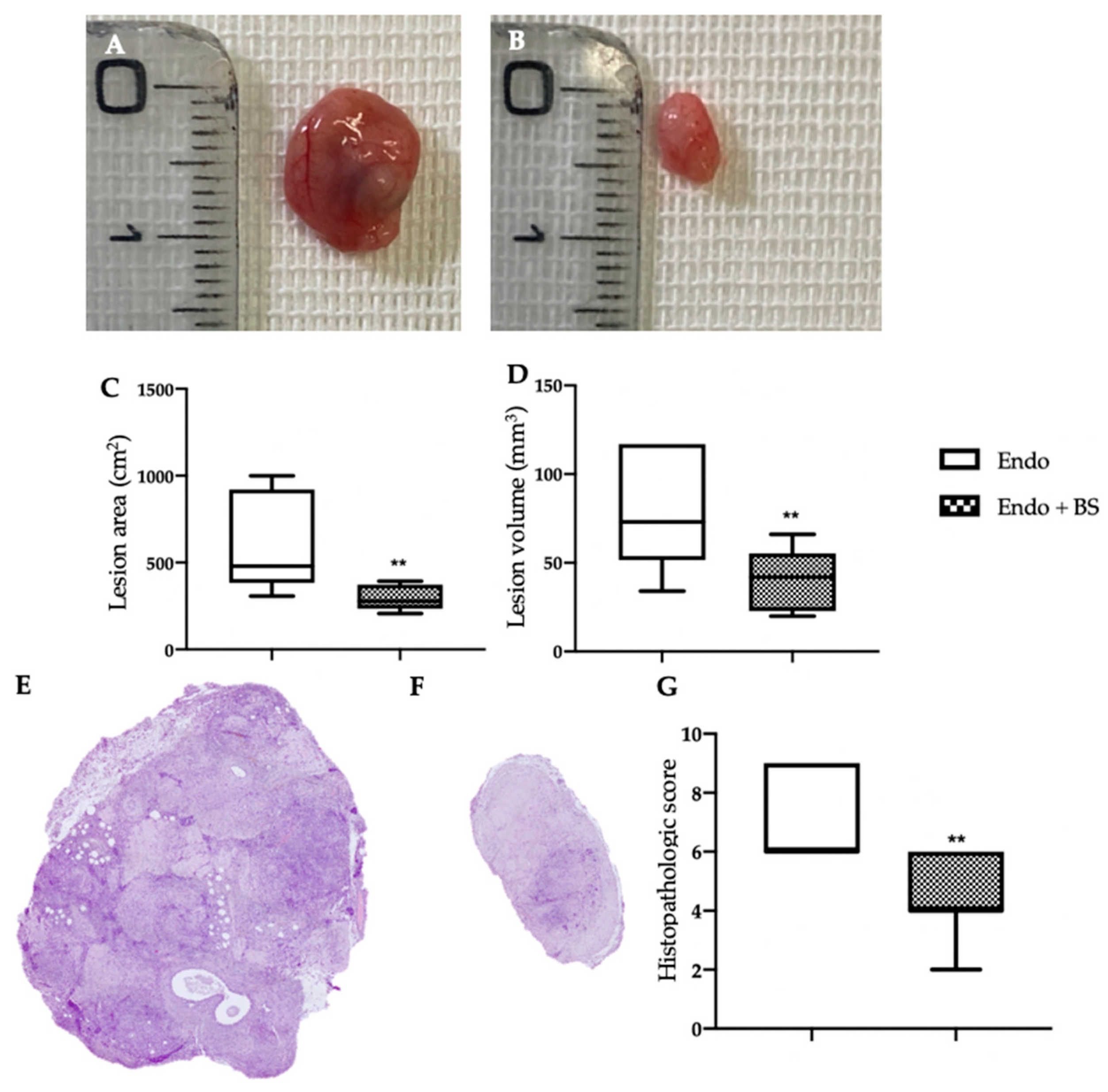
2.2. Effect of Oral BS Administration on Macroscopic and Histological Analysis

The macroscopic endometrioma analysis (Figure 2A,B) was in line with the hfUS examination. Lesions from the EMS group had major area (Figure 2C, ** p < 0.01 vs. control) and volume (Figure 2D, ** p < 0.01 vs. control), as compared to the one harvested from the EMS + BS group. The histological exam revealed that oral BS administration also modified lesions morphology (Figure 2G, ** p < 0.01 vs. control). Endometriotic lesions from EMS group showed characteristic stroma and glands (Figure 2E), while the oral BS administration (Figure 2F) significantly reduced the histopathological score (Figure 2G).

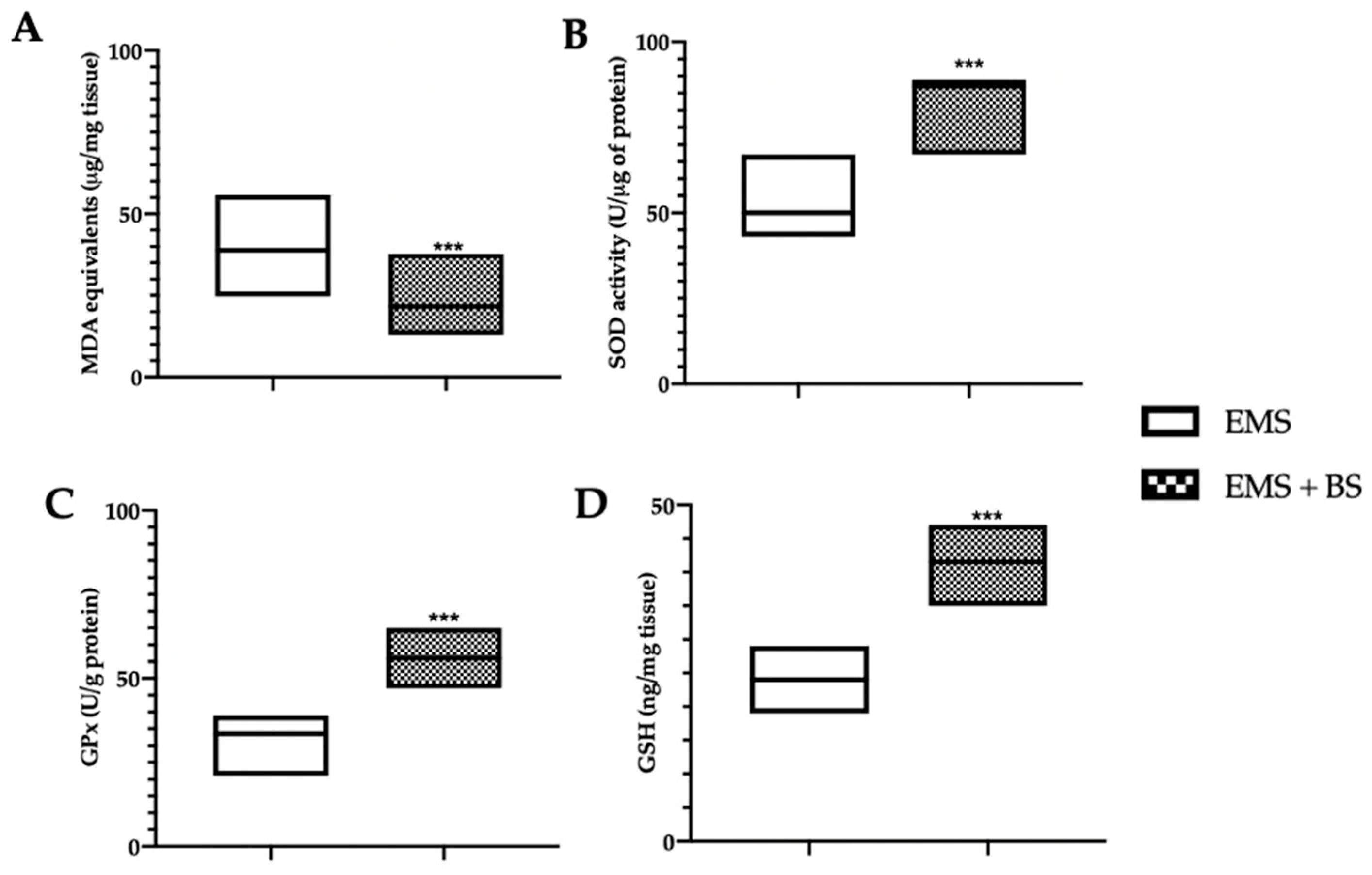
2.3. Effect of Oral BS Administration on the Oxidative Stress

Western blot analysis showed a significant increase in the expression of nuclear Nrf2 (Figure 3A, *** p < 0.01 vs. control) and cytosolic HO-1 (Figure 3B, *** p < 0.01 vs. control) and NQO-1 (Figure 3C, *** p < 0.01 vs. control) levels in the EMS + BS group, as compared to the EMS group. Moreover, oral BS administration significantly reduced malondialdehyde (MDA) levels (Figure 4A, *** p < 0.01 vs. control), increased superoxide dismutase (SOD) (Figure 4B, *** p < 0.01 vs. control) and glutathione peroxidase (GPx) (Figure 4C, *** p < 0.01 vs. control) activities and glutathione (GSH) (Figure 4D, *** p < 0.01 vs. control) levels.

2.4. Effect of Oral BS Administration on the Apoptosis Pathway

Western blot analysis showed that samples harvested from the EMS + BS group had a significant reduction in Bcl-2 (Figure 5A, * p < 0.01 vs. control) and Pro caspase 9 (Figure 5C, ** p < 0.01 vs. control) levels, as compared to EMS group. Levels of Bax (Figure 5B, * p < 0.01 vs. control) and cleaved caspase 9 (Figure 5D, ** p < 0.01 vs. control) were significantly increased in the EMS + BS samples, as compared to EMS. Additionally, Pro PARP expression was significantly reduced in EMS + BS group (Figure 6A, *** p < 0.01 vs. control), while cleaved PARP increased (Figure 6B, ** p < 0.01 vs. control), as compared to the EMS group. TUNEL staining confirmed these data. A significant increased number of TUNEL positive cells were detected in the EMS + BS group (Figure 6D,E, *** p < 0.01 vs. control), as compared to the EMS group (Figure 6C,E, *** p < 0.01 vs. control).

3. Discussion

The molecular mechanism underlying the pathophysiology of the EMS is controversial, as the therapeutic treatments. Several studies proposed the antioxidants as beneficial tools for EMS [31,32]. In this paper, BS, as an antioxidant agent and apoptosis inducer were used in an animal model of EMS. The pathology was induced and monitored by hfUS analysis. The macroscopic endometrioma analysis were in line with the hfUS examination. Additionally, BS administration modified endometriotic lesions histology, reducing glands and stroma tissue. Oxidative stress plays a key role in the establishment and development of the endometriosis [33]. Endometriotic cells showed an unbalanced oxidative/antioxidative, with an alteration of the ROS detoxification pathways [34]. The activity of Nrf2 is decisive to maintain intracellular oxidative stress status [35]. In fact, the Nrf2 transcription factor and its downstream proteins HO-1 and NQO1 were elected as one of the main pathways involved in the disease [36]. Nrf2 activity is normally restricted by its negative regulator Keap1. Increased oxidative stress induces Keap1 degradation, which in turn leads to Nrf2 translocation into the nucleus. Once translocated Nrf2 binds to the anti-oxidant response element (ARE) and promotes the transcription of its downstream antioxidant effectors [37]. In the endometriotic lesions the endogenous antioxidant systems are compromised and the oxidative/antioxidative equilibrium is unbalance. BS administration, well in line with the previous evidences [25], increased the Nrf2 nuclear levels and the expression of the downstream proteins HO-1 and NQO1. Additionally, it reduced lipid peroxidation and increased SOD and GPx activities, restoring the reduced GSH levels. Thus, BS restored the oxidative imbalance activating the endogenous antioxidant defense mechanisms.
The increased oxidative stress in the endometriotic lesions also impaired the apoptotic pathway [6]. It has been demonstrated that Nrf2-mediated modulation of apoptotic cell death has a key role in cell survival and drug resistance. In particular, impaired Nrf2 pathway results in increased expression of antiapoptotic protein Bcl-2 and reduced Bax levels, cytochrome c release from mitochondria, modulation of caspases, and DNA fragmentation [38]. Defective control of the programmed cell death has been shown to have an important role in the establishment of several disease including endometriosis [39]. Bcl-2 family proteins control the apoptotic mitochondria-dependent pathway. Several papers reported that the increase in the anti-apoptotic protein Bcl-2 hasten the progression of endometriosis [40]. The up-regulated Bcl-2 is accompanied with reduced Bax levels and caspases cleavage [41]. Caspases are endoproteases that have a key role in controlling apoptosis. In particular, it has been reported that in the endometriotic lesions the activation of caspase 9 is impaired [42]. As already confirmed by other studies [26,27,28,29,30], our results showed that BS caused a decrease in Bcl-2 expression and an increase in Bax levels and caspase 9 cleavage. The BS apoptotic effect was also confirmed by the cleavage of PARP, another specific marker of apoptosis, and by the TUNEL assay. PARP is a nuclear protein that promotes the transfer of ADP-ribose polymers onto itself and other nuclear enzymes in response to DNA strand breaks. TUNEL assays detect apoptotic cells by the terminal deoxynucleotidyl transferase (TdT)-mediated addition of labeled (X) deoxyuridine triphosphate nucleotides (X-dUTPs) to the 3′-OH end of DNA strand breaks. During apoptosis, cleavage of PARP-1 in fragments and TUNEL positive cells are a useful hallmark of apoptosis or cell death. Our experimental evidence showed that BS administration was able to restore the impaired apoptosis signaling. TUNEL staining confirmed these data.
There are several limitations in the current study. A normal rat’s uterine tissue transplant into another rat’s abdominal cavity was employed as the endometriosis model in this study. It was a poor representation of the ethology of endometriosis in humans. Indeed, the data were gathered using a synthetic model (no spontaneous lesions, no rat menstruation, no human lesion transplants). Rat models, on the other hand, have a long history of being employed extensively in endometriosis research and have also been approved as a model that captures the dynamics of the condition. It would be interesting to investigate the lesions for a longer duration in subsequent studies.

4. Materials and Methods

4.1. Animals

Sprague–Dawley rats (Envigo, Milan, Italy) were employed. The University of Messina Review Board for animal care (OPBA) approved the study. All animal experiments agree with the new Italian regulations (D.Lgs 2014/26), EU regulations (EU Directive 2010/63).

4.2. Model Induction

Rats were divided into two groups, donor or recipient. Donor animals were administered intraperitoneally with 10 IU pregnant mare’s serum gonadotropin (PMSG) to induce comparable estrogen levels among them. Donors were euthanized 41 h after the injection. The uterus was removed through a midline incision and washed in PBS before extrauterine tissue, including ovary and oviduct, was removed under a dissecting microscope. A longitudinal incision was made from one horn to the other. Tissue was transferred to a 1.5 mL centrifuge tube containing fresh PBS and minced with dissecting scissors. Minced tissue from all donors was pooled and the volume was adjusted to the equivalent of one uterus/500 uL of PBS. Recipient rats were injected intraperitoneally with the equivalent of tissue from one uterus in 500 uL of PBS (1:1 donor/recipient ratio) along the midventral line using a 18-gauge needle [43]. The disease was allowed to establish for seven days.

4.3. Experimental Groups

Seven days after the induction, recipient rats were assigned to the following groups (n = 20 for each group):
-
EMS group: animals were subjected to the above described induction and vehicle (2% gum acacia was orally administered on the seventh day until the fourteenth day);
-
EMS + BS group: animals were subjected to the above described induction and BS (100 mg/Kg) was orally administered on the seventh day until the fourteenth day;
-
Sham group: animals were subjected to the above described induction but they were intraperitoneally injected with 500 μL of PBS along the midventral line instead of endometriotic tissue.
BS dosage was based on previous studies [44].
The powder of BS was purchased from a company (Fontana standardized natural active principles, lot. S2111560, Canosa di Puglia, BT Italy).
Fourteen days from the endometriosis induction, animals were sacrificed and the endometriotic lesions were collected for the histological and molecular analyses (Figure 7).

4.4. Abdominal High-Frequency Ultrasound

Pelvic ultrasound was performed to monitor the development of the endometriotic lesions at seven and fourteen days from the implant. The analysis included the anterior and posterior pelvic areas to reach the lesions in both locations. The hair in the ventral abdomen was clipped from 1 cm cranial to the xyphoid cartilage to the caudal-most part of the pubis. Ultrasonographic exams were performed by the Esaote MYLAB OMEGA VET (Esaote Italia, Milan, Italy) on anesthetized rats (2% isoflurane) positioned in dorsal recumbency. Abdominal B-mode was performed with a high frequency linear array (4–15 MHz) transducer. Longitudinal and transverse scanning planes were employed for evaluation of different abdominal structures [45]. All analyses were performed as double blinded.

4.5. Biochemical Analysis

Lipid peroxidation was evaluated by the TBARS test, reading the MDA levels at 535 nm [46]. SOD activity was evaluated as already described [47] and expressed as U/g protein [48]. GSH levels were determined using a microplate reader at 412 nm [49]. GPx activity was evaluated as already described [50] and expressed as U/g protein [51].

4.6. Histological Examination

Endometriotic lesions were fixed in buffered formaldehyde solution, dehydrated and embedded in Paraplast [52,53]. Tissue slides were stained with H&E and evaluated using a Leica DM6 microscope (Leica Microsystems SpA, Milan, Italy) [54]. Histopathologic scores were evaluated with the formula P (persistence of epithelial cells in the explants) × I (intensity of glands) as already described [55]: P: 3 = well-preserved epithelial layer, 2 = moderately preserved epithelium with leukocyte infiltrating, 1 = poorly preserved epithelium (occasional epithelial cells only), and 0 = no epithelium; I: from 0 (no glands) to 3 (abundant glands). Lesion volume was calculated according to the formula: V = (length × width2) × 0.5.

4.7. Western Blot Analysis

Lesion samples were homogenized and Western blots were performed as already described [56]. Specific primary antibody anti-Bcl-2 (Santa Cruz Biotechnology (Santa Cruz, CA, USA), sc-7382, 1:2000), anti-Bax (Santa Cruz Biotechnology, sc-7480, 1:2000), anti-Pro caspase 9 (Santa Cruz Biotechnology, sc-56073, 1:1000), anti-cleaved caspase 9 (Cell Signalling (Danvers, MA, USA) 9509, 1:1000), anti-Pro PARP (Santa Cruz Biotechnology, sc-8007, 1:1000), anti-cleaved PARP (Cell Signalling 5625, 1:1000), anti-Nrf2 (Santa Cruz Biotechnology, sc-365949, 1:1000), anti-HO-1 (Santa Cruz Biotechnology, sc-390991, 1:2000), anti-NQO-1 (Santa Cruz Biotechnology, sc-32793, 1:2000), were mixed in 5% w/v nonfat dried milk solution and incubated overnight. Afterward, blots were incubated with peroxidase-conjugated anti-mouse IgG secondary antibody or peroxidase-conjugated ant-irabbit IgG (Jackson Immuno Research, Milan, Italy) for 1 h at room temperature [57]. Membranes were also blotted with the antibody against β-actin or lamin b1 [58]. Signals were detected with enhanced chemiluminescence detection system reagent (Super-SignalWest Pico Chemiluminescent Substrate) [59]. The relative expression of the protein bands was quantified by densitometry with Bio-Rad ChemiDoc XRS software (Bio-Rad, Milan, Italy) [60] and standardized to β-actin or lamin B1 levels. Images of blot signals were imported to analysis software (v2003, Image Quant TL) [61].

4.8. Terminal Deoxynucleotidyl Nick-End Labeling (TUNEL) Assay

Apoptosis was analyzed by a TUNEL assay using an in situ cell death detection kit (Roche 11684795910) [62].

4.9. Statistical Analysis

All values are expressed as mean ± standard error of the mean of N observations. The results were analyzed by t-test when comparing two groups while we used the one-way ANOVA followed by a Bonferroni post hoc test for multiple comparisons. A p-value of less than 0.05 was considered significant. * p < 0.05 vs. control, ** p < 0.01 vs. control, *** p < 0.001 vs. control.

5. Conclusions

BS is generally well tolerated, although it can have some side-effects [63,64,65]. Overall, this study was conducted to evaluate the effect of oral administration of BS in a rat model of endometriosis.
The results showed that BS works on the Nrf2 pathway to apply its antioxidant effects and that it induces early apoptosis acting on Bcl-2 and Bax expression and cleaving caspase 9 and PARP. Importantly, our results showed a response to the test material (Boswellia Serrata gum resin extract), allowing future studies to continue to develop this model.

Author Contributions

Conceptualization, R.F.; data curation, R.D. and D.I.; formal analysis, M.C. and R.S.; funding acquisition, S.C.; investigation, L.I.; methodology, R.C., E.G., and F.M.; supervision, R.D.P.; validation, D.D.P. and A.F.P. All authors have read and agreed to the published version of the manuscript.

Funding

This research received no external funding.

Institutional Review Board Statement

The study was conducted according to the guidelines of the Declaration of Helsinki and approved by the Institutional Review Board for animal care (OPBA) of the University of Messina.

Informed Consent Statement

Not applicable.

Data Availability Statement

The data presented in this study are available on request from the corresponding author.

Conflicts of Interest

The authors declare no conflict of interest.

References

  1. Jouhari, S.; Mohammadzadeh, A.; Soltanghoraee, H.; Mohammadi, Z.; Khazali, S.; Mirzadegan, E.; Lakpour, N.; Fatemi, F.; Zafardoust, S.; Mohazzab, A.; et al. Effects of silymarin, cabergoline and letrozole on rat model of endometriosis. Taiwan J. Obstet. Gynecol. 2018, 57, 830–835. [Google Scholar] [CrossRef]
  2. Fritzer, N.; Tammaa, A.; Haas, D.; Oppelt, P.; Renner, S.; Hornung, D.; Wölfler, M.; Ulrich, U.; Hudelist, G. When sex is not on fire: A prospective multicentre study evaluating the short-term effects of radical resection of endometriosis on quality of sex life and dyspareunia. Eur. J. Obstet. Gynecol. Reprod. Biol. 2016, 197, 36–40. [Google Scholar] [CrossRef] [PubMed]
  3. Van Langendonckt, A.; Casanas-Roux, F.; Donnez, J. Oxidative stress and peritoneal endometriosis. Fertil. Steril. 2002, 77, 861–870. [Google Scholar] [CrossRef] [PubMed]
  4. Foyouzi, N.; Berkkanoglu, M.; Arici, A.; Kwintkiewicz, J.; Izquierdo, D.; Duleba, A.J. Effects of oxidants and antioxidants on proliferation of endometrial stromal cells. Fertil. Steril. 2004, 82, 1019–1022. [Google Scholar] [CrossRef]
  5. Chen, N.; Du, B.; Zhou, H.; Shen, F.; Li, J.; Xie, Z. Abnormal expression of Nrf2 may play an important role in the pathogenesis and development of adenomyosis. PLoS ONE 2017, 12, e0182773. [Google Scholar] [CrossRef] [Green Version]
  6. Ekici, E.I.; Guney, M.; Naziroglu, M. Protective effect of cabergoline on mitochondrial oxidative stress-induced apoptosis is mediated by modulations of TRPM2 in neutrophils of patients with endometriosis. J. Bioenerg. Biomembr. 2020, 52, 131–142. [Google Scholar] [CrossRef] [PubMed]
  7. Tischner, D.; Woess, C.; Ottina, E.; Villunger, A. Bcl-2-regulated cell death signalling in the prevention of autoimmunity. Cell Death Dis. 2010, 1, e48. [Google Scholar] [CrossRef] [Green Version]
  8. Kulsoom, B.; Shamsi, T.S.; Afsar, N.A.; Memon, Z.; Ahmed, N.; Hasnain, S.N. Bax, Bcl-2, and Bax/Bcl-2 as prognostic markers in acute myeloid leukemia: Are we ready for Bcl-2-directed therapy? Cancer Manag. Res. 2018, 10, 403. [Google Scholar] [CrossRef] [Green Version]
  9. Eisenberg, D.M.; Davis, R.B.; Ettner, S.L.; Appel, S.; Wilkey, S.; Van Rompay, M.; Kessler, R.C. Trends in alternative medicine use in the United States, 1990-1997: Results of a follow-up national survey. JAMA 1998, 280, 1569–1575. [Google Scholar] [CrossRef] [Green Version]
  10. Bardaweel, S.K.; Shehadeh, M.; Suaifan, G.A.; Kilani, M.-V.Z. Complementary and alternative medicine utilization by a sample of infertile couples in Jordan for infertility treatment: Clinics-based survey. BMC Complement. Altern. Med. 2013, 13, 35. [Google Scholar] [CrossRef]
  11. Comar, K.M.; Kirby, D.F. Herbal remedies in gastroenterology. J. Clin. Gastroenterol. 2005, 39, 457–468. [Google Scholar] [CrossRef]
  12. Tindle, H.A.; Davis, R.B.; Phillips, R.S.; Eisenberg, D.M. Trends in use of complementary and alternative medicine by US adults: 1997-2002. Altern. Ther. Health Med. 2005, 11, 42. [Google Scholar]
  13. Cox, H.; Henderson, L.; Wood, R.; Cagliarini, G. Learning to take charge: Women’s experiences of living with endometriosis. Complement. Ther. Nurs. Midwifery 2003, 9, 62–68. [Google Scholar] [CrossRef]
  14. Fugh-Berman, A.; Kronenberg, F. Complementary and alternative medicine (CAM) in reproductive-age women: A review of randomized controlled trials. Reprod. Toxicol. 2003, 17, 137–152. [Google Scholar] [CrossRef]
  15. Wieser, F.; Cohen, M.; Gaeddert, A.; Yu, J.; Burks-Wicks, C.; Berga, S.L.; Taylor, R.N. Evolution of medical treatment for endometriosis: Back to the roots? Hum. Reprod. Update 2007, 13, 487–499. [Google Scholar] [CrossRef] [PubMed] [Green Version]
  16. Liu, J.-J.; Nilsson, Å.; Oredsson, S.; Badmaev, V.; Zhao, W.-Z.; Duan, R.-D. Boswellic acids trigger apoptosis via a pathway dependent on caspase-8 activation but independent on Fas/Fas ligand interaction in colon cancer HT-29 cells. Carcinogenesis 2002, 23, 2087–2093. [Google Scholar] [CrossRef] [Green Version]
  17. Han, R. Highlight on the studies of anticancer drugs derived from plants in China. Stem Cells 1994, 12, 53–63. [Google Scholar] [CrossRef] [PubMed]
  18. Sharma, S.; Thawani, V.; Hingorani, L.; Shrivastava, M.; Bhate, V.; Khiyani, R. Pharmacokinetic study of 11-keto β-Boswellic acid. Phytomedicine 2004, 11, 255–260. [Google Scholar] [CrossRef] [PubMed] [Green Version]
  19. Reddy, G.K.; Chandrakasan, G.; Dhar, S. Studies on the metabolism of glycosaminoglycans under the influence of new herbal anti-inflammatory agents. Biochem. Pharmacol. 1989, 38, 3527–3534. [Google Scholar] [CrossRef]
  20. Safayhi, H.; Mack, T.; Sabieraj, J.; Anazodo, M.I.; Subramanian, L.R.; Ammon, H. Boswellic acids: Novel, specific, nonredox inhibitors of 5-lipoxygenase. J. Pharmacol. Exp. Ther. 1992, 261, 1143–1146. [Google Scholar]
  21. Elshazly, S.M.; Abd El Motteleb, D.M.; Nassar, N.N. The selective 5-LOX inhibitor 11-keto-β-boswellic acid protects against myocardial ischemia reperfusion injury in rats: Involvement of redox and inflammatory cascades. Naunyn-Schmiedeberg’s Arch. Pharmacol. 2013, 386, 823–833. [Google Scholar] [CrossRef]
  22. Hartmann, R.M.; Morgan Martins, M.I.; Tieppo, J.; Fillmann, H.S.; Marroni, N.P. Effect of Boswellia serrata on antioxidant status in an experimental model of colitis rats induced by acetic acid. Dig. Dis. Sci. 2012, 57, 2038–2044. [Google Scholar] [CrossRef] [PubMed]
  23. Ali, E.N.; Mansour, S.Z. Boswellic acids extract attenuates pulmonary fibrosis induced by bleomycin and oxidative stress from gamma irradiation in rats. Chin. Med. 2011, 6, 36. [Google Scholar] [CrossRef] [PubMed] [Green Version]
  24. Rong, Z.-T.; Gong, X.-J.; Sun, H.-B.; Li, Y.-M.; Ji, H. Protective effects of oleanolic acid on cerebral ischemic damage in vivo and H2O2-induced injury in vitro. Pharm. Biol. 2011, 49, 78–85. [Google Scholar] [CrossRef]
  25. Ding, Y.; Chen, M.; Wang, M.; Wang, M.; Zhang, T.; Park, J.; Zhu, Y.; Guo, C.; Jia, Y.; Li, Y. Neuroprotection by acetyl-11-keto-β-boswellic acid, in ischemic brain injury involves the Nrf2/HO-1 defense pathway. Sci. Rep. 2014, 4, 7002. [Google Scholar] [CrossRef] [PubMed] [Green Version]
  26. Liu, J.-J.; Nilsson, A.; Oredsson, S.; Badmaev, V.; Duan, R.-D. Keto-and acetyl-keto-boswellic acids inhibit proliferation and induce apoptosis in Hep G2 cells via a caspase-8 dependent pathway. Int. J. Mol. Med. 2002, 10, 501–505. [Google Scholar] [CrossRef] [PubMed]
  27. Park, Y.S.; Lee, J.H.; Harwalkar, J.A.; Bondar, J.; Safayhi, H.; Golubic, M. Acetyl-11-Keto-ß-Boswellic acid (Akba) is cytotoxic for meningioma cells and inhibits phosphorylation of the extracellular-signal regulated kinase 1 and 2. In Eicosanoids and Other Bioactive Lipids in Cancer, Inflammation, and Radiation Injury 5; Springer: Berlin/Heidelberg, Germany, 2002; pp. 387–393. [Google Scholar]
  28. Syrovets, T.; Büchele, B.; Krauss, C.; Laumonnier, Y.; Simmet, T. Acetyl-boswellic acids inhibit lipopolysaccharide-mediated TNF-α induction in monocytes by direct interaction with IκB kinases. J. Immunol. 2005, 174, 498–506. [Google Scholar] [CrossRef] [PubMed] [Green Version]
  29. Winking, M.; Sarikaya, S.; Rahmanian, A.; Jödicke, A.; Böker, D.-K. Boswellic acids inhibit glioma growth: A new treatment option? J. Neuro-Oncol. 2000, 46, 97–103. [Google Scholar] [CrossRef] [PubMed]
  30. Zhao, W.; Entschladen, F.; Liu, H.; Niggemann, B.; Fang, Q.; Zaenker, K.S.; Han, R. Boswellic acid acetate induces differentiation and apoptosis in highly metastatic melanoma and fibrosarcoma cells. Cancer Detect. Prev. 2003, 27, 67–75. [Google Scholar] [CrossRef]
  31. Harlev, A.; Gupta, S.; Agarwal, A. Targeting oxidative stress to treat endometriosis. Expert Opin. Ther. Targets 2015, 19, 1447–1464. [Google Scholar] [CrossRef]
  32. Rosa e Silva, J.C.; do Amara, V.F.; Mendonca, J.L.; Rosa e Silva, A.C.; Nakao, L.S.; Poli Neto, O.B.; Ferriani, R.A. Serum markers of oxidative stress and endometriosis. Clin. Exp. Obstet. Gynecol. 2014, 41, 371–374. [Google Scholar] [CrossRef]
  33. Ngo, C.; Chereau, C.; Nicco, C.; Weill, B.; Chapron, C.; Batteux, F. Reactive oxygen species controls endometriosis progression. Am. J. Pathol. 2009, 175, 225–234. [Google Scholar] [CrossRef]
  34. Di Paola, D.; Natale, S.; Iaria, C.; Crupi, R.; Cuzzocrea, S.; Spanò, N.; Gugliandolo, E.; Peritore, A.F. Environmental Co-Exposure to Potassium Perchlorate and Cd Caused Toxicity and Thyroid Endocrine Disruption in Zebrafish Embryos and Larvae (Danio rerio). Toxics 2022, 10, 198. [Google Scholar] [CrossRef]
  35. Alfieri, A.; Srivastava, S.; Siow, R.C.; Modo, M.; Fraser, P.A.; Mann, G.E. Targeting the Nrf2–Keap1 antioxidant defence pathway for neurovascular protection in stroke. J. Physiol. 2011, 589, 4125–4136. [Google Scholar] [CrossRef]
  36. Lu, M.C.; Ji, J.A.; Jiang, Z.Y.; You, Q.D. The Keap1-Nrf2-ARE Pathway As a Potential Preventive and Therapeutic Target: An Update. Med. Res. Rev. 2016, 36, 924–963. [Google Scholar] [CrossRef] [PubMed]
  37. Dinkova-Kostova, A.T.; Kostov, R.V.; Canning, P. Keap1, the cysteine-based mammalian intracellular sensor for electrophiles and oxidants. Arch. Biochem. Biophys. 2017, 617, 84–93. [Google Scholar] [CrossRef] [PubMed] [Green Version]
  38. Niture, S.K.; Jaiswal, A.K. Nrf2 protein up-regulates antiapoptotic protein Bcl-2 and prevents cellular apoptosis. J. Biol. Chem. 2012, 287, 9873–9886. [Google Scholar] [CrossRef] [PubMed] [Green Version]
  39. Park, S.; Lim, W.; Bazer, F.W.; Song, G. Apigenin induces ROS-dependent apoptosis and ER stress in human endometriosis cells. J. Cell. Physiol. 2018, 233, 3055–3065. [Google Scholar] [CrossRef]
  40. McLaren, J.; Prentice, A.; Charnock-Jones, D.S.; Sharkey, A.M.; Smith, S.K. Immunolocalization of the apoptosis regulating proteins Bcl-2 and Bax in human endometrium and isolated peritoneal fluid macrophages in endometriosis. Hum. Reprod. 1997, 12, 146–152. [Google Scholar] [CrossRef] [Green Version]
  41. Talebi, H.; Farahpour, M.R.; Hamishehkar, H. The effectiveness of Rutin for prevention of surgical induced endometriosis development in a rat model. Sci. Rep. 2021, 11, 7180. [Google Scholar] [CrossRef]
  42. Cho, Y.J.; Lee, S.H.; Park, J.W.; Han, M.; Park, M.J.; Han, S.J. Dysfunctional signaling underlying endometriosis: Current state of knowledge. J. Mol. Endocrinol. 2018, 60, R97–R113. [Google Scholar] [CrossRef] [Green Version]
  43. Cordaro, M.; Trovato Salinaro, A.; Siracusa, R.; D’Amico, R.; Impellizzeri, D.; Scuto, M.; Ontario, M.L.; Interdonato, L.; Crea, R.; Fusco, R.; et al. Hidrox((R)) and Endometriosis: Biochemical Evaluation of Oxidative Stress and Pain. Antioxidants 2021, 10, 720. [Google Scholar] [CrossRef]
  44. Umar, S.; Umar, K.; Sarwar, A.H.M.G.; Khan, A.; Ahmad, N.; Ahmad, S.; Katiyar, C.K.; Husain, S.A.; Khan, H.A. Boswellia serrata extract attenuates inflammatory mediators and oxidative stress in collagen induced arthritis. Phytomedicine 2014, 21, 847–856. [Google Scholar] [CrossRef]
  45. Genovese, T.; Cordaro, M.; Siracusa, R.; Impellizzeri, D.; Caudullo, S.; Raffone, E.; Macri, F.; Interdonato, L.; Gugliandolo, E.; Interlandi, C.; et al. Molecular and Biochemical Mechanism of Cannabidiol in the Management of the Inflammatory and Oxidative Processes Associated with Endometriosis. Int. J. Mol. Sci. 2022, 23, 5427. [Google Scholar] [CrossRef] [PubMed]
  46. Fusco, R.; Cordaro, M.; Siracusa, R.; D’Amico, R.; Genovese, T.; Gugliandolo, E.; Peritore, A.F.; Crupi, R.; Impellizzeri, D.; Cuzzocrea, S.; et al. Biochemical Evaluation of the Antioxidant Effects of Hydroxytyrosol on Pancreatitis-Associated Gut Injury. Antioxidants 2020, 9, 781. [Google Scholar] [CrossRef]
  47. Cordaro, M.; Siracusa, R.; Fusco, R.; D’Amico, R.; Peritore, A.F.; Gugliandolo, E.; Genovese, T.; Scuto, M.; Crupi, R.; Mandalari, G.; et al. Cashew (Anacardium occidentale L.) Nuts Counteract Oxidative Stress and Inflammation in an Acute Experimental Model of Carrageenan-Induced Paw Edema. Antioxidants 2020, 9, 660. [Google Scholar] [CrossRef]
  48. D’Amico, R.; Trovato Salinaro, A.; Fusco, R.; Cordaro, M.; Impellizzeri, D.; Scuto, M.; Ontario, M.L.; Lo Dico, G.; Cuzzocrea, S.; Di Paola, R. Hericium erinaceus and coriolus versicolor modulate molecular and biochemical changes after traumatic brain injury. Antioxidants 2021, 10, 898. [Google Scholar] [CrossRef] [PubMed]
  49. Fusco, R.; Cordaro, M.; Siracusa, R.; Peritore, A.F.; D’Amico, R.; Licata, P.; Crupi, R.; Gugliandolo, E. Effects of hydroxytyrosol against lipopolysaccharide-induced inflammation and oxidative stress in bovine mammary epithelial cells: A natural therapeutic tool for bovine mastitis. Antioxidants 2020, 9, 693. [Google Scholar] [CrossRef]
  50. Rashid, K.; Das, J.; Sil, P.C. Taurine ameliorate alloxan induced oxidative stress and intrinsic apoptotic pathway in the hepatic tissue of diabetic rats. Food Chem. Toxicol. 2013, 51, 317–329. [Google Scholar] [CrossRef]
  51. Cordaro, M.; D’Amico, R.; Morabito, R.; Fusco, R.; Siracusa, R.; Peritore, A.F.; Impellizzeri, D.; Genovese, T.; Crupi, R.; Gugliandolo, E. Physiological and Biochemical Changes in NRF2 Pathway in Aged Animals Subjected to Brain Injury. Cell. Physiol. Biochem. 2021, 55, 160–179. [Google Scholar] [PubMed]
  52. D’Amico, R.; Fusco, R.; Cordaro, M.; Siracusa, R.; Peritore, A.F.; Gugliandolo, E.; Crupi, R.; Scuto, M.; Cuzzocrea, S.; Di Paola, R.; et al. Modulation of NLRP3 Inflammasome through Formyl Peptide Receptor 1 (Fpr-1) Pathway as a New Therapeutic Target in Bronchiolitis Obliterans Syndrome. Int. J. Mol. Sci. 2020, 21, 2144. [Google Scholar] [CrossRef] [PubMed] [Green Version]
  53. Fusco, R.; Gugliandolo, E.; Campolo, M.; Evangelista, M.; Di Paola, R.; Cuzzocrea, S. Effect of a new formulation of micronized and ultramicronized N-palmitoylethanolamine in a tibia fracture mouse model of complex regional pain syndrome. PLoS ONE 2017, 12, e0178553. [Google Scholar] [CrossRef] [PubMed]
  54. Peritore, A.F.; Crupi, R.; Scuto, M.; Gugliandolo, E.; Siracusa, R.; Impellizzeri, D.; Cordaro, M.; D’Amico, R.; Fusco, R.; Di Paola, R.; et al. The Role of Annexin A1 and Formyl Peptide Receptor 2/3 Signaling in Chronic Corticosterone-Induced Depression-Like behaviors and Impairment in Hippocampal-Dependent Memory. CNS Neurol. Disord. Drug Targets 2020, 19, 27–43. [Google Scholar] [CrossRef]
  55. Siracusa, R.; D’Amico, R.; Impellizzeri, D.; Cordaro, M.; Peritore, A.F.; Gugliandolo, E.; Crupi, R.; Salinaro, A.T.; Raffone, E.; Genovese, T. Autophagy and mitophagy promotion in a rat model of endometriosis. Int. J. Mol. Sci. 2021, 22, 5074. [Google Scholar] [CrossRef] [PubMed]
  56. Fusco, R.; Salinaro, A.T.; Siracusa, R.; D’Amico, R.; Impellizzeri, D.; Scuto, M.; Ontario, M.L.; Crea, R.; Cordaro, M.; Cuzzocrea, S. Hidrox® counteracts cyclophosphamide-induced male infertility through NRF2 pathways in a mouse model. Antioxidants 2021, 10, 778. [Google Scholar] [CrossRef]
  57. Fusco, R.; Cordaro, M.; Siracusa, R.; Peritore, A.F.; Gugliandolo, E.; Genovese, T.; D’Amico, R.; Crupi, R.; Smeriglio, A.; Mandalari, G.; et al. Consumption of Anacardium occidentale L. (Cashew Nuts) Inhibits Oxidative Stress through Modulation of the Nrf2/HO-1 and NF-kB Pathways. Molecules 2020, 25, 4426. [Google Scholar] [CrossRef]
  58. Peritore, A.F.; D’Amico, R.; Siracusa, R.; Cordaro, M.; Fusco, R.; Gugliandolo, E.; Genovese, T.; Crupi, R.; Di Paola, R.; Cuzzocrea, S.; et al. Management of Acute Lung Injury: Palmitoylethanolamide as a New Approach. Int. J. Mol. Sci. 2021, 22, 5533. [Google Scholar] [CrossRef]
  59. Cordaro, M.; Fusco, R.; D’Amico, R.; Siracusa, R.; Peritore, A.F.; Gugliandolo, E.; Genovese, T.; Crupi, R.; Mandalari, G.; Cuzzocrea, S.; et al. Cashew (Anacardium occidentale L.) Nuts Modulate the Nrf2 and NLRP3 Pathways in Pancreas and Lung after Induction of Acute Pancreatitis by Cerulein. Antioxidants 2020, 9, 992. [Google Scholar] [CrossRef]
  60. Cordaro, M.; Scuto, M.; Siracusa, R.; D’Amico, R.; Filippo Peritore, A.; Gugliandolo, E.; Fusco, R.; Crupi, R.; Impellizzeri, D.; Pozzebon, M.; et al. Effect of N-palmitoylethanolamine-oxazoline on comorbid neuropsychiatric disturbance associated with inflammatory bowel disease. FASEB J. 2020, 34, 4085–4106. [Google Scholar] [CrossRef]
  61. Crupi, R.; Palma, E.; Siracusa, R.; Fusco, R.; Gugliandolo, E.; Cordaro, M.; Impellizzeri, D.; De Caro, C.; Calzetta, L.; Cuzzocrea, S.; et al. Protective Effect of Hydroxytyrosol Against Oxidative Stress Induced by the Ochratoxin in Kidney Cells: In vitro and in vivo Study. Front. Vet. Sci. 2020, 7, 136. [Google Scholar] [CrossRef] [Green Version]
  62. D’Amico, R.; Cordaro, M.; Fusco, R.; Peritore, A.F.; Genovese, T.; Gugliandolo, E.; Crupi, R.; Mandalari, G.; Caccamo, D.; Cuzzocrea, S.; et al. Consumption of Cashew (Anacardium occidentale L.) Nuts Counteracts Oxidative Stress and Tissue Inflammation in Mild Hyperhomocysteinemia in Rats. Nutrients 2022, 14, 1474. [Google Scholar] [CrossRef] [PubMed]
  63. Yu, G.; Xiang, W.; Zhang, T.; Zeng, L.; Yang, K.; Li, J. Effectiveness of Boswellia and Boswellia extract for osteoarthritis patients: A systematic review and meta-analysis. BMC Complement. Med. Ther. 2020, 20, 225. [Google Scholar] [CrossRef] [PubMed]
  64. Suchita, W.; Raman, D.M.R.; Kaur, C.D. A Review on phytochemistry and pharmacological activities of Boswellia serrata: A natural remedy. Int. J. Pharmacogn. 2021, 8, 454–461. [Google Scholar]
  65. Aronson, J.K. Meyler’s Side Effects of Herbal Medicines; Elsevier: Amsterdam, The Netherlands, 2008. [Google Scholar]
Figure 1. Analysis of endometriotic lesions development: high-frequency ultrasound analysis (hfUS) analysis at 7 days from the surgery: EMS (A), EMS + BS (B), lesion number (C), lesion diameter (D); hfUS analysis at 14 days from the surgery: EMS (E), EMS + BS (F), lesion number (G), lesion diameter (H). For the analyses, n = 5 animals from each group were employed. A p-value of less than 0.05 was considered significant. ** p < 0.01 vs. control.
Ijms 23 15348 g001
Figure 2. Analysis of the effect of oral BS administration on lesions size and histology: Macroscopic analysis: EMS (A), EMS + BS (B); lesion area (C); lesion volume (D); histological analysis: EMS (E), EMS + BS (F), histopathologic score (G). For the analyses, n = 5 animals from each group were employed. A p-value of less than 0.05 was considered significant. ** p < 0.01 vs. control.
Ijms 23 15348 g002
Figure 3. Analysis of the effect of oral BS administration on Nrf2 pathway: Western blot analysis of: Nrf2 (A), HO-1 (B), NQO-1 (C) expression; for the analyses, n = 5 animals from each group were employed. A p-value of less than 0.05 was considered significant. *** p < 0.001 vs. control.
Ijms 23 15348 g003
Figure 4. Analysis of the effect of oral BS administration on oxidative stress: MDA levels (A), SOD activity (B), GPx activity (C), GSH levels (D). For the analyses, n = 5 animals from each group were employed. A p-value of less than 0.05 was considered significant. *** p < 0.001 vs. control.
Ijms 23 15348 g004
Figure 5. Analysis of the effect of oral BS administration on apoptosis: Western blot analysis of: Bcl-2 (A), Bax (B), Pro caspase 9 (C), cleaved caspase 9 (D). For the analyses, n = 5 animals from each group were employed. A p-value of less than 0.05 was considered significant. * p < 0.05 vs. control, ** p < 0.01 vs. control.
Ijms 23 15348 g005
Figure 6. Analysis of the effect of oral BS administration on apoptosis: Western blot analysis of: Pro PARP (A), cleaved PARP (B) expression; TUNEL staining (magnification 40×): EMS (C), EMS + BS (D), number of TUNEL positive cells (E). For the analyses, n = 5 animals from each group were employed. A p-value of less than 0.05 was considered significant. ** p < 0.01 vs. control, *** p < 0.001 vs. control.
Ijms 23 15348 g006
Figure 7. Study design figure.
Ijms 23 15348 g007
Publisher’s Note: MDPI stays neutral with regard to jurisdictional claims in published maps and institutional affiliations.

