Article

H2S Enhanced the Tolerance of Malus hupehensis to Alkaline Salt Stress through the Expression of Genes Related to Sulfur-Containing Compounds and the Cell Wall in Roots

State Key Laboratory of Crop Biology, College of Horticulture Science and Engineering, Shandong Agricultural University, 61 Daizong Street, Tai’an 271018, China
*
Authors to whom correspondence should be addressed.
These authors contributed equally to this work.
Int. J. Mol. Sci. 2022, 23(23), 14848; https://doi.org/10.3390/ijms232314848
Submission received: 23 October 2022 / Revised: 15 November 2022 / Accepted: 21 November 2022 / Published: 27 November 2022

Abstract

:
Malus is an economically important plant that is widely cultivated worldwide, but it often encounters saline–alkali stress. The composition of saline–alkali land is a variety of salt and alkali mixed with the formation of alkaline salt. Hydrogen sulfide (H2S) has been reported to have positive effects on plant responses to abiotic stresses. Our previous study showed that H2S pretreatment alleviated the damage caused by alkaline salt stress to Malus hupehensis Rehd. var. pingyiensis Jiang (Pingyi Tiancha, PYTC) roots by regulating Na+/K+ homeostasis and oxidative stress. In this study, transcriptome analysis was used to investigate the overall mechanism through which H2S alleviates alkaline salt stress in PYTC roots. Simultaneously, differentially expressed genes (DEGs) were explored. Transcriptional profiling of the Control-H2S, Control-AS, Control-H2S + AS, and AS-H2S + AS comparison groups identified 1618, 18,652, 16,575, and 4314 DEGs, respectively. Further analysis revealed that H2S could alleviate alkaline salt stress by increasing the energy maintenance capacity and cell wall integrity of M. hupehensis roots and by enhancing the capacity for reactive oxygen species (ROS) metabolism because more upregulated genes involved in ROS metabolism and sulfur-containing compounds were identified in M. hupehensis roots after H2S pretreatment. qRT-PCR analysis of H2S-induced and alkaline salt-response genes showed that these genes were consistent with the RNA-seq analysis results, which indicated that H2S alleviation of alkaline salt stress involves the genes of the cell wall and sulfur-containing compounds in PYTC roots.

1. Introduction

Soil salinization seriously threatens agricultural production and plant growth. In most studies of plant salt tolerance, NaCl is usually used as a salt stress factor. However, in real saline soil, the salt mixture includes NaCl, Na2SO4, NaHCO3, and Na2CO3. This mixture is alkaline, which is called alkaline salt [1]. The osmotic stress, ionic stress, and high pH caused by alkaline salt stress have a more direct toxic action than neutral salt stress [2,3]. High pH impedes growth and glycolysis more significantly than NaCl stress in maize [4,5]. Revealing the salt tolerance mechanism of plants and alleviating the damage caused to plants by salt stress are of great significance for the utilization of saline–alkali land and plant cultivation. However, knowledge of the mitigation of alkaline salt stress in plants and its mechanism is still limited.
Apple is an economically important plant that is widely cultivated worldwide. Cultivated apples are planted in the soil through their rootstocks. The root is the first organ of apple to be affected by alkaline salts. Alkaline stress was reported to significantly inhibit root activity and growth, induce an increase in the superoxide anion (O2) and H2O2 in rice and apple roots [6,7], and prevent plants from absorbing mineral nutrients [8]. The increase in glutamine synthetase activity and the soluble protein content improved the growth performance of wheat and switchgrass roots under alkaline salt stress [9,10]. Not only does the cell wall provide mechanical rigidity for plant tissues, but its physical and chemical properties can also affect cell growth. In addition, an increasing number of studies have shown that changes in the cell wall can regulate root growth under salt stress. Salt stress decreased the pectin content in cell walls, thus inhibiting soybean root growth [11]. The idea that XTH19 (xyloglucan endotransglucosylase/hydrolase) and XTH23 participate in the regulation of the development response of lateral roots to salt treatment was further confirmed in Arabidopsis, where BES1 functions directly upstream to control the expression levels of XTH19 and XTH23 [12]. Enhanced cell wall biosynthesis has also been shown to increase salt tolerance in broomcorn millet [13].
Hydrogen sulfide (H2S) is a small gaseous molecule that has physiological and signaling functions in plant tissues and cells [14]. To date, a variety of studies have demonstrated that H2S as a gaseous signal molecule plays a crucial role in many plant growth and abiotic stress responses [15]. Recently, there has been an important breakthrough in understanding how H2S is involved in the regulation of plant metabolism to enhance salt tolerance. H2S regulated the endogenous H2S metabolism, maintained the Na+/K+ balance and the oxidative stress response in cucumber [16], and also maintained ion homeostasis in rice [17] to alleviate the growth inhibition under salt stress. Moreover, evidence suggests that H2S interacts with nitric oxide (NO) to form a signal network, prevents K+ leakage and reestablishes redox balance in Medicago sativa [18], and reduces the Na+ concentration in wheat seedlings via major pathways of NSCCs and SOS1 [19]. However, most studies about the H2S modulation of alkaline and neutral salt stress mainly focused on the physiological mechanism of herbaceous plants [16,19]. In addition, research on fruit trees has also focused on the postharvest conservation biology of H2S [20], whereas limited work has been performed to explore the mechanism of H2S-mediated regulation of alkaline salt at the transcriptional level in fruit trees.
RNA-seq transcriptome analysis provides new ideas to reveal the mechanism of plant biological resistance to abiotic stress [21]. With this technology, the genes involved in salt and alkali tolerance have been successfully obtained from many horticultural plants, which have been proven to play a pivotal role in response to salt and alkali stress [22,23,24,25,26]. It is worth pointing out that these studies have mainly focused on the stress of simple salt or alkali conditions, and there is less information about how H2S alleviates alkaline salt stress in apple (Malus).
Malus hupehensis Rehd. var. pingyiensis Jiang (Pingyi Tiancha, PYTC), commonly used as the rootstock for apple cultivation, is resistant to waterlogging but sensitive to salinization [27]. We previously demonstrated that H2S pretreatment can alleviate the alkaline salt stress of PYTC roots [7]. Based on this, the current study investigated the effects of H2S and alkaline salt stress on gene expression profiles in M. hupehensis roots revealed by RNA-seq. The purposes of this study were to explore the mechanism through which H2S alleviates alkaline salt stress at the transcriptional level and to identify alkaline salt-responsive genes and the response to H2S regulation. These results will deepen the understanding of the role of H2S in alleviating alkaline salt stress in PYTC roots.

2. Results

2.1. Analysis of DEGs in the Roots of M. hupehensis in Response to H2S and Alkaline Salt Stress

Twelve cDNA libraries were collected from the Control, H2S, H2S + AS, and AS treatments with three replications and constructed using DNBSEQS Genome Analyzer deep sequencing. In total, 131.1 M (Control), 140.69 M (H2S), 142.64 M (H2S + AS), and 137.44 (AS) raw reads were obtained. After filtration, a total of 118.45 M, 125.79 M, 128.31 M, and 122.57 M clean reads were obtained from these respective libraries (Table 1). All clean reads had Phred-like quality Q20 > 96.53% and Q30 > 87.23%. As shown in Table S2, 72.23–73.85% and 66.94–69.55% of the clean reads were mapped to the M. hupehensis genome and gene, respectively, and only a small proportion were mapped uniquely to the genome and gene (Table S2). The transcript length and percent coverage distribution are shown in Figure 1A,B.

2.2. H2S Treatment Changed the DEG Expression Pattern of M. hupehensis Roots under Alkaline Salt Stress

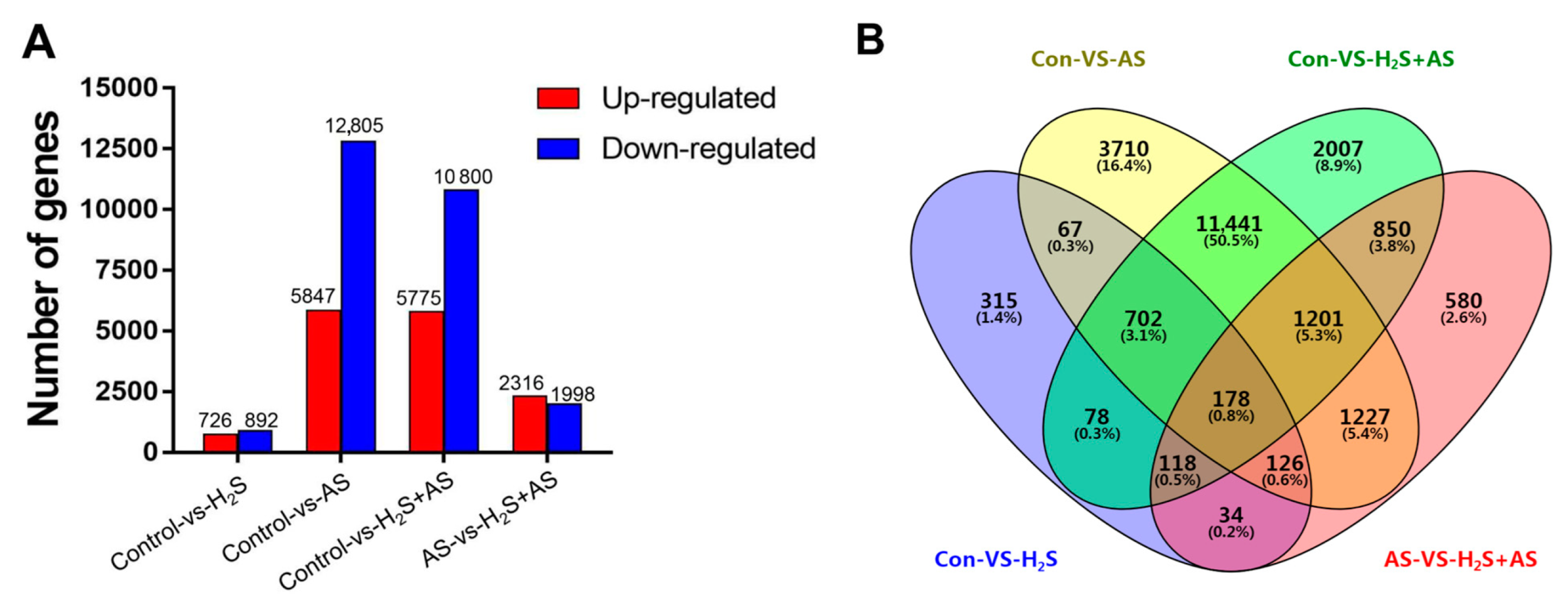
The normalized FPKM was used to quantify the transcription level of the reads, thus facilitating the comparison of mRNA standards within and between genotypes. The analysis of the distribution of the gene expression level revealed that most transcripts were stimulated by H2S pretreatment and alkaline salt stress, and the log10 (FPKM + 1) ranged from 0 to 5 as assessed by boxplot analysis (Figure S1). This analysis indicated that the gene expression level of this study was reliable for further analysis. To improve the accuracy of the differentially expressed genes (DEGs), DEGs (Q-value < 0.001 and |log2 (fold change)| > 2) were defined as genes that were highly enriched or depleted in one treatment relative to another treatment. In the Control-H2S + AS, Control-AS, Control-H2S, and AS-H2S + AS groups, we identified 5775 upregulated and 10,800 downregulated genes; 5947 upregulated and 12,805 downregulated genes; 726 upregulated and 892 downregulated genes; and 2316 upregulated and 1998 downregulated genes, respectively, in M. hupehensis roots (Figure 2A). Venn diagram analysis showed the specificity and overlap among differentially expressed genes between different comparison groups (Figure 2B). Numerous genes overlapped in expression under H2S pretreatment and in response to alkaline salt stress. For example, in the Control-H2S + AS and Control-AS groups, 13,522 differentially expressed genes overlapped.

2.3. GO Term and KEGG Pathway Enrichment Analysis of DEGs between H2S and Alkaline Salt Stress

In our study, GO annotation and statistical analysis indicated that the DEGs were described as three GO ontologies (cellular component, biological process, and molecular function) with approximately 35 terms in different comparison groups (Control-H2S, Control-H2S + AS, Control-AS, and AS-H2S + AS). In the different comparison groups, the biological processes were similar and included “metabolic process” and “cellular process”. The “membrane”, “cell”, “membrane part”, and “organelle” were the top four highest GO terms in the cellular component. In addition, we found a significantly high abundance of molecular function categories, i.e., “binding” and “catalytic activity” (Figure 3 and Figure S2).
The functional enrichment analysis showed that the GO significant enrichment period of each comparison group (Control-H2S, Control-H2S + AS, Control-AS, and AS-H2S + AS) was very similar. In the biological processes of the Control-H2S + AS, Control-AS, and Control-H2S groups, the overpowering majority was “cellular process”. In the cellular component, the overpowering majority was “cell”, and “binding” was the dominant term in the molecular functions. Considering the GO biological processes, the common DEGs were significantly enriched in “cellular process” and “metabolic process”, “cell” and “cell part” of the cellular component, and “catalytic activity” and “binding” of molecular functions. Otherwise, many enriched GO terms were observed to be different in the comparison groups, in the Control-H2S + AS group, “hydrogen peroxide metabolic process”, “oxidoreductase complex”, and “xyloglucan: xyloglucosyl transferase activity” GO terms were identified and highly enriched. In the Control-AS group, the “ion homeostasis” and “cell wall macromolecule catabolic process” terms were identified and were highly enriched (Tables S3–S5).
We assigned 1352, 13,238, 14,820, and 3597 genes to the comparison groups (Control-H2S, Control-H2S + AS, Control-AS, and AS-H2S + AS) for KEGG pathway annotation, and 19 pathways were classified based on five categories, i.e., including environmental information processing, metabolism, cellular processes, genetic information processing, and organismal systems (Figure 4 and Figure S2). KEGG analysis showed that the functions of most genes were annotated into sub-branches of “signal transduction”, “translation”, and “carbohydrate metabolism”. Together, these results supported the possibility that H2S could alleviate alkaline salt stress by the absorption and inhibition of solutes in the root tissues of M. hupehensis, thus enhancing root growth.
Interestingly, the DEGs of the Control-H2S, Control-AS, Control-H2S + AS, and AS-H2S + AS were matched to 124, 137, 137, and 132sub-branches of the KEGG pathway, respectively. The significantly enriched KEGG pathways in H2S-pretreated and alkaline salt samples are shown in Tables S6–S8. Furthermore, the KEGG enrichment analysis indicated that the enriched pathways with more genes than other pathways were similar in the Control-H2S + AS, Control-AS, and Control-H2S groups, including “phenylpropanoid biosynthesis”, “MAPK signaling pathway—plant”, “plant hormone signal transduction” and “plant–pathogen interaction”. These genes were involved in multiple mechanisms that might participate in the H2S alleviation of alkaline salt tolerance.

2.4. Transcriptome Analysis and PCR Validation in Response to H2S and Alkaline Salt Stress

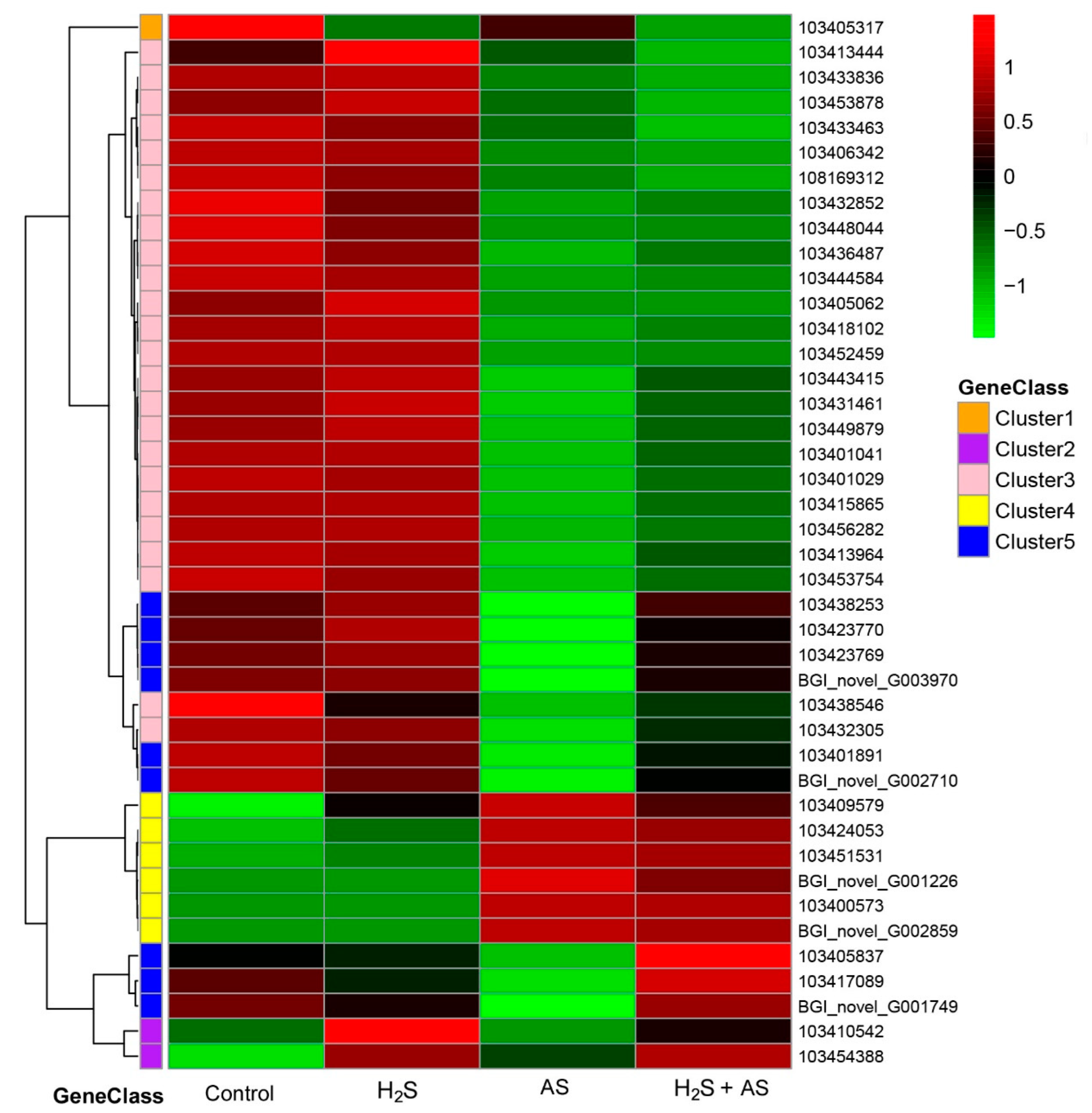
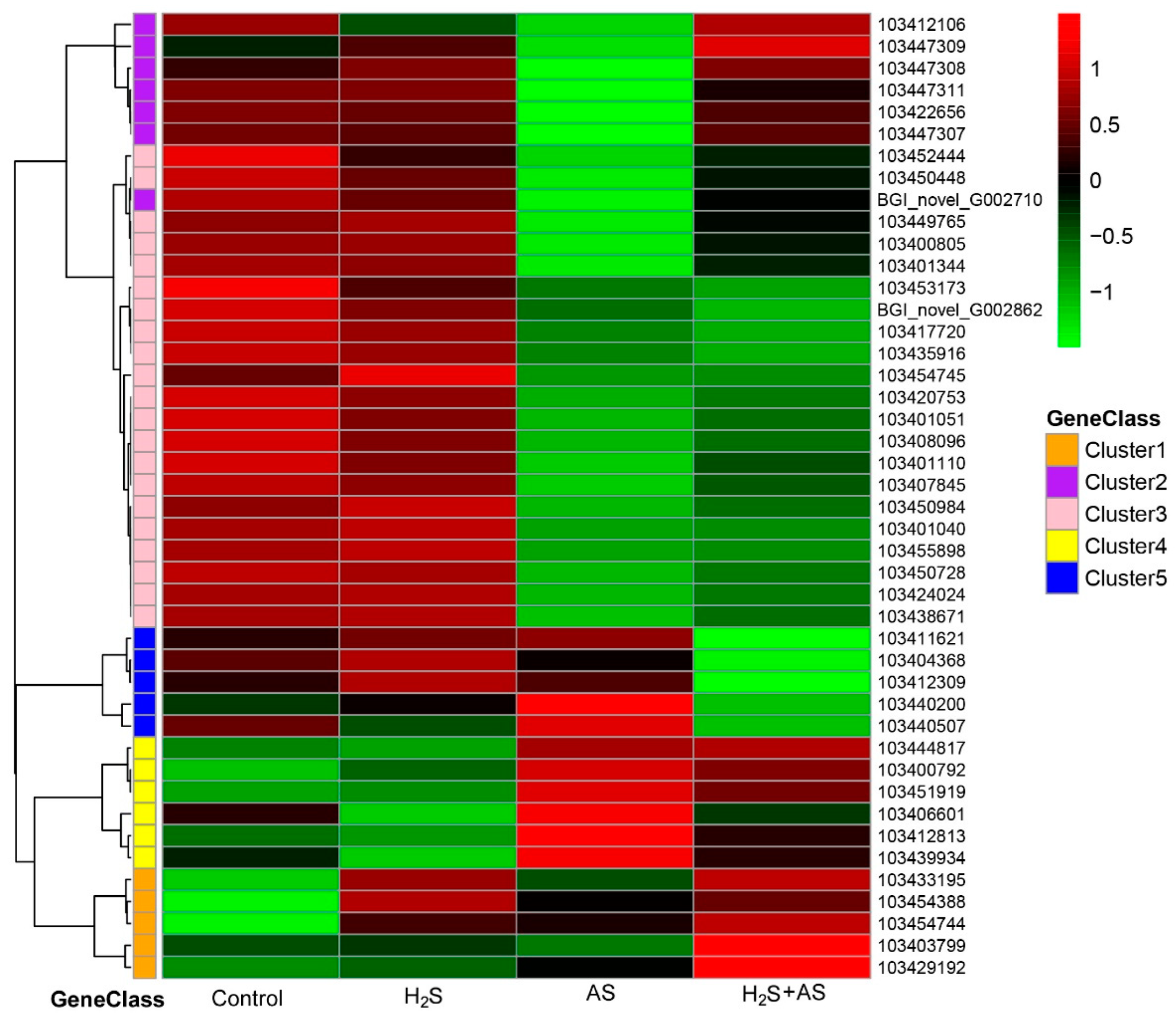
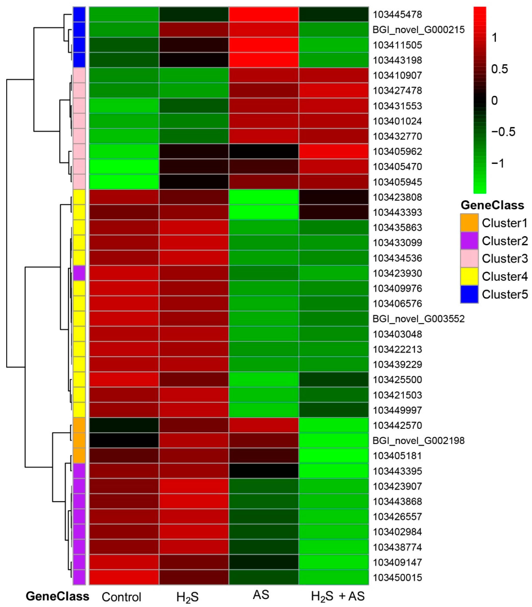
To further explore the molecular mechanism of H2S pretreatment alleviating alkaline salt stress in M. hupehensis root, genes related to the antioxidant system and S-containing compounds, cell wall metabolism, soluble sugar, and osmoprotectants were screened from differentially expressed genes under different treatments (Tables S9–S12). Then the expression patterns of differential genes were analyzed using a heatmap (Figure 5, Figure 6 and Figure 7).
In addition, we carried out Venn diagram analysis of differentially expressed genes (DEGs) among different groups and found 126 genes that were significantly stimulated or inhibited by H2S and differentially expressed by alkaline salt (AS) stress induction, but there was no difference between the control group and the treatment of H2S + AS stress (Figure S3A). We believe that these genes are H2S-regulated clusters involved in alleviating alkaline salt stress. Subsequently, we compared these genes with all the identified transcription factors (TFs) for Venn diagram analysis, and a total of 15 differential TFs were identified to be involved in H2S-mediated AS stress relief (Figure S3B). Then, we analyzed the expression characteristics of these transcription factors using heatmaps and found that four HSF family transcription factors were highly expressed after AS stress, while, after H2S treatment or H2S + AS treatment, they were downregulated (Figure S3C,D), suggesting that these TFs may play an important role in the process of H2S-mediated alkaline salt stress relief.
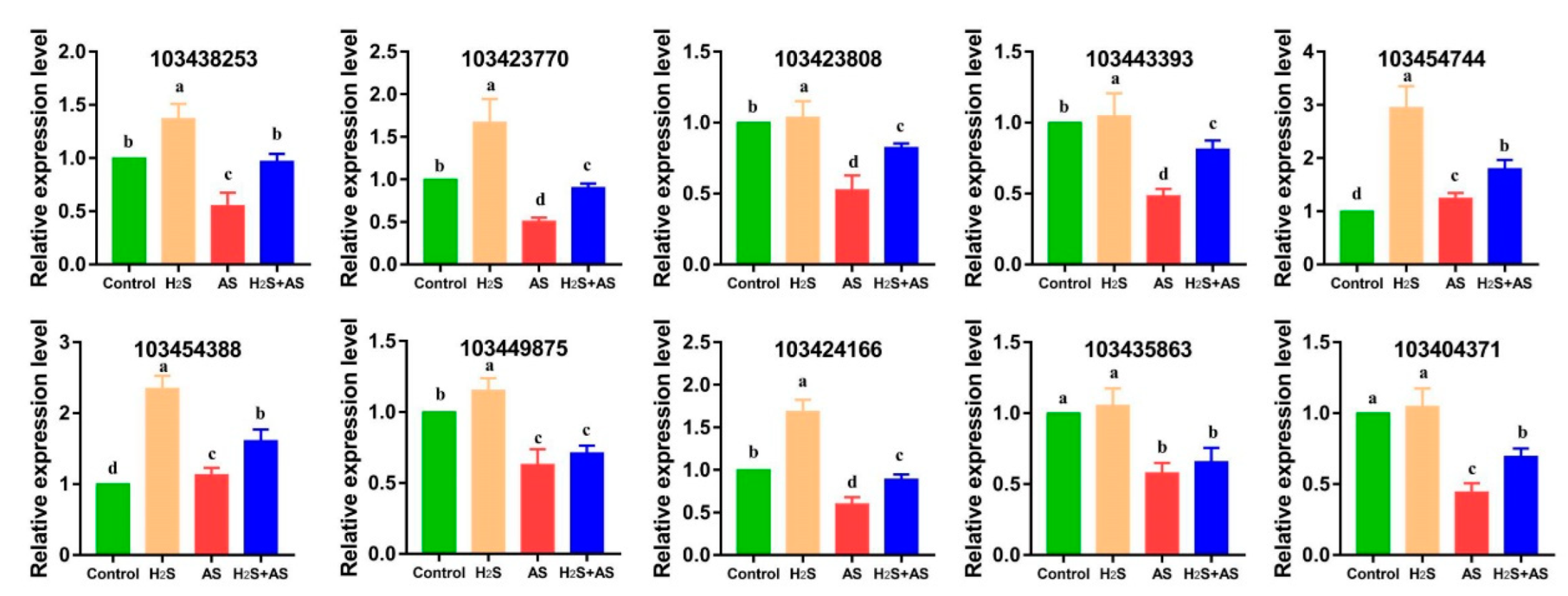
For the examination of the expression profiles of genes that responded to H2S and alkaline salt treatment in M. hupehensis roots, we randomly selected four sulfur-containing compounds, one reactive oxygen metabolism, three cell wall metabolism, and two energy metabolism genes for PCR verification. We found that these genes correlated well with the results achieved by RNA-seq analysis (Figure 8). Therefore, the qRT-PCR results demonstrated that the RNA-seq data were reliable. Actin was used for the data normalization of the reference genes. The primers of the selected genes are shown in Table S1.

3. Discussion

Many studies have demonstrated that H2S as a signal molecule can ameliorate or alleviate the adverse response of plants to salt/alkali stress. Our previous study revealed that H2S can alleviate the alkaline-salt-induced toxicity on M. hupehensis roots through the analysis of root morphology, Na+/K+ homeostasis, ROS accumulation, and oxidative stress at the biochemical and physiological levels [7]. Nevertheless, at the transcriptome level, knowledge of the regulation mechanism through which H2S alleviates alkaline salt stress in Malus hupehensis roots remains limited. In the present analysis, we revealed many differentially expressed genes at the transcriptome level and identified signaling pathways and metabolites that might be involved in H2S-mediated alleviation of alkaline salt stress tolerance. In addition, it will be helpful to better understand the mechanism through which H2S pretreatment regulates alkaline salts stress in Malus hupehensis roots.

3.1. H2S-Mediated Antioxidant System and S-Containing Compounds Alleviate Alkaline Salts in M. hupehensis Roots

Salt-stress-induced oxidative stress is an effect that promotes the generation of reactive oxygen species (ROS) [28], and an increase in the levels of ROS may cause harmful oxidative stress to plant cells and organelles [29]. Evidence has demonstrated that plants can activate both enzymatic (APX, CAT, POD, and SOD) and non-enzymatic (ascorbic acid, non-protein amino acids, and glutathione) pathways to reduce ROS-induced damage [29]. It is speculated that increasing the ROS-scavenging ability of plants can significantly improve the stress resistance of plants [30,31]. Thus, under H2S pretreatment and alkaline salt treatment, more antioxidant and detoxification-related genes should be isolated from M. hupehensis roots. As expected, we identified 32 DEGs involved in ROS metabolism under salt stress. However, we identified five DEGs involved in ROS scavenging (one upregulated and four downregulated genes) and superoxide radical scavenging (one upregulated) when exogenous H2S was applied to apple under alkaline salt stress. We isolated two downregulated (103411621, 103412309) and one upregulated (103407845) gene involved in ROS biosynthesis of AS-H2S + AS, implying that the ROS biosynthesis and level might decrease upon the addition of H2S to the alkaline salt treatment (Figure 5 and Table S9). Our previous study showed that H2S pretreatment can reduce the content of ROS in the roots of M. hupehensis [7].
It is paramount for plants to maintain their redox status by increasing sulfur (S) metabolism and by producing S-containing compounds in response to salt-stress-induced oxidative stress. Many sulfur-containing compounds, including H2S, glutathione, cysteine, methionine, and thioredoxin, play essential roles in plants in stressful environments [32,33]. Sulfur metabolism is the key pathway for the biosynthesis of S-containing compounds, including H2S, glutathione, cysteine, methionine, and thioredoxin, playing essential roles in plants in stressful environments [32,33]. The synthesis of cysteine is the final step of sulfate reduction in plants, which is almost the only reaction for reducing sulfur for metabolism in a demand-driven manner [34,35]. Salinity can induce a higher cysteine synthesis rate [36]. As an essential amino acid, methionine not only plays a central role in the initiation of plant mRNA translation but is also a basic metabolite of plant cells and can directly or indirectly regulate a variety of cellular processes [37]. Nazar et al. discovered that glutathione plays an important role in plant metabolism and can reduce the adverse effects of salt stress on plants [38]. Our study revealed that cysteine metabolism (15 DEGs), methionine metabolism (11 DEGs), glutathione metabolism (6 DEGs), and metabolism of S-containing compounds (77 DEGs) significantly responded at the transcriptomic level to alkaline salt stress. Moreover, the glutathione S-transferase DHAR1 (103417720; 103435916), glutathione S-transferase DHAR2-like (103433463), glutamate–cysteine ligase (103438546), and glutathione synthetase (103413964) genes were significantly downregulated, similar to the result for Catharanthus roseus, i.e., that salinity stress decreased the glutathione content [39]. In addition, our transcriptome analysis demonstrated that the genes involved in cysteine-type endopeptidase inhibitor (103428875; 103428868; 103434431; 103438575; 103438577) that responded to the pretreatment of H2S before alkaline salt stress were upregulated in M. hupehensis roots (Figure 5 and Table S10). This indicated that H2S pretreatment could alleviate alkaline salt stress by upregulating the expression of S-containing compounds in M. hupehensis roots. This also agrees with the increased requirement for ROS scavenging, because the alkaline-salt-induced production of ROS was greater than that with the addition of H2S to the alkaline salt treatment of M. hupehensis roots [7].

3.2. Role of Cell Wall Metabolism–Related Genes in H2S Alleviating Alkaline Salt Stress

The cell wall is of vital importance to the shape of the cell and provides the necessary mechanical strength and rigidity for plant tissues to support the turgor pressure. In addition, the deposition and modification of cell wall materials play an essential role in the plant response to environmental stress [40]. Numerous studies have reported that peroxidases can be induced by salt and drought stress [41,42,43], and the peroxidase content in the cell wall of cowpea was attributed to root growth under salinity conditions [43]. Kumar et al. found that increased peroxidase activity in the cell wall could improve the resistance of transgenic tobacco to salt and drought [44]. Here, we found 4 upregulated and 28 downregulated peroxidases in M. hupehensis roots under alkaline salt stress. In particular, there were 11 upregulated and 2 downregulated genes involved in hydrogen peroxide catabolism, implying that the cellulose concentration may have increased in these leaves due to increased biosynthesis with H2S pretreatment before alkaline salt stress compared to that with alkaline salt treatment alone (Figure 6 and Table S11). A previous report suggested that H2O2 and peroxidase play an important role in the synthesis of lignin and the formation of covalent bonds between lignin and carbohydrates in the cell wall [45]. Here, we obtained two upregulated genes (103450245 and 103402291) involved in lignin metabolism with the application of H2S (AS-H2S + AS). Thus, the lignin level might be elevated with the addition of H2S. Collectively, this reveals that peroxidases played a role in H2S- and alkaline-salt-induced lignin deposition by degrading H2O2.
Many reports have demonstrated that the changes in the physical and chemical properties of the cell wall are related to cell growth [11]. The function of the cell wall as a cell growth regulator under salt stress has been investigated in detail in some studies [46,47]. Polysaccharide is the main component of cell walls, which can further reflect the density of cell walls [48], and the cell wall density and hardening activity determine the growth rate of the root system [49]. Our transcriptome analyses of M. hupehensis roots revealed that the expression of genes related to cell wall density and stiffening was modified by alkaline salt stress. Here, we isolated three downregulated genes (103443393, 103423930, and 103406576) involved in hydroxyproline-rich glycoprotein metabolism and two downregulated genes encoding cell wall thickening (103443882, 103444840). Xyloglucosyl transferase (eight DEGs) was significantly downregulated in alkaline salt treatment (Figure 6 and Table S11). In addition, we observed that genes encoding hydroxyproline O-galactosyltransferase HPGT1-like (103443393) and xyloglucan endotransglucosylase/hydrolase protein 32 precursor (103449997) were upregulated with the addition of H2S. Xyloglucan is often modified by cell wall localization enzymes and participates in cell wall modification during cell elongation [50,51]. Xyloglucan endotransglucosylase 19 (XTH19) plays an important role in the lateral root development of Arabidopsis thaliana in response to salt stress [12]. These results showed that changes in the expression level of cell wall density and stiffening-related genes to maintain root elongation might be necessary for H2S to relieve alkaline salt stress in the root system of M. hupehensis.

3.3. H2S Induces Soluble Sugar and Osmoprotectant-Regulation-Related Genes in Response to Alkaline Salt Stress

There is much evidence that indicates that carbohydrate and energy metabolism are critical for plant development and response to stress, including the synthesis of protective substances and ROS scavenging, for resistance against alkaline salt stress in plant roots [52,53]. Starch and sucrose metabolism determines the level of soluble sugars and affects osmotic regulation [54,55]. Among them, the increased production of soluble sugars, including proline, fructose, glucose, trehalose, and sucrose, in cells can improve the stress resistance of plants [56,57].
Here, amino sugar metabolic (seven DEGs), proline biosynthetic (three DEGs), trehalose biosynthetic (seven DEGs), glucose metabolic (nine DEGs), and starch metabolic (seven DEGs) genes significantly responded at the transcriptomic level under alkaline salt stress. More downregulated than upregulated genes related to starch metabolism and glucose metabolism were isolated from M. hupehensis roots under alkaline salt stress (Table S12). Similarly, these DEGs were also found with the addition of H2S before the alkaline salt treatment. However, the addition of H2S before the alkaline salt treatment could led to the disappearance of some pathways and a reduction in the number of DEGs compared to alkaline salt stress alone (Table S12). Under alkaline salt stress, genes involved in trehalose metabolism, such as trehalose-phosphate phosphatase (TPP) (103408485), trehalose-phosphatase synthase (TPS) (103446295, 103451948, 103402996), and glucose catabolism (103433794, 103408593, 103433797) were upregulated, and genes encoding sucrose-phosphatase 2 isoform X1 (103430254) and sucrose-phosphate synthase 2 (103443444) were downregulated with the transcriptome sequencing. Previous studies have shown that some TPPs’ expression levels were increased under conditions of salt stress in A. thaliana [58]. Similarly, salt stress upregulated the expression of TPSs in Populus [59]. In addition, most transgenic plants overexpressing TPS and TPP genes have great tolerance to salt stress [60,61]. In our present study, some TPP and TPSs were upregulated under salt stress, while, in the addition of H2S before alkaline salt treatment, these gene were not significantly different from those in the control, which suggest that these genes play an important role in salt stress and the alleviation of alkaline salt stress by H2S.
On the other hand, genes involved in glucose homeostasis (103424166, 103431330, and 103414773), glucose-1-phosphate adenylyltransferase (103432152 and 103405088), and fructose 1,6-bisphosphate 1-phosphatase (103416291) were significantly upregulated when H2S was applied to apple under alkaline salt stress (Figure 7). This result indicates that H2S pretreatment before alkaline salt stress induced genes involved in carbohydrate and energy metabolism, which might be different from the case when only alkaline salt stress was applied to M. hupehensis roots. The significant changes in genes related to primary energy metabolism and the osmoprotection of soluble sugar induced by alkaline salt stress may inhibit plant growth and development. However, the addition of H2S increased the gene expression abundance of these metabolic pathways and alleviated the inhibition caused by alkaline salt stress.

4. Methods and Materials

4.1. Treatment of Plant Materials

PYTC (Malus hupehensis Rehd. var. pingyiensis Jiang) seedling stratification and H2S pretreatment and alkaline salt treatments were performed according to our previous research methods [7]. After seedling germination, five seedlings were cultivated in black plastic bowls (diameter 11 cm and height 9 cm) that contained clean river sand and were then grown in the greenhouse under a natural photoperiod at the National Research Center for Apple Engineering and Technology Shandong Agriculture University (SDAU), China.
From the eighth day after cultivation, the seedlings in the black plastic bowls were irrigated with fresh nutrition solution containing micronutrients and macronutrients every other day until the seedlings had six to seven leaves. Thereafter, we used 0.5 mM sodium hydrosulfide (NaHS, H2S donor) to pretreat the seedlings, and H2S was dissolved in the nutrient solution and changed every 24 h. The seedlings were adapted to the nutrient solution for one day and were then treated with mixed alkaline salt (NaCl:Na2SO4:NaHCO3:Na2CO3 = 1:9:9:1, molar ratio; salinity 150 mM; pH 8.47–8.83). The detailed steps of the H2S pretreatment and alkaline salt stress treatments were performed according to our previous research methods [7]. At the end of the experiment, root samples were carefully collected and frozen in liquid N2 immediately and then stored at −80 °C until extraction.
Experiments were performed 3 replications per treatment with 50 plants each in completely randomized design, and the 3 replicates of each treatment were named as Control-1, Control-2, Control-3 (Control); H2S-1, H2S-2, H2S-3 (H2S treatment); AS-1, AS-2, AS-3 (AS: alkaline salt treatment); H2S + AS-1, H2S + AS-2, H2S + AS-3 (H2S and alkaline salt treatment).

4.2. RNA Extraction and cDNA Library Preparation for Transcriptome Sequencing

Total RNA was extracted from M. hupehensis roots (Control, H2S, H2S + AS, AS) with the CTAB-PBIOZOL reagent (Hangzhou Bioer Technology Co. Ltd., Hangzhou, China) using the extraction method [62]. mRNA molecules were purified from total RNA using oligo (dT)-attached magnetic beads. Subsequently, cleaved RNA fragments were generated using a random hexamer primed for first-strand cDNA, followed by second-strand cDNA synthesis. The following steps were conducted after purification: end-repair, A addition, adaptor ligation, and amplification of the cDNA fragment by PCR. Libraries were validated on an Agilent Technologies 2100 Bioanalyzer. Twelve cDNA libraries of M. hupehensis roots were constructed using the BGISEQ500 platform (BGI, Shenzhen, China). Three biological replicates were used for all treatments.

4.3. RNA-Seq Read Mapping, Assembly, and Annotation of the Transcriptome

We obtained clean reads by filtering the low-quality reads, i.e., those contaminated with adapters and those with unknown base N from the raw reads. The software TRIMmomatic (version 0.36) was used to filter the reads, and SOAPnuke (version 1.4.0) software was used for statistics. The clean reads were mapped to the reference genome sequence using Hierarchical Indexing for Spliced Alignment of Transcripts (HISAT, version 2.1.0, http://www.ccb.jhu.edu/software/hisat (accessed on 24 June 2019)) [63] and gene sequencing was conducted using Bowtie2 (version 2.5, http://bowtie-bio.sourceforge.net/Bowtie2/index.shtml (accessed on 24 June 2019)) [64]. The gene expression levels were estimated through RNA-seq using RSEM software (version1.2.8 http://deweylab.biostat.wisc.edu/rsem/rsem-calculate-expression.html (accessed on 24 June 2019)) [65]. The gene expression level of the fragments (each contig) was normalized to FPKM (per kilobase per transcript per million mapped reads). We used density distribution analysis of the filtered transcripts, stacked bar charts, and a boxplot graph to assess the expression distribution among all samples.

4.4. Differentially Expressed Gene Annotation and Analysis

Differentially expressed gene analysis was performed [66]. Genes that had a fold change (FC) ≥ 2 and p-value ≤ 0.001 were considered as differentially expressed. The Pheatmap function in R software was used for hierarchical clustering analysis of the Control, H2S, H2S + AS, and AS libraries. To determine the significant enrichment of DEGs in GO terms and KEGG pathways, the threshold was selected as the corrected p-value < 0.05 [67].

4.5. Analysis and Validation of qRT-PCR

Total RNA from the roots was extracted as described above. The DEGs from M. hupehensis roots were randomly chosen for qRT-PCR analysis. We designed the specific primers (Table S1) for qRT-PCR using Primer Premier 5.0 software (Premier Biosoft Int., Palo Alto, CA, USA). The qRT-PCR analysis was performed using the LightCycler® 96 System (Roche Molecular Biochemicals, Lewes, UK) according to our previous research methods [62]. The 18S rRNA gene was quantified as an internal standard, and reference samples were obtained from M. hupehensis root control samples, which were set to 1. The 2−ΔΔCt method was used to calculate differential expression. Each biological replicate sample consisted of three technical replicates.

4.6. Experimental Design and Statistical Analysis

Comparative analyses of the expression levels of each gene among different treatments were carried out SPSS statistical software (version 18.0, SPSS Inc., Chicago, IL, USA) (p < 0.05) using one-way ANOVA tests followed by the least significant difference (LSD) test.

5. Conclusions

It is a complex process to maintain or promote plant root growth under alkaline salt stress. This work presents transcriptome sequencing to analyze the effect underlying the H2S regulation of alkaline salt stress tolerance in M. hupehensis roots. The GO terms and KEGG pathway enrichment showed the response mechanism of H2S applied to M. hupehensis roots under alkaline salt stress. Investigation of the transcriptomic data suggested the candidate functional DEGs that might contribute to H2S regulation of alkaline salt stress tolerance in M. hupehensis roots. Further analysis indicated that H2S alleviated alkaline salt stress, including the following aspects: (a) H2S pretreatment resulted in higher energy maintenance capacity and cell wall integrity of M. hupehensis roots. (b) H2S pretreatment induced higher numbers of upregulated genes involved in ROS metabolism and S-containing compounds and enhanced the capacity of ROS metabolism in M. hupehensis roots (Figure 9). To conclude, this information on novel genes and identified DEGs in M. hupehensis roots will be invaluable in future studies to elucidate the specific mechanism through which H2S regulates alkaline salt tolerance.

Supplementary Materials

The following supporting information can be downloaded at: https://www.mdpi.com/article/10.3390/ijms232314848/s1.

Author Contributions

Data curation, methodology, software, validation, and writing—original draft: H.L.; experiments, methodology, and software: M.H.; methodology and validation: J.S.; investigation and data management: Y.N.; methodology and writing—review and editing: W.Z.; funding acquisition, supervision, and writing—review and editing: H.Y. All authors have read and agreed to the published version of the manuscript.

Funding

This work was supported by the National Natural Science Foundation of China, grant numbers 32172517 and 31772251, and the National Key R&D Program of China, grant number 2019YFD1000103.

Institutional Review Board Statement

Not applicable.

Informed Consent Statement

Not applicable.

Data Availability Statement

The NCBI SRA database accession number was SRR13586165-SRR13586176.

Conflicts of Interest

The authors declare no conflict of interest.

References

  1. Kawanabe, S.; Zhu, T.C. Degeneration and conservation of aneurolepidium chinense grassland in Northern China. J. Jpn. Grassland Sci. 1991, 37, 91–99. [Google Scholar]
  2. Shi, D.C.; Wang, D.L. Effects of various salt-alkaline mixed stresses on Aneurolepidium chinense (Trin.) Kitag. Plant Soil 2005, 271, 15–26. [Google Scholar] [CrossRef]
  3. Yang, C.W.; Chong, J.N.; Li, C.Y.; Kim, C.M.; Shi, D.C.; Wang, D.L. Osmotic adjustment and ion balance traits of an alkali resistant halophyte Kochia sieversiana during adaptation to salt and alkali conditions. Plant Soil 2007, 294, 263–276. [Google Scholar] [CrossRef]
  4. Yu, S.; Yu, L.H.; Hou, Y.L.; Zhang, Y.F.; Guo, W.; Xue, Y.W. Contrasting effects of NaCl and NaHCO3 stresses on seed germination, seedling growth, photosynthesis, and osmoregulators of the common bean (Phaseolus vulgaris L.). Agronomy 2019, 9, 19. [Google Scholar] [CrossRef] [Green Version]
  5. Liu, P.; Zhu, Y.X.; Liu, H.; Liang, Z.J.; Zhang, M.Y.; Zou, C.Y.; Yuan, G.S.; Gao, S.B.; Pan, G.T.; Shen, Y.O.; et al. Acombination of a genome-wide association study and a transcriptome analysis reveals circRNAs as new regulators involved in the response to salt stress in maize. Int. J. Mol. Sci. 2022, 23, 9755. [Google Scholar] [CrossRef]
  6. Zhang, H.; Liu, X.L.; Zhang, R.X.; Yuan, H.Y.; Wang, M.M.; Yang, H.Y. Root Damage under alkaline stress is associated with reactive oxygen species accumulation in rice (Oryza sativa L.). Front. Plant Sci. 2017, 8, 1580. [Google Scholar] [CrossRef]
  7. Li, H.; Shi, J.Y.; Wang, Z.P.; Zhang, W.W.; Yang, H.Q. H2S pretreatment mitigates the alkaline salt stress on Malus hupehensis roots by regulating Na+/K+ homeostasis and oxidative stress. Plant Physiol. Bioch. 2020, 156, 233–241. [Google Scholar] [CrossRef]
  8. Capula-Rodríguez, R.; Valdez-Aguilar, L.A.; Cartmill, D.L.; Cartmill, A.D.; Alia-Tejacal, I. Supplementary calcium and potassium improve the response of tomato (Solanum lycopersicum L.) to simultaneous alkalinity, salinity, and boron stress. Commun. Soil Sci. Plant Anal. 2016, 47, 505–511. [Google Scholar]
  9. Zhang, K.H.; Tang, J.R.; Wang, Y.; Kang, H.Y.; Zeng, J. The tolerance to salinealkaline stress was dependent on the roots in wheat. Physiol. Mol. Biol. Plants 2020, 26, 947–954. [Google Scholar] [CrossRef]
  10. Zhang, P.; Duo, T.Q.; Wang, F.D.; Zhang, X.Z.; Yang, Z.Z.; Hu, G.F. De novo transcriptome in roots of switchgrass (Panicum virgatum L.) reveals gene expression dynamic and act network under alkaline salt stress. BMC Genom. 2021, 22, 82. [Google Scholar] [CrossRef]
  11. An, P.; Li, X.; Zheng, Y.; Matsuura, A.; Abe, J.; Eneji, A.E.; Tanimoto, E.; Inanaga, S. Effects of NaCl on root growth and cell wall composition of two soya bean cultivars with contrasting salt tolerance. J. Agron. Crop Sci. 2014, 200, 212–218. [Google Scholar] [CrossRef]
  12. Xu, P.P.; Fang, S.; Chen, H.Y.; Cai, W.M. The brassinosteroid-responsive xyloglucan endotransglucosylase/hydrolase 19 (XTH19) and XTH23 genes are involved in lateral root development under salt stress in Arabidopsis. Plant J. 2020, 104, 59–75. [Google Scholar] [CrossRef] [PubMed]
  13. Yuan, Y.H.; Li, J.; Ma, C.H.; Yang, Q.H.; Liu, C.J.; Feng, B.L. Salt-tolerant broomcorn millet (Panicum miliaceum L.) resists salt stress via modulation of cell wall biosynthesis and Na+ balance. Land Degrad. Dev. 2020, 32, 1–15. [Google Scholar] [CrossRef]
  14. Rausch, T.; Wachter, A. Sulfur metabolism: A versatile platform for launching defence operations. Trends Plant Sci. 2005, 10, 503–509. [Google Scholar] [CrossRef] [PubMed]
  15. Li, Z.G.; Min, X.; Zhou, Z.H. Hydrogen sulfide: A signal molecule in plant cross-adaptation. Front. Plant Sci. 2016, 7, 1621. [Google Scholar] [CrossRef] [PubMed] [Green Version]
  16. Jiang, J.L.; Tian, Y.; Li, L.; Yu, M.; Hou, R.P.; Ren, X.M. H2S alleviates salinity stress in cucumber by maintaining the Na+/K+ balance and regulating H2S metabolism and oxidative stress response. Front. Plant Sci. 2019, 10, 678. [Google Scholar] [CrossRef] [Green Version]
  17. Mostofa, M.G.; Saegusa, D.; Fujita, M.; Tran, L.S. Hydrogen sulfide regulates salt tolerance in rice by maintaining Na+/K+ balance, mineral homeostasis and oxidative metabolism under excessive salt Stress. Front. Plant Sci. 2015, 6, 1–14. [Google Scholar] [CrossRef] [PubMed] [Green Version]
  18. Lai, D.W.; Mao, Y.; Zhou, H.; Li, F.; Wu, M.Z.; Zhang, J. Endogenous hydrogen sulfifide enhances salt tolerance by coupling the reestablishment of redox homeostasis and preventing salt-induced K+ loss in seedlings of Medicago sativa. Plant Sci. 2014, 225, 117–129. [Google Scholar] [CrossRef]
  19. Deng, Y.Q.; Bao, J.; Yuan, F.; Liang, X.; Feng, Z.T.; Wang, B.S. Exogenous hydrogen sulfide alleviates salt stress in wheat seedlings by decreasing Na+ content. Plant Growth Regul. 2016, 79, 391–399. [Google Scholar] [CrossRef]
  20. Ziogas, V.; Molassiotis, A.; Fotopoulos, V.; Tanou, G. Hydrogen sulfide: A potent tool in postharvest fruit biology and possible mechanism of action. Front. Plant Sci. 2018, 9, 1375. [Google Scholar] [CrossRef] [Green Version]
  21. Wang, Z.; Gerstein, M.; Snyder, M. RNA-Seq: A revolutionary tool for transcriptomics. Nat. Rev. Genet. 2009, 10, 57–63. [Google Scholar] [CrossRef] [PubMed]
  22. Yang, L.F.; Jin, Y.H.; Huang, W.; Sun, Q.; Liu, F.; Huang, X.Z. Full-length transcriptome sequences of ephemeral plant Arabidopsis pumila provides insight into gene expression dynamics during continuous salt stress. BMC Genom. 2018, 19, 717–730. [Google Scholar] [CrossRef] [PubMed]
  23. Zhan, Y.H.; Wu, T.T.; Zhao, X.; Wang, Z.Q.; Chen, Y. Comparative physiological and full-length transcriptome analyses reveal the molecular mechanism of melatoninmediated salt tolerance in okra (Abelmoschus esculentus L.). BMC Plant Biol. 2021, 21, 180. [Google Scholar] [CrossRef] [PubMed]
  24. Zhang, W.W.; Song, J.F.; Yue, S.Q.; Duan, K.X.; Yang, H.Q. MhMAPK4 from Malus hupehensis Rehd. decreases cell death in tobacco roots by controlling Cd2+ uptake. Ecotox. Environ. Saf. 2019, 168, 230–240. [Google Scholar] [CrossRef]
  25. Cai, Z.C.; Wang, C.C.; Chen, C.H.; Zou, L.S.; Yin, S.X.; Liu, S.J.; Yuan, J.H.; Wu, N.; Liu, X.H. Comparative transcriptome analysis reveals variations of bioactive constituents in Lonicera japonica flowers under salt stress. Plant Physiol. Bioch. 2022, 15, 87–96. [Google Scholar] [CrossRef] [PubMed]
  26. Xu, Z.C.; Wang, M.; Ren, T.T.; Li, K.Y.; Li, Y.Q.; Marowa, P.; Zhang, C.S. Comparative transcriptome analysis reveals the molecular mechanism of salt tolerance in Apocynum venetum. Plant Physiol. Bioch. 2021, 167, 816–830. [Google Scholar] [CrossRef] [PubMed]
  27. Yang, H.Q.; Duan, K.X.; Zhang, W. Biology and physiology of Malus hupehensis for the apogamic plant resource. Acta. Hortic. 2008, 769, 441–447. [Google Scholar] [CrossRef]
  28. Zhu, J.K. Regulation of ion homeostasis under salt stress. Curr. Opin. Plant Biol. 2003, 6, 441–445. [Google Scholar] [CrossRef]
  29. Choudhury, F.K.; Rivero, R.M.; Blumwald, E.; Mittler, R. Reactive oxygen species, abiotic stress and stress combination. Plant J. 2017, 90, 856–867. [Google Scholar] [CrossRef]
  30. Gill, S.S.; Tuteja, N. Reactive oxygen species and antioxidant machinery in abiotic stress tolerance in crop plants. Plant Physiol. Biochem. 2010, 48, 909–930. [Google Scholar] [CrossRef]
  31. Wei, T.L.; Wang, Y.; Xie, Z.Z.; Guo, D.Y.; Chen, C.W.; Fan, Q.J.; Deng, X.D.; Liu, J.H. Enhanced ROS scavenging and sugar accumulation contribute to drought tolerance of naturally occurring autotetraploids in Poncirus trifoliata. Plant Biotechnol. J. 2018, 17, 1394–1407. [Google Scholar] [CrossRef] [PubMed] [Green Version]
  32. Droux, M. Sulfur assimilation and the role of sulfur in plant metabolism: A survey. Photosynth. Res. 2004, 79, 331–348. [Google Scholar] [CrossRef] [PubMed]
  33. Wirtz, M.; Droux, M. Synthesis of the sulfur amino acids: Cysteine and methionine. Photosynth. Res. 2005, 86, 345–362. [Google Scholar] [CrossRef] [PubMed]
  34. Kopriva, S.; Rennenberg, H. Control of sulphate assimilation and glutathione synthesis: Interaction with N and C metabolism. J. Exp. Bot. 2004, 55, 1831–1842. [Google Scholar] [CrossRef]
  35. Kopriva, S. Regulation of sulfate assimilation in Arabidopsis and beyond. Ann. Bot. 2006, 97, 479–495. [Google Scholar] [CrossRef] [Green Version]
  36. Romero, L.C.; Domínguez-Solis, J.R.; Gutierrez-Alcala, G.; Gotor, C. Salt regulation of O-acetylserine(thiol)lyase in Arabidopsis thaliana and increased tolerance in yeast. Plant Physiol. Biochem. 2001, 39, 643–647. [Google Scholar] [CrossRef]
  37. Amir, R. Current understanding of the factors regulating methionine content in vegetative tissues of higher plants. Amino Acids. 2010, 39, 917–931. [Google Scholar] [CrossRef]
  38. Nazar, R.; Iqbal, N.; Masood, A.; Syeed, S.; Khan, N.A. Understanding the significance of sulfur in improving salinity tolerance in plants. Environ. Exp. Bot. 2011, 70, 80–87. [Google Scholar] [CrossRef]
  39. Jaleel, C.A.; Manivannan, P.; Lakshmanan, G.M.; Sridharan, R.; Panneerselvam, R. NaCl as a physiological modulator of proline metabolism and antioxidant potential in Phyllanthus amarus. C. R. Biol. 2007, 330, 806–813. [Google Scholar] [CrossRef]
  40. Reiter, W.D. Biosynthesis and properties of the plant cell wall. Curr. Opin. Plant Biol. 2002, 5, 536–542. [Google Scholar] [CrossRef]
  41. Chai, L.; Wang, J.M.; Fan, Z.Y.; Liu, Z.B.; Li, X.F.; Yang, Y. Ascorbate peroxidase gene from Brassica napus enhances salt and drought tolerances in Arabidopsis thaliana. Afr. J. Biotechnol. 2011, 10, 18085–18091. [Google Scholar]
  42. Choi, H.W.; Hwang, B.K. The pepper extracellular peroxidase CaPO2 is required for salt, drought and oxidative stress tolerance as well as resistance to fungal pathogens. Planta 2012, 235, 1369–1382. [Google Scholar] [CrossRef] [PubMed]
  43. Maia, J.M.; Ferreira-Silva, S.L.; Voigt, E.L.; de Macedo, C.E.C.; Ponte, L.F.A.; Silveira, J.A.G. Activities of antioxidant enzymes and root growth inhibition in cowpea seedlings exposed to different salt levels. Acta Bot. Bras. 2012, 26, 2. [Google Scholar]
  44. Kumar, D.; Singh, P.; Yusuf, M.A.; Upadhyaya, C.P.; Roy, S.D.; Hohn, T.; Sarin, N.B. The Xerophyta viscosa Aldose Reductase (ALDRXV4) Confers Enhanced Drought and Salinity Tolerance to Transgenic Tobacco Plants by Scavenging Methylglyoxal and Reducing the Membrane Damage. Mol. Biotechnol. 2013, 54, 292–303. [Google Scholar] [CrossRef]
  45. Fry, S.C. Cross-linking of matrix polymers in the growing cell walls of angiosperms. Ann. Rev. Plant Physiol. 1986, 37, 165–186. [Google Scholar] [CrossRef]
  46. Hu, L.X.; Li, H.Y.; Chen, L.; Lou, Y.H.; Amombo, E. RNA-seq for gene identification and transcript profiling in relation to root growth of bermudagrass (Cynodon dactylon) under salinity stress. BMC Genom. 2015, 16, 575. [Google Scholar] [CrossRef] [Green Version]
  47. Zhong, H.; Läuchli, A. Changes of cell wall composition and polymer size in primary roots of cotton seedlings under high salinity. J. Exp. Bot. 1993, 44, 773–778. [Google Scholar] [CrossRef]
  48. Buchanan, B.; Gruissem, W.; Jones, R. Biochemistry and Molecular Biology of Plants; John Wiley & Sons: Hoboken, NJ, USA, 2005. [Google Scholar]
  49. Muller, B.; Reymond, M.; Tardieu, F. The elongation rate at the base of a maize leaf shows an invariant pattern during both the steady-stage elongation and the establishment of the elongation zone. J. Exp. Bot. 2001, 52, 1259–1268. [Google Scholar] [CrossRef]
  50. Reidy, B.; Nösberger, J.; Fleming, A. Differential expression of XET-related genes in the leaf elongation zone of F. pratensis. J. Exp. Bot. 2001, 52, 1847–1856. [Google Scholar] [CrossRef] [Green Version]
  51. Levy, S.; York, W.S.; Stuike-prill, R.; Meyer, B.; Staehelin, L.A. Simulations of the static and dynamic molecular conformations of xyloglucan. The role of the fucosylated sidechain in surface-specific sidechain folding. Plant J. 1991, 1, 195–215. [Google Scholar] [CrossRef]
  52. Du, J.; Guo, S.R.; Sun, J.; Shu, S.R. Proteomic and physiological analyses reveal the role of exogenous spermidine on cucumber roots in response to Ca(NO3)2 stress. Plant Mol. Biol. 2018, 97, 1–21. [Google Scholar] [CrossRef] [PubMed]
  53. Hernández-Walias, F.J.; García, M.; Moreno, M.; Giannoukos, I.; González, N.; Sanz-García, E.; Necira, K.; Canto, T.; Tenllado, F. Transgenerational tolerance to salt and osmotic stresses induced by plant virus infection. Int. J. Mol. Sci. 2022, 23, 12497. [Google Scholar] [CrossRef] [PubMed]
  54. Dahro, B.; Wang, F.; Peng, T.; Liu, J.H. PtrA/NINV, an alkaline/neutral invertase gene of Poncirus trifoliata, confers enhanced tolerance to multiple abiotic stresses by modulating ROS levels and maintaining photosynthetic efficiency. BMC Plant Biol. 2016, 16, 76. [Google Scholar] [CrossRef] [Green Version]
  55. Wei, T.L.; Wang, Y.; Liu, J.H. Comparative transcriptome analysis reveals synergistic and disparate defense pathways in the leaves and roots of trifoliate orange (Poncirus trifoliata) autotetraploids with enhanced salt tolerance. Hortic. Res.-Engl. 2020, 7, 88. [Google Scholar] [CrossRef]
  56. Rady, M.M.; Elrys, A.S.; Abo El-Maati, M.F.; Desoky, E.S.M. Interplaying roles of silicon and proline effectively improve salt and cadmium stress tolerance in Phaseolus vulgaris plant. Plant Physiol. Biochem. 2019, 139, 558–5680. [Google Scholar] [CrossRef] [PubMed]
  57. Mansour, M.M.F.; Ali, E.F. Evaluation of proline functions in saline conditions. Phytochemistry 2017, 140, 52–68. [Google Scholar] [CrossRef] [PubMed]
  58. Krasensky, J.; Broyart, C.; Rabanal, F.A.; Jonak, C. The redox-sensitive chloroplast trehalose-6-phosphate phosphatase AtTPPD regulates salt stress tolerance. Antioxidants Redox Signal. 2014, 21, 1289–1304. [Google Scholar] [CrossRef] [Green Version]
  59. Gao, Y.; Yang, X.; Yang, X.; Zhao, T.; An, X.; Chen, Z. Characterization and expression pattern of the trehalose-6-phosphate synthase and trehalose-6-phosphate phosphatase gene families in Populus. Int. J. Biol. Macromol. 2021, 187, 9–23. [Google Scholar] [CrossRef]
  60. Garg, A.K.; Kim, J.K.; Owens, T.G.; Ranwala, A.P.; Choi, Y.D.; Kochian, L.V.; Wu, R.J. Trehalose accumulation in rice plants confers high tolerance levels to different abiotic stresses. Proc. Natl. Acad. Sci. USA 2002, 99, 15898–15903. [Google Scholar] [CrossRef] [Green Version]
  61. Ge, L.F.; Chao, D.Y.; Shi, M.; Zhu, M.Z.; Gao, J.P.; Lin, H.X. Overexpression of the trehalose-6-phosphate phosphatase gene OsTPP1 confers stress tolerance in rice and results in the activation of stress responsive genes. Planta 2008, 228, 191–201. [Google Scholar] [CrossRef]
  62. Li, H.; Yu, T.T.; Ning, Y.S.; Li, H.; Zhang, W.W.; Yang, H.Q. Hydrogen sulfide alleviates alkaline salt stress by regulating the expression of microRNAs in Malus hupehensis Rehd. roots. Front. Plant Sci. 2021, 12, 663519. [Google Scholar] [CrossRef] [PubMed]
  63. Kim, D.; Langmead, B.; Salzberg, S.L. HISAT: A fast spliced aligner with low memory requirements. Nat. Methods 2015, 12, 357–360. [Google Scholar] [CrossRef] [PubMed] [Green Version]
  64. Langmead, B.; Salzberg, S.L. Fast gapped-read alignment with Bowtie 2. Nat. Methods 2012, 9, 357–359. [Google Scholar] [CrossRef] [PubMed] [Green Version]
  65. Li, B.; Dewey, C.N. RSEM: Accurate transcript quantification from RNA-Seq data with or without a reference genome. BMC Bioinform. 2011, 12, 323. [Google Scholar] [CrossRef] [PubMed]
  66. Wang, L.; Feng, Z.; Wang, X. DEGseq: An R package for identifying differentially expressed genes from RNA-seq data. Bioinformatics 2010, 26, 136–138. [Google Scholar] [CrossRef] [Green Version]
  67. Mao, X.Z.; Cai, T.; Olyarchuk, J.G.; Wei, L.P. Automated genome annotation and pathway identification using the KEGG Orthology (KO) as a controlled vocabulary. Bioinformatics 2005, 21, 3787–3793. [Google Scholar] [CrossRef]
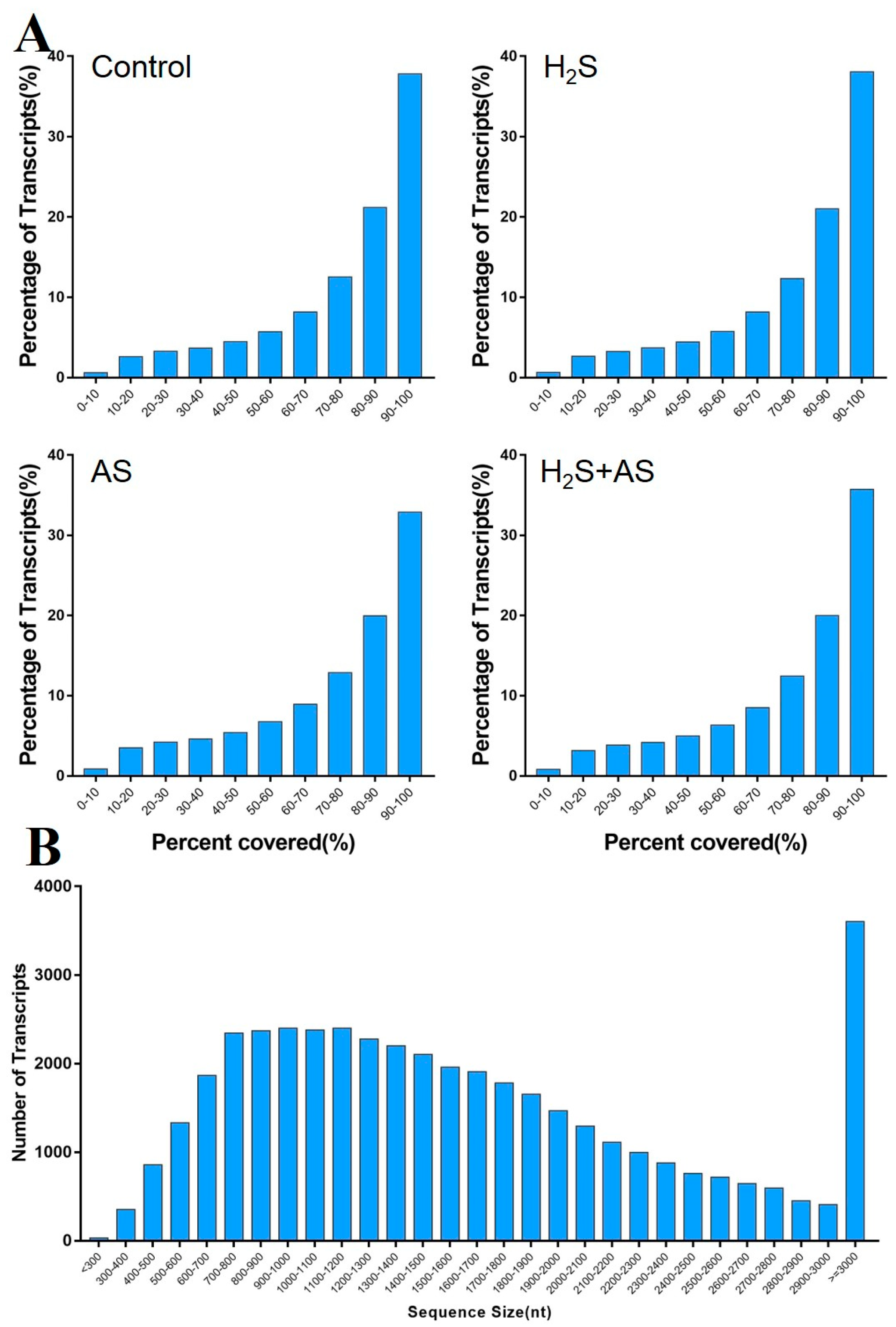
Figure 1. (A) Reads’ coverage of transcripts and (B) length distribution of known gene transcripts treated with H2S and alkaline salt stress.
Ijms 23 14848 g001
Figure 2. The differentially expressed genes (DEGs) of M. hupehensis after treatment with H2S and alkaline salt stress. The number of up- and downregulated DEGs between different comparison groups (A). Venn of the number of DEGs in different comparison groups (B).
Ijms 23 14848 g002
Figure 3. Gene ontology classification of differentially expressed genes. Blue, green, and red represent three GO ontologies: biological process, cellular component, and molecular function, respectively.
Ijms 23 14848 g003
Figure 4. KEGG pathway of differentially expressed genes. Blue, red, green, purple, and orange represent the different KEGG pathways: cellular processes, environmental information processes, genetic information processes, metabolism, and organism systems.
Ijms 23 14848 g004
Figure 5. H2S specifically induces S-containing compound genes to alleviate alkaline salts in M. hupehensis roots.
Ijms 23 14848 g005
Figure 6. H2S-mediated expression of ROS-defense-related genes under alkaline salt stress in M. hupehensis root.
Ijms 23 14848 g006
Figure 7. Role of cell wall metabolism–related genes in the mitigation of alkaline salt stress by H2S of M. hupehensis root.
Ijms 23 14848 g007
Figure 8. qRT-PCR analysis of DEGs in roots under different treatments. The 18S rRNA was used as a housekeeping gene for normalizing gene expression and to correct for sample-to-sample variation. The expression level of genes in the roots of plants grown under control conditions was assigned to 1. The bar represents standard deviation (SD) of the mean, different letters suggest significant differences among the treatments at p < 0.05.
Ijms 23 14848 g008
Figure 9. A model for the mechanisms underlying the hydrogen-sulfide-enhanced alkaline salt tolerance of Malus hupehensis Rehd. Hydrogen sulfide alleviates alkaline salt stress due to the activation of multifaceted defense machinery in the roots. In the roots, cell wall elongation increases because XTH gene expression is upregulated, and with the increase of POD gene expression, ROS scavenging ability was enhanced, thereby, effectively achieving osmotic regulation and detoxification of ROS.
Ijms 23 14848 g009
Table 1. Summary of the RNA-seq data collected from H2S and alkaline salt stress roots of M. hupehensis.
SampleRaw ReadsClean Reads (%)Clean BasesN (%)Adapter (%)Low Quality (%)Q20 (%)Q30 (%)
Control-145.09 M40.84 M (90.58%)6.1397,530 (0.22%)2,061,610 (4.57%)2,086,720 (4.63%)96.61%87.44%
Control-242.2 M38.23 M (90.5%)5.7389,922 (0.21%)1,862,696 (4.41%)2,061,138 (4.88%)96.62%87.53%
Control-343.77 M39.38 M (89.98%)5.9193,516 (0.21%)2,109,890 (4.82%)2,182,808 (4.99%)96.6%87.47%
H2S-147.19 M42.2 M (89.4%)6.33106,196 (0.23%)2,533,522 (5.37%)2,348,632 (4.98%)96.61%87.5%
H2S-246.48 M41.74 M (89.43%)6.26100,562 (0.22%)2,337,408 (5.03%)2,302,112 (4.95%)96.61%87.53%
H2S-347.02 M41.85 M (89.8%)6.28102,490 (0.22%)2,621,430 (5.58%)2,440,370 (5.19%)96.53%87.23%
H2S + AS-146.29 M41.82 M (89.02%)6.27100,578 (0.22%)2,083,188 (4.5%)2,282,972 (4.93%)96.64%87.56%
H2S + AS-247.33 M42.71 M (90.35%)6.41109,606 (0.23%)2,246,082 (4.73%)2,265,452 (4.79%)96.71%87.8%
H2S + AS-349.02 M43.78 M (90.24%)6.57108,808 (0.22%)2,832,818 (5.78%)2,305,498 (4.7%)96.69%87.75%
AS-147.94 M42.63 M (89.3%)6.39105,086 (0.22%)2,797,352 (5.84%)2,407,260 (5.02%)96.61%87.52%
AS-247.35 M42.12 M (88.97%)6.32100,798 (0.21%)3,043,710 (6.43%)2,078,552 (4.39%)96.8%88.12%
AS-342.15 M37.82 M (89.71%)5.6790,538 (0.21%)2,118,994 (5.03%)2,127,618 (5.05%)96.55%87.31%
AS: alkaline salt.
Publisher’s Note: MDPI stays neutral with regard to jurisdictional claims in published maps and institutional affiliations.