Article

Oleic Acid Facilitates Cd Excretion by Increasing the Abundance of Burkholderia in Cd-Exposed Mice

1
College of Food Science and Technology, Guangdong Ocean University, Zhanjiang 524088, China
2
Guangdong Provincial Key Laboratory of Aquatic Product Processing and Safety, Zhanjiang 524088, China
3
Guangdong Provincial Engineering Technology Research Center of Marine Food, Zhanjiang 524088, China
4
Key Laboratory of Advanced Processing of Aquatic Products of Guangdong Higher Education Institution, Zhanjiang 524088, China
5
Department of Wine, Food and Molecular Biosciences, Lincoln University, Lincoln, Canterbury 7647, New Zealand
*
Authors to whom correspondence should be addressed.
Int. J. Mol. Sci. 2022, 23(23), 14718; https://doi.org/10.3390/ijms232314718
Submission received: 28 October 2022 / Revised: 20 November 2022 / Accepted: 23 November 2022 / Published: 25 November 2022
(This article belongs to the Special Issue Microbial Metal Research)

Abstract

:
As a global pollutant, cadmium (Cd) can easily enter the body through food chains, threatening human health. Most Cd is initially absorbed in the gut, with the gut microbiota playing a pivotal role in reducing Cd absorption and accumulation. This study assessed the effects of three fatty acids on Cd accumulation and toxicity in Cd-exposed mice. The results showed that oleic acid (OA) was the most effective in facilitating Cd excretion in mice among these fatty acids. The use of OA led to reduced Cd accumulation in the organs and increased Cd content in the feces. The metagenomic analysis of the gut microbiota showed that the genus Burkholderia was the most significantly restored by OA in Cd-exposed mice. Burkholderia cepacia, as the type species for the genus Burkholderia, also exhibited strong Cd tolerance after treatment with OA. Furthermore, the electron microscopy analysis showed that most of the Cd was adsorbed on the surface of B. cepacia, where the extracellular polymeric substances (EPSs) secreted by B. cepacia play a key role, displaying a strong capacity for Cd adsorption. The peak at 2355 cm−1 and the total sulfhydryl group content of EPSs showed significant increases following co-treatment with Cd and OA. The results demonstrated the potential roles that gut Burkholderia may play in OA-mediated Cd excretion in mice.

Graphical Abstract

1. Introduction

As a global food contaminant, cadmium (Cd) can easily enter and accumulate in the human body [1,2]. Prolonged exposure to Cd will produce adverse health effects due to its accumulation [3]. Cd accumulation in the body can lead to multiple organ damages and health diseases, including cancers [4,5]. Most accumulated Cd is initially absorbed in the gut before being transported to other organs [6,7]. However, it would be difficult to eliminate Cd in vivo at short notice. The accumulative property of Cd may be ascribed to its high affinity to endogenous substrates, such as protein and DNA [8], and its low excretion rate [9]. Thus, the promotion of Cd excretion and reductions in Cd accumulation in the body have been hot areas of research for several years.
Several physical and chemical methods have been used for the removal of Cd from wastewater [10]. However, it would be unfeasible to promote Cd excretion from the human body via these physical and chemical methods due to their inherent drawbacks in terms of safety. Fortunately, evidence has shown that lipidic supplements exert a particular reductive effect against Cd accumulation [11,12]. Our previous study reported that oleic acid (OA) could significantly reduce Cd-induced multiple tissue damage, particularly in the liver and kidneys [13]. Additionally, changes in the lipidic content and fatty acids profiles were examined upon Cd exposure, and supplemental fatty acids were beneficial for mitigating Cd poisoning [14,15]. However, the reductive mechanism of fatty acids on Cd accumulation in the body is still unclear.
Cd is primarily absorbed through the gut [16,17]. The gut microbiota affects the heavy metal absorption and metabolism [18]. The gut microbiota plays a pivotal role in Cd absorption and accumulation [19]. Cd exposure can change the intestinal microbial composition [20] and destroy the gut microbial diversity [21]. In Cd-exposed mice, an impaired gut barrier can exacerbate Cd accumulation in multiple organs (liver, kidneys, and small intestine) [17]. Even worse, Cd can perturb the gut microbiota communities, aggravating the Cd accumulation and hindering Cd excretion [22]. The gut microbiota is considered a toxicological indicator in response to stress from environmental pollutants, such as Cd [23]. Moreover, the role of the gut microbiota as a protective factor against Cd accumulation in the liver, spleen, and kidneys of mice was indicated in an earlier study [24]. The oral administration of gut microbiota, such as Akkermansia muciniphila, could lead to reduced Cd toxicity [25]. On the other hand, Cd exposure altered the bacterial metabolism and accelerated oleic oil production in Caenorhabditis elegans [26]. According to a new study, dietary fatty acids can sustain the composition of gut microbiota [27]. Alpha-linolenic acid (ALA) has a significant regulatory effect on the gut microbiota of mice fed a high-fat diet [28]. In addition, dietary linoleic acid (LA) altered fecal microbiome diversity before and during pregnancy [29]. It has been hypothesized that these fatty acids could exert their reductive effects on Cd accumulation by sustaining some key members of the gut microbiota.
The aim of this study was to clarify the reductive mechanism of fatty acids on Cd accumulation in the body from the perspective of the gut microbiota. In this study, the effects of fatty acids on Cd accumulation and excretion were assessed. Furthermore, their effects on the gut microbiota were evaluated, and the key gut microorganism contributing the most to the reduction in Cd accumulation was identified. Eventually, the Cd-adsorption behavior of the key gut microorganism was characterized. Overall, this study may potentially provide a reliable and safe means for reducing Cd accumulation in the body.

2. Results

2.1. Effects of Fatty Acids on Cd Excretion in Cd-Exposed Mice

To compare the promotive effects of 3 fatty acids (OA, linoleic acid (LA), and alpha-linolenic acid (ALA)) on Cd excretion, the Cd levels in feces and organs in Cd-exposed mice after the oral administration of fatty acids were determined. The three fatty acids significantly increased the fecal Cd levels to varying degrees, and OA was the most effective in promoting Cd excretion (p < 0.001). Compared with the Cd group, the Cd concentrations in the feces of the mice in the OA-treated group increased by 71.61%, indicating that OA markedly enhanced the excretion of Cd via feces (Figure 1A). On the other hand, the Cd levels in multiple organs were significantly decreased after 7 days of oral administration with OA, particularly in the liver and kidneys (p < 0.05) (Figure 1B), suggesting that OA could abate Cd accumulation in the organs by promoting Cd excretion in the feces.

2.2. Effects of Oleic Acid (OA) on Gut Microbiota and B. cepacia against Cd

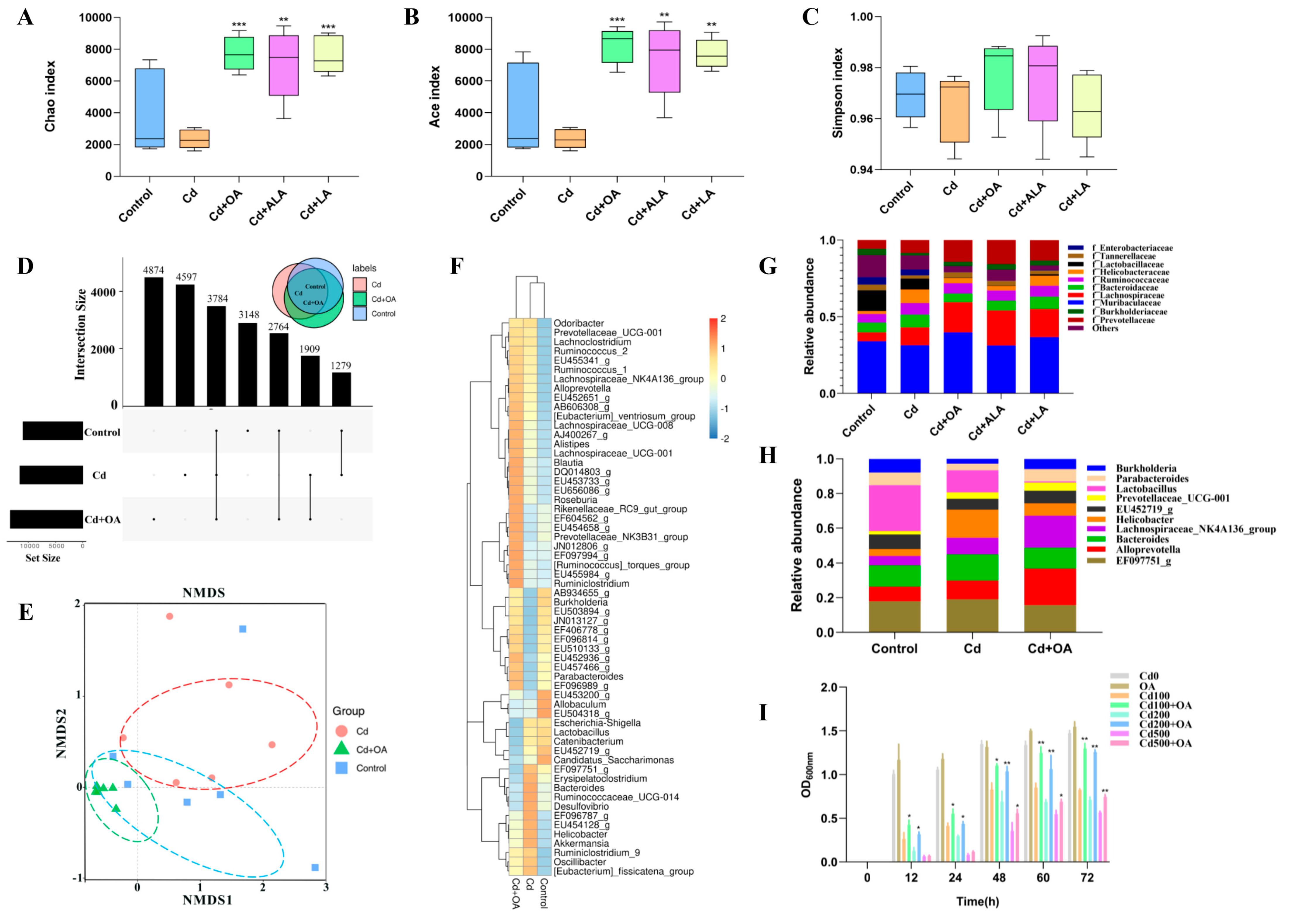
The effects of the fatty acids on the gut microbiota were analyzed using high-throughput 16S rRNA sequencing. Both microorganism community richness indices Chao and Ace of the gut microbiome significantly decreased after exposure to Cd (Figure 2A−C). In contrast, these two indices were restored following fatty acid supplementation (p < 0.05). However, the Simpson index indicated no significant differences between these five groups.
The numbers of shared OTUs of gut microbes at the 97% identity level in the UpSet diagram were compared between the groups. From the results shown in Figure 2D, the number of shared OTUs between the OA−treated and control groups was higher than that between the Cd−treated and control groups. In addition, the non−metric multidimensional scaling analysis (NMDS) in Beta diversity also showed that the overlapping regions between the OA−treated and the control groups were more extensive than that between the Cd−treated and control groups (Figure 2E). The results of both the UpSet diagrams and NMDS maps revealed that the structure of the gut microbiota in the OA−treated group was closer to the control group, accounting for the effect of OA on Cd toxicity, which the OA effectively restored.
To identify the critical gut microbes that contributed to the OA−mediated Cd excretion, the relative abundances of gut microbes at the family level in each group were compared. As shown in Figure 2F,G, compared to the control group, there were lower abundances of Muribaculaceae, Lactobacillaceae, Tannerellaceae, and Burkholderiaceae in the Cd group. However, the gut microbiota variations had been ameliorated due to the OA treatment, such as Muribaculaceae, Bacteroidaceae, Helicobacteraceae, Burkholderiaceae, and Tannerellaceae, among which Burkholderiaceae increased the most (79.77%). These results demonstrated that the OA significantly affected the abundance of gut microbiota inhibited by Cd, particularly Burkholderiaceae. Furthermore, the gut microbiota composition of the top 10 members at the genus level showed that Burkholderia from the Burkholderiaceae family increased most dramatically following OA supplementation (Figure 2H). B. cepacia has been described as the type species of the Burkholderia genus [30], and B. cepacia was reported to be as efficient for the bioaccumulation of Cd [31,32].
To confirm the effect of OA on B. cepacia in vitro, a standard strain B. cepacia (ATCC 25416) was employed in a Cd−sensitivity test (Figure 2I). Compared to the Cd group, the inhibited growth of B. cepacia cells was significantly restored by OA. Therefore, it can be concluded that OA could confer strong Cd resistance to B. cepacia, which may enable the restoration of the abundance of Burkholderia in Cd−exposed mice.

2.3. B. cepacia Facilitates Cd Excretion and Reduces Cd Accumulation in Cd-Exposed Mice

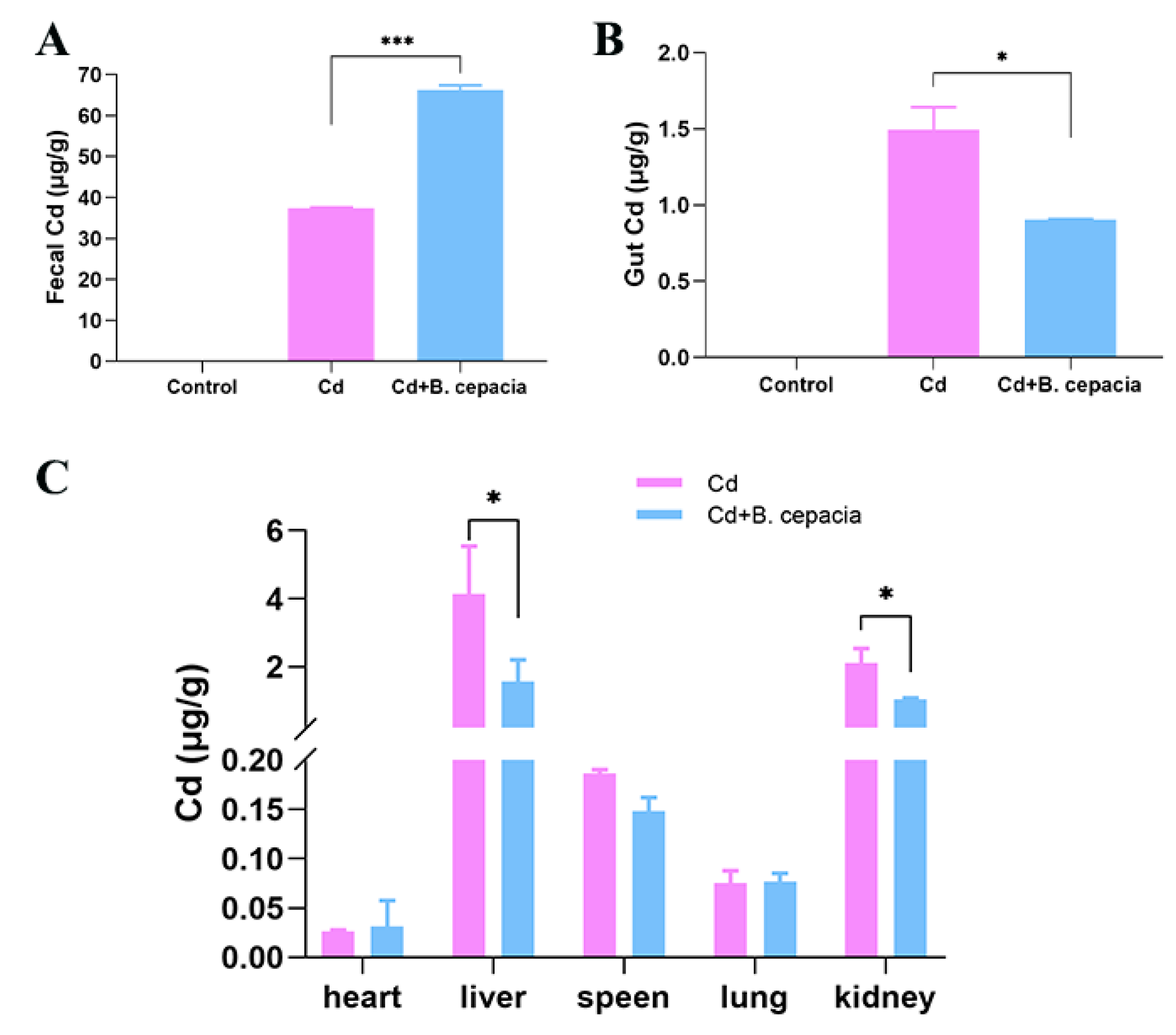
To assess the contribution of B. cepacia in the Cd excretion, B. cepacia was orally administrated to Cd-exposed mice, and the Cd contents in the feces, gut, and other organs were determined using ICP-MS. As shown in Figure 3, compared with the Cd group, the oral administration of B. cepacia resulted in the Cd levels significantly increasing in the feces but decreasing in the gut, liver, and kidneys. However, there was no significant change in the heart or lung tissues. These results demonstrated that B. cepacia plays an active role in reducing the residual tissue Cd contents while facilitating Cd excretion.

2.4. The Cd Content in B. cepacia and on Its Surface

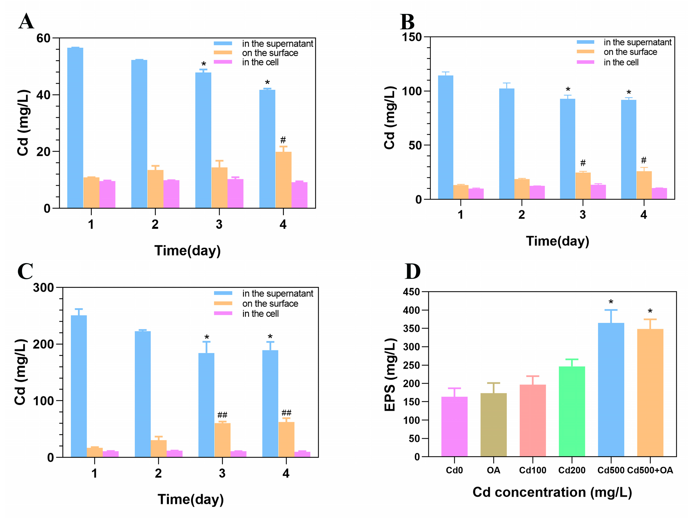
To clarify the role of B. cepacia in facilitating Cd excretion, the amount of Cd in the cell and on its surface was assessed. As shown in Figure 4, the Cd content in the supernatant gradually decreased as the cultivating time was extended. In contrast, the Cd content on the surface of B. cepacia showed an expected increase. Moreover, after being exposed to 500 mg/L of Cd for four days, less than 4% of Cd was absorbed intracellularly, and about 24% was adsorbed on the surface. When the concentration of Cd increased, the amount of total extracellular polymeric substances (EPSs) gradually increased. Compared to the Cd-free group, the EPSs in Cd 500 showed a significant increase (p < 0.05), which may be attributed to the increase in Cd content on the surface of B. cepacia. However, the OA did not further increase the EPS production under Cd exposure. These results suggested that extracellular adsorption and intracellular uptake occur for B. cepacia exposed to Cd, but extracellular adsorption plays a dominant role.

2.5. Analysis of the Cd Distribution in B. cepacia Using Electron Microscopy

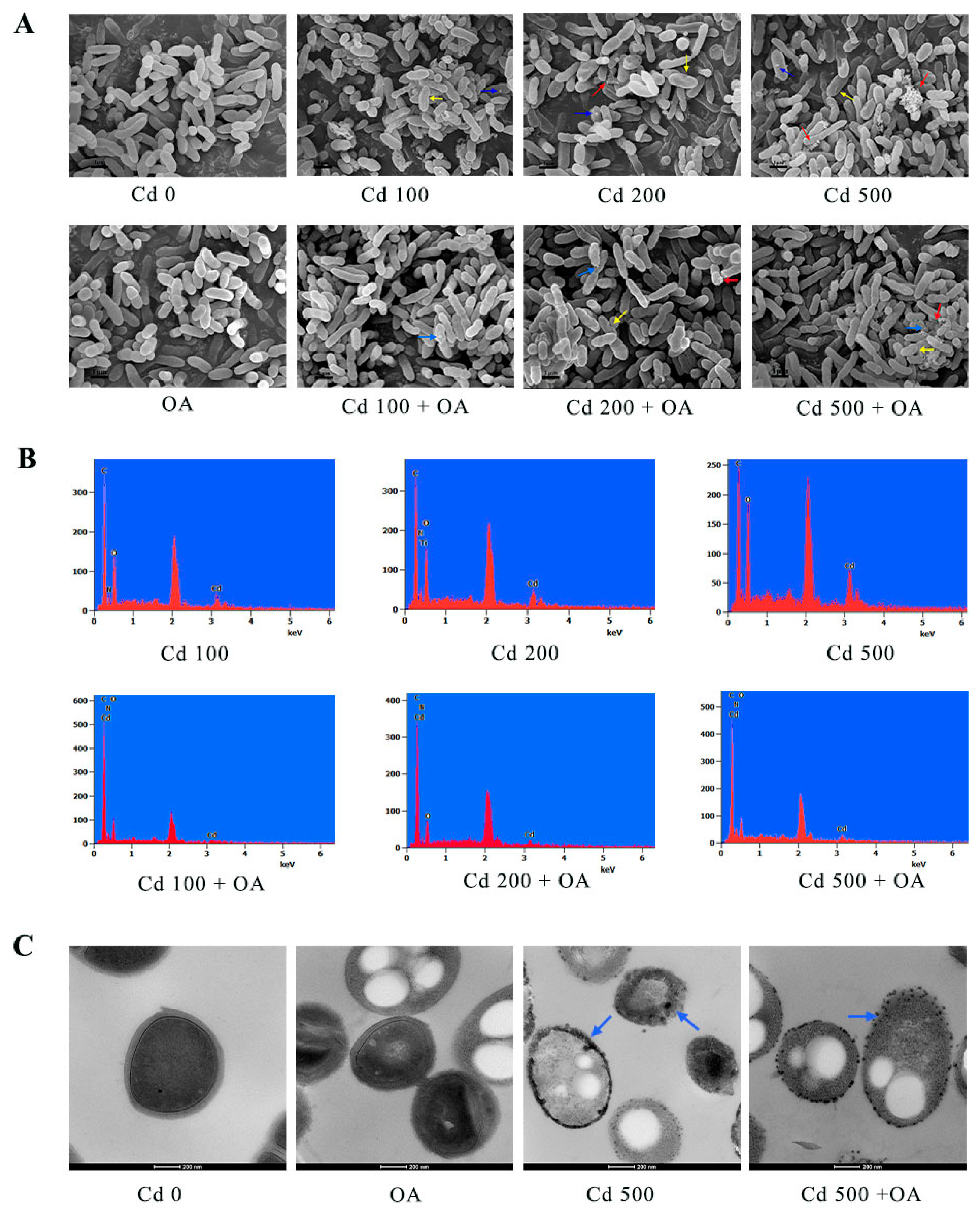
To monitor the adsorption of Cd by EPSs in B. cepacia, scanning electron microscopy (SEM) and transmission electron microscopy (TEM) were employed to characterize the cell morphology and Cd distribution. The plump and rod-shaped surface morphology for the B. cepacia cells was observed in the Cd 0 and OA groups, respectively (Figure 5A). After Cd exposure, significant depressions (as indicated by the yellow arrows) in the surface entrapped many cells, and these depressions became more severe with increasing Cd concentrations. In addition, EPSs and insoluble Cd particles (as indicated by the red and blue arrows, respectively) appeared on the cell surface, particularly in the Cd 500 group. In contrast, these surface depressions were relieved after the co-treatment of Cd and OA, exhibiting a relatively smooth surface compared to the Cd group. Meanwhile, the insoluble Cd particles and EPS-like flocculent materials were also observed in the Cd+OA groups. Cd was detected on the surface of B. cepacia exposed to Cd using an energy-dispersive spectroscopy (EDS) analysis (Figure 5B), which was confirmed by the TEM observation (Figure 5C).

2.6. Fourier Transform Infrared Spectroscopy (FTIR) Analysis of EPS Secreted by B. cepacia under Cd Stress

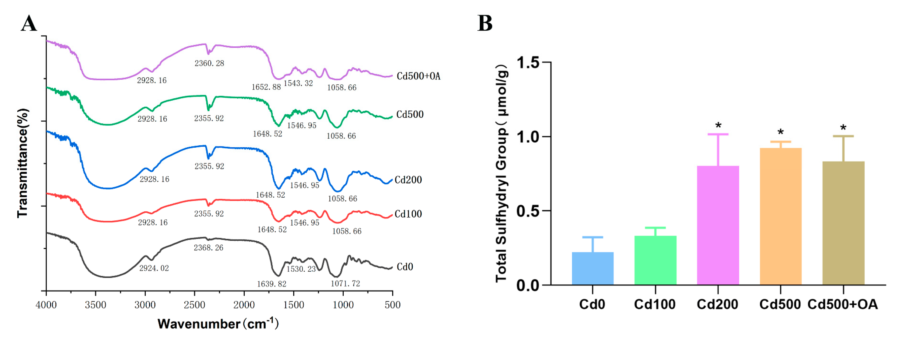
FTIR spectroscopy was carried out to further reveal the Cd−adsorption mechanism of EPSs and to characterize functional group changes on the EPSs before and after Cd exposure. As shown in Figure 6A, the peak at 1058 cm−1 (P=O) increased with Cd and OA in co-treatment. The peaks at 1648 cm−1 (C=O and C=N) and 1530 cm−1 (N−H) were shifted after exposure to Cd [33,34,35]. Notably, the characteristic peak at 2355 cm−1 (−SH) increased in the presence of Cd and OA, suggesting an interaction between Cd and the P=O or −SH groups of EPSs.
The sulfhydryl group of EPSs usually acts as the binding site for metals [36]. The total sulfhydryl group content in the EPSs confirmed the −SH group changes due to Cd. As shown in Figure 6B, the total sulfhydryl group of the EPSs in B. cepacia significantly increased after 200 or 500 mg/L of Cd exposure. However, the OA did not further increase the total sulfhydryl group in the EPSs. The result suggests that Cd rather than OA increases the sulfhydryl content in EPSs, which may contribute to the enhanced Cd adsorption capacity of B. cepacia.

3. Discussion

Cd accumulation in the body is an urgent problem to be solved. OA was reported to possess good anti-Cd activity [13]. The gut microbiota plays the key role in Cd absorption and accumulation. In this study, we found that fatty acids, particularly OA, increased the gut levels of Burkholderia, and facilitated Cd excretion.

3.1. OA Reduced the Cd Content by Facilitating Cd Excretion

As an essential food supplement, fatty acids usually act as an important means of defense against Cd stress [37,38]. A high-fat diet was found to decrease hepatic iron storage by affecting iron absorption [39]. Anethum graveolens seed oil was found to reduce Cd deposition in the liver of Cd-exposed mice [40]. In the present study, OA showed a marked Cd detoxification, increasing fecal Cd content while significantly reducing the Cd levels in the liver and kidneys. This result is in line with an earlier report, in which a high-fat diet effectively reduced heavy metal accumulation in the liver and kidneys by increasing the fecal excretion of heavy metals [41]. A previous study also showed that OA protected against Cd-induced tissue injuries in mice [42]. This evidence clearly indicates that OA is effective in Cd detoxification, as well as in reducing Cd accumulation and facilitating Cd excretion.

3.2. OA Facilitated Cd Excretion by Increasing the Abundance of Gut Burkholderia

The gut microbiota, a vital micro-ecosystem in the human body, plays an essential role in the high-fat-diet-mediated detoxification of heavy metals, and positively correlates with the excretion of heavy metals in mice [41]. From earlier findings, Cd exposure decreased the Alpha indices of the gut microbiome, reducing the microbial community’s richness and diversity [43,44]. In this study, orally administered OA significantly restored the diversity and abundance of gut microbiota, particularly the Burkholderia genus. As a type species for the genus Burkholderia, B. cepacia was well-protected by OA under Cd stress. Likewise, the OA-related synthetic gene OLE1 confers Cd resistance and alleviates Cd-induced damage [45,46]. Actually, B. cepacia, a Gram-negative and aerobic bacterial strain, has been reported to improve plant growth under Cd stress, showing a high Cd accumulation capacity in Cd-contaminating environments [30,47,48]. A similar result was observed in a previous report, in which Lactobacillus plantarum CCFM8610 effectively decreased the intestinal Cd absorption and Cd accumulation in tissues [49]. Thus, the evidence validates that OA may confer Cd resistance and increase the abundance of Burkholderia in the gut, contributing to facilitated Cd excretion.

3.3. The Cd Absorptive Ability of B. cepacia and Its EPSs

Bacteria are usually considered to have an extensive capacity to absorb metal ions from solutions [50]. Bacteria are used for the bioremediation of Cd-polluted water via different surface interaction mechanisms, including complexation and inorganic microprecipitation [51]. Most Cd is adsorbed onto the cell surface or the biofilm [52]. As the key elements of biofilm, EPSs play an important role in the biosorption of heavy metals [53,54]. In this study, oral administration of B. cepacia significantly reduced the Cd accumulation in Cd-exposed mice. More importantly, high Cd levels were detected on the surface of B. cepacia, which was confirmed via SEM, TEM, and EDS analyses. This result is in line with an earlier report, in which extracellular bioaccumulation was identified to be primarily responsible for Cd biosorption [31].
The early evidence also indicates that the increasing extracellular accumulation of Cd might be due to the production of EPSs [55]. EPSs exhibit strong metal binding and complexation potential, which effectively influences the mobility and bioavailability of heavy metal in the environment [54]. EPSs are secreted to participate in this process when exposed to Cd [31]. In the current study, the EPS production in B. cepacia gradually increased with the exposure concentration of Cd in a dose-dependent manner. Higher levels of EPSs were produced for the high-exposure concentrations of Cd, and a higher capacity for Cd adsorption was displayed. A similar finding was observed previously, in which the EPS production of B. cenocepacia YG-3 increased with the addition of Cd (between 100 and 500 mg/L) [56]. Consequently, the data indicated that B. cepacia possesses remarkable Cd absorption ability, removing Cd from the cellular environment by producing EPSs in response to Cd.

3.4. The Cd Absorption Mechanism of the EPS of B. cepacia and Its Sulfhydryl Group

As is well known, bacteria possess the capacity to absorb metal ions [50]. Bacteria were reported in the sorption of Cd via various surface interaction mechanisms [57]. The surface binding of metals is a kind of metal complexation reaction with functional groups of biomolecules [58]. Klebsiella aerogenes was reported to absorb Cd through biosynthesizing insoluble cadmium sulfide particles on the cell surface [59]. In this study, an abundance of insoluble Cd particles was observed on the surface of B. cepacia after being exposed to Cd. Some researchers also found that the extracellular sorption of Cd just was mainly attributed to functional groups on the cell wall [60]. A previous study reported that the extracellular sorption of Cd in Pseudomonas putida might be due to the production of EPSs [55].
The enhanced Cd-adsorption capacity of EPSs may be attributed to the increased adsorption sites or functional groups. Functional groups on the outer membrane with high affinity to metals were found to play a major role in the binding to Cd [61]. Functional groups of EPSs, such as phosphate and sulfhydryl groups, were reported to have a strong affinity for Cd [62,63]. In this study, the FTIR analysis of the EPSs showed that the peak at 2355 cm−1 (-SH) increased in the presence of Cd. A similar finding was observed in a previous report, in which the interaction between Cd and the EPSs of Pseudomonas aeruginosa produced a significant increase in the FTIR peak at 2365.09 cm−1 (-SH) [64]. -SH was deemed to be a soft base ligand capable of binding with Pb strongly by forming covalent bonds [65]. More importantly, Cd has a high affinity for sulfhydryl groups [66]. In a previous study, a significant increase was observed in the content of sulfhydryl groups in Tetraselmis suecica cultures with the increase in the exposure concentration of Cd [67]. Notably, the increased intracellular Cd in T. suecica correlated with the increasing sulfhydryl groups [67]. The detection of -SH stretching upon the interaction with Cd confirmed the involvement of the sulfhydryl functional group in Cd binding. The current study confirms that the total sulfhydryl group (-SH) content of the EPSs of B. cepacia significantly increases after Cd exposure.

4. Materials and Methods

4.1. Materials and Reagents

Cadmium chloride (CdCl2, 98%) was purchased from Chengdu Huaxia Chemical Reagent (Chengdu, China). Burkholderia Cepacia (ATCC 25416) was purchased from Beijing Beina Chuanglian Biotechnology Institute.

4.2. Animals and Experimental Design

A total of 48 mice (8 weeks) were purchased from SPF (Beijing, China) Biotechnology Co., Ltd. (SCXK2019-0010). All mice were fed with the standard commercial rat food and kept in cages under the condition of 12 h light/dark cycles, and they were given free access to food and water.
To investigate the attenuation of various fatty acids against Cd, 30 SPF mice were randomly divided into 5 groups (n = 6). In the control group, the experimental mice received normal drinking water without Cd. For the Cd-exposed group, mice were given drinking water containing 100 μmol/L of CdCl2. The mice in the fatty acid-treated groups were given drinking water containing 100 μmol/L of CdCl2 and followed by intragastric administration containing 40 μmol/g/d of each fatty acids. Treatment was continued for 7 days.
To investigate the attenuation of B. cepacia against Cd toxicity, 18 SPF mice were randomly divided into 3 groups (n = 6). In the control group, mice received normal drinking water without Cd. For the Cd group, mice were exposed to the drinking water containing 100 μmol/L of CdCl2. In the Cd + B. cepacia group, mice were given drinking water with 100 μmol/L of CdCl2 and an intragastric administration containing 1×10⁹ CFU of B. cepacia. Treatment was also continued for 7 days. The above doses of Cd and B. cepacia were in accordance with the study of Qixiao et al. [68]
Animals were given a standard laboratory diet and water. The experiment was approved by the Animal Ethics Committee of Guangdong Ocean University (No.: GDOU-LAE-2021-020).

4.3. Determination of Cd Contents in Feces and Tissues

The feces samples from each group were collected every day. After sacrifice, small sections of tissues (gut, heart, liver, spleen, lung, and kidneys) of mice were collected and stored at −80 °C. The feces and tissue samples were crushed, ground evenly, and digested with HNO3 using a graphite digestion system (GDANA DS-360, Guangzhou, China) [11]. Cd content was measured by an inductive coupled plasma mass spectrometer (ICP-MS, ThermoFisher, ICAPQ, Germany) according to the manufacturer’s instructions. All analyses were run in triplicate.

4.4. 16S rRNA Gene Sequencing of the Gut Microbiota

16S rRNA gene sequencing of the gut microbiota was performed as previously described [69]. Total DNA of fresh fecal samples in each group were extracted with the PowerMax (stool/soil) DNA isolation kit (MoBioLaboratories, Carlsbad, CA, USA) according to the manufacturer’s instructions. The NanoDrop ND-1000 spectrophotometer (Thermo Fisher Scientific, Waltham, MA, USA) and agarose gel electrophoresis were used for quantity and quality measurement, respectively. The 16S rRNA gene sequencing was amplified using the forward primer 515F (5′-GTGCCAGCMGCCGCGGTAA-3′) and the reverse primer 806R (5′-GGACTACHVGGGT WTCTAAT-3′) targeting the V4 region. The PCR products were purified with AMPure XP Beads (Beckman Coulter, Indianapolis, IN, USA), and quantified with the PicoGreen dsDNA Assay Kit (Invitrogen, Carlsbad, CA, USA) before paired-end 2 × 150 bp sequencing using the Illlumina Novaseq 6000 platform at GUHE Info technology Co., Ltd. (Hangzhou, China)
Raw tags were processed using Vsearch v2.4.4 after removing the Barcode and primer sequence for obtaining effective tags through quality filtration. Sequencing data were processed using Quantitative Insights Into Microbial Ecology (QIIME, v3.2.0), clustering high-quality reads into operational taxonomic units (OTUs) at the 97% identity level. QIIME and R packages (v3.2.0) were used for the sequence data analyses.

4.5. Cd Sensitivity Assay of B. cepacia by the Growth of B. cepacia Cells

The B. cepacia was cultured in Luria-Bertani (LB) broth for 24 h at 37 °C with shaking at 130 r/min. B. cepacia was cultivated in LB containing 0, 100, 200, and 500 mg/L of Cd levels or 2 mmol/L of OA, and the optical density (OD) was measured at 600 nm to evaluate Cd sensitivity.

4.6. Determination of Cd Contents Accumulated Intracellularly, Extracellularly, and in the Supernatant of B. cepacia

The Cd contents accumulated intracellularly, extracellularly, and in the supernatant of B. cepacia were determined as previously described [31]. The B. cepacia cells were cultivated in LB broth containing 0, 100, 200, and 500 mg/L of CdCl2 for 1 to 4 days. The supernatant was obtained by centrifugation at 12,000× g at 4 °C for 15 min and then filtrated through a 0.22 μm cellulose acetate membrane. The remaining bacterial pellets were washed with 0.1 mol/L of EDTA-2 Na for 3 h and then centrifuged to collect Cd bound to the cell surface. The residual pellets were digested with HNO3 to determine the Cd absorbed in the cells. Cd contents in the supernatant, on the cell surface, and within the cells were determined by ICP-MS. All experiments were run in triplicate.

4.7. EPS Extraction

EPS extraction was performed as previously described [48]. The B. cepacia was cultured aerobically in LB broth for 72 h. First, the bacteria supernatant after exposure with the different CdCl2 was separately collected by centrifugation at 5000× g at 4 °C for 10 min. Secondly, the same volume of cold ethanol was added into the supernatant and kept at −20 °C overnight for precipitation of EPSs. Then, the EPS pellet was collected by centrifugation at 9000× g at 4 °C for 15 min and further dissolved in distilled water and then was dialyzed at 40 °C for 48 h. Finally, the pure EPS was obtained by lyophilization.

4.8. Electron Microscopy Analysis

The cell precipitate was collected by centrifugation at 4500× g at 4 °C for 10 min, and slightly washed with PBS aiming to remove medium. It was then fixed in 2.5% glutaraldehyde solution at 4 °C overnight. Then, cells were dehydrated with ethanol gradient and finally lyophilized for scanning electron microscopy (SEM, JSM-7610F) observation equipped with energy-dispersive spectroscopy (EDS, NORAN System 7). For transmission electron microscopy (TEM, TECNAI G2 20 TWIN) observation, the bacteria were prepared the same as for SEM from collection to dehydration steps, then embedded and sectioned into ultra-thin sections, and finally stained for microscopical observation [56].

4.9. Fourier Transform Infrared Spectrometer (FTIR) Assay

The EPS of B. cepacia was freeze-dried and mixed with KBr at a 1:100 mass ratio. FTIR spectroscopy (TENSOR 27, BRUKER OPTICS) was employed to examine the changes in structural vibration information of EPSs and the interaction with Cd under different Cd concentrations at 4 cm−1 spectral resolution within 4000–500 cm−1 [70].

4.10. Statistical Analyses

The data are expressed as the mean ± standard deviation (SD). All statistical analyses were performed by using the Graphpad Prism version 8.0. The comparison between groups were performed using Student’s t test. A p value of <0.05 was considered to indicate statistical significance.

5. Conclusions

The data from this study confirm that OA is the most effective of the three free fatty acids in reducing Cd accumulation through facilitating Cd excretion in mice. OA facilitates Cd excretion by increasing the abundance of the Burkholderia genus in the gut microbiota. In addition, as the type species for the Burkholderia genus, B. cepacia conferred Cd tolerance by OA. More importantly, B. cepacia facilitated the Cd excretion through secreting EPSs with a high sulfhydryl group (-SH) content following Cd exposure. Finally, OA is a potentially suitable supplemental therapy to reduce Cd accumulation in the body.

Author Contributions

Conceptualization, Z.F. and J.W.; methodology, Z.F. and Y.C.; software, Y.C.; validation, Z.F., J.W. and L.S.; formal analysis, Y.C.; investigation, Y.C. and Y.L.; resources, Z.F. and Q.D.; data curation, Y.C.; writing—original draft preparation, Z.F., Y.C. and R.G.; writing—review and editing, Z.F.; visualization, Y.C.; supervision, Z.F.; project administration, Z.F.; funding acquisition, Z.F. All authors have read and agreed to the published version of the manuscript.

Funding

This work was supported by the National Natural Science Foundation of China (No. 32172215 and 31701706), the Natural Science Foundation of Guangdong Province (No. 2019A1515010809 and 2021A1515012443), and the Characteristic Innovation Project of Guangdong Province (No. 2018KTSCX089).

Institutional Review Board Statement

The animal experiments were approved by the Laboratory Animal Ethics Committee of Guangdong Ocean University (approval number: GDOU-LAE-2020-009).

Informed Consent Statement

Not applicable.

Data Availability Statement

The data presented in this study are available on request from the corresponding author.

Conflicts of Interest

The authors declare no conflict of interest.

References

  1. Bauenova, M.O.; Sadvakasova, A.K.; Mustapayeva, Z.O.; Kokociński, M.; Zayadan, B.K.; Wojciechowicz, M.K.; Balouch, H.; Akmukhanova, N.R.; Alwasel, S.; Allakhverdiev, S.I. Potential of microalgae Parachlorella kessleri Bh-2 as bioremediation agent of heavy metals cadmium and chromium. Algal Res. 2021, 59, 102463. [Google Scholar] [CrossRef]
  2. RoyChowdhury, A.; Datta, R.; Sarkar, D. Heavy Metal Pollution and Remediation. In Green Chemistry; NUS Biological Sciences: Singapore, 2018; pp. 359–373. [Google Scholar]
  3. Han, C.; Zhu, Y.; Yang, Z.; Fu, S.; Zhang, W.; Liu, C. Protective effect of Polygonatum sibiricum against cadmium-induced testicular injury in mice through inhibiting oxidative stress and mitochondria-mediated apoptosis. J. Ethnopharmacol. 2020, 261, 113060. [Google Scholar] [CrossRef] [PubMed]
  4. Yazihan, N.; Kocak, M.K.; Akcil, E.; Erdem, O.; Sayal, A. Role of midkine in cadmium-induced liver, heart and kidney damage. Hum. Exp. Toxicol. 2011, 30, 391–397. [Google Scholar] [CrossRef]
  5. Hao, R.; Song, X.; Sun-Waterhouse, D.; Tan, X.; Li, F.; Li, D. MiR-34a/Sirt1/p53 signaling pathway contributes to cadmium-induced nephrotoxicity: A preclinical study in mice. Environ. Pollut. 2021, 282, 117029. [Google Scholar] [CrossRef] [PubMed]
  6. Johri, N.; Jacquillet, G.; Unwin, R. Heavy metal poisoning: The effects of cadmium on the kidney. BioMetals 2010, 23, 783–792. [Google Scholar] [CrossRef] [PubMed]
  7. Park, J.D.; Cherrington, N.J.; Klaassen, C.D. Intestinal Absorption of Cadmium Is Associated with Divalent Metal Transporter 1 in Rats. Toxicol. Sci. 2002, 68, 288–294. [Google Scholar] [CrossRef] [PubMed]
  8. McMurray, C.T.; Tainer, J.A. Cancer, cadmium and genome integrity. Nat. Genet. 2003, 34, 239–241. [Google Scholar] [CrossRef] [PubMed]
  9. Hengstler, J.G.; Bolm-Audorff, U.; Faldum, A.; Janssen, K.; Reifenrath, M.; Götte, W.; Jung, D.; Mayer-Popken, O.; Fuchs, J.; Gebhard, S.; et al. Occupational exposure to heavy metals: DNA damage induction and DNA repair inhibition prove co-exposures to cadmium, cobalt and lead as more dangerous than hitherto expected. Carcinogenesis 2003, 24, 63–73. [Google Scholar] [CrossRef] [Green Version]
  10. Purkayastha, D.; Mishra, U.; Biswas, S. A comprehensive review on Cd(II) removal from aqueous solution. J. Water Process Eng. 2014, 2, 105–128. [Google Scholar] [CrossRef]
  11. Fan, R.; Hu, P.C.; Wang, Y.; Lin, H.Y.; Su, K.; Feng, X.S.; Wei, L.; Yang, F. Betulinic acid protects mice from cadmium chloride-induced toxicity by inhibiting cadmium-induced apoptosis in kidney and liver. Toxicol. Lett. 2018, 299, 56–66. [Google Scholar] [CrossRef] [PubMed]
  12. Fang, J.; Yin, H.; Yang, Z.; Tan, M.; Wang, F.; Chen, K.; Zuo, Z.; Shu, G.; Cui, H.; Ouyang, P.; et al. Vitamin E protects against cadmium-induced sub-chronic liver injury associated with the inhibition of oxidative stress and activation of Nrf2 pathway. Ecotoxicol. Environ. Saf. 2021, 208, 111610. [Google Scholar] [CrossRef] [PubMed]
  13. Wang, J.; Zhang, Y.; Fang, Z.; Sun, L.; Wang, Y.; Liu, Y.; Xu, D.; Nie, F.; Gooneratne, R. Oleic Acid Alleviates Cadmium-Induced Oxidative Damage in Rat by Its Radicals Scavenging Activity. Biol. Trace Elem. Res. 2019, 190, 95–100. [Google Scholar] [CrossRef] [PubMed]
  14. Grawe, K.P.; Pickova, J.; Dutta, P.C.; Oskarsson, A. Fatty acid alterations in liver and milk of cadmium exposed rats and in brain of their suckling offspring. Toxicol. Lett. 2004, 148, 73–82. [Google Scholar] [CrossRef] [PubMed]
  15. Nouairi, I.; Ghnaya, T.; Ben Youssef, N.; Zarrouk, M.; Habib Ghorbel, M. Changes in content and fatty acid profiles of total lipids of two halophytes: Sesuvium portulacastrum and Mesembryanthemum crystallinum under cadmium stress. J. Plant Physiol. 2006, 163, 1198–1202. [Google Scholar] [CrossRef] [PubMed]
  16. Inaba, T.; Kobayashi, E.; Suwazono, Y.; Uetani, M.; Oishi, M.; Nakagawa, H.; Nogawa, K. Estimation of cumulative cadmium intake causing Itai–itai disease. Toxicol. Lett. 2005, 159, 192–201. [Google Scholar] [CrossRef]
  17. Liu, Y.; Li, Y.; Liu, K.; Shen, J. Exposing to cadmium stress cause profound toxic effect on microbiota of the mice intestinal tract. PLoS ONE 2014, 9, e85323. [Google Scholar] [CrossRef]
  18. Duan, H.; Yu, L.; Tian, F.; Zhai, Q.; Fan, L.; Chen, W. Gut microbiota: A target for heavy metal toxicity and a probiotic protective strategy. Sci. Total Environ. 2020, 742, 140429. [Google Scholar] [CrossRef] [PubMed]
  19. Fazeli, M.; Hassanzadeh, P.; Alaei, S. Cadmium chloride exhibits a profound toxic effect on bacterial microflora of the mice gastrointestinal tract. Hum. Exp. Toxicol. 2011, 30, 152–159. [Google Scholar] [CrossRef] [PubMed]
  20. Zhang, S.; Jin, Y.; Zeng, Z.; Liu, Z.; Fu, Z. Subchronic Exposure of Mice to Cadmium Perturbs Their Hepatic Energy Metabolism and Gut Microbiome. Chem. Res. Toxicol. 2015, 28, 2000–2009. [Google Scholar] [CrossRef]
  21. Tinkov, A.A.; Gritsenko, V.A.; Skalnaya, M.G.; Cherkasov, S.V.; Aaseth, J.; Skalny, A.V. Gut as a target for cadmium toxicity. Environ. Pollut. 2018, 235, 429–434. [Google Scholar] [CrossRef] [PubMed]
  22. Yang, J.; Chen, W.; Sun, Y.; Liu, J.; Zhang, W. Effects of cadmium on organ function, gut microbiota and its metabolomics profile in adolescent rats. Ecotoxicol. Environ. Saf. 2021, 222, 112501. [Google Scholar] [CrossRef] [PubMed]
  23. Jin, Y.; Wu, S.; Zeng, Z.; Fu, Z. Effects of environmental pollutants on gut microbiota. Environ. Pollut. 2017, 222, 1–9. [Google Scholar] [CrossRef] [PubMed]
  24. Breton, J.; Daniel, C.; Dewulf, J.; Pothion, S.; Froux, N.; Sauty, M.; Thomas, P.; Pot, B.; Foligne, B. Gut microbiota limits heavy metals burden caused by chronic oral exposure. Toxicol. Lett. 2013, 222, 132–138. [Google Scholar] [CrossRef] [PubMed]
  25. Feng, S.; Liu, Y.; Huang, Y.; Zhao, J.; Zhang, H.; Zhai, Q.; Chen, W. Influence of oral administration of Akkermansia muciniphila on the tissue distribution and gut microbiota composition of acute and chronic cadmium exposure mice. FEMS Microbiol. Lett. 2019, 366, fnz160. [Google Scholar] [CrossRef] [PubMed]
  26. Ma, X.; Hou, M.; Liu, C.; Li, J.; Ba, Q.; Wang, H. Cadmium accelerates bacterial oleic acid production to promote fat accumulation in Caenorhabditis elegans. J. Hazard. Mater. 2022, 421, 126723. [Google Scholar] [CrossRef]
  27. Agans, R.; Gordon, A.; Kramer, D.L.; Perez-Burillo, S.; Rufián-Henares, J.A.; Paliy, O. Dietary Fatty Acids Sustain the Growth of the Human Gut Microbiota. Appl. Environ. Microbiol. 2018, 84, e01525-18. [Google Scholar] [CrossRef] [Green Version]
  28. Gao, X.; Chang, S.; Liu, S.; Peng, L.; Xie, J.; Dong, W.; Tian, Y.; Sheng, J. Correlations between α-Linolenic Acid-Improved Multitissue Homeostasis and Gut Microbiota in Mice Fed a High-Fat Diet. mSystems 2020, 5, e00391-20. [Google Scholar] [CrossRef]
  29. Shrestha, N.; Sleep, S.L.; Cuffe, J.S.M.; Holland, O.J.; McAinch, A.J.; Nitert, M.D.; Hryciw, D.H. Pregnancy and diet-related changes in the maternal gut microbiota following exposure to an elevated linoleic acid diet. Am. J. Physiol-Endoc. Metab. 2020, 318, E276–E285. [Google Scholar] [CrossRef]
  30. Serrato, R.V.; Sassaki, G.L.; Gorin, P.A.; Cruz, L.M.; Pedrosa, F.O.; Choudhury, B.; Carlson, R.W.; Iacomini, M. Structural characterization of an acidic exoheteropolysaccharide produced by the nitrogen-fixing bacterium Burkholderia tropica. Carbohydr. Polym. 2008, 73, 564–572. [Google Scholar] [CrossRef]
  31. Zhang, J.; Li, Q.; Zeng, Y.; Zhang, J.; Lu, G.; Dang, Z.; Guo, C. Bioaccumulation and distribution of cadmium by Burkholderia cepacia GYP1 under oligotrophic condition and mechanism analysis at proteome level. Ecotoxicol. Environ. Saf. 2019, 176, 162–169. [Google Scholar] [CrossRef]
  32. Gan, L.; Zhou, F.; Owens, G.; Chen, Z. Burkholderia cepacia immobilized on eucalyptus leaves used to simultaneously remove malachite green (MG) and Cr(VI). Colloids Surf. B. Biointerfaces 2018, 172, 526–531. [Google Scholar] [CrossRef]
  33. Abdel-Rahman, L.H.; Abu-Dief, A.M.; El-Khatib, R.M.; Abdel-Fatah, S.M. Some new nano-sized Fe(II), Cd(II) and Zn(II) Schiff base complexes as precursor for metal oxides: Sonochemical synthesis, characterization, DNA interaction, in vitro antimicrobial and anticancer activities. Bioorg. Chem. 2016, 69, 140–152. [Google Scholar] [CrossRef] [PubMed]
  34. Abdel-Rahman, L.H.; Ismail, N.M.; Ismael, M.; Abu-Dief, A.M.; Ahmed, E.A.-H. Synthesis, characterization, DFT calculations and biological studies of Mn(II), Fe(II), Co(II) and Cd(II) complexes based on a tetradentate ONNO donor Schiff base ligand. J. Mol. Struct. 2017, 1134, 851–862. [Google Scholar] [CrossRef]
  35. Khalaf, M.M.; Abd El-Lateef, H.M.; Gouda, M.; Sayed, F.N.; Mohamed, G.G.; Abu-Dief, A.M. Design, Structural Inspection and Bio-Medicinal Applications of Some Novel Imine Metal Complexes Based on Acetylferrocene. Materials 2022, 15, 4842. [Google Scholar] [CrossRef] [PubMed]
  36. Fein, J.B.; Yu, Q.; Nam, J.; Yee, N. Bacterial cell envelope and extracellular sulfhydryl binding sites: Their roles in metal binding and bioavailability. Chem. Geol. 2019, 521, 28–38. [Google Scholar] [CrossRef]
  37. Nouairi, I.; Ammar, W.B.; Youssef, N.B.; Daoud, D.B.M.; Ghorbal, M.H.; Zarrouk, M. Comparative study of cadmium effects on membrane lipid composition of Brassica juncea and Brassica napus leaves. Plant Sci. 2006, 170, 511–519. [Google Scholar] [CrossRef]
  38. Omotoso, O.; Owolabi, J.; Akpan, H.; Dare, B.; Ashamu, E.; Adelakun, S. Positive Anti-Cadmium Poisoning Anti-Oxidant Effects of Moringa Seed Oil and Cashew Nut Oil in Juvenile Wistar Rats Hippocampus. J. Adv. Med. Pharm. Sci. 2016, 5, 1–8. [Google Scholar] [CrossRef]
  39. Shin, S.; Lim, Y.; Chung, J.; Park, S.; Han, S.N. Effect of Korean pine nut oil on hepatic iron, copper, and zinc status and expression of genes and proteins related to iron absorption in diet-induced obese mice. J. Nutr. Health 2021, 54, 435–447. [Google Scholar] [CrossRef]
  40. Shafaei, N.; Barkhordar, S.M.A.; Rahmani, F.; Nabi, S.; Idliki, R.B.; Alimirzaei, M.; Karimi, E.; Oskoueian, E. Protective Effects of Anethum graveolens Seed’s Oil Nanoemulsion Against Cadmium-Induced Oxidative Stress in Mice. Biol. Trace Elem. Res. 2020, 198, 583–591. [Google Scholar] [CrossRef] [PubMed]
  41. Liu, T.; Liang, X.; Lei, C.; Huang, Q.; Song, W.; Fang, R.; Li, C.; Li, X.; Mo, H.; Sun, N.; et al. High-Fat Diet Affects Heavy Metal Accumulation and Toxicity to Mice Liver and Kidney Probably via Gut Microbiota. Front. Microbiol. 2020, 11, 1604. [Google Scholar] [CrossRef] [PubMed]
  42. Bhattacharjee, B.; Pal, P.K.; Chattopadhyay, A.; Bandyopadhyay, D. Oleic acid protects against cadmium induced cardiac and hepatic tissue injury in male Wistar rats: A mechanistic study. Life Sci. 2020, 244, 117324. [Google Scholar] [CrossRef]
  43. He, X.; Qi, Z.; Hou, H.; Qian, L.; Gao, J.; Zhang, X.X. Structural and functional alterations of gut microbiome in mice induced by chronic cadmium exposure. Chemosphere 2020, 246, 125747. [Google Scholar] [CrossRef]
  44. Ba, Q.; Li, M.; Chen, P.; Huang, C.; Duan, X.; Lu, L.; Li, J.; Chu, R.; Xie, D.; Song, H.; et al. Sex-Dependent Effects of Cadmium Exposure in Early Life on Gut Microbiota and Fat Accumulation in Mice. Environ. Health Perspect. 2017, 125, 437–446. [Google Scholar] [CrossRef] [PubMed]
  45. Fang, Z.; Chen, Z.; Wang, S.; Shi, P.; Shen, Y.; Zhang, Y.; Xiao, J.; Huang, Z. Overexpression of OLE1 enhances cytoplasmic membrane stability and confers resistance to cadmium in Saccharomyces cerevisiae. Appl. Environ. Microbiol. 2017, 83, e02319-16. [Google Scholar] [CrossRef] [PubMed] [Green Version]
  46. Huang, Z.; Yu, Y.; Fang, Z.; Deng, Y.; Shen, Y.; Shi, P. OLE1 reduces cadmium-induced oxidative damage in Saccharomyces cerevisiae. FEMS Microbiol. Lett. 2018, 365, fny193. [Google Scholar] [CrossRef] [PubMed] [Green Version]
  47. Wang, C.; Huang, Y.; Yang, X.; Xue, W.; Zhang, X.; Zhang, Y.; Pang, J.; Liu, Y.; Liu, Z. Burkholderia sp. Y4 inhibits cadmium accumulation in rice by increasing essential nutrient uptake and preferentially absorbing cadmium. Chemosphere 2020, 252, 126603. [Google Scholar] [CrossRef]
  48. You, L.-X.; Zhang, R.-R.; Dai, J.-X.; Lin, Z.-T.; Li, Y.-P.; Herzberg, M.; Zhang, J.-L.; Al-Wathnani, H.; Zhang, C.-K.; Feng, R.-W.; et al. Potential of cadmium resistant Burkholderia contaminans strain ZCC in promoting growth of soy beans in the presence of cadmium. Ecotoxicol. Environ. Saf. 2021, 211, 111914. [Google Scholar] [CrossRef]
  49. Zhai, Q.; Wang, G.; Zhao, J.; Liu, X.; Narbad, A.; Chen, Y.Q.; Zhang, H.; Tian, F.; Chen, W. Protective effects of Lactobacillus plantarum CCFM8610 against chronic cadmium toxicity in mice indicate routes of protection besides intestinal sequestration. Appl. Environ. Microbiol. 2014, 80, 4063–4071. [Google Scholar] [CrossRef] [Green Version]
  50. Beveridge, T.J. Role of cellular design in bacterial metal accumulation and mineralization. Annu. Rev. Microbiol. 1989, 43, 147–171. [Google Scholar] [CrossRef]
  51. Chakravarty, R.; Banerjee, P.C. Mechanism of cadmium binding on the cell wall of an acidophilic bacterium. Bioresour. Technol. 2012, 108, 176–183. [Google Scholar] [CrossRef] [PubMed]
  52. Xing, Y.; Luo, X.; Liu, S.; Wan, W.; Huang, Q.; Chen, W. Synergistic effect of biofilm growth and cadmium adsorption via compositional changes of extracellular matrix in montmorillonite system. Bioresour. Technol. 2020, 315, 123742. [Google Scholar] [CrossRef]
  53. Wang, Z.; Gao, M.; Wei, J.; Ma, K.; Zhang, J.; Yang, Y.; Yu, S. Extracellular polymeric substances, microbial activity and microbial community of biofilm and suspended sludge at different divalent cadmium concentrations. Bioresour. Technol. 2016, 205, 213–221. [Google Scholar] [CrossRef] [PubMed]
  54. Zhang, D.; Pan, X.; Mostofa, K.M.G.; Chen, X.; Mu, G.; Wu, F.; Liu, J.; Song, W.; Yang, J.; Liu, Y.; et al. Complexation between Hg(II) and biofilm extracellular polymeric substances: An application of fluorescence spectroscopy. J. Hazard. Mater. 2010, 175, 359–365. [Google Scholar] [CrossRef] [PubMed]
  55. Pabst, M.W.; Miller, C.D.; Dimkpa, C.O.; Anderson, A.J.; McLean, J.E. Defining the surface adsorption and internalization of copper and cadmium in a soil bacterium, Pseudomonas putida. Chemosphere 2010, 81, 904–910. [Google Scholar] [CrossRef] [PubMed]
  56. Wang, X.; Zhang, X.; Liu, X.; Huang, Z.; Niu, S.; Xu, T.; Zeng, J.; Li, H.; Wang, T.; Gao, Y.; et al. Physiological, biochemical and proteomic insight into integrated strategies of an endophytic bacterium Burkholderia cenocepacia strain YG-3 response to cadmium stress. Metallomics 2019, 11, 1252–1264. [Google Scholar] [CrossRef]
  57. Hansda, A.; Kumar, V. Anshumali A comparative review towards potential of microbial cells for heavy metal removal with emphasis on biosorption and bioaccumulation. World J. Microbiol. Biotechnol. 2016, 32, 170. [Google Scholar] [CrossRef]
  58. Fein, J.B.; Daughney, C.J.; Yee, N.; Davis, T.A. A chemical equilibrium model for metal adsorption onto bacterial surfaces. Geochim. Cosmochim. Acta 1997, 61, 3319–3328. [Google Scholar] [CrossRef]
  59. Holmes, J.D.; Smith, P.R.; Evans-Gowing, R.; Richardson, D.J.; Russell, D.A.; Sodeau, J.R. Energy-dispersive X-ray analysis of the extracellular cadmium sulfide crystallites of Klebsiella aerogenes. Arch. Microbiol. 1995, 163, 143–147. [Google Scholar] [CrossRef] [PubMed]
  60. Huang, F.; Guo, C.-L.; Lu, G.-N.; Yi, X.-Y.; Zhu, L.-D.; Dang, Z. Bioaccumulation characterization of cadmium by growing Bacillus cereus RC-1 and its mechanism. Chemosphere 2014, 109, 134–142. [Google Scholar] [CrossRef]
  61. Guibaud, G.; Comte, S.; Bordas, F.; Dupuy, S.; Baudu, M. Comparison of the complexation potential of extracellular polymeric substances (EPS), extracted from activated sludges and produced by pure bacteria strains, for cadmium, lead and nickel. Chemosphere 2005, 59, 629–638. [Google Scholar] [CrossRef] [PubMed]
  62. Maurya, A.; Kumar, R.; Yadav, P.; Singh, A.; Yadav, A.; Chowdhary, P.; Raj, A. Biofilm formation and extracellular polymeric substance (EPS) production by Bacillus haynesii and influence of hexavalent chromium. Bioresour. Technol. 2022, 352, 127109. [Google Scholar] [CrossRef] [PubMed]
  63. Raj, R.; Dalei, K.; Chakraborty, J.; Das, S. Extracellular polymeric substances of a marine bacterium mediated synthesis of CdS nanoparticles for removal of cadmium from aqueous solution. J. Colloid Interface Sci. 2016, 462, 166–175. [Google Scholar] [CrossRef] [PubMed]
  64. Chakraborty, J.; Das, S. Characterization and cadmium-resistant gene expression of biofilm-forming marine bacterium Pseudomonas aeruginosa JP-11. Environ. Sci. Pollut. Res. 2014, 21, 14188–14201. [Google Scholar] [CrossRef] [PubMed]
  65. Giles, N.M.; Watts, A.B.; Giles, G.I.; Fry, F.H.; Littlechild, J.A.; Jacob, C. Metal and Redox Modulation of Cysteine Protein Function. Chem. Biol. 2003, 10, 677–693. [Google Scholar] [CrossRef] [PubMed] [Green Version]
  66. Zalups, R.K.; Ahmad, S. Molecular handling of cadmium in transporting epithelia. Toxicol. Appl. Pharmacol. 2003, 186, 163–188. [Google Scholar] [CrossRef]
  67. Pérez-Rama, M.; Abalde Alonso, J.; Herrero López, C.; Torres Vaamonde, E. Cadmium removal by living cells of the marine microalga Tetraselmis suecica. Bioresour. Technol. 2002, 84, 265–270. [Google Scholar] [CrossRef]
  68. Zhai, Q.; Tian, F.; Zhao, J.; Zhang, H.; Narbad, A.; Chen, W. Oral Administration of Probiotics Inhibits Absorption of the Heavy Metal Cadmium by Protecting the Intestinal Barrier. Appl. Environ. Microbiol. 2016, 82, 4429–4440. [Google Scholar] [CrossRef] [PubMed] [Green Version]
  69. Wang, R.; Deng, Y.; Deng, Q.; Sun, D.; Fang, Z.; Sun, L.; Wang, Y.; Gooneratne, R. Vibrio parahaemolyticus Infection in Mice Reduces Protective Gut Microbiota, Augmenting Disease Pathways. Front. Microbiol. 2020, 11, 73. [Google Scholar] [CrossRef] [PubMed] [Green Version]
  70. D’Souza, L.; Devi, P.; Divya Shridhar, M.P.; Naik, C.G. Use of Fourier Transform Infrared (FTIR) Spectroscopy to Study Cadmium-Induced Changes in Padina Tetrastromatica (Hauck). Anal. Chem. Insights 2008, 3, 135–143. [Google Scholar] [CrossRef]
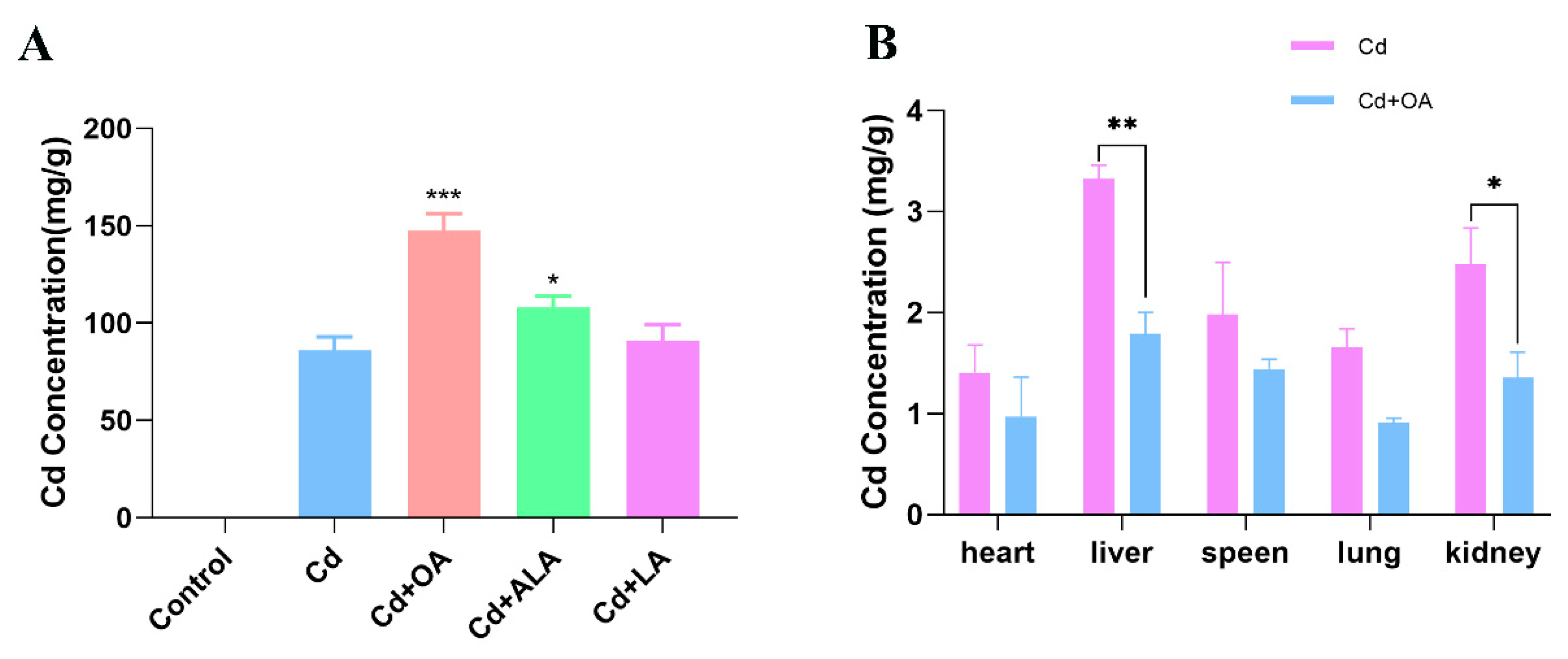
Figure 1. Effects of different fatty acids on the Cd contents in Cd-exposed mice. (A) The Cd contents in feces of Cd-exposed mice treated with three kinds of fatty acids for 7 days. (B) The Cd contents in different organs of Cd-exposed mice treated with OA. * p < 0.05, ** p < 0.01, and *** p < 0.001 versus the Cd group.
Ijms 23 14718 g001
Figure 2. Effects of OA on the gut microbiota. The Alpha diversity of the gut microbial community: Chao (A), Ace (B), and Simpson (C) index. (D) The OTUs results at 97% identity level in UpSet diagram of the control, Cd−treated, and OA−treated groups. (E) The Beta diversity of the gut microbial community by NMDS maps. (F) The microbial abundance heatmap (log10) at the genus level. (G) The relative abundance of the top 10 gut microbiota at the family level. (H) The relative abundance of the top 10 gut microbiota at the genus level. (I) The growth of B. cepacia cultivated in LB broth containing 0, 100, 200, and 500 mg/L of CdCl2 and 2 mmol/L of OA. * p < 0.05, ** p < 0.01, and *** p < 0.001 versus the corresponding Cd groups.
Ijms 23 14718 g002
Figure 3. Cd accumulation in Cd-exposed mice. (A) Total Cd content in feces; (B) Cd content in the gut; (C) Cd contents in the heart, liver, spleen, lung, and kidneys after treatment for 7 d. * p < 0.05 and *** p < 0.001 versus the Cd group.
Ijms 23 14718 g003
Figure 4. Cd bioaccumulation and production of EPS in B. cepacia after being exposed to (A) 100 mg/L, (B) 200 mg/L, and (C) 500 mg/L of CdCl2. * p < 0.05, # p < 0.05, and ## p < 0.01 versus corresponding group on day 1. (D) EPS production of B. cepacia after being exposed to 0, 100, 200, and 500 mg/L of CdCl2 and 2 mmol/L of OA. * p < 0.05 versus the Cd 0 group.
Ijms 23 14718 g004
Figure 5. SEM (A), EDS (B), and TEM (C) images of B. cepacia under Cd exposure as well as co-treatment with Cd+OA. The EPS, Cd, and cell depressions are indicated by the red, blue, and yellow arrows, respectively. The Cd precipitates adsorbed on the cell surface and accumulated intracellularly are indicated by the blue arrows.
Ijms 23 14718 g005
Figure 6. (A) FTIR spectroscopic analysis of EPSs produced by B. cepacia after being exposed to 0, 100, 200, and 500 mg/L of Cd and 2 mmol/L of OA. (B) The content of total sulfhydryl group in EPSs. * p < 0.05 versus control group.
Ijms 23 14718 g006
Publisher’s Note: MDPI stays neutral with regard to jurisdictional claims in published maps and institutional affiliations.