Article

ZmGLP1, a Germin-like Protein from Maize, Plays an Important Role in the Regulation of Pathogen Resistance

1
Key Laboratory of Biology and Sustainable Management of Plant Diseases and Pests of Anhui Higher Education Institutes, School of Plant Protection, Anhui Agricultural University, Hefei 230036, China
2
National Engineering Laboratory of Crop Stress Resistance Breeding, School of Life Sciences, Anhui Agricultural University, Hefei 230036, China
*
Author to whom correspondence should be addressed.
These authors contributed equally to this work.
Int. J. Mol. Sci. 2022, 23(22), 14316; https://doi.org/10.3390/ijms232214316
Submission received: 7 September 2022 / Revised: 10 November 2022 / Accepted: 12 November 2022 / Published: 18 November 2022
(This article belongs to the Section Molecular Plant Sciences)

Abstract

:
A gene encoding a protein similar to germin-like proteins (GLPs) was obtained from maize (Zea mays) and designated as ZmGLP1. Based on the ZmGLP1 conserved domain and phylogenetic status, ZmGLP1 was grouped into GLP subfamily b and has high similarity to OsGLP8-14 from Oryza sativa. ZmGLP1 is expressed in different maize tissues during different growth stages and is mainly expressed in the stems and leaves. The induced expression patterns confirmed that ZmGLP1 is differentially expressed under abiotic and hormone stress; it had an early response to jasmonic acid (JA) and ethephon (ET) but a late response to salicylic acid (SA) and was significantly upregulated under Bipolaris maydis infection. The overexpression of ZmGLP1 in Arabidopsis improved the resistance to biotrophic Pseudomonas syringae pv. tomato DC3000 (PstDC3000) and necrotrophic Sclerotinia sclerotiorum by inducing the expression of JA signaling-related genes. Moreover, the hydrogen peroxide (H2O2) content increased due to the overexpression of ZmGLP1 in Arabidopsis after pathogen infection. Compared to the wild-type control, the H2O2 content of ZmGLP1-overexpressing Arabidopsis infected by PstDC3000 increased significantly but was lower in transgenic plants infected with S. sclerotiorum. Furthermore, high-performance liquid chromatography–tandem mass (HPLC-MS/MS) spectrometry showed that the JA contents of ZmGLP1-overexpressing Arabidopsis markedly increased after pathogen infection. However, the improved resistance of ZmGLP1-overexpressing Arabidopsis pretreated with the JA biosynthetic inhibitor, sodium diethyldithiocarbamate trihydrate (DIECA), was suppressed. Based on these findings, we speculate that ZmGLP1 plays an important role in the regulation of Arabidopsis resistance to biotrophic PstDC3000 and necrotrophic S. sclerotiorum; the regulatory effects are achieved by inducing plant oxidative burst activity and activation of the JA signaling pathway.

1. Introduction

Maize is an important food crop, a primary source of animal feed, and an important industrial raw material used in alcohol and starch production worldwide. Maize has also been playing an increasingly important role in the global economy and agricultural production [1]. In recent years, due to the adjustment of planting structure, transformation of cultivation methods, substitution of maize varieties, and extreme climatic events, the occurrence of maize diseases has increased [2,3]. For example, the widespread cultivation of the susceptible variety Xianyu335 has led to an outbreak of northern corn leaf blight in northern China [4]. In the Huang-Huai-Hai plain of China, high temperatures and humidity have resulted in prime conditions for stalk rot and ear rot diseases [2]. These diseases have become restricting factors that prevent high and stable maize yields. Currently, the most economical and effective strategy for coping with disease stress is to explore new disease-resistant genes and cultivate new disease-resistant maize varieties.
Germin-like proteins (GLPs) are a class of disease-related proteins containing cupin domains that extensively exist in plants [5]. They are highly homologous to germin (GER) sequences and possess β-folded bucket conserved domains. GLPs are soluble proteins that belong to the cupin superfamily. To date, several GLPs have been cloned from many plant species, including Hordeum vulgare, Arabidopsis thaliana, Triticum aestivum, Oryza sativa, and Lilium regale Wilson [6,7,8]. The overexpression of GLP genes increases the resistance of transgenic plants to various pathogens [9,10]. For example, AhGLPs in peanut were significantly upregulated in response to Aspergillus flavus infection, indicating that AhGLPs are prevalent in the defense against A. flavus [11]. Zhang et al. identified LrGLP1, which confers L. regale Wilson resistance to Fusarium oxysporum, and found that LrGLP1-overexpressing tobacco increases disease resistance to F. oxysporum by enhancing the reactive oxygen species (ROS)-scavenging ability [12]. Barley HvGLP4 and wheat TaGLP4 significantly increase the activities of superoxide dismutase (SOD) and polyphenol oxidase (PPO) due to pathogen infection and clear ROS produced during pathogen invasion, resulting in the accumulation of hydrogen peroxide (H2O2), which activated specific H2O2-mediated defense response genes [13]. Pei et al. found that heterologous-expressing GhABP19 in Arabidopsis, a member of the cotton GLP family, increases Arabidopsis resistance to F. oxysporum by activating the jasmonic acid (JA)-mediated signaling pathway [14]. The function of GLP genes in some plants has been demonstrated [12,13,14]; however, the regulatory mechanism underlying GLP genes remains unclear.
Only a few studies on GLP genes in maize have been conducted. The numbers and characteristics of ZmGLP genes in the maize genome and expression changes during Setosphaeria turcica infection have been identified and reported [8]. Few studies on the signaling pathways involved in the resistance to different trophic pathogens of GLP genes have been reported. Therefore, the isolation and study of GLP genes in maize are important for enhancing our understanding of the resistance mechanisms involved in response to diseases and are crucial for facilitating the methods and development of disease-resistant maize varieties.
In a previous study, we found that the application of Bacillus subtilis DZSY21 enhances maize resistance to Bipolaris maydis and identified differentially expressed genes that respond to pathogen infection in interactions between DZSY21 and maize [15,16]; ZmGLP1 was identified among these genes. In this study, we isolated ZmGLP1 from maize, analyzed bioinformatic data and tissue-specific expression patterns, induced the expression of ZmGLP1, and clarified the function of this gene in response to infection with different trophic pathogens, including biotrophic PstDC3000 and necrotrophic S. sclerotiorum. Our findings will lay a foundation for clarifying the disease resistance function of ZmGLP1 in future studies.

2. Results

2.1. Identification and Physicochemical Property Analysis of ZmGLP1

The ZmGLP1 (GRMZM2G064096) CDS obtained from the Phytozome database (https://plants.ensembl.org/Zea_mays/Info/Index, accessed on 20 October 2019) had a full length of 639 bp, encoded 212 amino acids, and was located on chromosome 6. The predicted isoelectric point and molecular weight of the ZmGLP1 protein were 6.01 and 54.08 kDa, respectively. It harbored a typical Cupin_1 conserved domain (Figure 1A and Figure S1), which is characteristic of the plant GLP family. Based on the 132 GLP genes identified in maize, O. sativa, and Arabidopsis [6], a phylogenetic tree was constructed using MEGA7.0 software (Figure 1B). The tree revealed that the GLP family members in maize, O. sativa, and Arabidopsis are divided into six subfamilies (Groups a–f). Moreover, maize ZmGLP1 is clustered with subfamily b and has a close phylogenetic relationship with OsGLP8-14 from O. sativa (Figure 1B; Table S2).

2.2. Expression of ZmGLP1 in Different Maize Tissues

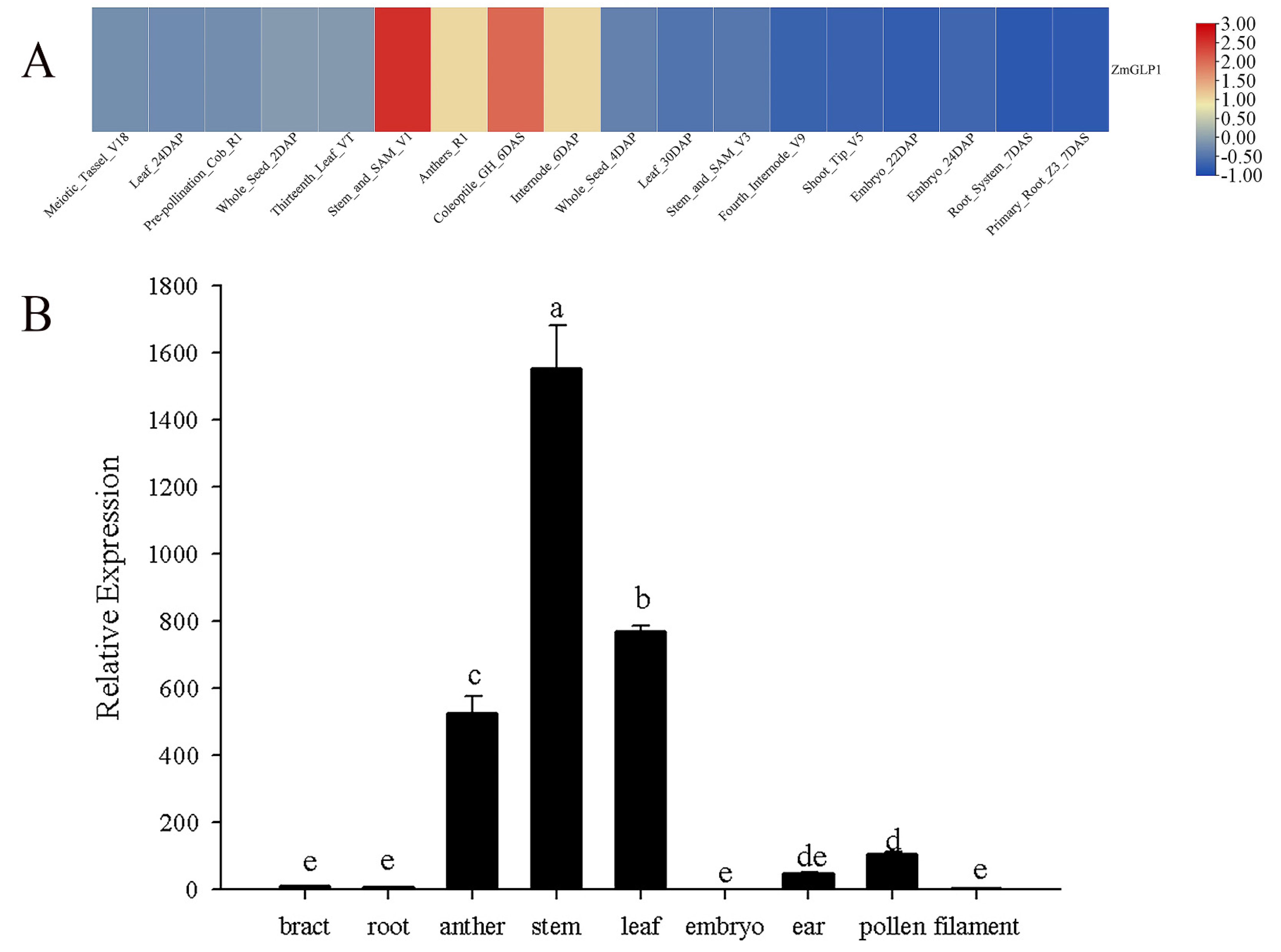
To clarify the tissue-specific expression pattern of the ZmGLP1 gene in maize, publicly available corn microarray data of different tissues were applied to draw the heat map (Figure 2A). Results revealed that ZmGLP1 is highly expressed in the stems, coleoptile, and anthers (Figure 2A). The above prediction results were verified by qRT-PCR, which found that ZmGLP1 is expressed in all maize growth stages. The expression level of ZmGLP1 is the highest in the stems, followed by the leaves and anthers, but lower in the roots, embryos, and filaments. These results are consistent with the chip data (Figure 2B).

2.3. Analysis of ZmGLP1 Expression Induced by Biotic Stress and Hormone Signals

SA, JA, and ET, which mediate plant defense responses against biological stress [17], were analyzed to determine the response of ZmGLP1 to pathogen infection. When compared to 0 h, the transcription level of ZmGLP1 gradually increased 6–12 h after SA application, and its expression increased by 4.0 times after 12 h and then declined rapidly (Figure 3A). ZmGLP1 gradually increased 3–24 h after JA application and reached its maximum level after 24 h, which was 10.0 times greater than that at 0 h (Figure 3B). The ZmGLP1 mRNA transcript level increased gradually from 3 to 12 h after ET application and was 3.43 times higher at 12 h (Figure 3C). ZmGLP1 may have different response times and expression patterns to these hormones, as ZmGLP1 had an early response to JA and ET (3 h) but a late response to SA (6 h).
The transcription level of ZmGLP1 decreased gradually 0–48 h after C. lunata and P. stewartii inoculation (Figure 3D,E) but slowly increased in B. maydis-treated plants after 0–48 h. At 48 h, the expression of ZmGLP1 was 2.50 times greater than that at 0 h after B. maydis infection (Figure 3F). These results indicate that ZmGLP1 might be involved in plant disease resistance, but its underlying mechanism requires further investigation.

2.4. Subcellular Localization Analysis of ZmGLP1

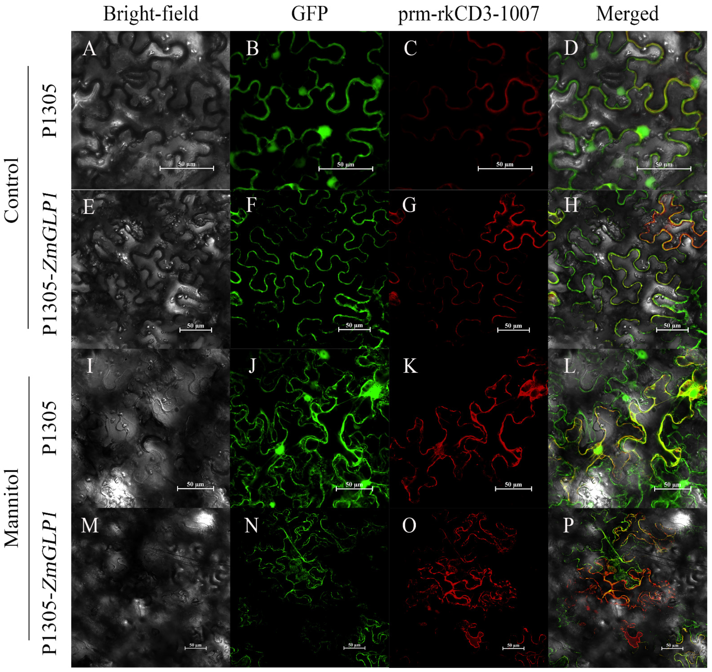
According to our predictions (http://psort.hgc.jp, accessed on 10 October 2020), the protein encoded by ZmGLP1 is located in the cell wall. To verify this, p1305-ZmGLP1-GFP and the plasma membrane marker pm-rk CD3-1007 were co-infiltrated in N. benthamiana, and the fusion protein was allowed to accumulate in the cells (Figure 4A–H). To further confirm the protein location, plasmolysis was performed using N. benthamiana leaf epidermal cells. Results revealed that 35S-GFP and pm-rk CD3-1007 shrank during plasmolysis treatment. However, p1305-ZmGLP1-GFP and pm-rk CD3-1007 were isolated during the plasmolysis treatment (Figure 4I–P), indicating that ZmGLP1-GFP is localized in the cell wall and plasma membrane.

2.5. Expression of ZmGLP1 in Arabidopsis Increases Resistance to PstDC3000

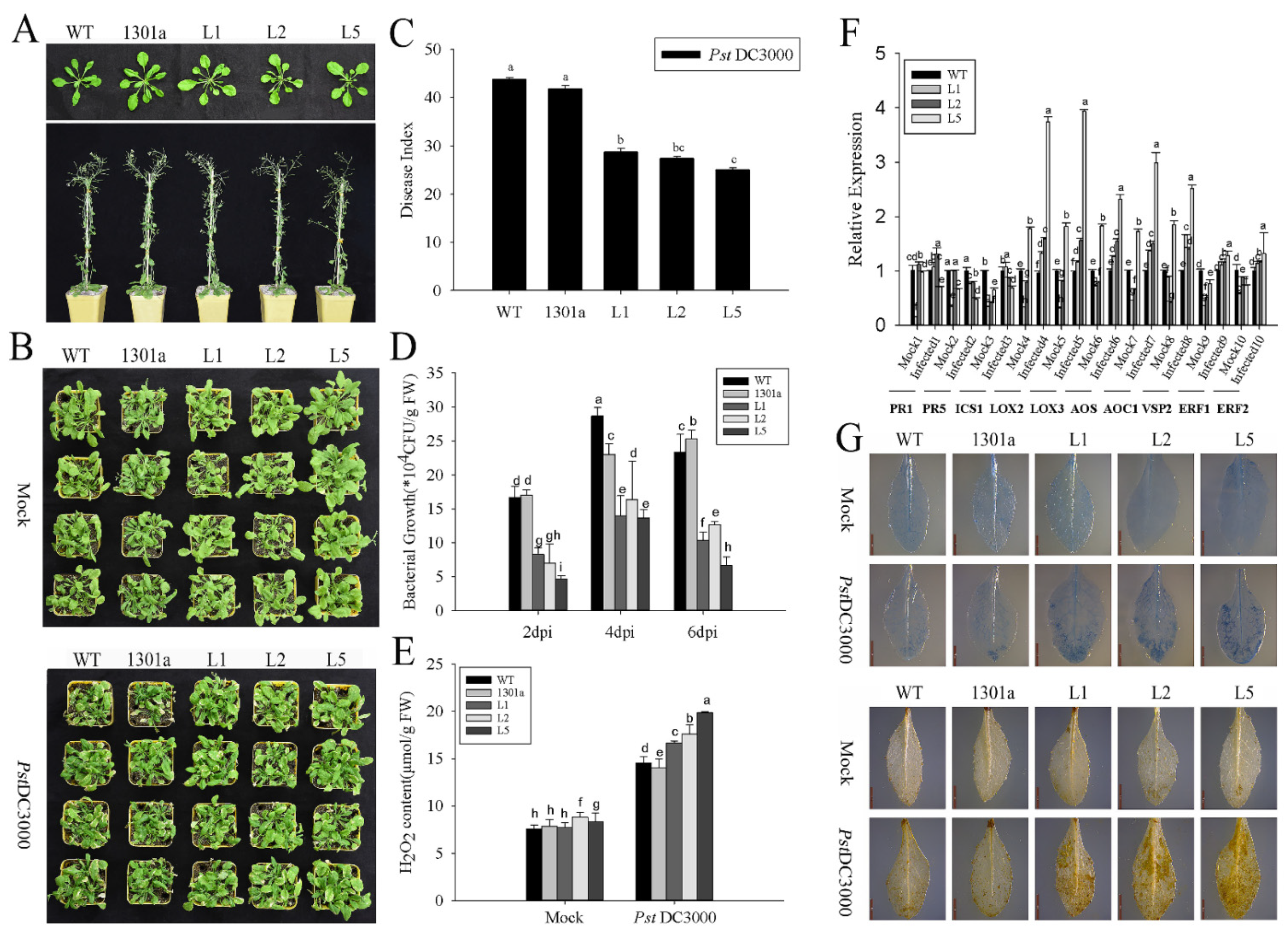
To elucidate the function of ZmGLP1, transgenic Arabidopsis plants overexpressing ZmGLP1 were obtained (Figure S2). Three independent T3 homozygous lines (L1, L2, and L5) were selected to analyze the functional characteristics of ZmGLP1 (Figure 5A). PstDC3000 and S. sclerotiorum were used to evaluate the disease resistance of ZmGLP1-overexpressing Arabidopsis to pathogen infection.
Symptoms appeared on the leaves of different groups 3 d after foliar spraying with PstDC3000 suspension (2 mL per plant). Yellow necrosis spots were observed on plant leaves after 7 d. Compared to the negative control, the leaves of transgenic Arabidopsis had fewer spots (Figure 5B). The disease indices of transgenic L1, L2, and L5 treated with PstDC3000 after 7 d were 28.46, 27.82, and 25.40, respectively, which showed a significant downtrend when compared to wild-type (43.36) and empty plasmid-overexpressing Arabidopsis (40.53) (Figure 5C). These results indicate that the expression of ZmGLP1 in Arabidopsis improves plant resistance to PstDC3000.
The bacterial populations on plant leaves of different groups at 2, 4, and 6 d after treatment with PstDC3000 were detected to clarify the relationship between the bacterial populations in transgenic Arabidopsis and disease resistance. Results revealed that the bacterial populations of transgenic L1, L2, and L5 were lower than those of wild-type and empty plasmid-overexpressing Arabidopsis at 2, 4, and 6 d. The cell count of L5 remained at 6.67 × 10⁴ CFU/g at 6 d, while those of the wild-type and empty plasmid-overexpressing Arabidopsis plants were 23.3 × 10⁴ and 25.3 × 10⁴ CFU/g at 6 d, respectively (Figure 5D). These results indicate that the overexpression of ZmGLP1 in Arabidopsis partially enhances plant resistance to PstDC3000.
As an important mechanism of plant defense responses against pathogens, plant allergic cell death limits the expansion of pathogens and leads to locally acquired resistance. In this study, different groups were stained with trypan blue 3 dpi with PstDC3000, which showed that more necrotic cells accumulated near the PstDC3000 infection site in the transgenic lines than in wild-type and empty plasmid-overexpressing Arabidopsis (Figure 5G), indicating that the overexpression of ZmGLP1 in Arabidopsis induces cell death due to PstDC3000 infection. This finding is consistent with a previous report [18]. Moreover, several yellowish-brown precipitates formed in the leaves of transgenic plants after DAB staining when compared to the control groups (Figure 5G), indicating that H2O2 accumulated in ZmGLP1-overexpressing Arabidopsis. Our quantitative results further showed that the H2O2 content in plants infected with PstDC3000 increased significantly when compared to the negative controls (Figure 5E). The H2O2 contents in transgenic L1, L2, and L5 reached 16.68, 17.75, and 19.90 µmol/g, respectively, which were higher than those in the wild-type (14.57 µmol/g) and empty plasmid-overexpressing (14.06 µmol/g) Arabidopsis (Figure 5E). Results revealed that ZmGLP1-overexpressing Arabidopsis resists PstDC3000 infection by inducing more cell death and accumulating H2O2.
To study the signaling pathways that ZmGLP1 participates in and thereby enhances resistance of transgenic Arabidopsis to PstDC3000, the expression levels of target plant genes in the SA, JA, or ET signaling pathways were analyzed [19]. At 24 h after spraying with PstDC3000, LOX2, LOX3, AOC1, AOS, and VSP2 were strongly induced in transgenic lines, and the expression levels of these genes in L5 were 3.75, 3.94, 2.98, 2.32, and 2.52 times higher than those in wild-type plants, respectively (Figure 5F). ERF1 and ERF2 were slightly induced, while PR1, PR5, and ICS1 were not induced in transgenic Arabidopsis (Figure 5F). Results revealed that LOX2, LOX3, and AOC1 were simultaneously highly expressed in ZmGLP1-overexpressing lines after PstDC3000 infection, indicating that ZmGLP1 may participate in plant defense mechanisms against biotrophic PstDC3000 by activating the JA-dependent signaling pathway.

2.6. ZmGLP1 Confers Resistance to S. sclerotiorum in Arabidopsis

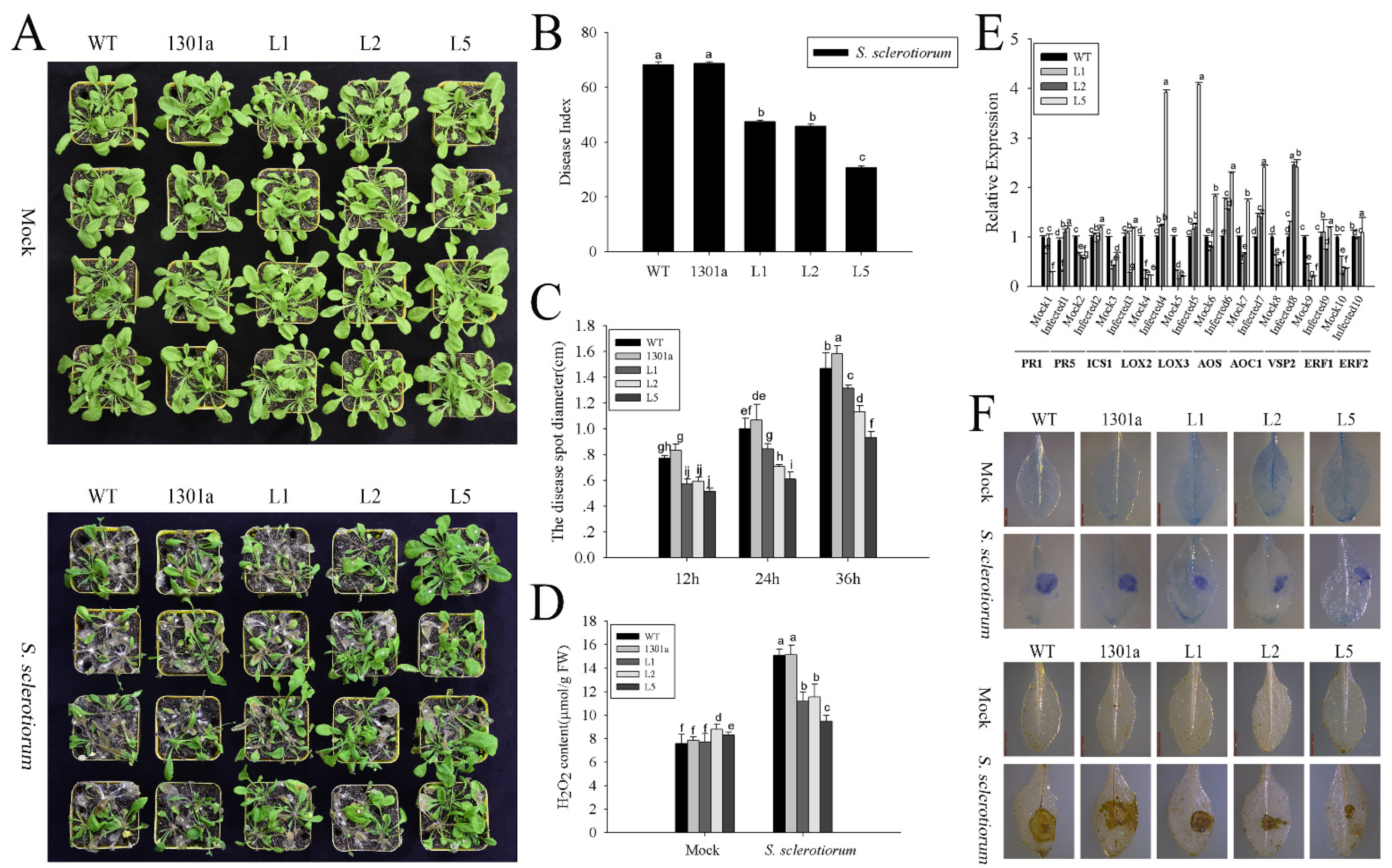
After clarifying disease resistance in ZmGLP1-overexpressing plants to PstDC3000, the function of ZmGLP1 in transgenic plants against pathogenic necrotrophic S. sclerotiorum was analyzed. At 12 h post infection with S. sclerotiorum, necrotic lesions appeared near the inoculation sites. The lesion diameters in transgenic plants were significantly smaller than those in wild-type and empty plasmid-overexpressing plants at 12, 24, and 36 h. The lesion diameter in L5 was 0.93 cm at 36 h, while that of the wild-type and empty plasmid-overexpressing Arabidopsis reached 1.46 and 1.58 cm at 36 h, respectively (Figure 6C). The disease indices of L1 (47.24), L2 (45.83), and L5 (30.21) were significantly lower than those of the wild-type (68.78) and empty plasmid-overexpressing Arabidopsis (69.35) at 3 dpi with S. sclerotiorum (Figure 6B). Transgenic L5 especially showed stronger disease resistance to S. sclerotiorum (Figure 6A,C). These results indicate that ZmGLP1, which is overexpressed in Arabidopsis, partially increases resistance to S. sclerotiorum.
Subsequently, the groups were inoculated with S. sclerotiorum for 3 d, then stained with trypan blue and DAB. We found fewer necrotic cells around the lesions in the transgenic lines compared to the wild-type and empty plasmid-overexpressing Arabidopsis. The lesions in ZmGLP1-overexpressing lines were smaller than those in other groups (Figure 6F). H2O2 accumulated on the surface of spots in different groups treated with S. sclerotiorum (Figure 6F). The H2O2 contents of the wild-type, empty plasmid-overexpressing, and transgenic L1, L2, and L5 plants increased significantly, reaching 15.10, 15.16, 11.20, 11.55, and 9.48 µmol/g, respectively, when compared to the negative controls (Figure 6D). However, the H2O2 contents of transgenic plants were lower than those of the wild-type and empty plasmid-overexpressing Arabidopsis (Figure 6D).
Finally, the key genes in transgenic plants were analyzed to investigate the disease resistance signaling pathway that ZmGLP1 participates in and thereby enhances resistance to S. sclerotiorum. At 6 h after infection with S. sclerotiorum, the target genes in the JA signaling pathway in transgenic L1, L2, and L5 were upregulated. LOX2, LOX3, AOC1, AOS, and VSP2 in transgenic L5 were strongly induced and were 3.93, 4.08, 2.41, 2.30, and 2.41 times higher than those in wild-type plants, respectively (Figure 6E). ERF1 and ERF2 in L5 were slightly induced and were 1.66 and 1.80 times greater than those in wild-type plants (Figure 6E). These results indicate that ZmGLP1 might participate in plant defense mechanisms against pathogenic fungi by activating the JA-dependent signaling pathway.

2.7. ZmGLP1-Overexpressing Arabidopsis Exhibits Resistance to PstDC3000 and S. sclerotiorum through the JA-Mediated Signaling Pathway

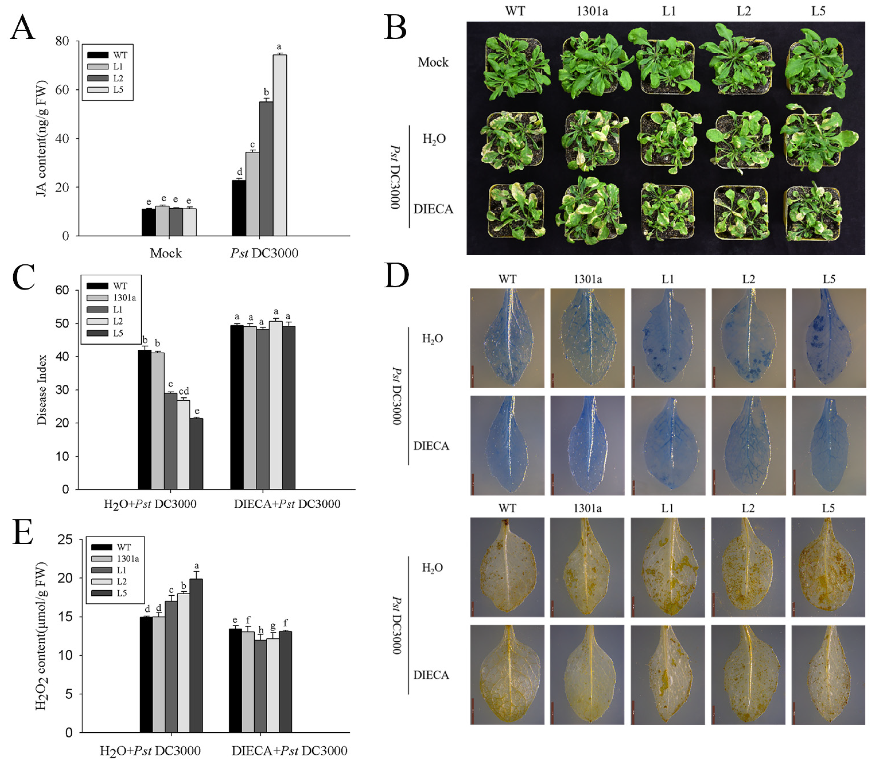
Preliminary results indicated that ZmGLP1 might participate in plant defense mechanisms against PstDC3000 and S. sclerotiorum through the JA signaling pathway. However, the dynamic content changes of endogenous JA in transgenic Arabidopsis before and after pathogen infection have yet to be determined. Therefore, the JA contents in ZmGLP1-overexpressing Arabidopsis were analyzed by HPLC-MS/MS. The retention time of the peaks of the JA extracts was consistent with standard JA, appearing at 8.09 ± 0.02 s (Figures S3 and S4). The JA contents of transgenic and wild-type plants did not exhibit apparent differences before PstDC3000 or S. sclerotiorum infection; however, the JA contents of transgenic L1, L2, and L5 accumulated rapidly after pathogen infection and reached 33.18, 52.59, and 71.71 μg/g FW at 24 h after PstDC3000 infection, respectively, which were significantly higher than those of wild-type plants (21.84 μg/g FW) (Figure 7A). Furthermore, the JA contents in L1, L2, and L5 infected with S. sclerotiorum were 22.23, 20.53, and 57.71 μg/g FW, respectively, which were also significantly higher than those of wild-type plants (18.59 μg/g FW) (Figure 8A). These results show that the rapid accumulation of endogenous JA in transgenic Arabidopsis enhances disease resistance to PstDC3000 and S. sclerotiorum as well as indicates that the overexpression of ZmGLP1 in Arabidopsis may activate the JA signaling pathway to enhance pathogen resistance.
DIECA is a JA biosynthesis inhibitor that effectively controls JA biosynthesis in plants. To confirm that ZmGLP1 enhances Arabidopsis resistance to PstDC3000 and S. sclerotiorum through the JA signaling pathway, different groups were pretreated with 200 μmol/L DIECA for 24 h. Then, plants were infected with PstDC3000 or S. sclerotiorum; disease indices were estimated at 7 and 3 dpi with PstDC3000 and S. sclerotiorum, respectively. As expected, ZmGLP1-overexpressing lines L1, L2, and L5 and empty plasmid-overexpressing and wild-type plants pretreated with DIECA exhibited more severe symptoms after inoculation with PstDC3000 (Figure 7B); the disease indices were 48.16, 50.68, 49.16, 49.05, and 49.40, respectively (Figure 7C), while the negative groups were 28.95, 26.77, 21.43, 41.12, and 41.96, respectively (Figure 7C). The disease indices of transgenic lines L1, L2, and L5 and wild-type and empty plasmid-overexpressing Arabidopsis after S. sclerotiorum infection were 74.41, 79.47, 78.62, 78.47, and 79.28, respectively (Figure 8B, C), while the disease indices of the negative groups were 49.92, 47.81, 33.86, 69.32, and 70.07, respectively (Figure 8C). These results indicate that the application of DIECA reduces disease resistance to PstDC3000 and S. sclerotiorum in transgenic plants.
Trypan blue was used to detect necrotic cells in plant leaves infected with PstDC3000 and S. sclerotiorum. Results revealed that hypersensitive reactions were inhibited in DIECA-pretreated Arabidopsis lines infected with PstDC3000, which had fewer necrotic cells around the infection site when compared to the negative controls (Figure 7D). Fewer yellowish-brown precipitates were observed in DIECA-pretreated Arabidopsis leaves after 3 dpi with PstDC3000 (Figure 7D). The H2O2 contents in the DIECA-pretreated groups significantly decreased when compared to the negative controls (Figure 7E).
Trypan blue and DAB staining were performed so we could observe the number of necrotic cells and the active site of peroxidase in DIECA-pretreated plants after 3 dpi with S. sclerotiorum. Results revealed that more necrotic cells and yellowish-brown precipitates aggregated near the lesions on DIECA-pretreated plant leaves when compared to the negative controls (Figure 8D). The H2O2 content of the DIECA-pretreated lines significantly increased when compared to the negative controls, exhibiting the opposite trend to DIECA-pretreated groups inoculated with PstDC3000 (Figure 8E). These results indicate that DIECA application reduces disease resistance in ZmGLP1-overexpressing plants to PstDC3000 and S. sclerotiorum, further indicating that the overexpression of ZmGLP1 in Arabidopsis enhances resistance to biotrophic PstDC3000 and necrotrophic S. sclerotiorum by activating the JA signaling pathway.

3. Materials and Methods

3.1. Bioinformatics Analysis of ZmGLP1

The ZmGLP1 coding sequence (CDS) and protein sequence were downloaded from the Phytozome database (http://phytozome.jgi.doe.gov/pz/portal.html, accessed on 20 October 2019). A conserved motif in the ZmGLP1 protein was detected by SMART (http://smart.embl-heidelberg.de/, accessed on 20 October 2019). Then, using MEGA v7.0 software, the obtained sequences were clustered by the neighbor-joining (NJ) method, and the constructed phylogenetic tree was optimized using the ITOL website (https://itol.embl.de/, accessed on 20 January 2021). The data required for drawing a heat map of the tissue-specific expression of ZmGLP1 were obtained from the Corn database (http://www.plexdb.org/plex.php?database=Corn, accessed on 20 January 2021); a heat map was drawn following previously described methods [20,21].

3.2. Vector Construction and Arabidopsis Transformation

ZmGLP1 was cloned from maize leaves by PCR using specific primers. The mRNA was cloned by RT-PCR (forward: ATG GCC AAA ATG GTG TTG; reverse: ACC GCT GCC GCC GAG CAC AC). The fragment was inserted (digested with BamHI and PstI) into the vector pCAMBIA1301 to generate p35S:ZmGLP1, and the designed primer sequences were as follows: ZmGLP1-BamHI: GGG GAT CCA TGG CCA AAA TGG TGT TGC TCT GC; ZmGLP1-PstI: GCC TGC AGA CCG CTG CCG CCG AGC A (Figure S5). Then, plasmid correctness was confirmed by enzyme digestion analysis and sequencing. ZmGLP1 transgenic Arabidopsis was generated by Agrobacterium-mediated genetic transformation [21]. Transgenic T3 homozygous Arabidopsis was used in the follow-up experiments.

3.3. Pathogen Cultivation

Pseudomonas syringae pv. tomato DC3000 (PstDC3000) was grown at 28 °C for 24 h in a beef protein liquid medium. S. sclerotiorum was incubated on PDA solid medium at 22 °C for 2 d under a 12/12 h light/dark photoperiod.

3.4. Plant Materials and Treatments

Maize (B73) plants and Nicotiana benthamiana were grown at 25 °C under a 16/8 h light/dark photoperiod and 60–70% relative humidity. Arabidopsis was grown at 23 °C under a 16/8 h light/dark photoperiod.
Analyses of ZmGLP1 expression induced by biotic stress and hormones and the tissue-specific expression of ZmGLP1 were conducted following previously described methods [21]. Plants at the three-leaf stage were treated with salicylic acid (SA) (1 mM), jasmonic acid (JA) (50 μM), or ethephon (ET) (1 mM) and infected with Curvularia lunata (1 × 10⁵ CFU/mL), Bipolaris maydis (1 × 10⁵ CFU/mL), or Pantoea stewartii (1 × 10⁶ CFU/mL). At 0, 3, 6, 12, 24, and 48 h after pathogen and hormone infection, maize leaves were collected for RNA extraction. Maize tissues at different growth stages were collected to extract RNA for the analysis of the tissue-specific expression of ZmGLP1. Each group consisted of three replicates (three maize plants per replicate).
Four-week-old Arabidopsis sprayed with PstDC3000 suspension (OD600 = 0.4) and plants treated with 10 mM MgCl2·6H2O were used as the negative controls. Disease severity was recorded 7 days post infection (dpi) with a scoring range of 0–4 [22]. Grades of 0, 1, 2, 3, and 4 indicated diseased leaf areas of 0%, 1–25%, 26–50%, 51–75%, and 76–100%, respectively. Three leaves were picked from each wild and transgenic Arabidopsis plant at the age of 4 weeks, and each leaf was inoculated on a quarter of an S. sclerotiorum dish (Φ = 6 mm); plants without S. sclerotiorum inoculation were used as the negative controls. The size of the lesion diameter was measured at 12, 24, and 36 h post infection, and the severity of the disease was recorded at 3 d with a scoring range of 0–4. Grades of 0, 1, 2, 3, and 4 indicated diseased leaf areas of 0%, 0–10%, 11–30%, 31–50%, and 50–100%, respectively [23]. There were 30 plants in a single treatment, and the plants within each treatment were divided into three replicates. The disease index was calculated to evaluate the disease resistance of plants [15]. Arabidopsis leaves were collected for RNA extraction at 24 h after infection with PstDC3000 and 6 h with S. sclerotiorum.
Bacterial numbers within the mesophyll tissues were estimated using the serial dilution coating method on different days after PstDC3000 infection [24]. Each group consisted of three replicates (three Arabidopsis plants per replicate).
To confirm that the overexpression of ZmGLP1 in Arabidopsis enhances pathogen resistance through the JA signaling pathway, four-week-old wild-type and transgenic Arabidopsis were treated with 200 μmol/L sodium diethyldithiocarbamate trihydrate (DIECA) containing 0.02% Tween 20. After 24 h, the plants were inoculated with PstDC3000 suspension and S. sclerotiorum. Plants pretreated with sterile distilled water containing 0.02% Tween 20 were used as the negative controls. A total of 30 plants in each treatment were divided into three replicates per treatment.

3.5. Quantitative PCR

Plant leaves harvested at different time points were ground in liquid nitrogen for RNA extraction with TRIzol reagent. The RNA quality was evaluated based on the concentration using electrophoresis strips. qRT-PCR was conducted using AceQ qPCR SYBR Green Master Mix (Vazyme Biotech Co., Ltd., Nanjing, China). Each PCR tube contained 10 μL AceQ qPCR SYBR Green Master Mix, 25 ng cDNA, and 1 μL of each primer [25] (Table S1). The thermal cycling conditions were as follows: 30 s at 94 °C, 40 cycles for 5 s at 94 °C, and 15 s at 55 °C. The data were processed following previously described methods [26].

3.6. Subcellular Localization Assay of ZmGLP1

The p1305-ZmGLP1-GFP vector was constructed (Figure S6). Through sequence analysis, we inserted the target fragment, which removed the terminator, into the front of the GFP tag by double digestion sites (XbaI and BamHI). The designed primer sequences were as follows: ZmGLP1-XbaI: GCT CTA GAA TGG CCA AAA TGG TGT TGC TCT GC; ZmGLP1-BamHI: ATG GAT CCA CCG CTG CCG CCG AGC A. Then, plasmid correctness was confirmed by enzyme digestion analysis and sequencing. The vector was transformed into Agrobacterium tumefaciens strain GV3101 for Agrobacterium-mediated transient expression. Four-week-old N. benthamiana was selected for infiltration treatment. The p1305-ZmGLP1-GFP or 35S-GFP vectors and a plasma membrane marker (pm-rk188 CD3-1007) were co-infiltrated in N. benthamiana leaves for 3 d [27]. Subsequently, N. benthamiana was subjected to plasmolysis, and the infiltrated leaves were incubated in 1 mM mannitol for 30 min. The fluorescence of the samples was observed under a Nikon AX confocal microscope (Nikon, Tokyo, Japan).

3.7. DAB Staining

To detect the degree of H2O2 accumulation in ZmGLP1-overexpressing Arabidopsis leaves at 3 dpi, DAB staining was applied to the leaves. Under the catalysis of peroxidase, H2O2 rapidly reacts with DAB to form brown compounds, thereby revealing brown spots on plant leaves. Therefore, the aggregation of H2O2 can be determined by observing whether brown spots are produced on the leaves. The size and color of the spots produced by H2O2 accumulation are often used to qualitatively analyze the accumulation of H2O2 in plants under stress.
At 3 dpi with PstDC3000 and S. sclerotiorum, Arabidopsis leaves were collected and completely immersed in a Petri dish filled with 10 mM DAB staining solution, soaked in a vacuum for 5 min, wrapped with tin foil, and gently placed on a horizontal shaker at 80 rpm for 4–5 h. Then, the DAB staining solution was changed to a bleach solution and transferred to a water bath at 90–95 °C for 15 min to rinse off the chlorophyll, thereby leaving a brown precipitate from the DAB and H2O2 reaction. Samples were left in the new bleach solution for 30 min and then placed for 4 d at 4 °C [28]. Arabidopsis leaves treated with 10 mM MgCl2·6H2O and uninoculated leaves were used as the negative controls for the PstDC3000 and S. sclerotiorum treatments, respectively.

3.8. Detection of H2O2

Wild-type and transgenic Arabidopsis leaves were harvested at 3 dpi and stored at −80 °C for detection of the H2O2 content using a Keming kit (Suzhou Keming Biotechnology Co., Ltd., Suzhou, China). Based on whether an orange complex was formed by the H2O2 and titanium ions in the acidic media, titanium sulfate spectrophotometry was used to determine the H2O2 concentrations in plants treated with the pathogens. Arabidopsis leaves treated with 10 mM MgCl2·6H2O and uninoculated leaves were used as the negative controls for the PstDC3000 and S. sclerotiorum treatments, respectively.

3.9. Trypan Blue Staining

Trypan blue staining was performed following previously described methods [28]. Briefly, leaves infected with pathogens for 3 d were transferred to a solution containing 2.5 mg/mL trypan blue, vacuum infiltrated for 10 min, boiled for 2 min, and kept on a horizontal shaker for 12 h. Finally, the solution was replaced with a 2.5 g/mL chloral hydrate solution for decolorization until colorless. Transgenic and wild-type leaves treated with 10 mM MgCl2·6H2O or uninoculated leaves were used as the negative controls for the PstDC3000 and S. sclerotiorum treatments, respectively.

3.10. Detection of JA in ZmGLP1-Overexpressing Arabidopsis

The JA contents of wild-type and transgenic Arabidopsis before and after 24 h infection with PstDC3000 and 6 h with S. sclerotiorum were monitored using a high-performance liquid chromatography–tandem mass (HPLC-MS/MS) spectrometer consisting of an Agilent 1290 HPLC system coupled to an MS/MS spectrometer (Applied Biosystems 6500 Quadrupole Trap) (Agilent, San Francisco, USA), which was operated in multiple-reaction monitoring mode (Nanjing Convinced-Test Technology Co., Ltd., Nanjing, China). The JA content was extracted using the isopropanol–water–hydrochloric acid extraction method. Samples were detected by gradient elution using 0.1% formic acid methanol (A) and 0.1% acetic acid in water (B) as the mobile phases. Each group consisted of three replicates (three Arabidopsis plants per replicate).

3.11. Statistical Analysis

DPS software was used for the statistical data analyses. Statistical differences between different samples using a P < 0.05 threshold were assessed by one-way ANOVA followed by Duncan’s post hoc test.

4. Discussion

A candidate disease-resistant gene, ZmGLP1, was investigated in this study. ZmGLP1 encodes a GLP and has the highest sequence similarity with OsGLP8-14 in rice, which encodes a cell-wall-related protein with SOD activity and plays an important role in plant disease resistance [29,30]. GLP genes are widely expressed in multiple organs of different plants, showing tissue specificity and functional diversity at different developmental stages [11,31]. Our results are consistent with previous reports, which found that ZmGLP1 is expressed in different maize tissues and mainly expressed in the stems, leaves, and anthers, suggesting that ZmGLP1 may play an important role in maize growth and development. Additionally, we used subcellular localization analysis and plasmolysis experiments combined with network positioning to predict the localization of ZmGLP1 in the cell wall and plasma membrane. In future studies, we intend to conduct protein fractionation coupled with Western blot analysis to confirm ZmGLP1 subcellular localization in subsequent tests.
Plant GLPs play key roles in plant responses to pathogenic microbial infection and external stress [5]. Thus far, 32, 43, 57, and 21 GLP genes have been identified in Arabidopsis, O. sativa, Z. mays, and H. vulgare, respectively [6,7,8]. Here, we identified certain feature genes in ZmGLP1-overexpressing Arabidopsis which are involved in the SA, JA, and ET signaling pathways [19] to clarify the function of ZmGLP1 in conferring plant disease resistance. The results demonstrated that the expression of key genes in ZmGLP1-overexpressing Arabidopsis, namely, LOX2, LOX3, AOC1, and AOS, is strongly induced after PstDC3000 and S. sclerotiorum infection. LOX2, LOX3, AOC1, and AOS are biosynthetic marker genes of the JA signaling pathway. Thus, we speculated that ZmGLP1-overexpressing Arabidopsis might enhance its resistance by activating the JA signaling pathway in response to pathogen infection. Other studies showed that the JA contents in ZmGLP1-overexpressing plants rapidly accumulated 24 h after infection with PstDC3000 and 6 h with S. sclerotiorum. However, after the application of the JA biosynthetic inhibitor DIECA, ZmGLP1 transgenic plants did not exhibit pathogen resistance. Our findings showed that the overexpression of ZmGLP1 promoted the accumulation of endogenous JA and conferred plant resistance to PstDC3000 and S. sclerotiorum, suggesting that ZmGLP1 may be involved in disease resistance, depend on the JA signaling pathway, and be conversely regulated by JA. In future studies, we will utilize the mutation coi1, which is the receptor of JA, to confirm the function of JA signaling in ZmGLP1-mediated defense responses.
It is well known that JA negatively regulates Arabidopsis defense responses to PstDC3000 [32,33]. In recent years, some studies have reported interesting results that contradict previous reports. De Torres Zabala et al. revealed that JAZ5 and JAZ10 function cooperatively to attenuate phytotoxicity mediated by COR and moderately restrict PstDC3000 growth; the JA in Arabidopsis leaves did not accumulate until late in the infection process challenged with COR-deficient P. syringae or in the more resistant JA receptor mutant coi1. [34]. The ectopic expression of MdHIR4 in Arabidopsis and N. benthamiana enhances resistance to PstDC3000. Furthermore, the interaction between MdHIR4 and AtJAZs proteins (AtJAZ3, AtJAZ4, and AtJAZ9) implies that MdHIR4 mediates biotic stress through the JA signaling pathway [35]. Additionally, the overexpression of PevD1 in Arabidopsis, which is isolated from Verticillium dahlia, triggered Arabidopsis resistance to PstDC3000 by affecting the JA signaling pathway, three critical regulators of JA biosynthesis were upregulated, and the JA levels in transgenic Arabidopsis slightly increased [36]. In our study, we found that the ectopic expression of ZmGLP1 plays a significant role in the regulation of resistance to biotrophic PstDC3000 in Arabidopsis due to its ability to activate the JA pathway, which corroborates previous studies.
When plants interact with pathogenic microorganisms, the expression levels of GLP genes in plants are significantly upregulated, which induces an “oxidative burst” response. ROS burst is the main immune response in plant resistance to pathogen invasion, which directly inhibits pathogen growth and limits pathogen invasion by strengthening and thickening the cell wall. Some studies have uncovered the function of GLPs in inducing the “oxidative burst” response [37]. During the process of pathogen infection, HvGLP4 in H.vulgare, TaGLP4 in T. aestivum, and OsRGLP2 and OsGLP1 in O. sativa induce the burst of ROS, resulting in the accumulation of H2O2, which thereby activates genes involved in H2O2-mediated defense responses and improves disease resistance [13,28,38]. Therefore, the H2O2 contents in ZmGLP1-overexpressing Arabidopsis after pathogen infection were detected in this study. We found that the H2O2 contents in ZmGLP1-overexpressing Arabidopsis increased significantly after PstDC3000 and S. sclerotiorum infection when compared to the negative controls. Interestingly, the H2O2 contents in transgenic plants were significantly higher than those in the wild-type plants infected with PstDC3000 (Figure 5E), while the H2O2 contents in transgenic plants were lower than those in the control groups after S. sclerotiorum infection (Figure 6D). Changes in the H2O2 contents in transgenic plants, which responded to PstDC3000 and S. sclerotiorum infection, exhibited the opposite trend. We speculated that ZmGLP1-overexpressing Arabidopsis might elicit different ROS responses to different types of pathogen infection. When PstDC3000, a biotrophic pathogen, infects Arabidopsis, the intracellular receptor kinase, RIPK, recognizes the lipopolysaccharide of PstDC3000 and phosphorylated NADPH oxidase RBOHD to induce oxidative burst, which initiates plant defense signal transmission, strengthens the cell wall, causes an allergic reaction, and leads to programmed cell death, thereby inhibiting the infection of pathogenic bacteria [39,40]. However, ROS play the opposite role in the infection of necrotrophic pathogens [41,42]. As a necrotrophic pathogen, S. sclerotiorum kills host cells by secreting toxins or cytolytic enzymes and invades the host. It uses host ROS to enhance its pathogenicity, thereby reducing the level of ROS in the host, which may prevent cell death and inhibit the growth of S. sclerotiorum [43]. These results support previous findings.
In summary, our data reveal that ZmGLP1 overexpression confers disease resistance to biotrophic PstDC3000 and necrotrophic S. sclerotiorum by inducing oxidative burst activity and promoting JA accumulation in plants, which preliminarily explicates the disease resistance signaling pathway induced by ZmGLP1. Future studies should aim to confirm the function of the JA signaling pathway in ZmGLP1-mediated defense responses by utilizing the Arabidopsis mutation coi1 to enhance our understanding of the regulatory mechanism underlying the JA signaling pathway induced by ZmGLP1 overexpression as well as identify the interacting proteins of ZmGLP1.

Supplementary Materials

The following supporting information can be downloaded at: https://www.mdpi.com/article/10.3390/ijms232214316/s1. Figure S1: Molecular characterization of ZmGLP1; Figure S2: De-tection of ZmGLP1 transgenic Arabidopsis thaliana; Figure S3: Chromatogram of JA extracted from Arabidopsis; Figure S4: Chromatogram of JA extracted from Arabidopsis; Figure S5: Construction of the p1301-ZmGLP1 vector; Figure S6: Construction of the p1305-ZmGLP1-GFP vector; Table S1: GLP gene family in Oryza sativa and Arabidopsis and Maize; Table S2: Primers of qRT-PCR used in this study.

Author Contributions

Conceptualization, L.M. and L.G.; methodology, X.Y.; software, X.Y.; validation, L.M., L.G. and X.Y.; formal analysis, L.M.; investigation, L.G.; resources, L.X.; data curation, L.G.; writing—original draft preparation, L.M.; writing—review and editing, T.D.; visualization, W.S.; supervision, L.X.; project administration, T.D.; funding acquisition, T.D. All authors have read and agreed to the published version of the manuscript.

Funding

This research was funded by the General Program of Natural Science Foundation of Anhui Province, China (2008085MC93).

Institutional Review Board Statement

Not applicable.

Informed Consent Statement

Not applicable.

Data Availability Statement

Not applicable.

Conflicts of Interest

The authors declare no conflict of interest.

References

  1. Lu, X.; Liu, J.; Ren, W.; Yang, Q.; Chai, Z.; Chen, R.; Wang, L.; Zhao, J.; Lang, Z.; Wang, H.; et al. Gene-indexed mutations in maize. Mol. Plant 2018, 11, 496–504. [Google Scholar] [CrossRef] [PubMed] [Green Version]
  2. Duan, C.; Song, F.; Sun, S.; Guo, C.; Zhu, Z.; Wang, X. Characterization and molecular mapping of two novel genes resistant to pythium stalk rot in maize. Phytopathology 2019, 109, 804–809. [Google Scholar] [CrossRef] [PubMed]
  3. Sun, X.; Qi, X.; Wang, W.; Liu, X.; Zhao, H.; Wu, C.; Chang, X.; Zhang, M.; Chen, H.; Gong, G. Etiology and symptoms of maize leaf spot caused by Bipolaris spp. in Sichuan, 2020, China. Pathogens 2020, 9, 229. [Google Scholar] [CrossRef] [PubMed] [Green Version]
  4. Pu, Z. Dynamic analysis on the disease progression of maize leaf blight between Xianyu335 and improved Xianyu335 in Western Region of Heilongjiang Province. J. Maize Sci. 2013, 21, 119–123. [Google Scholar] [CrossRef]
  5. Ilyas, M.; Naqvi, S.M.S.; Mahmood, T. In silico analysis of transcription factor binding sites in promoters of germin-like protein genes in rice. Arch. Biol. Sci. 2016, 68, 863–876. [Google Scholar] [CrossRef] [Green Version]
  6. Li, L.; Xu, X.; Chen, C.; Shen, Z. Genome-wide characterization and expression analysis of the germin-like protein family in rice and Arabidopsis. Int. J. Mol. Sci. 2016, 17, 1622. [Google Scholar] [CrossRef] [Green Version]
  7. Zimmermann, G.; Baumlein, H.; Mock, H.P.; Himmelbach, A.; Schweizer, P. The multigene family encoding germin-like proteins of barley. Regulation and function in Basal host resistance. Plant Physiol. 2006, 142, 181–192. [Google Scholar] [CrossRef] [Green Version]
  8. Liu, Y.J.; Jia, M.X.; Zhou, H.; Zhang, L.; Liu, C.; Si, H.L.; Liu, Y.Q.; Gu, S.Q.; Gong, X.D.; Dong, J.G. Genome-wide Identification of GLP Gene Family in Maize (Zea mays) and It’s Expression Analysis When Maize is Exposed to Setosphaeria turcica. Chin. J. Agric. Biotechnol. 2021, 29, 13. [Google Scholar] [CrossRef]
  9. Breen, J.; Bellgard, M. Germin-like proteins (GLPs) in cereal genomes: Gene clustering and dynamic roles in plant defence. Funct. Integr. Genom. 2010, 10, 463–476. [Google Scholar] [CrossRef] [Green Version]
  10. Majeed, N.; Javaid, B.; Deeba, F.; Naqvi, S.; Douches, D. Enhanced Fusarium oxysporum f. sp. tuberosi Resistance in Transgenic Potato Expressing a Rice GLP Superoxide Dismutase Gene. Am. J. Potato Res. 2018, 95, 383–394. [Google Scholar] [CrossRef]
  11. Wang, T.; Chen, X.; Zhu, F.; Li, H.; Li, L.; Yang, Q. Characterization of peanut gennin-like proteins, AhGLPs inplant development and defense. PLoS ONE 2013, 8, e61722. [Google Scholar] [CrossRef] [Green Version]
  12. Zhang, N.; Guan, R.; Yang, Y.; Bai, Z.; Ge, F.; Liu, D. Isolation and characterization of a Fusarium oxysporum-resistant gene LrGLP1 from Lilium regale Wilson. In Vitro Cell Dev. Biol.-Plant 2017, 53, 461–468. [Google Scholar] [CrossRef]
  13. Christensen, A.B.; Thordal-Christensen, H.; Zimmermann, G.; Gjetting, T.; Lyngkjær, M.F.; Dudler, R.; Schweizer, P. The germinlike protein GLP4 exhibits superoxide dismutase activity and is an important component of quantitative resistance in wheat and barley. Mol. Plant-Microbe Interact. 2004, 17, 109–117. [Google Scholar] [CrossRef] [PubMed] [Green Version]
  14. Pei, Y.; Li, X.; Zhu, Y.; Ge, X.; Sun, Y.; Liu, N. Gh ABP19, a novel germin-like protein from Gossypium hirsutum, plays an important role in the regulation of resistance to Verticillium and Fusarium wilt pathogens. Front. Plant Sci. 2019, 10, 583. [Google Scholar] [CrossRef] [PubMed] [Green Version]
  15. Ding, T.; Su, B.; Chen, X.; Xie, S.; Gu, S.; Wang, Q.; Huang, D.; Jiang, H. An Endophytic Bacterial Strain Isolated from Eucommia ulmoides Inhibits Southern Corn Leaf Blight. Front. Microbiol. 2017, 8, 903–915. [Google Scholar] [CrossRef] [Green Version]
  16. Zhou, Q.; Gu, S.Y.; Xu, L.; Tan, G.J. Effects of endophyte DZSY21 colonization on DNA methylation levels in Maize. J. Hefei Norm. Univ. 2019, 3, 32–37. [Google Scholar] [CrossRef]
  17. Bari, R.; Jones, J. Role of plant hormones in plant defence responses. Plant Mol. Biol. 2009, 69, 473–488. [Google Scholar] [CrossRef]
  18. Dickman, M.B.; Fluhr, R. Centrality of Host Cell Death in Plant-Microbe Interactions. Annu. Rev. Phytopathol. 2013, 51, 543–570. [Google Scholar] [CrossRef]
  19. Clarke, J.D.; Volko, S.M.; Ledford, H.; Dong, A.X. Roles of salicylic acid, jasmonic acid, and ethylene in cpr-induced resistance in Arabidopsis. Plant Cell 2000, 12, 2175–2190. [Google Scholar] [CrossRef] [Green Version]
  20. Paradis, E.; Claude, J.; Strimmer, K. APE: Analyses of Phylogenetics and Evolution in Rlanguage. Bioinformatics 2004, 20, 289–290. [Google Scholar] [CrossRef]
  21. Hang, T.; Ling, X.; He, C.; Xie, S.; Ding, T. Isolation of the zmers4 gene from maize and its functional analysis in transgenic plants. Front. Microbiol. 2021, 12, 632908. [Google Scholar] [CrossRef] [PubMed]
  22. Zeng, H.H. Chitosan Oligosaccharide-Induced Resistance to Pseudomonas syringaepv Tomato DC3000 in Arabidopsis and a Preliminary Study on the Resistance Mechanism; Dalian Ocean University: Dalian, China, 2016. [Google Scholar]
  23. Gao, X.N. Control of Sclerotinia sclerotiorum in Rapeseed by Endophytic Bacterial Strain Em7; Northwest Sci-Tech University: Shenzhen, China, 2012. [Google Scholar]
  24. Katagiri, F.; Thilmony, R.; He, S.Y. The Arabidopsis thaliana-Pseudomonas syringae interaction. Arab. Book 2002, 1. [Google Scholar] [CrossRef] [PubMed] [Green Version]
  25. Zha, K.; Xie, H.; Ge, M.; Wang, Z.; Wang, Y.; Si, W.; Gu, L. Expression of Maize MADS Transcription Factor ZmES22 Negatively Modulates Starch Accumulation in Rice Endosperm. Int. J. Mol. Sci. 2019, 20, 483–499. [Google Scholar] [CrossRef] [Green Version]
  26. Livak, K.J.; Schmittgen, T.D. Analysis of relative gene expression data using realtime quantitative PCR and the 2-DDCt method. Methods 2001, 25, 402–408. [Google Scholar] [CrossRef] [PubMed]
  27. Nelson, B.; Cai, X.; Nebenfuhr, A. A multicolored set of in vivo organelle markers for co-localization studies in Arabidopsis and other plants. Plant J. 2007, 51, 1126–1136. [Google Scholar] [CrossRef] [PubMed]
  28. Bhadauria, V.; Miraz, P.; Kennedy, R.; Banniza, S.; Wei, Y. Dual trypan-aniline blue fluorescence staining methods for studying fungus-plant interactions. Biotech. Histochem. 2010, 85, 99–105. [Google Scholar] [CrossRef] [PubMed]
  29. Banerjee, J.; Maiti, M.K. Functional role of rice germin-like protein 1 in regulation of plant height and disease resistance. Biochem. Biophys. Res. Commun. 2010, 394, 178–183. [Google Scholar] [CrossRef]
  30. Banerjee, J.; Das, N.; Dey, P.; Maiti, M.K. Transgenically expressed rice germin-like protein1 in tobacco causes hyper-accumulation of H2O2 and reinforcement of the cell wall components. Biochem. Biophys. Res. Commun. 2010, 402, 637–643. [Google Scholar] [CrossRef]
  31. Ilyas, M.; Rasheed, A.; Mahmood, T. Functional characterization of germin and germin-like protein genes in various plant species using transgenic approaches. Biotechnol. Lett. 2016, 38, 1405–1421. [Google Scholar] [CrossRef]
  32. Feys, B.J.F.; Benedetti, C.E.; Turner, P.J.G. Arabidopsis mutants selected for resistance to the phytotoxin coronatine are male sterile, insensitive to methyl jasmonate, and resistant to a bacterial pathogen. Plant Cell 1994, 6, 751–759. [Google Scholar] [CrossRef]
  33. Kloek, A.P.; Verbsky, M.L.; Sharma, S.B.; Schoelz, J.E.; Vogel, J.; Klessig, D.F.; Kunkel, B.N. Resistance to Pseudomonas syringae conferred by an Arabidopsis thaliana coronatine-insensitive (coi1) mutation occurs through two distinct mechanisms. Plant J. 2001, 26, 509–522. [Google Scholar] [CrossRef]
  34. de Torres Zabala, M.; Zhai, B.; Jayaraman, S.; Eleftheriadou, G.; Winsbury, R.; Yang, R.; Truman, W.; Tang, S.; Smirnoff, N.; Grant, M. Novel JAZ co-operativity and unexpected JA dynamics underpin Arabidopsis defence responses to Pseudomonas syringae infection. New Phytol. 2016, 209, 1120–1134. [Google Scholar] [CrossRef] [PubMed] [Green Version]
  35. Zhao, X.Y.; Qi, C.H.; Jiang, H.; Zheng, P.F.; Hao, Y.J. Functional identification of apple on MdHIR4 in biotic stress. Plant Sci. 2018, 283, 396–406. [Google Scholar] [CrossRef] [PubMed]
  36. Liu, M.; Khan, N.U.; Wang, N.; Yang, X.; Qiu, D. The protein elicitor PevD1 enhances resistance to pathogens and promotes growth in Arabidopsis. Int. J. Biol. Sci. 2016, 12, 931–943. [Google Scholar] [CrossRef] [PubMed] [Green Version]
  37. Averyanov, A. Oxidative burst and plant disease resistance. Front. Biosci. 2008, 1, 142. [Google Scholar] [CrossRef]
  38. Munir, F.; Hayashi, S.; Batley, J.; Naqvi, S.M.S.; Mahmood, T. Germin-like protein 2 gene promoter from rice is responsive to fungal pathogens in transgenic potato plants. Funct. Integr. Genom. 2016, 16, 19–27. [Google Scholar] [CrossRef]
  39. Li, Q.Y. The 919-Position Glutamate of RBOHD Regulates the Molecular Mechanism of Lipopolysaccharide-Induced Reactive Oxygen Species Production; Zhejiang University: Hangzhou, China, 2019. [Google Scholar]
  40. Li, P.; Zhao, L.; Qi, F.; Htwe, N.M.; Li, Q.; Zhang, D.; Lin, F.; Shang-Guan, K.; Liang, Y. The receptor-like cytoplasmic kinase ripk regulates broad-spectrum ros signaling in multiple layers of plant immune system. Mol. Plant 2021, 14, 10. [Google Scholar] [CrossRef]
  41. Dickman, M.B.; Park, Y.K.; Oltersdorf, T.; Li, W.; Clemente, T.; French, R. Abrogation of disease development in plants expressing animal antiapoptotic genes. Proc. Natl. Acad. Sci. USA 2001, 98, 6957–6962. [Google Scholar] [CrossRef] [Green Version]
  42. Eri, M.G.; Alex, L. The hypersensitive response facilitates plant infection by the necrotrophic pathogen botrytis cinerea. Curr. Biol. 2000, 10, 13. [Google Scholar] [CrossRef] [Green Version]
  43. Elad, Y.; Williamson, B.; Tudzynski, P.; Delen, N. Botrytis: Biology, Pathology, and Control; Kluwer Academic Publishers: Dordrecht, The Netherlands, 2004; pp. 119–141. [Google Scholar] [CrossRef]
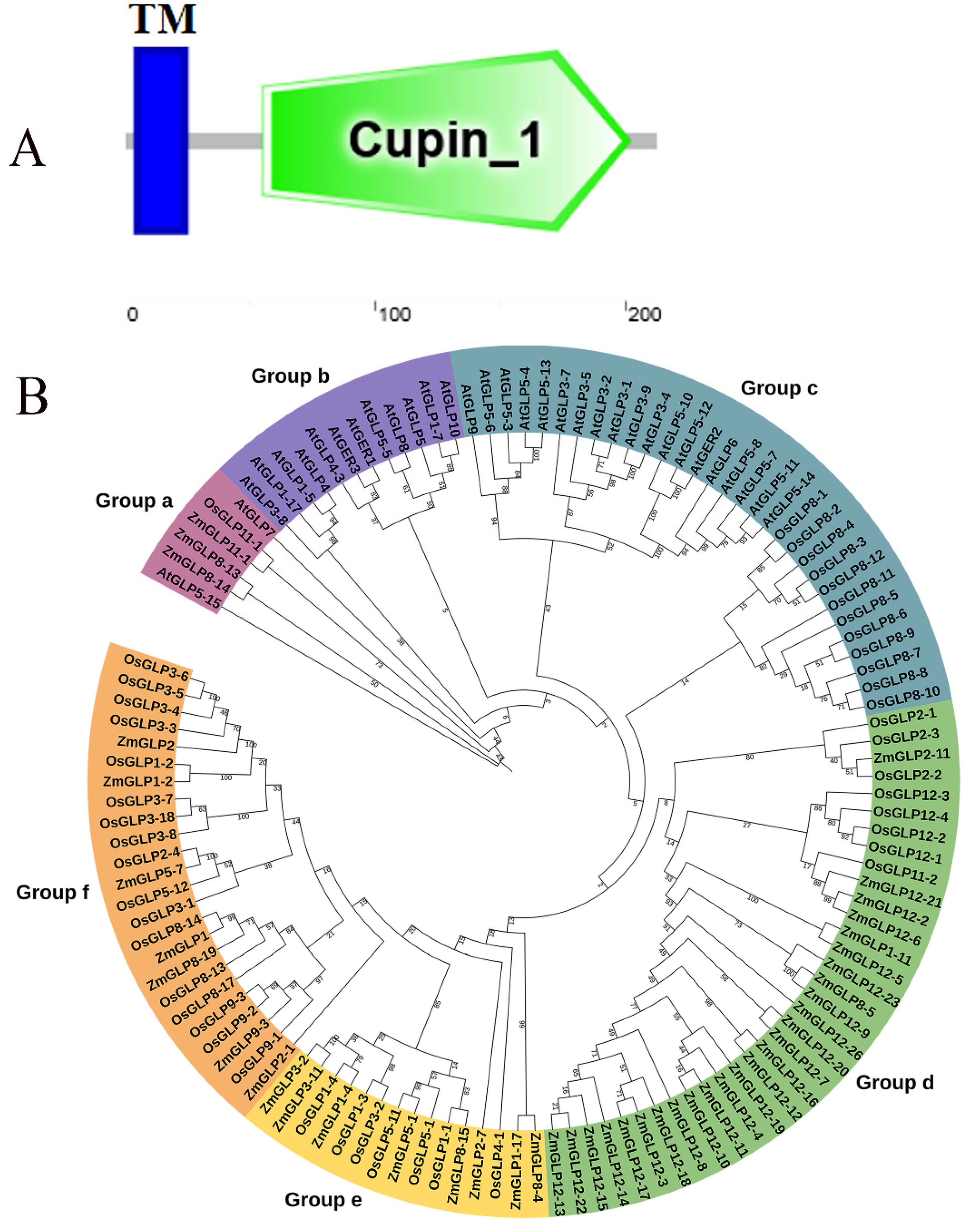
Figure 1. Characteristic analysis of ZmGLP1. (A) The conserved domain analysis of ZmGLP1. (B) Phylogenetic analysis of GLPs gene family from At (Arabidopsis thaliana), Os (Oryza sativa), and Zm (Zea mays). GLP genes are divided into 6 categories. ZmGLP1 is marked in red. Bootstrap values of 1000 replicates are shown as percentages at the branch nodes. Bar = 0.1. Subfamilies are represented by orange, yellow, green, blue, bluish-purple, and purple branch lines.
Ijms 23 14316 g001
Figure 2. Expression analysis of ZmGLP1 in different maize tissues. (A) Heat map of tissue-specific expression of ZmGLP1 in maize. DAS = days after sowing; DAP = days after pollination. (B) Relative expression of ZmGLP1 in different maize tissues. Different letters in subfigure (B) indicated significant difference between different samples by Duncan’s post-hoc test. Data are presented as the mean ± SD based on three biological replicates.
Ijms 23 14316 g002
Figure 3. Relative expression levels of ZmGLP1 under six different treatments over time. (A) Salicylic acid application, (B) jasmonic acid application, (C) ethephon application, (D) the conidial suspension of Curvularia lunata, (E) Pantoea stewartii suspensions, (F) the conidial suspension of Bipolaris maydis. ZmActin1 and GADPH were used as reference genes for normalization. Different letters in subfigures (AF) indicated significant difference between different samples by Duncan’s post-hoc test. Data are presented as the mean ± SD based on three biological replicates.
Ijms 23 14316 g003
Figure 4. ZmGLP1 location in the cell wall and plasma membrane. (AD) Subcellular localization images of positive control 35S-GFP in N. benthamiana. (EH) Subcellular localization of p1305-ZmGLP1-GFP in N. benthamiana. (IL) Images of 35S-GFP epidermal cells after fugitive wall isolation. (MP) Image of p1305-ZmGLP1-GFP signal in N. benthamiana epidermal cells after plasmolysis. Bar = 50 µm.
Ijms 23 14316 g004
Figure 5. Expression of ZmGLP1 in Arabidopsis increases resistance to PstDC3000. (A) Growth status of Arabidopsis leaves in different groups at four and seven weeks old. (B) Phenotypes of the different groups treated with PstDC3000 at 7 d. (C) The disease indexes of groups at 7 d. (D) The population density of bacteria of different groups. (E) H2O2 content of different groups at 3 d. (F) The expression levels of target plant genes of the salicylic acid, jasmonic acid, or ethephon pathway at 24 h. AtActin2 and AtTUB4 were used as reference genes for normalization. (G) Trypan blue staining and DAB staining in Arabidopsis leaves of different groups at 3 d. Different letters in subfigures (CF) indicated significant difference between different samples by Duncan’s post-hoc test. Data are presented as the mean ± SD based on three biological replicates.
Ijms 23 14316 g005
Figure 6. ZmGLP1 increases resistance to Sclerotinia sclerotiorum in Arabidopsis. (A) Phenotypes of different groups before and after being treated with Sclerotinia sclerotiorum at 3 d. (B) The disease indexes of different groups at 3 d. (C) The disease spot diameter of the Arabidopsis leaves in different groups treated with Sclerotinia sclerotiorum at 12, 24, and 36 h. (D) H2O2 content in Arabidopsis leaves of different groups at 3 d. (E) The expression levels of target plant genes of the salicylic acid, jasmonic acid, or ethephon pathway at 6 h. AtActin2 and AtTUB4 were used as reference genes for normalization. (F) Trypan blue staining and DAB staining of different groups of leaves at 3 d. Different letters in subfigures (BE) indicated significant difference between different samples by Duncan’s post-hoc test. Data are presented as the mean ± SD based on three biological replicates.
Ijms 23 14316 g006
Figure 7. Overexpression of ZmGLP1 in Arabidopsis enhances resistance to PstDC3000 through the jasmonic-acid-mediated signaling pathway. (A) Jasmonic acid contents in the different groups after PstDC3000 infection. (B) Phenotypes of the different groups at 7 d post PstDC3000 infection, which were pretreated with DIECA for 24 h. (C) Disease indexes of the different groups. (D) Trypan blue staining and DAB staining of the different groups. (E) Detection of H2O2 content of Arabidopsis leaves at 3 d post PstDC3000 infection in different groups shown in (B). Different letters in subfigures (A,C,E) indicated significant difference between different samples by Duncan’s post-hoc test.Data are presented as the mean ± SD based on three biological replicates.
Ijms 23 14316 g007
Figure 8. ZmGLP1 transgenic Arabidopsis enhances resistance to Sclerotinia sclerotiorum through the jasmonic-acid-mediated signaling pathway. (A) Jasmonic acid contents in the different groups at 6 h post Sclerotinia sclerotiorum infection. (B) Phenotypes of the different groups at 3 d post Sclerotinia sclerotiorum, which were pretreated with DIECA for 24 h. (C) Disease indexes of the different groups. (D) Trypan blue staining and DAB staining of the different groups. (E) Detection of H2O2 content of Arabidopsis leaves at 3 d in different groups. Different letters in subfigures (A,C,E) indicated significant difference between different samples by Duncan’s post-hoc test. Data are presented as the mean ± SD based on three biological replicates.
Ijms 23 14316 g008
Publisher’s Note: MDPI stays neutral with regard to jurisdictional claims in published maps and institutional affiliations.