Article

Self-Organization of Fullerene Derivatives in Solutions and Biological Cells Studied by Pulsed Field Gradient NMR

by
Irina A. Avilova
1,
Alexander V. Chernyak
1,2,
Yuliya V. Soldatova
1,
Alexander V. Mumyatov
1,
Olga A. Kraevaya
1,
Ekaterina A. Khakina
3,
Pavel A. Troshin
1 and
Vitaliy I. Volkov
1,2,*
1
Federal Research Center for Problems of Chemical Physics and Medicinal Chemistry RAS, 142432 Chernogolovka, Russia
2
Scientific Center in Chernogolovka RAS, 142432 Chernogolovka, Russia
3
A.N. Nesmeyanov Institute of Organoelement Compounds RAS, 119334 Moscow, Russia
*
Author to whom correspondence should be addressed.
Int. J. Mol. Sci. 2022, 23(21), 13344; https://doi.org/10.3390/ijms232113344
Submission received: 8 October 2022 / Revised: 24 October 2022 / Accepted: 28 October 2022 / Published: 1 November 2022
(This article belongs to the Special Issue Ion and Molecule Transport in Membrane Systems 4.0)

Abstract

:
Fullerene derivatives are of great interest in various fields of science and technology. Fullerene derivatives are known to have pronounced anticancer and antiviral activity. They have antibacterial properties. Their properties are largely determined by association processes. Understanding the nature and properties of associates in solvents of various types will make it possible to make significant progress in understanding the mechanisms of aggregation of molecules of fullerene derivatives in solutions. Thus, this work, aimed at studying the size and stability of associates, is relevant and promising for further research. The NMR method in a pulsed field gradient was used, which makes it possible to directly study the translational mobility of molecules. The sizes of individual molecules and associates were calculated based on the Stokes–Einstein model. The lifetime of associates was also estimated. The interaction of water-soluble C60 fullerene derivatives with erythrocytes was also evaluated. The values of self-diffusion coefficients and the lifetime of molecules of their compounds in cell membranes are obtained. It is concluded that the molecules of fullerene derivatives are fixed on the cell surface, and their forward movement is controlled by lateral diffusion.

1. Introduction

Fullerene is a three-dimensional allotropic modification of carbon (Cn, where n = 20…540). A fullerene molecule is a convex closed polyhedron consisting of an even number of atoms forming pentagons and hexagons. Currently, a large number of types of fullerenes are known, but the most common and studied is the C60 molecule. At the vertices of a truncated icosahedron are 60 carbon atoms, and the faces are 20 hexagons and 12 pentagons. Fullerene derivatives are widely used in various fields of science and technology [1,2].
Water-soluble fullerene derivatives (WSFD) show considerable promise in the field of biomedical applications. These are substances with unique properties. They can be used as a targeted component of drug delivery systems. It is known from the literature data that some of the fullerene derivatives exhibited pronounced anticancer activity [3,4,5,6], antiviral (including anti-HIV) [7,8,9,10,11], and antibacterial [4,12,13] activity. It was also shown that fullerenes and fullerene-dye hybrid structures have a pronounced antitumor photodynamic effect [14,15,16].
The biological activity of water-soluble fullerene derivatives is explained by the peculiarity of their structure. Water-soluble fullerene derivatives are amphiphilic compounds consisting of a hydrophobic carbon framework surrounded by hydrophilic addends [17,18,19]. As a result, these compounds are lipophilic. On the one hand, WSFDs interact with the lipid matrix of the cell membrane; on the other hand, WSFDs interact specifically with protein active sites.
There are many publications devoted to the behavior of WSFD in proteins, model and native biological membranes [20,21,22,23,24]. However, at the same time, only a few works are devoted to studying the interaction of water-soluble fullerene derivatives with red blood cell (RBC) membranes [25,26]. Although RBCs as a biological system are of interest since the structure of their membrane is similar to the membranes of other cells. In [25,26], fullerenols C60(OH)24 and C60(OH)36 containing polar hydroxyl groups were studied. The authors showed that fullerenols are predominantly associated with surface proteins of the plasma membrane; however, they can also be incorporated into the membrane. It has also been demonstrated that fullerenol affects membrane ATPases and can modulate ion transport across membranes.
The amphiphilic nature of the molecules of water-soluble fullerene derivatives causes their aggregation in aqueous solutions. As a result of self-organization processes, nanosized hollow spherical vesicles are formed [27,28,29]. It is reasonable to assume that the biological properties of these molecules will be influenced by the processes of self-aggregation and the size of the resulting clusters. Therefore, the study of self-organization in solutions of fullerene derivatives is of fundamental importance.
The processes of self-organization of fullerene derivatives in water and organic solutions are studied using various experimental methods: dynamic light scattering [7,30,31,32], atomic force microscopy [7,30,33,34], transmission electron microscopy [7,32,33,35,36]. However, these methods are rather indirect.
The most direct way to estimate the size of aggregates in solutions from diffusion coefficients is using the Stokes–Einstein model. Diffusion NMR spectroscopy is informative for measuring molecular diffusion parameters [37,38]. For example, in [39,40,41,42], the 1H DOSY NMR method was used to study supramolecular systems based on fullerenes. This method made it possible to obtain information about the processes of formation of supramolecular systems by analyzing the self-diffusion coefficients of the initial compounds and finished complexes. It was shown that the mobility of supramolecular systems is lower than the mobility of the initial molecules. In paper [43], the 1H DOSY NMR method was used to establish the formation of fullerene polyesters, which was also confirmed by a decrease in the diffusion coefficient.
A special place in the field of diffusion NMR spectroscopy is occupied by the pulsed field gradient NMR technique (PFG NMR). In the PFG NMR experiment, individual diffusion components could be selected from the NMR spectra using Fourier transform, and the partial diffusion coefficients can be estimated, which makes it possible to apply this method to study the association of fullerene derivatives in organic and aqueous media.
Another remarkable feature of PFG NMR is the possibility of its application to study the translational mobility of molecules in biological systems (cells, models) [22,23,44,45,46,47,48,49,50,51,52,53]. It does not have a destructive effect on cells and, at the same time, allows obtaining of qualitative and quantitative information related to the self-diffusion processes of molecules in biosystems.
Thus, the following conclusions can be drawn. First, water-soluble fullerene derivatives are promising compounds for biomedical research and pharmaceutical applications. Second, the investigation of these substances’ state in aqueous solutions, as well as their interaction with biological systems (for example, RBCs), is very important. Third, the pulsed field gradient NMR technique makes it possible to noninvasively study the translational mobility of fullerene derivatives molecules in solutions and biological cells.
Herein we have presented the experimental results of fullerene derivative association in solutions and erythrocytes obtained with the pulsed field gradient technique.

2. Results and Discussion

2.1. Fullerene Derivatives in Solutions

The Stokes–Einstein model is usually used to describe the aggregation of molecules in solutions. The hydrodynamic radius rH of a spherical particle is calculated using the self-diffusion coefficient D according to Equation (1) [54,55,56,57,58]:
D = k T n π η r H ,
where k is the Boltzmann constant, T is the absolute temperature, and η is the solvent viscosity.
In the case of slip molecules, the value of n is equal to four. For the stick-shaped molecules, n is equal to 6.
The applicability of the Stokes–Einstein model for calculating the size of particles formed by molecules of fullerene derivatives was tested using the fluorinated fullerene C60F36. The structure of this compound is well known; the van der Waals diameter is 1.23 nm [58]. From the experimental data of PFG NMR of 19F nuclei, the value of the self-diffusion coefficient of C60F36 molecules was obtained, which was 6.5 10−10 m2/s [59]. The hydrodynamic diameter 2rH, calculated from Equation (1), is equal to the van der Waals diameter at n = 6 within the measurement error. Thus, it seems appropriate to apply Equation (2) for hydrodynamic radii of particles formed by molecules of fullerene derivatives estimation:
r H = k T 6 π η D

2.1.1. Nonpolar Fullerene Derivatives

The molecular structures of fullerene derivatives that do not contain polar groups (IV) are shown in Figure 1.
For compounds IV, spin echo attenuation of 1H nuclei was measured in the solvents indicated in Section 3.1. The dependences of the spin echo attenuation amplitude A(2τ, τ1, g) vs. the square of the pulsed field gradient amplitude g2 (diffusion decays) were analyzed. The diffusion decays were exponential shapes and fitted well using Equation (3):
A 2 τ , τ 1 , g = A 2 τ , τ 1 , 0 exp γ 2 g 2 δ 2 t d D s ,
which is an instance of Equation (9) presented in Section 3.3 at m = 1.
The exponential character of diffusion decays of compounds IV indicates that the mobility of the molecules of fullerene derivatives is characterized by a single self-diffusion coefficient Ds. Since the NMR spectra of the studied compounds are well resolved, the diffusion decays of different spectral lines belonging to different functional groups were analyzed. The resulting decays turned out to be almost identical.
Table 1 presents the self-diffusion coefficients obtained for the molecules of compounds IV in various solvents. The sizes of particles dH = 2·rH are calculated using the Stokes–Einstein Equation (2).
The hydrodynamic diameters of the molecules of compounds IV are in the range of 1.2–1.4 nm. It should be noted that the type of solvent does not affect the particle sizes, which are close to the van der Waals diameter of the fullerene. Thus, fullerene derivatives containing non-polar groups are not prone to the formation of aggregates but are in solutions as isolated molecules.

2.1.2. Polar Fullerene Derivatives

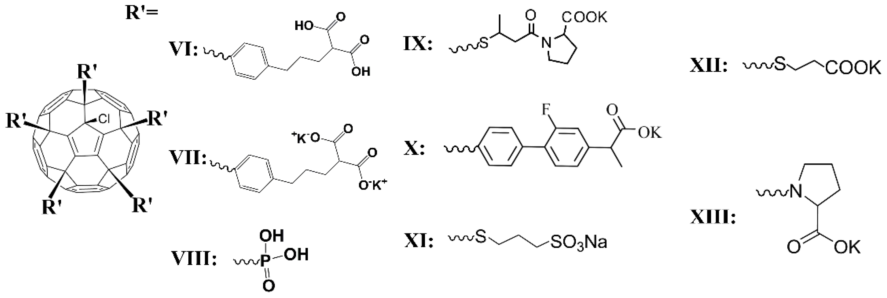
Another picture was observed for fullerene derivatives containing polar groups. The molecular structures of fullerene derivatives containing polar groups are shown in Figure 2.
The exponential diffusion decays were observed for solutions of VI in deuterated DMSO-d6, acetone-d6, and DMSO-D2O mixtures. The fullerene self-diffusion coefficients and hydrodynamic diameters calculated from Equations (2) and (3), respectively, are shown in Table 2.
The calculated particle size formed by the molecules of compound VI in solutions of deuterated DMSO and acetone was 2.3–2.4 nm. The obtained value exceeds the size of the C60 fullerene molecule twice as much. Presumably, it can be explained by the presence of five bulk addends on the C60 fullerene framework, which increases the molecular hydrodynamic diameter. Another option is the formation of self-assembling dimers by compound VI due to the occurrence of hydrogen bonds between the -COOH groups of two complementary molecules, as is well known for organic acids.
At the same time, in a mixture of DMSO-D2O solvents, for the molecules of compound VI, a noticeable aggregation was observed, which can be seen in Table 2. Probably, stable aggregates of various sizes, including D2O molecules, are formed in the system. In addition, the observed exponential diffusion decay characterizes an average aggregate self-diffusion coefficient.
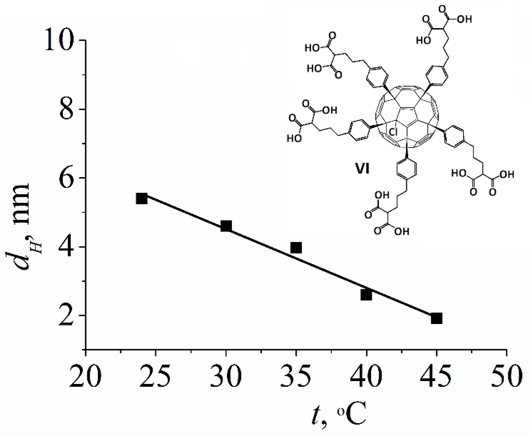
Associates formed by the molecules of compound VI possess low stability, which was confirmed using the dependence of the hydrodynamic diameter dH on temperature. Figure 3 shows that with the increasing temperature of the solution from 25 to 45 °C, a decrease in particle size was observed.

2.1.3. Water-Soluble Fullerene Derivatives

The results obtained for compounds VIIXIII in aqueous solutions should be considered separately.

Compound VII

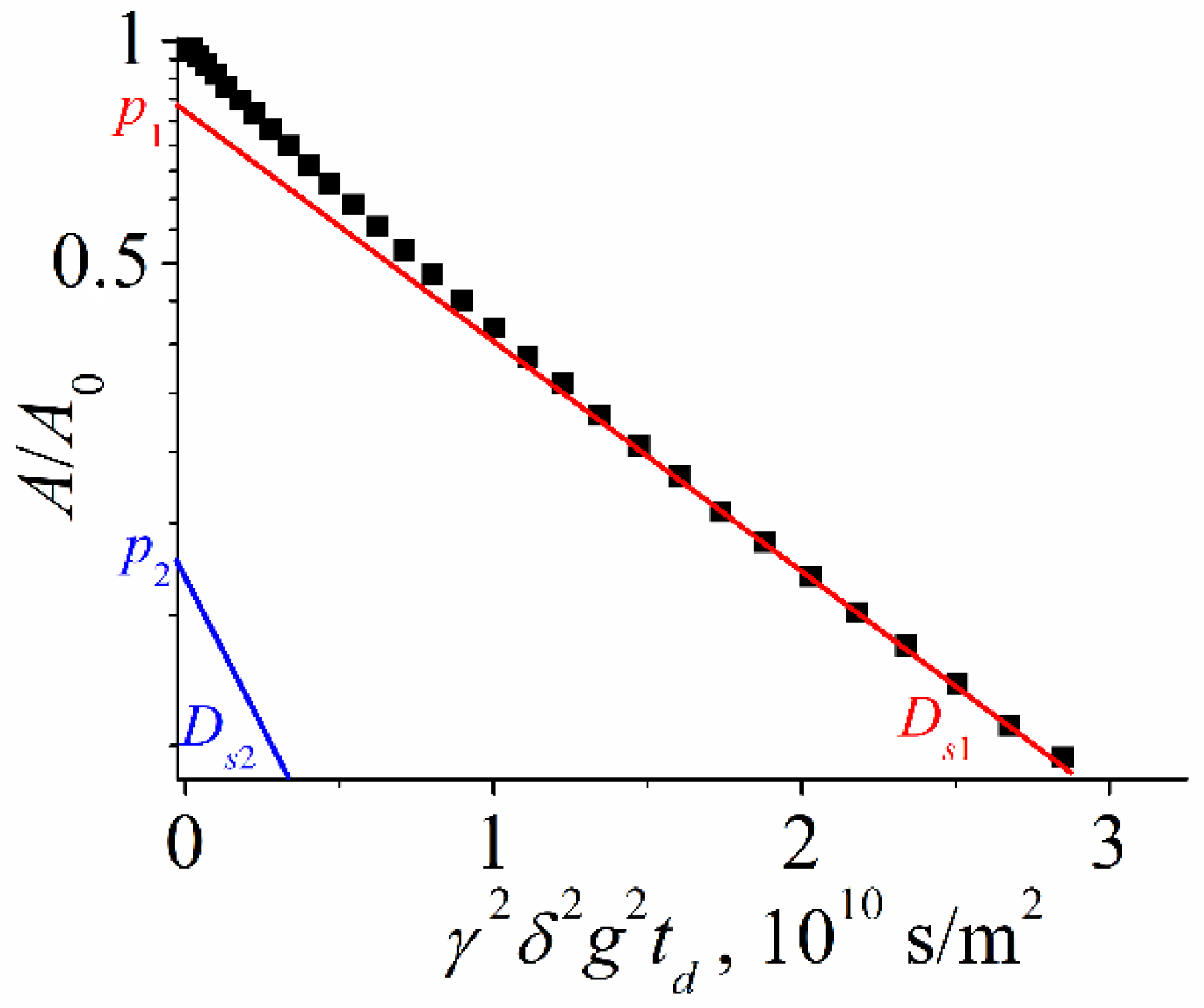
Water-soluble fullerene derivatives (e.g., compound VII in Figure 2) demonstrate the strongest tendency to self-organization in solutions. The self-diffusion of the potassium salt VII in D2O was investigated. The diffusion decays were approximated by the sum of two exponential components according to Equation (4) [59] (m is equal to two in Equation (9)) as it is shown in Figure 4:
A g = p 1 exp γ 2 g 2 δ 2 t d D s 1 + p 2 exp γ 2 g 2 δ 2 t d D s 2 ,
where p1, Ds1 and p2, Ds2—are the relative parts (phase populations) and partial self-diffusion coefficients of the first and the second component, respectively.
The hydrodynamic diameters dH based on each self-diffusion coefficient were calculated from Equation (2) and are given in Table 3.
From the data in Table 3, we can conclude that there are two different types of clusters formed by the fullerene derivative in the solution. Based on the obtained values of hydrodynamic diameters, it can be assumed that isolated molecules (dH = 2 nm) and stable aggregates (dH about 5.5 nm) are observed. The values of the self-diffusion coefficients did not depend on the solution concentration; they were constant in the concentration range from 6 to 24 mg/mL. The increasing of aggregate populations with concentration rising was observed.

Compound VIII

The results obtained for the fullerene derivative VIII show a completely different behavior of molecules in the solution. It is assumed that for the molecules of this compound, the aggregate lifetime is shorter than the diffusion time. This ensures fast exchange between isolated molecules and aggregates of compound VIII. Therefore, the obtained diffusion decays are exponential. Their analysis revealed average self-diffusion coefficients characterizing the translational mobility of various types of compound VIII aggregates.
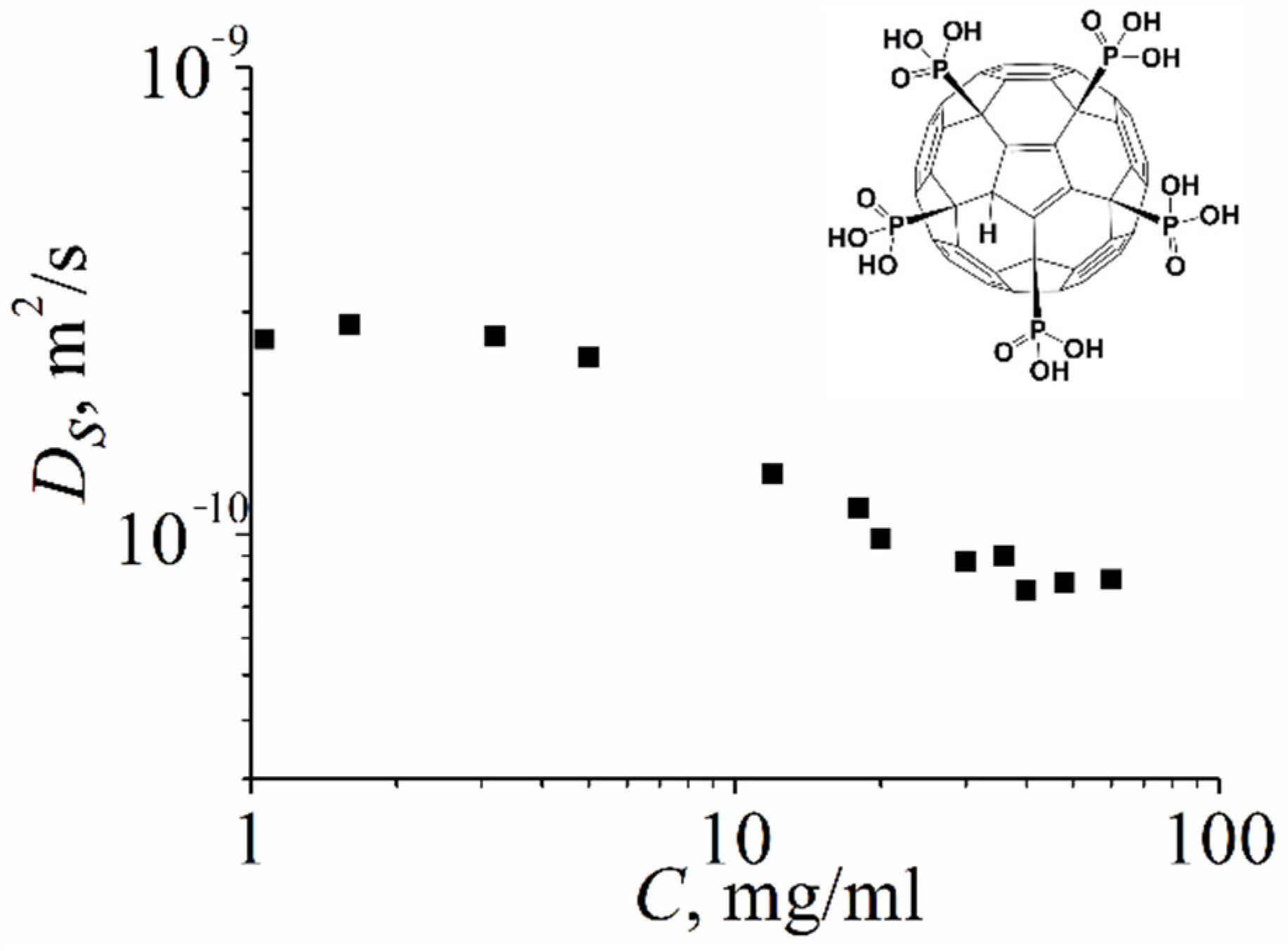
Figure 5 shows the dependence of the self-diffusion coefficient of molecules of compound VIII on the concentration of an aqueous solution. At low solution concentrations (less than 3 mg/mL), the dH value was (1.3 ± 0.15) nm, which corresponds to the size of an isolated molecule. With an increase in concentration, a decrease in the self-diffusion coefficient was observed, which indicates the formation of aggregates by the molecules of the fullerene derivative VIII. At a solution concentration of 40 mg/mL, the maximum value of the hydrodynamic diameter dH = (4.5 ± 0.5) nm was obtained, which does not change with a further increase in concentration.

Compound IX

For compound IX, aqueous solutions of the concentrations indicated in paragraph 4.1 were prepared, and NMR experiments were performed. The diffusion decays of fullerenes (Figure 6) were approximated using bi-exponential curves according to Equation (4).
From the analysis of diffusion decays, the self-diffusion coefficients of the molecules of compound IX were obtained, the values of which were Ds1 = (6.5 ± 0.3) 10−11 m2/s and Ds2 = (4.0 ± 0.5) 10−10 m2/s.
By analogy with the previous compounds, it is assumed that Ds1 and Ds2 characterize particles of the fullerene derivative of different sizes. From Equation (2), the particle sizes were calculated, the values of which were dH1 = (5.2 ± 0.4) nm and dH2 = (1.0 ± 0.06) nm. The obtained values indicate that the molecules of compound IX in an aqueous solution are represented by isolated and aggregated molecules.
The experimental data obtained for compound IX give the possibility to estimate the lifetime of aggregates formed by molecules in an aqueous solution. The lifetime of fullerene aggregates was estimated from the temperature dependence of the low self-diffusion coefficient population on diffusion time td p2(td) according to the procedure proposed in [37].
Figure 7 shows an example of dependence p2(td). The characteristic times of the curve tail were about 1 s, which was in good agreement with the values of the spin lattice relaxation times (T1 = 0.9 s) for this compound, measured independently. Figure 7 also shows the result of the spin lattice relaxation component subtracting, which is a straight line. Their slopes were used to calculate the lifetime τ according to Equation (5):
p 2 = p f exp t d τ + p s exp t d T 1 ,
where τ is the lifetime, T1 is fullerene spin lattice relaxation time, pf + ps = p2(0).
The lifetime values τ are 140 ms, 180 ms, and 200 ms for compound IX solution concentrations of 64.4 mg/mL, 21.3 mg/mL, and 5.3 mg/mL, respectively.

Compound X

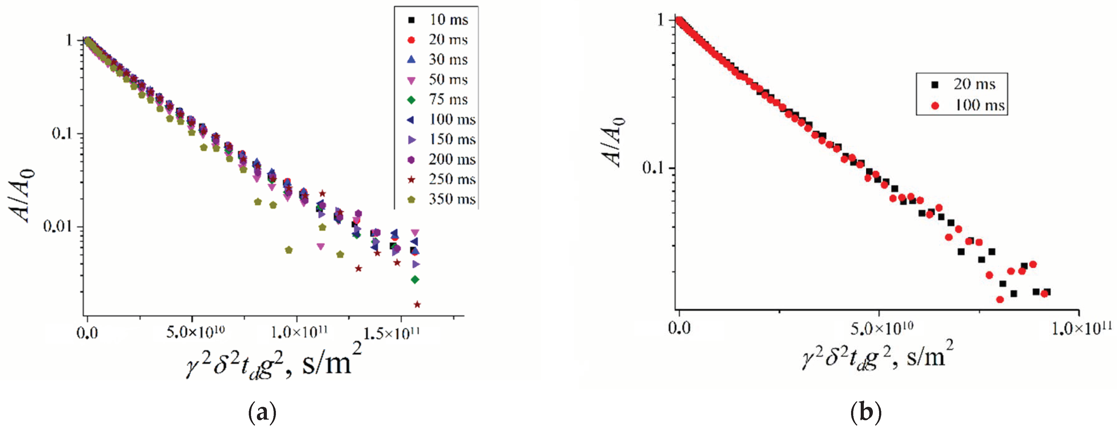
For compound X, aqueous solutions of the concentrations indicated in Section 3.1 were prepared. NMR studies were performed for 1H and 19F nuclei. The diffusion decays of 1H (Figure 8a) and 19F (Figure 8b) nuclei at different diffusion times td are shown.
These diffusion decays are bi-exponential shapes. As shown in Figure 8, self-diffusion coefficients Ds1 and Ds2 are td independent. It may be proposed that two types of molecule associations are observed. The exchange rate between these two associates was slow compared to diffusion time. Self-diffusion coefficients decrease with solution concentration increasing. Hydrodynamic diameters calculated from Stokes–Einstein by Equation (2) are given in Table 4. The lesser diameter is close to individual molecule diameters, but the largest diameter characterizes associate size.
The particle sizes calculated from 1H and 19F self-diffusion data are agreed well. The associate size, as well as the relative part of associated molecules, increases with the solution concentration increasing. The diameter increases from 7.6 nm to 9.8 nm and from 7.6 nm to 10.6 nm; the associate part increases from 0.67 to 0.8 and from 0.69 to 0.83 for 1H and 19F data, correspondingly, with concentration increasing from 6 to 50 mg/mL.

Compound XI

For compound XI, aqueous solutions of the concentrations indicated in Section 3.1 were prepared. NMR studies were performed on 1H nuclei. These derivative diffusion decays are bi exponential which was evidence of two sizes of particles: individual molecules dH2 = (0.9 ± 0.1) nm and associated molecules dH1 = (5.6 ± 0.3) nm.
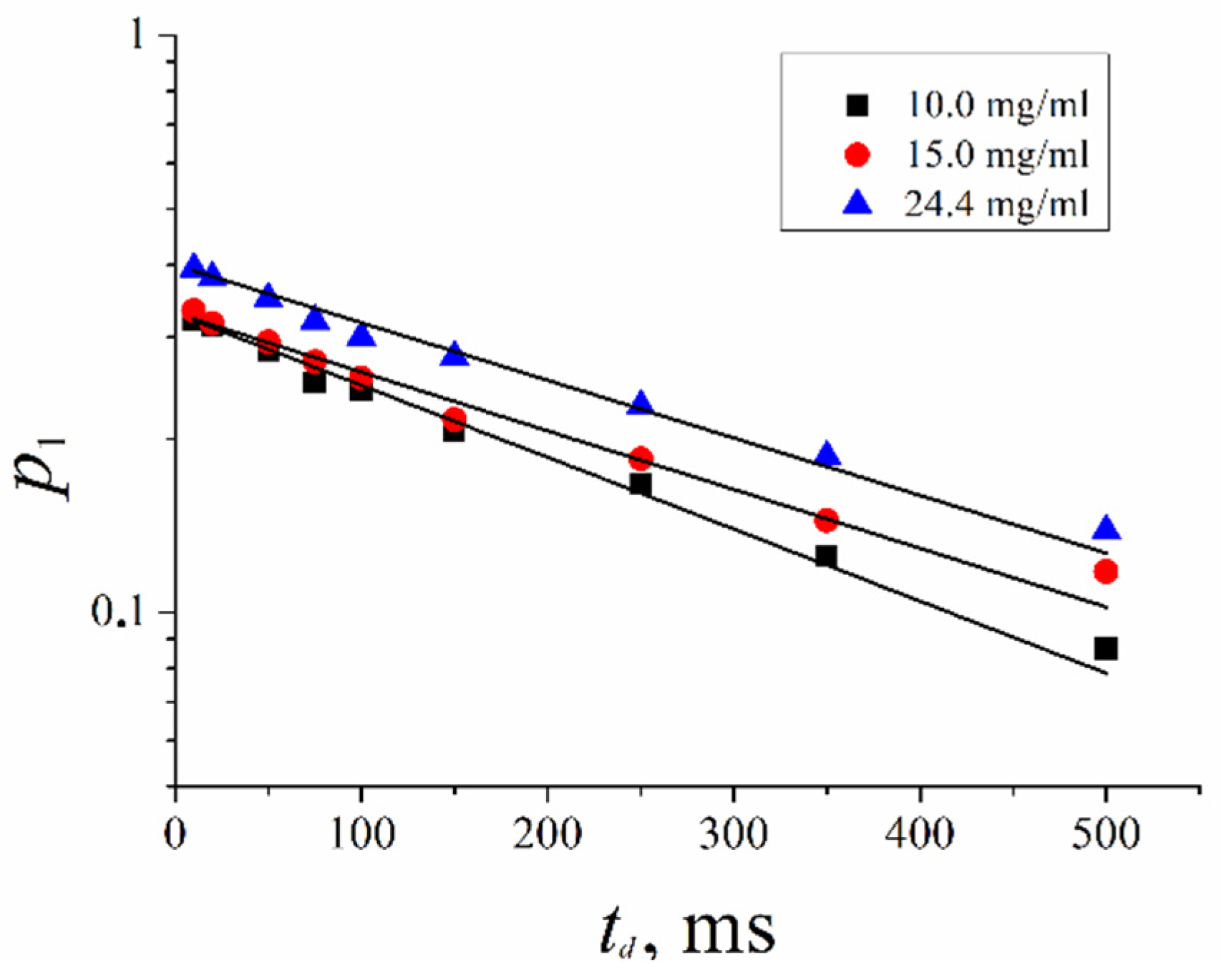
As shown in Figure 9, the low diffusion associated molecule component population p1 decreases with diffusion time td increasing. In Figure 9, p1(td) dependence is shown.
This dependence is exponential, as described by Equation (6). The lifetime of associates formed by molecules of compound XI in aqueous solutions was calculated.
p t d = p 0 exp t d τ
The lifetime of associates τ are (350 ± 10), (430 ± 20) and (440 ± 20) ms at solution concentration C = 10, 15 and 24.4 mg/mL, accordingly. The lifetime of associates increased insignificantly with solution concentration increasing.

Compound XII

The diffusion decay of compound XII is bi-exponential, which is due to isolated (self-diffusion coefficient Ds2 = (4.3 ± 0.8)·10−10 m2/s) and associated molecules (self-diffusion coefficient Ds1 = (7.5 ± 1.5)·10−11 m2/s). The dependence of diffusion decay shape on diffusion time td and concentration was negligible, indicating a slow exchange between isolated and associated molecules compared to the maximum value of td (250 ms). The isolated molecule size dH2 = 1.0 ± 0.2 nm, and the associate size was dH1 = 5.0 ± 1.0 nm. The relative part of associates increases with concentration growth from 0.56 to 0.73.

Compound XIII

The 1H diffusion decay for molecules of compound XIII is also bi-exponential, and decay shape depends on diffusion time and solution concentration C insignificantly. Self-diffusion coefficients Ds1 and Ds2 are (1.3 ± 0.1)·10−10 m2/s and (5.8 ± 0.2)·10−10 m2/s, accordingly. Hydrodynamical diameters are dH2 = (0.7 ± 0.1) nm—isolated molecules and dH1 = (2.8 ± 0.2) nm—associates. The associated molecules’ relative part increases from 0.59 to 0.70, with concentration increasing from 3 to 40 mg/mL.

2.2. Water-Soluble Fullerene Derivatives in Red Blood Cells

Water-soluble fullerene derivatives (WSFDs) are biologically active species. Therefore, one of the fundamental problems is to reveal the mechanism of fullerene derivative interaction with Red Blood Cells (RBCs).
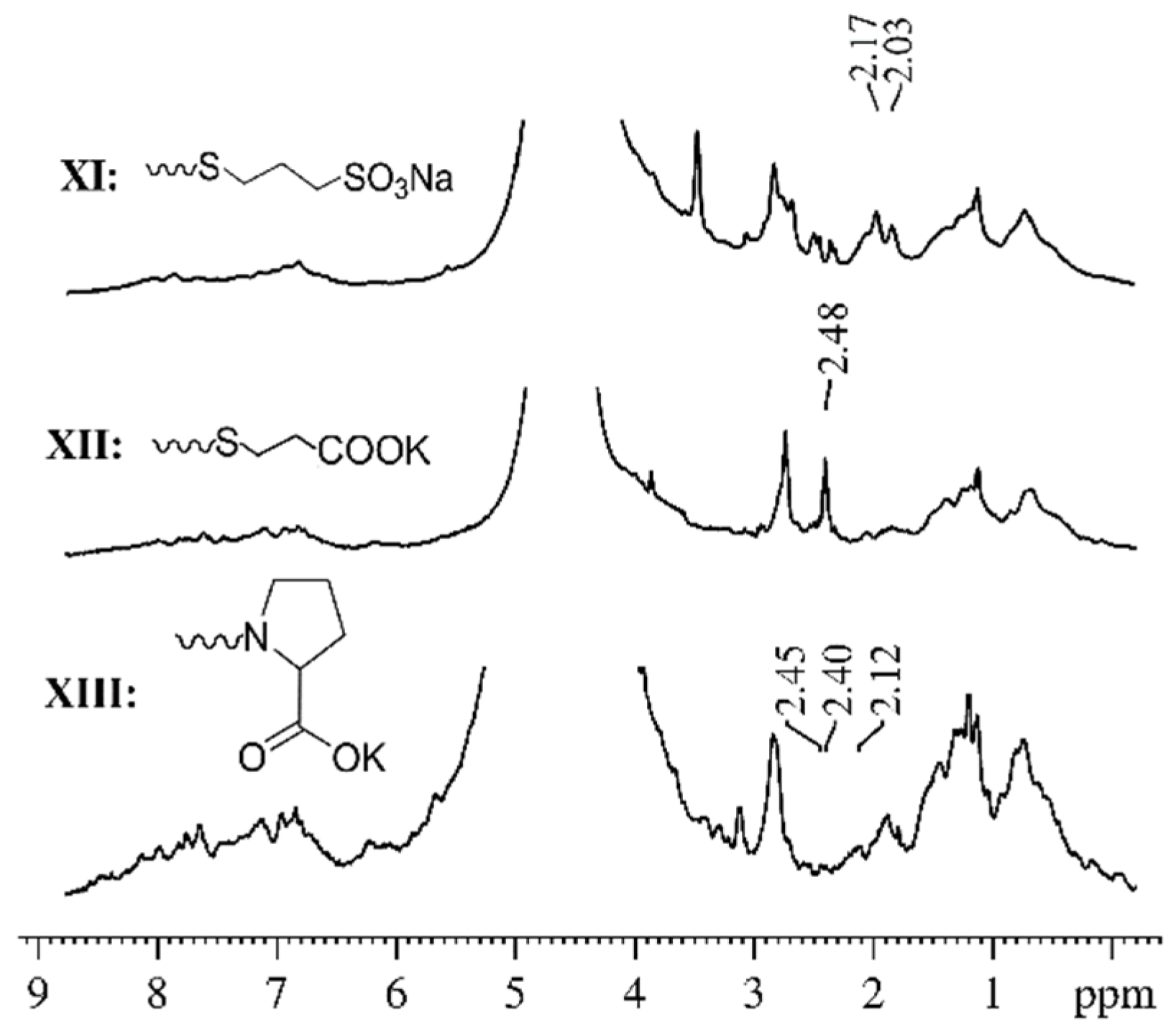
We have previously performed a detailed study of the translational mobility of compound XI in various biological systems: liposomes, shadows, and erythrocytes [53]. In this work, analysis of the high-resolution NMR spectra of the following systems was conducted: an aqueous solution of compound XI; RBCs suspension; suspensions of RBCs, RBCs shadows, and liposomes with the addition of compound XI. Using the analysis of 1H spectra at different amplitudes of the magnetic field gradient, we showed that the signals from the protons of compound XI could be uniquely identified in the spectra of suspensions. These signals do not overlap with the proton signals of the membrane components. It was also shown that, at a high amplitude of the magnetic field gradient, in the spectra of 1H suspension of RBCs with added compound XI, a signal of WSFD molecules was well observed. At the same time, proton signals of compound XI molecules are absent in an aqueous solution under the same experimental conditions. A similar picture was obtained for compounds XII and XIII. Figure 10 shows examples of the 1H spectra of the systems under study (erythrocyte suspensions with added compounds XIXIII). 1H chemical shifts of compounds are slightly changing in suspensions of RBCs compared to solutions due to fullerene derivative interaction with RBCs.
The isolated 1H signals of compound molecules give the possibility to register the diffusion decays of WSFD molecules in a suspension of RBCs correctly. The obtained experimental data made it possible to extract information about the translational mobility of WSFD molecules in cell suspensions. The obtained diffusion decays were complex and were decomposed into three exponent components according to Equation (9) at m = 3. At the same time, in aqueous solutions of compounds XIXIII, two exponential diffusion decays are observed. The values of the self-diffusion coefficients of the molecules of compounds XIXIII are presented in Table 5 [44].
As shown in Table 5, the translational mobility of molecules of compounds XIXIII in a suspension of RBCs is characterized by three diffusion coefficients, the values of which differ by an order of magnitude. However, only two self-diffusion coefficients have been identified in the aqueous solutions of these compounds. Extraction of the third coefficient of self-diffusion, the value of which differs significantly from the other two coefficients, suggests that the movement of the molecules of compounds XIXIII in the cell suspension was hindered.
It is known from the literature data that the lateral diffusion coefficient DL, which characterizes the mobility of lipid molecules in the cell membrane, has a value of about (5–7)∙10−12 m2/s [44,52,53]. The observed “low” coefficient of self-diffusion of the molecules of compounds XIXIII was close to a lateral diffusion coefficient. Thus, the complex nature of the obtained experimental data can be explained by the fact that in the erythrocyte suspension, the molecules of compounds XIXIII are in the aqueous phase of the system or are bound to the erythrocyte cell membrane. The penetration of molecules of compounds XIXIII into the membrane was observed as a result of the appearance of a slow diffusion component. At the same time, in the aqueous phase, the molecules of compounds XIXIII are in an isolated and associated form.
The analysis of the dependence of relative parts slow moving fullerene derivative molecules p1 on diffusion time td makes it possible to estimate the lifetimes τ of fullerene derivative molecules in the erythrocyte membrane from Equation (6). Lifetimes τ for compounds XIXIII are given in Table 6.
The dependences of the relative parts p1 of molecules of compound XI on the diffusion time td at different concentrations are shown in Figure 11. From the analysis of these dependences, the lifetime calculation was carried out according to Equation (6). The obtained values of the lifetimes are presented in Table 7.
As seen from Table 7, the lifetime τ1 of compound XI molecules in the cell membrane increases with the increase of its concentration in the RBCs suspension.

3. Materials and Methods

3.1. Fullerene Derivatives

Methanofullerenes I, II, and IV were reported recently [60,61]. Compound III represents a well-known n-type material for organic solar cells abbreviated as PCBM [62]. Fullerene derivatives V and VIII were synthesized recently in the reaction of chlorofullerene C60Cl6 and triethyl phosphite [17]. Fullerene derivatives VI and VII were reported as products of the Friedel–Crafts arylation of C60Cl6 [63]. Compounds IX and XI were synthesized as described in [64]. Fullerene derivatives X, XII, and XIII were obtained and characterized as reported in our previous papers [19], [65], [66], respectively.
The molecular mobility of compounds IV was studied in various solvents: CDCl3 (I, II, III, V), CS2 (I, II), and C6D5CD3 (II, IV). For investigation of the molecular mobility of compound VI in solution, deuterated DMSO-d6, acetone-d6, and mixtures of DMSO-D2O were used. Compounds VIIXIII are highly soluble in water due to the presence of five hydrophilic groups. Therefore, solutions of various concentrations in deuterium water were prepared for them. The concentrations were from 6 to 24 mg/mL for VII; from 2 to 50 mg/mL for VIII; from 5 to 64 mg/mL for IX; from 6 to 50 mg/mL for X; from 10 to 24 mg/mL for XI; from 10 to 47 mg/mL for XII; from 3 to 40 mg/mL for XIII.

3.2. Red Blood Cells

Blood samples were taken from anesthetized mice by decapitation. The 0.11 molar sodium citrate aqueous solutions were used as an anticoagulant. The citrate solution to blood ratio was one to five by volume. The collected blood samples were centrifuged for 15 min at 1000 rpm, and the plasma was removed with decantation. The erythrocyte sediment was resuspended in NaCl solution (salt concentration was 0.85 g per 100 mL) containing 5 mM of Na-phosphate buffer with pH = 7.4. The centrifugation and decantation procedures were repeated triply; the duration of the second and third centrifugations was 7 min. The fresh buffered NaCl solution was used for each resuspending step. The obtained erythrocyte sediment was stored at 4 °C for no more than 36 h. The fullerene derivatives were added to the RBCs suspension. The final concentration of WSFD in suspension was 1.19·10−2, 9.12·10−3, and 1.99·10−3 M for compound XI; 1.2·10−2 M for compound XII and XIII.

3.3. PFG NMR Technique

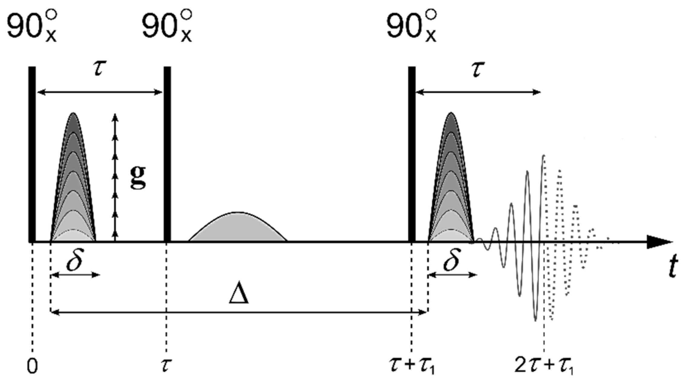
The self-diffusion coefficients were measured with the pulsed field gradient technique for 1H, 19F, and 31P nuclei at the frequencies of 400.13, 376.498, and 161.976 MHz, correspondingly. The measurements were conducted on Bruker Avance-III—400 NMR spectrometer equipped with the diff-60 gradient unit. The pulsed field gradient stimulated echo sequence shown in Figure 12 was applied in the experiments. Three 90° pulses produce a stimulated spin-echo at the time of 2τ + τ1 (where τ and τ1 are the time intervals between the first and the second or the second and the third 90° pulses, respectively). The magnetic field gradient pulses of amplitude g and duration δ were applied after the first and the third 90° pulses. The gradient strength was varied linearly in 64 steps within the range from 0.1 to 27 T/m. The integrated intensities of the spectrum lines were used to obtain the dependence of the echo signal attenuation with respect to g2 (diffusion decay) [44,49,50,51,52,67].
For the molecules undergoing unhindered isotropic Brownian motion, the evolution of the spin echo signal is described by the following Equation (3):
A 2 τ , τ 1 , g = A 2 τ , τ 1 , 0 exp γ 2 g 2 δ 2 t d D s ,
where γ is gyromagnetic ratio, td = Δ−δ/3 is the diffusion time, and Ds is the self-diffusion coefficient, τ, τ1 and g are shown in Figure 12; A(2τ, τ1, 0) is expressed by the equation:
2 τ , τ 1 , 0 = A 0 2 exp 2 τ T 2 τ 1 T 1
where A(0) is the signal intensity after the first radio frequency (RF) pulse (Figure 12). T1 and T2 are the spin-lattice and spin-spin relaxation times, respectively. While measuring the echo signal evolution, τ and τ1 are fixed, and only the dependence of A as a function of g is analyzed, which is called the diffusion decay.
In the case of non-exponential diffusion decay decays, the experimental curves:
A g = A 2 τ , τ 1 , g A 2 τ , τ 1 , 0 ,
are usually deconvoluted in several exponential components, which are described by Equation (3). For the multiphase system consisting of m phases in the case of slow (compared to td) molecular exchange between the phases:
A g = i = 1 m p i exp ( γ 2 g 2 δ 2 t d D s i )
where Dsi is the self-diffusion coefficient of the i-th component and:
p i = p i exp 2 τ T 2 i τ 1 T 1 i / i = 1 m p i exp 2 τ T 2 i τ i T 1 i , i = 1 m p i = 1
Here pi is the relative amount of the nuclei belonging to the molecules characterized by the self-diffusion coefficient Dsi. The value pi is called the population of the i-th phase. For the long T1 and T2 values, it is usually assumed that pipi. The details of the experimental curve decomposition in several exponential diffusion decays were described previously [44].
We have decomposed diffusion decays on two or three exponential components; the values m in Equation (9) were two or three, accordingly. Between different phases (individual and associated molecules), a molecular exchange occurs. The lifetime in phase is τ. In the case of fast exchange rate τ << td (td is diffusion time), the exponential diffusion decay is observed, which is characterized by the average self-diffusion coefficient:
D = ƩpiDsi
For slow rate exchange conditions, τ >> td multi-exponential diffusion decay shape according to Equation (9) is observed, and pi and Dsi do not depend on diffusion time td.
If phase exchange time is comparable with diffusion time τtd, the slowly moving molecule phase population exponentially decreases with increasing diffusion time, and lifetime τ may be estimated [49,50].

4. Conclusions

The self-organization of a series of differently functionalized fullerene derivatives dissolved in the solvents of different polarity and suspension of RBCs was investigated with pulsed field gradient NMR spectroscopy of 1H, 19F, 31P nuclei. No aggregation was observed for the fullerene derivatives containing nonpolar functional groups IV in carbon disulfide, deuterated chloroform, and toluene-d8 solutions. The hydrodynamic diameters calculated from Stokes–Einstein equation are equal to individual molecule van der Waals diameters (1.2–1.4 nm). Therefore, Stokes–Einstein model is correct for hydrodynamic size calculations. The fullerene derivatives bearing polar functional groups such as -COOH, -COOK, -P(O)(OH)2, -SO3Na showed a strong tendency to aggregate. The revealed diameters of the aggregates varied from 2.2 nm to 9.6 nm depending on the solvent and temperature. The water-soluble fullerene derivatives VIIXIII demonstrated the most stable aggregates with a diameter of about 5 nm. An analysis of the concentration dependences showed that in solutions, the number of associated molecules of water-soluble derivatives of fullerenes increases with increasing concentration. Aggregated lifetimes were estimated from the analysis of diffusion decay shape dependence on the diffusion time. Associate lifetime increases with increasing fullerene derivatives aqueous solution concentration.
Self-diffusion of water-soluble fullerene derivatives XIXIII in RBCs was characterized using 1H PFG NMR. Were obtained and analyzed the diffusion decays of the molecules of compounds XIXIII in a suspension of RBCs. It was found that a fraction of fullerene derivative molecules shows a self-diffusion coefficient of about (5–6)·10−12 m2/s, independent of fullerene derivative type, which matched the coefficient of the lateral diffusion of lipids in the RBCs membrane (DL = (5–7) 10−12 m2/s). This experimental finding evidences the absorption of the fullerene derivatives by RBC. The obtained results suggest that fullerene derivative molecules are probably fixed on the RBC surface. The average lifetime of the fullerene derivative molecule on RBC was estimated as 440 ± 70 ms for compound XI, 470 ± 70 ms for compound XII, and 1200 ± 300 ms for compound XIII. An experimental dependence of the lifetime of molecules of compound XI in the RBC membrane on the concentration of the compound in suspension was obtained. It was shown that with increasing concentration, the lifetime also increases.
Thus, pulsed field gradient NMR was shown to be a versatile technique for the investigation of self-organization and interactions of the fullerene derivatives with blood cells, providing essential information which could be projected on their behavior in-vivo after intravenous administration while screening as potential drug candidates.

Author Contributions

Statement of work, writing—review and editing V.I.V.; investigated derivatives of fullerenes A.V.M., O.A.K., E.A.K. and P.A.T.; investigated red blood cells Y.V.S.; experimental work and processing of results A.V.C. and I.A.A. All authors have read and agreed to the published version of the manuscript.

Funding

This research was performed with the support of State Assignment of the Federal Research Center of Problem of Chemical Physics and Medicinal Chemistry RAS (state registration No. 0089-2019-0010/AAAA-A19-119071190044-3 and No. AAAA-A19-119071890015-6).

Acknowledgments

NMR measurements were performed using equipment from the Multi-User Analytical Center of the Federal Research Center of Problem of Chemical Physics and Medicinal Chemistry RAS and Science Center in Chernogolovka, RAS. We are grateful to I.I. Voronov and A.V. Zhilenkov for the synthesis of several fullerene derivatives.

Conflicts of Interest

The authors declare no conflict of interest.

References

  1. Langa, F.; Nierengarten, J.-F. Fullerene Principles and Applications; RCS: Cambridge, UK, 2007; 398p. [Google Scholar] [CrossRef]
  2. Penkova, A.V.; Acquah, S.F.A.; Piotrovskiy, L.B.; Markelov, D.A.; Semisalova, A.S.; Kroto, H.W. Fullerene derivatives as nano-additives in polymer composites. Russ. Chem. Rev. 2017, 86, 530. [Google Scholar] [CrossRef]
  3. Nakajima, N.; Nishi, C.; Li, F.-M.; Ikada, Y. Photo-Induced Cytotoxicity of Water-Soluble Fullerene. Fuller. Sci. Technol. 1996, 4, 1–19. [Google Scholar] [CrossRef]
  4. Tabata, Y.; Ishii, T.; Aoyama, T.; Oki, R.; Hirano, Y.; Ogawa, O.; Ikada, Y. Sonodynamic Effect of Polyethylene glecol-conjugated Fullerene on Tumor. In Perspectives of Fullerene Nanotechnology; Osawa, E., Ed.; Kluver Academic Publishers: Amsterdam, The Netherlands, 2001; pp. 185–199. [Google Scholar]
  5. Mashino, T.; Nishikawa, D.; Takanashi, K.; Usui, U.; Yamori, T.; Seki, M.; Endo, T.; Mochizuki, M. Antibacterial and antiproliferative activity of cationic fullerene derivatives. Bioorg. Med. Chem. Lett. 2003, 13, 4395–4397. [Google Scholar] [CrossRef] [PubMed]
  6. Fernandes, N.B.; Shenoy, R.U.K.; Kajampady, M.K.; DCruz, C.E.M.; Shirodkar, R.K.; Kumar, L.; Verma, R. Fullerenes for the treatment of cancer: An emerging tool. Environ. Sci. Pollut. Res. Int. 2022, 29, 58607–58627. [Google Scholar] [CrossRef]
  7. Friedman, S.H.; Ganapathi, P.S.; Rubin, Y.; Kenyon, G.L. Optimizing the binding of fullerene inhibitors of the HIV-1 protease through predicted increases in hydrophobic desolvation. J. Med. Chem. 1998, 41, 2424–2429. [Google Scholar] [CrossRef]
  8. Bosi, S.; Da Ros, T.; Spalluto, G.; Balzarini, J.; Prato, M. Synthesis and anti-HIV properties of new water-soluble bis-functionalized[60]fullerene derivatives. Bioorg. Med. Chem. Lett. 2003, 13, 4437. [Google Scholar] [CrossRef]
  9. Mashino, T.; Shimotohno, K.; Ikegami, N.; Nishikawa, D.; Okuda, K.; Takanashi, K.; Nakamura, S.; Mochizuki, M. Human immunodeficiency virus-reverse transcriptase inhibition and hepatitis C virus RNA-dependent RNA polymerase inhibition activities of fullerene derivatives. Bioorg. Med. Chem. Lett. 2005, 15, 1107. [Google Scholar] [CrossRef]
  10. Castro, E.; Garcia, A.H.; Zavalaa, G.; Echegoyen, L. Fullerenes in biology and medicine. J. Mater. Chem. B 2017, 5, 6523. [Google Scholar] [CrossRef]
  11. Khalikov, S.K.; Sharipova, D.; Zafarov, S.Z.; Umarkhon, M.; Alieva, S.V. Synthesis and Characterization of Fullero-C60 a-Amino Acids with Antiviral Properties. Chem. Nat. Compd. 2017, 53, 121–127. [Google Scholar] [CrossRef]
  12. Bosi, S.; Da Ros, T.; Castellano, S.; Bafni, E.; Prato, M. Antimycobacterial activity of ionic fullerene derivatives. Bioorg. Med. Chem. Lett. 2000, 10, 1043–1045. [Google Scholar] [CrossRef]
  13. Su, Y.; Ding, M.; Dong, H.; Hu, Y.; Yang, D.; Shao, J.; Huang, B. Recent advances in nanozymes for combating bacterial infection. Mater. Chem. Front. 2022, 6, 2596–2609. [Google Scholar] [CrossRef]
  14. Belik, A.Y.; Rybkin, A.Y.; Voronov, I.V.; Goryachev, N.S.; Volyniuk, D.; Grasulevicius, J.V.; Troshin, P.A.; Kotelnikov, A.I. Non-covalent complexes of polycationic fullerene C60 derivative with xanthene dyes—Spectral and photochemical properties in water and in liposomes. Dyes Pigments 2017, 139, 65–72. [Google Scholar] [CrossRef]
  15. Mikhailov, P.A.; Kraevaya, O.A.; Belik, A.Y.; Rybkin, A.Y.; Khakina, E.A.; Goryachev, N.S.; Usol’tseva, L.I.; Romanenko, Y.V.; Koifman, O.I.; Gushchina, O.I.; et al. Synthesis, photophysical properties, and photochemical activity of the water-soluble dyad based on fullerene C60 and chlorin e6 derivatives. Dokl. Phys. Chem. 2017, 477, 226–230. [Google Scholar] [CrossRef]
  16. Belik, A.Y.; Rybkin, A.Y.; Goryachev, N.S.; Sadkov, A.P.; Filatova, N.V.; Buyanovskaya, A.G.; Talanova, V.N.; Klemenkova, Z.S.; Romanova, V.S.; Koifman, M.O.; et al. Nanoparticles of water-soluble dyads based on amino acid fullerene C60 derivatives and pyropheophorbide: Synthesis, photophysical properties, and photodynamic activity. Spectrochim. Acta Part A Mol. Biomol. Spectrosc. 2021, 260, 119885. [Google Scholar] [CrossRef]
  17. Yurkova, A.A.; Khakina, E.A.; Troyanov, S.I.; Chernyak, A.V.; Shmygleva, L.; Peregudov, A.A.; Martynenko, V.M.; Dobrovolskiy, Y.A.; Troshin, P.A. Arbuzov chemistry with chlorofullerene C60Cl6: A powerful method for selective synthesis of highly functionalized [60]fullerene derivatives. Chem. Commun. 2012, 48, 8916. [Google Scholar] [CrossRef]
  18. Khakina, E.A.; Kraevaya, O.A.; Popova, M.L.; Peregudov, A.S.; Troyanov, S.I.; Chernyak, A.V.; Martynenko, V.M.; Kulikov, A.V.; Scholse, D.; Troshin, P.A. Synthesis of different types of alkoxy fullerene derivatives from chlorofullerene C60Cl6. Org. Biomol. Chem. 2017, 15, 773–777. [Google Scholar] [CrossRef]
  19. Kraevaya, O.A.; Peregudov, A.S.; Godovikov, I.A.; Shchurik, E.V.; Martynenko, V.M.; Shestakov, A.F.; Balzarini, J.; Schols, D.; Troshin, P.A. Direct arylation of C60Cl6 and C70Cl8 with carboxylic acids: A synthetic avenue to water-soluble fullerene derivatives with promising antiviral activity. Chem. Commun. 2020, 56, 1179–1182. [Google Scholar] [CrossRef]
  20. Ros, T.D.; Prato, M. Medicinal chemistry with fullerenes and fullerene derivatives. Chem. Commun. 1999, I.8, 663–669. [Google Scholar] [CrossRef]
  21. Rokitskaya, T.I.; Antonenko, Y.N. Fullerenol C60(OH)24 increases ion permeability of lipid membranes in a pH-dependent manner. Biochim. Biophys. Acta 2016, 1858, 1165–1174. [Google Scholar] [CrossRef]
  22. Wattraint, O.; Sarazin, C. Diffusion measurements of water, ubiquinone and lipid bilayer inside a cylindrical nanoporous support: A stimulated echo pulsed-field gradient MAS-NMR investigation. Biochim. Biophys. Acta 2005, 1713, 65–72. [Google Scholar] [CrossRef] [Green Version]
  23. Leal, C.; Sandström, D.; Nevsten, P.; Topgaard, D. Local and translational dynamics in DNA–lipid assemblies monitored by solid-state and diffusion NMR. Biochim. Biophys. Acta 2008, 1778, 214–228. [Google Scholar] [CrossRef] [PubMed] [Green Version]
  24. Tardy-Laporte, C.; Arnold, A.A.; Genard, B.; Gastineau, R.; Morançais, M.; Mouget, J.-L.; Tremblay, R.; Marcotte, I. A 2H solid-state NMR study of the effect of antimicrobial agents on intact Escherichia coli without mutating. Biochim. Biophys. Acta 2013, 1828, 614–622. [Google Scholar] [CrossRef] [PubMed] [Green Version]
  25. Grebowski, J.; Krokosz, A.; Puchala, M. Fullerenol C60(OH)36 could associate to band 3 protein of human erythrocyte membranes. Biochim. Biophys. Acta 2013, 1828, 2007–2014. [Google Scholar] [CrossRef] [PubMed] [Green Version]
  26. Grebowski, J.; Krokosz, A.; Puchala, M. Membrane fluidity and activity of membrane ATPases in human erythrocytes under the influence of polyhydroxylated fullerene. Biochim. Biophys. Acta 2013, 1828, 241–248. [Google Scholar] [CrossRef] [PubMed] [Green Version]
  27. Chen, M.; Zhou, S.; Guo, L.; Wang, L.; Yao, F.; Hu, Y.; Li, H.; Hao, J. Aggregation Behavior and Antioxidant Properties of Amphiphilic Fullerene C60 Derivatives Cofunctionalized with Cationic and Nonionic Hydrophilic Groups. Langmuir 2019, 35, 6939–6949. [Google Scholar] [CrossRef]
  28. Harano, K.; Nakamura, E. Interfacial Chemistry of Conical Fullerene Amphiphiles in Water. Acc. Chem. Res. 2019, 52, 2090–2100. [Google Scholar] [CrossRef]
  29. Li, J.; Chen, M.; Zhou, S.; Li, H.; Hao, J. Self-assembly of fullerene C60-based amphiphiles in solutions. Chem. Soc. Rev. 2022, 51, 3226–3242. [Google Scholar] [CrossRef]
  30. Nakamura, E.; Isobe, H. Functionalized Fullerenes in Water. The First 10 Years of Their Chemistry, Biology, and Nanoscience. Acc. Chem. Res. 2003, 36, 807. [Google Scholar] [CrossRef]
  31. Zhang, G.; Liu, Y.; Liang, D.; Gan, L.; Li, Y. Facile Synthesis of Isomerically Pure Fullerenols and Formation of Spherical Aggregates from C60(OH)8. Angew. Chem. Int. Ed. 2010, 49, 5293. [Google Scholar] [CrossRef]
  32. E, Y.; Bai, L.; Fan, L.; Han, M.; Zhang, X.; Yang, S. Electrochemically generated fluorescent fullerene[60] nanoparticles as a new and viable bioimaging platform. J. Mater. Chem. 2011, 21, 819–823. [Google Scholar] [CrossRef]
  33. Wang, G.; Zhao, G.; Yan, L. Investigation on the aggregation properties of cationic [60]fullerene derivative. Chin. Sci. Bull. 2004, 49, 1441. [Google Scholar] [CrossRef]
  34. Liao, Q.; Qu, X.; Chen, L.; Jin, X. Aggregates of Polymer-Substituted Fullerenes. J. Phys. Chem. B 2006, 110, 7153. [Google Scholar] [CrossRef] [PubMed]
  35. Kato, H.; Böttcher, C.; Hirsch, A. Sugar Balls: Synthesis and Supramolecular Assembly of [60]Fullerene Glycoconjugates. Eur. J. Org. Chem. 2007, 2007, 2659–2666. [Google Scholar] [CrossRef]
  36. Braun, M.; Hartnagel, U.; Ravanelli, E.; Schade, B.; Böttcher, C.; Vostrowsky, O.; Hirsch, A. Amphiphilic [5:1]- and [3:3]-Hexakisadducts of C60. Eur. J. Org. Chem. 2004, 2004, 1983–2001. [Google Scholar] [CrossRef]
  37. Avilova, I.A.; Chernyak, A.V.; Zhilenkov, A.V.; Troshin, P.A.; Volkov, V.I. The self-organization of water soluble fullerene derivatives studied by pulsed field gradient NMR. Mendeleev Commun. 2016, 26, 146. [Google Scholar] [CrossRef]
  38. Hong, Y.S.; Kim, K.C.; Volkov, V.I.; Skirda, V.D.; Lee, C.-H. Structural and dynamic properties of polyoxyethylene sorbitan monooleate micelle in water dispersion studied by pulsed field gradient NMR. Appl. Magn. Reson. 2005, 29, 351. [Google Scholar] [CrossRef]
  39. Zhu, Y.-Y.; Li, C.; Li, G.-Y.; Jiang, X.-K.; Li, Z.-T. Hydrogen-Bonded Aryl Amide Macrocycles:  Synthesis, Single-Crystal Structures, and Stacking Interactions with Fullerenes and Coronene. J. Org. Chem. 2008, 73, 1745–1751. [Google Scholar] [CrossRef]
  40. Ksenofontov, A.A.; Bichan, N.G.; Khodov, I.A.; Antina, E.V.; Berezin, M.B.; Vyugin, A.I. Novel non-covalent supramolecular systems based on zinc(II) bis(dipyrromethenate)s with fullerenes. J. Mol. Liq. 2018, 269, 327–334. [Google Scholar] [CrossRef]
  41. Ksenofontov, A.A.; Lukanov, M.M.; Bichan, N.G.; Khodov, I.A.; Kudryakova, N.O.; Ksenofontova, K.V.; Antina, E.V. Non-covalent supramolecular systems with photoinduced electron transfer based on zinc bis(dipyrromethenate)s and C60. Dyes Pigments 2021, 185, 108918. [Google Scholar] [CrossRef]
  42. Hirao, T.; Fujii, N.; Iwabe, Y.; Haino, T. Self-sorting behavior in supramolecular fullerene polymerization directed by host–guest complexation between calix[5]arene and C60. Chem. Commun. 2021, 57, 11831–11834. [Google Scholar] [CrossRef]
  43. Chen, S.; Chen, D.; Lu, M.; Zhang, X.; Li, H.; Zhang, X.; Yang, X.; Li, X.; Tu, Y.; Li, C.Y. Incorporating Pendent Fullerenes with High Refractive Index Backbones: A Conjunction Effect Method for High Refractive Index Polymers. Macromolecules 2015, 48, 8480–8488. [Google Scholar] [CrossRef]
  44. Volkov, V.I.; Chernyak, A.V.; Avilova, I.A.; Slesarenko, N.A.; Melnikova, D.L.; Skirda, V.D. Molecular and Ionic Diffusion in Ion Exchange Membranes and Biological Systems (Cells and Proteins) Studied by NMR. Membranes 2021, 11, 385. [Google Scholar] [CrossRef]
  45. Gianolio, E.; Ferrauto, G.; Di Gregorio, E.; Aime, S. Re-evaluation of the water exchange lifetime value across red blood cell membrane. Biochim. Biophys. Acta 2016, 1858, 627–631. [Google Scholar] [CrossRef] [PubMed]
  46. Lindblom, G.; Orädd, G. Lipid lateral diffusion and membrane heterogeneity. Biochim. Biophys. Acta 2009, 1788, 234–244. [Google Scholar] [CrossRef] [PubMed] [Green Version]
  47. Waldeck, A.R.; Kuchel, P.W.; Lennon, A.J.; Chapman, B.E. NMR diffusion measurements to characterise membrane transport and solute binding. Prog. Nucl. Magn. Reson. Spectrosc. 1997, 30, 39–68. [Google Scholar] [CrossRef]
  48. Anselmi, C.; Bernardi, F.; Centini, M.; Gaggelli, E.; Gaggelli, N.; Valensin, D.; Valensin, G. Interaction of ferulic acid derivatives with human erythrocytes monitored by pulsed field gradient NMR diffusion and NMR relaxations studies. Chem. Phys. Lipids 2005, 134, 109–117. [Google Scholar] [CrossRef]
  49. Cho, C.-H.; Hong, Y.-S.; Kang, K.; Volkov, V.I.; Skirda, V.D.; Lee, C.-Y.J.; Lee, C.-H. Water self-diffusion in Chlorella sp. studied by pulse field gradient NMR. Magn. Reson. Imaging 2003, 21, 1009–1017. [Google Scholar] [CrossRef]
  50. Suh, K.-J.; Hong, Y.-S.; Skirda, V.D.; Volkov, V.I.; Lee, C.-Y.; Lee, C.-H. Water self-diffusion behavior in yeast cells studied by pulsed field gradient NMR. Biophys. Chem. 2003, 104, 121–130. [Google Scholar] [CrossRef]
  51. Avilova, I.A.; Vasil’ev, S.G.; Rimareva, L.V.; Serba, E.M.; Volkova, L.D.; Volkov, V.I. Water metabolism in cells of Saccharomyces cerevisiae of races Y-3137 and Y-3327, according to pulsed-field gradient NMR data. Russ. J. Phys. Chem. A 2015, 89, 710. [Google Scholar] [CrossRef]
  52. Avilova, I.A.; Smolina, A.V.; Kotelnikov, A.I.; Kotelnikova, R.A.; Loskutov, V.V.; Volkov, V.I. Self-diffusion of water and blood lipids in mouse erythrocytes. Appl. Magn. Reson. 2016, 47, 335–347. [Google Scholar] [CrossRef]
  53. Avilova, I.A.; Khakina, E.A.; Kraevaya, O.A.; Kotelnikov, A.I.; Kotelnikova, R.A.; Troshin, P.A.; Volkov, V.I. Self-diffusion of water-soluble fullerene derivatives in mouse erythrocytes. Biochim. Biophys. Acta BBA-Biomembr. 2018, 1860, 1537–1543. [Google Scholar] [CrossRef] [PubMed]
  54. Spernol, A.; Wirtz, K. Zur Mikroreibung in Flüssigkeiten. Z. Nat. A 1953, 8, 522–532. [Google Scholar] [CrossRef] [Green Version]
  55. Gierer, A.; Wirtz, K. Molekulare Theorie der Mikroreibung. Z. Nat. A 1953, 8, 532–538. [Google Scholar] [CrossRef] [Green Version]
  56. Perrin, F. Mouvement brownien d’un ellipsoide—I. Dispersion diélectrique pour des molécules ellipsoidales. J. Phys. Radium 1936, 7, 497–511. [Google Scholar] [CrossRef]
  57. Edward, J.T. Molecular volumes and the Stokes-Einstein equation. J. Chem. Educ. 1970, 47, 261. [Google Scholar] [CrossRef]
  58. Kawasaki, S.; Aketa, T.; Touhara, H.; Okino, F.; Boltalina, O.V.; Gol’dt, I.V.; Troyanov, S.I.; Taylor, R. Crystal Structures of the Fluorinated Fullerenes C60F36 and C60F48. J. Phys. Chem. B 1999, 103, 1223. [Google Scholar] [CrossRef]
  59. Chernyak, A.V.; Avilova, I.A.; Khakina, E.A.; Mumyatov, A.V.; Zabrodin, V.A.; Troshin, P.A.; Volkov, V.I. Supramolecular Self-Organization of Fullerene Derivatives in Solutions Studied by Pulsed Field Gradient NMR Technique. Appl. Magn. Reson. 2016, 47, 859–868. [Google Scholar] [CrossRef]
  60. Mumyatov, A.V.; Susarova, D.K.; Mukhacheva, O.A.; Troshin, P.A.; Razumov, V.F. Fullerene Derivatives with Reduced Electron Affinity and a Photovoltaic Cell Using the Same. European Patent Application No. EP 12180839.8A, 17 August 2012. [Google Scholar]
  61. Mumyatov, A.V.; Goryachev, A.E.; Prudnov, F.A.; Mukhacheva, O.A.; Sagdullina, D.K.; Chernyak, A.V.; Troyanov, S.I.; Troshin, P.A. Monocyclopropanated fullerene derivatives with decreased electron affinity as promising electron acceptor materials for organic solar cells. Synth. Met. 2020, 270, 116565. [Google Scholar] [CrossRef]
  62. Hummelen, J.C.; Knight, B.W.; LePeq, F.; Wudl, F.; Yao, J.; Wilkins, C.L. Preparation and Characterization of Fulleroid and Methanofullerene Derivatives. J. Org. Chem. 1995, 60, 532–538. [Google Scholar] [CrossRef]
  63. Voronov, I.I.; Martynenko, V.M.; Chernyak, A.V.; Balzarini, J.; Schols, D.; Troshin, P.A. Synthesis and Antiviral Activity of Water-Soluble Polycarboxylic Derivatives of [60]Fullerene Loaded with 3,4-Dichlorophenyl Units. Chem. Biodivers. 2018, 15, e1800293. [Google Scholar] [CrossRef]
  64. Hsieh, F.-Y.; Zhilenkov, A.V.; Voronov, I.I.; Khakina, E.A.; Mischenko, D.V.; Troshin, P.A.; Hsu, S.-h. Water-soluble fullerene derivatives as brain drugs: Surface chemistry determines neuroprotective and antitumor effects. ACS Appl. Mater. Interfaces 2017, 9, 11482–11492. [Google Scholar] [CrossRef] [PubMed]
  65. Khakina, E.A.; Yurkova, A.A.; Peregudov, A.S.; Troyanov, S.I.; Trush, V.; Vovk, A.I.; Mumyatov, A.V.; Martynenko, V.M.; Balzarini, J.; Troshin, P.A. Highly selective reactions of C60Cl6 with thiols for synthesis of functionalized [60]fullerene derivatives. Chem. Commun. 2012, 48, 7158–7160. [Google Scholar] [CrossRef] [PubMed] [Green Version]
  66. Wong, C.-W.; Zhilenkov, A.V.; Kraevaya, O.A.; Mischenko, D.V.; Troshin, P.A.; Hsu, S.-h. Toward Understanding the Antitumor Effects of Water-Soluble Fullerene Derivatives on Lung Cancer Cells: Apoptosis or Autophagy Pathways? J. Med. Chem. 2019, 62, 7111–7125. [Google Scholar] [CrossRef] [PubMed]
  67. Anisimov, A. Gradient NMR Method for Studies of Water Translational Diffusion in Plants. Membranes 2021, 11, 487. [Google Scholar] [CrossRef] [PubMed]
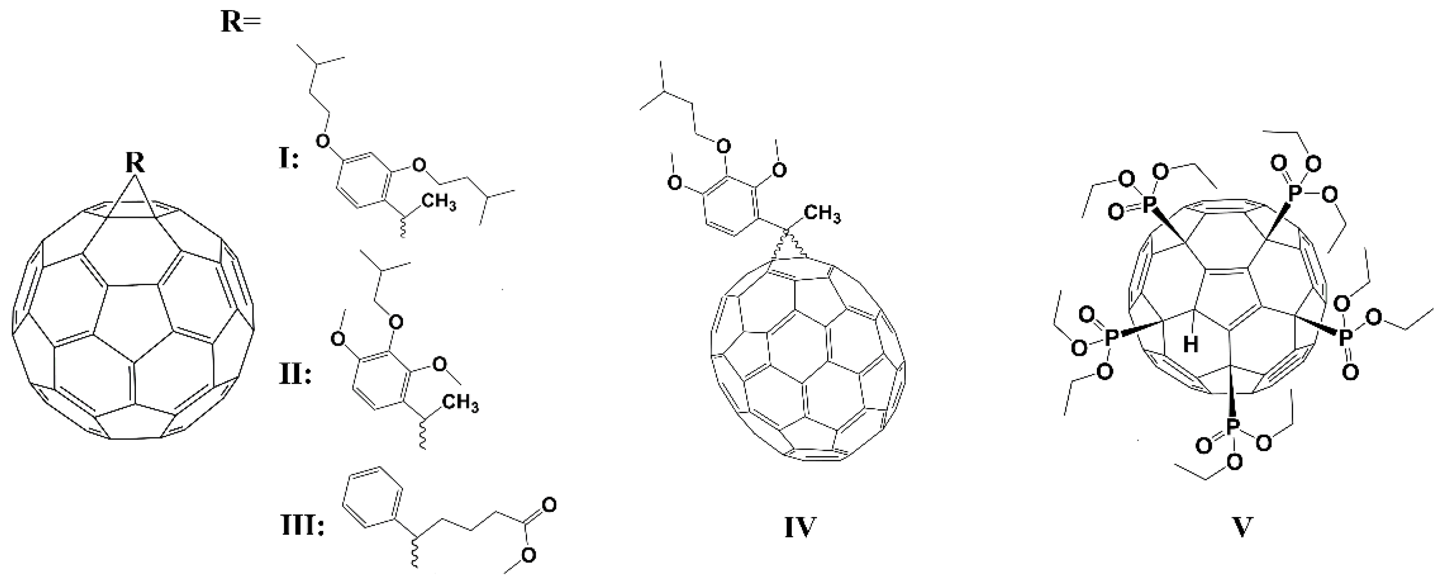
Figure 1. The molecular structure of compounds IV.
Ijms 23 13344 g001
Figure 2. The molecular structure of compounds VIXIII.
Ijms 23 13344 g002
Figure 3. Temperature dependence of the hydrodynamic diameter dH characterizing the association of the fullerene derivative VI in DMSO/D2O (3:1 m/m) solution. The concentration of VI was 8 mg/mL.
Ijms 23 13344 g003
Figure 4. Diffusion decay of the fullerene derivative VII in D2O solution at td = 20 ms, C = 6 mg/mL.
Ijms 23 13344 g004
Figure 5. The concentration dependence of self-diffusion coefficients of compound VIII in D2O measured on 31P nuclei.
Ijms 23 13344 g005
Figure 6. Diffusion decays of the fullerene derivative IX at different diffusion times td. The values of td are indicated in insertion. C = 64.4 mg/ml. Reprinted with permission from Ref. [37]. Copyright © 2016 Published by Elsevier B.V.
Ijms 23 13344 g006
Figure 7. 1—the dependence of part p2(td) on diffusion time td; 2—the dependence p2(td) after subtraction of spin–lattice relaxation component. Reprinted with permission from Ref. [37]. Copyright © 2016 Published by Elsevier B.V.
Ijms 23 13344 g007
Figure 8. 10 Diffusion decays of 1H and 19F nuclei of compound X at different diffusion times td: (a)—1H nuclei; (b)–19F nuclei. The values of td show in insertions. The aqueous solution concentration is 50 mg/mL.
Ijms 23 13344 g008
Figure 9. Dependences of associate molecules populations of compound XI on diffusion times. The values of concentrations are indicated in insertion.
Ijms 23 13344 g009
Figure 10. The 1H spectra of the RBCs suspensions with added compounds XIXIII.
Ijms 23 13344 g010
Figure 11. The dependences of the relative parts p1 of molecules of compound XI on the diffusion time td at various concentrations. The concentrations indicate in the insertion.
Ijms 23 13344 g011
Figure 12. Stimulated echo pulse sequence with the magnetic field gradient pulses. Here, τ is the time interval between the first and the second RF pulses, and τ1 is the time interval between the second and the third ones. D is the interval between the gradient pulses, δ is the duration of the equivalent rectangular magnetic field gradient pulses, g is the amplitude of the magnetic field gradient pulse, and g0 is the amplitude of the constant magnetic field gradient. Reprinted with permission from Ref. [44].
Ijms 23 13344 g012
Table 1. The self-diffusion coefficients Ds and the diameters dH calculated from Equation (2) for the fullerene derivatives IV.
CompoundSolventDs, m2/sdH, nm
ICDCl35.9·10−101.3 ± 0.1
CS21.0·10−91.2 ± 0.1
IICDCl36.3·10−101.3 ± 0.1
CS21.1·10−91.2 ± 0.1
C6D5CD36.6·10−101.2 ± 0.1
IIICDCl36.9·10−101.2 ± 0.1
IVC6D5CD36.1·10−101.2 ± 0.1
VCDCl35.8·10−101.4 ± 0.1
Table 2. The self-diffusion coefficients Ds and the diameters dH from Equation (2) for fullerene derivative VI, the solution concentration C = 8 mg/mL.
SolventDs, m2/sdH, nm
acetone-d65.7·10−102.4 ± 0.2
DMSO8.5·10−112.3 ± 0.2
The molar ratio DMSO:D2O = 3:12.1·10−115.4 ± 0.5
The molar ratio DMSO:D2O = 1:12.3·10−116.3 ± 0.6
The molar ratio DMSO:D2O = 1:32.3·10−119.6 ± 0.9
Table 3. The self-diffusion coefficients Ds1, Ds2, populations p1, p2, and the hydrodynamic diameters dH1, dH2 of the aggregates formed by the compound VII in deuterated water solutions.
C, mg/mLDs, m2/spdH, nm
Ds1Ds2p1p2dH1dH2
67.1·10−112.1·10−100.600.405.5 ± 0.51.8 ± 0.2
126.6·10−111.7·10−100.700.305.9 ± 0.62.3 ± 0.2
147.1·10−111.7·10−100.800.205.5 ± 0.52.3 ± 0.2
246.9·10−112.0·10−100.870.135.7 ± 0.61.9 ± 0.2
Table 4. Diameters of particles of compound X calculated on the basis of Equation (2) from self-diffusion coefficients measured for 1H and 19F nuclei.
C, mg/mL1H19F
dH1, nmdH2, nmdH1, nmdH2, nm
67.6 ± 0.40.8 ± 0.17.6 ± 0.41.0 ± 0.1
87.6 ± 0.41.0 ± 0.17.8 ± 0.41.0 ± 0.1
127.4 ± 0.41.1 ± 0.18.2 ± 0.41.1 ± 0.1
167.6 ± 0.41.1 ± 0.18.0 ± 0.41.1 ± 0.1
248.0 ± 0.41.3 ± 0.18.4 ± 0.41.6 ± 0.1
307.9 ± 0.41.5 ± 0.18.8 ± 0.41.9 ± 0.1
509.8 ± 0.42.4 ± 0.110.6 ± 0.42.4 ± 0.1
Table 5. Self-diffusion coefficients of WSFD molecules in aqueous solutions and RBCs suspension. Ds1w and Ds2w—aqueous solution; Ds1s, Ds2s, Ds3s—RBCs suspension. Reprinted with permission from Ref. [44].
CompoundAqueous SolutionRBCs Suspension
Ds1w·1011, m2/sDs2w·1010, m2/sDs1s·1012, m2/sDs2s·1011, m2/sDs3s·1010, m2/s
XI7.4 ± 0.74.1 ± 0.45.5 ± 0.83.9 ± 0.65.5 ± 0.8
XII7.5 ± 1.54.3 ± 0.85.0 ± 1.04.4 ± 0.97.1 ± 1.4
XII4.9 ± 0.51.2 ± 0.16.0 ± 1.03.8 ± 0.68.0 ± 1.0
Table 6. The self-diffusion coefficients Ds1s, population p1 at td→0, p1(0), and lifetimes τ of WSFD molecules in RBCs. Reprinted with permission from Ref. [44].
CompoundDs1s·1012, m2/sp1(0)τ, ms
XI5.5 ± 0.80.33440 ± 70
XII5.0 ± 1.00.13470 ± 70
XIII6.0 ± 1.00.061200 ± 300
Table 7. The values of the lifetime of the molecules of compound XI in the RBC membrane at various concentrations.
C, Mτ1, ms
1.99·10−3280 ± 15
9.12·10−3300 ± 20
1.19·10−2440 ± 70
Publisher’s Note: MDPI stays neutral with regard to jurisdictional claims in published maps and institutional affiliations.