Review

Bioactive Lipodepsipeptides Produced by Bacteria and Fungi †

Department of Chemical Sciences, University of Naples Federico II, Complesso Universitario Monte Sant’Angelo, Via Cintia 4, 80126 Naples, Italy
In the memory of Prof. Nicola Sante Iacobellis.
Int. J. Mol. Sci. 2022, 23(20), 12342; https://doi.org/10.3390/ijms232012342
Submission received: 7 September 2022 / Revised: 4 October 2022 / Accepted: 7 October 2022 / Published: 15 October 2022

Abstract

:
Natural products are a vital source for agriculture, medicine, cosmetics and other fields. Lipodepsipeptides (LPDs) are a wide group of natural products distributed among living organisms such as bacteria, fungi, yeasts, virus, insects, plants and marine organisms. They are a group of compounds consisting of a lipid connected to a peptide, which are able to self-assemble into several different structures. They have shown different biological activities such as phytotoxic, antibiotic, antiviral, antiparasitic, antifungal, antibacterial, immunosuppressive, herbicidal, cytotoxic and hemolytic activities. Their biological activities seem to be due to their interactions with the plasma membrane (MP) because they are able to mimic the architecture of the native membranes interacting with their hydrophobic segment. LPDs also have surfactant properties. The review has been focused on the lipodepsipeptides isolated from fungal and bacterial sources, on their biological activity, on the structure–activity relationships of some selected LPD subgroups and on their potential application in agriculture and medicine. The chemical and biological characterization of lipodepsipeptides isolated in the last three decades and findings that resulted from SCI-FINDER research are reported. A critical evaluation of the most recent reviews dealing with the same argument has also been described.

1. Introduction

Natural products are the most important source to find compounds with different biological activities and new carbon skeletons, and thus, they have vital importance for agriculture, medicine, cosmetics and other fields. The new naturally occurring compounds also allow the resistance phenomena to be overcome and have potential applications as new eco-friendly solutions in various fields [1,2].
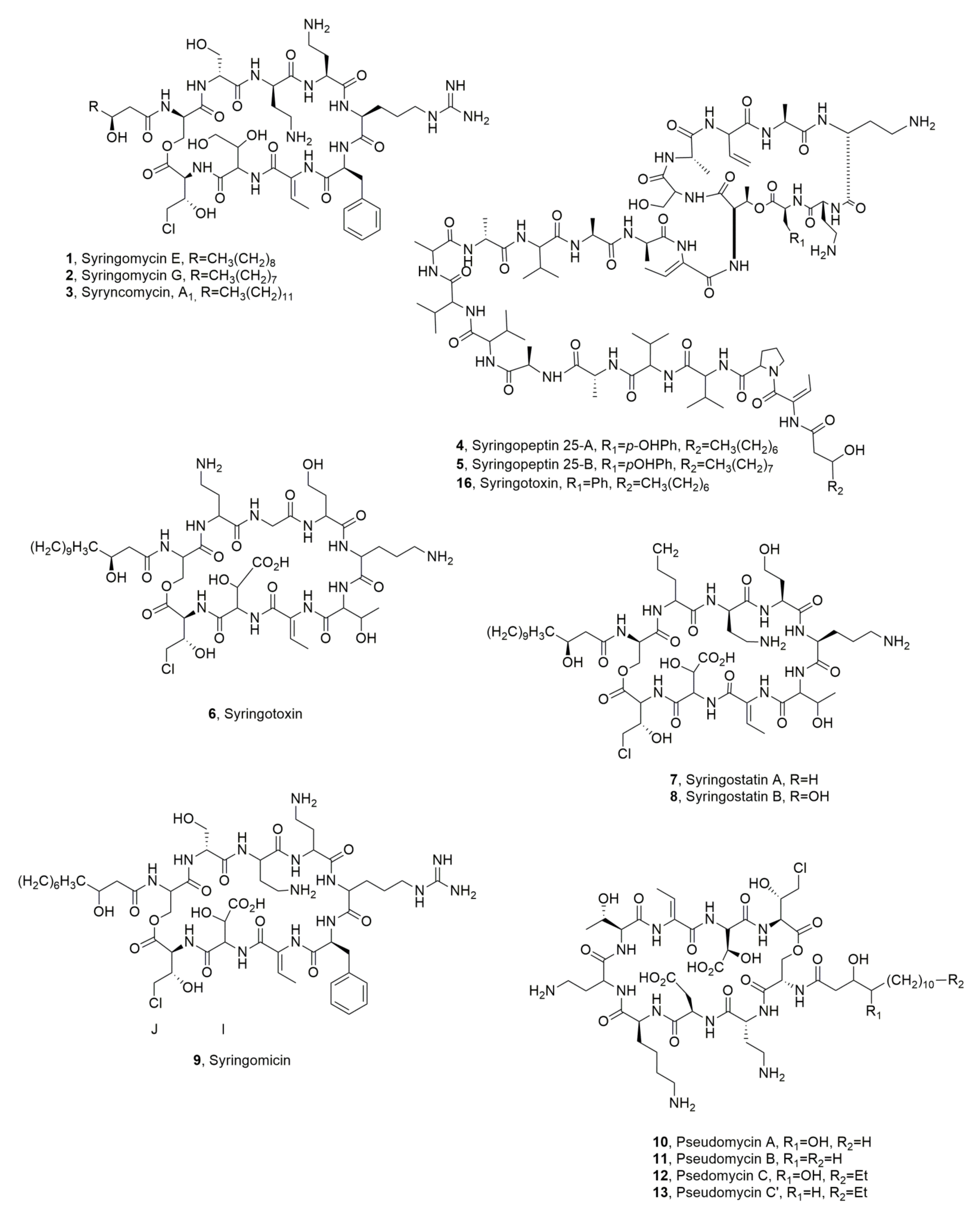
Among these classes of natural bioactive metabolites, there are the lipodepsipeptides (LPDs), which are biologically active metabolites produced by different bacteria, fungi and marine organisms. They also include cyclic lipodepsipeptides which are subject to many studies due to their important and new biological activities. Cyclic LPDs are constituted by three moieties: (i) a macrocyclic peptide lactone; (ii) a linear peptide; iii) fatty acid. These lipodepsipeptides contain unusual amino acids also with an opposite stereochemistry (D) in respect to that of the common natural spread having a L stereochemistry. They are classified according to their primary structures into two groups (Figure 1). The first group, represented by the phytotoxic cyclic lipodepsinonapeptides constituted by a polar peptide head and a hydrophobic 3-hydroxy fatty acid tail. This group include syringomycins, syringostatins, syringotoxins, and pseudomycins. The four lipodepsinonapeptide subgroups differ in the amino acid sequence between the positions 2 and 6. The 3-hydroxy fatty acyl group is a derivative of either decanoic (syringomycins), or dodecanoic (syringomycins and syringostatins), or tetradecanoic (all four lipodepsinonapeptides), or hexadecanoic (pseudomycins) acid. Some pseudomycins are acylated by either 3,4-dihydroxytetradecanoate or 3,4-dihydroxyhexadecanoate (Figure 1). Furthermore, the three known syringomycins (SRs) only differ themselves by the length of the 3-hydroxy fatty acid moiety, which is either decanoic acid for SRA1, or dodecanoic acid for SRE, or tetradecanoic acid for SRG. Through an amide bond, the carboxylic groups of these acids linked the N-terminal serine residue, which in turn is bonded to 4-chlorothreonine at the C terminus through an ester bond generating a macrocyclic lactone ring [3].
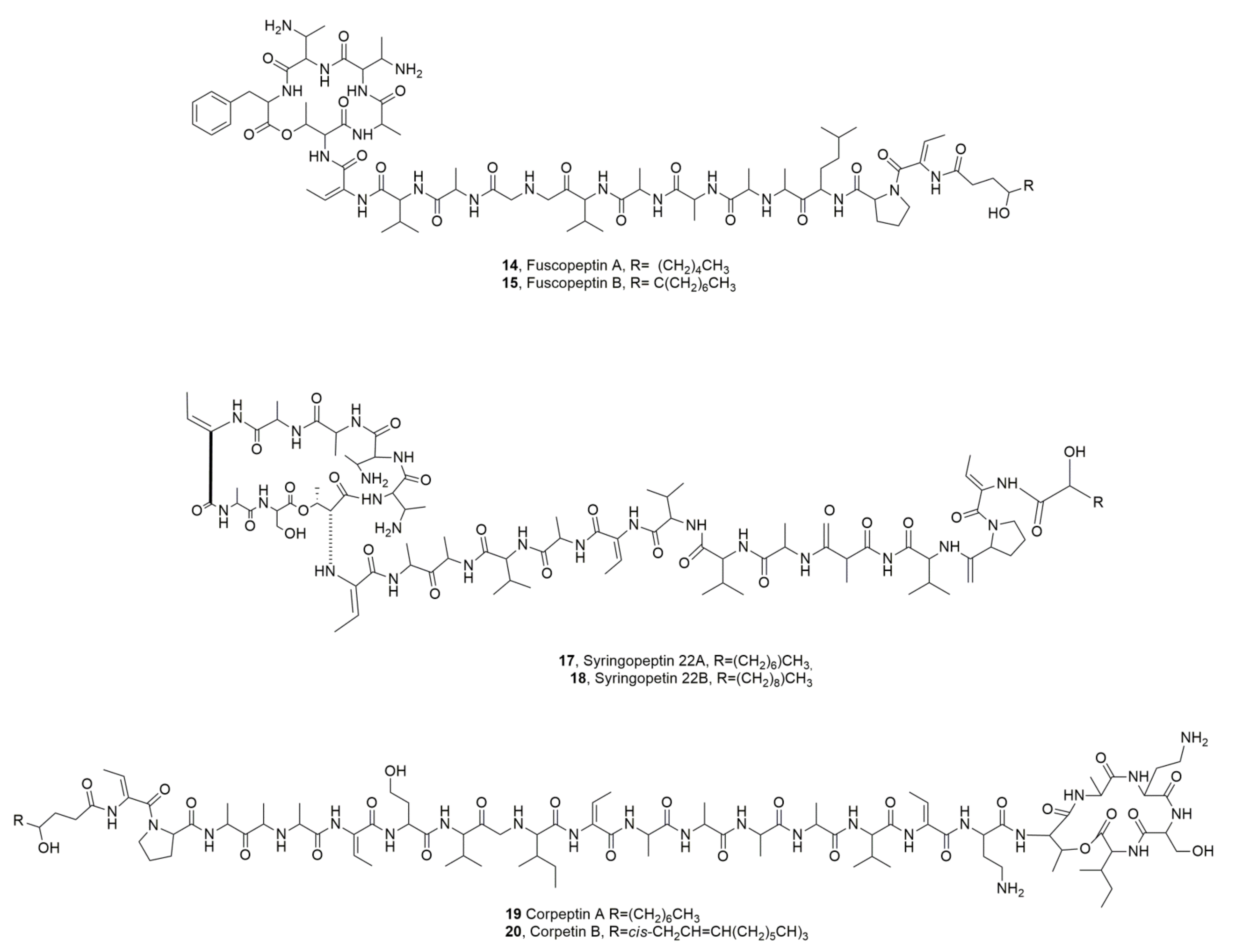
The second group of LPDs, which is represented by phytotoxic syringopeptins (SPs) (Figure 2), was isolated from Pseudomonas syringae pv. syringae strains [4]. Syringopeptins, different from lipodepsinonapeptides, contain either 22 or 25 amino acids depending on the specific bacterial strain producer. The N-terminal amino acid, which is the 2,3-dehydro-2-aminobutyric acid, is acylated by either 3-hydroxydecanoic or 3-hydroxydodecanoic acid. The lactone ring was generated by the ester bond between allothreonine and the C-terminal tyrosine. A high percentage of hydrophobic amino acids are found in the syringopeptin peptide chain, most of which possess a D-configuration [5]. The octapeptide cationic loop formed by a lactone ring, together with the hydrophobic tail, are responsible for their function as a membrane-permeabilizing tool which significantly affected the biological activity [5]. Syringopeptins, which are produced by several strains of P. syringae pv. syringae differed in the peptide sequences [4,6]. For example, a strain of P. syringae pv. syringae isolated from laurel produced syringopeptin 25A with phenylalanine as the C-terminal amino acid instead of tyrosine [7]. Strain SC1 of P. syringae pv. syringae isolated from sugarcane produces a form of SP22 that differs from the previously SP25A for the substitution of Leu residue at amino acid positions 4 and 7 and for the 2-aminodehydropropionic acid (dehydroalanine) at position 9 [8].
Regarding LPDs produced by either bacteria or fungi, there are no references available before 1990. However, lipodepsipedptides are partially referenced in the last three decades in some previously published reviews. In particular, the pharmacological activity of LPDs and their potential as promising lead structures for the development of novel synthetically derived drugs were described by Bionda and Cudic [9]. Some of those compounds are either already marketed (daptomycin 37) or in advanced stages of clinical development (ramoplanin 32) for the treatment of complicated infections caused by multidrug-resistant bacterial strains. The development of new antibiotics based on LPDs is of vital importance because MRSA (methicillin resistant Staphylococcus aureus) strains are rapidly increasing, [10,11,12,13,14,15].
LPDs were previously and extensively described in another review based on 135 references regarding their activity as antifungal compounds. In particular, LPDs belonging to the aureobasidin and echinocandin classes were discussed together with aureobasidin A showing in vitro activity against Candida and Cryptococcus sp. Echinocandins cyclic hexapeptides had been reported as cidal agents inhibiting the synthesis of β-(1,3)-glucan [16].
A table (Table 1) reporting compound names, ring sizes, microbial producers, biological activities, and related references is provided for each LPDs described in the text.

2. Source, Isolation, and Biological Activity of Bacterial Lipodepsipeptides

The LPDs reported in this section as well as in the two successive ones are chronologically presented except when different lipodepsipeptides were isolated from the same source or belong to the same subgroup. The data, which are critically described, are based on the direct knowledge of work carried out extensively in the field by the research group of prof. A. Ballio, of whom the author was a pupil working on several fungal phytotoxins. The other data were obtained by SciFinder research on LPDs in the last three decades using appropriate key words. The LPDs as syringotoxins, syringomycins, pseudomycins, syringostatins and syringopeptins, preliminary and briefly described above, fuscopeptins, tolaasins and corpeptins and others, will be described in this section.
The first article on the partially chemical and biological characterization of a syrigomicin was published by Sinden et al. on 1998 [17]. They LPDs produced by Pseudomonas syringae, which causes bacterial canker disease of peach trees, were purified using a bioguided purification protocol testing the activity against the model fungus Geotrichum candidum. In fact, previously P. syringae van Hall, which was pathogenic on stone fruit trees, was described as a producer of a metabolite with a wide antimicrobial activity spectrum. Furthermore, the virulence of several bacterial isolates was correlated with the antimicrobial activity toward G. candidum. The metabolites present in the culture’s filtrates were responsible for the observed phytotoxicity [18]. Most of both the antibiotic and phytotoxic activities of this bacterium were due to its ability to produce syringomycins (SR) [19]. The same bacterium also showed the ability to produce phytotoxic syringomycins or syringotoxins but their structures at that time were not determined [20].
Three SRs, named SR-A1, SR-E and SR-G (13, Figure 3) were successively isolated from P. syringae pv. syringae. The last two LPDs (2 and 3) were the most abundant of the mixtures separated by HPLC [21].
Further studies on the culture filtrates of the same bacterium highlighted the presence of other several and more bioactive metabolites. Among them, the main component, as estimated by HPLC analysis, were a lipodepsinonapeptide named syringostatin (ST) and two more hydrophobic lipodepsieicosipentapeptides, named syringopeptins (SP25A and SP25B, 4 and 5, Figure 3). The biological activities of the metabolite mixtures were different between ST and SPs.
In fact, pure ST exhibited high antifungal and moderate phytotoxic activities, while SP25A was strongly phytotoxic and had a very poor antifungal activity. The biological activity of SP25-B seemed to be very similar to that of SP25A [4].
Syringomycin E and syringopeptins SP25A (1 and 4) were also produced by the saprophytic fluorescent strain of P. syringae (strain Ml) isolated from wheat. This strain grew in planta without affecting germination or inducing disease symptoms in wheat [22].
Syringotoxin (6, Figure 3) was produced by P. syringae pv. syringae isolated from various citrus species. Its structure was determined by Ballio et al. [23]. NMR and molecular dynamic calculation studies allowed for the determination of its conformation in solution. In fact, the evaluation of NOE correlations observed in its ROESY spectrum, recorded in acetonitrile/water, allowed the determination of the nature and number of intramolecular hydrogen bonds and the predominant conformation of ST [24].
Syringostatins A and B (7 and 8, Figure 3), two novel phytotoxic LPDs, which also showed antifungal activity, were isolated from P. syringea pv. syringae SY12 lilac blights in Japan [25].
A new syringomycin (9, Figure 1) was isolated from the same bacterial strain SC1, which was obtained from sugar cane in Japan. The bacterial culture filtrates showed antimicrobial activity. LPD 9 differs from syringomycin E in the sequence of units J and I and the α-peptide linkage of unit I [26].
Pseudomycins A, B, C, C’ (1013, Figure 3) were isolated together with syringopeptins 25A and 25B (4 and 5), from the culture filtrates of P. syringae MSU 16H [27] which is a transposon-generated mutant of a wild-type strain. The strain MSU 16H showed a higher ability to protect than the wild-type strain isolated from elms infected with Ceratocystis ulmi, the causal agent of Dutch elm disease [28]. The biological activities of pseudomycin A (10) were compared to that of syringomycin E (1). Compound 10 showed phytotoxicity and activity in vitro and in vivo towards some fundamental processes of plant plasma membrane similar to those of syringomycin E [29].
Two new LPDs were isolated from Pseudomonas fuscovaginae, which is the pathogen responsible for the bacterial sheath brown rot in rice and other gramineae such as Hordeum vulgare, Triticum aestivum, Avena sativa and Zea mays [30]. The two LPDs that were close to the above described syringopeptins (SPs), were named fuscopeptins A and B (14 and 15, Figure 4) Compounds 14 and 15 themselves differ only in the fatty acid residue which is a 3-hydroxyoctanoate and a 3-hydroxydecanoate, respectively. Both LPDs (14 and 15) showed phytotoxic and antifungal activity [31]. Their conformation in solution was also obtained by a NOE-NMR study [32].
A new syringopeptin (16, Figure 3) was purified from culture filtrates of P. syringae pv. syringae, which was obtained from a twig dieback of Laurel (Laurus nobilis L.). LPD 16 is very close to syringopeptin 25A, which differs for the substitution of tyrosine residue as C-terminal of the macrocyclic lactone with a phenyl one. Compound 16 showed phytotoxic activity [7].
Syringopeptins SP22A and SP22B (17 and 18, Figure 4) were isolated together with syringomycin SRE, SRG, and SRA1 from P. syringae pv. syringae strain B301D. Both LPDs 17 and 18 in tobacco protoplast assays, caused lysis of protoplasts at 50 ng/mL and showed potent biosurfactant activity at 0.8 mg/mL. The activities of both LPDS 17 and 18 were very similar to those SRs produced by the same bacterium. These results demonstrated that both LPDs secreted by P. syringae pv. syringae are cytotoxic to plant cells at nanomolar concentrations and induced necrosis due to the formation of ion channels that are freely permeable to divalent cations [33].
Two LPDs, named corpeptins A and B (19 ad 20, Figure 4) were isolated from Pseudomonas corrugata, the causal agent of tomato pith necrosis, in which both corpeptins play an important role. The two LPDs themselves differs only for the acyl residue which is a 3-hydroxydecanoyl in LPD 19 and cis-3-hydroxy-5-dodecenoyl in LPD 20. Both LPDs 19 and 20 when assayed in a tobacco leaf induced chlorosis with the LPD 19 being more phytotoxic than compound 20. Their antimicrobial activities were very similar to those of SPs. LPDs 19 and 20 when tested against the B. megaterium showed minimal inhibitory concentrations of 3.75 and 4.20 μM, which are comparable to those for SPs [34], while against Rhodotorula pilimanae their activity was negligible [35]. Successively, from the same bacterium another LPD was isolated and named cormycin A (21, Figure 5). LPD 21 showed significant, antibiotic phytotoxic and red-blood-cell lysis. Some studies on its structure in solution were also carried out by NOESY NMR experiments and computational calculations [36].
Putisolvins I and II (22 and 23, Figure 5) are two biosurfactant LPDs produced by Pseudomonas putida strain PCL1445 when the bacterium was isolated from roots of plants and grown on a site polluted with polycyclic aromatic hydrocarbons. LPDs 22 and 23, which themselves differ in the second amino acid from the C-terminus, which is valine for putisolvin I, and isoleucine for putisolvin II. Both LPDs showed a significant antibiofilm activity [37].
Syringopeptins 508A and 508B (24 and 25, Figure 5) were produced by Pseudomonas syringae pv. lachrymans strain 508, which was isolated in an apple orchard (New York) where it acted in antagonism with Venturia inequalis, the causal agent of apple scab [38]. LPDs 24 and 25, which are very close to syringopeptins 22A and 22B (17 and 18), showed growth-inhibition against Mycobacterium smegmatis, another gram-positive bacterium, and yeasts [39].
A group of closely related LPDs, named A54145, was produced by Streptomyces fradiae [40,41]. A representative structure of this group was reported as compound 26 (Figure 5). This group of LPDs, which is Ca2+-dependent, are an emerging class of antibiotics for the treatment of infections caused by gram-positive pathogens. The group A54145 is close to daptomycin, and the calcium-dependent antibiotic (CDA) produced by Streptomyces roseosporus and Streptomyces coelicolor, respectively. A54145 had antibacterial activity against strains of Staphylococcus, Streptococcus, Clostridium and Enterococci. Furthermore, three components of the LPDs mixture (26), which were enzymatically deacylated, were re-acylated with different fatty acids and showed antibiotic activity against S. aureus and Streptococcus pyogenes in mice [42].
Pseudodesmins A and B (27 and 28, Figure 6) were isolated from a Pseudomonas tolaassii, which was obtained from the mucus layer in the skin of the black belly salamander (Desmognathus quadramaculatus). Pseudodesmins A and B are very close to the viscosin group [43]. In this group it needs to be noted that viscosinamide (29, Figure 6) differs from the other members in the substitution of glutamic acid at position 2 with a glutamine residue. It can be noted that pseudodesmin A differs from WLIP (White Line Inducing Principle, see below paragraph 3) only for the substitution of D-Glu with a DGln residue. Pseudodesmin A can thus be considered as WLIPamide. Furthermore, LPD 27 differs from viscosinamide, only for the stereochemistry of the Leu at position 5 being D rather than L. Pseudodesmin B (28) having L-Val at position 9 differs by at least two substitutions from any previously known viscosin group member. Massetolide E, another member of the same group, had an L-Val9, an L-Leu instead than a D-Leu at position 5 and a D-Glu instead of D-Gln [44].
The viscosin group in addition to pseudodesmins A and B and viscosamide (2629) and WLIP is constituted by viscosin, massetolides A-H and pseudophomins A and B which all consist of nine amino acid residues, seven of which form the cyclic structure via lactone bond formation between the C-terminus and the side chain of a D-alloThr at position 3. They are produced by several Pseudomonas species, including Pseudomonas viscosa, Pseudomonas libanensis, which produced viscosin [45], different strains of Pseudomonas fluorescens which synthesized viscosin and viscosinamide [46,47], the massetolides [48] and the pseudophomins [49] and Pseudomonas reactans, which produced WLIP [50].
Viscosin (30, Figure 6) was isolated for the first time from P. viscosa by Kochi group (1951) [45] during a systematic use of microorganisms able to produce antibiotic metabolites. The LPD 30 named at that time “P-preparation,’’ was obtained as crystals and appeared to be heat stable and soluble in ethanol, methanol, ether, acetone, and alkaline phosphate buffer. Some preliminary investigations assigned an acidic polypeptide structure to 30, which showed a therapeutic effect in guinea pigs showing tuberculosis symptoms. When more large amounts of this LPD, which was then named viscosin, were prepared, it was possible to demonstrate its antibiotic activity against mycobacteria, its ability to protect embryonated eggs infected with chicken bronchitis virus and to highlight its slight suppressive effect on the infection progress in mice infected with influenza A virus [45].
Successively, the LPD 30 was also isolated as a surfactant from a P. fluorescens strain and showed a surface tension value of 26.5 mN m−1 and a critical micelle concentration of 0.15 mg/mL [51].
The structure of viscosin (30) was determined for the first time using essentially spectroscopic and degradative chemical methods from Laycock et al. (1991) [46] when it was isolated from a strain of P. fluorescence biovars II e IV, which are the causal agents of head rot in broccoli. This is a destructive disease which causes heavy losses in Eastern Canada and other regions of the world. Moreover, its surfactant activity was confirmed. Successively LPD 30 was also isolated as surfactant from P. libanensis M9-3. Viscosin had a critical micelle concentration (cmc) of 54 mg L−1, its minimum surface tension between air and water at the cmc was 28 mN m−1 and it is able to form stable emulsions even at low concentrations (7.5 mg L−1). The physicochemical properties recorded for LPD 30 are similar to other biosurfactants such as the well-known rhamnolipid and surfactin. Viscosin, without associated toxicity, also inhibited migration of the metastatic prostate cancer cell line, PC-3M [52].
Viscosamide (29), above cited, is produced by P. fluorescens DR54 and showed biosurfactant and antibiotic properties [47].
Massetolides A-H (3138, Figure 6) were isolated together viscosin (30) from two Pseudomonas sp. strains obtained from a marine alga and a marine tube worm. In particular, LPDs 3134 were isolated from the first organism while LPDs 3538 were produced by the other one [53]. Massetolide A (31) and viscosin (30) showed in vitro antibiotic activity against Mycobacterium tuberculosis and Mycobacterium avium-intracellulare. In addition, massetolides I-K (3941, Figure 6) were biosynthesized by incorporating nonprotein amino acids. In fact, feeding experiments using L-butyrine and L-norvaline cyclopropylalanine as precursors generated massetolides I, J and K (3941) as the main analogues, respectively [53]. Some studies on the biosynthesis of massetolide A were carried out using P. fluorescens. LPD 37 is important in the ability of this bacterium to form biofilm [54].
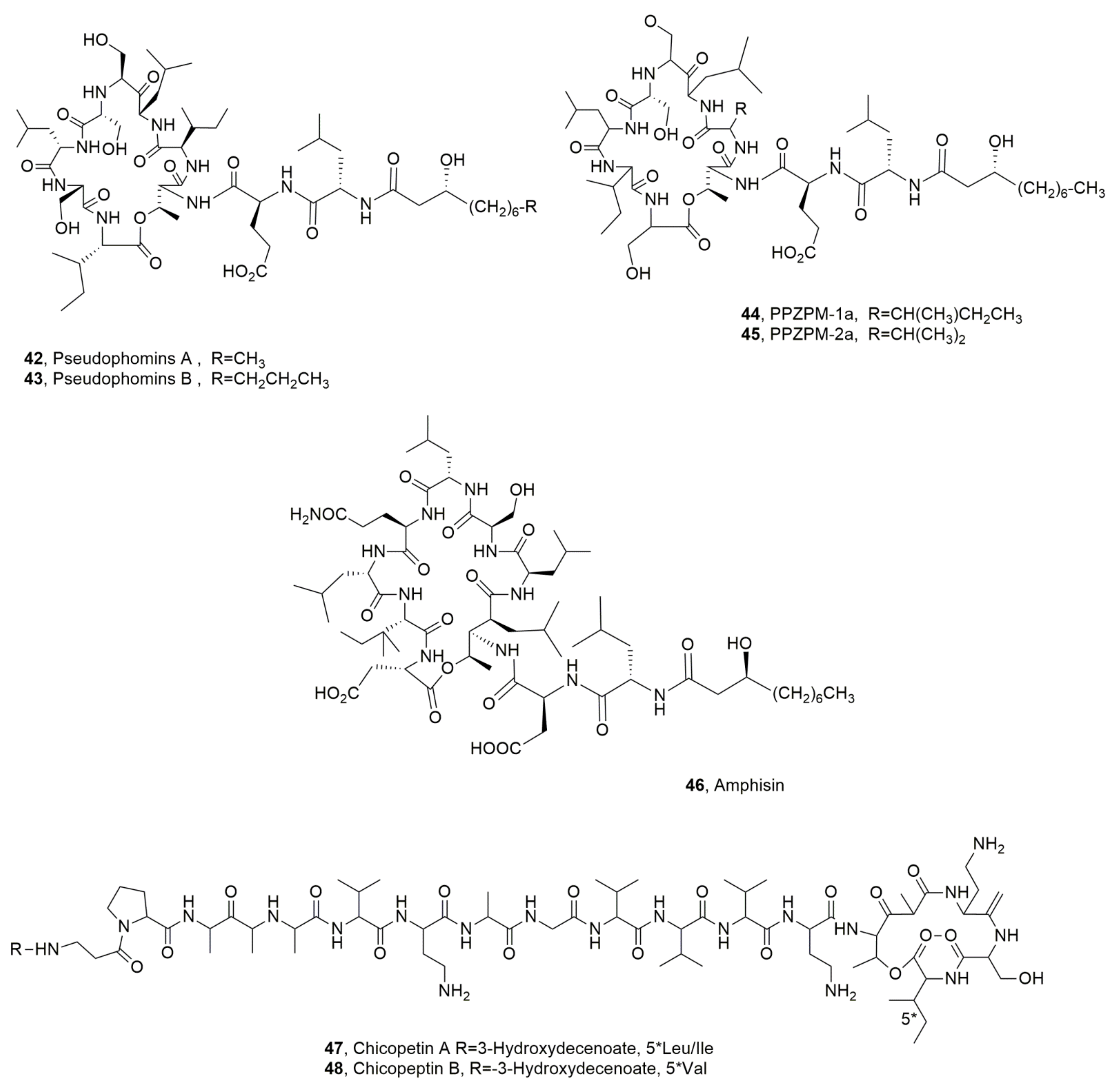
Pseudophomins A and B (42 and 43, Figure 7) were isolated from P. fluorescens strain BRG100, a plant pathogen proposed for weed biocontrol. Pseudophomin B (43), which is the main metabolite, exhibited a higher antifungal activity against the phytopathogens Phoma lingam, Leptosphaeria maculans and Sclerotinia sclerotiorum than pseudophomin A. Indeed, pseudophomin A (42) was a stronger root germination inhibitor of green foxtail (Setaria viridis) than pseudophomin B [49].
PPZPM-1a and PPZPM-2a (44 and 45, Figure 7) are the main LPDs among the mixture of more than 30 cyclodepsipetides produced by Pseudomonas sp. JX090307, which was isolated from hyphae of the phytopathogenic oomycete Phytophthora alni spp. alni and appeared close to Pseudomonas orientalis. The cell extract of Pseudomonas sp. JX090307 showed antifungal activity against Verticillium dahlia, some strains of P. alni spp. alni and different fungal pathogens of forest tree species. The LPDs belonging PPZPM group and thus also compounds 42 and 43, are constituted by a β-hydroxy fatty acid bonded to a peptide moiety containing 10 amino acids, eight of which generated the macrocyclic lactone, and this later represented the different features with the LPDs viscosin and amphisin groups [55]. Amphisin (46, Figure 7) is a lipoundecapeptide produced by Pseudomonas sp. strain DSS73, whose structure was determined by X-ray analysis. Amphisin showed biosurfactant and antifungal activity [56]. LPD 46 has diverse applications but is essentially used as a biosurfactant in bioremediation methods to decontaminate dredged harbor sediments as it can increase the bioavailability or mobility of contaminants in an aqueous phase. Amphisin increased the efficacy in releasing polycyclic aromatic hydrocarbons (PAHs) strongly adsorbed to sediments when compared to a synthetic anionic surfactant, [57].
Pseudomonas chicorii is a pathogen that causes necrotic leaf and stem lesions on several agrarian plants, including lettuce celery, chrysanthemum, tomato, coffee and soybean [58,59,60,61,62]. This bacterium produces phytotoxic metabolite whose role in pathogenesis is not yet demonstrated [63,64,65,66]. Two LPDs, named cichopeptins A and B (46 and 47, Figure 7) were isolated from the culture filtrate of P. chicorii. LPDs 46 and 47 showed antibacterial activity against Bacillus megaterium but not on Rhodotorula mucilaginosa and different levels of phytotoxic on the host plant above cited [67].
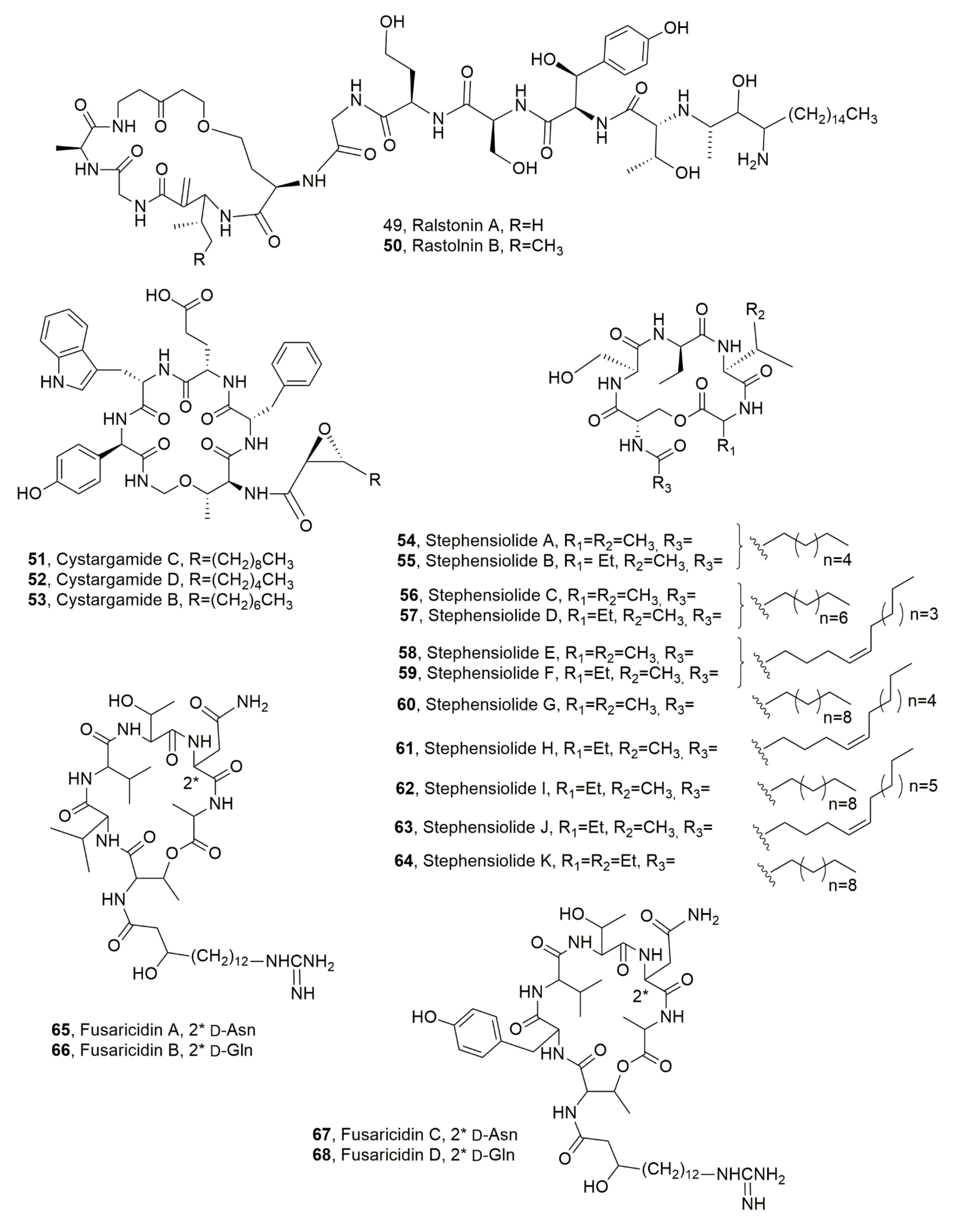
Two LPDs, named ralstonins A and B (49 and 50, Figure 8) were isolated from Ralstonia solanacearum, a β-proteobacterium-inducing lethal disease called “bacterial wilt”, which is observed in more than 200 plant species in tropical, subtropical, and warm temperature regions of the world [68,69,70]. Compounds 49 and 50 are not common among LPDs being constituted of 11 amino acids (containing unique amino acids such as β-hydroxytyrosine and dehydroalanine) and a 3-amino-2-hydroxyoctadecanoic acid. Ralstonins exhibit chlamydospore-inducing activity and moderate phytotoxicity [71].
Cystargamides C and D (51 and 52, Figure 8) were isolated, together with previously known cystargamide B (53, Figure 8) [72] from a tidal mudflat-derived Streptomyces sp. JMS132 collected at Beolgyo, South Korea. The LPDs 5153 showed an antioxidant effect in the DPPH and ABTS assay [73].
Eleven LPDs, named stephensiolides A-K (5464, Figure 8) were produced by a Serratia sp. found within the midgut and salivary glands of Anopheles stephensi mosquitoes. The latter, that contains the Plasmodium parasite, has a microbiota that can influence both the vector and the parasite. LPDs 5464 showed antibiotic activity and facilitate bacterial surface motility [74].
Recently a quantitative determination and pharmacokinetic study of fusaridicin A (65, Figure 8) in mice plasma and tissues was performed using ultra-high performance liquid chromatography-tandem mass spectrometry [75]. LPD 65 belong to a group of LPDs, named fusaricidins or LI-F antibiotics, which were isolated from Bacillus polymyxa, as potential compounds for the development of antibacterial and antifungal agents [9,76,77,78]. In fact, fusaricidins demonstrated strong in vitro antibacterial activity against gram-positive bacteria such as S. aureus, Micrococcus luteus, and B. subtilis. The same group of LPDs showed strong antifungal activities against a broad range of pathogens including Candida spp., Aspergillus spp., Penicillium spp., Fusarium oxysporum and Cryptococcus neoformans [9,77,78,79]. Fusaricidins A-D (6568, Figure 8) were produced by P. polymyxa KT-8 strain and showed significant antibiotic activity [77,78]. LPD 64, among the four isolated fusaricidins, exhibited the strongest activity, towards some gram-positive bacteria e.g., S. aureus and pathogenic fungi e.g., C. neoforman [77,78,80,81,82,83,84].
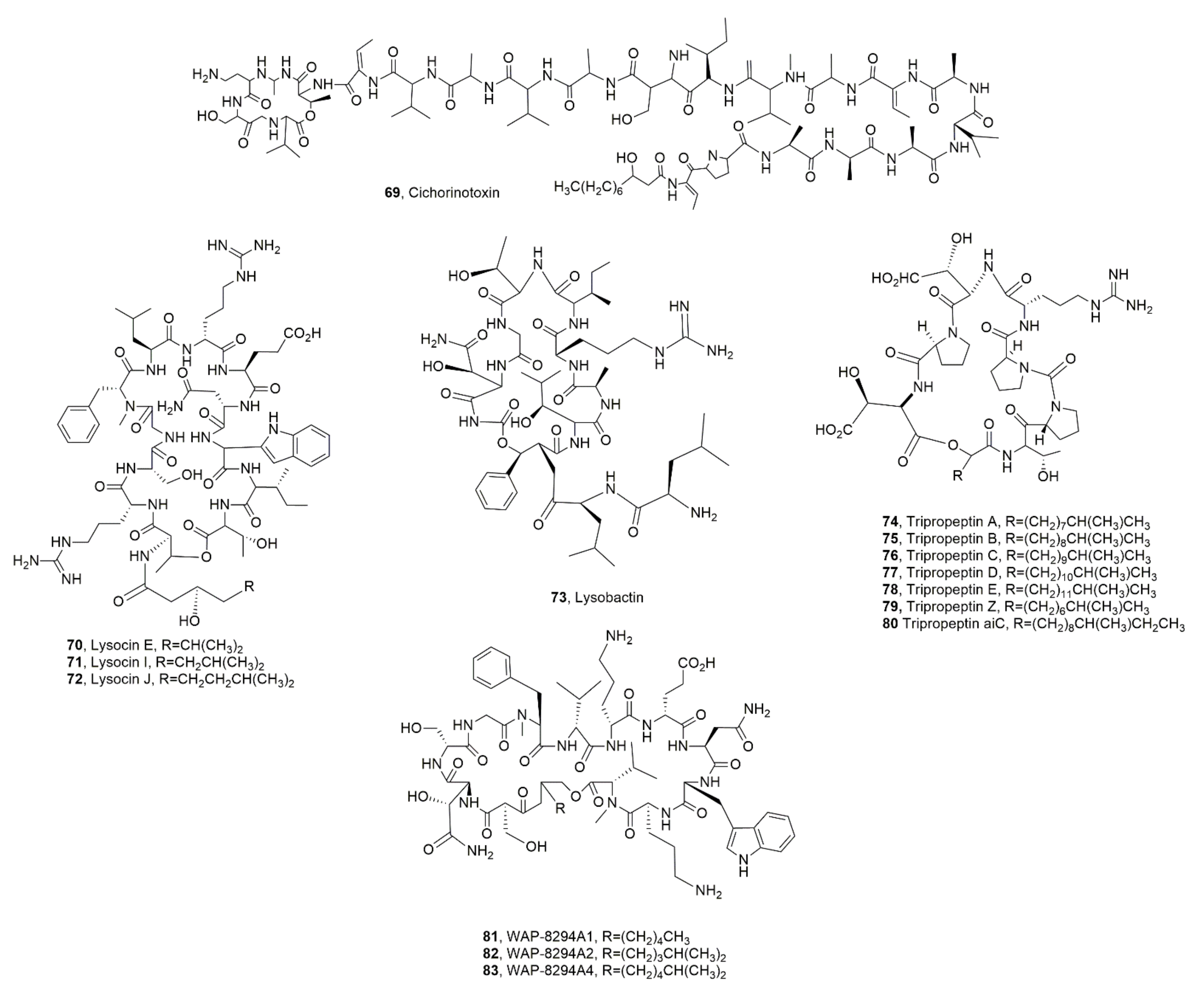
A LPD, named cichorinotoxin (69, Figure 9) was isolated from P. cichorii, which is responsible for varnish spots on lettuces causing serious losses to lettuce production during the summer season in Japan. LPD 69 induce lettuce rot [85].
Lysocin E (70, Figure 9) was isolated from Lysobacter sp. 3655, and belong to lysocins, which are a group of LPDs showing strong activity against MRSA strains [86,87] associated with a novel mode of action [88]. This antibiotic activity was also shown against Mycobacterium spp. in vitro and in silkworm infection models [87]. Successively, lysolicins I and J (71 and 72, Figure 9) were isolated together LPD 70 from Lysobacter enzymogenes, whose crude extract showed strong antibacterial activity against several gram-positive bacteria [89]. Lysobacter are gram-negative bacteria ubiquitously diffused in soil and water [90]. Several Lysobacter species produce extracellular lytic enzymes and new bioactive metabolites, which determine their potential both as biocontrol agents and as producers of new drug leads. These metabolites include cyclic peptides, cephem-type β-lactams and polycyclic tetramate macrolactams (PoTeM) [91]. Among LPDs there are lysobactin, the cyclic lipodepsipeptides of tripropeptin family, WAP-8294A family, lysocin family. For some of them including lysocin a total synthesis was realized and mode of action was deeply studied [86,87,92,93,94,95,96,97,98].
Lysobactin (73, Figure 9), also known as katanosin B, was produced by Lysobacter sp. and its antibiotic activity against aerobic and anaerobic gram-positive bacteria, including Staphylococci, Streptococci, corynebacteria, clostridia and various other gram-positive anaerobic bacteria, was higher by 2- to 4-folds than that of vancomycin. LPD 73 only had a weak activity against aerobic and anaerobic gram-negative bacteria [99,100].
Tripropetins A-E and Z (7478 and 79, Figure 9), were isolated as antimicrobial metabolites from Lysobacter sp. strain BMK333-48F3 [92,93,101]. They showed strong activity against MRSA and VRE clinical bacteria strains. In addition, tripropetin C (76) blocks the lipid cycle of cell wall biosynthesis by complex formation with undecaprenyl pyrophosphate [102]. Successively, a new LPD, belonging to the same group and named tripropeptin aiC (80, Figure 9), was isolated from the same Lysobacter strain. LPD 80 showed the same antibiotic activity of the other tripropetins [103].
The LPDs belonging to the WAP-8294A group were isolated from Lysobacter sp. WAP-8294 and showed antibiotic activity essentially towards gram-positive bacteria. WAP-8294A2 (82, Figure 9) was the major component, while A1, A4, (81 and 83, Figure 9) Ax8, Ax9 and Ax13 were the minor ones. These LPDs exhibited antibiotic activity essentially against MRSA strains in vitro [104]. Successively, WAP-8294A1 (82) was also isolated from Lysobacter antibioticus ATCC 29479 [98].
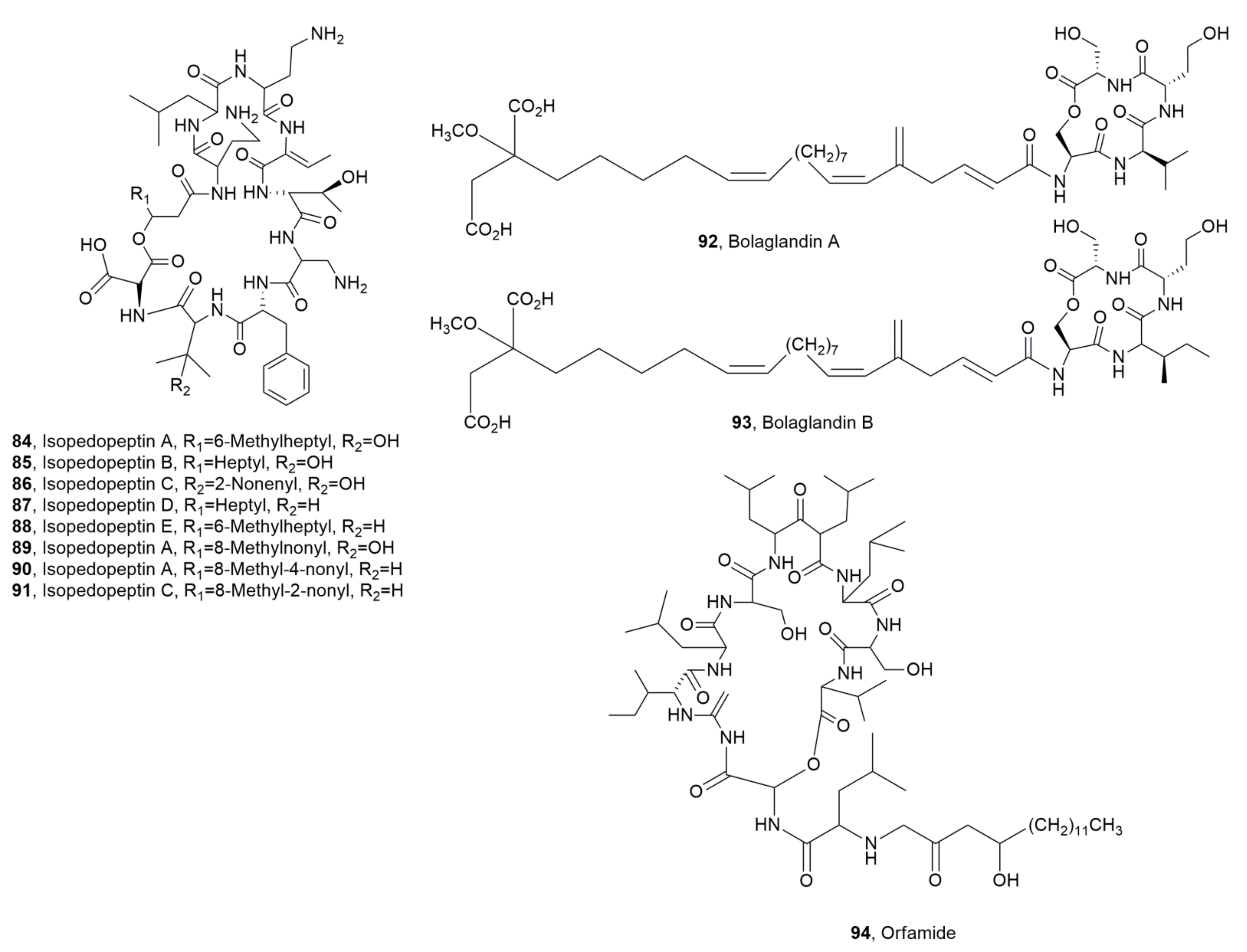
Isopedopeptins A-H (8491, Figure 10), were isolated from Pedobacter cryoconitis strain UP508, which was collected from a soil sample. The LPDs 8491 essentially showed antibiotic activity against gram-negative bacteria. Among them isopedopeptin B (85), had not only antibacterial activity, but also cytotoxicity and hemolytic properties. In fact, it exhibited good activity against strains of WHO top-priority gram-negative bacteria, i.e., carbapenem resistant Acinetobacter baumannii, Escherichia coli, Pseudomonas aeruginosa, and against colistin-resistant strains of A. baumannii, E. coli, and Klebsiella pneumonia [105].
Bolagladins A and B (92 and 93, Figure 10), were considered two unusual LPDs as they contained citrate-derived fatty acid and a rare dehydro-β-alanine residue. LPDs 92 and 93 were produced by two Burkholderia gladioli strains isolated from the lungs of cystic fibrosis patients. LPDs 84 and 85 did not show activity against any of the ESKAPE panel of bacterial pathogens, Mycobacterium bovis BCG, or Candida albicans [106].
Orfamide H (94, Figure 10) was isolated from Pseudomonas protegens CHA0, which was obtained from tobacco roots growing in suppressive soil [107]. LPD 94 showed antifungal activity inhibiting the appressoria formation of Magnaporthe oryzae, the causal agent of the blast severe disease in rice [108].
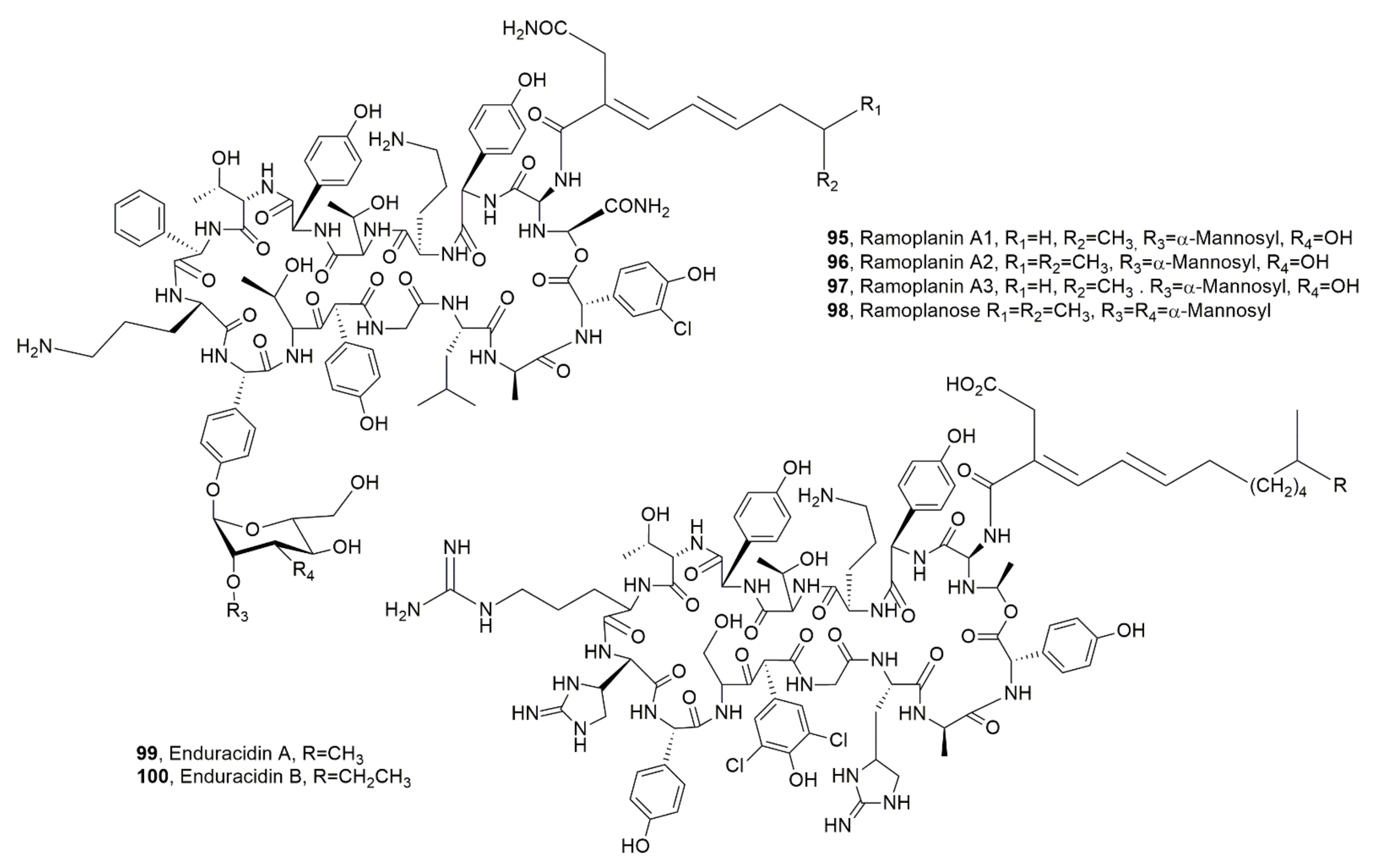
Ramoplanins A1, A2, A3 and ramoplanose (9598, Figure 11) and the enduracidins A and B (99 and 100, Figure 10) were isolated from Actinoplanes ATCC 33076 [109,110] and from Streptomyces fungicidicus B5477, respectively [111]. Their structures are quite close [112]. Their antibiotic activity is based on their ability to avoid peptidoglycan (PG) cross-link generation inducing bacterial death consequent to osmotic lysis of the cell wall. In particular, LPD ramplanin A2 (96) induce membrane depolarization in S. aureus, as a complementary mode of action for the disruption of lipid membrane integrity [113]. Ramoplanin A2 (96) prompt interest as it can overcome the resistance developed by some gram-positive bacteria to antibiotics based on glycopeptides, macrolides, and penicillins, but it is not orally absorbed and showed mild to moderate hemolytic efficacy when administered intravenously.
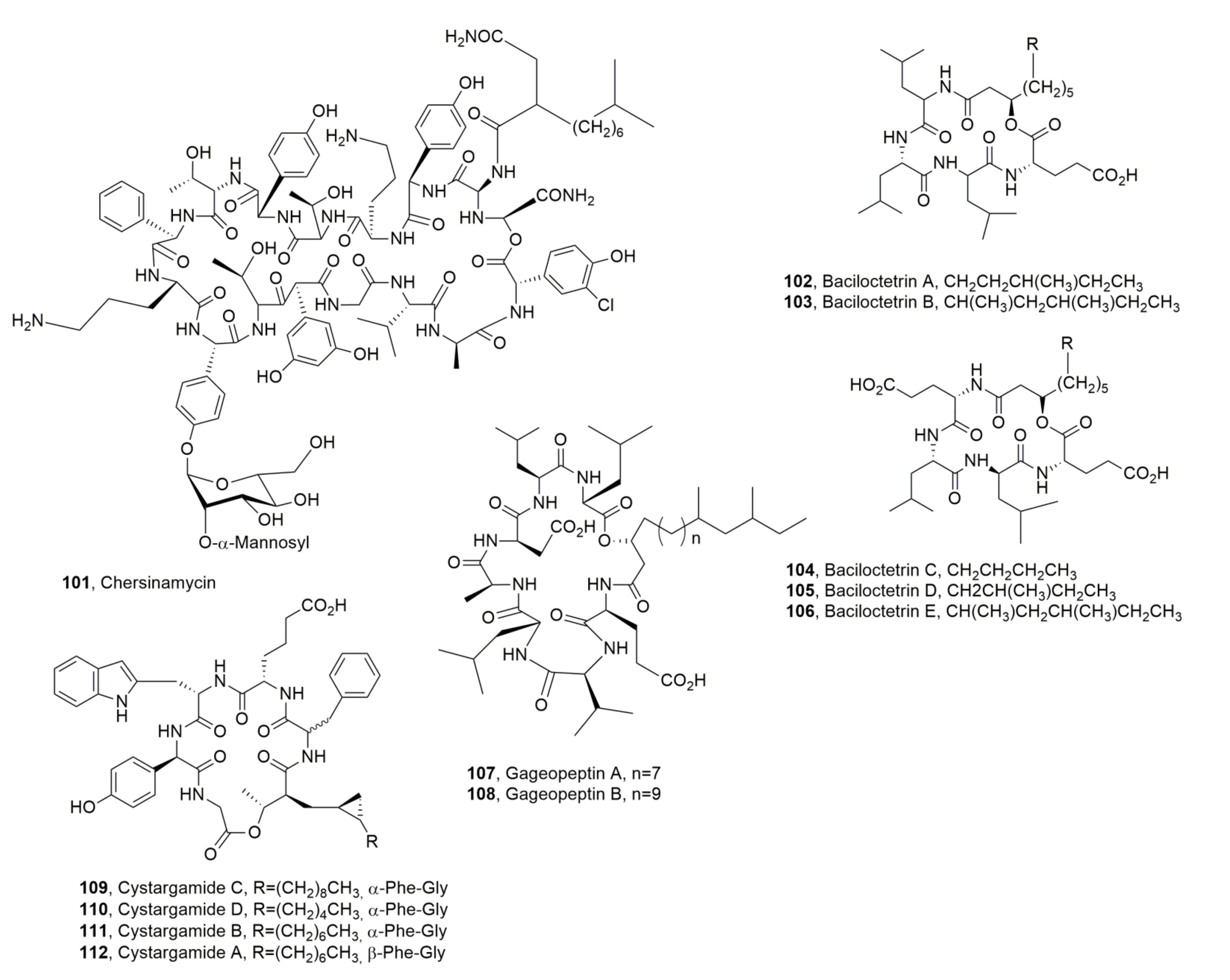
In addition, when dispensed by intraperitoneal injection its macrolactone is easily hydrolyzed [114]. Enduracidins A and, B, which had similar activity, but showed decreased water solubility, were approved in the United States only as a growth-promoting feed additive for livestock [115,116]. Ramoplanins and enduracinins were also produced by Micromonospora chersina strain DSM 44151, Actinoplanes orientalis strain DSM 40040, and Actinoplanes balhimycina FH 1894 strain DSM 44591, but only M. chersina produced in few amounts of the LPD named chersinamycin (101, Figure 12) [113].
Baciloctetrins A and B (102 and 103, Figure 12) were produced by B. subtilis isolated from a marine sponge sample collected from the Gageo reef, Republic of Korea. The antibiotic activity of LPDs 102 and 103 was evaluated against clinically isolated MRSA strains (ATCC25923, XU212, SA1199B, RN4220, and EMRSA15) [117]. Successively, three LPDs, named bacilotetrins C-E (104106, Figure 12), were isolated from the same bacterium, which showed antibiotic activity against Mycoplasma hyorhinis. The latter is the main causal agent of polyserositis and arthritis in swine and is a common contaminant in laboratories. Compounds 104106 showed activity, with an MIC value of 31 µg/mL, which was two folds stronger than that of the positive control, BioMycoX®. The same author also revised the structure of baciloctetrins A and B (102 and 103) [118]. Previously, two other LPDs, named gageopeptins A and B (107 and 108, Figure 12) were isolated from the same strain of B. subtitlis. LPDs 107 and 108 were able to impair the motility of zoospores of Phytophthora capsici in dose- and time-dependent manners. LPDs 107 and 108 also showed moderate antibacterial and good antifungal activities [119].
Cystargamides C and D (109110, Figure 12) were isolated together with the already known cystargamide B (111, Figure 12) [72] from a marine actinomycete mixture of Streptomyces sp. (98.8% identical to Streptomyces malachitofuscus), which was collected at Beolgyo, South Korea. The LPDs 109111 showed an antioxidant effect in the DPPH (2,2-diphenyl-1-picrylhydrazyl) and ABTS (2,2-azino-bis (3-ethylbenzothiazoline-6-sulfonic acid) radical-scavenging assay [73]. Previously, cystargamide A (112, Figure 12) had been isolated from Kitasatospora cistarginea. LPD 112 differed from cystargamide B for the stereochemistry of pheylglicine residue [120].

3. Source, Isolation, and Biological Activity of Fungal Lipodepsipeptides

Although the LPDs produced by fungi are less extended in respect to those ones synthesized by bacteria as reported in Section 2, they are interesting due to their chemical and biological properties.
They are chronologically described in this section with the exception of the case of LPDs belonging to the same subgroup.
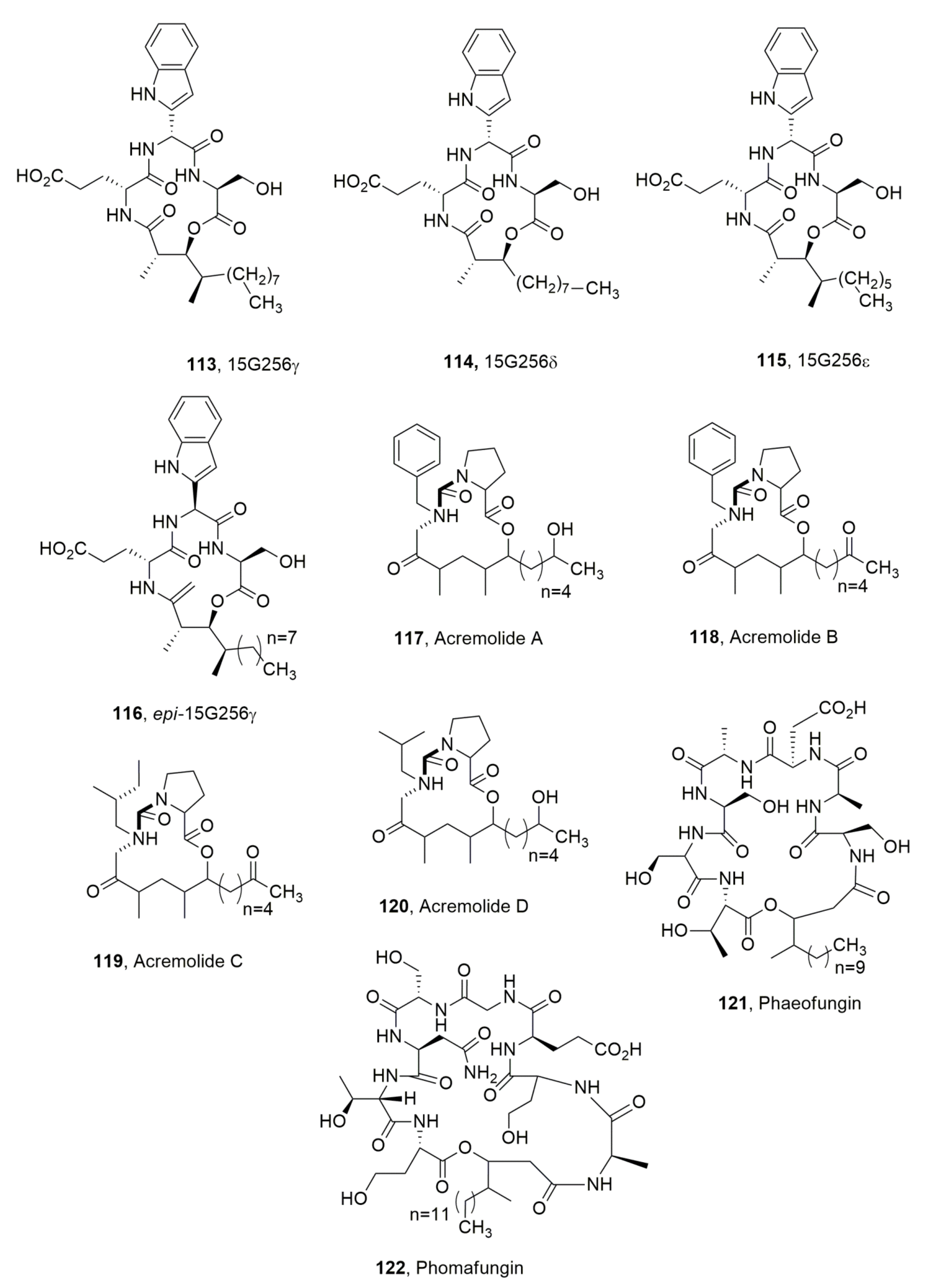
The lipodepsipeptides 15G256γ, 15G256δ and 15G256ε (113115, Figure 13) were isolated together with some polylactones from the marine fungus Hypoxylon oceanicum. Their structures are characterized by the presence of the unusual ketotryptophan amino acid, which is responsible for the epimerization occurring in solutions of the α-carbon, as occurring in the conversion of 113 in its epimer (116, Figure 13). LPDs 113115 showed antifungal activity with compound 113 being the stronger one [121].
A novel family of LPDs, named acremolides A-D (117120, Figure 13), were isolated together with the known 19-O-acetylchaetoglobosin D, 19-O-acetylchaetoglobosin B and aromatic metabolite RKB 3564S from an Australian estuarine strain of an Acremonium sp. (MST-MF588a). Among all of these, the two chetoglobusins are the only compounds showing cytotoxic properties [122]. The acremolides did not show cytotoxic activity but they were not tested for synergic activity [123].
Phaeofungin (121, Figure 13) was isolated from Phaeosphaeria sp. (F-167,953) obtained from the stems and leaves of a Sedum sp. (Crassulaceae), which was collected in Motilleja, Albacete, Spain. Moreover, a second strain (F-262,327, E-000531145) of the same fungus producing phaeofungin was isolated from the stems and leaves of Teucrium sp. (Lamiaceae) which was also collected in Spain but at Serrania de Cuenca, Cuenca. The whole fungal extract showed activity against wild-type C. albicans and Aspergillus fumigatus, two important human pathogens [124]. LPD 121 is very close to phomafungin (122, Figure 13) produced by Phoma sp. which was isolated from soil collected in Séréhini, Grand Comore, Union of the Comoros and showed a broad spectrum of antifungal activity against Candida spp., A. fumigatus and Trichophyton mentagrophytes [125]. LPD 121 induced ATP release in wild type of C. albicans strains, while LPD 122 did not [124].
Verlamelins A and B (123 and 124, Figure 14) were isolated from the entomopathogenic fungus Lecanicillium sp. obtained from a chilli thrips cadaver. Both LPDs 123 and 124 showed antifungal activity against Cochliobolus miyabeanus and Alternaria solani, while verlamelin B was less active than the others against Fusarium oxysporum, Cladosporium cucumerinum and Ustilago maydis thus suggesting an important role played by the methyl group on the first amino-acid residue connected to the fatty acid moiety [126].
Ophiotine (125, Figure 14), was isolated, together with artthrichitin, arthrichitins B and C (126128, Figure 14) and xanthomide Z from a fungus obtained from cysts of the nematode Heterodera filipjevi, which showed affinities to the genus Ophiosphaerella. LPD 125 showed moderate nematicidal activity against the host nematode, while xanthomide Z exhibited very weak activity. Arthrichitin C (126) showed weak cytotoxicity against several cancer cell lines [127].
Colletotrichamides A-E (129133, Figure 14) were isolated from Colletotrichum gloeosporioides JS419, which was obtained from Suaeda japonica Makino. The latter was collected from a swamp in Suncheon, South Korea. All the LPDS 129133 were tested for their protective activity on HT22 hippocampal cell death induced by glutamate. Compounds 130, 131, and 133 were found to be active, with LPD 128 being the most active metabolite [128].
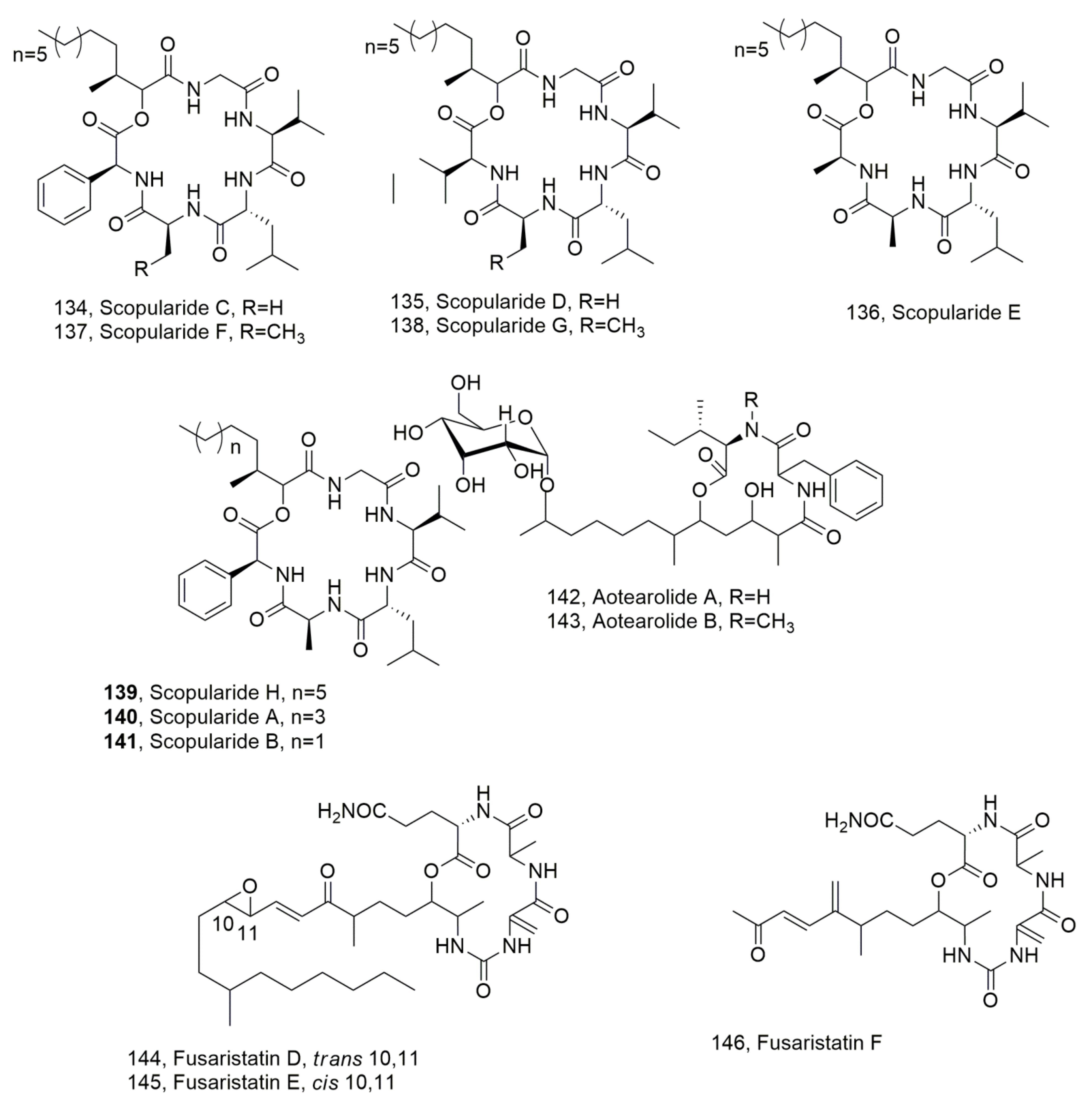
Scopularides C-G (134138, Figure 15) were isolated from Beauvaria sp. CMB F585, while scopularide H (139, Figure 15) was obtained from Scopulariopsis sp. CMB F115 together with the already known scopularides A and B (140 and 141, Figure 15) [129]. All the strains were obtained from the gastrointestinal tract of Mugil mullet fish. LPDs 137 and 138 were also previously isolated from Scopulariopsis brevicalis. When tested for antibiotic and cytotoxic activity all LPDs were inactive [130].
Aotearolides A and B (142 and 143, Figure 15) were isolated together with the known 1H-indole-3-carbohaldehyde and 2-(1H-indole-3-yl)acetic acid from Colletotrichum aotearoa, which is an endophytic fungus of Huperzia serrata [131].
Fusaristatins D-F (144146, Figure 15), were isolated together with (-)-chlamydospore and eight known compounds from Fusarium sp. BZCB-CA, which is an endophytic fungus collected from the Chinese plant Bothriospermum chinesis. They did not show biological activity [132]. Previously, fusaristatins A and B (147 and 148, Figure 16) were isolated from Fusarium sp. YG collected from Maackia chinensis and showed cytotoxic activity against lung cancer cells [133]. More recently, fusaristatin C (149, Figure 16) was isolated from Pithomyces sp. collected from Caribbean octoral Eunica fusca and did not show antimicrobial or cytotoxic activity [134].

4. Lipodepsipeptides Produced by Bacteria Pathogens of Mushrooms

Some species of pathogenic bacteria are responsible for severe diseases in edible mushrooms such as brown blotch in Agaricus bisporus and yellowing of Pleurotus ostreatum induced by P. tolaasii [135]. Other bacteria are involved in A. bisporus and Pleurotus sp. diseases including Pleurotus gyngerii, which is cultivated in the Southern Italy [136,137]. Burkholderia gladioli pv. agaricicola is responsible for cavity diseases in white button mushrooms [138]. All these bacteria are able to produce bioactive LPDs showing phytotoxic as well as other interesting biological activities. Pseudomonas gladioli pv. agaricicola, P. tolaasii and P. reactans, which are all pathogens for cultivated mushrooms, produce lipodepsipeptides with different biological activities.
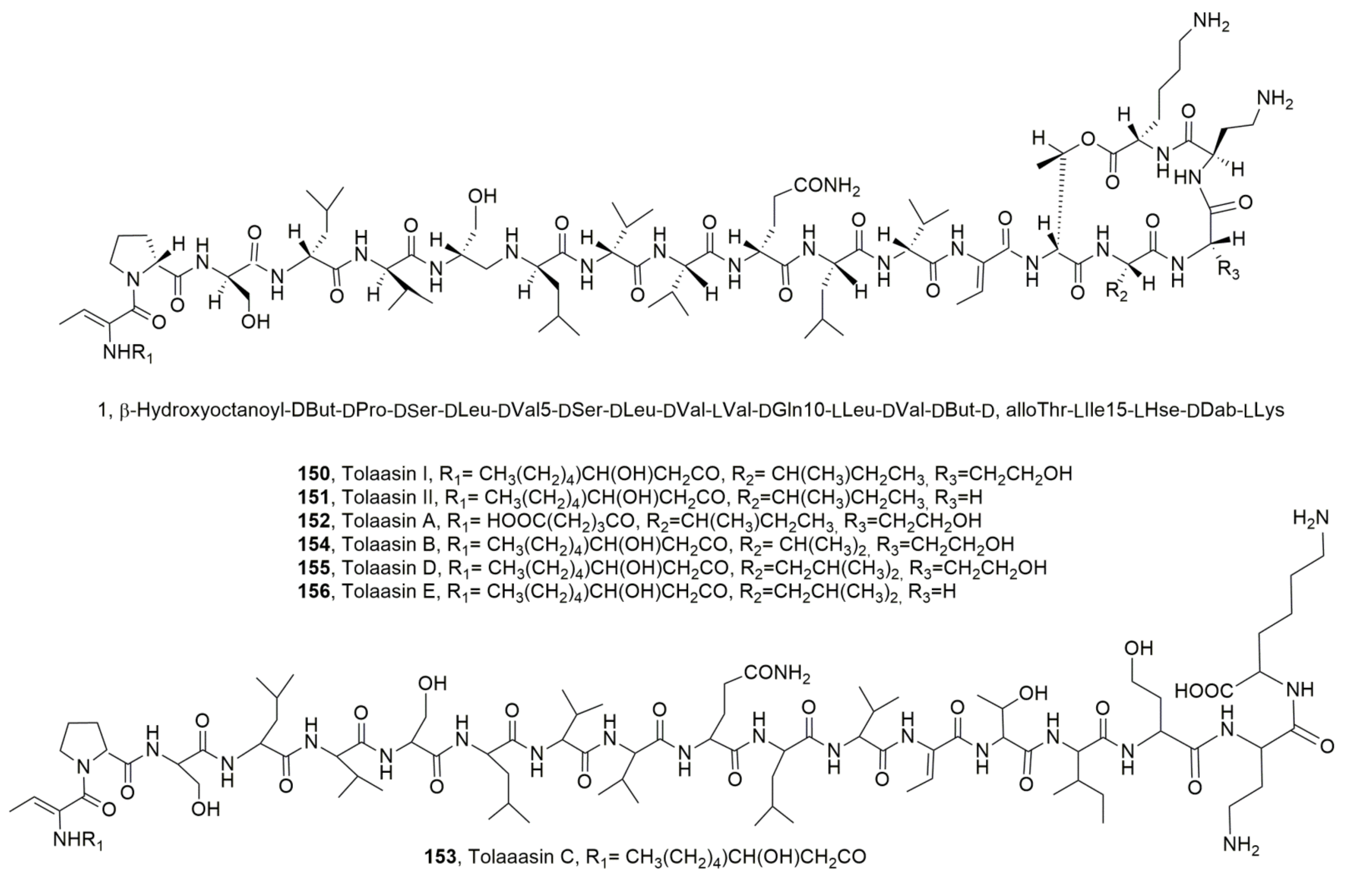
The main bioactive lipodepsipeptides produced by P. tolaasii are tolaasins I and II (150 and 151, Figure 17), whose structure was determined by Nutkins et al. (1991) [139]. LPDs 150 and 151 themselves differed in the substitution of homoserine residue (Hse16) of macrocyclic lactone with a glycine residue. Successively from the same bacterium four other close LPDs, named tolaasins A, B, D and E (152, 154, 155 and 156, Figure 17) were isolated [140]. Although tolaasins A, B, D and E showed the same macrocyclic lactone ring, they showed differences in the peptide moiety and maintained the same β-hydroxyocatnoyl ϕ chain at the N-terminus except for tolaasin A, in which the acyl moiety is a γ-carboxybutanoyl ϕ residue. In addition, tolaasin C (154, Figure 17) was also isolated and showed the opening of the macrocyclic ring while the peptide sequence was the same as tolaasins I. Thus, it could be generated by hydrolysis of LPD 150. All the LPDs 150153, 155 and 156 showed antimicrobial activity against fungi, bacteria and yeasts [140].
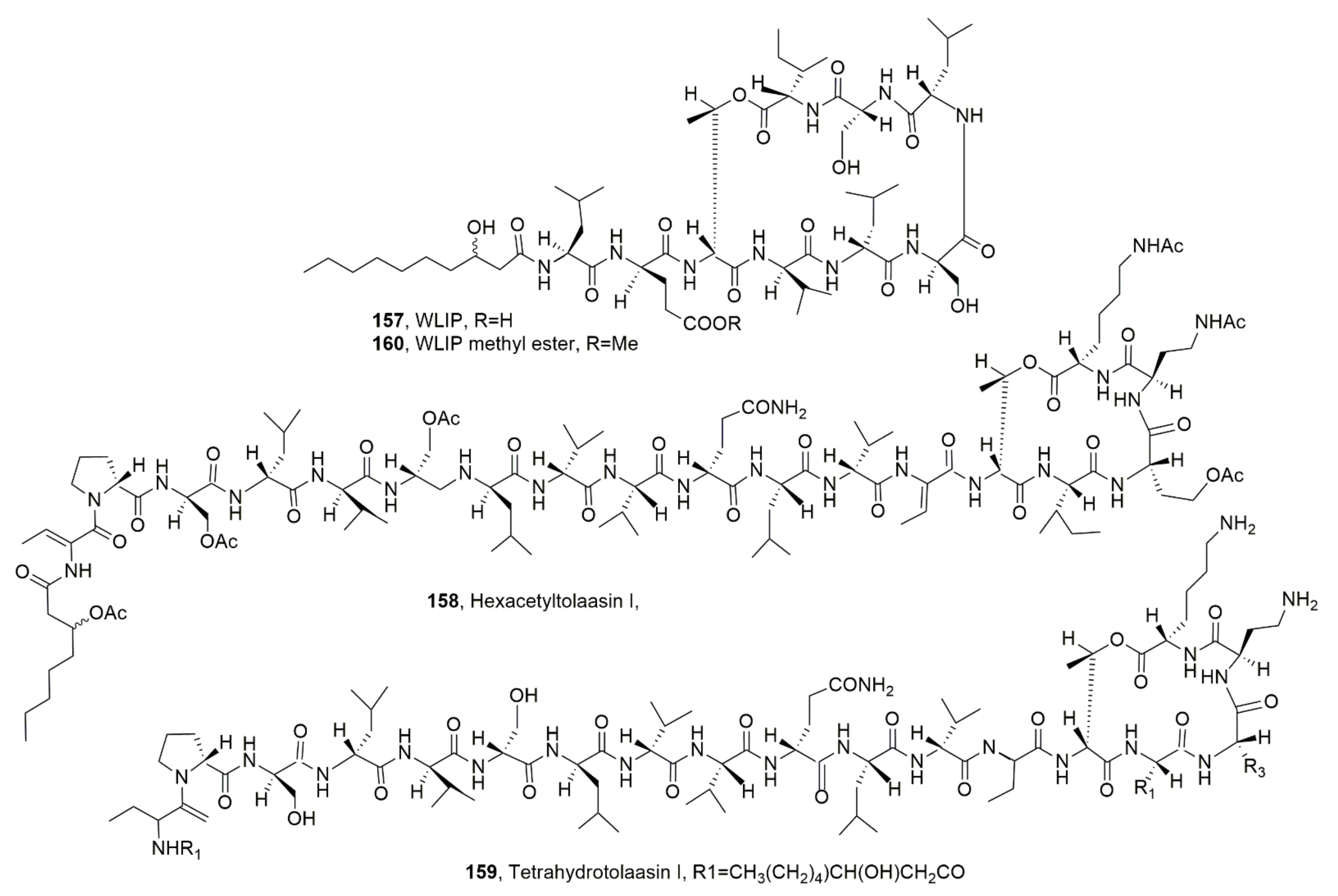
P. reactans produced as the main LPD according to the so-called White Line Inducing Principle (WLIP, 157, Figure 18), which in respect to tolaasins showed a shorter peptide side chain [50].
Tolaasin I and WLIP were compared for their different biological activity. Both LPD 150 and 157 inhibited the growth of mushrooms including A. bisporus, Letinus edodes and Pleurotus spp.-chromista, as well as that of gram-positive bacteria. LPD 150 showed antimicrobial activity against gram-negative bacteria including Escherichia, Erwinia, Agrobacterium, Pseudomonas and Xanthomas and antimicrobial activity against mushrooms, while WLIP affected only Erwinia carotova subp. carotova. Both LPDs induced hemolysis of red blood cells with a strongest effect exhibited by WLIP [137]. Both LPDs also affected the lipid membrane inducing calcein release from large unicellular vesicles [141].
Considering the food importance of mushroom species damaged by the above cited Pseudomonas strains and the consequent heavy economic losses, recently some attempts were made to avoid these severe problems. Some positive results were obtained using helper bacteria belonging to the Mycetocola genus which inactivated tolaasin by hydrolyzing the lactone ring and thus generating a liner peptide [142]. A different mechanism of tolaasin detoxification seemed to be operated by Microbacterium foliorum, which hydrolyzed the peptide chain in two points [143].
Tolaasins I, II, A, D and E, the hexacetyl- and tetrahydro-derivatives of tolaasin I, WLIP, its methyl ester and some cyclic dipeptides were assayed for their antimicrobial activity against bacteria and fungi pathogens for some important agrarian plants such as Pseudomonas caryophilly, Pseudomonas syringae pv. panici, Pseudomonas syringae pv. tabaci, P. syringae pv. syrinage, Pseudomonas syringae pv. japonica, B. subtilis, B. megaterium, E. coli and Colletotrichum truncatum. Among the LPDs the strongest antimicrobial activity was shown by tolaasin D [144].

5. Structure–Activity Relationship of SAR Studies

In this section the results of SAR studies carried out with some selected LPD groups from different sources and with different activities were reported.
In comparison with tolaasin I and II (150 and 151), Tolaasins A-E (152156) when assayed against the gram-positive bacteria B. megaterium and Rodococcus fascians, the yeast R. pilimanae, the gram-negative bacteria E. coli and E. c. sups. carotovorasis and the fungus Rizoctonia solani inhibited the growth of the bacteria, except for tolaasin C, although differences among their specific activities were observed. Furthermore, tolaasin D was the most effective while tolaasins I and II were the least effective. Among the microorganisms tested, B. megaterium and R. fascians were the most sensitive to tolaasins A/B and E. Similar results were observed when testing all the LPDs against the fungus R. solani. The gram-negative bacteria were unaffected while the growth of R. pilimanae, that was less sensitive in respect to bacteria, was inhibited only by tolaasins I, II, and D. The results suggested the importance of both the lactone and the N-terminus acyl moiety. In fact, tolaasin A, which has pentanedioic acid instead of α-hydroxyoctanoic acid, and tolaasin C, which is a linear peptide, were inactive and less active in respect to LPD 150, respectively. An aspect that also appeared to be important was the nature of the amino acid at position 15 as the substitution of isoleucine with a valine or leucine residue in tolaasins B and D, respectively, compared to the parent LPD 150, induced a decrease or an increase of the antimicrobial, respectively. Similarly, the presence of leucine in position 15 in tolaasin E, in respect to LPD 151, determined the reduction in the activity. This last result must be considered additionally taking into account the structural differences between LPDs 151 and 150 consistent in the presence of a glycine residue instead of homoserine at position 16 [140]. More recently, as reported above, tolaasins I, II, A, D and E, the hexacetyl- and tetrahydro-derivatives of tolaasin I (158 and 159, Figure 18), WLIP, its methyl ester (160, Figure 18) and some cyclic dipeptides were assayed for their antimicrobial activity against bacteria and fungi pathogens for some important agrarian plants. Among the LPDs and derivatives tested, only LPDs 150, 151 and 155 and the tetrahydrotolaasin I inhibited all the bacteria and the fungus tested, while E. coli growth was not inhibited. Tolaasin E (156) and the hexacetyltolaasin I did not show activity against B. subtilis, B. megaterium, and E. coli but inhibited all the pathogenic bacteria and C. truncatum. The highest antimicrobial activity was shown by tolaasin D (155) while the less toxic ones appeared to be tolaasin E (156) and the two derivatives of tolaasin I [144]. These results showed that the amino acid residue at position 16 is not important for the antimicrobial activity as the L-homoserine present in LPD 150 was substituted by and L-serine in LPD 151 and both compounds have a similar antimicrobial activity. L-Hse is also present at the same position in tolaasin D (155). Thus, the increased activity shown by the latter LPD, with respect to the parents LPDs 150 and 151, could be due to the presence of a different amino acid residue at position 15, which is L-leucine (L-Leu) in LPD 155 and L-isoleucine in the other two LPDS. However, the presence in the same lipodepsipeptide of L-Leu and L-Ser at positions 15 and 16, as observed in LPD 156, induces a noteworthy decrease in antimicrobial activity. The acetylation of the hydroxyl group of both macrolactone and the petide chain as well as the hydrogenation of some residues of the latter determined a marked reduction of the activity [144]. In comparison ti LPDs 150, 151 and 155, WLIP (157), which differs from tolaasins for all the three moieties such as the fatty acid, the linear side peptide chain and the macrocyclic lactone, did not inhibit the growth of all pathogenic bacteria, but was active against gram-positive strains B. subtilis and B. megaterium and exhibited antifungal activity against C. truncatum. Similar activity was shown by WLIP methyl ester, suggesting that the ester group could probably be hydrolyzed under the physiological conditions [144].
As reported above P. s. pv. syringae synthesized both small cyclic lipodepsinonapeptides such as the syringomycins (13) and the larger cyclic LPDs syringopeptins SP22 or SP25 (4 and 5). The first LPD group inhibit a broad spectrum of fungi, but particularly yeasts through lipid-dependent membrane interaction while the others showed essentially phytotoxicity and inhibition of gram-positive bacteria. LPDs SP22A and SP25A, compared to LPD 1, were less effective in inhibiting Saccharomyces cerevisiae and C. albicans. The same differences were observed for the ability to cause cellular K+ and Ca2+ fluxes in S. cerevisiae. Furthermore, syringopeptins were able to form larger single channels in the target yeast plasma membrane but using the same mode of action of syringomycin E. Thus, the difference in efficacy to inhibit the yeasts could be attributed to their different hydrophobicity, with SP22A and SP25A being more hydrophobic than syringomicin E and consequently, they interact more strongly with the yeast cell wall [145].
The viscosin included groups of LPDs produced by Pseudomonas bacteria showing a range of biological activities. Their oligopeptide moieties are composed of both L- and D-amino acids. The only residue present in both L or D configuration is Leu. The D/L switch has a determinantal impact on the LPD conformation in solution and consequently on the SAR results. On the basis of Leu configuration, the viscosins were divided into two subgroups, the L and the D ones. In some specific subgroups such as massetolides (L-Leu5) and the pseudophomins (D-Leu5), another variation of configuration needs to be considered and regards the isoleucine at position 4, which are D-allo-Ile4 and D-Ile4, respectively. The impact of the D-Leu5/L-Leu5 variation among the viscosin (30) LPD group was also studied deeply using viscosamide (29), a closely related minor metabolite. In fact, the structures in solutions of pseudodesmin A (27) and viscosinamide A (29) were calculated, showing that the overall peptide fold remains the same, but the surface distribution of the hydrophobic side chains, and thus amphipathicity, appeared affected. Consequently, the D/L switch appeared to be a tool to modulate the biosurfactant properties and biological function of this LPD group [146].
Among the different LPDs belonging to the tripropeptins group, that are reported above as A-E and Z, tripropeptin C (TPPC), which is the main component, showed potent antibiotic activity against a variety of different gram-positive pathogens, including MRSA strains, vancomycin-resistant Enterococcus faecalis/faecium (VRE) and penicillin-resistant Streptococcus pneumoniae. Furthermore, TPPC also showed a good toxicological profile. TPPC inhibited peptidoglycan biosynthesis in a different way than the drugs that currently target peptidoglycan biosynthesis, including vancomycin and bacitracin and was thus proposed as a potential new class of antibiotic against MRSA strains and vancomycin-resistant E. faecalis/faecium. On this basis, a SAR study focused on finding derivatives with increased antibiotic activity and/or selectivity was prompted. Thus, several derivatives of TPPC were hemisynthesized by chemical modification such as that of the two carboxyl groups. In particular, the carboxyl groups were firstly converted in the corresponding methyl ester and later by reduction into the corresponding primary alcohols. The carboxyl groups were also converted into the corresponding amides using amino acids which had different chain lengths. All the derivatives were assayed, in comparison to TPPC, against 10 clinical Methicillin-sensitive S. aureus (MSSA) and 10 clinical Methicillin-resistant S. aureus (MRSA) strains. Unfortunately, all of the modifications completed yielded derivatives with a decreased antibacterial activity in respect to that of TPPC suggesting the importance of the presence of the two free carboxyl groups to impart the antibiotic activity [147].
Table 1. Lipodepsipeptides isolated from bacterial and fungi in the last 3 decades.
LpodepsipeptideSourceBiological ActivityReferences
Lipodepsipeptides produced by bacteria
Syringomicyn E (1)Pseudomonas syrungae pv. syringaePhytotoxic, Antifungal[21]
Syringomicyn G (2)
Syringomicyn A1 (3)
Syringopeptin SP25A (4)Phytotoxic[4]
Syringopeptin SP25B (5)
Syringotoxin (6)No Activity
Syringostatin A (7)Pseudomonas syringae pv. syringae SY12Phytotoxic[25]
Syringostatin B (8)
Syrimgomicin (9)Pseudomonas syringae pv. syringae SC1Antimicrobial[26]
Pseudomycin A (10)Pseudomonas syringae MSU 16HPhytotoxic, Antifungal[27,29]
Pseudomycin B (11)No Activity[27]
Pseudomycin C (12)
Pseudomycin C’ (13)
Fuscopeptin A (14)Pseudomonas fuscovaginaePhytotoxic, Antifungal[31]
Fuscopeptin B (15)
Syringopeptin (16)Pseudomonas syringae pv. syringaePhytotoxic[7]
Syringopeptin SP22A (17)Phytotoxic, Cytotoxic[33]
Syringopeptin SP22B (18)
Corpeptins A (19)Pesudomonas corrugataPhytotoxiv, Antibiotic [35]
Corpeptins B (20)
Putisolvin I (21)Pseudomonas putidaAntibiofilm[37]
Putisolvin II (22)
Cormycin A (23)Pseudomonas corrugataPhytotoxic, Antibiotic, Red-blood-cell lysis [36]
Syringopeptin 508A (24)P. syringae pv. lachrymansAntibiotic[39]
Syringopeptin 508B (25)
LPDs A54145 (26)Streptomyces fradiae[40,41]
Pseudodesmin A (27)Pseudomonas tolaassii[44]
Pseudodesmin A (28)
Viscosamide (29)Pseudomonas fluorescensBiosurfactant, Antibiotic[47]
Viscosin (30)Pseudomonas viscosa,
Pseudomonas fluorescens
Pseudomonas libanensis
Antibiotic
Phytotoxic, Surfactant
Surfactant, Anticancer
[45]
[51]
[52]
Massetolide A (31) PseudomonasAntibiotic[53]
Massetolide B (32)No Activity
Massetolide C (33)
Massetolide D (34)
Massetolide E (35)
Massetolide F (36)
Massetolide G (37)
Massetolide H (38)
Massetolide I (39)
Massetolide J (40)
Massetolide K (41)
Pseudophomin A (42)Pseudomonas fluorescensPhytotoxic, Antifungal [49]
Pseudophomin B (43)
PPZPM-1a (44)Pseudomonas sp. JX090307No activity[55]
PPZPM-2a (45)
Amphisin (46)Pseudomonas sp. Biosurfactant, Antifungal [56]
[57]
Cichopeptin A (47)Pseudomonas cichoriiPhytotoxic, Antibiotic[67]
Cichopeptin B (48)
Ralstonin A (49)Ralstonia solanacearumChlamydospore-inducing activity, Phytotoxicity[71]
Ralstonin B (50)
Cystargamide C (51)Streptomyces sp.Antioxidant[73]
Cystargamide D (52)
Cystargamide B (53)[72]
Stephensiolide A (54)Serratia sp.Antibiotic, Induction of bacterial motility[74]
Stephensiolide B (55)
Stephensiolide C (56)
Stephensiolide D (57)
Stephensiolide E (58)
Stephensiolide F (59)
Stephensiolide G (60)
Stephensiolide H (61)
Stephensiolide I (62)
Stephensiolide J (63)
Stephensiolide K (64)
Fusaricidin A (65)Paenibacillus polymyxaAntimicrobial [77]
Fusaricidin B (66)[78]
Fusaricidin C (67)
Fusaricidin D (68)
Cichorinotoxin (69)Pseudomonas cichoriiPhytotoxic[85]
Lysocin E (70)Lysobater sp. Antibiotic [86,89]
Lysocin I (71)Lysobacter enzymogenes[89]
Lysocin J (72)
Lysobactin (73)Lysobacter sp.[99,100]
Tripropetin A (74)Antmicrobial[92,93,101]
Tripropetin B (75)
Tripropetin C (76)
Tripropetin D (77)
Tripropetin E (78)
Tripropetin Z (79)
Tripropetin aiC (80)[102]
WAP-8294A1 (81)
Lysobacter antibioticus
Antibiotic
[104]
[98]
WAP-8294A2 (82)Lysobacter sp. Antibiotic [104]
WAP-8294A4 (83)
Isopedopeptin A (84) Antibiotic[105]
Isopedopeptin B (85)Pedobacter cryoconitisAntibiotic, Cytotoxic Hemolytic
Isopedopeptin C (86)Antibiotic
Isopedopeptin D (87)
Isopedopeptin E (88)
Isopedopeptin F (89)
Isopedopeptin G (90)
Isopedopeptin H (91)
Bolagladin A (92)Burkholderia gladioliNo activity[106]
Bolagladin B (93)
Orfamide H (94)Pseudomonas protegensAntifungal[108]
Ramoplanin A1 (95) Actinoplanes, Actinoplanes orientalis,
Actinoplanes balhimycin,
Micromonospora chersina
Antibiotic[112]
Ramoplanin A2 (96)Antibiotic, Hemolytic
Ramoplanin A3 (97)Antibotic
Ramoplanose (98)
Enduracidin A (99)Streptomyces fungicidicus, A. orientalis,
A. balhimycin, M. chersina
Enduracidin B (100)
Chersinamycin (101)Micromonospora chersina
Baciloctetrin A (102)Bacillus subtilis[117]
Baciloctetrin B (103)
Baciloctetrin C (104)Anti-micoplasma[118]
Baciloctetrin D (105)
Baciloctetrin E (106)
Gageopeptin A (107)Impaire fungal zoospore motility, Antibiotiv[119]
Gageopeptin B (108)
Cystargamide C (109)Streptomyces sp.Antioxidant[73]
Cystargamide D (110)
Cystargamide B (111)[72,73]
Lipodepsipeptides produced by fungi
Cystargamide A (112)Kitasatospora cistargineaNo activity[120]
15G256γ (113)Hypoxylon ocenicumAntifungal[113]
15G256δ (114)
15G256ε (115)
Epi-15G256ε (116)Chemical derivative of 113No activity
Acremolide A (117)Acremonium sp.[123]
Acremolide B (118)
Acremolide C (119)
Acremolide D (120)
Phaeofungin (121)Phaeosphaeria sp.Antifungal[124]
Phomafungin (122)Phoma sp. [125]
Verlamelin A (123)Lecanicillium sp.[126]
Verlamelin B (124)
Ophiotine (125)Fungus of Ophiosphaerella genusNematocidal [127]
Arthrichitin B (126) No activity
Arthrichitin C (127) Cytotoxic
Artrichitin (128) No activity
Colletotrichamide A (129)Colletotrichum gleosporioides[128]
Colletotrichamide B (130)Protective on HT22 cell hippocampal cell death
Colletotrichamide C (131)
Colletotrichamide D (132)No activity
Colletotrichamide E (133)Protective om HT22 cell hippocampal cell death
Scopularide C (134)Beauvaria sp.No activity[130]
Scopularide D (135)
Scopularide E (136)
Scopularide F (137)
Scopularide G (138)
Scopularide H (139)Scopulariopsis sp.
Scopularide A (140)Scopulariopsis brevicaulis [129]
Scopularide B (141)
Aotearolide A (142)Colletotrichum aotearoaNo activity[131]
Aotearolide B (143)
Fusaristatin D (144)Fusarium sp. BZCB-CA[132]
Fusaristatin E (145)
Fusaristatin F (146)
Fusaristatin A (147)Fusarium sp. YG-45Cytotoxic[133]
Fusaristatin B (148)
Fusaristatin C (149)Pithomyces sp.No activity[134]
Lipodepsipeptides produced by bacteria pathogen of mushrooms
Tolaasiin I (150)Psudomonas tolaasiiPhytotoxic,

Antimicrobial
Hemolytic
[139]
[140]
[144]
[137]
Tolaasiin II (151)
Tolaasiin A (152)Antimicrobial[140,144]
Tolaasiin B (153)[140]
Tolaasiin C (154)[140,144]
Tolaasiin D (155)
Tolaasiin E (156)
WLIP (157)Pseudomonas reactansPhytotoxic,
Antimicrobial,
Hemolytic
[50]
[137,145]
[139]

6. Conclusions

The review reports on the lipodepsipeptides isolated from bacteria and fungi including pathogens and endophytes. The different sources, chemical structures and biological activities of LPDs were described in detail. The differences between the different groups of LPDs and between those belonging to the same one were also highlighted in terms of structural features and biological activity. A separate section was organized to describe the LPDs isolated from Pseudomonas sp. pathogens for mushrooms which showed severe disease damage induced on their hosts and the consequent heavy economical losses. The same procedure was also carried out when LPDs were produced as phytotoxins produced by bacteria and fungi pathogens for important agrarian plants. The biological activity that LPDs showed against some human diseases was also described as well as the study carried out by synthesizing derivatives to perform SAR studies focused on increased activity and/or selectivity, as was the case in regard to antimicrobial activity. The potential practical application of some LPDs in medicine was also highlighted.

Funding

This research received no external funfing.

Institutional Review Board Statement

Not applicable.

Informed Consent Statement

Not applicable.

Data Availability Statement

Not applicable.

Acknowledgments

A. Evidente is associated with Istituto di Chimica Biomolecolare, CNR, Pozzuoli, Italy.

Conflicts of Interest

The author declares no conflict of interest.

References

  1. Newman, D.J.; Cragg, G.M. Natural products as sources of new drugs over the nearly four decades from 01/1981 to 09/2019. J. Nat. Prod. 2020, 83, 770–803. [Google Scholar] [CrossRef] [PubMed] [Green Version]
  2. Cimmino, A.; Masi, M.; Evidente, M.; Superchi, S.; Evidente, A. Fungal phytotoxins with potential herbicidal activity: Chemical and biological characterization. Nat. Prod. Rep. 2015, 32, 1629–1653. [Google Scholar] [CrossRef] [PubMed]
  3. Bender, C.L.; Alarcon-Chaidez, F.; Gross, D.C. Pseudomonas syringae phytotoxins: Mode of action, regulation, and biosynthesis by peptide and polyketide synthetases. Microbiol. Mol. Biol. Rev. 1999, 63, 266–292. [Google Scholar] [CrossRef] [PubMed] [Green Version]
  4. Ballio, A.; Barra, D.; Bossa, F.; Collina, A.; Grgurina, I.; Marino, G.; Moneti, G.; Paci, M.; Pucci, P.; Segre, A.; et al. Syringopeptins, new phytotoxic lipodepsipeptides of Pseudomonas syringae pv. syringae. FEBS Lett. 1991, 291, 109–112. [Google Scholar] [CrossRef] [Green Version]
  5. Ballio, A.; Bossa, F.; Di Giorgio, D.; Di Nola, A.; Manetti, C.; Paci, M.; Scaloni, A.; Segre, A.L. Solution conformation of the Pseudomonas syringae pv. syringae phytotoxic lipodepsipeptide syringopeptin 25-A: Twodimensional NMR, distance geometry and molecular dynamics. Eur. J. Biochem. 1995, 234, 747–758. [Google Scholar] [CrossRef] [PubMed]
  6. Jones, W.T.; Harvey, D.; Mitchell, R.E.; Ryan, G.B.; Bender, C.L.; Reynolds, P.H.S. Competitive ELISA employing monoclonal antibodies specific for coronafacoyl amino acid conjugates. Food Agric. Immunol. 1997, 9, 67–76. [Google Scholar] [CrossRef] [Green Version]
  7. Scaloni, A.; Camoni, L.; Di Giorgio, D.; Scortichini, M.; Cozzolino, R.; Ballio, A. A new syringopeptin produced by a Pseudomonas syringae pv. syringae strain isolated from diseased twigs of laurel. Physiol. Mol. Plant Pathol. 1997, 51, 259–264. [Google Scholar] [CrossRef]
  8. Isogai, A.; Iguchi, H.; Nakayama, J.; Kusai, A.; Takemoto, J.Y.; Suzuki, A. Structural analysis of new syringopeptins by tandem mass spectrometry. Biosci. Biotechnol. Biochem. 1995, 59, 1374–1376. [Google Scholar] [CrossRef] [Green Version]
  9. Bionda, N.; Cudic, P. Cyclic lipodepsipeptides in novel antimicrobial drug discovery. Croat. Chem. Acta 2011, 84, 315–329. [Google Scholar] [CrossRef]
  10. Guimarães, D.O.; Momesso, L.D.S.; Pupo, M.T. Antibiotics: Therapeutic importance and perspectives for the discovery and development of new agents. Quim. Nova 2010, 33, 667–679. [Google Scholar] [CrossRef]
  11. Schneider, T.; Sahl, H.-G. Lipid II and other bactoprenol-bound cell wall precursors as drug targets. Curr. Opin. Investig. Drugs 2010, 11, 157–164. [Google Scholar] [PubMed]
  12. Bionda, N.; Pitteloud, J.-P.; Cudic, P. Cyclic lipodepsipeptides: A new class of antibacterial agents in the battle against resistant bacteria. Future Med. Chem. 2013, 5, 1311–1330. [Google Scholar] [CrossRef] [PubMed] [Green Version]
  13. Roscetto, E.; Masi, M.; Esposito, M.; Di Lecce, R.; Delicato, A.; Maddau, L.; Calabrò, V.; Evidente, A.; Catania, M.R. Anti-biofilm activity of the fungal phytotoxin sphaeropsidin A against clinical isolates of antibiotic-resistant bacteria. Toxins 2020, 12, 444. [Google Scholar] [CrossRef]
  14. Cimmino, A.; Roscetto, E.; Masi, M.; Tuzi, A.; Radjai, I.; Gahdab, C.; Paolillo, R.; Guarino, A.; Catania, M.R.; Evidente, A. Sesquiterpene lactones from Cotula cinerea with antibiotic activity against clinical isolates of Enterococcus faecalis. Antibiotics 2021, 10, 819. [Google Scholar] [CrossRef]
  15. Masi, M.; Roscetto, E.; Cimmino, A.; Catania, M.R.; Surico, G.; Evidente, A. Farnesane-type sesquiterpenoids with antibiotic activity from Chiliadenus lopadusanus. Antibiotics 2021, 10, 148. [Google Scholar] [CrossRef] [PubMed]
  16. Balkovec, J.M. Section review: Anti-infectives: Lipopeptide antifungal agents. Expert Opin. Investig. Drugs 1994, 3, 65–82. [Google Scholar] [CrossRef]
  17. Sinden, S.L.; DeVay, J.E.; Backman, P.A. Properties of syringomycin, a wide spectrum antibiotic and phytotoxin produced by Pseudomonas syringae, and its role in the bacterial canker disease of peach trees. Physiol. Plant Pathol. 1971, 1, 199–213. [Google Scholar] [CrossRef]
  18. Deyay, J.E.; Lukezic, F.L.; Sinden, S.L.; English, H.; Coplin, D.L. A biocide produced by pathogenic isolates of Pseudomonas syringae and its possible role in the bacterial canker disease of peach trees. Phytopathology 1968, 58, 95–101. [Google Scholar]
  19. Ballio, A.; Barra, D.; Bossa, F.; DeVay, J.E.; Grgurina, I.; Iacobellis, N.S.; Marino, G.; Pucci, P.; Simmaco, M.; Surico, G. Multiple forms of syringomycin. Physiol. Mol. Plant Pathol. 1988, 33, 493–496. [Google Scholar] [CrossRef]
  20. Isogai, A.; Fukuchi, N.; Yamashita, S.; Suyama, K.; Suzuki, A. Syringostatins, novel phytotoxins produced by Pseudomonas syringae pv. syringae. Agric. Biol. Chem. 1989, 53, 3117–3119. [Google Scholar] [CrossRef] [Green Version]
  21. Segre, A.; Bachmann, R.C.; Ballio, A.; Bossa, F.; Grgurina, I.; Iacobellis, N.S.; Marino, G.; Pucci, P.; Simmaco, M.; Takemoto, J.Y. The structure of syringomycins A1, E and G. FEBS Lett. 1989, 255, 27–31. [Google Scholar] [CrossRef]
  22. Adetuyi, F.C.; Isogai, A.; Di Giorgio, D.; Ballio, A.; Takemoto, J.Y. Saprophytic Pseudomonas syringae strain Ml of wheat produces cyclic lipodepsipeptides. FEMS Microbiol. Lett. 1995, 131, 63–67. [Google Scholar] [CrossRef] [PubMed]
  23. Ballio, A.; Bossa, F.; Collina, A.; Gallo, M.; Iacobellis, N.S.; Paci, M.; Pucci, P.; Scaloni, A.; Simmaco, M. Structure of syringotoxin, a bioactive metabolite of Pseudomonas syringae pv. syringae. FEBS Lett. 1990, 269, 377–380. [Google Scholar] [CrossRef] [Green Version]
  24. Ballio, A.; Collina, A.; Nola, A.D.; Manetti, C.; Pad, M.; Segre, A. Determination of structure and conformation in solution of syringotoxin, a lipodepsipeptide from Pseudomonas syringae pv. syringae by 2D NMR and molecular dynamics. Struct. Chem. 1994, 5, 43–50. [Google Scholar] [CrossRef]
  25. Isogai, A.; Fukuchi, N.; Yamashita, S.; Suyama, K.; Suzuki, A. Structures of syringostatins A and B, novel phytotoxins produced by Pseudomonas syringae pv. syringae isolated from lilac blights. Tetrahedron Lett. 1990, 31, 695–698. [Google Scholar] [CrossRef]
  26. Fukuchi, N.; Isogai, A.; Yamashita, S.; Suyama, K.; Takemoto, J.Y.; Suzuki, A. Structure of phytotoxin syringomycin produced by a sugar cane isolate of Pseudomonas syringae pv. syringae. Tetrahedron Lett. 1990, 31, 1589–1592. [Google Scholar] [CrossRef]
  27. Harrison, L.; Teplow, D.B.; Rinaldi, M.; Strobel, G. Pseudomycins, a family of novel peptides from Pseudomonas syringae possessing broad-spectrum antifungal activity. J. Gen. Microbiol. 1991, 137, 2857–2865. [Google Scholar] [CrossRef] [Green Version]
  28. Lam, B.S.; Strobel, G.A.; Harrison, L.A.; Lam, S.T. Transposon mutagenesis and tagging of fluorescent Pseudomonas: Antimycotic production is necessary for control of Dutch elm disease. Proc. Natl. Acad. Sci. USA 1987, 84, 6447–6451. [Google Scholar] [CrossRef] [Green Version]
  29. Ballio, A.; Bossa, F.; Di Giorgio, D.; Ferranti, P.; Paci, M.; Pucci, P.; Scaloni, A.; Segre, A.; Strobel, G.A. Novel bioactive lipodepsipeptides from Pseudomonas syringae: The pseudomycins. FEBS Lett. 1994, 355, 96–100. [Google Scholar] [CrossRef] [Green Version]
  30. Duveiller, E.; Maraite, H. Bacterial sheath rot of wheat caused by Pseudomonas fuscovaginae in the highlands of Mexico. Plant Dis. 1990, 74, 932–935. [Google Scholar] [CrossRef]
  31. Ballio, A.; Bossa, F.; Camoni, L.; Di Giorgio, D.; Flamand, M.C.; Maraite, H.; Nitti, G.; Pucci, P.; Scaloni, A. Structure of fuscopeptins, phytotoxic metabolites of Pseudomonas fuscovaginae. FEBS Lett. 1996, 381, 213–216. [Google Scholar] [CrossRef]
  32. Baré, S.; Coiro, V.M.; Scaloni, A.; Di Nola, A.; Paci, M.; Segre, A.L.; Ballio, A. Conformations in solution of the fuscopeptins: Phytotoxic metabolites of Pseudomonas fuscovaginae. Eur. J. Biochem. 1999, 266, 484–492. [Google Scholar] [CrossRef] [PubMed] [Green Version]
  33. Hutchison, M.L.; Gross, D.C. Lipopeptide phytotoxins produced by Pseudomonas syringae pv. syringae: Comparison of the biosurfactant and ion channel-forming activities of syringopeptin and syringomycin. Mol. Plant Microbe Interact. 1997, 10, 347–354. [Google Scholar] [CrossRef] [PubMed] [Green Version]
  34. Lavermicocca, P.; Iacobellis, N.S.; Simmaco, M.; Graniti, A. Biological properties and spectrum of activity of Pseudomonas syringae pv. syringae toxins. Physiol. Mol. Plant Pathol. 1997, 50, 129–140. [Google Scholar] [CrossRef]
  35. Emanuele, M.C.; Scaloni, A.; Lavermicocca, P.; Jacobellis, N.S.; Camoni, L.; Di Giorgio, D.; Pucci, P.; Pace, M.; Segre, A.; Ballio, A. Corceptins, new bioactive lipodepsipeptides from cultures of Pseudomonas corrugata. FEBS Lett. 1998, 433, 317–320. [Google Scholar] [CrossRef] [Green Version]
  36. Scaloni, A.; Dalla Serra, M.; Amodeo, P.; Mannina, L.; Vitale, R.M.; Segre, A.L.; Cruciani, O.; Lodovichetti, F.; Greco, M.L.; Greco, M.L.; et al. Structure, conformation and biological activity of a novel lipodepsipeptide from Pseudomonas corrugata: Cormycin A. Biochem. J. 2004, 384, 25–36. [Google Scholar] [CrossRef] [Green Version]
  37. Kuiper, I.; Lagendijk, E.L.; Pickford, R.; Derrick, J.P.; Lamers, G.E.M.; Thomas-Oates, J.E.; Lugtenberg, B.J.J.; Bloemberg, G.V. Characterization of two Pseudomonas putida lipopeptide biosurfactants, putisolvin I and II, which inhibit biofilm formation and break down existing biofilms. Mol. Microbiol. 2004, 51, 97–113. [Google Scholar] [CrossRef]
  38. Burr, T.J.; Matteson, M.C.; Smith, C.A.; Corral-Garcia, M.R.; Huang, T.Z. Effectiveness of bacteria and yeasts from apple orchards as biological control agents of apple scab. Biol. Control 1996, 6, 151–157. [Google Scholar] [CrossRef]
  39. Grgurina, I.; Bensaci, M.; Pocsfalvi, G.; Mannina, L.; Cruciani, O.; Fiore, A.; Fogliano, V.; Sorensen, K.N.; Takemoto, J.Y. Novel cyclic lipodepsipeptide from Pseudomonas syringae pv. lachrymans strain 508 and syringopeptin antimicrobial activities. Antimicrob. Agents Chemother. 2005, 49, 5037–5045. [Google Scholar] [CrossRef] [Green Version]
  40. Boeck, L.D.; Papiska, H.R.; Wetzel, R.W.; Mynderse, J.S.; Fukuda, D.S.; Mertz, F.P.; Berry, D.M. A54145, a new lipopeptide antibiotic complex: Discovery, taxonomy, fermentation and HPLC. J. Antibiot. 1990, 43, 587–593. [Google Scholar] [CrossRef] [Green Version]
  41. Miao, V.; Brost, R.; Chapple, J.; She, K.; Gal, M.-F.C.-L.; Baltz, R.H. The lipopeptide antibiotic A54145 biosynthetic gene cluster from Streptomyces fradiae. J. Ind. Microbiol. Biotechnol. 2006, 33, 129–140. [Google Scholar] [CrossRef] [PubMed]
  42. Counter, F.T.; Allen, N.E.; Fukuda, D.S.; Hobbs, J.N.; Ott, J.; Ensminger, P.W.; Mynderse, J.S.; Preston, D.A.; Wu, C.Y. A54145, a new lipopeptide antibiotic complex: Microbiological evaluation. J. Antibiot. 1990, 43, 616–622. [Google Scholar] [CrossRef] [PubMed] [Green Version]
  43. Nybroe, O.; Sorensen, J. Production of cyclic lipopeptides by fluorescent pseudomonads. In Pseudomonas Macromolecules; Ramos, J.L., Ed.; Kluwer Academic/Plenum: New York, NY, USA, 2004; Volume IIId, pp. 147–172. [Google Scholar]
  44. Sinnaeve, D.; Michaux, C.; Van Hemel, J.; Vandenkerckhove, J.; Peys, E.; Borremans, F.A.; Sas, B.; Wouters, J.; Martins, J.C. Structure and X-ray conformation of pseudodesmins A and B, two new cyclic lipodepsipeptides from Pseudomonas bacteria. Tetrahedron 2009, 65, 4173–4181. [Google Scholar] [CrossRef] [Green Version]
  45. Kochi, M.; Weiss, D.W.; Pugh, L.H.; Groupé, V. Viscosin, a new antibiotic. Bacteriol. Proc. 1951, 1, 29–30. [Google Scholar]
  46. Laycock, M.V.; Hildebrand, P.D.; Thibault, P.; Walter, J.A.; Wright, J.L.C. Viscosin, a potent peptidolipid biosurfactant and phytopathogenic mediator produced by a pectolytic strain of Pseudomonas fluorescens. J. Agric. Food Chem. 1991, 39, 483–489. [Google Scholar] [CrossRef]
  47. Nielsen, T.H.; Christophersen, C.; Anthoni, U.; Sørensen, J. Viscosinamide, a new cyclic depsipeptide with surfactant and antifungal properties produced by Pseudomonas fluorescens DR54. J. Appl. Microbiol. 1999, 87, 80–90. [Google Scholar] [CrossRef] [Green Version]
  48. De Souza, J.T.; De Boer, M.; De Waard, P.; Van Beek, T.A.; Raaijmakers, J.M. Biochemical, genetic, and zoosporicidal properties of cyclic lipopeptide surfactants produced by Pseudomonas fluorescens. Appl. Environ. Microbiol. 2003, 69, 7161–7172. [Google Scholar] [CrossRef] [Green Version]
  49. Pedras, M.S.C.; Ismail, N.; Quail, J.W.; Boyetchko, S.M. Structure, chemistry, and biological activity of pseudophomins A and B, new cyclic lipodepsipeptides isolated from the biocontrol bacterium Pseudomonas fluorescens. Phytochemistry 2003, 62, 1105–1114. [Google Scholar] [CrossRef]
  50. Mortishire-Smith, R.J.; Nutkins, J.C.; Packman, L.C.; Brodey, C.L.; Rainey, P.B.; Johnstone, K.; Williams, D.H. Determination of the structure of an extracellular peptide produced by the mushroom saprotroph Pseudomonas reactans. Tetrahedron 1991, 47, 3645–3654. [Google Scholar] [CrossRef]
  51. Neu, T.R.; Härtner, T.; Poralla, K. Surface active properties of viscosin: A peptidolipid antibiotic. Appl. Microbiol. Biotechnol. 1990, 32, 518–520. [Google Scholar] [CrossRef]
  52. Saini, H.S.; Barragán-Huerta, B.E.; Lebrón-Paler, A.; Pemberton, J.E.; Vázquez, R.R.; Burns, A.M.; Marron, M.T.; Seliga, C.J.; Gunatilaka, A.A.L.; Maier, R.M. Efficient purification of the biosurfactant viscosin from Pseudomonas libanensis strain M9-3 and its physicochemical and biological properties. J. Nat. Prod. 2008, 71, 1011–1015. [Google Scholar] [CrossRef] [PubMed]
  53. Gerard, J.; Lloyd, R.; Barsby, T.; Haden, P.; Kelly, M.T.; Andersen, R.J. Massetolides A-H, antimycobacterial cyclic depsipeptides produced by two pseudomonads isolated from marine habitats. J. Nat. Prod. 1997, 60, 223–229. [Google Scholar] [CrossRef] [PubMed]
  54. De Bruijn, I.; de Kock, M.J.D.; de Waard, P.; van Beek, T.A.; Raaijmakers, J.M. Massetolide A biosynthesis in Pseudomonas fluorescens. J. Bacteriol. 2008, 190, 2777–2789. [Google Scholar] [CrossRef] [PubMed] [Green Version]
  55. Weißhoff, H.; Hentschel, S.; Zaspel, I.; Jarling, R.; Krause, E.; Pham, T.L.H. PPZPMs-A novel group of cyclic lipodepsipeptides produced by the Phytophthora alni associated strain Pseudomonas sp. JX090307-The missing link between the viscosin and amphisin group. Nat. Prod. Commun. 2014, 9, 989–996. [Google Scholar]
  56. Soerensen, D.; Nielsen, T.H.; Christophersen, C.; Soerensen, J.; Gajhede, M. Cyclic lipoundecapeptide amphisin from Pseudomonas sp. strain DSS73. Acta Crystallogr. Sect. C Cryst. Struct. Commun. 2001, 57, 1123–1124. [Google Scholar] [CrossRef] [Green Version]
  57. Groboillot, A.; Portet-Koltalo, F.; Le Derf, F.; Feuilloley, M.J.; Orange, N.; Poc, C.D. Novel application of cyclolipopeptide amphisin: Feasibility study as additive to remediate polycyclic aromatic hydrocarbon (PAH) contaminated sediments. Int. J. Mol. Sci. 2011, 12, 1787–1806. [Google Scholar] [CrossRef] [Green Version]
  58. Bradbury, J.F. Guide to Plant Pathogenic Bacteria; Oxford University Press: Oxford, UK, 1986. [Google Scholar]
  59. Jagger, I.C. A bacterial, leaf spot disease of celery (Abstract). Phytopathology 1914, 4, 395. [Google Scholar]
  60. Jones, J.B.; Raju, B.C.; Engelhard, A.W. Effects of temperature and leaf wetness on development of bacterial spot of geraniums and chrysanthemums incited by Pseudomonas cichorii. Plant Dis. 1984, 68, 248–251. [Google Scholar] [CrossRef]
  61. Wilkie, P.J.; Dye, D.W. Pseudomonas cichorii causing tomato and celery diseases in New Zealand. N. Z. J. Agric. Res. 1974, 17, 123–130. [Google Scholar] [CrossRef] [Green Version]
  62. Yu, S.-M.; Lee, Y.H. First report of Pseudomonas cichorii associated with leaf spot on soybean in South Korea. Plant Dis. 2012, 96, 142. [Google Scholar] [CrossRef]
  63. Hu, F.P.; Young, J.M.; Fletcher, M.J. Preliminary description of biocidal (syringomycin) activity in fluorescent plant pathogenic Pseudomonas species. J. Appl. Microbiol. 1998, 85, 365–371. [Google Scholar] [CrossRef] [PubMed]
  64. Lazzaroni, S.; Evidente, A.; Surico, G. Toxic metabolites and lipopolysaccharides from Pseudomonas cichorii. In Pseudomonas syringae and Related Pathogens; Springer: Dordrecht, The Netherlands, 2003; pp. 233–243. [Google Scholar]
  65. Pauwelyn, E.; Huang, C.J.; Ongena, M.; Leclère, V.; Jacques, P.; Bleyaert, P.; Budzikiewicz, H.; Schäfe, M.; Höfte, M. New linear lipopeptides produced by Pseudomonas cichorii SF1-54 are involved in virulence, swarming motility, and biofilm formation. Mol. Plant Microbe Interact. 2013, 26, 585–598. [Google Scholar] [CrossRef] [PubMed] [Green Version]
  66. Shirakawa, T.; Ozaki, K. Ultrastructural changes of lettuce tissue due to Pseudomonas cichorii and cichorin. Ann. Phytopathol. Soc. Jpn. 1993, 59, 316. [Google Scholar]
  67. Huang, C.-J.; Pauwelyn, E.; Ongena, M.; Debois, D.; Leclère, V.; Jacques, P.; Bleyaert, P.; Höfte, M. Characterization of cichopeptins, new phytotoxic cyclic lipodepsipeptides produced by Pseudomonas cichorii SF1-54 and their role in bacterial midrib rot disease of lettuce. Mol. Plant Microbe Interact. 2015, 28, 1009–1022. [Google Scholar] [CrossRef] [Green Version]
  68. Genin, S.; Denny, T.P. Pathogenomics of the Ralstonia solanacearum species complex. Annu. Rev. Phytopathol. 2012, 50, 67–89. [Google Scholar] [CrossRef]
  69. Brumbley, S.M.; Carney, B.F.; Denny, T.P. Phenotype conversion in Pseudomonas solanacearum due to spontaneous inactivation of PhcA, a putative LysR transcriptional regulator. J. Bacteriol. 1993, 175, 5477–5487. [Google Scholar] [CrossRef] [Green Version]
  70. Clough, S.J.; Schell, M.A.; Denny, T.P. Evidence for involvement of a volatile extracellular factor in Pseudomonas solanacearum virulence gene expression. Mol. Plant Microbe Interact. 1994, 7, 621–630. [Google Scholar] [CrossRef]
  71. Murai, Y.; Mori, S.; Konno, H.; Hikichi, Y.; Kai, K. Ralstonins A and B, lipopeptides with chlamydospore-inducing and phytotoxic activities from the plant pathogen Ralstonia solanacearum. Org. Lett. 2017, 19, 4175–4178. [Google Scholar] [CrossRef]
  72. Kitani, S.; Yoshida, M.; Boonlucksanawong, O.; Panbangred, W.; Anuegoonpipat, A.; Kurosu, T.; Ikuta, K.; Igarashi, Y.; Nihira, T. Cystargamide B, a cyclic lipodepsipeptide with protease inhibitory activity from Streptomyces sp. J. Antibiot. 2018, 71, 662–666. [Google Scholar] [CrossRef]
  73. Seo, J.; Shin, Y.-H.; Jo, S.J.; Du, Y.E.; Um, S.; Kim, Y.R.; Moon, K. Cystargamides C and D, new cyclic lipopeptides from a tidal mudflat-derived Streptomyces sp. JMS132. Front. Microbiol. 2022, 13, 904954. [Google Scholar] [CrossRef]
  74. Ganley, J.G.; Carr, G.; Ioerger, T.R.; Sacchettini, J.C.; Clardy, J.; Derbyshire, E.R. Discovery of antimicrobial lipodepsipeptides produced by a Serratia sp. within mosquito microbiomes. ChemBioChem 2018, 19, 1590–1594. [Google Scholar] [CrossRef] [PubMed]
  75. Haron, M.H.; Avula, B.; Shi, Q.; Li, X.C.; Ashfaq, M.K.; Bae, J.Y.; Shaohua Guanc, S.; Hincheec, M.; Khana, I.A.; Khan, S.I. Quantitative determination and pharmacokinetic study of fusaricidin A in mice plasma and tissues using ultra-high performance liquid chromatography-tandem mass spectrometry. J. Pharm. Biomed. Anal. 2019, 170, 187–192. [Google Scholar] [CrossRef]
  76. Kuroda, J.; Fukai, T.; Konishi, M.; Uno, J.; Kurusu, K.; Nomura, T. LI-F Antibiotics, a family of antifungal cyclic depsipeptides produced by Bacillus polymyxa L-1129. Heterocycles 2000, 53, 1533–1549. [Google Scholar] [CrossRef]
  77. Kajimura, Y.; Kaneda, M. Fusaricidin A, a new depsipeptide antibiotic producedby Bacillusa polymyxa KT-8: Taxonomy, fermentation, isolation, structureelucidation and biological activity. J. Antibiot. 1996, 49, 129–135. [Google Scholar] [CrossRef] [PubMed] [Green Version]
  78. Kajimura, Y.; Kaneda, M. Fusaricidin B, C and D, new depsipeptide antibioticsproduced by Bacillusa Polymyxa KT-8: Isolation, structure elucidation and biological activity. J. Antibiot. 1997, 50, 220–228. [Google Scholar] [CrossRef]
  79. Kurusu, K.; Ohba, K.; Arai, T.; Fukushima, K. New peptide antibiotics LI-F03, F04, F05, F07, and F08,produced by Bacillus polymyxa. J. Antibiot. 1987, 40, 1506–1514. [Google Scholar] [CrossRef] [PubMed] [Green Version]
  80. Choi, S.-K.; Park, S.-Y.; Kim, R.; Lee, C.-H.; Kim, J.F.; Park, S.-H. Identification andfunctional analysis of the fusaricidin biosynthetic gene of Paenibacillus polymyxa E681. Biochem. Biophys. Res. Commun. 2008, 365, 89–95. [Google Scholar] [CrossRef]
  81. Cochrane, J.R.; McErlean, C.S.P.; Jolliffe, K.A. Total synthesis and assignment of the side chain stereochemistry of LI-F04a: An antimicrobial cyclicdepsipeptide. Org. Lett. 2010, 12, 3394–3397. [Google Scholar] [CrossRef]
  82. Beatty, P.H.; Jensen, S.E. Paenibacillus polymyxa produces fusaricidin-type antifungal antibiotics active against Leptosphaeria maculans, the causativeagent of blackleg disease of canola. Can. J. Microbiol. 2002, 48, 159–169. [Google Scholar] [CrossRef]
  83. Yu, W.-B.; Yin, C.-Y.; Zhou, Y.; Ye, B.-C. Prediction of the mechanism of action of fusaricidin on Bacillus subtilis. PLoS ONE 2012, 7, e50003. [Google Scholar] [CrossRef]
  84. Vater, J.; Herfort, S.; Doellinger, J.; Weydmann, M.; Dietel, K.; Faetke, S.; Lasch, P. Fusaricidins from Paenibacillus polymyxa M-1, a family of lipohexapeptides of unusual complexity-A mass spectrometric study. J. Mass Spectrom. 2017, 52, 7–15. [Google Scholar] [CrossRef] [PubMed]
  85. Komatsu, H.; Shirakawa, T.; Uchiyama, T.; Hoshino, T. Chemical structure of cichorinotoxin, a cyclic lipodepsipeptide that is produced by Pseudomonas cichorii and causes varnish spots on lettuce. Beilstein J. Org. Chem. 2019, 15, 299–309. [Google Scholar] [CrossRef] [PubMed]
  86. Hamamoto, H.; Urai, M.; Ishii, K.; Yasukawa, J.; Paudel, A.; Murai, M.; Kaji, T.; Kuranaga, T.; Hamase, K.; Katsu, T.; et al. Lysocin E is a new antibiotic that targets menaquinone in the bacterial membrane. Nat. Chem. Biol. 2015, 11, 127–133. [Google Scholar] [CrossRef]
  87. Panthee, S.; Hamamoto, H.; Paudel, A.; Sekimizu, K. Lysobacter species: A potential source of novel antibiotics. Arch. Microbiol. 2016, 198, 839–845. [Google Scholar] [CrossRef]
  88. Yu, L.; Du, F.; Chen, X.; Zheng, Y.; Morton, M.; Liu, F.; Du, L. Identification of the biosynthetic gene cluster for the anti-MRSA lysocins through gene cluster activation using strong promoters of housekeeping genes and production of new analogs in Lysobacter sp. 3655. ACS Synth. Biol. 2020, 9, 1989–1997. [Google Scholar] [CrossRef]
  89. Santiago, M.; Lee, W.; Fayad, A.A.; Coe, K.A.; Rajagopal, M.; Do, T.; Hennessen, F.; Srisuknimit, V.; Müller, R.; Meredith, T.C.; et al. Genome-wide mutant profiling predicts the mechanism of a Lipid II binding antibiotic. Nat. Chem. Biol. 2018, 14, 601–608. [Google Scholar] [CrossRef]
  90. Christensen, P.; Cook, F.D. Lysobacter, a new genus of non-fruiting, gliding bacteria with a high base ratio. Int. J. Syst. Bacteriol. 1978, 28, 367–393. [Google Scholar] [CrossRef] [Green Version]
  91. Xie, Y.; Wright, S.; Shen, Y.; Du, L. Bioactive natural products from Lysobacter. Nat. Prod. Rep. 2012, 29, 1277–1287. [Google Scholar] [CrossRef] [Green Version]
  92. Hashizume, H.; Igarashi, M.; Hattori, S.; Hori, M.; Hamada, M.; Takeuchi, T. Tripropeptins, novel antimicrobial agents produced by Lysobacter sp. I. Taxonomy, isolation and biological activities. J. Antibiot. 2001, 54, 1054–1059. [Google Scholar] [CrossRef]
  93. Hashizume, H.; Hirosawa, S.; Sawa, R.; Muraoka, Y.; Ikeda, D.; Naganawa, H.; Igarashi, M. Tripropeptins, novel antimicrobial agents produced by Lysobacter sp. II. Structure elucidation. J. Antibiot. 2004, 57, 52–58. [Google Scholar] [CrossRef] [Green Version]
  94. Itoh, H.; Tokumoto, K.; Kaji, T.; Paudel, A.; Panthee, S.; Hamamoto, H.; Sekimizu, K.; Inoue, M. Total synthesis and biological mode of action of WAP-8294A2: A menaquinonetargeting antibiotic. J. Org. Chem. 2018, 83, 6924–6935. [Google Scholar] [CrossRef] [PubMed]
  95. Zhang, W.; Li, Y.; Qian, G.; Wang, Y.; Chen, H.; Li, Y.-Z.; Liu, F.; Shen, Y.; Du, L. Identification and characterization of the anti-methicillin-resistant Staphylococcus aureus WAP-8294A2 biosynthetic gene cluster from Lysobacter enzymogenes OH11. Antimicrob. Agents Chemother. 2011, 55, 5581–5589. [Google Scholar] [CrossRef] [Green Version]
  96. Murai, M.; Kaji, T.; Kuranaga, T.; Hamamoto, H.; Sekimizu, K.; Inoue, M. Total synthesis and biological evaluation of the antibiotic lysocin E and its enantiomeric, epimeric, and N-demethylated analogues. Angew. Chem. Int. Ed. Engl. 2015, 54, 1556–1560. [Google Scholar] [CrossRef] [PubMed]
  97. Itoh, H.; Tokumoto, K.; Kaji, T.; Paudel, A.; Panthee, S.; Hamamoto, H.; Sekimizu, K.; Inoue, M. Development of ahigh-throughput strategy for discovery of potent analogues of antibiotic lysocin E. Nat. Commun. 2019, 10, 2992. [Google Scholar] [CrossRef] [PubMed] [Green Version]
  98. Sang, M.; Wang, H.; Shen, Y.; de Almeida, N.R.; Conda-Sheridan, M.; Li, S.; Li, Y.; Du, L. Identification of an anti-MRSA cyclic lipodepsipeptide, WBP29479A1, by genome mining of Lysobacter antibioticus. Org. Lett. 2019, 21, 6432–6436. [Google Scholar] [CrossRef]
  99. Bonner, D.P.; O’Sullivan, J.; Tanaka, S.K.; Clark, J.M.; Whitney, R.R. Lysobactin, a novel antibacterial agent produced by Lysobacter sp. II. Biological properties. J. Antibiot. 1988, 41, 1745–1751. [Google Scholar] [CrossRef] [Green Version]
  100. Lee, W.; Schaefer, K.; Qiao, Y.; Srisuknimit, V.; Steinmetz, H.; Muller, R.; Kahne, D.; Walker, S. The mechanism ofaction of lysobactin. J. Am. Chem. Soc. 2016, 138, 100–103. [Google Scholar] [CrossRef] [Green Version]
  101. Hashizume, H.; Hattori, S.; Igarashi, M.; Akamatsu, Y. Tripropeptin E, a new tripropeptin group antibiotic produced by Lysobacter sp. BMK333-48F3. J. Antibiot. 2004, 57, 394–399. [Google Scholar] [CrossRef] [Green Version]
  102. Hashizume, H.; Sawa, R.; Harada, S.; Igarashi, M.; Adachi, H.; Nishimura, Y.; Nomoto, A. Tripropeptin C blocks the lipid cycle of cell wall biosynthesis by complex formation with undecaprenyl pyrophosphate. Antimicrob. Agents Chemother. 2011, 55, 3821–3828. [Google Scholar] [CrossRef] [Green Version]
  103. Hashizume, H.; Igarashi, M.; Sawa, R.; Adachi, H.; Nishimura, Y.; Akamatsu, Y. A new type of tripropeptin with anteiso-branched chain fatty acid from Lysobacter sp. BMK333-48F3. J. Antibiot. 2008, 61, 577–582. [Google Scholar] [CrossRef] [Green Version]
  104. Kato, A.; Nakaya, S.; Kokubo, N.; Aiba, Y.; Ohashi, Y.; Hirata, H. A new anti-MRSA antibiotic complex, WAP8294A. I. Taxonomy, isolation and biological activities. J. Antibiot. 1998, 51, 929–935. [Google Scholar] [CrossRef] [PubMed] [Green Version]
  105. Nord, C.; Bjerketorp, J.; Levenfors, J.J.; Cao, S.; Stromstedt, A.A.; Guss, B.; Larsson, R.; Hughes, D.; Öberg, B.; Broberg, A. Isopedopeptins A–H: Cationic cyclic lipodepsipeptides from Pedobacter cryoconitis UP508 targeting WHO top-priority Carbapenem-resistant bacteria. ACS Chem. Biol. 2020, 15, 2937–2944. [Google Scholar] [CrossRef] [PubMed]
  106. Dashti, Y.; Nakou, I.T.; Mullins, A.J.; Webster, G.; Jian, X.; Mahenthiralingam, E.; Challis, G.L. Discovery and biosynthesis of bolagladins: Unusual lipodepsipeptides from Burkholderia gladioli clinical isolates. Angew. Chem. 2020, 132, 21737–21745. [Google Scholar] [CrossRef]
  107. Stutz, E.; Defago, G.; Kern, H. Naturally occurring fluorescent pseudomonads involved in suppression of black root rot of tobacco. Phytopathology 1986, 76, 181–185. [Google Scholar] [CrossRef]
  108. Ma, Z.; Zhang, S.; Liang, J.; Sun, K.; Hu, J. Isolation and characterization of a new cyclic lipopeptide orfamide H from Pseudomonas protegens CHA0. J. Antibiot. 2020, 73, 179–183. [Google Scholar] [CrossRef]
  109. Cavalleri, B.; Pagani, H.; Volpe, G.; Selva, E.; Parenti, F. A-16686, a new antibiotic from Actinoplanes I. Fermentation, isolation and preliminary physico-chemical characteristic. J. Antibiot. 1984, 37, 309–317. [Google Scholar] [CrossRef] [Green Version]
  110. Parenti, F.; Ciabatti, R.; Cavalleri, B.; Kettenring, J. Ramoplanin: A review of its discovery and its chemistry. Drug Exp. Clin. Res. 1990, 16, 451–455. [Google Scholar]
  111. Higashide, E.; Hatano, K.; Shibata, M.; Nakazawa, K. Enduradicinin, a new antiobiotioc. I Streptomyces fungicidicus no. B 5477, and enduracidin production organism. J. Antibiot. 1968, 21, 126–137. [Google Scholar] [CrossRef] [Green Version]
  112. Morgan, K.T.; Zheng, J.; McCafferty, D.G. Discovery of six ramoplanin family gene clusters and the lipoglycodepsipeptide chersinamycin. ChemBioChem 2021, 22, 176–185. [Google Scholar] [CrossRef]
  113. Cheng, M.; Huang, J.X.; Ramu, S.; Butler, M.S.; Cooper, M.A. Ramoplanin at bactericidal concentrations induces bacterial membrane depolarization in Staphylococcus aureus. Antimicrob. Agents Chemother. 2014, 58, 6819–6827. [Google Scholar] [CrossRef] [Green Version]
  114. Ciabatti, R.; Maffioli, S.I.; Panzone, G.; Canavesi, A.; Michelucci, E.; Tiseni, P.S.; Marzorati, E.; Checchia, A.; Giannone, M.; Jabes, D.; et al. Synthesis and preliminary biological characterization of new semisynthetic derivatives of ramoplanin. J. Med. Chem. 2007, 50, 3077–3085. [Google Scholar] [CrossRef] [PubMed]
  115. Goto, S.; Kuwahara, S.; Okubo, N.; Zenyoji, H. In vitro and in vitro evaluation of enduracidin, a new peptide antibiotic substance. J. Antibiot. 1968, 21, 119–125. [Google Scholar] [CrossRef] [PubMed] [Green Version]
  116. Tsuchiya, K.; Kondo, M.; Oishi, T.; Yamazaki, I. Enduracidin, a new antibiotic. 3. In vitro and in vivo antimicrobial activity. J. Antibiot. 1968, 21, 147–153. [Google Scholar] [CrossRef]
  117. Tareq, F.S.; Shin, H.J. Bacilotetrins A and B, anti-staphylococcal cyclic-lipotetrapeptides from a marine-derived Bacillus subtilis. J. Nat. Prod. 2017, 80, 2889–2892. [Google Scholar] [CrossRef]
  118. Lee, H.-S.; Shin, H.J. Anti-mycoplasma activity of bacilotetrins C-E, cyclic lipodepsipeptides from the marine-derived Bacillus subtilis and structure revision of bacilotetrins A and B. Mar. Drugs 2021, 19, 528. [Google Scholar] [CrossRef]
  119. Tareq, F.S.; Hasan, C.M.; Lee, H.S.; Lee, Y.J.; Lee, J.S.; Surovy, M.Z.; Islam, M.T.; Shin, H.J. Gageopeptins A and B, new inhibitors of zoospore motility of the phytopathogen Phytophthora capsici from a marine-derived bacterium Bacillus sp. 109GGC020. Bioorganic Med. Chem. Lett. 2015, 25, 3325–3329. [Google Scholar] [CrossRef]
  120. Gill, K.A.; Berrué, F.; Arens, J.C.; Kerr, R.G. Isolation and structure elucidation of cystargamide, a lipopeptide from Kitasatospora cystarginea. J. Nat. Prod. 2014, 77, 1372–1376. [Google Scholar] [CrossRef]
  121. Schlingmann, G.; Milne, L.; Williams, D.R.; Carter, G.T. Cell wall active antifungal compounds produced by the marine fungus Hypoxylon oceanicum LL-15G256 II. Isolation and structure determination. J. Antibiot. 1998, 51, 303–316. [Google Scholar] [CrossRef]
  122. Osada, H.; Kakeya, H.; Hayashi, Y.; Shoji, M.; Uchida, S.P. Japanese Patent 101483, 2004.
  123. Ratnayake, R.; Fremlin, L.J.; Lacey, E.; Gill, J.H.; Capon, R.J. Acremolides A–D, lipodepsipeptides from an Australian marine-derived fungus, Acremonium sp. J. Nat. Prod. 2008, 71, 403–408. [Google Scholar] [CrossRef]
  124. Singh, S.B.; Ondeyka, J.; Harris, G.; Herath, K.; Zink, D.; Vicente, F.; Bills, G.; Collado, J.; Platas, G.; González del Val, A.; et al. Isolation, structure, and biological activity of phaeofungin, a cyclic lipodepsipeptide from a Phaeosphaeria sp. using the genome-wide Candida albicans fitness test. J. Nat. Prod. 2013, 76, 334–345. [Google Scholar] [CrossRef]
  125. Herath, K.; Harris, G.; Jayasuriya, H.; Zink, D.; Smith, S.; Vicente, F.; Bills, G.; Collado, J.; González, A.; Jiang, B.; et al. Isolation, structure and biological activity of phomafungin, a cyclic lipodepsipeptide from a widespread tropical Phoma sp. Bioorg. Med. Chem. 2009, 17, 1361–1369. [Google Scholar] [CrossRef] [PubMed]
  126. Ishidoh, K.I.; Kinoshita, H.; Igarashi, Y.; Ihara, F.; Nihira, T. Cyclic lipodepsipeptides verlamelin A and B, isolated from entomopathogenic fungus Lecanicillium sp. J. Antibiot. 2014, 67, 459–463. [Google Scholar] [CrossRef] [PubMed] [Green Version]
  127. Helaly, S.E.; Ashrafi, S.; Teponno, R.B.; Bernecker, S.; Dababat, A.A.; Maier, W.; Stadler, M. Nematicidal cyclic lipodepsipeptides and a xanthocillin derivative from a phaeosphariaceous fungus parasitizing eggs of the plant parasitic nematode Heterodera filipjevi. J. Nat. Prod. 2018, 81, 2228–2234. [Google Scholar] [CrossRef] [PubMed]
  128. Bang, S.; Lee, C.; Kim, S.; Song, J.H.; Kang, K.S.; Deyrup, S.T.; Nam, S.-J.; Xia, X.; Shim, S.H. Neuroprotective glycosylated cyclic lipodepsipeptides, colletotrichamides A–E, from a halophyte-associated fungus, Colletotrichum gloeosporioides JS419. J. Org. Chem. 2019, 84, 10999–11006. [Google Scholar] [CrossRef]
  129. Yu, Z.; Lang, G.; Kajahn, I.; Schmaljohann, R.; Imhoff, J.F. Scopularides A and B, cyclodepsipeptides from a marine sponge-derived fungus, Scopulariopsis brevicaulis. J. Nat. Prod. 2008, 71, 1052–1054. [Google Scholar] [CrossRef]
  130. Elbanna, A.H.; Khalil, Z.G.; Bernhardt, P.V.; Capon, R.J. Scopularides revisited: Molecular networking guided exploration of lipodepsipeptides in Australian marine fish gastrointestinal tract-derived fungi. Mar. Drugs 2019, 17, 475. [Google Scholar] [CrossRef]
  131. Wu, Y.; Zhang, J.; Xiang, W.; Wang, M.; Lu, C. Two new lipodepsipeptides from the Endophytic Fungus Colletotrichum aotearoa Y41. Rec. Nat. Prod. 2019, 13, 199–204. [Google Scholar] [CrossRef]
  132. Ariantari, N.P.; Frank, M.; Gao, Y.; Stuhldreier, F.; Kiffe-Delf, A.L.; Hartmann, R.; Simon-Patrick Hofert, S.P.; Christoph, J.; Wesselborg, S.; Müller, W.E.G.; et al. Fusaristatins D–F and (7S, 8R)-(−)-chlamydospordiol from Fusarium sp. BZCB-CA, an endophyte of Bothriospermum chinense. Tetrahedron 2021, 85, 132065. [Google Scholar] [CrossRef]
  133. Shiono, Y.; Tsuchinari, M.; Shimanuki, K.; Miyajima, T.; Murayama, T.; Koseki, T.; Laatsch, H.; Funakoshi, T.; Takanami, K.; Suzuki, K. Fusaristatins A and B, two new cyclic lipodepsipeptide from an endophytic Fusarium sp. J. Antbiot. 2007, 60, 309–316. [Google Scholar] [CrossRef] [Green Version]
  134. MacIntyre, L.W.; Marchbank, D.H.; Correa, H.; Kerr, R.G. Fusaristatin C, a cyclic lipodepsipeptide from Pithomyces sp. RKDO 1698. J. Nat. Prod. 2018, 81, 2768–2772. [Google Scholar] [CrossRef]
  135. Paine, P.B. Studies in bacteriosis II. A brown blotch disease of cultivated mushroom. Ann. Appl. Biol. 1919, 5, 206–219. [Google Scholar] [CrossRef] [Green Version]
  136. Munsch, P.; Alatossava, T. Several pseudomonads, associated with the cultivated mushrooms Agaricus bisporus or Pleurotus sp., are hemolytic. Microbiol. Res. 2002, 157, 311–315. [Google Scholar] [CrossRef] [PubMed]
  137. Lo Cantore, P.; Lazzaroni, S.; Coraiola, M.; Serra, M.D.; Cafarchia, C.; Evidente, A.; Iacobellis, N.S. Biological characterization of white line-inducing principle (WLIP) produced by Pseudomonas reactans NCPPB1311. Mol. Plant Microbe Interact. 2006, 19, 1113–1120, (and literature therein cited). [Google Scholar] [CrossRef] [PubMed] [Green Version]
  138. Chowdhury, P.R.; Heinemann, J.A. The general secretory pathway of Burkholderia gladioli pv. agaricicola BG164R is necessary for cavity disease in white button mushrooms. Appl. Environ. Microbiol. 2006, 72, 3558–3565. [Google Scholar] [CrossRef] [Green Version]
  139. Nutkins, J.C.; Mortishire-Smith, R.J.; Packman, L.C.; Brodey, C.L.; Rainey, P.B.; Johnstone, K.; Williams, D.H. Structure determination of tolaasin, an extracellular lipodepsipeptide produced by the mushroom pathogen, Pseudomonas tolaasii Paine. J. Am. Chem. Soc. 1991, 113, 2621–2627. [Google Scholar] [CrossRef]
  140. Bassarello, C.; Lazzaroni, S.; Bifulco, G.; Lo Cantore, P.; Iacobellis, N.S.; Riccio, R.; Gomez-Paloma, L.; Evidente, A. Tolaasins A–E, five new lipodepsipeptides produced by Pseudomonas tolaasii. J. Nat. Prod. 2004, 67, 811–816. [Google Scholar] [CrossRef]
  141. Coraiola, M.; Lo Cantore, P.; Lazzaroni, S.; Evidente, A.; Iacobellis, N.S.; Dalla Serra, M. WLIP and tolaasin I, lipodepsipeptides from Pseudomonas reactans and Pseudomonas tolaasii, permeabilise model membranes. Biochim. Biophys. Acta 2006, 1758, 1713–1722. [Google Scholar] [CrossRef] [Green Version]
  142. Hermenau, R.; Kugel, S.; Komor, A.J.; Hertweck, C. Helper bacteria halt and disarm mushroom pathogens by linearizing structurally diverse cyclolipopeptides. Proc. Natl. Acad. Sci. USA 2020, 117, 23802–23806. [Google Scholar] [CrossRef]
  143. Tomita, S.; Kajikawa, A.; Igimi, S.; Shinohara, H.; Yokota, K. Insights into detoxification of tolaasins, the toxins behind mushroom bacterial blotch, by Microbacterium foliorum NBRC 103072T. PhytoFrontiers 2021, 1, 267–275. [Google Scholar] [CrossRef]
  144. Castaldi, S.; Cimmino, A.; Masi, M.; Evidente, A. Bacterial lipodepsipeptides and some of their derivatives and cyclic dipeptides as potential agents for biocontrol of pathogenic bacteria and fungi of agrarian plants. J. Agric. Food Chem. 2022, 70, 4591–4598. [Google Scholar] [CrossRef]
  145. Bensaci, M.F.; Gurnev, P.A.; Bezrukov, S.M.; Takemoto, J.Y. Fungicidal activities and mechanisms of action of Pseudomonas syringae pv. syringae lipodepsipeptide syringopeptins 22A and 25A. Front. Microbiol. 2011, 2, 216. [Google Scholar] [CrossRef] [PubMed] [Green Version]
  146. Geudens, N.; De Vleeschouwer, M.; Fehr, K.; Rokni-Zadeh, H.; Ghequire, M.G.K.; Madder, A.; De Mot, R.; Jos Martins, J.C.; Davy Sinnaeve, D. Impact of a stereocentre inversion in cyclic lipodepsipeptides from the viscosin group: A comparative study of the viscosinamide an pconformation and self-assembly. ChemBioChem 2014, 15, 2736–2746. [Google Scholar] [CrossRef]
  147. Hirosawa, S.; Takahashi, Y.; Hashizume, H.; Miyake, T.; Akamatsu, Y. Synthesis and antibacterial activity of tripropeptin C derivatives modified at the carboxyl groups. J. Antibiot. 2014, 67, 265–268. [Google Scholar] [CrossRef] [PubMed]
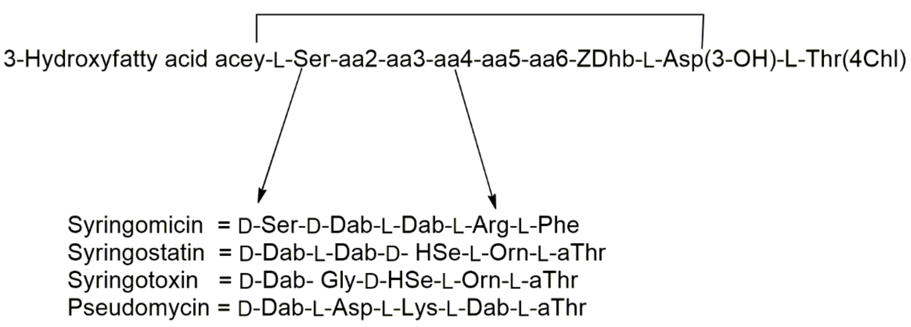
Figure 1. Structures of syringomycin, syringostatin, syringotoxin, and pseudomycin. Abbreviations of non-standard amino acids: Asp(3-OH), 3-hydroxyaspartic acid; Dab, 2,4-diaminobutyric acid; Dhb, 2,3-dehydroaminobutyric acid; Hse, homoserine; Orn, ornithine; Thr(4-Chl), 4-chlorothreonine; aThr, allothreonine.
Ijms 23 12342 g001
Figure 2. Structures of syringopeptin forms SP22 and SP25. The fatty acid can be either 3-hydroxydecanoic or 3-hydroxydodecanoic acid. Abbreviations of non-standard amino acids: Dab, 2,4-diaminobutyric acid; Dhb, 2,3-dehydroaminobutyric acid; aThr, allothreonine. D-Amino acids are common in both SP22 (13 of 22 residues) and SP25 (15 of 25 residues).
Ijms 23 12342 g002
Figure 3. Lipodepsipeptides produced by different strains of Pseudomonas syringae pv. syringae (113 and 16).
Ijms 23 12342 g003
Figure 4. Lipodepsipeptides produced by Pseudomonas fuscovaginae (14 and 15), Pseudomonas syringae pv. syringae (17 and 18), and Pseudomonas corrugata (19 and 20).
Ijms 23 12342 g004
Figure 5. Structures of lipodepsipeptides produced by Pseudomonas corrugata (21) Pseudomonas putida (22 and 23) and Pseudomonas syringae pv. lachrymans (24 and 25) and Streptomyces fradiae (26).
Ijms 23 12342 g005
Figure 6. Structures of lipodepsipedtide produced by Pseudomonas tolaasii (27 and 28), Pseudomonas fluorescens and Pseudomonas libanensis (30), and P. fluorescens (2941).
Ijms 23 12342 g006
Figure 7. Structures of lipodepsipeptide produced by Pseudomonas fluorescens (42 and 43), Pseudomonas sp. JX090307 (44 and 45), Pseudomonas sp. (46), and Pseudomonas cichorii (47 and 48).
Ijms 23 12342 g007
Figure 8. Structures of lipodepsipeptides produced by Ralstonia solanacearum (49 and 50), Streptomyces sp. (5153), Serratia sp. (5464) and Paenibacillus polymyxa (6568).
Ijms 23 12342 g008
Figure 9. Lipodepsipeptides produced by Pseudomonas cichorii (69), and Lysobacter sp. (7083).
Ijms 23 12342 g009
Figure 10. Structure of lipodepsipeptides produced by Pedobacter cryoconitis (8491), by Burkholderia gladioli (92 and 93), and by Pedobacter protegens (94).
Ijms 23 12342 g010
Figure 11. Structures of lipodepsipeptides produced by Actinoplanes (9598) and Streptomyces fungicidicus (99 and 100).
Ijms 23 12342 g011
Figure 12. Structures of lipodepsipeptides produced by Micromonospora chersina (101), Bacillus subtilis (102108), and Streptomyces sp. (109112).
Ijms 23 12342 g012
Figure 13. Lipodepsipeptides produced by Hypxylon oceanicum (113115), Acremonium sp. (117120), Phaeoshaeria sp. (121) and Phoma sp. (122).
Ijms 23 12342 g013
Figure 14. Lipodepsipeptide produced by Lecanicillium sp. (123 and 124), Ophiosphaerella genus (125128), and Colletotrichum gloesporioides (129133).
Ijms 23 12342 g014
Figure 15. Structure of lipodepsipediteds produced by Beauvaria sp. (134138), Scopulariopsis sp. (139141), Colletotrichum aotearoa (142 and 143), and Fusarium sp. (144146).
Ijms 23 12342 g015
Figure 16. Structure of lipodepsipeptide produced by Fusarium sp. (147 and 148) and Pithomyces sp. (149).
Ijms 23 12342 g016
Figure 17. Structure of lipodepsipeptides produced by Pseudomonas tolaasii (150156).
Ijms 23 12342 g017
Figure 18. Structure of WLIP produced by Pseudomonas reactans and its methyl ester (157 and 160) and the hexacetyl- and tetrahydro-derivative of tolaasiin I (158 and 159).
Ijms 23 12342 g018
Publisher’s Note: MDPI stays neutral with regard to jurisdictional claims in published maps and institutional affiliations.