Review

Effects of Isorhamnetin on Diabetes and Its Associated Complications: A Review of In Vitro and In Vivo Studies and a Post Hoc Transcriptome Analysis of Involved Molecular Pathways

1
Alliance for Research on the Mediterranean and North Africa (ARENA), University of Tsukuba, Tsukuba 305-8572, Japan
2
Laboratory of Aromatic and Medicinal Plants, Center of Biotechnology, Technopark of Borj Cedria, BP 901, Hammam-Lif 2050, Tunisia
3
Faculty of Life and Environmental Sciences, University of Tsukuba, Tsukuba 305-8575, Japan
*
Author to whom correspondence should be addressed.
Int. J. Mol. Sci. 2022, 23(2), 704; https://doi.org/10.3390/ijms23020704
Submission received: 30 November 2021 / Revised: 6 January 2022 / Accepted: 6 January 2022 / Published: 9 January 2022
(This article belongs to the Special Issue The Effect of Phytochemicals and Food Bioactive Compounds on Diabetes)

Abstract

:
Diabetes mellitus, especially type 2 (T2DM), is a major public health problem globally. DM is characterized by high levels of glycemia and insulinemia due to impaired insulin secretion and insulin sensitivity of the cells, known as insulin resistance. T2DM causes multiple and severe complications such as nephropathy, neuropathy, and retinopathy causing cell oxidative damages in different internal tissues, particularly the pancreas, heart, adipose tissue, liver, and kidneys. Plant extracts and their bioactive phytochemicals are gaining interest as new therapeutic and preventive alternatives for T2DM and its associated complications. In this regard, isorhamnetin, a plant flavonoid, has long been studied for its potential anti-diabetic effects. This review describes its impact on reducing diabetes-related disorders by decreasing glucose levels, ameliorating the oxidative status, alleviating inflammation, and modulating lipid metabolism and adipocyte differentiation by regulating involved signaling pathways reported in the in vitro and in vivo studies. Additionally, we include a post hoc whole-genome transcriptome analysis of biological activities of isorhamnetin using a stem cell-based tool.

1. Introduction

Plants have been used as traditional medicines in almost all continents of the world since ancient times. Scientific research into the reasons for their medicinal uses has led to the exploration of bioactive molecules. Extremophile plants that grow in extreme environmental conditions are considered a good potential source of bioactive molecules of interest. In fact, these environmental constraints are at the origin of dysfunctional oxygen metabolism, which leads to oxidative stress by increasing reactive oxygen species (ROS) [1,2]. Some plants, such as halophytes, have a powerful antioxidant system to eliminate these harmful compounds. Among the bioactive molecules of interest, there are phenolic compounds. Several studies have valued plants rich in polyphenols with or without restrictive conditions in the laboratory [3,4,5,6,7,8,9,10]. Several biological effects are attributed to extracts rich in polyphenols, such as anti-inflammatory and anticancer activities [11,12,13]. Other studies showed the antioxidant, the antimicrobial [8,14,15,16] as well as anti-obesity, anti-diabetic and anti-hepatic steatotic [17,18,19] effects of isorhamnetin. Among these polyphenols, flavonoids are distinguished. This group includes several subgroups, such as flavonols. Isorhamnetin is one of the major compounds of flavonols. Isorhamnetin is a monomethoxyflavone or an O-methylated flavonol from the class of flavonoids. It is quercetin in which a methoxy group replaces the hydroxy group at position 3’. Some isorhamnetin derivatives are present in nature, such as isorhamnetin 3-O-β-d-glucopyranoside, isorhamnetin 3-O-neohesperidoside, and isorhamnetin 3-O-rutinoside from Calendula officinalis L. [20]. Isorhamnetin presents significant biological properties such as antioxidant [21], anticancer [22], antimicrobial [23], antiviral [24], anti-inflammatory and anti-diabetic effects [21,25,26,27,28,29,30,31,32,33].
In this review, the first focus is on the origin, chemical structure, isolation and extraction methods, as well as the phytochemical aspect of isorhamnetin. Then, in the second part, we focus on describing the potential anti-diabetic effects of this flavonol through reducing diabetes-related disorders by decreasing glucose levels, ameliorating the oxidative status, alleviating inflammation, and modulating lipid metabolism and adipocyte differentiation. Finally, we performed a secondary analysis of our previously published whole-genome microarray data to explore diabetes-related bioactivities of isorhamnetin in a stem cell-based tool. We also aim to highlight the effect of this molecule on the regulation of the involved signaling pathways by reporting the in vitro and in vivo studies used in this field of research. In this review, we exposed the characteristics of the anti-diabetic activity of isorhamnetin in comparison with quercetin which is considered to be a metabolite of isorhamnetin and an important reference in the natural treatment of diabetes.

2. General Overview on Bioactive Molecules in Particular Polyphenols and Flavonoids

2.1. Oxidative Stress as an Origin of Bioactive Molecules in Plants

Several plants can be subjected to various environmental conditions (salinity, drought, UV rays, heavy metals, extreme temperatures, nutrient deficiency, air pollution and pathogen attacks). These constraints are at the origin of dysfunctions of oxygen metabolism, which generate oxidative stress by increasing reactive oxygen species (ROS). The oxygen molecule (O2) plays an important role in photosynthetic organisms. Originally, in higher plants and algae, gas exchange involving electrons occurs in chloroplasts by capturing carbon dioxide during the day and producing oxygen. This gas exchange involves electrons. Indeed, in the case of severe environmental constraints, a large part of oxygen is not reduced and can thus generate ROS in some organelles from plant cells [1,2] due to an imbalance in the proper functioning of chloroplasts and the transfer of electrons [1]. Moreover, under the environmental constraints as mentioned above, many ROSs are produced, such as hydroxyl radical (.OH), the superoxide anion radical (O2.−), alkoxyl and peroxyl radicals (RO. and RO2, respectively), hydrogen peroxide (H2O2), hypochlorite radical (OCl), singlet oxygen (O2), nitric oxide radical (NO) and other lipid peroxides (such as malondialdehyde and 4-hydroxynonenal) [2,34,35]. Sometimes, ROS may have a role in cell signaling in the physiological behavior of plants, for example in the process of seed growth and development, development of tissues, and the transition from cellular proliferation to cell elongation during the earlier stages of differentiation [36]. At a high level, these molecules cause molecular damage such as lipid peroxidation of membranes, alteration of proteins and DNA, and cell death [1,35,37]. Some plants, such as halophytes, have the capacity to adapt well to these conditions through a powerful antioxidant system. Halophytes are known to be a source of secondary metabolites such as polyphenols [3,4,5,6,7,8,9,10]. These authors show that under severe conditions, polyphenols are synthesized to play an important role in protecting against stress-induced oxidative damage. The biosynthesis, the content and the activities of these phenolic compounds are a function of several extrinsic (light, temperature, salinity and dryness) and intrinsic (genotype, organ and stage of development) parameters influencing their content and distribution in plants [8,38,39]. For example, in Pyracantha coccinea, some flavonoids as flavanones, flavones, and flavonols are present in the shoots during the vegetative phase and in the roots exclusively during the reproductive one [40]. On the other hand, several authors have shown that these phenolic compounds have other biological properties such as anti-inflammatory and anticancer activities [11,12,13]. Other studies showed the antioxidant, antimicrobial [8,14,15,16] as well as anti-diabetic [18,41,42] effects of phenolic extracts.

2.2. Classification of Natural Antioxidants

The antioxidant system can be classified according to the nature of these components into enzymatic or non-enzymatic compounds. The first include superoxide dismutase (SOD), catalase (CAT), ascorbate peroxidase, and glutathione reductase [1,35,42,43,44]. The genes relating to these enzymes revealed their importance during the postharvest physiological deterioration of storage root and in response to osmotic stress and abscisic acid as well as Xanthomonas axonopodis infection [44]. The second group contains mainly phenolic compounds, carotenoids, vitamins and osmolytes [1]. Phenolic compounds are characterized by the presence of one or more benzene rings and differ in the complexity of the base molecule, the number and location of the hydroxyl and the degree of polymerization. These compounds are secondary metabolites that fall into three broad groups: phenolic acids (derivatives of benzoic and cinnamic acids), flavonoids (flavonols, flavanols, flavanones, flavones, anthocyanins) and tannins (hydrolyzable tannins and proanthocyanidins). In addition to these molecules, stilbenes, lignans and coumarins are also distinguished [45].

2.3. Origins and Biochemical Structure of Flavonoids, in Particular, Isorhamnetin

Phenolic compounds are secondary metabolites prevalent in plants. These compounds are with an aromatic ring with one or more hydroxyl groups (OH) and contain molecules ranging from simple phenolic acids to polymerized compounds such as tannins. The synthesis of phenolic compounds is a complex process that goes through several stages. Phenolic compounds are bioactive molecules with two origin pathways: in one side the shikimic acid and on the other side, the phenylpropanoid molecules. The biosynthesis of flavonoids such as isorhamnetin is based on these pathways. In fact, on the one hand, shikimate gives the basic skeleton of polyphenols which have one or more benzene rings (C6) carrying one or more hydroxyl functions. On the other hand, there is the synthesis of the C6-C3 base formed by the condensation of phenylalanine to cinnamic acid (Figure 1) [46].
More precisely, shikimic acid is at the base of several reactions, representing the skeleton of aromatic amino acids, which are the initiators of phenolic compounds. The first step consists of a combination of two molecules: phosphoenolpyruvate and erythrose 4-phosphate, which after four reactions, lead to forming the first skeleton of phenolics: the shikimate or shikimic acid (C7H9O5). This later presents the first ring, which characterizes phenolics with two hydroxyl groups. The shikimate undergoes six reactions which end in the first amino acid: phenylalanine. Thanks to two key enzymes, phenylalanine ammonia-lyase (PAL) and cinnamate-4-hydroxylase (C4H), phenylalanine successively forms cinnamate and p-coumarate.. In this step, the level of PAL activity could quantitatively regulate the accumulation of phenolic compounds. The p-coumarate molecule is at the origin of coumarin derivatives. The pathway for the synthesis of phenylpropanoid molecules is characterized by the presence of a key enzyme called 4-coumarate CoA ligase (4CL), which catalyzes in the presence of the thiol function of the coenzyme A (CoA) the p-coumaric acid into 4-coumaroyl CoA (C30H42N7O18P3S). The aromatic A-ring of flavonoids provides from the condensation of three molecules of malonyl-CoA (-C6).
Subsequently, the 4-coumaroyl CoA produced the naringenin chalcone, explaining the link between the aromatic B-ring and the 3C one of chalcone (C6-C3-). Chalcone is a key element in this topic because it is a precursor of all flavonoids based upon a fifteen-carbon skeleton consisting of two benzene rings. Afterward, chalcone is converted to naringenin (also called flavanone or trihydroxyflavone) by the action of chalcone isomerase (CHI). On the one hand, flavones such as apigenin, acacetin, chrysin or luteolin are synthesized from naringenin in the presence of flavone synthase (FNS). On the other hand, different flavonols and succinic acid compounds are produced from naringenin in the presence of two enzymes: flavanone 3-dioxygenase (F3D) and flavonol synthase (FS). Apart from flavonol derivatives, other compounds, such as kaempferol, myricetin, and quercetin, are also produced. By a methyl group transfer from the S-adenosyl-L-methionine, the isorhamnetin is produced in the presence of the flavone 3′-O-methyltransferase (FMT) [47,48]. Then, isorhamnetin is a monomethoxyflavone or an O-methylated flavonol from the class of flavonoids. It is a quercetin (precursor) in which the hydroxy group at position 3’ is replaced by a methoxy group. Some isorhamnetin derivatives are present in nature, such as isorhamnetin 3-O-β-d-glucopyranoside, isorhamnetin 3-O-neohesperidoside, and isorhamnetin 3-O-rutinoside from Calendula officinalis L. [20].
In fact, flavonoids are considered one of the most important groups of the polyphenol family. They have a structure based on a diphenyl propane type with two benzene rings (ring A and B, see Figure 2) linked by a three-carbon chain that forms a closed pyran ring (C ring). Therefore, their structure is referred to as C6-C3-C6. O-glycosylation positions are C7 in flavones, isoflavones, flavanones, and flavonols, and C3 in flavonols and anthocyanidins. C-glycosylation positions are C6 and C8 in flavones [49]. In addition, the importance of the antioxidant role of phenolic compounds is related to the degree of hydroxylation of the molecule. Flavonoids include the isoflavones, flavones, flavanones and their glycosides and flavonols as isorhamnetin, which is also named 3’-methoxy quercetin and 3-methyl quercetin [50]. In plants, an enzyme, the UDP-dependent glycosyltransferases, is responsible for the glycoside form of isorhamnetin (isorhamnetin 3-O-glucoside). This enzyme utilizes nucleotide diphosphate sugars, usually uridine diphosphate (UDP)-sugars, to transfer the methyl group to the cycle and associate the glycoside function with isorhamnetin [51].

2.4. Isolation and Analyses of Isorhamnetin Originated from Medicinal Plants

The distribution of isorhamnetin in medicinal plants is very wide and the methods of extraction and analyses are varied. Isorhamnetin derivatives are particularly wanted. The hydroxyl and methyl groups help in their characterization. Some methods are used to extract isorhamnetin, among them based on fractionation, using chemometric approaches, enzyme, and supercritical fluid extraction (SFE-CO2). Firstly, fractionation can be used to simplify the extraction by removing all oil and lipophilic pigments from lipid-containing samples. The defatted sample is also sonicated before maceration in a mixture of methanol and water. Chromatography can then be used for analyses of phenolic compounds, especially flavonoids [49]. For these last compounds, LC-MS is used and both electrospray ionization (ESI) and atmospheric-pressure chemical ionization are frequently applied, and the best sensitivity for flavonoids is generally achieved in the negative-ion mode [49]. For example, the phytochemistry of Calligonum azel Maire plant fractions, collected from the Tunisian desert, have been assessed and an ultra-high performance liquid chromatography coupled to quadruple time of flight mass spectrometry UHPLC-ESI-QTOF was used to identify phenolics among them flavones and flavanols, which were the most abundant phenolic compounds identified [52]. More specifically, the presence of isorhamnetin glucoside and isorhamnetin glucosyl-rhamnoside was confirmed as the major compounds in the leaves of the edible halophyte Mesembryanthemum edule using LC/ESI-MS/MS technology [3]. This work was followed by others who have used LC-ESI-TOF-MS to characterize many polyphenols; among them, flavonoids were identified from aerial parts of the full flowering stage of halophytes such as Arthrocnemum indicum [5], Tamarix gallica [16], Glaucium flavum [13], and Salsola kali [8]. Similar to last works, LC-ESI-TOF-MS and GC-MS profiling of Artemisia herba-alba was performed to identify phenolics, such as flavones, flavonols and flavonoid alkaloids [7]. Another work was carried out on Pancratium maritimum and the analysis by HPLC-DAD-ESI/MS revealed the presence of flavonoids including flavonol as isorhamnetin with their pentoside and hexoside conjugates such as isorhamnetin di-hexoside [53]. As a non-fatty compound, flavonols could be isolated by hexane. Then, a polar solvent such as ethanol can be used. A study on Limoniastrum guyonianum using HPLC showed the presence of many phenolics, among them isorhamnetin-3-O-rutinoside [54].
Far from phenolic extracts, phenolic compounds are also found in oils such as olive oil [55]. These compounds have the ability to protect oils against oxidation and improve the nutritional value of the oil. Isorhamnetin, as a phenolic compound, has also been detected in an oil extract from Tunisian black cumin (Nigella sativa L.) seeds obtained with a green solvent such as 2-methyl tetrahydrofuran (MeTHF) as an alternative to petroleum or hexane-based solvent to extract oil-enriched phenolic compounds [10]. The presence of isorhamnetin was approved by HPLC analysis. Analyses using Liquid Chromatography with Diode Array Detector (LC-DAD) revealed a high amount of isorhamnetin ranging between 6.3 and 6.6 (µg/g oil) after oil extraction from black cumin by MeTHF and hexane, respectively. In addition, using H nuclear magnetic resonance (HNMR) and C nuclear magnetic resonance (CNMR), some flavonol glycosides such as kaempferol-3-O-rutinoside (nicotiflorin) and isorhamnetin-3-O-rutinoside (narcissin) were characterized from the aerial parts of Peucedanum aucheri Boiss collected in Marivan city, Kurdistan province, Iran [56]. The NMR method detects the property that certain atomic nuclei of the compound interact with a magnetic field. This property, which is to produce magnetic resonance at a certain frequency, provides information on the structure of the molecule. Secondly, mathematical models are chosen to evaluate the efficiency of flavonol extraction, especially to understand the best way to obtain isorhamnetin-3-O-rutinoside [20]. A multivariate factorial analysis was conducted using flowers of Calendula officinalis. Linear, quadratic, full cubic and special cubic models were analyzed. At the final full cubic was the most appropriate one, allowing greater efficiency in the extraction of isorhamnetin-3-O-rutinoside by 60%. For this study, the enzymes, Rapidase Maxi Fruit and Viscozyme, were used under some important factors affecting the enzyme activity under supercritical CO2 conditions such as pressure, temperature, pH, time and aqueous ethanol solution. Finally, the supercritical fluid extraction method was used to extract phenolic compounds such as flavonols. Many years ago, the supercritical fluid extraction procedure was used on the Eucalyptus globulus bark for the first time, using pure and modified CO2 with water, ethyl acetate and ethanol [57]. Authors showed that the supercritical CO2 combined with ethanol could extract significant amounts of phenolic compounds, including isorhamnetin. HPLC-MS quantification determined some flavonols, such as isorhamnetin-hexoside (0.26 g·g−1 of extract) and simple isorhamnetin (14.29 mg·g−1 of extract). Ethanol was used as a co-solvent. The quantification was carried out with high-pressure liquid chromatography equipped with a photodiode array detector. Isorhamnetin 3-O-glucosyl-rhamnosyl-rhamnoside, isorhamnetin 3-O-glucosyl-rhamnosyl-pentoside, isorhamnetin 3-O-glucosyl-rhamnoside, and isorhamnetin 3-O-glucosyl-pentoside were the most abundant flavonols extracted from O. ficus-indica supercritical extracts. As mentioned before, high-pressure liquid chromatography or HPLC is a commonly used method for analyzing phenolic compounds. In apple juice and pear juice, isorhamnetin 3-O-glucoside was detected. In fact, the separation of the flavonol glycosides in a “Brettacher” apple extract by HPLC and mass spectrometry revealed the presence of two glycoside forms such as isorhamnetin 3-O-glucose and isorhamnetin 3-O-galactoside [58].

3. General Overview of Biological Activities of Isorhamnetin

Oxidative stress is an endogenous and exogenous process that plays a role in aging. Oxidative stress occurs due to dysregulation in the antioxidant defences resulting from the imbalanced production of ROS and nitrogen species [59]. Several researchers have shown that long-term exposure to free radicals contributes to the development of chronic diseases, such as cancer [60], diabetes [61], cardiovascular problems [62], and neurodegenerative disease [63]. Therefore, the study of the biological activities of isorhamnetin can be initiated by investigating the involvement of this molecule in the antioxidant phenomenon. Isorhamnetin belongs to the flavonol class known for its antioxidant potential [64,65]. Moreover, several studies have shown that this flavonol has remarkable antioxidant activity, scavenging DPPH radical and ABTS radical, and can inhibit lipid peroxidation [66,67,68]. Wu et al. showed that isorhamnetin and isorhamnetin-3-glucuronide could inhibit the human breast cancer MCF-7 cell proliferation [69]. Wei et al. proved that isorhamnetin inhibits the proliferation of a cervical cancer cell line HeLa [70]. The mechanism of the anti-proliferative effect of isorhamnetin was thoroughly related to cell cycle arrest at the G2/M stage by the activation of the ATM-Chk2 pathway. Li and colleagues revealed that isorhamnetin could also inhibit the growth of the gefitinib resistant PC9 cells (PC9-IR) by downregulating BCL-2 gene expression and PCNA protein expression, inhibiting DNA synthesis and upregulating P53, BAX and CASP3 gene expression [22]. Along with this, Aonuma et al. [71] demonstrated that isorhamnetin might efficiently suppress hypertrophy and fibrosis induced by angiotensin II in cardiac tissue by regulating transforming growth factor (TGF-β) pathways. Furthermore, it might have appreciated properties on potential clinical consequences of cardiovascular diseases by the renin-angiotensin system regulation. Isorhamnetin exerted a neuroprotective effect against ischemic injury in mice by reducing infarct volume and Casp3 activity (a biomarker of apoptosis) and improving neurological function repossession. Likewise, the mice treated with this flavonol showed decreased cerebral edema, enhanced blood–brain barrier function, and upregulated gene expression of tight junction proteins, including Ocln, Zo-1, and Cldn-5 [72]. Ishola et al. reported that isorhamnetin ameliorates scopolamine-induced spatial and non-spatial learning and memory impairment in vivo [73]. Likewise, it could reduce malondialdehyde (MDA) and nitrite production by increasing glutathione (GSH) level, SOD, and CAT activities in the prefrontal cortex and hippocampus. The anti-inflammatory effect of isorhamnetin and related mechanisms have been widely studied [21]. Yang et al. [32] demonstrated that isorhamnetin could alleviate LPS-induced acute lung injury by inhibiting Cox-2 expression in male BALB/c mice [74]. Similarly, the in vivo study of Dou et al. [75] showed that it could alleviate bowel disease by activating PXR and promoting the upregulation of PXR-mediated metabolism of probiotics and the downregulation of nuclear factor-kappa B (NF-κB) signal transduction [75].
Furthermore, isorhamnetin has been reported to prevent pulmonary tuberculosis [76]. Additionally, it may ameliorate acute kidney injury by inhibiting the NF-κB signaling pathway activation [31]. Additionally, isorhamnetin exhibits anti-thrombus [77], antihypertensive [78], anti-inflammatory [26], anti-osteoporosis [79], antiplatelet activity [80], hepatoprotective [81], and anti-hypoxia [82] effects. It has also been shown that isorhamnetin could boost innate immunity [83]. However, we have not found a wide range of publications that confirm antimicrobial and antiviral activities of isorhamnetin. Still, some studies reported bactericidal effects of plant extracts containing isorhamnetin or its derivatives [84,85,86]. Antiviral activities of isorhamnetin were observed against influenza [87], SARS-CoV-2 spike [24], and herpes simplex [88] (see the summary in Figure 3). In summary, this interesting molecule is an immense source of biological activities. More than what we have mentioned, it is endowed with a critical anti-diabetic activity that we study in the next section of this review.

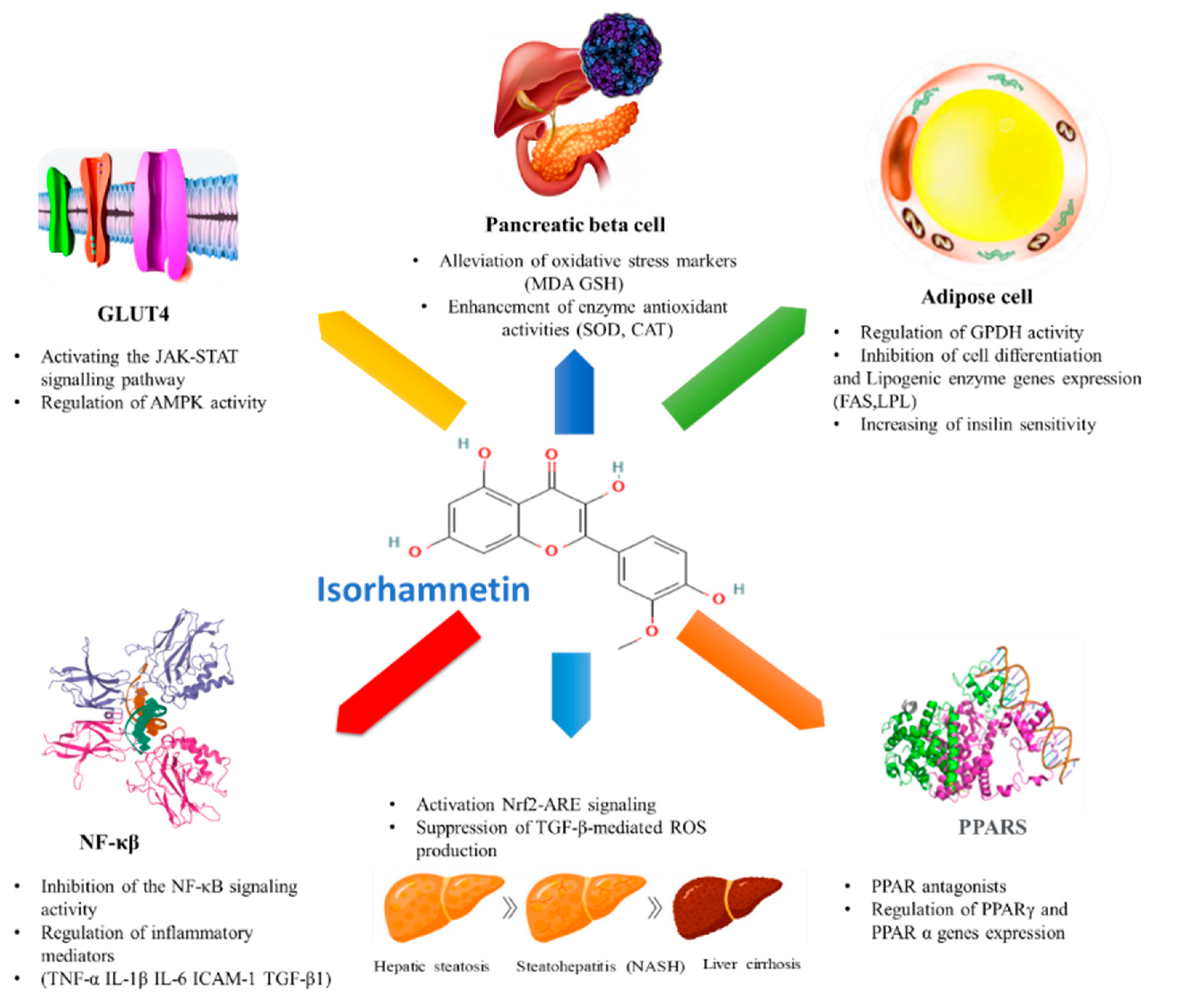
4. Anti-Diabetic Effect of Isorhamnetin

Isorhamnetin has been reported to alleviate shared metabolic complications in type 1 diabetes mellitus (T1DM) and type 2 diabetes mellitus (T2DM), but until the present, it is more aligned with T2DM. For example, Matboli et al. showed that isorhamnetin used at three different doses (10, 20, 40 mg/kg for 3 weeks) possessed anti-diabetic action by regulating the insulin pathway at the microscopic, molecular and protein levels in the streptozotocin/high fat diet-induced T2DM rat models [89]. Authors in this study concluded that isorhamnetin could be used as an alternative and/or potential complementary treatment in T2DM through modulation of insulin resistance signaling pathway-related gene expression. However, regarding T1DM, the effect of isorhamnetin is not yet well studied with in vitro and in vivo models.

4.1. General Overview on Diabetes and Its Link with Metabolic Syndrome

Metabolic syndrome is a constellation of cardiometabolic risk factors first described 90 years ago [90]. In 1988, the notion of syndrome X appeared [91]. It includes abdominal obesity, insulin resistance, glucose intolerance (or hyperglycemia), hypertension, and dyslipidemia. The combination of these risk factors is linked to an increased risk of developing T2DM and cardiovascular diseases. Diabetes mellitus (DM) is a prevalent metabolic disease characterized by abnormally elevated blood glucose levels. It is classified into type 1 (T1DM) and type 2 (T2DM). The first category of diabetes (T1DM) is typically associated with failure in insulin production resulting from the destruction of pancreatic β-cells by T-cell-mediated autoimmunity. However, T2DM is more specific to insulin resistance and function [92]. The causes of metabolic syndrome are generally poorly understood but involve genetic factors linked to the environment. Among the genetic factors, one can quote those determining the corpulence, the distribution of the fatty mass, the hyperinsulinemia, and various metabolisms (lipoproteins) [93]. The constitutive elements of these factors tend to derive from multivalent logic. The environmental factors, the sedentary lifestyle, smoking, excess calories provided in the form of lipids, and added sugars, in particular, are better known. Many other factors have been recently discovered, such as the presence of inflammatory cells in adipose tissue and alterations in the secretion of adipokines [93]. It is, therefore, necessary to find an adequate and safe treatment given the severe consequences of this disease on health, including diabetic nephropathy, retinopathy, neuropathy, cardiovascular diseases, and diabetic foot ulcer [92].

4.2. Effect of Isorhamnetin on Associated Metabolic Pathways

Several in vitro and in vivo studies showed that isorhamnetin might modulate carbohydrate metabolism and exhibit anti-diabetic activities by involving different mechanisms. Researchers have suggested that isorhamnetin promotes carbohydrate metabolism from the stages of digestion and intestinal absorption [94], improves glucose uptake by the liver and muscles [19,95], protects the pancreatic β-cells and alleviates insulin secretion impairment [96]. In addition, isorhamnetin regulates adipose tissue differentiation, growth and maturation [29]. For anti-diabetic potential, researchers have investigated isorhamnetin isolated from different medicinal plants. In Table 1, we have listed the plant sources and extraction procedures of isorhamnetin that regulated diabetes-associated different intracellular signaling pathways. In the rest of this review, we will try to explain the impact of this molecule and its metabolic precursor (quercetin) [97] on the diabetes-associated mechanisms.

4.2.1. Effect of Isorhamnetin on Glucose Transporters

Glucose transporters (GLUT) are a large group of membrane proteins that transport glucose from epithelial cells to blood and from blood to cells passing the intestinal barrier in the gradient direction by passive transport. Some of them are insulin-dependent, notably GLUT4. It is a major insulin-regulated glucose transporter and coordinator of insulin action in adipose. In fact, it is well established that insulin, which enhances glucose uptake, also stimulates the recruitment of GLUT4 to the cell membrane of insulin-sensitive tissue. In a study underlying the anti-diabetic properties of quercetin and isorhamnetin, the authors proved that physiological concentrations of isorhamnetin promoted GLUT4 translocation to the plasma membrane in L6 myotubes through different mechanisms without altering GLUT4 expression [95]. In fact, isorhamnetin could activate the JAK-STAT signaling pathway at 1 nM and 10 nM, allowing glucose transporter translocation induction. JAK-STAT is a signal transduction system composed of a transmembrane receptor, coupled to a Janus Kinase (JAK) enzyme and a STAT-type protein. When a ligand binds to the receptor, it changes its conformation, activating the enzyme JAK, which phosphorylates the STAT protein, inducing a transduction cascade and activating transcription of specific genes [100]. In an in silico study, Selvaraj [101] showed that certain flavonoids, including isorhamnetin, can strongly interact with the active site of GLUT4 protein through H-bond interaction. This finding lets us look for the influence of this molecule on glucose transporters, particularly GLUT 4. By comparison, quercetin stimulates the insulin and AMPK-dependent pathway in the same L6 cells. In fact, Eid et al. have shown that an 18 h treatment of cultured rat L6 skeletal muscle cells with a dose of 50 µM quercetin could stimulate AMPK and increase GULT4 translocation [102]. Likewise, quercetin demonstrated divergent effects on insulin-mediated GLUT4 translocation in adipocytes under basal and insulin-resistant conditions related to its regulation of AMPK activity [103]. Moreover, Eid et al. confirmed that quercetin aglycone, extracted from Vaccinium vitis berries, could enhance muscle cell glucose uptake [98]. Guided fractionation of berry extract based on glucose uptake in muscle cells demonstrated that quercetin-3-O-glycosides were the main active compounds. At 50 μM, these compounds enhanced basal glucose uptake by up to 59% following an 18 h treatment, an effect significantly greater than that of 100 nM insulin.

4.2.2. Effect of Isorhamnetin on Peroxisome Proliferator-Activated Receptors (PPARs)

PPARs belong to the transcription nuclear receptor factors class II family with an important action in the glucose and lipid metabolism regulation [104]. They are expressed in various cell types: pancreatic cells, hepatic cells, and adipose cells, thus controlling a wide range of biological processes by the modulation of related genes’ expressions [105]. In mammalian cells, three isoforms are present: PPARα, PPARβ/δ and PPARγ, which regulate genes implicated in glucose and lipid transport, synthesis and fatty acid oxidation [106,107]. Many clinical studies are targeting PPARs for metabolic disorders and diabetes treatments. Thiazolidinediones (TZDs: pioglitazone and rosiglitazone) are a class of oral anti-diabetic drugs that reduce high blood sugar levels and improve the lipid profile of T2DM patients. A huge number of hypotheses suggest that the anti-diabetic effect of TZDs is exerted via PPARγ. Fibrates that target PPARα have also been used to treat hyperlipidemia and diabetes. Isorhamnetin has been reported to exert similar effects as the mentioned drugs, mainly as PPARs antagonists or agonists [28], which ameliorates metabolic complications induced by a high-fat diet or leptin deficiency [33]. Pre-adipocyte cell line model 3T3-L1 cells are widely used in obesity and diabetes research [108,109]. Obesity-induced diabetes is characterized by hypertrophy and hyperplasia of adipocytes. Pretreatment of 3T3-L1 cells with different doses of isorhamnetin could inhibit adipocyte differentiation by decreasing triglyceride (TG) accumulation and glycerol-3-phosphate dehydrogenase (GPDH) activity [29]. At the molecular level, isorhamnetin could also regulate the mRNA expression of the major adipocyte markers, mainly the transcription factors PPARγ and CCAAT/enhancer-binding protein-α (C/EBPα), the master co-working regulators of adipogenesis and cell differentiation [29,30]. These findings were also concordant with studies revealing that Nitraria retusa isorhamnetin-rich extract could reduce the fat accumulation in 3T3-L1 adipocytes in a dose-dependent manner [18]. The single-molecule isorhamnetin could also significantly inhibit the differentiation rate of 3T3-L1 cells, leading to reducing lipid droplet content by reducing cell size and number. In addition, oral administration of the Nitraria retusa extract could regulate PPARγ and lipogenic enzymes target genes LPL and FAS [17]. Importantly, a recent study revealed that isorhamnetin could significantly reduce fat amount in animal body via PPARα-dependent pathway [27].

4.2.3. Effect of Isorhamnetin on Hepatic Enzymes

The liver is an organ that plays a key role in carbohydrate homeostasis [110]. Beyond the micro and macrovascular complications of diabetes, the T2DM patient presents more certain hepatic complications with, in the first place, hepatic steatosis. In T2DM, the risk of progression to fibrosis and hepatic inflammation is greater [111], exposing a certain number of diabetic patients to significant clinical consequences, particularly with hepatocarcinoma [112]. In parallel, hepatic complications in diabetic patients have been associated with a higher risk of cardiovascular events, which may have practical consequences in terms of optimization of cardiovascular prevention [113]. We focus on the most important liver complications related to diabetes: fatty liver diseases, hepatic fibrosis, and hepatocellular carcinoma (HCC).

Non-Alcoholic Steatohepatitis

Fatty liver disease or non-alcoholic steatohepatitis (NASH) represents a potential hepatic complication. It is characterized as an abnormal accumulation of TGs inside liver cells. In type 2 diabetes, insulin resistance leads to the accumulation of toxic fatty acids. When this accumulation occurs in liver cells it causes non-alcoholic fatty liver disease (NAFLD) and in severe forms steatosis or NASH [111,114]. Ganbold et al. have shown that isorhamnetin ameliorated hepatic steatosis and fibrosis in a NASH mouse model exhibits similar features of human NASH [19]. In fact, compared to the control mouse group, liver samples from the NASH-induced group showed acute fat accumulation overtaking 37% of the oil red O positive area. However, isorhamnetin treatment in the NASH group could reduce oil red O stained area to 22%. In addition, isorhamnetin treatment alleviated NASH-induced gene expression alteration. The authors have shown that isorhamnetin efficiently improved the altered lipid metabolic process in NASH and inhibited de novo lipogenesis.

Hepatic Fibrosis

Hepatic fibrosis is excessive scarring resulting from the build-up of connective tissue in the liver. It is caused by excessive production and/or insufficient degradation of extracellular matrix. There are many causes of liver fibrosis. Among them is NASH origin. When there is resistance to insulin, fatty acids build up in the liver, causing toxicity and inflammation. This is accompanied by the production of cytokines and reactive oxygen species. In fibrosis, cytokines are responsible for the activation of hepatic stellate cells (HSCs) and will activate an excessive production of collagen fibers [115,116]. The trigger is chronic aggression, especially in the event of an inflammatory component [115]. TGF-β is a crucial mediator of HSC activation and extracellular matrix accumulation, leading to fibrosis [117]. Therefore, blocking this pathway could be a potential strategy for liver fibrosis [118]. In this context, Yang et al. demonstrated that isorhamnetin inhibited HSC activation and prevented TGF-β-induced expression of fibrogenic genes, including α-SMA, PAI-1, and COL1A1 [74]. In fact, the authors revealed that the inhibitory effect of this flavonol was a result of the canonical TGF-β/Smad signaling pathway inhibition. Additionally, isorhamnetin activated Nrf2-ARE signaling and suppressed TGF-β-mediated ROS production, which also contributed to the inhibition of fibrogenic gene expression. Furthermore, isorhamnetin significantly suppressed carbon tetrachloride (CCl4)-induced fibrosis in mice. Liu et al. also showed that isorhamnetin could alleviate CCl4 and bile duct ligation (BDL)-induced liver fibrosis by inhibiting TGF-β1 production and HSC activation [119]. Likewise, Lee et al. demonstrated that isorhamnetin, extracted from Oenanthe javanica, could reduce fibrosis in rat HSCs by blocking the extracellular signal regulated kinase (ERK) signaling pathway and inhibiting the proliferation and collagen synthesis of HSC-T6 cells [99]. In fact, pretreatment of HSC-T6 cells with this flavonol repressed serum-induced ERK phosphorylation, the same way as a MEK inhibitor (PD98059).

Hepatocellular Carcinoma

Hepatocellular carcinoma is often discovered late at the metastasis stage (in 64% of cases); the most frequent secondary locations are the lung, lymph nodes, kidneys, and adrenal glands [120]. As mentioned above, with the high accumulation of fatty acid in liver cells, physiological disturbances appear over time such as NASH, then inflammation (with production of cytokines and ROS), then in more advanced cases cirrhosis and in severe forms carcinoma [121,122]. The role of isorhamnetin is deduced by its anti-tumor potential activities. Isorhamnetin, extracted from Hippophae rhamnoides L., showed potent anti-tumor activity in hepatocellular carcinoma BEL-7402 cells, exerting strong cytotoxicity with IC50 at the concentration of 74.4 ± 1.13 µg/mL and 72 h incubation [123]. Cytotoxicity of this flavonol on cancer cells depends on isorhamnetin cellular accumulation. Instead, the pibenzimol hydrochloride staining of isorhamnetin-treated cells displayed condensed and fragmented nuclei and apoptotic bodies. Furthermore, flow cytometry analysis used to evaluate cell cycle progression and differentiate between apoptosis or necrosis, revealed that 13.77% of isorhamnetin-treated BEL-7402 cells (with 50 µg/mL for 48 h) appeared in the hypodiploid peak. Similar work by Wei et al. reported that isorhamnetin inhibited the cell cycle progression of the cervical cancer cell line HeLa, by arresting the G2/M phase via activating the ataxia-telangiectasia mutated Chk2 pathway [75]. In another work and by making a comparison, isorhamnetin-3-O-glucoside, quercetin 3-O-rhamnoside-7-O-glucoside and kaempferol-3-O-glucoside-7-O-rhamnoside, extracted from Cleome droserifolia, reduced HepG2, a liver hepatocellular carcinoma cell, cell viability and anchorage-independent cell growth in a dose- and time-dependent manner but with a highest effect with the quercetin derivative. Furthermore, they exhibited a reduced migration capacity of HepG2 cells [124].

4.2.4. Effect of Isorhamnetin on Pancreatic β-Cell Dysfunction

Insulin is a hormone naturally secreted by the body that helps our cells, particularly muscle cells, liver cells, adipose cells, and brain cells, absorb glucose from food to be converted into energy for all biological processes and functions [125,126]. In normal conditions, insulin is secreted by pancreatic β-cells, then released into the bloodstream. β-cell dysfunction is characterized by impaired insulin secretion, the critical feature of T2DM, both in its onset and progression [127]. Metabolic stress-induced β-cell dysfunction in T2DM can also be associated with high levels of saturated fats in obesity, leading to increased inflammation and oxidative stress within β-cells. Therefore, impairment of insulin secretion is more severe than insulin resistance [128]. Taken together, new anti-diabetic therapeutic strategies are needed to prevent or recover pancreatic β-cell dysfunction. In this context, Wang et al. suggested that isorhamnetin, derived from Vernonia anthelmintica herb, could suppress the proliferation of pancreatic cancer cells (colorectal adenocarcinoma cell line) via arresting the cell cycle in the S phase, which may be an alternative way to prevent pancreatic carcinoma and its metabolic complications [129]. Consistently, another report has shown that isorhamnetin and a glycosylated form of isorhamnetin (isolated from Salicornia herbacea plant) could promote glucose-stimulated insulin secretion in insulin-secreting rat insulinoma (INS-1) pancreatic β-cells without affecting cell viability [96]. Moreover, it could stimulate insulin secretion via phosphorylation of total ERK, insulin receptor substrate-2 (IRS-2), phosphatidylinositol 3-kinase (PI3K), Akt, and activated pancreatic and duodenal homeobox-1 (PDX-1) [96]. The enhancement of these markers may alleviate the pancreatic cell dysfunction causing diabetes.
Furthermore, Grdović et al. reported that isorhamnetin originated from the Castanea sativa plant species has a protective effect on streptozotocin-induced oxidative damage and β-cell apoptotic death [130]. This beneficial effect was correlated strongly to the antioxidant potential of this molecule by alleviating the cellular biomarkers of oxidative stress, i.e., MDA and intracellular GSH levels, and enhancing the activities of antioxidant enzymes SOD and CAT. The effect was also confirmed at the molecular level by the decreased NF-kB transcription factor stimulated by cell oxidative stress, and thus suggestive of a protective effect within β-cells. Consistently, quercetin, which is metabolized in isorhamnetin, could reduce glucose levels by ameliorating insulin secretion in the streptozotocin-induced type 1 diabetes model in rats [131]. In the same animal model, quercetin showed a protective effect on β-cell integrity by reducing oxidative stress markers [132]. It could also regenerate the islets in pancreatic cells [133].

4.2.5. Effect of Isorhamnetin on NF-κB

NF-κB is a protein of the superfamily of transcription factors involved in the immune and cellular stress responses and is considered as the major mediator of the secretion of inflammatory markers such as cytokines, chemokines, and different key enzymes [130,134,135]. The role of NF-κβ in the pathophysiology of diabetes and its linked vascular complications has been investigated [136]. Hyperglycaemia activates this nuclear factor that in turn induces the secretion and over-expression of pro-inflammatory cytokines, mainly TNF-α, interleukins, TGF-β, and Bcl2, causing vascular complications such as neuropathy, nephropathy, retinopathy, and cardiomyopathy [137]. Many experimental investigations on the preventive effect of flavonols on diabetes-induced vascular dysfunction have demonstrated that isorhamnetin could inhibit the activation of NF-κβ through inducing the activity of pancreatic antioxidant enzymes [138]. Another study reported the inhibitory effect of isorhamnetin on iNOS expression and NO production in activated macrophages, leading to the blocking of NF-κβ [139]. Moreover, Qiu et al. have studied the renoprotective effect of the flavonol isorhamnetin in the T2DM rat model through the modulation of the NF-κβ signaling pathway [31]. Authors have shown that this molecule inhibited the NF-κB signaling activity by increasing the production of NF-κB p65, phospho-NF-κB p65, and phospho-IκBα and inflammatory mediators TNF-α, IL-1β, IL-6, ICAM-1, and TGF-β1, as well as decreasing the NF-κB p65 DNA-binding activity. In addition, it could alleviate oxidative cell stress by regulating MDA and SOD levels, leading to recovery of renal damages in diabetic rats.

5. A Post Hoc Transcriptome Analysis Predicts the Potential Effect of the Isorhamnetin on Diabetes in a Stem Cell-Based Tool

We have established a stem cell-based tool using a perinatal stem cell, human amniotic epithelial cells (hAECs), to evaluate the bioactivities of natural compounds employing whole-genome microarray analysis [71,140,141,142,143,144]. In recent years, an increasingly large number of bioactive compounds of medicinal plants have been screened for their potential therapeutic and preventive effects. In this context, stem cell-based approaches using human pluripotent stem cells (hPSCs) receive great attention as physiologically more relevant in vitro human models for drug screening and validation of thousands of compounds in both academic research and the pharmaceutical industry [145,146,147]. However, hPSCs, including embryonic stem cells (hESCs) and induced pluripotent stem cells (hiPSCs), have limited cell resources, require invasive extraction procedures, expensive cell reprogramming, and critical maintenance procedures, as well as pose ethical constraints, and therefore are less favorable as a practical source for drug screening. On the other hand, hAECs are derived from discarded term placenta, a medical waste product. They do not require invasive harvesting procedures and have minimum ethical concerns. Furthermore, hAECs are derived from the pluripotent epiblasts and thus maintain ESC-like multilineage differentiation potential and can be differentiated into cells from all three germ layers [148,149,150,151].
It is worth noting that upon appropriate differentiation protocol, hAECs can be differentiated into hepatocyte-like cells [152,153,154,155], cholangiocyte [156], and, most importantly, pancreatic β-like insulin-producing cells [157,158,159,160]. Transplantation of hAEC-induced pancreatic cells into streptozotocin-induced diabetic mice could normalize the blood glucose level [161]. hAECs [162], as well as exosomes derived from hAECs [163], could accelerate diabetic wound healing via promoting angiogenesis and fibroblast function and reducing inflammation. Incorporating hAECs into islet organoids [164] and shielding native islets with a layer of hAECs [165] could enhance islet engraftment and revascularization in diabetic mice models. Additionally, hAEC-derived hepatocyte-like cells, as well as hAEC itself, have been reported to have therapeutic efficacy in liver diseases, including hepatic fibrosis [166,167], cirrhosis [168], and hepatic failure [169].
Considering the complex pathophysiology of DM, hAEC may not be an ideal in vitro model to study the anti-diabetic effects of compounds. However, because of its stem cell-like properties, it can be used for the initial screening of target compounds. We have previously explored antifibrotic [71] and hepatic differentiation-inducing [170] potential of isorhamnetin in hAECs. In the present study, we have performed a targeted secondary analysis of our previously published data [71] to explore the potential functionalities of isorhamnetin in diabetes (Figure 4). Data analysis was conducted for three biological replicates of day 10 control (n = 3) and isorhamnetin-treated (n = 3) hAECs. The cells were grown on 3D cell culture. Control cells were maintained in placental basal epithelial cell medium (Promo Cell, Cat. #C-26140) in absence of any differentiation medium or growth factors, whereas treatment cells were supplemented with 20 mM of isorhamnetin (Sigma-Aldrich, Japan) for 10 days. Differentially expressed genes (DEGs) are referred to as genes with a linear fold change > 2 and p-value < 0.05 (one-way between-subjects ANOVA). A total of 303 DEGs were identified; among them, 60 were upregulated and 243 were downregulated. Details of methodology have been explained elsewhere [71,170]. All microarray data are available at Gene Expression Omnibus (GEO) under accession number: GSE153149 (https://www.ncbi.nlm.nih.gov/geo/query/acc.cgi?acc=GSE153149, accessed on 24 November 2021).

5.1. Cell Type Signature Gene Sets

In our previous studies on hAECs, we found that different types of compounds could direct the differentiation of hAECs towards different cell lineages, such as a caffeic acid ester, rosmarinic acid [142] and a caffeoylquinic acid derivative 3,4,5-Tri-O-Caffeoylquinic Acid (TCQA) [140] which could enhance neural cell differentiation, whereas an anthocyanin, cyanidin-3-O-glucoside (Cy3G), induced adipocyte differentiation [143] in hAECs. We observed that the bioactivities or functionalities of natural compounds could generally be predicted from the enriched cell types by the DEGs.
We examined the significantly enriched cell type signature data sets using the Molecular Signatures Database (MSigDB) ver. 7.4 of GSEA online software (https://software.broadinstitute.org/gsea/index.jsp; accessed on 26 November 2021) [171]. These gene sets contain cluster marker signature genes for cell types identified in human tissue single-cell sequencing studies and facilitate the cell type assignments in data sets, such as experiments of developing organoid models.
We found that the most significantly enriched cell type gene set was pancreatic mesenchymal stromal cells [172] (Figure 4A). Additionally, pancreatic ductal and endothelial cell types were significantly enriched [172]. Pancreatic signature genes in isorhamnetin-treated hAECs are involved in epithelial-mesenchymal transition, TGF-β signaling, TNF-α signaling via NF-kB, KRAS signaling, and fatty acid metabolism. Several hepatic signature gene sets were also significantly enriched, such as HSCs, kupffer cells, bile duct cells [173], and fetal liver mesothelial cells [174]. There were also several significantly enriched skeletal muscle signature gene sets, including fibrillin 1+fibro-adipogenic progenitor (FBN1+FAP) cells, fibro-adipogenic progenitor (FAP) cells, and skeletal muscle pericytes [175]. Biological functions of hepatic signature genes in isorhamnetin-treated hAECs include several inflammatory response pathways, whereas skeletal muscle signature genes regulate wound healing, collagen fibril organization, and MAPK cascade. Significance was measured as the false discovery rate, an analog of hypergeometric p-value after Benjamini and Hochberg correction for multiple hypothesis testing (FDR q-value < 0.05).

5.2. Significantly Enriched Hallmark Gene Sets

Next, we examined the significantly enriched hallmark gene sets on MSigDB (retrieved on 26 November 2021). Hallmark gene sets represent specific, well-defined biological states or processes generated based on identifying gene set overlaps and retaining genes that display coherent expression. The hallmarks have a collection of 50 gene sets condensed from over 4000 overlapping gene sets and thus have reduced noise and redundancy [176].
Significantly enriched hallmark gene sets include genes defining epithelial-mesenchymal transition, genes upregulated in response to hypoxia, genes regulated by NF-kB in response to TNF, genes up and downregulated by KRAS activation, genes mediating apoptosis by activation of caspases, genes involved in myogenesis, genes upregulated in response to TGF-β1, genes upregulated by STAT5 in response to IL-2 stimulation, genes defining inflammatory response, genes involved in p53 pathways and networks, and genes encoding proteins involved in glycolysis and gluconeogenesis (Figure 4B). Significance was considered at FDR q-value < 0.05.
An interesting finding is enrichment of KRAS activation by the DEGs of the isorhamnetin-treated hAECs. Several KRAS-induced gene expressions were found to be significantly downregulated by isorhamnetin, such as MMP9, TSPAN1, and ITGBL1. Hyperglycemia triggers genomic instability leading to KRAS mutations in pancreatic cells [177] and has also been associated with increased risk and invasiveness of pancreatic [178] and colon cancers [179]. Wang et al. reported that isorhamnetin suppresses proliferation of pancreatic adenocarcinoma cell line PANC-1 through downregulating Ras/MAPK signaling pathway activity [134]. Therefore, as mentioned in Section 4.2.4, the effect of isorhamnetin on the KRAS-induced risk of cancers in diabetes, especially pancreatic cancers, is worth further exploration.

5.3. Significantly Enriched Pathways

Further pathway analysis of the DEGs was conducted using the Comparative Toxicogenomics Database (CTD) (http://ctdbase.org/; accessed on 29 November 2021) [180]. CTD represents Kyoto Encyclopedia of Genes and Genomes (KEGG) and REACTOME pathways. We found that several inflammatory pathways, collagen formation and assembly, PI3K-Akt signaling pathway, and AGE-RAGE signaling pathway in diabetic complications were significantly enriched (Figure 4C).
Advanced glycation end products (AGEs) are produced through the non-enzymatic glycation and oxidation of proteins, lipids, and nucleic acids. The receptors for advanced glycation end products (RAGE) belong to the immunoglobulin superfamily. AGE/RAGE signaling is a complex and intricate cascade that activates multiple intracellular signal pathways involving protein kinase C, NADPH oxidase, and MAPKs, resulting in NF-κB -induced expression of IL-1, IL-6, TNF-α, VCAM-1, and VEGF. Particularly, AGE/RAGE signaling has been implicated in diabetes-mediated vascular calcification through activation of TGF-β mediated fibrosis, NFκB, and ERK1/2 pathways [181,182,183,184]. We found that isorhamnetin significantly decreased AGE/RAGE signaling-related gene expression, such as COL1A1, COL1A2, COL4A6, FN1, MMP2, and SERPINE1. As discussed in the earlier section, isorhamnetin’s antifibrotic effects have been well documented [71,74,99,119], and therefore, it can be asserted that isorhamnetin may also have beneficial effects in diabetes-induced vascular pathology.

5.4. Significantly Enriched Metabolic Diseases and Related Gene Expressions

Curated gene–disease association data were retrieved from the CTD (retrieved on 29 November 2021). We curated only significantly enriched metabolic diseases. The significance of enrichment was calculated by the hypergeometric distribution adjusted by the Bonferroni method. Significantly enriched metabolic diseases included DM, glucose and lipid metabolism disorders, hyperglycemia, and obesity (Figure 4D). Heatmap shows that PPARs, TGFs, TNFs, ILs, collagen, and apoptosis-inducing gene expressions were significantly downregulated in isorhamnetin-treated hAECs (Figure 4E). On the other hand, insulin receptors, lipoprotein lipases, and apoptosis inhibitors were significantly upregulated in isorhamnetin-treated hAECs.
Our targeted microarray data analysis of isorhamnetin-treated hAECs also confirmed the potential of isorhamnetin in regulating biological functions related to DM and its associated complications.

6. Bioavailability and Intestinal Absorption of Isorhamnetin Aglycone and Its Glycosylated Derivatives

With the presence of various categories of flavonoids in nature, it is interesting to analyze the presence of these compounds in the body. When we talk about the metabolism of isorhamnetin and its availability in the human body after consumption, we are talking about the origin, the metabolism and the transport. This is based on an observation made by chromatography of some flavonoids such as flavonols in human serum. Liquid chromatography-mass spectrometry provides insight into the bioavailability of certain flavonoids in their aglycone and glycoside forms in the body [49]. Mass spectrometry can be used to determine flavonoids in biological samples [185]. A study reported 23 mixed sulfate, methyl, glucuronide, and glucose derivatives of quercetin in both urine and plasma of human volunteers 1 h after ingestion of lightly fried red onions. This study detected glycosides of both quercetin and isorhamnetin in plasma [186].
Several factors play a role in the entry of nutrients through the digestive tract. For example, enzymes from the intestinal microbiota affect the entry of phenolic compounds. A study carried out on the ginkgo leaf extracts in a mice model showed the importance of gut microbiota on the bioavailability and absorption from the gastrointestinal tract of some bioactive molecules, especially isorhamnetin [187]. In this step, enzymes of gut microbiota produce flavonoid aglycones and a variety of ring fission products. Analyses of the whole blood samples indicated that the uptake of isorhamnetin was increased by antibacterial treatment, suggesting that gut microbiota enzymes have a negative effect on the pharmacokinetics of natural molecules, such as isorhamnetin. Antibacterial or probiotic consumption may increase the bioavailability of the glycoside form of isorhamnetin. In addition, the in vitro biotransformation rates and residence times of bioactive molecules differed between normal, diabetic, and diabetic nephropathy rats [188].
On the other hand, different membrane transporters control the transport of flavonoids, such as the sodium-dependent glucose transporter 1 (SGLT1) and the multidrug resistance-associated proteins 2 and 3 (MRP2 and MRP3) [189]. In this context, MRP transporters regulate the transcellular and the paracellular transport pathways of isorhamnetin [190]. Inside cells, the transport of isorhamnetin from the apical to the basal side was 6.8–9.3-fold higher. In Figure 5, the anti-diabetic effects of isorhamnetin are summarized.

7. Conclusions

Isorhamnetin is a phenolic compound of the flavonoid family, more precisely of the flavonols. Originally it is a quercetin molecule but has undergone methylation. Isorhamnetin is distributed in the plant kingdom in many wild and cosmetic plants. In addition, several medicinal plants produce this molecule and several studies have attested to its anti-diabetic effect among other biological activities. All these data thus show the interest of isorhamnetin in the therapeutic industry. From this perspective, it would be very interesting to explore the effect of isorhamnetin and its derivatives, isolated especially from natural resources, on metabolic disorders. It is also necessary to highlight and review the clinical studies performed in this context using flavonoid-rich fractions and natural products in order to avoid the impact of side effects caused by synthetic and chemical drugs.

Author Contributions

Conceptualization, H.I.; resources, H.I.; writing—original draft preparation, F.Z.K., M.B. and F.F.; writing—review and editing, H.I.; visualization, F.Z.K., M.B. and F.F.; funding acquisition, H.I. All authors have read and agreed to the published version of the manuscript.

Funding

This research was partially supported by Japan Science and Technology Agency (JST); Science and Technology Research Partnership for Sustainable Development (SATREPS, Grant No. JPMJSA1506).

Institutional Review Board Statement

Not applicable.

Informed Consent Statement

Not applicable.

Data Availability Statement

Microarray data are deposited in the Gene Expression Omnibus (GEO) under Accession Number: GSE153149 (https://www.ncbi.nlm.nih.gov/geo/query/acc.cgi?acc=GSE153149, accessed on 24 November 2021).

Conflicts of Interest

The authors declare no conflict of interest. The funder had no role in the design of the study; in the collection, analyses, or interpretation of data; in the writing of the manuscript, or in the decision to publish the results.

References

  1. Gill, S.S.; Tuteja, N. Reactive oxygen species and antioxidant machinery in abiotic stress tolerance in crop plants. Plant Physiol. Biochem. 2010, 48, 909–930. [Google Scholar] [CrossRef] [PubMed]
  2. Corpas, F.J.; Gupta, D.K.; Palma, J.M. Production sites of reactive oxygen species (ROS) in organelles from plant cells. In Reactive Oxygen Species and Oxidative Damage in Plants under Stress; Springer: Berlin/Heidelberg, Germany, 2015; pp. 1–22. [Google Scholar]
  3. Falleh, H.; Oueslati, S.; Guyot, S.; Dali, A.B.; Magné, C.; Abdelly, C.; Ksouri, R. LC/ESI-MS/MS characterisation of procyanidins and propelargonidins responsible for the strong antioxidant activity of the edible halophyte Mesembryanthemum edule L. Food Chem. 2011, 127, 1732–1738. [Google Scholar] [CrossRef]
  4. Trabelsi, N.; Oueslati, S.; Falleh, H.; Waffo-Téguo, P.; Papastamoulis, Y.; Mérillon, J.-M.; Abdelly, C.; Ksouri, R. Isolation of powerful antioxidants from the medicinal halophyte Limoniastrum guyonianum. Food Chem. 2012, 135, 1419–1424. [Google Scholar] [CrossRef] [PubMed]
  5. Boulaaba, M.; Mkadmini, K.; Tsolmon, S.; Han, J.; Smaoui, A.; Kawada, K.; Ksouri, R.; Isoda, H.; Abdelly, C. In vitro antiproliferative effect of Arthrocnemum indicum extracts on Caco-2 cancer cells through cell cycle control and related phenol LC-TOF-MS identification. Evid.-Based Complement. Altern. Med. 2013, 2013, 529375. [Google Scholar] [CrossRef] [Green Version]
  6. Karker, M.; De Tommasi, N.; Smaoui, A.; Abdelly, C.; Ksouri, R.; Braca, A. New sulphated flavonoids from Tamarix africana and biological activities of its polar extract. Planta Med. 2016, 82, 1374–1380. [Google Scholar] [CrossRef] [Green Version]
  7. Bourgou, S.; Rebey, I.B.; Mkadmini, K.; Isoda, H.; Ksouri, R.; Ksouri, W.M. LC-ESI-TOF-MS and GC-MS profiling of Artemisia herba-alba and evaluation of its bioactive properties. Food Res. Int. 2017, 99, 702–712. [Google Scholar] [CrossRef]
  8. Boulaaba, M.; Medini, F.; Hajlaoui, H.; Mkadmini, K.; Falleh, H.; Ksouri, R.; Isoda, H.; Smaoui, A.; Abdelly, C. Biological activities and phytochemical analysis of phenolic extracts from Salsola kali L. Role of endogenous factors in the selection of the best plant extracts. S. Afr. J. Bot. 2019, 123, 193–199. [Google Scholar] [CrossRef]
  9. Najjaa, H.; Abdelkarim, B.A.; Doria, E.; Boubakri, A.; Trabelsi, N.; Falleh, H.; Tlili, H.; Neffati, M. Phenolic composition of some Tunisian medicinal plants associated with anti-proliferative effect on human breast cancer MCF-7 cells. EuroBiotechnol. J. 2020, 4, 104–112. [Google Scholar] [CrossRef]
  10. Bourgou, S.; Bettaieb Rebey, I.; Ben Kaab, S.; Hammami, M.; Dakhlaoui, S.; Sawsen, S.; Msaada, K.; Isoda, H.; Ksouri, R.; Fauconnier, M.-L. Green Solvent to Substitute Hexane for Bioactive Lipids Extraction from Black Cumin and Basil Seeds. Foods 2021, 10, 1493. [Google Scholar] [CrossRef]
  11. Oueslati, S.; Ksouri, R.; Falleh, H.; Pichette, A.; Abdelly, C.; Legault, J. Phenolic content, antioxidant, anti-inflammatory and anticancer activities of the edible halophyte Suaeda fruticosa Forssk. Food Chem. 2012, 132, 943–947. [Google Scholar] [CrossRef]
  12. Boulaaba, M.; Tsolmon, S.; Ksouri, R.; Han, J.; Kawada, K.; Smaoui, A.; Abdelly, C.; Isoda, H. Anticancer effect of Tamarix gallica extracts on human colon cancer cells involves Erk1/2 and p38 action on G 2/M cell cycle arrest. Cytotechnology 2013, 65, 927–936. [Google Scholar] [CrossRef] [PubMed] [Green Version]
  13. Boulaaba, M.; Zar Kalai, F.; Dakhlaoui, S.; Ezzine, Y.; Selmi, S.; Bourgou, S.; Smaoui, A.; Isoda, H.; Ksouri, R. Antioxidant, antiproliferative and anti-inflammatory effects of Glaucium flavum fractions enriched in phenolic compounds. Med. Chem. Res. 2019, 28, 1995–2001. [Google Scholar] [CrossRef]
  14. Medini, F.; Fellah, H.; Ksouri, R.; Abdelly, C. Total phenolic, flavonoid and tannin contents and antioxidant and antimicrobial activities of organic extracts of shoots of the plant Limonium delicatulum. J. Taibah Univ. Sci. 2014, 8, 216–224. [Google Scholar] [CrossRef] [Green Version]
  15. Bouraoui, N.; Oueslati, S.; Falleh, H.; Harbaoui, F.; Ksouri, R.; Legault, J.; Lachaâl, M. Antioxidant, antimicrobial, anti-inflammatory and anticancer activities of Carthamus tinctorius flowers. Planta Med. 2011, 77, PM136. [Google Scholar] [CrossRef]
  16. Boulaaba, M.; Snoussi, M.; Saada, M.; Mkadmini, K.; Smaoui, A.; Abdelly, C.; Ksouri, R. Antimicrobial activities and phytochemical analysis of Tamarix gallica extracts. Ind. Crops Prod. 2015, 76, 1114–1122. [Google Scholar] [CrossRef] [Green Version]
  17. Kalai, F.Z.; Han, J.; Ksouri, R.; Abdelly, C.; Isoda, H. Oral administration of Nitraria retusa ethanolic extract enhances hepatic lipid metabolism in db/db mice model ‘BKS. Cg-Dock7m+/+ Leprdb/J’through the modulation of lipogenesis–lipolysis balance. Food Chem. Toxicol. 2014, 72, 247–256. [Google Scholar] [CrossRef] [Green Version]
  18. Zar Kalai, F.; Han, J.; Ksouri, R.; El Omri, A.; Abdelly, C.; Isoda, H. Antiobesity effects of an edible halophyte Nitraria retusa Forssk in 3T3-L1 preadipocyte differentiation and in C57B6J/L mice fed a high fat diet-induced obesity. Evid.-Based Complement. Altern. Med. 2013, 2013, 368658. [Google Scholar] [CrossRef] [Green Version]
  19. Ganbold, M.; Owada, Y.; Ozawa, Y.; Shimamoto, Y.; Ferdousi, F.; Tominaga, K.; Zheng, Y.-W.; Ohkohchi, N.; Isoda, H. Isorhamnetin alleviates steatosis and fibrosis in mice with nonalcoholic steatohepatitis. Sci. Rep. 2019, 9, 1–11. [Google Scholar] [CrossRef] [Green Version]
  20. Moraes, M.L.L.; da Silva, H.D.T.; Blanes, L.; Doble, P.; Tavares, M.F.M. Optimization of chemometric approaches for the extraction of isorhamnetin-3-O-rutinoside from Calendula officinalis L. J. Pharm. Biomed. Anal. 2016, 125, 408–414. [Google Scholar] [CrossRef]
  21. Seo, K.; Yang, J.H.; Kim, S.C.; Ku, S.K.; Ki, S.H.; Shin, S.M. The antioxidant effects of isorhamnetin contribute to inhibit COX-2 expression in response to inflammation: A potential role of HO-1. Inflammation 2014, 37, 712–722. [Google Scholar] [CrossRef]
  22. Li, C.; Yang, X.; Hu, J.-B.; Liao, J.-Z. Isorhamnetin suppresses the growth of gefitinib resistant human lung cancer PC9 cells. Her. Med. 2012, 31, 831–834. [Google Scholar]
  23. Chauhan, A.K.; Kim, J.; Lee, Y.; Balasubramanian, P.K.; Kim, Y. Isorhamnetin Has Potential for the Treatment of Escherichia coli-Induced Sepsis. Molecules 2019, 24, 3984. [Google Scholar] [CrossRef] [PubMed] [Green Version]
  24. Zhan, Y.; Ta, W.; Tang, W.; Hua, R.; Wang, J.; Wang, C.; Lu, W. Potential antiviral activity of isorhamnetin against SARS-CoV-2 spike pseudotyped virus in vitro. Drug Dev. Res. 2021, 82, 1124–1130. [Google Scholar] [CrossRef] [PubMed]
  25. Gong, G.; Guan, Y.-Y.; Zhang, Z.-L.; Rahman, K.; Wang, S.-J.; Zhou, S.; Luan, X.; Zhang, H. Isorhamnetin: A review of pharmacological effects. Biomed. Pharmacother. 2020, 128, 110301. [Google Scholar] [CrossRef] [PubMed]
  26. Antunes-Ricardo, M.; Gutiérrez-Uribe, J.A.; Martínez-Vitela, C.; Serna-Saldívar, S.O. Topical anti-inflammatory effects of isorhamnetin glycosides isolated from Opuntia ficus-indica. BioMed Res. Int. 2015, 2015, 847320. [Google Scholar] [CrossRef] [Green Version]
  27. Farias-Pereira, R.; Savarese, J.; Yue, Y.; Lee, S.-H.; Park, Y. Fat-lowering effects of isorhamnetin are via NHR-49-dependent pathway in Caenorhabditis elegans. Curr. Res. Food Sci. 2020, 2, 70–76. [Google Scholar] [CrossRef]
  28. Kim, B.; Choi, Y.E.; Kim, H.S. Eruca sativa and its flavonoid components, quercetin and isorhamnetin, improve skin barrier function by activation of peroxisome proliferator-activated receptor (PPAR)-α and suppression of inflammatory cytokines. Phytother. Res. 2014, 28, 1359–1366. [Google Scholar] [CrossRef]
  29. Lee, J.; Jung, E.; Lee, J.; Kim, S.; Huh, S.; Kim, Y.; Kim, Y.; Byun, S.Y.; Kim, Y.S.; Park, D. Isorhamnetin represses adipogenesis in 3T3-L1 cells. Obesity 2009, 17, 226–232. [Google Scholar] [CrossRef]
  30. Lee, J.; Lee, J.; Jung, E.; Hwang, W.; Kim, Y.-S.; Park, D. Isorhamnetin-induced anti-adipogenesis is mediated by stabilization of β-catenin protein. Life Sci. 2010, 86, 416–423. [Google Scholar] [CrossRef] [PubMed]
  31. Qiu, S.; Sun, G.; Zhang, Y.; Li, X.; Wang, R. Involvement of the NF-κB signaling pathway in the renoprotective effects of isorhamnetin in a type 2 diabetic rat model. Biomed. Rep. 2016, 4, 628–634. [Google Scholar] [CrossRef] [Green Version]
  32. Yang, B.; Li, X.-P.; Ni, Y.-F.; Du, H.-Y.; Wang, R.; Li, M.-J.; Wang, W.-C.; Li, M.-M.; Wang, X.-H.; Li, L. Protective effect of isorhamnetin on lipopolysaccharide-induced acute lung injury in mice. Inflammation 2016, 39, 129–137. [Google Scholar] [CrossRef] [PubMed]
  33. Zhang, Y.; Gu, M.; Cai, W.; Yu, L.; Feng, L.; Zhang, L.; Zang, Q.; Wang, Y.; Wang, D.; Chen, H. Dietary component isorhamnetin is a PPARγ antagonist and ameliorates metabolic disorders induced by diet or leptin deficiency. Sci. Rep. 2016, 6, 1–12. [Google Scholar] [CrossRef] [PubMed]
  34. Kapoor, D.; Singh, S.; Kumar, V.; Romero, R.; Prasad, R.; Singh, J. Antioxidant enzymes regulation in plants in reference to reactive oxygen species (ROS) and reactive nitrogen species (RNS). Plant Gene 2019, 19, 100182. [Google Scholar] [CrossRef]
  35. Hasanuzzaman, M.; Bhuyan, M.; Zulfiqar, F.; Raza, A.; Mohsin, S.M.; Mahmud, J.A.; Fujita, M.; Fotopoulos, V. Reactive oxygen species and antioxidant defense in plants under abiotic stress: Revisiting the crucial role of a universal defense regulator. Antioxidants 2020, 9, 681. [Google Scholar] [CrossRef] [PubMed]
  36. Choudhary, A.; Kumar, A.; Kaur, N. ROS and oxidative burst: Roots in plant development. Plant Divers. 2020, 42, 33–43. [Google Scholar] [CrossRef] [PubMed]
  37. Xie, H.-T.; Wan, Z.-Y.; Li, S.; Zhang, Y. Spatiotemporal production of reactive oxygen species by NADPH oxidase is critical for tapetal programmed cell death and pollen development in Arabidopsis. Plant Cell 2014, 26, 2007–2023. [Google Scholar] [CrossRef] [Green Version]
  38. Jallali, I.; Megdiche, W.; M’Hamdi, B.; Oueslati, S.; Smaoui, A.; Abdelly, C.; Ksouri, R. Changes in phenolic composition and antioxidant activities of the edible halophyte Crithmum maritimum L. with physiological stage and extraction method. Acta Physiol. Plant 2012, 34, 1451–1459. [Google Scholar] [CrossRef]
  39. Medini, F.; Ksouri, R.; Falleh, H.; Megdiche, W.; Trabelsi, N.; Abdelly, C. Effects of physiological stage and solvent on polyphenol composition, antioxidant and antimicrobial activities of Limonium densiflorum. J. Med. Plants Res. 2011, 5, 6719–6730. [Google Scholar]
  40. Fico, G.; Bilia, A.R.; Morelli, I.; Tome, F. Flavonoid distribution in Pyracantha coccinea plants at different growth phases. Biochem. Syst. Ecol. 2000, 6, 673–678. [Google Scholar] [CrossRef]
  41. Feten, Z.K.; Mondher, B.; Najla, T.; Pierre, W.T.; Michel, M.J.; Abderrazak, S.; Chedly, A.; Riadh, K. Phenolic content and biological activities of Limonium densiflorum crude extract and its methanolic fraction: Influence of genotype and fractionation. Int. J. Med. Plants 2013, 105, 16. [Google Scholar]
  42. Akyol, T.Y.; Yilmaz, O.; Uzilday, B.; Uzilday, R.Ö.; Türkan, İ. Plant response to salinity: An analysis of ROS formation, signaling, and antioxidant defense. Tuk. J. Bot. 2020, 44, 13. [Google Scholar]
  43. Li, L.; Yi, H. Effect of sulfur dioxide on ROS production, gene expression and antioxidant enzyme activity in Arabidopsis plants. Plant Physiol. Biochem. 2012, 58, 46–53. [Google Scholar] [CrossRef]
  44. Shang, S.; Tang, Y.; Dai, J.; Wu, C.; Yan, Y.; Tie, W.; Li, M.; Yang, J.; Zeng, J.; Chen, M.; et al. Genomic Analysis of the Principal Members of Antioxidant Enzymes in Simulated Stresses Response and Postharvest Physiological Deterioration in Cassava. Trop. Plant Biol. 2021, 14, 419–428. [Google Scholar] [CrossRef]
  45. Karou, D.; Dicko, M.H.; Simpore, J.; Traore, A.S. Antioxidant and antibacterial activities of polyphenols from ethnomedicinal plants of Burkina Faso. Afr. J. Biotechnol. 2005, 4, 823–828. [Google Scholar]
  46. Cheynier, V.; Comte, G.; Davies, K.M.; Lattanzio, V.; Martens, S.J. Plant phenolics: Recent advances on their biosynthesis, genetics, and ecophysiology. Plant Physiol. Biochem. 2013, 72, 1–20. [Google Scholar] [CrossRef] [PubMed]
  47. Ghedira, K. Les flavonoïdes: Structure, propriétés biologiques, rôle prophylactique et emplois en thérapeutique. Phytotherapy 2005, 3, 162–169. [Google Scholar] [CrossRef]
  48. Tohge, T.; Watanabe, M.; Hoefgen, R.; Fernie, A.R. Shikimate and Phenylalanine Biosynthesis in the Green Lineage. Front. Plant Sci. 2013, 4, 62. [Google Scholar] [CrossRef] [PubMed] [Green Version]
  49. Niessen, W.M.A. Liquid Chromatography Mass Spectrometry, 3rd ed.; Cazes, J., Ed.; Taylor & Francis Group: Abingdon, UK, 2006; Volume 97. [Google Scholar]
  50. Kougan, G.B.; Tabopda, T.; Kuete, V.; Verpoorte, R. Simple Phenols, Phenolic Acids, and Related Esters from the Medicinal Plants of Africa. In Medicinal Plant Research in Africa; Elsevier: Amsterdam, The Netherlands, 2013; pp. 225–249. [Google Scholar]
  51. Kim, B.-G.; Sung, S.H.; Jung, N.R.; Chong, Y.; Ahn, J.-H. Biological synthesis of isorhamnetin 3-O-glucoside using engineered glucosyltransferase. J. Mol. Catal. B Enzym. 2010, 63, 194–199. [Google Scholar] [CrossRef]
  52. Bannour, M.; Fellah, B.; Rocchetti, G.; Ashi-Smiti, S.; Lachenmeier, D.W.; Lucini, L.; Khadhri, A. Phenolic profiling and antioxidant capacity of Calligonum azel Maire, a Tunisian desert plant. Food Res. Int. 2017, 101, 148–154. [Google Scholar] [CrossRef] [PubMed]
  53. Rokbeni, N.; M’rabet, Y.; Cluzet, S.; Richard, T.; Krisa, S.; Boussaid, M.; Boulila, A. Determination of phenolic composition and antioxidant activities of Pancratium maritimum L. from Tunisia. Ind. Crops Prod. 2016, 94, 505–513. [Google Scholar] [CrossRef]
  54. Bettaib, J.; Talarmin, H.; Kalai, F.Z.; Giroux-Metges, M.-A.; Ksouri, R. Limoniastrum guyonianum prevents H2O2-induced oxidative damage in IEC-6 cells by enhancing enzyamtic defense, reducing glutathione depletion and JNK phosphorylation. Biomed. Pharmacother. 2017, 95, 1404–1411. [Google Scholar] [CrossRef]
  55. Serreli, G.; Deiana, M. Biological relevance of extra virgin olive oil polyphenols metabolites. Antioxidants 2018, 7, 170. [Google Scholar] [CrossRef] [Green Version]
  56. Dehaghani, Z.A.; Asghari, G.; Dinani, M.S. Isolation and Identification of Nicotiflorin and Narcissin from the Aerial Parts of Peucedanum aucheri Boiss. J. Agric. Sci. Technol. A 2017, 7, 45–51. [Google Scholar]
  57. Santos, S.A.O.; Villaverde, J.J.; Silva, C.M.; Neto, C.P.; Silvestre, A.J.D. Supercritical fluid extraction of phenolic compounds from Eucalyptus globulus Labill bark. J. Supercrit. Fluids 2012, 71, 71–79. [Google Scholar] [CrossRef]
  58. Schieber, A.; Keller, P.; Streker, P.; Klaiber, I.; Carle, R. Detection of isorhamnetin glycosides in extracts of apples (Malus domestica cv. Brettacher) by HPLC-PDA and HPLC-APCI-MS/MS. Phytochem. Anal. 2002, 13, 87–94. [Google Scholar] [CrossRef]
  59. Liguori, I.; Russo, G.; Curcio, F.; Bulli, G.; Aran, L.; Della-Morte, D.; Gargiulo, G.; Testa, G.; Cacciatore, F.; Bonaduce, D. Oxidative stress, aging, and diseases. Clin. Interv. Aging 2018, 13, 757. [Google Scholar] [CrossRef] [Green Version]
  60. Hayes, J.D.; Dinkova-Kostova, A.T.; Tew, K.D. Oxidative stress in cancer. Cancer Cell 2020, 38, 167–197. [Google Scholar] [CrossRef]
  61. Wei, W.; Liu, Q.; Tan, Y.; Liu, L.; Li, X.; Cai, L. Oxidative stress, diabetes, and diabetic complications. Hemoglobin 2009, 33, 370–377. [Google Scholar] [CrossRef]
  62. Madamanchi, N.R.; Vendrov, A.; Runge, M.S. Oxidative stress and vascular disease. Arterioscler. Thromb. Vasc. Biol. 2005, 25, 29–38. [Google Scholar] [CrossRef] [Green Version]
  63. Barnham, K.J.; Masters, C.L.; Bush, A.I. Neurodegenerative diseases and oxidative stress. Nat. Rev. Drug Discov. 2004, 3, 205–214. [Google Scholar] [CrossRef]
  64. De Souza Farias, S.A.N.; da Costa, K.S.; Martins, J.o.B. Analysis of Conformational, Structural, Magnetic, and Electronic Properties Related to Antioxidant Activity: Revisiting Flavan, Anthocyanidin, Flavanone, Flavonol, Isoflavone, Flavone, and Flavan-3-ol. ACS Omega 2021, 6, 8908–8918. [Google Scholar] [CrossRef] [PubMed]
  65. Ronsisvalle, S.; Panarello, F.; Longhitano, G.; Siciliano, E.A.; Montenegro, L.; Panico, A. Natural flavones and flavonols: Relationships among antioxidant activity, glycation, and metalloproteinase inhibition. Cosmetics 2020, 7, 71. [Google Scholar] [CrossRef]
  66. Seo, S.; Seo, K.; Ki, S.H.; Shin, S.M. Isorhamnetin inhibits reactive oxygen species-dependent hypoxia inducible factor (HIF)-1α accumulation. Biol. Pharm. Bull. 2016, 39, 1830–1838. [Google Scholar] [CrossRef] [PubMed] [Green Version]
  67. Pengfei, L.; Tiansheng, D.; Xianglin, H.; Jianguo, W. Antioxidant properties of isolated isorhamnetin from the sea buckthorn marc. Plant Foods Hum. Nutr. 2009, 64, 141–145. [Google Scholar] [CrossRef] [PubMed]
  68. Zuo, A.; Yanying, Y.; Li, J.; Binbin, X.; Xiongying, Y.; Yan, Q.; Shuwen, C. Study on the relation of structure and antioxidant activity of isorhamnetin, quercetin, phloretin, silybin and phloretin isonicotinyl hydrazone. Free Radic. Antioxid. 2011, 1, 39–47. [Google Scholar] [CrossRef] [Green Version]
  69. Wu, Q.; Kroon, P.A.; Shao, H.; Needs, P.W.; Yang, X. Differential effects of quercetin and two of its derivatives, isorhamnetin and isorhamnetin-3-glucuronide, in inhibiting the proliferation of human breast-cancer MCF-7 cells. J. Agric. Food Chem. 2018, 66, 7181–7189. [Google Scholar] [CrossRef]
  70. Wei, J.; Su, H.; Bi, Y.; Li, J.; Feng, L.; Sheng, W. Anti-proliferative effect of isorhamnetin on HeLa cells through inducing G2/M cell cycle arrest. Exp. Ther. Med. 2018, 15, 3917–3923. [Google Scholar] [CrossRef]
  71. Aonuma, K.; Ferdousi, F.; Xu, D.; Tominaga, K.; Isoda, H. Effects of isorhamnetin in human amniotic epithelial stem cells in vitro and its cardioprotective effects in vivo. Front. Cell Dev. Biol. 2020, 8, 578197. [Google Scholar] [CrossRef]
  72. Zhao, J.-J.; Song, J.-Q.; Pan, S.-Y.; Wang, K. Treatment with isorhamnetin protects the brain against ischemic injury in mice. Neurochem. Res. 2016, 41, 1939–1948. [Google Scholar] [CrossRef]
  73. Ishola, I.O.; Osele, M.O.; Chijioke, M.C.; Adeyemi, O.O. Isorhamnetin enhanced cortico-hippocampal learning and memory capability in mice with scopolamine-induced amnesia: Role of antioxidant defense, cholinergic and BDNF signaling. Brain Res. 2019, 1712, 188–196. [Google Scholar] [CrossRef]
  74. Yang, J.H.; Kim, S.C.; Kim, K.M.; Jang, C.H.; Cho, S.S.; Kim, S.J.; Ku, S.K.; Cho, I.J.; Ki, S.H. Isorhamnetin attenuates liver fibrosis by inhibiting TGF-β/Smad signaling and relieving oxidative stress. Eur. J. Pharmacol. 2016, 783, 92–102. [Google Scholar] [CrossRef]
  75. Dou, W.; Zhang, J.; Li, H.; Kortagere, S.; Sun, K.; Ding, L.; Ren, G.; Wang, Z.; Mani, S. Plant flavonol isorhamnetin attenuates chemically induced inflammatory bowel disease via a PXR-dependent pathway. J. Nutr. Biochem. 2014, 25, 923–933. [Google Scholar] [CrossRef] [Green Version]
  76. Jnawali, H.N.; Jeon, D.; Jeong, M.-C.; Lee, E.; Jin, B.; Ryoo, S.; Yoo, J.; Jung, I.D.; Lee, S.J.; Park, Y.-M. Antituberculosis activity of a naturally occurring flavonoid, isorhamnetin. J. Nat. Prod. 2016, 79, 961–969. [Google Scholar] [CrossRef]
  77. Ibarra, M.; Pérez-Vizcaíno, F.; Cogolludo, A.; Duarte, J.; Zaragozá-Arnáez, F.; López-López, J.G.; Tamargo, J. Cardiovascular effects of isorhamnetin and quercetin in isolated rat and porcine vascular smooth muscle and isolated rat atria. Planta Med. 2002, 68, 307–310. [Google Scholar] [CrossRef] [PubMed]
  78. Chang, Z.; Wang, J.l.; Jing, Z.c.; Ma, P.; Xu, Q.b.; Na, J.r.; Tian, J.; Ma, X.; Zhou, W.; Zhou, R. Protective effects of isorhamnetin on pulmonary arterial hypertension: In vivo and in vitro studies. Phytother. Res. 2020, 34, 2730–2744. [Google Scholar] [CrossRef]
  79. Yang, L.; Chen, Q.; Wang, F.; Zhang, G. Antiosteoporotic compounds from seeds of Cuscuta chinensis. J. Ethnopharmacol. 2011, 135, 553–560. [Google Scholar] [CrossRef]
  80. Rodríguez, L.; Badimon, L.; Méndez, D.; Padró, T.; Vilahur, G.; Peña, E.; Carrasco, B.; Vogel, H.; Palomo, I.; Fuentes, E. Antiplatelet Activity of Isorhamnetin via Mitochondrial Regulation. Antioxidants 2021, 10, 666. [Google Scholar] [CrossRef]
  81. Dong, G.-Z.; Lee, J.-H.; Ki, S.H.; Yang, J.H.; Cho, I.J.; Kang, S.H.; Zhao, R.J.; Kim, S.C.; Kim, Y.W. AMPK activation by isorhamnetin protects hepatocytes against oxidative stress and mitochondrial dysfunction. Eur. J. Pharmacol. 2014, 740, 634–640. [Google Scholar] [CrossRef] [PubMed]
  82. Zhang, D.; Lei, J.; Hong, E.-K.; Lu, D.; Yuan, W.; Yang, Z.; Ming, C. Anti-hypoxia effects of the ethanol extract of Oxytropis ochrocephala. Legume Res.-Int. J. 2016, 39, 914–920. [Google Scholar] [CrossRef] [Green Version]
  83. Wang, H.; Zhang, Q.; Cheng, M.L.; Ma, L.; Meng, Q.Z.; Duan, L.; Chen, Y.; Tan, J.W.; Chen, M.; Liang, T.T. Effect of the Miaoyao Fanggan sachet-derived isorhamnetin on TLR2/4 and NKp46 expression in mice. J. Ethnopharmacol. 2012, 144, 138–144. [Google Scholar] [CrossRef] [PubMed]
  84. Rigano, D.; Formisano, C.; Basile, A.; Lavitola, A.; Senatore, F.; Rosselli, S.; Bruno, M. Antibacterial activity of flavonoids and phenylpropanoids from Marrubium globosum ssp. libanoticum. Phytother. Res. 2007, 21, 395–397. [Google Scholar] [CrossRef]
  85. Ren, X.; Bao, Y.; Zhu, Y.; Liu, S.; Peng, Z.; Zhang, Y.; Zhou, G. Isorhamnetin, hispidulin, and cirsimaritin identified in Tamarix ramosissima barks from southern Xinjiang and their antioxidant and antimicrobial activities. Molecules 2019, 24, 390. [Google Scholar] [CrossRef] [Green Version]
  86. Bhattacharya, D.; Ghosh, D.; Bhattacharya, S.; Sarkar, S.; Karmakar, P.; Koley, H.; Gachhui, R. Antibacterial activity of polyphenolic fraction of Kombucha against Vibrio cholerae: Targeting cell membrane. Lett. Appl. Microbiol. 2018, 66, 145–152. [Google Scholar] [CrossRef] [PubMed]
  87. Dayem, A.A.; Choi, H.Y.; Kim, Y.B.; Cho, S.-G. Antiviral effect of methylated flavonol isorhamnetin against influenza. PLoS ONE 2015, 10, e0121610. [Google Scholar] [CrossRef] [PubMed] [Green Version]
  88. Kim, H.-K.; Kang, B.-J.; Park, K.-J.; Ko, B.-S.; Whang, W.-K. Anti-Herpes Simplex Virus type I (HSV-1) Effect of Isorhamnetin 3-0-beta-D-Glucopyranoside Isolated from Brassica rapa. Yakhak Hoeji 1998, 42, 607–612. [Google Scholar]
  89. Matboli, M.; Saad, M.; Hasanin, A.H.; Saleh, L.A.; Baher, W.; Bekhet, M.M.; Eissa, S. New insight into the role of isorhamnetin as a regulator of insulin signaling pathway in type 2 diabetes mellitus rat model: Molecular and computational approach. Biomed. Pharmacother. 2021, 135, 111176. [Google Scholar] [CrossRef] [PubMed]
  90. Kylin, E. Studien ueber das Hypertonie-Hyperglyka “mie-Hyperurika” miesyndrom. Zent. Inn. Med. 1923, 44, 105–127. [Google Scholar]
  91. Reaven, G.M. Role of insulin resistance in human disease. Diabetes 1988, 37, 1595–1607. [Google Scholar] [CrossRef] [PubMed]
  92. Tan, S.Y.; Wong, J.L.M.; Sim, Y.J.; Wong, S.S.; Elhassan, S.A.M.; Tan, S.H.; Lim, G.P.L.; Tay, N.W.R.; Annan, N.C.; Bhattamisra, S.K. Type 1 and 2 diabetes mellitus: A review on current treatment approach and gene therapy as potential intervention. Diabetes Metab. Syndr. Clin. Res. Rev. 2019, 13, 364–372. [Google Scholar] [CrossRef]
  93. Bruce, K.D.; Hanson, M.A. The developmental origins, mechanisms, and implications of metabolic syndrome. J. Nutr. Biochem. 2010, 140, 648–652. [Google Scholar] [CrossRef] [Green Version]
  94. Oboh, G.; Ademiluyi, A.O.; Akinyemi, A.J.; Henle, T.; Saliu, J.A.; Schwarzenbolz, U. Inhibitory effect of polyphenol-rich extracts of jute leaf (Corchorus olitorius) on key enzyme linked to type 2 diabetes (α-amylase and α-glucosidase) and hypertension (angiotensin I converting) in vitro. J. Funct. Foods 2012, 4, 450–458. [Google Scholar] [CrossRef]
  95. Jiang, H.; Yamashita, Y.; Nakamura, A.; Croft, K.; Ashida, H. Quercetin and its metabolite isorhamnetin promote glucose uptake through different signalling pathways in myotubes. Sci. Rep. 2019, 9, 2690. [Google Scholar] [CrossRef] [Green Version]
  96. Lee, D.; Park, J.Y.; Lee, S.; Kang, K.S. In Vitro Studies to Assess the α-Glucosidase Inhibitory Activity and Insulin Secretion Effect of Isorhamnetin 3-O-Glucoside and Quercetin 3-O-Glucoside Isolated from Salicornia herbacea. Processes 2021, 9, 483. [Google Scholar] [CrossRef]
  97. Manach, C.; Texier, O.; Régérat, F.; Agullo, G.; Demigné, C.; Rémésy, C. Dietary quercetin is recovered in rat plasma as conjugated derivatives of isorhamnetin and quercetin. J. Nutr. Biochem. 1996, 7, 375–380. [Google Scholar] [CrossRef]
  98. Eid, H.M.; Martineau, L.C.; Saleem, A.; Muhammad, A.; Vallerand, D.; Benhaddou-Andaloussi, A.; Nistor, L.; Afshar, A.; Arnason, J.T.; Haddad, P.S. Stimulation of AMP-activated protein kinase and enhancement of basal glucose uptake in muscle cells by quercetin and quercetin glycosides, active principles of the antidiabetic medicinal plant Vaccinium vitis-idaea. Mol. Nutr. Food Res. 2010, 54, 991–1003. [Google Scholar] [CrossRef]
  99. Lee, M.-K.; Yang, H.-K.; Ha, N.-R.; Sung, S.-H.; Kim, Y.-C. Isorhamnetin from Oenanthe javanica attenuates fibrosis in rat hepatic stellate cells via inhibition of ERK signaling pathway. Nat. Prod. Sci. 2008, 14, 81–85. [Google Scholar]
  100. Heim, M. The Jak–STAT pathway: Specific signal transduction from the cell membrane to the nucleus. Eur. J. Clin. Investig. 1996, 26, 1–12. [Google Scholar] [CrossRef] [PubMed]
  101. Selvaraj, J. Identification of new antidiabetic agents targeting GLUT4 protein using in silico analysis. Int. J. Green Pharm. 2019, 12, S876. [Google Scholar] [CrossRef]
  102. Eid, H.M.; Nachar, A.; Thong, F.; Sweeney, G.; Haddad, P.S. The molecular basis of the antidiabetic action of quercetin in cultured skeletal muscle cells and hepatocytes. Pharmacogn. Mag. 2015, 11, 74. [Google Scholar] [PubMed] [Green Version]
  103. Xu, M.; Hu, J.; Zhao, W.; Gao, X.; Jiang, C.; Liu, K.; Liu, B.; Huang, F. Quercetin differently regulates insulin-mediated glucose transporter 4 translocation under basal and inflammatory conditions in adipocytes. Mol. Nutr. Food Res. 2014, 58, 931–941. [Google Scholar] [CrossRef]
  104. Calkin, A.C.; Thomas, M.C. PPAR agonists and cardiovascular disease in diabetes. PPAR Res. 2008, 2008, 245410. [Google Scholar] [CrossRef] [Green Version]
  105. Holm, L.J.; Mønsted, M.Ø.; Haupt-Jorgensen, M.; Buschard, K. PPARs and the development of type 1 diabetes. PPAR Res. 2020, 2020, 6198628. [Google Scholar] [CrossRef]
  106. Lamichane, S.; Dahal Lamichane, B.; Kwon, S.-M. Pivotal roles of peroxisome proliferator-activated receptors (PPARs) and their signal cascade for cellular and whole-body energy homeostasis. Int. J. Mol. Sci. 2018, 19, 949. [Google Scholar] [CrossRef] [Green Version]
  107. Varga, T.; Czimmerer, Z.; Nagy, L. PPARs are a unique set of fatty acid regulated transcription factors controlling both lipid metabolism and inflammation. Biochim. Biophys. Acta -Mol. Basis Dis. 2011, 1812, 1007–1022. [Google Scholar] [CrossRef] [PubMed]
  108. Krishnapuram, R.; Dhurandhar, E.J.; Dubuisson, O.; Hegde, V.; Dhurandhar, N.V. Doxycycline-regulated 3T3-L1 preadipocyte cell line with inducible, stable expression of adenoviral E4orf1 gene: A cell model to study insulin-independent glucose disposal. PLoS ONE 2013, 8, e60651. [Google Scholar] [CrossRef] [PubMed]
  109. Ruiz-Ojeda, F.J.; Rupérez, A.I.; Gomez-Llorente, C.; Gil, A.; Aguilera, C.M. Cell models and their application for studying adipogenic differentiation in relation to obesity: A review. Int. J. Mol. Sci. 2016, 17, 1040. [Google Scholar] [CrossRef] [PubMed] [Green Version]
  110. Sherwin, R.S. Role of liver in glucose homeostasis. Diabetes Care 1980, 3, 261–265. [Google Scholar] [CrossRef]
  111. Roden, M. Mechanisms of disease: Hepatic steatosis in type 2 diabetes—Pathogenesis and clinical relevance. Nat. Clin. Pract. Endocrinol. Metab. 2006, 2, 335–348. [Google Scholar] [CrossRef] [PubMed]
  112. Koyama, Y.; Brenner, D.A. Liver inflammation and fibrosis. J. Clin. Investig. 2017, 127, 55–64. [Google Scholar] [CrossRef]
  113. Marks, J.B.; Raskin, P. Cardiovascular risk in diabetes: A brief review. J. Diabetes Its Complicat. 2000, 14, 108–115. [Google Scholar] [CrossRef]
  114. Marra, F.; Gastaldelli, A.; Baroni, G.S.; Tell, G.; Tiribelli, C. Molecular basis and mechanisms of progression of non-alcoholic steatohepatitis. Trends Mol. Med. 2008, 14, 72–81. [Google Scholar] [CrossRef]
  115. Ahmad, A.; Ahmad, R. Understanding the mechanism of hepatic fibrosis and potential therapeutic approaches. Saudi J. Gastroenterol. 2012, 18, 155. [Google Scholar] [CrossRef]
  116. Sánchez-Valle, V.; Chavez-Tapia, N.C.; Uribe, M.; Méndez-Sánchez, N. Role of oxidative stress and molecular changes in liver fibrosis: A review. Curr. Med. Chem. 2012, 19, 4850–4860. [Google Scholar] [CrossRef]
  117. Shek, F.W.; Benyon, R.C. How can transforming growth factor beta be targeted usefully to combat liver fibrosis? Eur. J. Gastroenterol. Hepatol. 2004, 16, 123–126. [Google Scholar] [CrossRef]
  118. Liu, X.; Hu, H.; Yin, J.Q. Therapeutic strategies against TGF-β signaling pathway in hepatic fibrosis. Liver Int. 2006, 26, 8–22. [Google Scholar] [CrossRef] [PubMed]
  119. Liu, N.; Feng, J.; Lu, X.; Yao, Z.; Liu, Q.; Lv, Y.; Han, Y.; Deng, J.; Zhou, Y. Isorhamnetin inhibits liver fibrosis by reducing autophagy and inhibiting extracellular matrix formation via the TGF-β1/Smad3 and TGF-β1/p38 MAPK pathways. Mediat. Inflamm. 2019, 2019, 6175091. [Google Scholar] [CrossRef] [PubMed] [Green Version]
  120. Katyal, S.; Oliver, J.H., III; Peterson, M.S.; Ferris, J.V.; Carr, B.S.; Baron, R.L. Extrahepatic metastases of hepatocellular carcinoma. Radiology 2000, 216, 698–703. [Google Scholar] [CrossRef]
  121. Singh, M.K.; Das, B.K.; Choudhary, S.; Gupta, D.; Patil, U.K. Diabetes and hepatocellular carcinoma: A pathophysiological link and pharmacological management. Biomed. Pharmacother. 2018, 106, 991–1002. [Google Scholar] [CrossRef] [PubMed]
  122. Yang, J.D.; Ahmed, F.; Mara, K.C.; Addissie, B.D.; Allen, A.M.; Gores, G.J.; Roberts, L.R. Diabetes is associated with increased risk of hepatocellular carcinoma in patients with cirrhosis from nonalcoholic fatty liver disease. Hepatology 2020, 71, 907–916. [Google Scholar] [CrossRef]
  123. Teng, B.-s.; Lu, Y.-H.; Wang, Z.-T.; Tao, X.-Y.; Wei, D.-Z. In vitro anti-tumor activity of isorhamnetin isolated from Hippophae rhamnoides L. against BEL-7402 cells. Pharmacol. Res. 2006, 54, 186–194. [Google Scholar] [CrossRef] [PubMed]
  124. Youness, R.; Assal, R.; Gad, M.; Motaal, A.A. Hijacking hepatocellular carcinoma (HCC) tumour progression through restoring TP53/miR-15a/miR-16 tumour suppressor axis by a novel quercetin glycoside. ESMO Open 2018, 3, A160. [Google Scholar] [CrossRef] [Green Version]
  125. Burganova, G.; Bridges, C.; Thorn, P.; Landsman, L. The Role of Vascular Cells in Pancreatic Beta-Cell Function. Front. Endocrinol. 2021, 12, 442. [Google Scholar] [CrossRef]
  126. Marchetti, P.; Ferrannini, E. International Textbook of Diabetes Mellitus; Wiley-Blackwell: Hoboken, NJ, USA, 2015; Volume 2, p. 1240. [Google Scholar]
  127. Marchetti, P.; Bugliani, M.; De Tata, V.; Suleiman, M.; Marselli, L. Pancreatic beta cell identity in humans and the role of type 2 diabetes. Front. Cell Dev. Biol. 2017, 5, 55. [Google Scholar] [CrossRef] [PubMed] [Green Version]
  128. Cerf, M.E. Beta cell dysfunction and insulin resistance. Front. Endocrinol. 2013, 4, 37. [Google Scholar] [CrossRef] [PubMed] [Green Version]
  129. Wang, J.-L.; Quan, Q.; Ji, R.; Guo, X.-Y.; Zhang, J.-M.; Li, X.; Liu, Y.-G. Isorhamnetin suppresses PANC-1 pancreatic cancer cell proliferation through S phase arrest. Biomed. Pharmacother. 2018, 108, 925–933. [Google Scholar] [CrossRef] [PubMed]
  130. Grdović, N.; Dinić, S.; Arambašić, J.; Mihailović, M.; Uskoković, A.; Marković, J.; Poznanović, G.; Vidović, S.; Zeković, Z.; Mujić, A. The protective effect of a mix of Lactarius deterrimus and Castanea sativa extracts on streptozotocin-induced oxidative stress and pancreatic β-cell death. Br. J. Nutr. 2012, 108, 1163–1176. [Google Scholar] [CrossRef] [Green Version]
  131. Vessal, M.; Hemmati, M.; Vasei, M. Antidiabetic effects of quercetin in streptozocin-induced diabetic rats. Comp. Biochem. Physiol. Part C Toxicol. Pharmacol. 2003, 135, 357–364. [Google Scholar] [CrossRef]
  132. Coskun, O.; Kanter, M.; Korkmaz, A.; Oter, S. Quercetin, a flavonoid antioxidant, prevents and protects streptozotocin-induced oxidative stress and β-cell damage in rat pancreas. Pharmacol. Res. 2005, 51, 117–123. [Google Scholar] [CrossRef]
  133. Hii, C.; Howell, S. Effects of flavonoids on insulin secretion and 45Ca2+ handling in rat islets of Langerhans. J. Endocrinol. 1985, 107, 1–8. [Google Scholar] [CrossRef]
  134. He, Z.; King, G.L. Microvascular complications of diabetes. Endocrinol. Metab. Clin. 2004, 33, 215–238. [Google Scholar] [CrossRef]
  135. Patel, S.; Santani, D. Role of NF-κB in the pathogenesis of diabetes and its associated complications. Pharmacol. Rep. 2009, 61, 595–603. [Google Scholar] [CrossRef]
  136. Suryavanshi, S.; Kulkarni, Y. NF-κβ: A potential target in the management of vascular complications of diabetes. Front. Pharmacol. 2017, 8, 798. [Google Scholar] [CrossRef] [Green Version]
  137. Singh, V.P.; Bali, A.; Singh, N.; Jaggi, A.S. Advanced glycation end products and diabetic complications. Korean J. Physiol. Pharmacol. 2014, 18, 1–14. [Google Scholar] [CrossRef] [Green Version]
  138. Leo, C.-H.; Woodman, O.L. Flavonols in the prevention of diabetes-induced vascular dysfunction. J. Cardiovasc. Pharmacol. 2015, 65, 532. [Google Scholar] [CrossRef] [PubMed]
  139. Hämäläinen, M.; Nieminen, R.; Vuorela, P.; Heinonen, M.; Moilanen, E. Anti-inflammatory effects of flavonoids: Genistein, kaempferol, quercetin, and daidzein inhibit STAT-1 and NF-κB activations, whereas flavone, isorhamnetin, naringenin, and pelargonidin inhibit only NF-κB activation along with their inhibitory effect on iNOS expression and NO production in activated macrophages. Mediat. Inflamm. 2007, 2007, 045673. [Google Scholar]
  140. Bejaoui, M.; Ferdousi, F.; Zheng, Y.-W.; Oda, T.; Isoda, H. Regulating cell fate of human amnion epithelial cells using natural compounds: An example of enhanced neural and pigment differentiation by 3,4,5-tri-O-caffeoylquinic acid. Cell Commun. Signal. 2021, 19, 26. [Google Scholar] [CrossRef] [PubMed]
  141. Ferdousi, F.; Kondo, S.; Sasaki, K.; Uchida, Y.; Ohkohchi, N.; Zheng, Y.-W.; Isoda, H. Microarray analysis of verbenalin-treated human amniotic epithelial cells reveals therapeutic potential for Alzheimer’s Disease. Aging 2020, 12, 5516. [Google Scholar] [CrossRef]
  142. Ferdousi, F.; Sasaki, K.; Ohkohchi, N.; Zheng, Y.-W.; Isoda, H. Exploring the Potential Role of Rosmarinic Acid in Neuronal Differentiation of Human Amnion Epithelial Cells by Microarray Gene Expression Profiling. Front. Neurosci. 2019, 13, 779. [Google Scholar] [CrossRef] [Green Version]
  143. Takahashi, S.; Ferdousi, F.; Zheng, Y.-W.; Oda, T.; Isoda, H. Human Amniotic Epithelial Cells as a Tool to Investigate the Effects of Cyanidin 3-O-Glucoside on Cell Differentiation. Int. J. Mol. Sci. 2021, 22, 3768. [Google Scholar] [CrossRef]
  144. Ferdousi, F.; Furuya, K.; Sasaki, K.; Zheng, Y.-W.; Oda, T.; Isoda, H. DNA Microarray-Based Global Gene Expression Profiling in Human Amniotic Epithelial Cells Predicts the Potential of Microalgae-Derived Squalene for the Nervous System and Metabolic Health. Biomedicines 2022, 10, 48. [Google Scholar] [CrossRef]
  145. Grskovic, M.; Javaherian, A.; Strulovici, B.; Daley, G.Q. Induced pluripotent stem cells—Opportunities for disease modelling and drug discovery. Nat. Rev. Drug Discov. 2011, 10, 915–929. [Google Scholar] [CrossRef] [PubMed]
  146. Laustriat, D.; Gide, J.; Peschanski, M. Human Pluripotent Stem Cells in Drug Discovery and Predictive Toxicology; Portland Press Ltd.: London, UK, 2010. [Google Scholar]
  147. Thomson, H. Bioprocessing of embryonic stem cells for drug discovery. Trends Biotechnol. 2007, 25, 224–230. [Google Scholar] [CrossRef]
  148. Gottipamula, S.; Sridhar, K.N. Large-scale Isolation, Expansion and Characterization of Human Amniotic Epithelial Cells. Int. J. Stem Cells 2018, 11, 87–95. [Google Scholar] [CrossRef] [PubMed] [Green Version]
  149. Miki, T.; Lehmann, T.; Cai, H.; Stolz, D.B.; Strom, S.C. Stem cell characteristics of amniotic epithelial cells. Stem Cells 2005, 23, 1549–1559. [Google Scholar] [CrossRef] [Green Version]
  150. Miki, T.; Marongiu, F.; Dorko, K.; Ellis, E.C.; Strom, S.C. Isolation of amniotic epithelial stem cells. Curr. Protoc. Stem Cell Biol. 2010, 12, 1E.3.1–1E.3.10. [Google Scholar]
  151. Murphy, S.; Rosli, S.; Acharya, R.; Mathias, L.; Lim, R.; Wallace, E.; Jenkin, G. Amnion epithelial cell isolation and characterization for clinical use. Curr. Protoc. Stem Cell Biol. 2010, 13, 1E.6.1–1E.6.25. [Google Scholar] [CrossRef]
  152. Díaz-Prado, S.; Muiños-López, E.; Hermida-Gómez, T.; Rendal-Vázquez, M.E.; Fuentes-Boquete, I.; de Toro, F.J.; Blanco, F.J. Multilineage differentiation potential of cells isolated from the human amniotic membrane. J. Cell. Biochem. 2010, 111, 846–857. [Google Scholar] [CrossRef] [PubMed] [Green Version]
  153. Furuya, K.; Zheng, Y.-W.; Sako, D.; Iwasaki, K.; Zheng, D.-X.; Ge, J.-Y.; Liu, L.-P.; Furuta, T.; Akimoto, K.; Yagi, H. Enhanced hepatic differentiation in the subpopulation of human amniotic stem cells under 3D multicellular microenvironment. World J. Stem Cells 2019, 11, 705. [Google Scholar] [CrossRef]
  154. Marongiu, F.; Gramignoli, R.; Dorko, K.; Miki, T.; Ranade, A.R.; Paola Serra, M.; Doratiotto, S.; Sini, M.; Sharma, S.; Mitamura, K. Hepatic differentiation of amniotic epithelial cells. Hepatology 2011, 53, 1719–1729. [Google Scholar] [CrossRef] [Green Version]
  155. Maymó, J.L.; Riedel, R.; Pérez-Pérez, A.; Magatti, M.; Maskin, B.; Dueñas, J.L.; Parolini, O.; Sánchez-Margalet, V.; Varone, C.L. Proliferation and survival of human amniotic epithelial cells during their hepatic differentiation. PLoS ONE 2018, 13, e0191489. [Google Scholar] [CrossRef] [Green Version]
  156. Moritoki, Y.; Ueno, Y.; Kanno, N.; Yamagiwa, Y.; Fukushima, K.; Gershwin, M.E.; Shimosegawa, T. Amniotic epithelial cell–derived cholangiocytes in experimental cholestatic ductal hyperplasia. Hepatol. Res. 2007, 37, 286–294. [Google Scholar] [CrossRef]
  157. Okere, B.; Alviano, F.; Costa, R.; Quaglino, D.; Ricci, F.; Dominici, M.; Paolucci, P.; Bonsi, L.; Iughetti, L. In vitro differentiation of human amniotic epithelial cells into insulin-producing 3D spheroids. Int. J. Immunopathol. Pharmacol. 2015, 28, 390–402. [Google Scholar] [CrossRef] [PubMed] [Green Version]
  158. Zhao, Y.-J.; Fang, N.; Chen, D.-X.; Yu, L.-M.; Yu, H.-F.; Zhao, C.-H. Study on inducing differentiation of human amniotic epithelial cells into insulin secreting cells in vitro. Zhongguo Ying Yong Sheng Li Xue Za Zhi Zhongguo Yingyong Shenglixue Zazhi Chin. J. Appl. Physiol. 2012, 28, 139–143. [Google Scholar]
  159. Hou, Y.; Huang, Q.; Liu, T.; Guo, L. Human amnion epithelial cells can be induced to differentiate into functional insulin-producing cells. Acta Biochim. Biophys. Sin. 2008, 40, 830–839. [Google Scholar] [CrossRef]
  160. Szukiewicz, D.; Pyzlak, M.; Stangret, A.; Rongies, W.; Maslinska, D. Decrease in expression of histamine H 2 receptors by human amniotic epithelial cells during differentiation into pancreatic beta-like cells. Inflamm. Res. 2010, 59, 205–207. [Google Scholar] [CrossRef] [PubMed]
  161. Wei, J.P.; Zhang, T.S.; Kawa, S.; Aizawa, T.; Ota, M.; Akaike, T.; Kato, K.; Konishi, I.; Nikaido, T. Human amnion-isolated cells normalize blood glucose in streptozotocin-induced diabetic mice. Cell Transplant. 2003, 12, 545–552. [Google Scholar] [CrossRef] [Green Version]
  162. Zheng, Y.; Zheng, S.; Fan, X.; Li, L.; Xiao, Y.; Luo, P.; Liu, Y.; Wang, L.; Cui, Z.; He, F. Amniotic epithelial cells accelerate diabetic wound healing by modulating inflammation and promoting neovascularization. Stem Cells Int. 2018, 2018, 1082076. [Google Scholar] [CrossRef] [PubMed]
  163. Wei, P.; Zhong, C.; Yang, X.; Shu, F.; Xiao, S.; Gong, T.; Luo, P.; Li, L.; Chen, Z.; Zheng, Y. Exosomes derived from human amniotic epithelial cells accelerate diabetic wound healing via PI3K-AKT-mTOR-mediated promotion in angiogenesis and fibroblast function. Burn. Trauma 2020, 8, tkaa020. [Google Scholar] [CrossRef]
  164. Lebreton, F.; Lavallard, V.; Bellofatto, K.; Bonnet, R.; Wassmer, C.H.; Perez, L.; Kalandadze, V.; Follenzi, A.; Boulvain, M.; Kerr-Conte, J. Insulin-producing organoids engineered from islet and amniotic epithelial cells to treat diabetes. Nat. Commun. 2019, 10, 4491. [Google Scholar] [CrossRef] [Green Version]
  165. Lebreton, F.; Bellofatto, K.; Wassmer, C.H.; Perez, L.; Lavallard, V.; Parnaud, G.; Cottet-Dumoulin, D.; Kerr-Conte, J.; Pattou, F.; Bosco, D. Shielding islets with human amniotic epithelial cells enhances islet engraftment and revascularization in a murine diabetes model. Am. J. Transplant. 2020, 20, 1551–1561. [Google Scholar] [CrossRef]
  166. Lim, R.; Hodge, A.; Moore, G.; Wallace, E.M.; Sievert, W. A pilot study evaluating the safety of intravenously administered human amnion epithelial cells for the treatment of hepatic fibrosis. Front. Pharmacol. 2017, 8, 549. [Google Scholar] [CrossRef] [PubMed] [Green Version]
  167. Manuelpillai, U.; Lourensz, D.; Vaghjiani, V.; Tchongue, J.; Lacey, D.; Tee, J.-Y.; Murthi, P.; Chan, J.; Hodge, A.; Sievert, W. Human amniotic epithelial cell transplantation induces markers of alternative macrophage activation and reduces established hepatic fibrosis. PLoS ONE 2012, 7, e38631. [Google Scholar] [CrossRef] [Green Version]
  168. Lin, J.S.; Zhou, L.; Sagayaraj, A.; Jumat, N.H.B.; Choolani, M.; Chan, J.K.Y.; Biswas, A.; Wong, P.C.; Lim, S.G.; Dan, Y.Y. Hepatic differentiation of human amniotic epithelial cells and in vivo therapeutic effect on animal model of cirrhosis. J. Gastroenterol. Hepatol. 2015, 30, 1673–1682. [Google Scholar] [CrossRef]
  169. Liu, Q.-W.; Liu, Q.-Y.; Li, J.-Y.; Wei, L.; Ren, K.-K.; Zhang, X.-C.; Ding, T.; Xiao, L.; Zhang, W.-J.; Wu, H.-Y. Therapeutic efficiency of human amniotic epithelial stem cell-derived functional hepatocyte-like cells in mice with acute hepatic failure. Stem Cell Res. Ther. 2018, 9, 321. [Google Scholar] [CrossRef] [PubMed]
  170. Uchida, Y.; Ferdousi, F.; Zheng, Y.-W.; Oda, T.; Isoda, H. Global Gene Expression Profiling Reveals Isorhamnetin Induces Hepatic-Lineage Specific Differentiation in Human Amniotic Epithelial Cells. Front. Cell Dev. Biol. 2020, 8, 578036. [Google Scholar] [CrossRef]
  171. Subramanian, A.; Tamayo, P.; Mootha, V.K.; Mukherjee, S.; Ebert, B.L.; Gillette, M.A.; Paulovich, A.; Pomeroy, S.L.; Golub, T.R.; Lander, E.S. Gene set enrichment analysis: A knowledge-based approach for interpreting genome-wide expression profiles. Proc. Natl. Acad. Sci. USA 2005, 102, 15545–15550. [Google Scholar] [CrossRef] [Green Version]
  172. Muraro, M.J.; Dharmadhikari, G.; Grün, D.; Groen, N.; Dielen, T.; Jansen, E.; Van Gurp, L.; Engelse, M.A.; Carlotti, F.; De Koning, E.J. A single-cell transcriptome atlas of the human pancreas. Cell Syst. 2016, 3, 385–394.e3. [Google Scholar] [CrossRef] [PubMed] [Green Version]
  173. Aizarani, N.; Saviano, A.; Mailly, L.; Durand, S.; Herman, J.S.; Pessaux, P.; Baumert, T.F.; Grün, D. A human liver cell atlas reveals heterogeneity and epithelial progenitors. Nature 2019, 572, 199–204. [Google Scholar] [CrossRef]
  174. Cao, J.; O’Day, D.R.; Pliner, H.A.; Kingsley, P.D.; Deng, M.; Daza, R.M.; Zager, M.A.; Aldinger, K.A.; Blecher-Gonen, R.; Zhang, F. A human cell atlas of fetal gene expression. Science 2020, 370, eaba7612. [Google Scholar] [CrossRef]
  175. Rubenstein, A.B.; Smith, G.R.; Raue, U.; Begue, G.; Minchev, K.; Ruf-Zamojski, F.; Nair, V.D.; Wang, X.; Zhou, L.; Zaslavsky, E. Single-cell transcriptional profiles in human skeletal muscle. Sci. Rep. 2020, 10, 229. [Google Scholar] [CrossRef] [Green Version]
  176. Liberzon, A.; Birger, C.; Thorvaldsdóttir, H.; Ghandi, M.; Mesirov, J.P.; Tamayo, P. The molecular signatures database hallmark gene set collection. Cell Syst. 2015, 1, 417–425. [Google Scholar] [CrossRef] [Green Version]
  177. Hu, C.-M.; Tien, S.-C.; Hsieh, P.-K.; Jeng, Y.-M.; Chang, M.-C.; Chang, Y.-T.; Chen, Y.-J.; Chen, Y.-J.; Eva, Y.-H.L.; Lee, W.-H. High glucose triggers nucleotide imbalance through O-GlcNAcylation of key enzymes and induces KRAS mutation in pancreatic cells. Cell Metab. 2019, 29, 1334–1349.e10. [Google Scholar] [CrossRef]
  178. Menini, S.; Iacobini, C.; De Latouliere, L.; Manni, I.; Vitale, M.; Pilozzi, E.; Pesce, C.; Cappello, P.; Novelli, F.; Piaggio, G. Diabetes promotes invasive pancreatic cancer by increasing systemic and tumour carbonyl stress in Kras G12D/+ mice. J. Exp. Clin. Cancer Res. 2020, 39, 152. [Google Scholar] [CrossRef]
  179. Al-Qahtani, W.S.; Al-Olayan, E.; Albani, F.G.; Suliman, R.S.; Aljarba, N.H.; Al-Humaidhi, E.; Almurshedi, A.S.; Domiaty, D.M.; Alduwish, M.A.; Al-Otaibi, A.M. Utility of KRAS Gene and Clinicopathological Features in the Assessment of the Risk of Type 2 Diabetes in the Etiology of Colon Cancer. Glob. Med. Genet. 2020, 7, 35–40. [Google Scholar] [CrossRef] [PubMed]
  180. Davis, A.P.; Grondin, C.J.; Johnson, R.J.; Sciaky, D.; Wiegers, J.; Wiegers, T.C.; Mattingly, C.J. Comparative toxicogenomics database (CTD): Update 2021. Nucleic Acids Res. 2021, 49, D1138–D1143. [Google Scholar] [CrossRef]
  181. Giacco, F.; Brownlee, M. Oxidative stress and diabetic complications. Circ. Res. 2010, 107, 1058–1070. [Google Scholar] [CrossRef] [Green Version]
  182. Soman, S.; Raju, R.; Sandhya, V.K.; Advani, J.; Khan, A.A.; Harsha, H.; Prasad, T.K.; Sudhakaran, P.; Pandey, A.; Adishesha, P.K. A multicellular signal transduction network of AGE/RAGE signaling. J. Cell Commun. Signal. 2013, 7, 19–23. [Google Scholar] [CrossRef] [PubMed] [Green Version]
  183. Stitt, A.W.; Li, Y.M.; Gardiner, T.A.; Bucala, R.; Archer, D.B.; Vlassara, H. Advanced glycation end products (AGEs) co-localize with AGE receptors in the retinal vasculature of diabetic and of AGE-infused rats. Am. J. Pathol. 1997, 150, 523. [Google Scholar] [PubMed]
  184. Tanikawa, T.; Okada, Y.; Tanikawa, R.; Tanaka, Y. Advanced glycation end products induce calcification of vascular smooth muscle cells through RAGE/p38 MAPK. J. Vasc. Res. 2009, 46, 572–580. [Google Scholar] [CrossRef]
  185. Prasain, J.K.; Wang, C.-C.; Barnes, S. Mass spectrometric methods for the determination of flavonoids in biological samples. Free Radic. Biol. Med. 2004, 37, 1324–1350. [Google Scholar] [CrossRef] [PubMed]
  186. Mullen, W.; Boitier, A.; Stewart, A.; Crozier, A. Flavonoid metabolites in human plasma and urine after the consumption of red onions: Analysis by liquid chromatography with photodiode array and full scan tandem mass spectrometric detection. J. Chromatogr. A 2004, 1058, 163–168. [Google Scholar] [CrossRef]
  187. Choi, M.S.; Kim, J.-K.; Kim, D.-H.; Yoo, H.H. Effects of Gut Microbiota on the Bioavailability of Bioactive Compounds from Ginkgo Leaf Extracts. Metabolites 2019, 9, 132. [Google Scholar] [CrossRef] [PubMed] [Green Version]
  188. Tang, D.; Yu, Y.; Zheng, X.; Wu, J.; Li, Y.; Wu, X.; Du, Q.; Yin, X. Comparative investigation of in vitro biotransformation of 14 components in Ginkgo biloba extract in normal, diabetes and diabetic nephropathy rat intestinal bacteria matrix. J. Pharm. Biomed. Anal. 2014, 100, 1–10. [Google Scholar] [CrossRef] [PubMed]
  189. Walle, T. Absorption and metabolism of flavonoids. Free Radic. Biol. Med. 2004, 36, 829–837. [Google Scholar] [CrossRef]
  190. Duan, J.; Xie, Y.; Luo, H.; Li, G.; Wu, T.; Zhang, T. Transport characteristics of isorhamnetin across intestinal Caco-2 cell monolayers and the effects of transporters on it. Food Chem. Toxicol. 2014, 66, 313–320. [Google Scholar] [CrossRef] [PubMed]
Figure 1. Simplified biosynthetic of isorhamnetin by (A) the shikimic and (B) the phenylpropanoid pathways. PAL: phenylalanine ammonia lyase, C4H: cinnamate 4-hydroxylase, 4CL: 4-coumaroyl-coenzyme A ligase, CHS: chalcone synthase, CHI: chalcone-flavanone isomerase, FNS: flavone synthase, F3D: flavanone 3-dioxygenase, FS: flavonol synthase, FMT: flavone 3′-O-methyltransferase.
Ijms 23 00704 g001
Figure 2. (A): Basic skeleton of flavonoids, (B): Isorhamnetin.
Ijms 23 00704 g002
Figure 3. Overview of biological activities of isorhamnetin. NB. Circles do not represent any hierarchical relationship.
Ijms 23 00704 g003
Figure 4. Whole-genome microarray analysis predicts the potential effect of the isorhamnetin on diabetes in a stem cell-based tool of hAEC. (A) Significantly enriched cell type signature gene sets (MSigDB of GSEA; https://www.gsea-msigdb.org/gsea/index.jsp, accessed on 26 November 2021); (B) significantly enriched hallmark gene sets (GSEA); (C) significantly enriched pathways (CTD; http://ctdbase.org/, accessed on 29 November 2021); (D) significantly enriched metabolic diseases (CTD); (E) heatmap for DM-associated gene expression. All data are available at Gene Expression Omnibus (GEO) under accession number: GSE153149 (https://www.ncbi.nlm.nih.gov/geo/query/acc.cgi?acc=GSE153149, accessed on 24 November 2021).
Ijms 23 00704 g004
Figure 5. Anti-diabetic effects of isorhamnetin (NF-κβ and PPARS images were downloaded from Protein Data Bank (https://www.rcsb.org/, accessed on 10 November 2021), other images were freely downloaded from free picture database (https://fr.freepik.com/, accessed on 10 November 2021).
Ijms 23 00704 g005
Table 1. Isorhamnetin resources cited in the current review.
Plant SourcesExtraction MethodsAction ModesReferences
Vaccinium vitis-idaeaFractionation guided
80% ethanol > ethyl acetate > water
Enhances muscle cell glucose uptake[98]
Nitraria retusaMaceration in water-ethanol solutionModulation of lipogenesis–lipolysis balance[17]
Nitraria retusaMaceration in water-ethanol solution3T3-L1 preadipocyte differentiation regulation[18]
Corchorus olitoriusSoxhlet extraction with methanol, the residue from the above soluble extraction was hydrolyzed directly with 200 mL of 4 M NaOH solution. Then, the mixture was adjusted to pH 2 with concentrated HCl and the bound phytochemicals were extracted with ethyl acetate.- α-Amylase inhibition
- α-Glucosidase inhibition
- Angiotensin I converting enzyme (ACE) inhibition
- Degradation of deoxyribose
[95]
Salicornia herbaceaFractionation guided ethanol under reflux > ethyl
acetate ethyl acetate fraction was eluted on a silica gel chromatography by gradient elution with chloroform and methanol
- α-Glucosidase
- Regulating the expression of ERK, PI3K, AKT, IRS-2, and PDX-1
[96]
Eruca sativaExtraction with ethanol by the accelerated solvent extractor, at 50 °C, 1500 psi for 20 minActivation of PPAR-α and suppression of inflammatory cytokines[28]
Oenanthe javanicaIsorhamnetin was isolated from the aerial part of O. javanica and its purity was higher than 95.0%Attenuation of fibrosis in rat hepatic stellate cells via inhibition of ERK signaling pathway[99]
Publisher’s Note: MDPI stays neutral with regard to jurisdictional claims in published maps and institutional affiliations.

