Article

A Single Oral Vitamin D3 Bolus Reduces Inflammatory Markers in Healthy Saudi Males

by
Shareefa A. AlGhamdi
1,2,3,*,
Nusaibah N. Enaibsi
1,
Hadeil M. Alsufiani
1,2,3,
Huda F. Alshaibi
1,3,
Sawsan O. Khoja
1,2 and
Carsten Carlberg
4,5,*
1
Department of Biochemistry, Faculty of Sciences, King Abdulaziz University, Jeddah 21589, Saudi Arabia
2
Vitamin D Pharmacogenomics Research Group, King Abdulaziz University, Jeddah 21589, Saudi Arabia
3
Experimental Biochemistry Unit, King Fahd Medical Research Center, King Abdulaziz University, Jeddah 21589, Saudi Arabia
4
Institute of Animal Reproduction and Food Research, Polish Academy of Sciences, 10-748 Olsztyn, Poland
5
Institute of Biomedicine, School of Medicine, University of Eastern Finland, 70211 Kuopio, Finland
*
Authors to whom correspondence should be addressed.
Int. J. Mol. Sci. 2022, 23(19), 11992; https://doi.org/10.3390/ijms231911992
Submission received: 9 September 2022 / Revised: 5 October 2022 / Accepted: 6 October 2022 / Published: 9 October 2022
(This article belongs to the Special Issue The Role of Vitamin D in Human Health and Diseases 2.0)

Abstract

:
Vitamin D deficiency has increased in the general population and is a public health issue. Vitamin D plays an important role in regulating the immune system, e.g., by modulating the production of inflammatory cytokines. In most countries, the recommended maximal daily dose of vitamin D3 is 4000 IU (100 µg) per day. In this study, we investigated whether a single vitamin D3 bolus can reduce the levels of the inflammatory markers interleukin (IL) 6, IL8 and tumor necrosis factor (TNF) within one month. Fifty healthy Saudi males were recruited from the local community in Jeddah city and were orally supplemented with a single dose of 80,000 IU vitamin D3. Serum samples were collected at time points 0, 1 and 30 days, and serum levels of IL6, IL8 and TNF, parathyroid hormone (PTH), 25-hydroxyvitamin D3 (25(OH)D3), triglycerides, cholesterol, calcium (Ca2+) and phosphate (PO4−) were determined. On average, the vitamin D3 bolus resulted in a significant increase in vitamin D status as well as in a significant decrease in the levels of inflammatory cytokines even one month after supplementation without changing serum Ca2+, PO4− or lipid levels. In conclusion, single high-dose vitamin D3 supplementation is safe for reducing inflammation markers and may lead to an update of current recommendations for vitamin D intake, in order to prevent critical health problems.

1. Introduction

Inflammation is primarily the reaction of the innate immune system to microbial and non-microbial molecules, referred to as pathogen- and damage-associated molecular patterns, such as surface molecules of bacteria or nucleic acids of viruses, noxious chemicals or physical trauma [1]. Acute inflammation is part of the normal protective response of the host and is resolved after 1–2 weeks [2]. In contrast, in the case of chronic inflammation lasting months, years or even decades, the host is unable to resolve the cause of the response, which is in most cases non-microbial molecules, e.g., deriving from a lipid overload [3]. This often results in the onset of autoinflammatory or autoimmune disorders [4]. Moreover, most chronic diseases such as type 2 diabetes, obesity, neurodegenerative diseases, cancer and arteriosclerosis are associated with chronic inflammation. Since cancer and cardiovascular diseases are the main causes of mortality in most countries of the world, chronic inflammation significantly reduces health span and needs to be avoided [5,6,7].
There are a number of different, effective anti-inflammatory medications including both steroidal and nonsteroidal anti-inflammatory drugs. These drugs have a high therapeutic efficacy toward acute inflammation; however, they are not effective in curing or treating chronic inflammatory diseases, such as inflammatory bowel disease (Crohn’s disease and ulcerative colitis), osteoarthritis, bronchiectasis and rheumatoid arthritis [7]. Due to the undesirable side effects associated with nonsteroidal anti-inflammatory drugs and steroidal medications, an increased interest has been prompted for natural compounds, including herbal remedies and dietary supplements, as these have been used for centuries to reduce inflammation and pain [8]. Thus, changes in lifestyle and diet help to minimize the effects of inflammation and its stimulators.
Vitamin D deficiency is associated with a variety of illnesses, many of which are linked to inflammation [9,10]. The multiple effects of vitamin D3 on the immune response suggest that the molecule might be a promising candidate for treating various immune-related disorders. Clinical studies have shown that vitamin D3 plays an important role in regulating the innate immune response against different pathogens [11]. In addition, the adaptive immune response in a variety of inflammatory and autoimmune diseases can be regulated by vitamin D3 [12]. These findings suggest the beneficial effects of vitamin D3 supplementation on reducing the risk and adverse consequences of inflammatory diseases. A number of studies have investigated how vitamin D3 reduces the risk of viral infection [13,14,15]. For example, low levels of vitamin D3 are associated with the release of major proinflammatory cytokines, such as IL6 [16]. The major challenges of severe forms of SARS-CoV-2 infections are cytokine storms, in which innate immunity loses control and produces excessive amounts of cytokines such as IL6 and TNF [17]. Vitamin D3 supplementation can inhibit the progression of COVID-19, while vitamin D deficiency promotes acute dyspnea [18]. Thus, there are many reasons to raise vitamin D status, defined by the serum 25(OH)D3 concentrations, of the general population.
Vitamin D3 supplementation is mostly performed in the form of daily tablets with doses between 400 and 4000 IU (10–100 µg), but the lower doses used are often inadequate, and adherence to daily intake is not optimal [19]. Thus, a monthly bolus may a convenient alternative to daily supplementation [20]. Nevertheless, there is a lack of systematic evaluation of high-dose vitamin D treatments. Individuals with an initial 25(OH)D3 serum level of less than 50 nM responded very well to a vitamin D3 bolus of 500,000 IU, as their vitamin D status increased to an optimal level of more than 100 nM [21,22,23]. In contrast, a monthly dose of 50,000 IU vitamin D3 takes about 3 months to reach satisfactory 25(OH)D3 levels [24]. In the vitamin D intervention studies VitDbol (https://clinicaltrials.gov/ct2/show/NCT02063334, accessed on 20 July 2022) and VitDHiD (https://clinicaltrials.gov/ct2/show/NCT03537027, accessed on 20 July 2022), a single vitamin D3 bolus of 80,000 IU was proven to be a safe treatment for healthy individuals in Finland [25,26,27,28]. Therefore, we chose the same protocol for investigating the effect of a single vitamin D3 bolus on the inflammatory markers IL6, IL8 and TNF among healthy male subjects from Saudi Arabia.

2. Results

2.1. Characteristics of the Participants

The demographic characteristics of the participants (Table 1) indicate a mean age of 30.3 years, a mean body mass index (BMI) of 27.6 kg/m2 and a mean WHR (waist-to-hip ratio) of 0.91. Thus, 66% of the participants were overweight or even obese. Moreover, 38% were smokers. None of the participants were taking any medication, and 4% were taking supplements, but none of them took vitamin D supplementation over the last three months prior to this study.

2.2. Biochemical Parameters

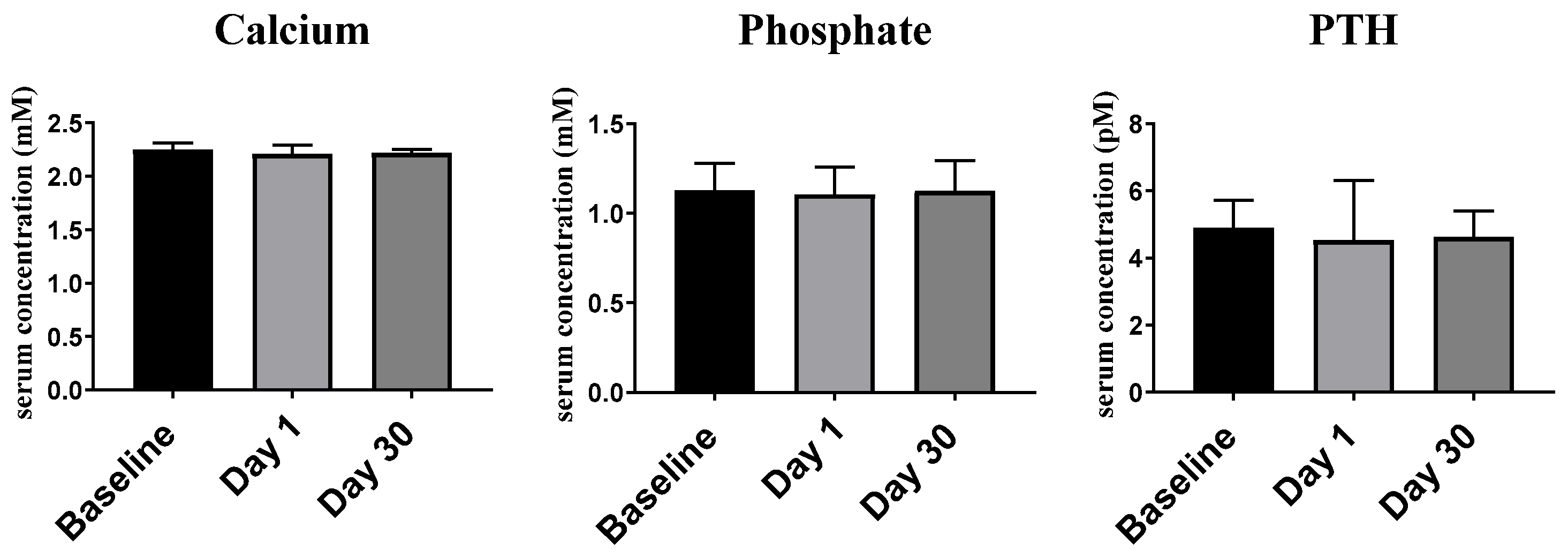
2.2.1. Ca2+, PO4− and PTH

For serum Ca2+ levels, the median as well as 25th and 75th percentiles were, at day 0 (baseline): 2.21 (2.18–2.31) mM, at day 1, 2.21 (2.17–2.29) mM, and at day 30, 2.22 (2.13–2.25) mM (Figure 1). No statistically significant differences between the days of measurement were observed (p > 0.05). Similarly, the vitamin D3 bolus resulted, neither for serum PO4− (day 0: 1.13 (0.98–1.28) mM, day 1: 1.11 (0.99–1.26) mM and day 30: 1.13 (0.90–1.29) mM) nor for PTH (day 0: 4.90 (3.95–5.72) pM, day 1: 4.53 (3.79–6.30) pM and day 30: 4.63 (3.30–5.40) pM), in any significant differences (p > 0.05).

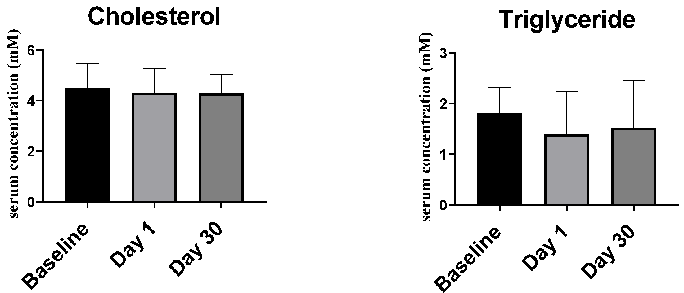
2.2.2. Lipid Profile

Concerning the serum lipid profile (Figure 2), no significant difference was observed for cholesterol concentrations at day 0 (4.49 ± 0.14 mM), day 1 (4.31 ± 0.140 mM) and day 30 (4.28 ± 0.11 mM), nor for triglyceride levels at day 0 (1.82 (1.14–2.33) mM), day 1 (1.39 (1.09–2.23) mM) and day 30 (1.52 (1.15–2.46) mM).

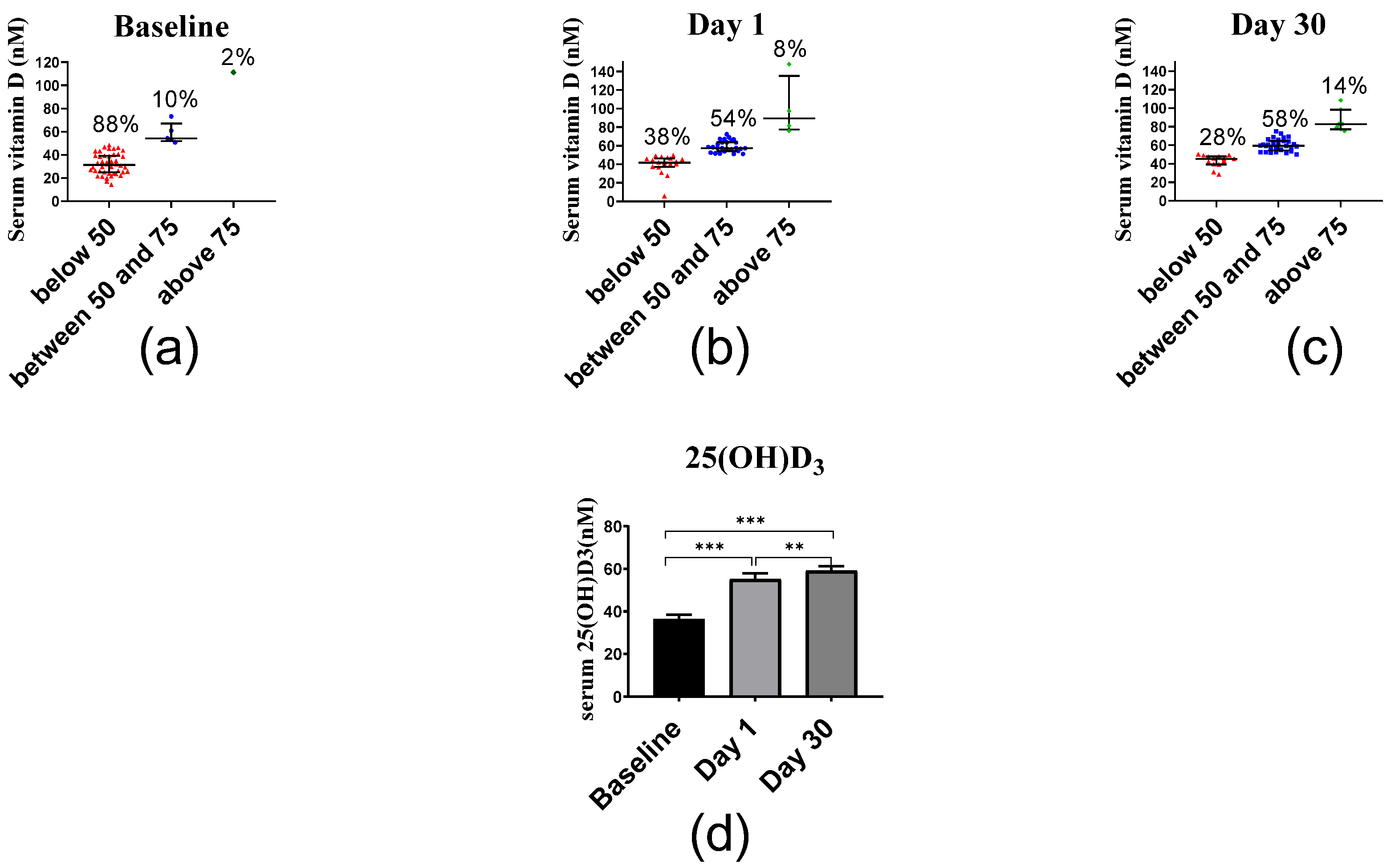
2.2.3. Serum Vitamin D

At baseline, most study participants had serum vitamin D levels less than 50 nM, i.e., they were vitamin D deficient (Figure 3). Compared to baseline (33.05 (25.40–43.30) nM), the 25(OH)D3 levels at day 1 (53.95 (44.70–63.93) nM) and day 30 (58.15 (49.03–66.93) nM) increased significantly (baseline vs. day 1, p < 0.001, baseline vs. day 30, p < 0.001 and day 1 vs. day 30, p < 0.01).

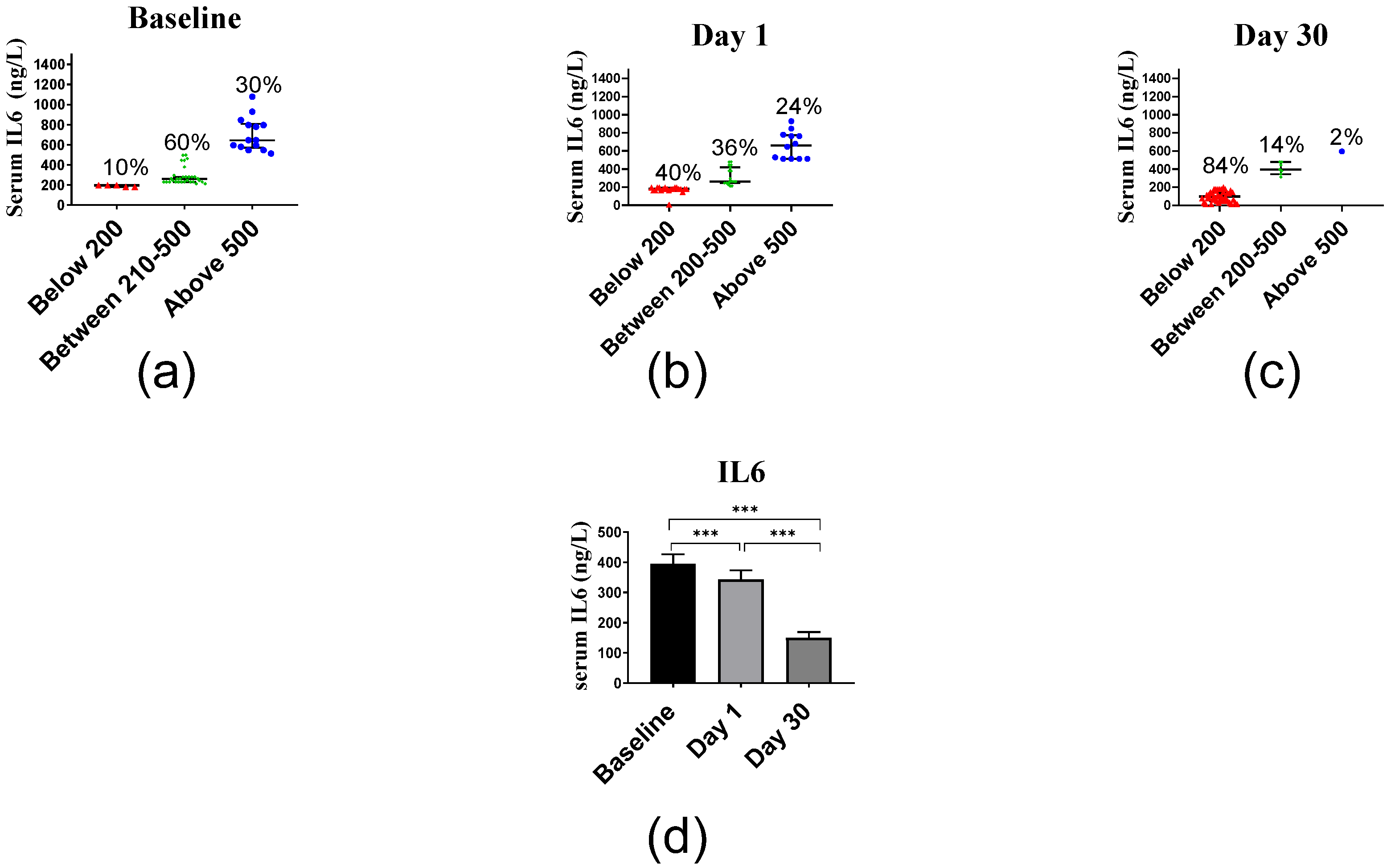
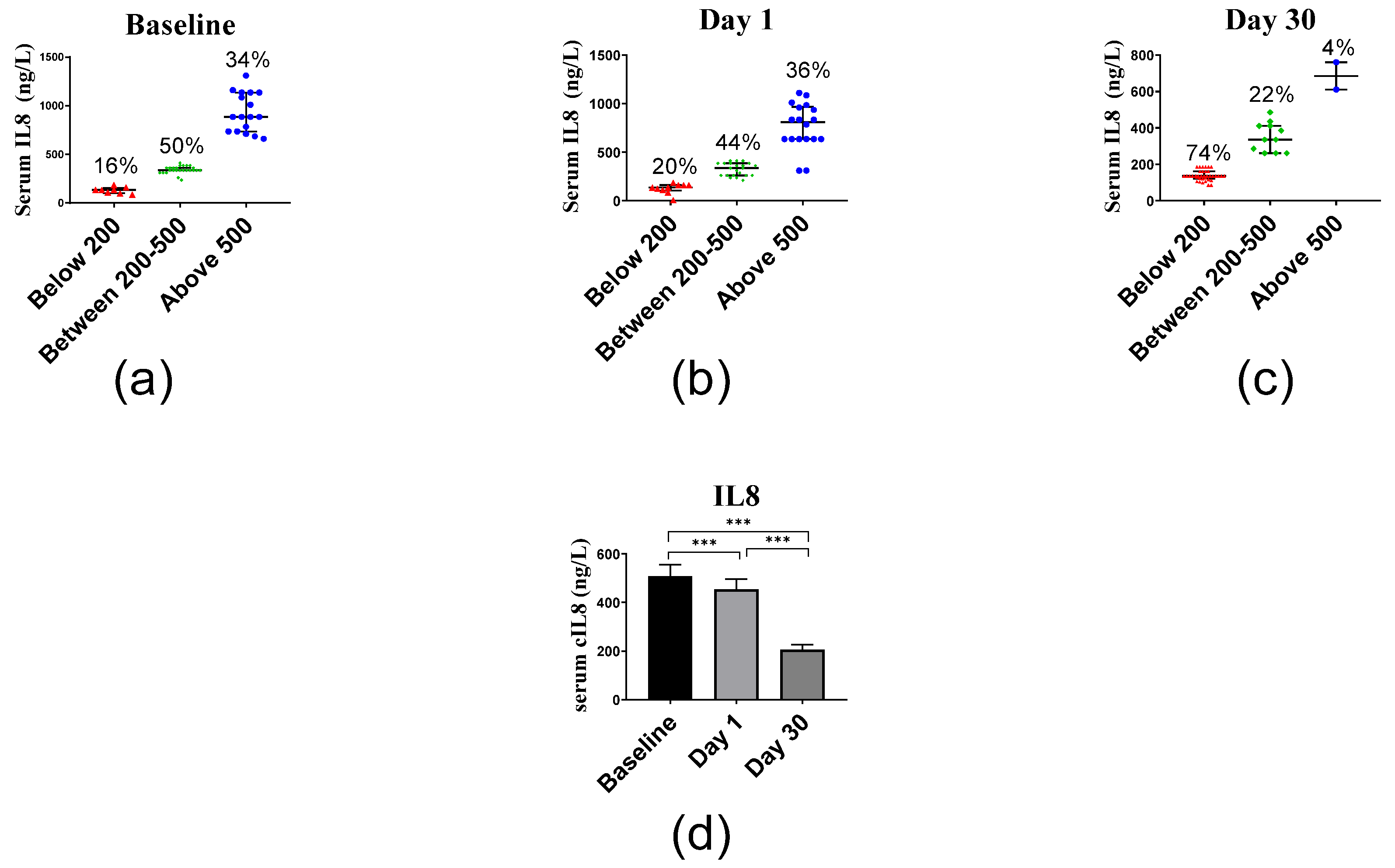
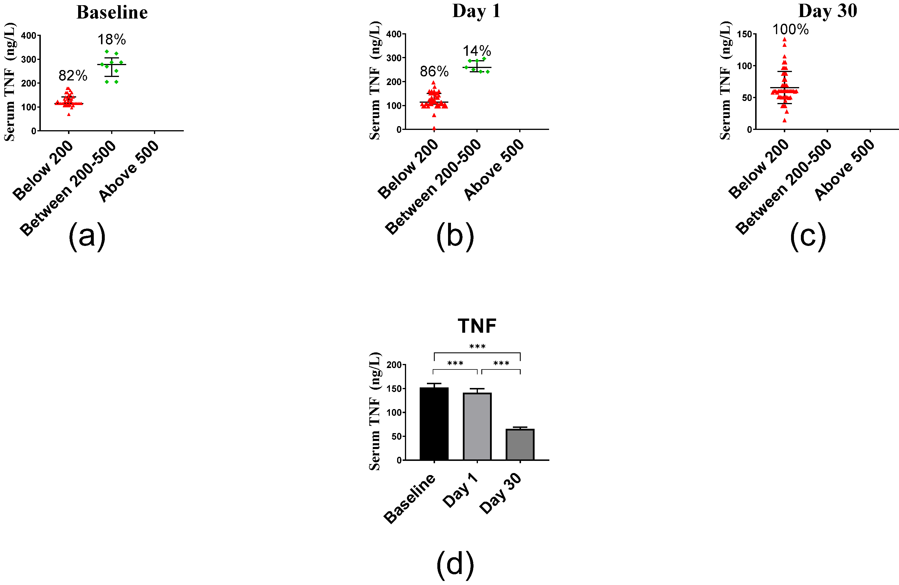
2.2.4. Inflammatory Markers

The average IL6 serum levels of 394.83 ng/L at baseline decreased at day 1 to 342.70 ng/L and at day 30 even to 149.83 ng/L (Figure 4). Median and interquartile of IL6 levels at days 0, 1 and 30 were 278.83 (228.83–545.50) ng/L, 245.50 (178.83–487.83) ng/L and 112.17 (58.00–178.83) ng/L, respectively. The decrease in IL6 levels between baseline vs. day 1 (p < 0.001), baseline vs. day 30 (p < 0.001) and day 1 vs. day 30 (p < 0.001) was significant.
The average IL8 serum levels of 508.38 ng/L at baseline decreased at day 1 to 443.90 ng/L and at day 30 even to 206.90 ng/L (Figure 5). Median and interquartile range of IL8 levels were 360.75 (310.75–748.00) ng/L, 360.75 (235.75–635.75) ng/L and 135.75 (134.50–260.75) ng/L at days 0, 1 and 30, respectively, and differed significantly (p < 0.001).
The average TNF serum levels of 151.95 ng/L at baseline decreased at day 1 to 141.37 ng/L and at day 30 even to 65.62 ng/L (Figure 6). Median and interquartile range of TNF levels were 123.55 (114.45–162.18) ng/L, 123.55 (105.36–159.91) ng/L and 59.91 (49.91–78.09) ng/L at days 0, 1 and 30, respectively. The decrease in IL6 levels was significant between baseline vs. day 1 (p < 0.001), baseline vs. day 30 (p < 0.001) and day 1 vs. day 30 (p < 0.001).
The relationships between inflammatory markers were determined by calculating the correlation coefficients between their serum levels (Table 2). There was a significant positive correlation between IL6 and IL8 concentrations at baseline, day 1 and day 30 and significant positive correlation between IL6 and TNF as well as IL8 and TNF concentrations at baseline and day 1. There were no significant correlations between IL6 and TNF nor IL8 and TNF levels at day 30.

3. Discussion

The participants of this study are representative of the general population in Saudi Arabia. BMI ranged between 20 and 39 kg/m2 (27.5 ± 4.7), indicating that most individuals were overweight or even obese. This may be due to reduced outdoor activities and lack of interest in participating in physical activities due to warm weather in Saudi Arabia. Moreover, it parallels the observation that the prevalence of obesity has increased dramatically in many countries around the world [29]. Saudi Arabia has become increasingly westernized in recent decades and today has one of the highest prevalence of overweight and obesity [30]. This increase in average BMI has contributed to the development of other diseases, such as diabetes, high blood pressure, osteoarthritis, cardiovascular disease and certain types of cancers [31]. Furthermore, the participants were characterized by an average low vitamin D status, as is often observed with obese people [32].
In the present study, we observed baseline serum levels of the cytokines IL6, IL8 and TNF of 395, 508 and 152 ng/L, respectively. This represents an extreme release of pro-inflammatory cytokines [33]. Previous studies have suggested a strong positive correlation between obesity and pro-inflammatory markers, partly reflecting the fact that adipocytes and their associated immune cells are the source of many pro-inflammatory adipokines [34]. Furthermore, studies have found that the increased release of cytokines and decreased adiponectin levels play an essential role in the pathogenesis of obesity [35]. Vgontzas et al. [36] demonstrated elevated levels of both TNF and IL6 in obese subjects, smokers and type 2 diabetes patients. Obesity results in the increased secretion not only of TNF but also of resistin, IL1β and IL6 [35,37]. Thus, this study confirms that overweight and obese people are characterized by increased levels of inflammatory markers.
Inflammation is a crucial etiology in the development of chronic diseases, such as heart diseases, metabolic syndrome and type 2 diabetes [38]. Moreover, during the COVID-19 pandemic, vitamin D deficiency was linked to cytokine storms and was positively correlated with the severity of the disease [39]. The definition of recommended 25(OH)D3 serum levels is still under debate, but the majority of researchers in the vitamin D field as well as medical laboratories consider a serum level of 30 ng/mL (75 nM) as a critical threshold [40]. Below this level, a person is considered vitamin D insufficient or even deficient (below 20 ng/mL (50 nM)). The present results are in agreement with previous studies conducted in Saudi Arabia [41,42]. Epidemiological data show that vitamin D deficiency is dominant in 30% of males and 35% of females [43,44]. This observation may be related to inadequate exposure to sunlight, inadequate consumption of dairy products, skin photo-type and reduced outdoor activities [45].
The present research demonstrates that a single bolus of vitamin D3 (80,000 IU) changes the vitamin D status from deficient to more normal levels. Approximately 76% of the participants gained serum vitamin D levels of 50 nM and above after one month (day 30). However, a different response was observed where the participants can be divided into low, mid and high responders [46,47]. Accordingly, high and mid responders may already be sufficiently supplemented with a single vitamin D3 bolus, while low responders may need a monthly bolus treatment for a longer time, in order to reach an optimal concentration of higher than 100 nM.
Since vitamin D3 is a key regulator of Ca2+ and PO4− homeostasis and controls in an antagonistic fashion, the peptide hormone PTH being important for bone metabolism [48], it is important to test whether the vitamin D3 bolus applied in this study affects respective serum levels. A dose of 80,000 IU vitamin D3 given once to the participants resulted in no significant differences in Ca2+, PO4− and PTH serum concentrations at any of the studied three time points. These results are supported by the findings of Kearns et al. [49] and suggest that the vitamin D3 bolus was safe.
In the current study, a single vitamin D3 bolus reduced serum concentrations of IL6. This observation is in agreement with the cohort study on Irish adults conducted by Laird et al. [50], who observed significant negative correlations between vitamin D and inflammatory markers such as IL6 and C-reactive protein (CRP). Moreover, several in vitro studies indicated that 1,25(OH)2D3 inhibits the secretion of IL6 in different cell types. The present findings are in agreement with Hashemi et al., who reported reduced IL17A and IL6 levels and increased IL10 levels in mRNA expression in multiple sclerosis patients after vitamin D3 supplementation [51]. IL6 levels are upregulated during inflammation of the central nervous system, which results in neuronal damage, especially in axons [52]. Vitamin D3 supplementation was found to improve multiple sclerosis by downregulation of both IL17A and IL6 levels [51]. Furthermore, a downregulation of IL6 levels was found after vitamin D3 supplementation in type 2 diabetic patients [53].
Another finding of this study was the significant reduction of TNF concentrations. The present findings agree with in vitro studies where vitamin D reduced the synthesis and secretion of TNF [54,55]. In addition, several clinical trials with patients suffering from chronic diseases have reported a positive effect of vitamin D on reducing TNF levels [56]. Collectively, the present data confirmed that vitamin D affects the TNF signaling pathway. The most significant changes occurred after 30 days, which may suggest a long-term effect of a single high dose of vitamin D3 supplement. Vitamin D3 bolus supplementation also reduced IL8 serum levels. This observation is supported by Dauletbaev et al. [57], who examined the ability of vitamin D to downregulate IL8 production in macrophages. Vitamin D upregulates the transcription of the anti-inflammatory gene DUSP1 (dual specificity phosphatase 1), which partly controls the production of the inflammatory chemokine IL8 [57]. In general, the association between 25(OH)D3 serum concentration with multiple inflammatory markers including IL6, IL8 and TNF has been addressed in numerous human studies in diseased subjects [58,59]. In contrast, only a few studies have been conducted with healthy subjects addressing the possible relation between vitamin D and inflammatory markers [60]. For example, a study accomplished in Kuwait on younger females (aged 19–47) analyzed a wider spectrum of cytokines (IL1β, IL6, IL8, IL7, IFNγ, TNF, IL4, IL10 and IL13) but only reported an inverse correlation between 25(OH)D3 serum levels with TNF and IL8 concentration, while CRP levels were elevated [61]. The strong positive correlation between the inflammatory markers IL6, IL8 and TNF may indicate an interrelationship between their production. Taken together, these results indicate that proinflammatory cytokines interact and that a single high dose of vitamin D3 is able to reduce these inflammatory markers.

4. Materials and Methods

4.1. Study Design

Fifty healthy Saudi males aged between 18 and 60 were recruited from the staff of King Abdul Aziz University and King Fahd Medical Research Center and their families. This study was conducted from January to December 2019. The study protocol was approved by the King Abdulaziz University Hospital Ethics Committee (reference no. 30-18, 5 February 2018). All procedures were conducted in compliance with the institution’s ethical guidelines. All participants gave their written informed consent prior to entering the study.
The selection criteria were based upon the following: no intake of vitamin D3 supplement for at least the past three months. Apart from this, individuals suffering from systemic diseases including chronic liver or chronic kidney diseases were excluded.
After overnight fasting, blood samples were collected (day 0) to determine the baseline levels of the biochemical variables. Then, a high dose of vitamin D3 (80,000 IU) was given orally to all participants only once. After 24 h, blood samples were collected again (day 1). No further vitamin D3 supplementation was given to the participants. The third set of blood samples was collected after one month (day 30).

4.2. Questionnaire and Anthropometric Measurements

The questionnaire was designed after consultation with experts in this field, in order to collect general demographic information and their medical history. Since several health problems may affect vitamin D digestion, absorption, metabolism and excretion, the participants were questioned especially about liver and kidney diseases. Other questions regarded the usage of vitamin D3 supplementation, multivitamins, including minerals such as Ca2+. One category was used to describe the smoking habits of the participants. The demographic features that were assessed were sex, age, weight, BMI, waist circumference, hip circumference and WHR. Subjects were indicated as having abdominal fat accumulation if their WHR > 0.9.

4.3. Serum Specimen

Blood samples were collected into a plain tube and allowed to clot for 30 min at room temperature. Afterward, samples were then centrifuged at 3000 rpm for 10 min to separate the serum. To avoid freezing and thawing, the sera were divided into aliquots and kept at −80 °C until further analysis. The vitamin D status was determined by using CIMA-based kit (Abbott, Sligo, Ireland). The lower detection limit was 3.4 ng/mL (8.5 nM). The inflammatory cytokines were measured using ELISA kits (BT Lab, Birmingham, United Kingdom). All biochemical variables were measured using fully automated systems in the chemistry lab at King Abdulaziz University Hospital.

4.4. Statistical Analysis

Analyses of the data were conducted using Statistical package for Social Science (SPSS) version 23.0 for windows (SPSS Inc., Chicago, IL, USA), whereas GraphPad Prism 7 software was used to represent graphs. Characteristics of study participants were reported as means and SD. Variables specified with measurement were summarized as SD and medians with 25th and 75th percentiles as well as minimum and maximum values were analyzed by nonparametric tests. Nonparametric tests were reported as median with 25th and 75th percentiles and were used because the assumptions for one-way ANOVA and t tests were not met. Friedman’s ANOVA (F) was applied to compare more than two dependent (related) sets of days after vitamin D supplementation; then, Wilcoxon signed-rank (W) test equivalent to the paired t test was used to compare each two pairs of days. Correlations were carried out using Spearman’s correlation (r). Results were considered significant if p < 0.05.

5. Conclusions

The present study shows the positive short- and long-term effects on improving inflammatory markers in a population of healthy Saudi males (n = 50) after the intake of a single oral vitamin D3 bolus. The population is characterized by high serum concentrations of IL6, IL8 and TNF, particularly in those individuals with high BMI. Therefore, the present results may have an important implication on future recommendations for vitamin D3 supplementation, for instance, taking only one vitamin D3 bolus per month.
Nevertheless, further studies should include larger population sizes (n > 50) of male and female participants in cohorts (e.g., married/unmarried, employed/unemployed, high/low education) from all over Saudi Arabia to guarantee the representativeness of the study and to confirm the validity of the present results. This also includes cellular and molecular studies that may reveal the underlying mechanisms of the observed effects.

Author Contributions

Conceptualization, C.C.; methodology, N.N.E.; software, H.F.A.; validation, H.M.A.; formal analysis, S.A.A.; resources, S.O.K.; data curation, S.A.A.; writing—original draft preparation, S.A.A.; writing—review and editing, C.C.; supervision, S.A.A. and H.M.A.; project administration, S.O.K. and H.F.A.; funding acquisition, S.O.K. All authors have read and agreed to the published version of the manuscript.

Funding

This research was funded by the Deanship of Scientific Research (DSR)—King Abdulaziz University—Saudi Arabia under grant (RG: 21-130-38). C.C. received funding from the European Union’s Horizon 2020 Research and Innovation Program under grant agreement no. 952601.

Institutional Review Board Statement

The study was conducted in accordance with the Declaration of Helsinki and was approved by the King Abdulaziz University Hospital Ethics Committee. All procedures were conducted in compliance with the institution’s ethical guidelines (reference no. 30-18, 5 February 2018).

Informed Consent Statement

Informed consent was obtained from all subjects involved in the study. Written informed consent was obtained from the patient(s) to publish this paper.

Acknowledgments

The authors thank the deanship of scientific research (DSR). This work was supported by the Deanship of Scientific Research (DSR)—King Abdulaziz University—Saudi Arabia under grant (RG: 21-130-38). We greatly thank the study participants for their participation and cooperation.

Conflicts of Interest

The authors declare no conflict of interest.

References

  1. Chen, L.; Deng, H.; Cui, H.; Fang, J.; Zuo, Z.; Deng, J.; Li, Y.; Wang, X.; Zhao, L. Inflammatory responses and inflammation-associated diseases in organs. Oncotarget 2018, 9, 7204–7218. [Google Scholar] [CrossRef] [Green Version]
  2. Pahwa, R.; Goyal, A.; Jialal, I. Chronic Inflammation; StatPearls Publishing: Treasure Island, FL, USA, 2022. [Google Scholar]
  3. Curley, S.; Gall, J.; Byrne, R.; Yvan-Charvet, L.; McGillicuddy, F.C. Metabolic inflammation in obesity-at the crossroads between fatty acid and cholesterol metabolism. Mol. Nutr. Food Res. 2021, 65, e1900482. [Google Scholar] [CrossRef]
  4. Krainer, J.; Siebenhandl, S.; Weinhäusel, A. Systemic autoinflammatory diseases. J. Autoimmun. 2020, 109, 102421. [Google Scholar] [CrossRef]
  5. Joshipura, K.J.; Hu, F.B.; Manson, J.E.; Stampfer, M.J.; Rimm, E.B.; Speizer, F.E.; Colditz, G.; Ascherio, A.; Rosner, B.; Spiegelman, D.; et al. The effect of fruit and vegetable intake on risk for coronary heart disease. Ann. Intern. Med. 2001, 134, 1106–1114. [Google Scholar] [CrossRef]
  6. Gandini, S.; Merzenich, H.; Robertson, C.; Boyle, P. Meta-analysis of studies on breast cancer risk and diet: The role of fruit and vegetable consumption and the intake of associated micronutrients. Eur. J. Cancer 2000, 36, 636–646. [Google Scholar] [CrossRef]
  7. Yoon, J.H.; Baek, S.J. Molecular targets of dietary polyphenols with anti-inflammatory properties. Yonsei Med. J. 2005, 46, 585–596. [Google Scholar] [CrossRef] [Green Version]
  8. Maroon, J.C.; Bost, J.W.; Maroon, A. Natural anti-inflammatory agents for pain relief. Surg. Neurol. Int. 2010, 1, 80. [Google Scholar] [CrossRef] [PubMed] [Green Version]
  9. Dogan-Sander, E.; Mergl, R.; Willenberg, A.; Baber, R.; Wirkner, K.; Riedel-Heller, S.G.; Röhr, S.; Schmidt, F.M.; Schomerus, G.; Sander, C. Inflammation and the association of vitamin D and depressive symptomatology. Nutrients 2021, 13, 1972. [Google Scholar] [CrossRef]
  10. Pittas, A.G.; Harris, S.S.; Stark, P.C.; Dawson-Hughes, B. The effects of calcium and vitamin D supplementation on blood glucose and markers of inflammation in nondiabetic adults. Diabetes Care 2007, 30, 980–986. [Google Scholar] [CrossRef] [Green Version]
  11. Bui, L.; Zhu, Z.; Hawkins, S.; Cortez-Resendiz, A.; Bellon, A. Vitamin D regulation of the immune system and its implications for COVID-19: A mini review. SAGE Open Med. 2021, 9, 20503121211014073. [Google Scholar] [CrossRef] [PubMed]
  12. Tiosano, D.; Wildbaum, G.; Gepstein, V.; Verbitsky, O.; Weisman, Y.; Karin, N.; Eztioni, A. The role of vitamin D receptor in innate and adaptive immunity: A study in hereditary vitamin D-resistant rickets patients. J. Clin. Endocrinol. Metab. 2013, 98, 1685–1693. [Google Scholar] [CrossRef] [PubMed]
  13. Gruber-Bzura, B.M. Vitamin D and influenza—Prevention or therapy? Int. J. Mol. Sci. 2018, 19, 2419. [Google Scholar] [CrossRef] [Green Version]
  14. Rondanelli, M.; Miccono, A.; Lamburghini, S.; Avanzato, I.; Riva, A.; Allegrini, P.; Faliva, M.A.; Peroni, G.; Nichetti, M.; Perna, S. Self-care for common colds: The pivotal role of vitamin D, vitamin C, zinc, and Echinacea in three main immune interactive clusters (physical barriers, innate and adaptive immunity) involved during an episode of common colds—Practical advice on dosages and on the time to take these nutrients/botanicals in order to prevent or treat common catolds. Evid. Based Complement. Alternat. Med. 2018, 2018, 5813095. [Google Scholar] [CrossRef] [Green Version]
  15. Gombart, A.F.; Pierre, A.; Maggini, S. A review of micronutrients and the immune system–working in harmony to reduce the risk of infection. Nutrients 2020, 12, 236. [Google Scholar] [CrossRef] [Green Version]
  16. Roffe-Vazquez, D.N.; Huerta-Delgado, A.S.; Castillo, E.C.; Villarreal-Calderón, J.R.; Gonzalez-Gil, A.M.; Enriquez, C.; Garcia-Rivas, G.; Elizondo-Montemayor, L. Correlation of vitamin D with inflammatory cytokines, atherosclerotic parameters, and lifestyle factors in the setting of heart failure: A 12-month follow-up study. Int. J. Mol. Sci. 2019, 20, 5811. [Google Scholar] [CrossRef] [PubMed] [Green Version]
  17. Maigoro, A.Y.; An, D.; Lee, S. Exploring the link between vitamin D deficiency and cytokine storms in COVID-19 patients: An in silico analysis. J. Med. Food 2022, 25, 130–137. [Google Scholar] [CrossRef] [PubMed]
  18. Ali, N. Role of vitamin D in preventing of COVID-19 infection, progression and severity. J. Infect. Public Health 2020, 13, 1373–1380. [Google Scholar] [CrossRef]
  19. Giustina, A.; Adler, R.A.; Binkley, N.; Bollerslev, J.; Bouillon, R.; Dawson-Hughes, B.; Ebeling, P.R.; Feldman, D.; Formenti, A.M.; Lazaretti-Castro, M.; et al. Consensus Statement from 2nd International Conference on Controversies in Vitamin D. Rev. Endocr. Metab. Disord. 2020, 21, 89–116. [Google Scholar] [CrossRef] [Green Version]
  20. Ish-Shalom, S.; Segal, E.; Salganik, T.; Raz, B.; Bromberg, I.L.; Vieth, R. Comparison of daily, weekly, and monthly vitamin D3 in ethanol dosing protocols for two months in elderly hip fracture patients. J. Clin. Endocrinol. Metab. 2008, 93, 3430–3435. [Google Scholar] [CrossRef] [PubMed] [Green Version]
  21. Ross, A.C.; Manson, J.E.; Abrams, S.A.; Aloia, J.F.; Brannon, P.M.; Clinton, S.K.; Durazo-Arvizu, R.A.; Gallagher, J.C.; Gallo, R.L.; Jones, G.; et al. The 2011 report on dietary reference intakes for calcium and vitamin D from the Institute of Medicine: What clinicians need to know. J. Clin. Endocrinol. Metab. 2011, 96, 53–88. [Google Scholar] [CrossRef]
  22. Holick, M.F.; Binkley, N.C.; Bischoff-Ferrari, H.A.; Gordon, C.M.; Hanley, D.A.; Heaney, R.P.; Murad, M.H.; Weaver, C.M. Endocrine society evaluation, treatment, and prevention of vitamin D deficiency: An Endocrine Society clinical practice guideline. J. Clin. Endocrinol. Metab. 2011, 96, 1911–1930. [Google Scholar] [CrossRef] [Green Version]
  23. Shirvani, A.; Kalajian, T.A.; Song, A.; Holick, M.F. Disassociation of vitamin D’s calcemic activity and non-calcemic genomic activity and individual responsiveness: A randomized controlled double-blind clinical trial. Sci. Rep. 2019, 9, 17685. [Google Scholar] [CrossRef] [PubMed] [Green Version]
  24. Bacon, C.J.; Gamble, G.D.; Horne, A.M.; Scott, M.A.; Reid, I.R. High-dose oral vitamin D3 supplementation in the elderly. Osteoporos. Int. 2009, 20, 1407–1415. [Google Scholar] [CrossRef]
  25. Neme, A.; Seuter, S.; Malinen, M.; Nurmi, T.; Tuomainen, T.-P.; Virtanen, J.K.; Carlberg, C. In vivo transcriptome changes of human white blood cells in response to vitamin D. J. Steroid Biochem. Mol. Biol. 2019, 188, 71–76. [Google Scholar] [CrossRef] [PubMed]
  26. Vukić, M.; Neme, A.; Seuter, S.; Saksa, N.; de Mello, V.D.F.; Nurmi, T.; Uusitupa, M.; Tuomainen, T.-P.; Virtanen, J.K.; Carlberg, C. Relevance of vitamin D receptor target genes for monitoring the vitamin D responsiveness of primary human cells. PLoS ONE 2015, 10, e0124339. [Google Scholar] [CrossRef] [Green Version]
  27. Seuter, S.; Virtanen, J.K.; Nurmi, T.; Pihlajamäki, J.; Mursu, J.; Voutilainen, S.; Tuomainen, T.-P.; Neme, A.; Carlberg, C. Molecular evaluation of vitamin D responsiveness of healthy young adults. J. Steroid Biochem. Mol. Biol. 2017, 174, 314–321. [Google Scholar] [CrossRef]
  28. Carlberg, C.; Seuter, S.; Nurmi, T.; Tuomainen, T.-P.; Virtanen, J.K.; Neme, A. In vivo response of the human epigenome to vitamin D: A proof-of-principle study. J. Steroid Biochem. Mol. Biol. 2018, 180, 142–148. [Google Scholar] [CrossRef] [Green Version]
  29. Blüher, M. Obesity: Global epidemiology and pathogenesis. Nat. Rev. Endocrinol. 2019, 15, 288–298. [Google Scholar] [CrossRef] [PubMed]
  30. DeNicola, E.; Aburizaiza, O.S.; Siddique, A.; Khwaja, H.; Carpenter, D.O. Obesity and public health in the kingdom of Saudi Arabia. Rev. Environ. Health 2015, 30, 191–205. [Google Scholar] [CrossRef] [PubMed]
  31. Guh, D.P.; Zhang, W.; Bansback, N.; Amarsi, Z.; Birmingham, C.L.; Anis, A.H. The incidence of co-morbidities related to obesity and overweight: A systematic review and meta-analysis. BMC Public Health 2009, 9, 88. [Google Scholar] [CrossRef]
  32. Ding, C.; Gao, D.; Wilding, J.; Trayhurn, P.; Bing, C. Vitamin D signalling in adipose tissue. Br. J. Nutr. 2012, 108, 1915–1923. [Google Scholar] [CrossRef] [PubMed] [Green Version]
  33. Matute Wilander, A.; Kåredal, M.; Axmon, A.; Nordander, C. Inflammatory biomarkers in serum in subjects with and without work related neck/shoulder complaints. BMC Musculoskelet. Disord. 2014, 15, 103. [Google Scholar] [CrossRef] [PubMed] [Green Version]
  34. Kershaw, E.E.; Flier, J.S. Adipose tissue as an endocrine organ. J. Clin. Endocrinol. Metabol. 2004, 89, 2548–2556. [Google Scholar] [CrossRef]
  35. Zorena, K.; Jachimowicz-Duda, O.; Ślęzak, D.; Robakowska, M.; Mrugacz, M. Adipokines and obesity. Potential link to metabolic disorders and chronic complications. Int. J. Mol. Sci. 2020, 21, 3570. [Google Scholar] [CrossRef]
  36. Vgontzas, A.N.; Papanicolaou, D.A.; Bixler, E.O.; Hopper, K.; Lotsikas, A.; Lin, H.M.; Kales, A.; Chrousos, G.P. Sleep apnea and daytime sleepiness and fatigue: Relation to visceral obesity, insulin resistance, and hypercytokinemia. J. Clin. Endocrinol. Metab. 2000, 85, 1151–1158. [Google Scholar] [CrossRef]
  37. Kern, L.; Mittenbühler, M.J.; Vesting, A.J.; Ostermann, A.L.; Wunderlich, C.M.; Wunderlich, F.T. Obesity-induced TNFα and IL-6 signaling: The missing link between obesity and inflammation-driven liver and colorectal cancers. Cancers 2018, 11, 24. [Google Scholar] [CrossRef] [Green Version]
  38. Furman, D.; Campisi, J.; Verdin, E.; Carrera-Bastos, P.; Targ, S.; Franceschi, C.; Ferrucci, L.; Gilroy, D.W.; Fasano, A.; Miller, G.W.; et al. Chronic inflammation in the etiology of disease across the life span. Nat. Med. 2019, 25, 1822–1832. [Google Scholar] [CrossRef] [PubMed]
  39. Song, P.; Li, W.; Xie, J.; Hou, Y.; You, C. Cytokine storm induced by SARS-CoV-2. Clin. Chim. Acta 2020, 509, 280–287. [Google Scholar] [CrossRef]
  40. Holick, M.F. Vitamin D deficiency. N. Engl. J. Med. 2007, 357, 266–281. [Google Scholar] [CrossRef] [PubMed]
  41. Ardawi, M.S.M.; Sibiany, A.M.; Bakhsh, T.M.; Qari, M.H.; Maimani, A.A. High prevalence of vitamin D deficiency among healthy Saudi Arabian men: Relationship to bone mineral density, parathyroid hormone, bone turnover markers, and lifestyle factors. Osteoporos. Int. 2012, 23, 675–686. [Google Scholar] [CrossRef]
  42. Elsammak, M.Y.; Al-Wossaibi, A.A.; Al-Howeish, A.; Alsaeed, J. High prevalence of vitamin D deficiency in the sunny eastern region of Saudi Arabia: A hospital-based study. East. Mediterr. Heal. J. 2011, 17, 317–322. [Google Scholar] [CrossRef]
  43. Alsuwadia, A.O.; Farag, Y.M.; Al Sayyari, A.A.; Mousa, D.H.; Alhejaili, F.F.; Al-Harbi, A.S.; Housawi, A.A.; Mittal, B.V.; Singh, A.K. Prevalence of vitamin D deficiency in Saudi Adults. Saudi Med. J. 2013, 34, 814–818. [Google Scholar]
  44. Farhat, K.H.; Arafa, M.A.; Rabah, D.M.; Amin, H.S.; Ibrahim, N.K. Vitamin D status and its correlates in Saudi male population. BMC Public Health 2019, 19, 211. [Google Scholar] [CrossRef] [PubMed] [Green Version]
  45. Hussain, A.N.; Alkhenizan, A.H.; El Shaker, M.; Raef, H.; Gabr, A. Increasing trends and significance of hypovitaminosis D: A population-based study in the kingdom of Saudi Arabia. Arch. Osteoporos. 2014, 9, 190. [Google Scholar] [CrossRef] [PubMed]
  46. Carlberg, C. Molecular approaches for optimizing vitamin D supplementation. Vitam. Horm. 2016, 100, 255–271. [Google Scholar] [CrossRef]
  47. Carlberg, C.; Raunio, H. From pharmacogenomics to integrated personal omics profiling: A gap in implementation into healthcare. Per. Med. 2014, 11, 625–629. [Google Scholar] [CrossRef]
  48. Levine, B.S.; Rodríguez, M.; Felsenfeld, A.J. Serum calcium and bone: Effect of PTH, phosphate, vitamin D and uremia. Nefrologia 2014, 34, 658–669. [Google Scholar] [CrossRef]
  49. Kearns, M.D.; Binongo, J.N.G.; Watson, D.; Alvarez, J.A.; Lodin, D.; Ziegler, T.R.; Tangpricha, V. The effect of a single, large bolus of vitamin D in healthy adults over the winter and following year: A randomized, double-blind, placebo-controlled trial. Eur. J. Clin. Nutr. 2015, 69, 193–197. [Google Scholar] [CrossRef] [Green Version]
  50. Laird, E.; McNulty, H.; Ward, M.; Hoey, L.; McSorley, E.; Wallace, J.M.W.; Carson, E.; Molloy, A.M.; Healy, M.; Casey, M.C.; et al. Vitamin D deficiency is associated with inflammation in older Irish adults. J. Clin. Endocrinol. Metab. 2014, 99, 1807–1815. [Google Scholar] [CrossRef] [Green Version]
  51. Hashemi, R.; Morshedi, M.; Jafarabadi, M.A.; Altafi, D.; Hosseini-Asl, S.S.; Rafie-Arefhosseini, S. Anti-Inflammatory effects of dietary vitamin D3 in patients with multiple sclerosis. Neurol. Genet. 2018, 4, e278. [Google Scholar] [CrossRef] [Green Version]
  52. Erta, M.; Quintana, A.; Hidalgo, J. Interleukin-6, a major cytokine in the central nervous system. Int. J. Biol. Sci. 2012, 8, 1254–1266. [Google Scholar] [CrossRef]
  53. Giulietti, A.; van Etten, E.; Overbergh, L.; Stoffels, K.; Bouillon, R.; Mathieu, C. Monocytes from type 2 diabetic patients have a pro-inflammatory profile. 1,25-dihydroxyvitamin D3 works as anti-inflammatory. Diabetes Res. Clin. Pract. 2007, 77, 47–57. [Google Scholar] [CrossRef]
  54. Grossmann, R.E.; Zughaier, S.M.; Liu, S.; Lyles, R.H.; Tangpricha, V. Impact of vitamin D supplementation on markers of inflammation in adults with cystic fibrosis hospitalized for a pulmonary exacerbation. Eur. J. Clin. Nutr. 2012, 66, 1072–1074. [Google Scholar] [CrossRef] [Green Version]
  55. Dadaei, T.; Safapoor, M.H.; Asadzadeh Aghdaei, H.; Balaii, H.; Pourhoseingholi, M.A.; Naderi, N.; Zojaji, H.; Azimzadeh, P.; Mohammadi, P.; Zali, M.R. Effect of vitamin D3 supplementation on TNF-α serum level and disease activity index in Iranian IBD patients. Gastroenterol. Hepatol. Bed Bench 2015, 8, 49–55. [Google Scholar]
  56. Borazan, A.; Üstün, H.; Cefle, A.; Sekitmez, N.; Yilmaz, A. Comparative efficacy of oral and intravenous calcitriol treatment in haemodialysis patients: Effects on serum biochemistry and cytokine levels. J. Int. Med. Res. 2003, 31, 489–496. [Google Scholar] [CrossRef]
  57. Dauletbaev, N.; Herscovitch, K.; Das, M.; Chen, H.; Bernier, J.; Matouk, E.; Bérubé, J.; Rousseau, S.; Lands, L.C. Down-regulation of IL-8 by high-dose vitamin D is specific to hyperinflammatory macrophages and involves mechanisms beyond up-regulation of DUSP1. Br. J. Pharmacol. 2015, 172, 4757–4771. [Google Scholar] [CrossRef] [Green Version]
  58. Zhang, Y.; Fang, F.; Tang, J.; Jia, L.; Feng, Y.; Xu, P.; Faramand, A. Association between vitamin D supplementation and mortality: Systematic review and meta-analysis. BMJ 2019, 366, 14673. [Google Scholar] [CrossRef] [Green Version]
  59. Hopkins, M.H.; Owen, J.; Ahearn, T.; Fedirko, V.; Flanders, W.D.; Jones, D.P.; Bostick, R.M. Effects of supplemental vitamin D and calcium on biomarkers of inflammation in colorectal adenoma patients: A randomized, controlled clinical trial. Cancer Prev. Res. 2011, 4, 1645–1654. [Google Scholar] [CrossRef] [Green Version]
  60. Jakobsen, J.; Andersen, E.A.W.; Christensen, T.; Andersen, R.; Bügel, S. Vitamin D vitamers affect vitamin D status differently in young healthy males. Nutrients 2017, 10, 12. [Google Scholar] [CrossRef] [Green Version]
  61. Azizieh, F.; Alyahya, K.; Raghupathy, R. Association between levels of vitamin D and inflammatory markers in healthy women. J. Inflamm. Res. 2016, 9, 51–57. [Google Scholar] [CrossRef]
Figure 1. Serum concentration of Ca2+, PO4− and PTH. The median serum concentrations were compared between days 0, 1 and 30 of vitamin D3 supplementation. The error bars represent interquartile range. Data were analyzed by non-parametric repeated measure ANOVA followed by Wilcoxon signed-rank multiple comparison test.
Ijms 23 11992 g001
Figure 2. Serum cholesterol and triglyceride concentrations. The mean serum cholesterol and triglyceride concentrations at baseline, day 1 and day 30 are displayed. The error bars represent standard error of mean (SEM) and interquartile range, respectively. Data were analyzed by one-way (ANOVA) followed by Tukey’s multiple comparison and by non-parametric repeated measure ANOVA followed by Wilcoxon signed-rank multiple comparison test, respectively.
Ijms 23 11992 g002
Figure 3. Serum 25(OH) D3 concentrations before and after vitamin D3 supplementation. (a) Baseline levels, (b) day 1 levels, and (c) day 30 levels. (d) Columns represent medians of serum 25(OH)D3, and error bars indicate interquartile range. Data were analyzed by non-parametric repeated measure ANOVA followed by Wilcoxon signed-rank multiple comparison test. *** p < 0.001, ** p < 0.01.
Ijms 23 11992 g003
Figure 4. Serum IL6 levels before and after vitamin D supplementation. (a) Baseline levels, (b) day 1 levels, and (c) day 30 levels. (d) Columns represent the median serum IL6 concentration at baseline, day 1 and day 30, and error bars indicate interquartile range. Data were analyzed by non-parametric repeated measure ANOVA followed by Wilcoxon signed-rank multiple comparison test. *** p < 0.001.
Ijms 23 11992 g004
Figure 5. Serum IL8 levels before and after vitamin D supplementation: (a) baseline levels, (b) day 1 levels, and (c) day 30 levels. (d) Columns represent the median serum IL6 concentration at baseline, day 1 and day 30, and error bars indicate interquartile range. Data were analyzed by non-parametric repeated measure ANOVA followed by Wilcoxon signed-rank multiple comparison test, and p values are shown where the difference between days was determined to be statistically significant. *** p < 0.001.
Ijms 23 11992 g005
Figure 6. Serum TNF levels before and after vitamin D supplementation. (a) Baseline levels, (b) day 1 levels, and (c) day 30 levels. (d) Columns represent the median of TNF concentrations, and error bars indicate interquartile range. Data were analyzed by non-parametric repeated measure ANOVA followed by Wilcoxon signed-rank multiple comparison test. *** p < 0.001.
Ijms 23 11992 g006
Table 1. Demographic characteristics of studied participants. SD, standard deviation.
MeanSDRange
Age (years)30.3±6.218–53
Age N (%)All participants (n = 50)
20 to less than 40 years47 (94)
40 to less than 60 years3 (6)
60 years0 (0)
MeanSDRange
Height (cm)174.3±6.2160–188
Body weight (kg)83.8±15.253–121
BMI (kg/m2)27.6±4.720.1–39.1
BMI classificationAll participants (n = 50)
Underweight0 (0)
Normal (18.5–24.99)17 (34%)
Overweight (25–29.99)16 (32%)
Obese (>30)17 (34%)
MeanSDRange
Waist circumference (cm)94.4±14.859–126
Hip circumference (cm)104.9±15.739–128
WHR0.91±0.130.67–1.51
Health status N (%)All participants (n = 50)
Liver disease0 (0%)
Kidney diseases0 (0%)
Healthy50 (100%)
Taking any medicines N (%)All participants (n = 50)
Yes0 (0)
No50 (100%)
Broken any bones N (%)
Yes35 (70%)
No15 (30%)
Taking any supplements N (%)
Minerals2 (4%)
Vitamin D0 (0%)
Ca2+0 (0)%
Nothing48 (96%)
Awareness to vitamin D (if taking test of vitamin D concentration) N (%)
Yes15 (30%)
No35 (70%)
Sun exposure N (%)
Less than 5 min/day1 (2%)
5–15 min/day7 (14%)
15–30 min/day12 (24%)
More than 30 min/day30 (60%)
Exercise rate N (%)
Always15 (30%)
Usually30 (60%)
Rarely5 (10%)
Smoke status N (%)
Smoker19 (38%)
Non-smoker31 (62%)
Table 2. Spearman correlation coefficients (r) between inflammatory markers.
Inflammatory
Markers
Baseline, IL6
n = 50
Day 1, IL6
n = 50
Day 30, IL6
n = 50
rprprp
IL80.50 **<0.010.51 **<0.010.54 **<0.01
TNF0.64 **<0.010.56 **<0.010.030.82
Inflammatory
Markers
Baseline, IL8
n = 50
Day 1, IL8
n = 50
Day 30, IL8
n = 50
rprprp
TNF0.50 **<0.010.43 **<0.010.050.74
IL6 and IL8, IL6 and TNF as well as IL8 and TNF concentrations at baseline, day 1 and day 30. ** p < 0.01.
Publisher’s Note: MDPI stays neutral with regard to jurisdictional claims in published maps and institutional affiliations.

