Article

Assessment of DDAH1 and DDAH2 Contributions to Psychiatric Disorders via In Silico Methods

by
Alena A. Kozlova
1,†,
Anastasia N. Vaganova
2,†,
Roman N. Rodionov
3,
Raul R. Gainetdinov
2 and
Nadine Bernhardt
1,*
1
Department of Psychiatry and Psychotherapy, University Hospital Carl Gustav Carus, Technische Universität Dresden, 01307 Dresden, Germany
2
Institute of Translational Biomedicine, Saint-Petersburg State University, 199034 Saint-Petersburg, Russia
3
Department of Internal Medicine III, Technische Universität Dresden, 01307 Dresden, Germany
*
Author to whom correspondence should be addressed.
These authors contributed equally to this work.
Int. J. Mol. Sci. 2022, 23(19), 11902; https://doi.org/10.3390/ijms231911902
Submission received: 2 September 2022 / Revised: 30 September 2022 / Accepted: 4 October 2022 / Published: 7 October 2022
(This article belongs to the Special Issue Nitric Oxide Synthases: Function and Regulation)

Abstract

:
The contribution of nitric oxide synthases (NOSs) to the pathophysiology of several neuropsychiatric disorders is recognized, but the role of their regulators, dimethylarginine dimethylaminohydrolases (DDAHs), is less understood. This study’s objective was to estimate DDAH1 and DDAH2 associations with biological processes implicated in major psychiatric disorders using publicly accessible expression databases. Since co-expressed genes are more likely to be involved in the same biologic processes, we investigated co-expression patterns with DDAH1 and DDAH2 in the dorsolateral prefrontal cortex in psychiatric patients and control subjects. There were no significant differences in DDAH1 and DDAH2 expression levels in schizophrenia or bipolar disorder patients compared to controls. Meanwhile, the data suggest that in patients, DDAH1 and DDHA2 undergo a functional shift mirrored in changes in co-expressed gene patterns. This disarrangement appears in the loss of expression level correlations between DDAH1 or DDAH2 and genes associated with psychiatric disorders and reduced functional similarity of DDAH1 or DDAH2 co-expressed genes in the patient groups. Our findings evidence the possible involvement of DDAH1 and DDAH2 in neuropsychiatric disorder development, but the underlying mechanisms need experimental validation.

1. Introduction

Mental disorders remain among the top ten leading causes of burden worldwide, which makes research to establish causal pathways imperative for effective prevention and treatment [1]. In recent years, nitric oxide (NO) signaling has been implicated in the pathophysiology of several mental illnesses, such as schizophrenia and affective disorders, comprising bipolar disorder and major depressive disorder [2]. For example, in schizophrenia patients, NO metabolism is impaired in various organs, including the brain [3,4,5], and high NO levels are found in post-mortem samples of the prefrontal cortex and hippocampus [6]. NO is a gaseous molecule acting as the second messenger of the NMDA receptor, thereby regulating glutamatergic transmission [7]. NO further interacts with the dopaminergic and serotonergic systems [8] and is involved in the storage, uptake, and release of transmitters, such as acetylcholine, noradrenaline, GABA, taurine, and glycine [9]. In the brain, NO regulates synaptic plasticity, neurodevelopment, and cerebral blood flow [10]. Meanwhile, excessive amounts of free radical NO lead to neurotoxicity and neurodegeneration [10,11].
There are three isoforms of enzymes generating NO: the neuronal (nNOS or NOS1), the inducible (iNOS or NOS2), and the endothelial NO synthases (eNOS or NOS3) [9]. Genetic variations in NOS genes have been associated with several psychiatric conditions: (i) NOS1 polymorphisms are associated with an increased risk of schizophrenia development [12]; (ii) NOS1 and NOS3 alleles are involved in modifying an individual’s susceptibility to bipolar disorder, depression, or risk of suicide attempts, and impact glutamatergic neurotransmission [13,14,15,16,17,18,19]; (iii) some NOS3 variants demonstrate a protective role in bipolar disorder [18]; (iv) NOS2 involvement in psychiatric diseases was demonstrated in animal models [1] and population studies [20], but the knowledge of this association is limited.
In addition, the NOS1 gene is methylated differently in schizophrenic patients and healthy individuals [21]. NOS1 coupling to the NMDA receptor is regulated by the NOS1 adapter protein (NOS1AP) [13], whose polymorphisms are associated with schizophrenia [22] and the severity of posttraumatic stress disorder [23]. Thus, there is increasing evidence that NOS1 and NOS3 are promising drug targets for treating schizophrenia and affective disorders [24,25,26].
NOS activity and NO levels are regulated by dimethylarginine dimethylaminohydrolases (DDAHs). There are two known isoforms whose amino acid sequences are 50% identical. DDAH1 is responsible for the degradation of N (omega), N (omega) dimethyl-L-arginine (ADMA), the major competitive inhibitor of NOS [27]. DDAH2 also contributes to the regulation of NO levels, although it is still being debated if through ADMA degradation or ADMA-independent mechanisms. Both isoforms of DDAHs are expressed in the brain in a regional and cell-type complementary fashion [28].
DDAH1 alleles are associated with the risk of developing autistic spectrum disorder or obsessive-compulsive disorder [29], and animal models of autism and schizophrenia endophenotypes present with increased DDAH1 levels [30,31]. At the same time, experiments in mice fed an Mg-restricted diet resulted in depression-like behavior and decreased DDAH1 expression [32]. However, the data on DDAH1 expression levels in different brain structures and psychiatric conditions are sparse. Thus far, reduced DDAH1 expression has been found in the anterior cingulate cortex in schizophrenic patients [33], whereas in the prefrontal cortex, a downregulation was transient and detectable only in the first years after the onset of the disease [34]. In addition, a strong downregulation of the hsa-miR-219-5p that is suggested to control DDAH1 expression was observed in schizophrenia patients [35].
DDAH2 gene variants are associated with schizophrenia and bipolar disorder susceptibility [36]. In schizophrenia patients, the DDAH2 gene is aberrantly methylated in both the prefrontal cortex and blood, and DDAH2 brain mRNA levels are significantly increased [37]. Further, loss of methylation was shown in schizophrenia patients with suicide attempts [38].
Loss or overexpression of NOS or other genes directly involved in this pathway may define altered NO-signaling in psychiatric disorders. The impact of the spatial expression pattern and the level of co-expressed genes may also be relevant. For example, malformation of NOS1 positive GABAergic interneurons was described in schizophrenia [39]. In addition, the mRNA for NPY, expressed by many NOS1/NADPH-d GABA-ergic neurons, is selectively decreased in neurons within the superficial white matter of subjects with psychosis. While NO facilitates blood flow through the cortical microvasculature, NPY mediates microvessel constriction; therefore, its deregulation leads to microcirculatory dysfunction [40].
Thus, associations of NOS gene mutations and expression deregulation with psychiatric disorders are well studied. However, relatively less is known about the contribution of other modulators of NO-mediated processes in the pathogenesis of these diseases. Suggesting that the context-dependent consequences of NO-signaling may differ in the cortex of psychiatric and non-psychiatric subjects, we attempted to estimate the differences of DDAHs co-expressed gene patterns in publicly available datasets. We performed a careful functional analysis of these co-expressed gene sets. Specifically, we evaluated evidence of DDAH1 and DDAH2 involvement in regulating processes associated with major psychotic disorders, schizophrenia, and bipolar disorder.

2. Results

2.1. DDAH1 and DDAH2 mRNAs Are Represented in the Dorsolateral Prefrontal Cortex in Non-Psychiatric Controls and Psychotic Patients

DDAH1 and DDAH2 mRNA were identified in all dorsolateral prefrontal cortex samples in the selected datasets (refer to Table 1) in both patients and non-psychiatric controls. The DDAH1 expression levels were greater than DDAH2. However, no significant differences in the DDAH1 and DDAH2 expression levels were identified when their expression was compared between the control group and patients with either bipolar affective disorder or schizophrenia (refer to Figure 1a). Slight upregulation of DDAH2 was identified in the tissue samples from patients with bipolar disorder compared to the control group in the GSE112523 dataset. However, this finding did not remain statistically significant after the adjustment (Padj > 0.05).
The DDAH1 and DDAH2 expression values are congruent in different datasets. DDAH2 expression is lower than DDAH1; however, its estimation is more prone to fluctuations and bias, particularly in the dataset GSE87194, where expression is lower than the other datasets.
The dataset GSE112523 combines data from subjects with schizophrenia, bipolar affective disorder, and non-psychiatric controls; thus, it was further analyzed to compare DDAH1 and DDAH2 co-expressed gene sets in these psychiatric disorders. The dataset contains dorsolateral prefrontal cortex samples (mainly BA46 area) of seven patients with schizophrenia, ten patients with bipolar disorders, and seventeen non-psychiatric control subjects. Study group data are summarized in Table 2.

2.2. Genome-Wide Co-Expression Analysis of DDAH1 and DDAH2 Co-Expressed Genes

The distribution of the Pearson correlation coefficient demonstrates that the median values for all three study groups fluctuate near the zero level. In addition, a predominance of positive correlation coefficients was observed for both DDAH1 and DAAH2 in the schizophrenia group (refer to Figure 1b).

2.3. Functional Analysis of DDAH1 and DDAH2 Co-Expressed Genes

Suggesting the semantic similarity score between two genes mirrors the functional linkage of these genes, the most DDAH1 and DAAH2 co-expressed genes (r > 0.8, p > 0.05) were selected for the comparative semantic similarity analysis of GO biological process terms, with which these genes were annotated. Genes selected based on the correlation levels were used as clusters for further analysis. Both DDAH1 and DDAH2 co-expressed genes in control subjects have higher (p  <  0.001) functional relationships compared to bipolar disorder or schizophrenic patients (refer to Figure 1c).
As a number of direct and indirect protein–protein interactions of DDAH1 and DDAH2 are identified and represented in public databases such as STRING [42], BioGRID [43], MINT [44], and HPRT [45,46], we compared our co-expression pattern with the data deposited in these resources. This approach also allows us to more precisely identify the genes, which may be functionally linked with DDAHs in our co-expressed gene sets and to select them for further analysis. We analyzed the protein–protein interaction databases to select all genes for which interactions with DDAH1 or DDAH2 were previously identified. From now on, we refer to these genes collectively as the “DDAH1 cluster” and “DDAH2 cluster”, respectively (see Material and Methods for the sources and cluster formation and Supplementary S1, S2 for the lists of genes included in these clusters).
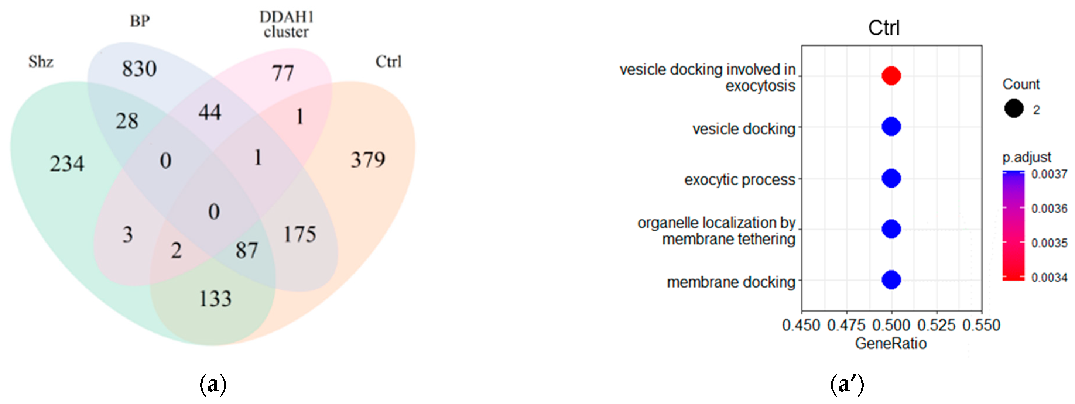
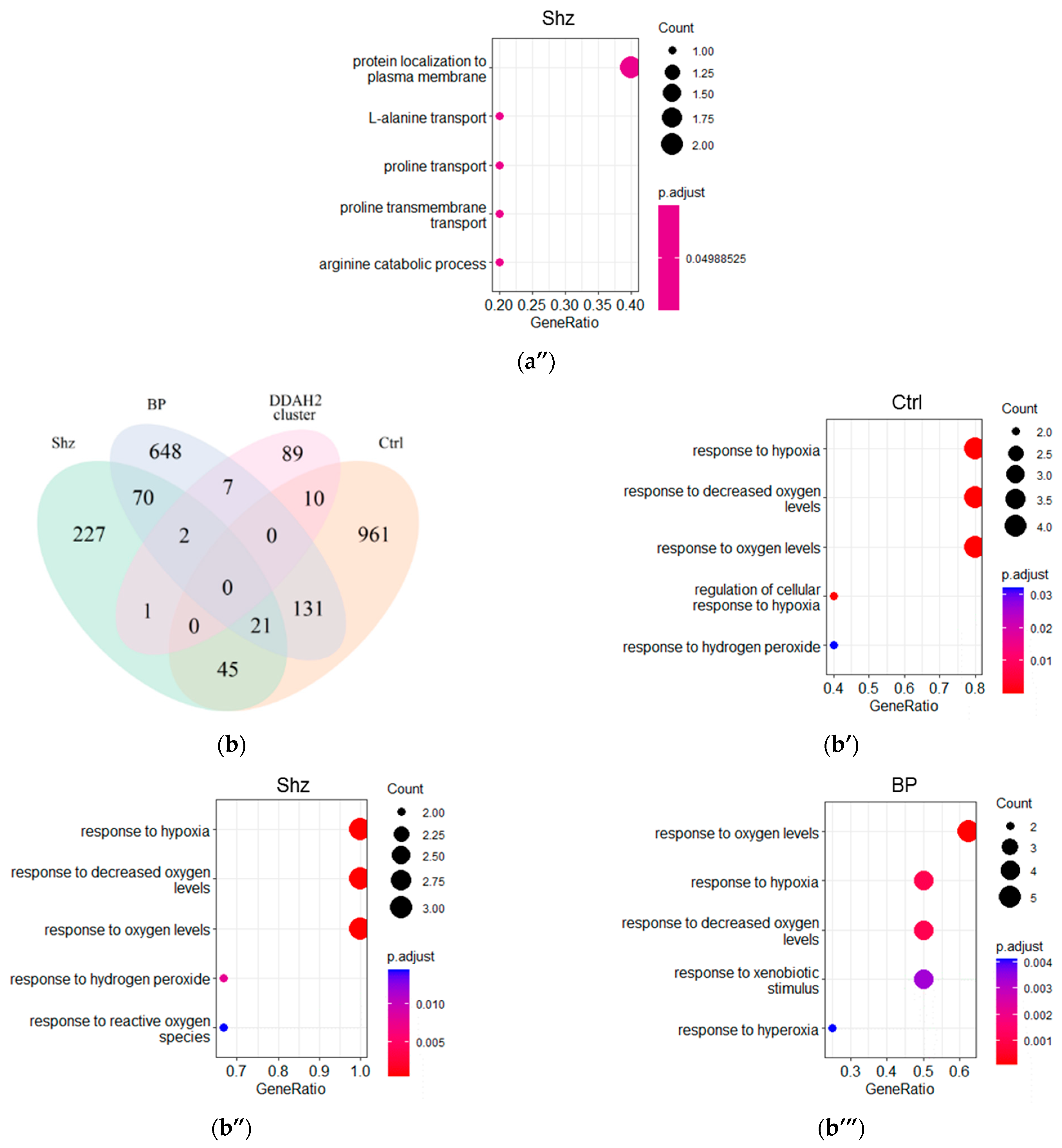
To designate the genes of the “DDAH1 cluster” and “DDAH2 cluster” that are co-expressed with DDAH1 or DDAH2, respectively, in the control group, bipolar disorder patients and schizophrenia patients, we compared these clusters with the gene sets derived from our co-expression analysis (cut-off r > 0.3, p < 0.05). All study groups had low overlap between the “DDAH1 cluster” or “DDAH2 cluster” and sets of genes co-expressed with DDAH1 or DDAH2, respectively. However, Venn diagrams show that DDAH1 or DDAH2 co-expressed gene sets in each group include some genes involved in DDAHs-related biological processes. The greatest overlap between the co-expressed gene set and the “DDAH1 cluster” was identified in patients with bipolar affective disorder. Forty-five common genes were identified between these two gene sets (refer to Figure 2a). In contrast, the DDAH2 co-expressed gene set in controls includes ten genes from the “DDAH2 cluster”, whereas in the samples from patients with bipolar affective disorder and schizophrenia, the overlap was even lower (refer to Figure 2b).

2.4. GO Term Enrichment Results

Despite the small number of genes included in the “DDAH1 cluster” or “DDAH2 cluster” and co-expressed with DDAH1 or DDAH2, respectively, in the tissue samples studied in the analyzed dataset, we performed GO term enrichment analysis in these narrow gene subsets to explain their specific biological function. We found that the large group of genes for which involvement in DDAH1-related functions was previously identified (n = 45) is co-expressed with DDAH1 in the bipolar affective disorder group stochastically, and no significant GO term enrichment results were revealed in this gene set. In contrast, several GO groups were enriched in the constricted clusters of DDAH functionally associated genes, co-expressing with DDAH1 in schizophrenic patients and controls or with DDAH2 in all study groups.
In the group of schizophrenia patients, DDAH1 co-expressed genes of the “DDAH1-cluster” (n = 5) are found to associate with protein localization and amino-acid metabolism and transport (refer to Figure 2a”, Supplementary data S3, Figure S1A). In contrast, in the control group (n = 4), the terms describing exocytosis and cell response were predominant (refer to Figure 2a’, Supplementary data S3, Figure S1B). DDAH2 co-expressed and functionally linked gene cluster enrichment results were congruent in different groups, with specific features in all cases. The most enriched GO terms in the control group (n = 10) and patients (n = 9 in patients with bipolar disorder and n = 3 in schizophrenic patients) describe the response to hypoxic conditions (refer to Figure 2b’–b’”, Supplementary data S3, Figure S2A–C).

2.5. Identification of Enriched Transcription Factors and Other Protein Binding Motives in Promoters of DDAH1/DDAH2 Co-Expressed Genes

To uncover whether the DDAH1 and DDAH2 co-expressed genes are regulated by common transcription factors in patients and non-psychiatric controls, we compared the enriched transcription factors-binding motives in genes co-expressed with DDAH1 and DDAH2 (i.e., r > 0.3, p < 0.05), respectively, in the different study groups.
In healthy subjects, genes whose promoters contain short motif CG, which is recognized by zinc finger-CxxC proteins, are enriched in the DDAH1 co-expressed gene set (refer to Table 3, Supplementary data S4, Table S4.1). This association is completely lost in both the schizophrenia and bipolar disorder group. In the control groups’ transcriptomic data, genes whose promoters contain the AGGGGGA motif, which is recognized by several C2H2 zinc finger transcription factors, are enriched in the DDAH2 co-expressed gene set (refer to Table 3, Supplementary Sdata 4, Table S4.2). In contrast, in patients with bipolar affective disorder, several types of promoters, including the promoters C2H2 zinc finger transcription factors binding sites, are over-represented in the DDAH2 co-expressed gene set (refer to Table 3, Supplementary data S4, Table S4.3). In the meantime, we did not observe any over-represented motives in the promoters of DDAH2 co-expressed genes in patients with schizophrenia.

2.6. Disease Ontology Gene Set Enrichment Analysis

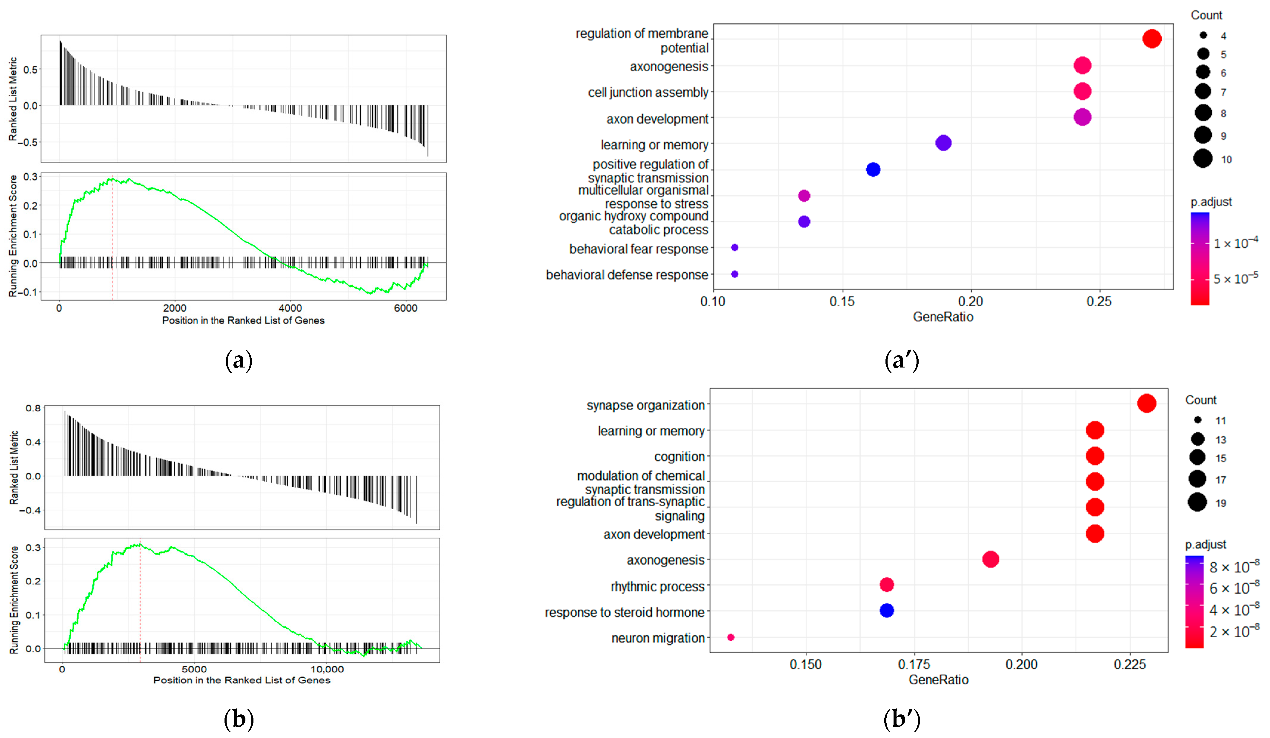
For disease ontology terms, 74 terms that predominantly characterize gene involvement in neoplastic disease (benign tumors and cancer) were enriched in DDAH1 co-expressed genes in the control group (refer to Supplementary data S5, Table S5.1). In genes co-expressed with DDAH1 in samples from schizophrenia patients, five terms corresponding to non-cancerous disease were enriched (refer to Supplementary data S5, Table S5.2). No DO terms were enriched in genes co-expressed with DDAH1 in the patients with bipolar affective disorder. In the context of the mental health terms, only one term, “DOID:0060037: a developmental disorder of mental health” (refer to Figure 3a), is significantly over-represented in the set of genes co-expressed with DDAH1 in the control group. Genes that regulate membrane potential, axonogenesis, and cell junction assembly contribute most to the enrichment result for this term (i.e., enrichment core; refer to Figure 3a’). No other associations with mental disorders were identified in DDAH1 co-expressed genes in any study group.
Over one hundred DO terms were enriched in DDAH2 co-expressed gene groups in samples from patients with bipolar disorder and non-psychiatric subjects (refer to Supplementary data S5, Tables S5.3 and S5.4). Conversely, in genes co-expressed with DDAH2 in samples from schizophrenic patients, only eighteen terms are enriched (refer to Supplementary data S5, Table S5.5). As in the DDAH1 co-expressed gene set, genes of the term “DOID:0060037: a developmental disorder of mental health” are enriched in the control group. In addition, the genes corresponding to the term “DOID:0060041: autism spectrum disorder”/“DOID:12849:autistic disorder” are significantly over-represented in this set (refer to Figure 3b,b”). The functional characteristics of the enrichment core of “DOID:0060037: a developmental disorder of mental health” in DDAH2 co-expressed genes in non-psychiatric subjects differ slightly from DDAH1 co-expressed genes. The enrichment core genes are associated with synapse organization and cognitive functions such as learning, memory, and cognition (refer to Figure 3b’). Curiously, the top three over-represented functions in “DOID0060041:autism spectrum disorder”/“DOID:12849 autistic disorder” in the enrichment core are the same (refer to Figure 3b’”).

3. Discussion

This study found DDAH1 and DDAH2 expression in all dorsolateral prefrontal cortex samples from patients and non-psychiatric control subjects. The expression level of DDAH1 was considerably higher than the DDAH2 expression levels in all subjects. We did not observe changes in DDAH1 expression levels here. In line, normal DDAH1 expression levels have previously been shown in chronic schizophrenic patients, while DDAH1 upregulation was found in patients with short-term schizophrenia [33]. For DDAH2 expression, upregulation is reported in association with schizophrenia patient-specific methylations and promoter region SNPs [36]. Other studies have also shown that DDAH2 mRNA levels were significantly elevated in brain tissue in schizophrenia, although the brain region was not specified in this study [37]. DDAH2 expression upregulation has also been described in the prefrontal cortex of patients with bipolar disorder [47]. While a similar trend was found in the present study, it does not reach statistical significance. The discrepancy between our results and published data may be attributed to the differences in study groups and applied methods.
Affective disorders such as major depression or bipolar disorder are associated with an aberrant expression pattern of NOS in the dorsolateral prefrontal cortex. Deregulation does, however, not influence the expression level of NOS1-, NOS2-, or NOS3-mRNA in whole cortex samples, but it was accompanied by changes in protein localization in cortical layers [48]. Thus, NO deregulation in the brain of psychiatric patients does not solely depend on gene expression levels. It also may be associated with the disturbance of expression patterns in the complex multicellular cortex structure and disease-associated deregulation of biological processes in the cortex. Considering that the gene’s co-expression mirrors the similar biologic function of these genes [49], we attempted to compare DDAH1 and DDAH2 co-expression patterns in control subjects and patients with psychiatric disorders.
The Pearson correlation coefficient is useful for estimating gene co-expression [50], revealing that subsets of genes for which co-expression with DDAH1 or DDAH2 is predicted in different groups hardly overlap. As the GO semantic similarity score between genes is related to the involvement of their products in the protein–protein interaction network [51], the functional relationship between DDAH1 or DDAH2 co-expressed genes appears to be higher in non-psychiatric controls. This difference may be related to the deregulation of DDAH-associated processes. Further analysis of DDAH co-expressed genes may uphold this assumption.
To increase the stringency of the selection of DDAHs interacting partners in each study group, we selected genes with the correlation coefficient (r) > 0.3, p < 0.05, which are suggested to interact with DDAHs. The overlap between sets of genes that are co-expressed with DDAHs in different conditions and “DDAH1 cluster” or “DDAH2 cluster” was low. Meanwhile, the occurrence of low overlap between an experimentally identified gene co-expression pattern and protein–protein interaction data described in the literature or databases was observed and discussed in previous studies [52,53].
In the non-psychiatric control subjects, these genes are involved in vesicular transport in both directions. In contrast, in schizophrenic patients, this association is lost, and genes associated with protein localization and amino acid metabolism predominate. Enrichment of any biological process in DDAH1-interacting genes co-expressed with DDAH1 is completely lost in patients with bipolar disorder patients. The significance of NO-signaling for normal transcytosis functioning, i.e., vesicular traffic across the interior of cells in the blood–brain barrier, has been demonstrated [54]. In addition, NO diffusion stimulates the release of vesicles in the synaptic cleft [55]. Thus, the association of DDAH1 co-regulated genes with vesicle transport seems reasonable. Psychiatric disorders such as schizophrenia and bipolar disorder may be associated with brain-blood barrier dysfunction [56]. Disruption of eNOS is one of the suggested reasons for increased blood–brain barrier permeability [57]. Losing the association of DDAH1 co-expressed genes with the vesicular traffic (exocytosis and endosome trafficking) may mirror this or any other aspect of NO-dependent pathway disruption in the prefrontal cortex in psychiatric patients.
The association of DDAH2 co-expressed genes with the response to the hypoxic stress identified in this study is expected in light of considerable evidence of DDAH2 upregulation in hypoxic conditions. The growth of DDAH2 expression levels in response to hypoxia was described in monocytes [58,59], endothelium [60], and myotubes [61]. In the studied group, DDAH2 co-expressed genes in the prefrontal cortex are stably associated with the response to hypoxia, despite the psychiatric diagnosis.
Deregulation of DDAH1 and DDAH2-associated processes in psychiatric patients was also confirmed in the analysis of protein-binding motif enrichment in their promoter regions. In schizophrenic patients, the over-representation of any specific promoter motive is completely lost, both in DDAH1 and DDAH2 co-expressed genes. In patients with bipolar disorder, the enrichment is also lost in the DDAH1 co-expressed gene set. While, in genes co-expressed with DDAH2 in the prefrontal cortex of patients with bipolar disorder, several protein-binding patterns are significantly over-represented. Notably, AP-2 transcription factors play essential roles in sleep regulation in the nematode Caenorhabditis elegans and the fruit fly Drosophila melanogaster [62]. The AP-2 paralogous transcription factors Tfap2a and Tfap2b control sleep behavior in mice, allowing for bidirectional control of sleep quality [63]. Over-representation may thus link prefrontal DDAH2 functionality with sleep disturbance, a core symptom of bipolar disorder [64]. Genes co-expressed with DDAH1 in non-psychiatric subjects frequently harbor the CG motif in their promoters. Human CxxC-binding domains display different structures and selectivity [65]. The CG pattern is the sole DNA-binding domain of CGBP, which is implicated in the expression of genes associated with CpG islands and the regulation of cytosine methylation [66]. ShinyGO software also demonstrated the enrichment of TET1 CxxC-binding protein, which binds predominantly on CGCGAT motifs [65], whose role in the expression regulation is also dualistic. However, TET1 binds and represses CpG-rich promoters by interacting with the polycomb repressive complex 2 [67]. In the brain structure, it is involved in regulating synapse development and functioning, memory, neuronal death and repair, and neuro-glial communication. The lost association of DDAH1 expression with CG promoter motif genes may mirror the overexpression of TET1 in cortical structures in patients with schizophrenia or bipolar disorder [68]. As the co-expressed genes are suggested to share their function, the demonstrated loss of correlation between expression of DDAH1 and genes harboring CG patterns in their promoters may distort the DDAH1 role in memory, learning, and neuron functioning. Still, further experimental work is needed on this matter.
The C2H2 zinc finger MZF-1 binding pattern is over-represented in DDAH2 co-expressed genes in non-psychiatric subjects and patients with bipolar disorder, but in schizophrenic patients, this association is lost. The transcription factor MZF-1 is a known tumor suppressor [69]. A significant portion of genes specifically expressed in the cortex, hindbrain, and midbrain harbor MZF-1 binding sites in their promoters [70], but the significance of MZF-1 expression in the central nervous system remains not well understood. In oxygen–glucose deprivation conditions, MZF-1 mediates the protective effect of human umbilical cord blood cells on both neurons and oligodendrocytes in mixed cultures [71,72]. This transcription factor also seems involved in gene regulation after peripheral nerve injury [73]. However, in contrast to non-psychiatric controls and schizophrenia, many other enriched promoter motifs in genes co-expressed with DDAH2 are found in patients with bipolar disorder samples.
Of note, neither TET1 binds the DDAH1 promoter, nor does MZF1 regulate DDAH2 expression directly, as summarized in the Chip Seq Atlas [74] or Signaling Pathways Project [75]. Thus, the DDAHs co-expression with TET1- or MZF1-regulated genes needs an explanation, which is more complex than the co-regulation of the same transcription factors motivating further research in this direction.
Disease Ontology was designed for researchers to study gene–disease relationships [76]. It has a hierarchical structure [77]; thus, the “DOID:0060037: developmental disorder of mental health” term covers the term “DOID0060041: autism spectrum disorder” and other terms corresponding to a learning disability, intellectual disability, attention deficit hyperactivity disorder, communication disorder, eating disorder and some other specific developmental disorders. The co-expression of DDAH1 and DDAH2 with the genes annotated with these terms was identified in dorsolateral prefrontal cortex samples in non-psychiatric controls but was lost both in schizophrenic and bipolar disorder patients.
Developmental mental disorders, including autism spectrum disorders, demonstrate behavior and cognitive disabilities, which are also intrinsic to major psychoses [78]. At the same time, schizophrenia and bipolar disorder patients exhibit a developmental lag [79], and the deregulation of genes involved in neurogenesis and neurodifferentiation was identified in schizophrenia patients at disease onset [34]. The genetic and molecular backgrounds of these diseases share numerous similarities. Genes with a documented association with neurodevelopmental and neuropsychiatric disorders are predominantly involved in transcription, synaptic transmission, cell–cell communication, ion transmembrane transport, intracellular signaling pathways, cell cycle, metabolic processes, nervous system development, and neuron death [78,80,81,82,83]. The common genetic etiology mirrors the high comorbidity of these psychiatric diseases and developmental mental disorders [84]. Hence, in the latest version of the Diagnostic and Statistical Manual of Mental Disorders, Fifth Edition (DSM-5), schizophrenia has been listed in proximity to neurodevelopmental disorders [85]. Currently, schizophrenia and bipolar disorder may be considered neurodevelopmental disorders with a widening of the neurodevelopmental spectrum [86] the loss of co-expression between genes characterized by the term “DOID:0060037: a developmental disorder of mental health”, including genes involved in synaptogenesis, axonogenesis and cognitive functions, and DDAHs is of particular interest.
Thus far, most of the evidence on DDAH/ADMA axis relevance for neuropsychiatric disorders comes from case-control clinical studies and measurements of peripheral ADMA and NO levels without offering in-depth mechanistic insight. Nevertheless, connections between the DDAH/ADMA axis and oxidative stress markers, as well as molecules important for cognitive processes, have been shown and support the findings of this study. For example, plasma levels of the oxidative stress-induced lipid peroxidation product 4-HNE were increased and correlated positively with plasma ADMA levels in depression [87] and schizophrenia patients [88]. In terms of cognition, a correlation between plasma ADMA levels and cognitive deficits has been established, with decreases in ADMA levels leading to improvements in working memory and attention [89,90]. ADMA infusion decreases BDNF, a factor highly associated with cognitive functions [91]. Lastly, the G allele of DDAH2 (−449 G/C) was positively associated with leukoaraiosis and high ADMA levels [92]. Progression of leukoaraiosis, a condition frequently met in neuropsychiatric disorders, relates to cognitive decline and thus could explain the link of DDAH2-cluster with cognition (learning and memory, cognition, synapse organization). In addition, ADMA levels in patients with leukoaraiosis were significantly higher than those in healthy controls [93], and these high concentrations of ADMA were associated with cognitive dysfunction in leukoaraiosis patients [94].
Our findings must be seen with some limitations. (i) The transcriptomic datasets are generated with different sequencing depths. Although all measures were taken to normalize the data, full uniformity and overcoming batch effects are unattainable. (ii) Only a few datasets in the GEO [95] repository were relevant for the study. Further study groups are relatively small and heterogeneous, and patient information is restricted. (iii) The mRNA abundance has a limited capacity as the indicator of downstream expression. The gene expression level and activity of gene products depend on multiple factors, including RNA stability, modifications, the translation rate and protein turnover, localization in the cell, and availability of ligands or co-interacting proteins. (iv) In addition, the size of gene clusters selected for the GO enrichment analysis was small and sometimes appeared less than the clusters used for the enrichment analysis. However, considering that the enrichment results are more informative than raw data about the GO groups in which the identified genes are included, we include GO enrichment results in this paper. Despite this limitation, we received statistically significant results, with gene ratios of 0.5–1 for several GO groups. (v) The used protein–protein interactions databases STRING [42], BioGRID [43], and HPRT [45,46] may be misleading for DDAH2 co-expressed genes because they are based on text mining and the assumption that DDAH2 is purely metabolizing ADMA, which is still debated. (vi) The analyzed data were restricted to the dorsolateral prefrontal cortical area and therefore do not allow for generalization to other brain regions. (v) Finally, our study lacks experimental validation. Despite all these limitations, the present work provides preliminary evidence that DDAH1 and DDAH2 are co-regulated with genes involved in mental disorder development and derangement. Experimental studies are now needed for confirmation and to gain mechanistic insight.

4. Materials and Methods

4.1. Public Resources and Databases

The expression data were derived from the public database of Gene Expression Omnibus (GEO) [95]. We used the terms “schizophrenia” and “bipolar disorder”; the filter “Expression profiling by high throughput sequencing” for the series type; and the filter “Homo sapiens” for the organism. Datasets that comprise the prefrontal cortex samples were selected (refer to Table 1). Unfortunately, the data for other brain structures were unrepresented or represented by a single dataset, making it impossible to compare them, and we did not include them.
The dataset GSE112523 combining data for the subjects with schizophrenia, bipolar disorder, and non-psychiatric controls was selected for further analysis of DDAH1 and DDAH2 co-expressed genes. This dataset was generated by the 75 bp paired-end sequencing, which was performed on an Illumina NextSeq 500 sequencer [41].
The genes of DDAH1- and DDAH2-interacting proteins (“DDAH1 cluster” and “DDAH2 cluster”, refer to Supplementary data S1, S2) for the comparative analysis were selected from the public databases STRING [42], MINT [44], BioGRID [43], and HPRD [45,46]. STRING database was searched for the human data in the full STRING network (i.e., for both functional and physical protein associations), and filtered for the data received by the text mining, experiments, databases, co-expression, or co-occurrence by the basic settings options. BioGRID data were filtered for the interactions identified in human studies. All other databases were searched with default settings. Then, the genes of all identified proteins were included in the “DDAH1 cluster” or “DDAH2 cluster”.

4.2. Data Normalization and Statistical Analysis

Raw counts were count per million (CPM)-normalized by edgeR package [96]. CPM values above the threshold level 1 were considered positive. The distribution of CPM-normalized expression levels in the analyzed samples was visualized by the beeswarm R package.
To estimate the differential gene expression, raw counts were normalized using the Trimmed Mean of M-values (TMM) method by the edgeR package [96] to avoid batch effects. Differentially expressed genes were identified by the glmQLFTest test using the edgeR package [96]. p values were adjusted for multiple testing corrections using the Benjamini–Hochberg method. Genes were considered differentially expressed if adjusted p values (Padj) < 0.05.

4.3. Measurement of Co-Expression

Before co-expression measurement, CPM-normalized data were filtered to exclude genes that are expressed below the threshold (i.e., CPM = 1 for all samples in the study groups). Data for different study groups were filtered independently. DDAH1 and DDAH2 co-expressed genes were selected by Pearson’s correlation coefficient (r > 0.3, p < 0.05). Genes co-expressed with DDAH1 or DDAH2 in the different study groups were included in separate gene clusters (i.e., DAAH1-co-expressed genes in the control group, DAAH1-co-expressed genes in schizophrenic patients, DAAH1-co-expressed genes in patients with bipolar disorder, DAAH2-co-expressed genes in the control group, DAAH2-co-expressed genes in schizophrenic patients, and DAAH2-co-expressed genes in patients with bipolar disorder). The comparative analysis of the selected clusters was performed as described below.

4.4. Analysis of Functional Semantic Similarity between Genes

Gene Ontology (GO) semantic similarity was calculated by Wang’s method in the GOSemSim package [97] employing the “Biological process” GO terms. The difference in semantic similarity scores in different gene clusters was estimated by Brown–Forsythe and Games–Howell post hoc tests.

4.5. Function and Enrichment Analysis

The clusters of DDAHs co-expressed genes, identified in different study groups, were compared to each other, and to the “DDAH1 cluster” or “DAAH2 cluster”, which were identified by searching public databases as described above. DDAH1 co-expressed gene clusters were compared in the control group, schizophrenic patients, and patients with bipolar disorder, and then matched with the “DDAH1 cluster”. Similarly, the DDAH2 co-expressed gene clusters were evaluated. The overlap between identified gene clusters was visualized by the VennDiagram R package. To increase the stringency of DDAHs interacting patterns, the genes that were common for DDAHs co-expressed groups and the “DDAH1 cluster” or “DAAH2 cluster” were selected for further analysis, as it was described elsewhere [52,98].
Enriched motifs in promoters of the studied gene sets were identified using the ShinyGo 0.76 [99] web tool (available at http://bioinformatics.sdstate.edu/go/, accessed 10 June 2022). The upstream 300 bp region was specified as the promoter.
GO enrichment analysis (identification of GO terms that are significantly enriched by the genes of the selected set) was performed in the identified co-expressed gene clusters, and visualization of results was performed by the clusterProfiler Bioconductor package [100].
Disease Ontology (DO) enrichment analysis was performed in gene lists ranging from the highest value of the Pearson correlation of the gene expression level with the levels of DDAH1 or DDAH2 expression to the lowest. The clusterProfiler [100] and DOSE [101] R packages were used.
We considered significant enrichment results only for GO biological process terms, transcription factors, or DO terms with a false discovery rate value of <0.05.

5. Conclusions

Our results suggest a possible involvement of DDAH1 and DDAH2 in the pathophysiology of psychiatric disorders. While mRNA levels in the dorsolateral prefrontal cortex of psychiatric patients remain unchanged, a functional shift occurs that is reflected in dramatic changes in the expression of genes whose products interact with DDAH1/2. We found that correlations between expression levels of DDAH1 or DDAH2 and genes associated with mental illness are lost in cortical samples from psychiatric patients. DDAH1 and DDAH2 co-expressed genes were generally less integrated into shared functions in psychiatric patients than in non-psychiatric controls. Furthermore, the overlap between genes co-expressed with DDAHs in control subjects and psychiatric patients is low, possibly due to the complex deregulation of transcription factor activity. These data suggest that DDAHs are associated with the processes that form the molecular basis of mental and cognitive functions and thus may be potential therapeutic targets in psychiatric disorders.

Supplementary Materials

The following supporting information can be downloaded at: https://www.mdpi.com/article/10.3390/ijms231911902/s1: S1. Title: DDAH1 cluster; S2. Title: DDAH2 cluster; S3. Title: Gene ontology (GO) enrichment analysis of genes involved in the functional interaction with DDAH1 or DDAH2: Figures S1 and S2; S4. Title: Enrichment analysis of promotor motifs in genes co-expressed with DDAH1 or DDAH2: Tables S4.1–S4.3; S5. Title: Disease Ontology (DO) terms enrichment analysis of genes co-expressed with DDAH1 or DDAH2: Tables S5.1–S5.5.

Author Contributions

Conceptualization, A.N.V., A.A.K., N.B. and R.R.G.; methodology, A.N.V.; validation, A.N.V., R.N.R. and R.R.G.; formal analysis, A.N.V.; investigation, A.N.V.; data curation, A.N.V.; writing—original draft preparation, A.N.V. and A.A.K.; writing—review and editing, A.N.V., A.A.K., R.R.G., R.N.R. and N.B.; visualization, A.A.K. and A.N.V.; supervision, N.B. and R.R.G.; project administration, N.B.; funding acquisition, N.B. and R.R.G. All authors have read and agreed to the published version of the manuscript.

Funding

This work was supported by the DFG IRTG 2773/1 and the woman habilitation promotion initiative from the Medical Faculty awarded to N.B. A.A.K. received research scholarships from the German Academic Exchange Service and the Gesellschaft von Freunden und Förderern der TU Dresden. A.N.V. and R.R.G. were supported by project ID: 93018770 of St. Petersburg State University.

Institutional Review Board Statement

Not applicable.

Informed Consent Statement

Not applicable.

Data Availability Statement

All whole tissue sample data are available in the GEO database (https://www.ncbi.nlm.nih.gov/geo/ (accessed on 10 December 2021); the detailed information is listed in Table 1).

Conflicts of Interest

The authors declare no conflict of interest.

References

  1. GBD 2019 Mental Disorders Collaborators. Global, Regional, and National Burden of 12 Mental Disorders in 204 Countries and Territories, 1990–2019: A Systematic Analysis for the Global Burden of Disease Study 2019. Lancet Psychiatry 2022, 9, 137–150. [Google Scholar] [CrossRef]
  2. McNeill, R.V.; Kehrwald, C.; Brum, M.; Knopf, K.; Brunkhorst-Kanaan, N.; Etyemez, S.; Koreny, C.; Bittner, R.A.; Freudenberg, F.; Herterich, S.; et al. Uncovering Associations between Mental Illness Diagnosis, Nitric Oxide Synthase Gene Variation, and Peripheral Nitric Oxide Concentration. Brain. Behav. Immun. 2022, 101, 275–283. [Google Scholar] [CrossRef]
  3. Jagannathan, K.; Calhoun, V.D.; Gelernter, J.; Stevens, M.C.; Liu, J.; Bolognani, F.; Windemuth, A.; Ruaño, G.; Assaf, M.; Pearlson, G.D. Genetic Associations of Brain Structural Networks in Schizophrenia: A Preliminary Study. Biol. Psychiatry 2010, 68, 657–666. [Google Scholar] [CrossRef] [PubMed] [Green Version]
  4. Kittel-Schneider, S.; Reuß, M.; Meyer, A.; Weber, H.; Gessner, A.; Leistner, C.; Kopf, J.; Schmidt, B.; Hempel, S.; Volkert, J.; et al. Multi-Level Biomarker Analysis of Nitric Oxide Synthase Isoforms in Bipolar Disorder and Adult ADHD. J. Psychopharmacol. 2015, 29, 31–38. [Google Scholar] [CrossRef] [PubMed]
  5. Minutolo, G.; Petralia, A.; Dipasquale, S.; Aguglia, E. Nitric Oxide in Patients with Schizophrenia: The Relationship with the Severity of Illness and the Antipsychotic Treatment. Expert Opin. Pharmacother. 2012, 13, 1989–1997. [Google Scholar] [CrossRef] [PubMed]
  6. Morales-Medina, J.C.; Aguilar-Alonso, P.; Di Cerbo, A.; Iannitti, T.; Flores, G. New Insights on Nitric Oxide: Focus on Animal Models of Schizophrenia. Behav. Brain Res. 2021, 409, 113304. [Google Scholar] [CrossRef] [PubMed]
  7. Garthwaite, J.; Garthwaite, G.; Palmer, R.M.; Moncada, S. NMDA Receptor Activation Induces Nitric Oxide Synthesis from Arginine in Rat Brain Slices. Eur. J. Pharmacol. 1989, 172, 413–416. [Google Scholar] [CrossRef]
  8. Kiss, J.P.; Vizi, E.S. Nitric Oxide: A Novel Link between Synaptic and Nonsynaptic Transmission. Trends Neurosci. 2001, 24, 211–215. [Google Scholar] [CrossRef]
  9. Nasyrova, R.F.; Ivashchenko, D.V.; Ivanov, M.V.; Neznanov, N.G. Role of Nitric Oxide and Related Molecules in Schizophrenia Pathogenesis: Biochemical, Genetic and Clinical Aspects. Front. Physiol. 2015, 6, 139. [Google Scholar] [CrossRef] [Green Version]
  10. Liu, P.; Jing, Y.; Collie, N.D.; Dean, B.; Bilkey, D.K.; Zhang, H. Altered Brain Arginine Metabolism in Schizophrenia. Transl. Psychiatry. 2016, 6, e871. [Google Scholar] [CrossRef]
  11. Quan, L.; Uyeda, A.; Muramatsu, R. Central Nervous System Regeneration: The Roles of Glial Cells in the Potential Molecular Mechanism Underlying Remyelination. Inflamm. Regen. 2022, 42, 7. [Google Scholar] [CrossRef]
  12. Rose, E.J.; Greene, C.; Kelly, S.; Morris, D.W.; Robertson, I.H.; Fahey, C.; Jacobson, S.; O’Doherty, J.; Newell, F.N.; McGrath, J.; et al. The NOS1 Variant Rs6490121 Is Associated with Variation in Prefrontal Function and Grey Matter Density in Healthy Individuals. NeuroImage 2012, 60, 614–622. [Google Scholar] [CrossRef]
  13. Freudenberg, F.; Alttoa, A.; Reif, A. Neuronal Nitric Oxide Synthase (NOS1) and Its Adaptor, NOS1AP, as a Genetic Risk Factors for Psychiatric Disorders. Genes Brain Behav. 2015, 14, 46–63. [Google Scholar] [CrossRef]
  14. Ginsberg, S.D.; Hemby, S.E.; Smiley, J.F. Expression Profiling in Neuropsychiatric Disorders: Emphasis on Glutamate Receptors in Bipolar Disorder. Pharmacol. Biochem. Behav. 2012, 100, 705–711. [Google Scholar] [CrossRef] [PubMed] [Green Version]
  15. González-Castro, T.B.; Genis-Mendoza, A.D.; Tovilla-Zárate, C.A.; Juárez-Rojop, I.E.; López-Narvaez, M.L.; Pérez-Hernández, N.; Rodríguez-Pérez, J.M.; Martínez-Magaña, J.J. Association between Polymorphisms of NOS1, NOS2 and NOS3 Genes and Suicide Behavior: A Systematic Review and Meta-Analysis. Metab. Brain Dis. 2019, 34, 967–977. [Google Scholar] [CrossRef]
  16. Oliveira, J.; Debnath, M.; Etain, B.; Bennabi, M.; Hamdani, N.; Lajnef, M.; Bengoufa, D.; Fortier, C.; Boukouaci, W.; Bellivier, F.; et al. Violent Suicidal Behaviour in Bipolar Disorder Is Associated with Nitric Oxide Synthase 3 Gene Polymorphism. Acta Psychiatr. Scand. 2015, 132, 218–225. [Google Scholar] [CrossRef] [PubMed]
  17. Reif, A.; Herterich, S.; Strobel, A.; Ehlis, A.-C.; Saur, D.; Jacob, C.P.; Wienker, T.; Töpner, T.; Fritzen, S.; Walter, U.; et al. A Neuronal Nitric Oxide Synthase (NOS-I) Haplotype Associated with Schizophrenia Modifies Prefrontal Cortex Function. Mol. Psychiatry 2006, 11, 286–300. [Google Scholar] [CrossRef] [PubMed] [Green Version]
  18. Reif, A.; Strobel, A.; Jacob, C.P.; Herterich, S.; Freitag, C.M.; Töpner, T.; Mössner, R.; Fritzen, S.; Schmitt, A.; Lesch, K.-P. A NOS-III Haplotype That Includes Functional Polymorphisms Is Associated with Bipolar Disorder. Int. J. Neuropsychopharmacol. 2006, 9, 13–20. [Google Scholar] [CrossRef] [Green Version]
  19. Sarginson, J.E.; Deakin, J.W.; Anderson, I.M.; Downey, D.; Thomas, E.; Elliott, R.; Juhasz, G. Neuronal Nitric Oxide Synthase (NOS1) Polymorphisms Interact with Financial Hardship to Affect Depression Risk. Neuropsychopharmacology 2014, 39, 2857–2866. [Google Scholar] [CrossRef] [Green Version]
  20. Wigner, P.; Czarny, P.; Synowiec, E.; Bijak, M.; Białek, K.; Talarowska, M.; Galecki, P.; Szemraj, J.; Sliwinski, T. Variation of Genes Involved in Oxidative and Nitrosative Stresses in Depression. Eur. Psychiatry 2018, 48, 38–48. [Google Scholar] [CrossRef] [PubMed]
  21. Wockner, L.F.; Noble, E.P.; Lawford, B.R.; Young, R.M.; Morris, C.P.; Whitehall, V.L.J.; Voisey, J. Genome-Wide DNA Methylation Analysis of Human Brain Tissue from Schizophrenia Patients. Transl. Psychiatry 2014, 4, e339. [Google Scholar] [CrossRef] [Green Version]
  22. Weber, H.; Klamer, D.; Freudenberg, F.; Kittel-Schneider, S.; Rivero, O.; Scholz, C.-J.; Volkert, J.; Kopf, J.; Heupel, J.; Herterich, S.; et al. The Genetic Contribution of the NO System at the Glutamatergic Post-Synapse to Schizophrenia: Further Evidence and Meta-Analysis. Eur. Neuropsychopharmacol. 2014, 24, 65–85. [Google Scholar] [CrossRef] [Green Version]
  23. Bruenig, D.; Morris, C.P.; Mehta, D.; Harvey, W.; Lawford, B.; Young, R.M.; Voisey, J. Nitric Oxide Pathway Genes (NOS1AP and NOS1) Are Involved in PTSD Severity, Depression, Anxiety, Stress and Resilience. Gene 2017, 625, 42–48. [Google Scholar] [CrossRef] [PubMed] [Green Version]
  24. Anirudh, C.S.; Pathak, A.K.; Sinha, P.; Jainarayanan, A.K.; Jain, S.; Brahmachari, S.K. Multi-Scale Analysis of Schizophrenia Risk Loci: Integrating Centenarian Genomes and Spatio-Temporal Expression Profiles Suggests the Need for Adjunctive Therapeutic Interventions for Neuropsychiatric Disorders. bioRxiv 2018. [Google Scholar] [CrossRef]
  25. Shinkai, T.; Ohmori, O.; Hori, H.; Nakamura, J. Allelic Association of the Neuronal Nitric Oxide Synthase (NOS1) Gene with Schizophrenia. Mol. Psychiatry 2002, 7, 560–563. [Google Scholar] [CrossRef] [PubMed] [Green Version]
  26. Zhou, Q.-G.; Zhu, X.-H.; Nemes, A.D.; Zhu, D.-Y. Neuronal Nitric Oxide Synthase and Affective Disorders. IBRO Rep. 2018, 5, 116–132. [Google Scholar] [CrossRef] [PubMed]
  27. Tran, C.T.L.; Fox, M.F.; Vallance, P.; Leiper, J.M. Chromosomal Localization, Gene Structure, and Expression Pattern of DDAH1: Comparison with DDAH2 and Implications for Evolutionary Origins. Genomics 2000, 68, 101–105. [Google Scholar] [CrossRef] [PubMed]
  28. Kozlova, A.A.; Ragavan, V.N.; Jarzebska, N.; Lukianova, I.V.; Bikmurzina, A.E.; Rubets, E.; Suzuki-Yamamoto, T.; Kimoto, M.; Mangoni, A.A.; Gainetdinov, R.R.; et al. Divergent Dimethylarginine Dimethylaminohydrolase Isoenzyme Expression in the Central Nervous System. Cell Mol. Neurobiol. 2021, 42, 2273–2288. [Google Scholar] [CrossRef] [PubMed]
  29. Guo, W.; Samuels, J.F.; Wang, Y.; Cao, H.; Ritter, M.; Nestadt, P.S.; Krasnow, J.; Greenberg, B.D.; Fyer, A.J.; McCracken, J.T.; et al. Polygenic Risk Score and Heritability Estimates Reveals a Genetic Relationship between ASD and OCD. Eur. Neuropsychopharmacol. 2017, 27, 657–666. [Google Scholar] [CrossRef]
  30. Cieślik, P.; Siekierzycka, A.; Radulska, A.; Płoska, A.; Burnat, G.; Brański, P.; Kalinowski, L.; Wierońska, J.M. Nitric Oxide-Dependent Mechanisms Underlying MK-801- or Scopolamine-Induced Memory Dysfunction in Animals: Mechanistic Studies. Int. J. Mol. Sci. 2021, 22, 12282. [Google Scholar] [CrossRef] [PubMed]
  31. Cortelazzo, A.; De Felice, C.; Guy, J.; Timperio, A.M.; Zolla, L.; Guerranti, R.; Leoncini, S.; Signorini, C.; Durand, T.; Hayek, J. Brain Protein Changes in Mecp2 Mouse Mutant Models: Effects on Disease Progression of Mecp2 Brain Specific Gene Reactivation. J. Proteom. 2020, 210, 103537. [Google Scholar] [CrossRef] [PubMed]
  32. Whittle, N.; Li, L.; Chen, W.-Q.; Yang, J.-W.; Sartori, S.B.; Lubec, G.; Singewald, N. Changes in Brain Protein Expression Are Linked to Magnesium Restriction-Induced Depression-like Behavior. Amino Acids 2011, 40, 1231–1248. [Google Scholar] [CrossRef]
  33. Clark, D.; Dedova, I.; Cordwell, S.; Matsumoto, I. A Proteome Analysis of the Anterior Cingulate Cortex Gray Matter in Schizophrenia. Mol. Psychiatry 2006, 11, 459–470. [Google Scholar] [CrossRef] [PubMed] [Green Version]
  34. Narayan, S.; Tang, B.; Head, S.R.; Gilmartin, T.J.; Sutcliffe, J.G.; Dean, B.; Thomas, E.A. Molecular Profiles of Schizophrenia in the CNS at Different Stages of Illness. Brain Res. 2008, 1239, 235–248. [Google Scholar] [CrossRef] [PubMed] [Green Version]
  35. Hass, J.; Walton, E.; Wright, C.; Beyer, A.; Scholz, M.; Turner, J.; Liu, J.; Smolka, M.N.; Roessner, V.; Sponheim, S.R.; et al. Associations between DNA Methylation and Schizophrenia-Related Intermediate Phenotypes—A Gene Set Enrichment Analysis. Prog. Neuro-Psychopharmacol. Biol. Psychiatry 2015, 59, 31–39. [Google Scholar] [CrossRef] [PubMed] [Green Version]
  36. Pineda-Cirera, L.; Cabana-Domínguez, J.; Lee, P.H.; Fernàndez-Castillo, N.; Cormand, B. Identification of Genetic Variants Influencing Methylation in Brain with Pleiotropic Effects on Psychiatric Disorders. Prog. Neuro-Psychopharmacol. Biol. Psychiatry 2022, 113, 110454. [Google Scholar] [CrossRef]
  37. Wu, X.; Ye, J.; Wang, Z.; Zhao, C. Epigenetic Age Acceleration Was Delayed in Schizophrenia. Schizophr. Bull. 2021, 47, 803–811. [Google Scholar] [CrossRef] [PubMed]
  38. Bani-Fatemi, A.; Jeremian, R.; Wang, K.Z.; Silveira, J.; Zai, C.; Kolla, N.J.; Graff, A.; Gerretsen, P.; Strauss, J.; De Luca, V. Epigenome-Wide Association Study of Suicide Attempt in Schizophrenia. J. Psychiatr. Res. 2018, 104, 192–197. [Google Scholar] [CrossRef] [PubMed]
  39. Reif, A.; Schecklmann, M.; Eirich, E.; Jacob, C.P.; Jarczok, T.A.; Kittel-Schneider, S.; Lesch, K.-P.; Fallgatter, A.J.; Ehlis, A.-C. A Functional Promoter Polymorphism of Neuronal Nitric Oxide Synthase Moderates Prefrontal Functioning in Schizophrenia. Int. J. Neuropsychopharmacol. 2011, 14, 887–897. [Google Scholar] [CrossRef]
  40. Connor, C.M.; Crawford, B.C.; Akbarian, S. White Matter Neuron Alterations in Schizophrenia and Related Disorders. Int. J. Dev. Neurosci. 2011, 29, 325–334. [Google Scholar] [CrossRef]
  41. Pai, S.; Li, P.; Killinger, B.; Marshall, L.; Jia, P.; Liao, J.; Petronis, A.; Szabó, P.E.; Labrie, V. Differential methylation of enhancer at IGF2 is associated with abnormal dopamine synthesis in major psychosis. Nat. Commun. 2019, 10, 2046. [Google Scholar] [CrossRef] [Green Version]
  42. Szklarczyk, D.; Gable, A.L.; Nastou, K.C.; Lyon, D.; Kirsch, R.; Pyysalo, S.; Doncheva, N.T.; Legeay, M.; Fang, T.; Bork, P.; et al. The STRING Database in 2021: Customizable Protein–Protein Networks, and Functional Characterization of User-Uploaded Gene/Measurement Sets. Nucleic Acids Res. 2021, 49, D605–D612. [Google Scholar] [CrossRef]
  43. Oughtred, R.; Rust, J.; Chang, C.; Breitkreutz, B.; Stark, C.; Willems, A.; Boucher, L.; Leung, G.; Kolas, N.; Zhang, F.; et al. The BioGRID Database: A Comprehensive Biomedical Resource of Curated Protein, Genetic, and Chemical Interactions. Protein Sci. 2021, 30, 187–200. [Google Scholar] [CrossRef] [PubMed]
  44. Licata, L.; Briganti, L.; Peluso, D.; Perfetto, L.; Iannuccelli, M.; Galeota, E.; Sacco, F.; Palma, A.; Nardozza, A.P.; Santonico, E.; et al. MINT, the Molecular Interaction Database: 2012 Update. Nucleic Acids Res. 2012, 40, D857–D861. [Google Scholar] [CrossRef]
  45. Keshava Prasad, T.S.; Goel, R.; Kandasamy, K.; Keerthikumar, S.; Kumar, S.; Mathivanan, S.; Telikicherla, D.; Raju, R.; Shafreen, B.; Venugopal, A.; et al. Human Protein Reference Database—2009 Update. Nucleic Acids Res. 2009, 37, D767–D772. [Google Scholar] [CrossRef] [PubMed] [Green Version]
  46. Peri, S.; Navarro, J.D.; Amanchy, R.; Kristiansen, T.Z.; Jonnalagadda, C.K.; Surendranath, V.; Niranjan, V.; Muthusamy, B.; Gandhi, T.K.B.; Gronborg, M.; et al. Development of Human Protein Reference Database as an Initial Platform for Approaching Systems Biology in Humans. Genome Res. 2003, 13, 2363–2371. [Google Scholar] [CrossRef] [PubMed] [Green Version]
  47. Wang, J.; Qu, S.; Wang, W.; Guo, L.; Zhang, K.; Chang, S.; Wang, J. A Combined Analysis of Genome-Wide Expression Profiling of Bipolar Disorder in Human Prefrontal Cortex. J. Psychiatr. Res. 2016, 82, 23–29. [Google Scholar] [CrossRef] [PubMed]
  48. Gao, S.-F.; Qi, X.-R.; Zhao, J.; Balesar, R.; Bao, A.-M.; Swaab, D.F. Decreased NOS1 Expression in the Anterior Cingulate Cortex in Depression. Cereb. Cortex 2013, 23, 2956–2964. [Google Scholar] [CrossRef] [PubMed] [Green Version]
  49. Eisen, M.B.; Spellman, P.T.; Brown, P.O.; Botstein, D. Cluster Analysis and Display of Genome-Wide Expression Patterns. Proc. Natl. Acad. Sci. USA 1998, 95, 14863–14868. [Google Scholar] [CrossRef] [PubMed] [Green Version]
  50. Furlotte, N.A.; Kang, H.M.; Ye, C.; Eskin, E. Mixed-Model Coexpression: Calculating Gene Coexpression While Accounting for Expression Heterogeneity. Bioinformatics 2011, 27, i288–i294. [Google Scholar] [CrossRef]
  51. Gan, M. Correlating Information Contents of Gene Ontology Terms to Infer Semantic Similarity of Gene Products. Comput. Math. Methods Med. 2014, 2014, e891842. [Google Scholar] [CrossRef] [PubMed] [Green Version]
  52. Dho, S.E.; Silva-Gagliardi, N.; Morgese, F.; Coyaud, E.; Lamoureux, E.; Berry, D.M.; Raught, B.; McGlade, C.J. Proximity interactions of the ubiquitin ligase Mind bomb 1 reveal a role in regulation of epithelial polarity complex proteins. Sci. Rep. 2019, 9, 12471. [Google Scholar] [CrossRef] [Green Version]
  53. Sumithra, B.; Saxena, U.; Das, A.B. A comprehensive study on genome-wide coexpression network of KHDRBS1/Sam68 reveals its cancer and patient-specific association. Sci Rep 2019, 9, 11083. [Google Scholar] [CrossRef] [Green Version]
  54. Martins, T.; Burgoyne, T.; Kenny, B.-A.; Hudson, N.; Futter, C.E.; Ambrósio, A.F.; Silva, A.P.; Greenwood, J.; Turowski, P. Methamphetamine-Induced Nitric Oxide Promotes Vesicular Transport in Blood–Brain Barrier Endothelial Cells. Neuropharmacology 2013, 65, 74–82. [Google Scholar] [CrossRef] [PubMed] [Green Version]
  55. Picón-Pagès, P.; Garcia-Buendia, J.; Muñoz, F.J. Functions and Dysfunctions of Nitric Oxide in Brain. Biochim. Biophys. Acta BBA Mol. Basis Dis. 2019, 1865, 1949–1967. [Google Scholar] [CrossRef] [PubMed]
  56. Pong, S.; Lizano, P.; Karmacharya, R. Investigating Blood-Brain Barrier Dysfunction in Schizophrenia Using Brain Microvascular Endothelial Cells Derived From Patient-Specific Stem Cells. Biol. Psychiatry 2020, 87, S189–S190. [Google Scholar] [CrossRef]
  57. Najjar, S.; Pahlajani, S.; De Sanctis, V.; Stern, J.N.H.; Najjar, A.; Chong, D. Neurovascular Unit Dysfunction and Blood–Brain Barrier Hyperpermeability Contribute to Schizophrenia Neurobiology: A Theoretical Integration of Clinical and Experimental Evidence. Front Psychiatry 2017, 8, 83. [Google Scholar] [CrossRef] [Green Version]
  58. Lambden, S.; Martin, D.; Tomlinson, J.; Mythen, M.; Leiper, J. Role of Dimethylarginine Dimethylaminohydrolase 2 in the Regulation of Nitric Oxide Synthesis in Animal and Observational Human Models of Normobaric Hypoxia. Lancet 2016, 387, S62. [Google Scholar] [CrossRef]
  59. Lambden, S.; Martin, D.; Vanezis, K.; Lee, B.; Tomlinson, J.; Piper, S.; Boruc, O.; Mythen, M.; Leiper, J. Hypoxia Causes Increased Monocyte Nitric Oxide Synthesis Which Is Mediated by Changes in Dimethylarginine Dimethylaminohydrolase 2 Expression in Animal and Human Models of Normobaric Hypoxia. Nitric Oxide 2016, 58, 59–66. [Google Scholar] [CrossRef] [PubMed] [Green Version]
  60. Harbaum, L.; Glatzel, A.; Klose, H.; Böger, R.H.; Lüneburg, N. Modulation of Symmetric Dimethyarginine Formation by Apelin in Human Pulmonary Endothelial Cells. Eur. Respir. J. 2015, 46, PA859. [Google Scholar] [CrossRef]
  61. Williams, G.; Shi-Wen, X.; Abraham, D.; Selvakumar, S.; Baker, D.M.; Tsui, J.C.S. Nitric Oxide Manipulation: A Therapeutic Target for Peripheral Arterial Disease? Cardiol. Res. Pract. 2012, 2012, e656247. [Google Scholar] [CrossRef] [PubMed] [Green Version]
  62. Turek, M.; Lewandrowski, I.; Bringmann, H. An AP2 Transcription Factor Is Required for a Sleep-Active Neuron to Induce Sleep-like Quiescence in C. Elegans. Curr. Biol. 2013, 23, 2215–2223. [Google Scholar] [CrossRef] [PubMed] [Green Version]
  63. Hu, Y.; Korovaichuk, A.; Astiz, M.; Schroeder, H.; Islam, R.; Barrenetxea, J.; Fischer, A.; Oster, H.; Bringmann, H. Functional Divergence of Mammalian TFAP2a and TFAP2b Transcription Factors for Bidirectional Sleep Control. Genetics 2020, 216, 735–752. [Google Scholar] [CrossRef] [PubMed]
  64. Hensch, T.; Wozniak, D.; Spada, J.; Sander, C.; Ulke, C.; Wittekind, D.A.; Thiery, J.; Löffler, M.; Jawinski, P.; Hegerl, U. Vulnerability to Bipolar Disorder Is Linked to Sleep and Sleepiness. Transl. Psychiatry 2019, 9, 294. [Google Scholar] [CrossRef] [PubMed] [Green Version]
  65. Xu, C.; Liu, K.; Lei, M.; Yang, A.; Li, Y.; Hughes, T.R.; Min, J. DNA Sequence Recognition of Human CXXC Domains and Their Structural Determinants. Structure 2018, 26, 85–95.e3. [Google Scholar] [CrossRef] [Green Version]
  66. Lee, J.-H.; Voo, K.S.; Skalnik, D.G. Identification and Characterization of the DNA Binding Domain of CpG-Binding Protein. J. Biol. Chem. 2001, 276, 44669–44676. [Google Scholar] [CrossRef] [Green Version]
  67. Wu, H.; Zhang, Y. Mechanisms and Functions of Tet Protein-Mediated 5-Methylcytosine Oxidation. Genes Dev. 2011, 25, 2436–2452. [Google Scholar] [CrossRef] [Green Version]
  68. Dong, E.; Gavin, D.P.; Chen, Y.; Davis, J. Upregulation of TET1 and Downregulation of APOBEC3A and APOBEC3C in the Parietal Cortex of Psychotic Patients. Transl. Psychiatry 2012, 2, e159. [Google Scholar] [CrossRef] [Green Version]
  69. Gaboli, M.; Kotsi, P.A.; Gurrieri, C.; Cattoretti, G.; Ronchetti, S.; Cordon-Cardo, C.; Broxmeyer, H.E.; Hromas, R.; Pandolfi, P.P. Mzf1 Controls Cell Proliferation and Tumorigenesis. Genes Dev. 2001, 15, 1625–1630. [Google Scholar] [CrossRef] [Green Version]
  70. Bellora, N.; Farré, D.; Albà, M.M. Positional Bias of General and Tissue-Specific Regulatory Motifs in Mouse Gene Promoters. BMC Genom. 2007, 8, 459. [Google Scholar] [CrossRef]
  71. Rowe, D.D.; Leonardo, C.C.; Hall, A.A.; Shahaduzzaman, M.D.; Collier, L.A.; Willing, A.E.; Pennypacker, K.R. Cord Blood Administration Induces Oligodendrocyte Survival through Alterations in Gene Expression. Brain Res. 2010, 1366, 172–188. [Google Scholar] [CrossRef] [Green Version]
  72. Shahaduzzaman, M.D.; Mehta, V.; Golden, J.E.; Rowe, D.D.; Green, S.; Tadinada, R.; Foran, E.A.; Sanberg, P.R.; Pennypacker, K.R.; Willing, A.E. Human Umbilical Cord Blood Cells Induce Neuroprotective Change in Gene Expression Profile in Neurons after Ischemia through Activation of Akt Pathway. Cell Transplant. 2015, 24, 721–735. [Google Scholar] [CrossRef] [Green Version]
  73. Chandran, V.; Coppola, G.; Nawabi, H.; Omura, T.; Versano, R.; Huebner, E.A.; Zhang, A.; Costigan, M.; Yekkirala, A.; Barrett, L.; et al. A Systems-Level Analysis of the Peripheral Nerve Intrinsic Axonal Growth Program. Neuron 2016, 89, 956–970. [Google Scholar] [CrossRef] [Green Version]
  74. Zhaonan, Z.; Tazro, O.; Fumihito, M.; Shinya, O. ChIP-Atlas 2021 Update: A Data-Mining Suite for Exploring Epigenomic Landscapes by Fully Integrating ChIP-Seq, ATAC-Seq and Bisulfite-Seq Data. Nucleic Acids Res. 2022, 50, W175–W182. [Google Scholar] [CrossRef]
  75. Ochsner, S.A.; Abraham, D.; Martin, K.; Ding, W.; McOwiti, A.; Kankanamge, W.; Wang, Z.; Andreano, K.; Hamilton, R.A.; Chen, Y.; et al. The Signaling Pathways Project, an Integrated ‘omics Knowledgebase for Mammalian Cellular Signaling Pathways. Sci. Data 2019, 6, 252. [Google Scholar] [CrossRef] [PubMed] [Green Version]
  76. Osborne, J.D.; Flatow, J.; Holko, M.; Lin, S.M.; Kibbe, W.A.; Zhu, L.; Danila, M.I.; Feng, G.; Chisholm, R.L. Annotating the Human Genome with Disease Ontology. BMC Genom. 2009, 10, S6. [Google Scholar] [CrossRef] [Green Version]
  77. Schriml, L.M.; Arze, C.; Nadendla, S.; Chang, Y.-W.W.; Mazaitis, M.; Felix, V.; Feng, G.; Kibbe, W.A. Disease Ontology: A Backbone for Disease Semantic Integration. Nucleic Acids Res. 2012, 40, D940–D946. [Google Scholar] [CrossRef] [Green Version]
  78. Guan, J.; Cai, J.J.; Ji, G.; Sham, P.C. Commonality in Dysregulated Expression of Gene Sets in Cortical Brains of Individuals with Autism, Schizophrenia, and Bipolar Disorder. Transl. Psychiatry 2019, 9, 152. [Google Scholar] [CrossRef] [PubMed] [Green Version]
  79. Bora, E. Developmental Trajectory of Cognitive Impairment in Bipolar Disorder: Comparison with Schizophrenia. Eur. Neuropsychopharmacol. 2015, 25, 158–168. [Google Scholar] [CrossRef]
  80. Cristino, A.S.; Williams, S.M.; Hawi, Z.; An, J.-Y.; Bellgrove, M.A.; Schwartz, C.E.; Costa, L.d.F.; Claudianos, C. Neurodevelopmental and Neuropsychiatric Disorders Represent an Interconnected Molecular System. Mol. Psychiatry 2014, 19, 294–301. [Google Scholar] [CrossRef] [PubMed]
  81. Nomura, J.; Mardo, M.; Takumi, T. Molecular Signatures from Multi-Omics of Autism Spectrum Disorders and Schizophrenia. J. Neurochem. 2021, 159, 647–659. [Google Scholar] [CrossRef] [PubMed]
  82. O’Connell, K.S.; McGregor, N.W.; Lochner, C.; Emsley, R.; Warnich, L. The Genetic Architecture of Schizophrenia, Bipolar Disorder, Obsessive-Compulsive Disorder and Autism Spectrum Disorder. Mol. Cell. Neurosci. 2018, 88, 300–307. [Google Scholar] [CrossRef] [PubMed]
  83. De Silva, P.N. Do Patterns of Synaptic Pruning Underlie Psychoses, Autism and ADHD? BJPsych Adv. 2018, 24, 212–217. [Google Scholar] [CrossRef]
  84. Jensen, M.; Girirajan, S. Mapping a Shared Genetic Basis for Neurodevelopmental Disorders. Genome Med. 2017, 9, 109. [Google Scholar] [CrossRef] [PubMed] [Green Version]
  85. Parellada, M.; Gomez-Vallejo, S.; Burdeus, M.; Arango, C. Developmental Differences Between Schizophrenia and Bipolar Disorder. Schizophr. Bull. 2017, 43, 1176–1189. [Google Scholar] [CrossRef] [PubMed] [Green Version]
  86. Fitzgerald, M. The Future of Psychiatry and Neurodevelopmental Disorders: A Paradigm Shift; IntechOpen: London, UK, 2019; ISBN 978-1-78923-826-6. [Google Scholar]
  87. Selley, M.L. Increased (E)-4-Hydroxy-2-Nonenal and Asymmetric Dimethylarginine Concentrations and Decreased Nitric Oxide Concentrations in the Plasma of Patients with Major Depression. J. Affect. Disord. 2004, 80, 249–256. [Google Scholar] [CrossRef]
  88. Telo, S.; Gurok, M.G. Asymmetric Dimethylarginine (ADMA), 4-OH-Nonenal and Vitamin E Levels in Chronic Schizophrenic Patients. Psychiatry Res. 2016, 240, 295–299. [Google Scholar] [CrossRef]
  89. Yang, Y.-J.; Xiong, J.-W.; Zhao, Y.; Zhan, J.-Q.; Chen, H.-B.; Yan, K.; Hu, M.-R.; Yu, B.; Wei, B. Increased Plasma Asymmetric Dimethylarginine Is Associated with Cognitive Deficits in Patients with Schizophrenia. Psychiatry Res. 2016, 246, 480–484. [Google Scholar] [CrossRef]
  90. Yu, Z.; Zhao, Y.; Zhan, J.; Luo, T.; Xiong, J.; Yu, B.; Wei, B.; Yang, Y. Treatment Responses of Cognitive Function and Plasma Asymmetric Dimethylarginine to Atypical Antipsychotic in Patients With Schizophrenia. Front. Psychiatry 2019, 9, 733. [Google Scholar] [CrossRef] [Green Version]
  91. Kielstein, H.; Suntharalingam, M.; Perthel, R.; Song, R.; Schneider, S.M.; Martens-Lobenhoffer, J.; Jäger, K.; Bode-Böger, S.M.; Kielstein, J.T. Role of the Endogenous Nitric Oxide Inhibitor Asymmetric Dimethylarginine (ADMA) and Brain-Derived Neurotrophic Factor (BDNF) in Depression and Behavioural Changes: Clinical and Preclinical Data in Chronic Kidney Disease. Nephrol. Dial. Transplant. 2015, 30, 1699–1705. [Google Scholar] [CrossRef]
  92. Fan, Y.; Gao, Q.; Guan, J.-X.; Liu, L.; Hong, M.; Jun, L.; Wang, L.; Ding, H.-F.; Jiang, L.-H.; Hou, B.-Y.; et al. DDAH2 (-449 G/C) G Allele Is Positively Associated with Leukoaraiosis in Northeastern China: A Double-Blind, Intergroup Comparison, Case-Control Study. Neural. Regen. Res. 2021, 16, 1592–1597. [Google Scholar] [CrossRef] [PubMed]
  93. Guan, J.; Yan, C.; Gao, Q.; Li, J.; Wang, L.; Hong, M.; Zheng, X.; Song, Z.; Li, M.; Liu, M.; et al. Analysis of Risk Factors in Patients with Leukoaraiosis. Medicine 2017, 96, e6153. [Google Scholar] [CrossRef]
  94. Gao, Q.; Fan, Y.; Mu, L.-Y.; Ma, L.; Song, Z.-Q.; Zhang, Y.-N. S100B and ADMA in Cerebral Small Vessel Disease and Cognitive Dysfunction. J. Neurol. Sci. 2015, 354, 27–32. [Google Scholar] [CrossRef] [PubMed]
  95. Barrett, T.; Wilhite, S.E.; Ledoux, P.; Evangelista, C.; Kim, I.F.; Tomashevsky, M.; Marshall, K.A.; Phillippy, K.H.; Sherman, P.M.; Holko, M.; et al. NCBI GEO: Archive for Functional Genomics Data Sets--Update. Nucleic Acids Res. 2013, 41, D991–D995. [Google Scholar] [CrossRef] [Green Version]
  96. Robinson, M.D.; McCarthy, D.J.; Smyth, G.K. EdgeR: A Bioconductor Package for Differential Expression Analysis of Digital Gene Expression Data. Bioinformatics 2010, 26, 139–140. [Google Scholar] [CrossRef] [PubMed] [Green Version]
  97. Yu, G.; Li, F.; Qin, Y.; Bo, X.; Wu, Y.; Wang, S. GOSemSim: An R Package for Measuring Semantic Similarity among GO Terms and Gene Products. Bioinformatics 2010, 26, 976–978. [Google Scholar] [CrossRef] [PubMed] [Green Version]
  98. Chen, S.-H.; Lin, H.-H.; Li, Y.-F.; Tsai, W.-C.; Hueng, D.-Y. Clinical Significance and Systematic Expression Analysis of the Thyroid Receptor Interacting Protein 13 (TRIP13) as Human Gliomas Biomarker. Cancers 2021, 13, 2338. [Google Scholar] [CrossRef] [PubMed]
  99. Ge, S.X.; Jung, D.; Yao, R. ShinyGO: A Graphical Gene-Set Enrichment Tool for Animals and Plants. Bioinformatics 2020, 36, 2628–2629. [Google Scholar] [CrossRef]
  100. Wu, T.; Hu, E.; Xu, S.; Chen, M.; Guo, P.; Dai, Z.; Feng, T.; Zhou, L.; Tang, W.; Zhan, L.; et al. ClusterProfiler 4.0: A Universal Enrichment Tool for Interpreting Omics Data. Innovation 2021, 2, 100141. [Google Scholar] [CrossRef] [PubMed]
  101. Yu, G.; Wang, L.-G.; Yan, G.-R.; He, Q.-Y. DOSE: An R/Bioconductor Package for Disease Ontology Semantic and Enrichment Analysis. Bioinformatics 2015, 31, 608–609. [Google Scholar] [CrossRef] [PubMed]
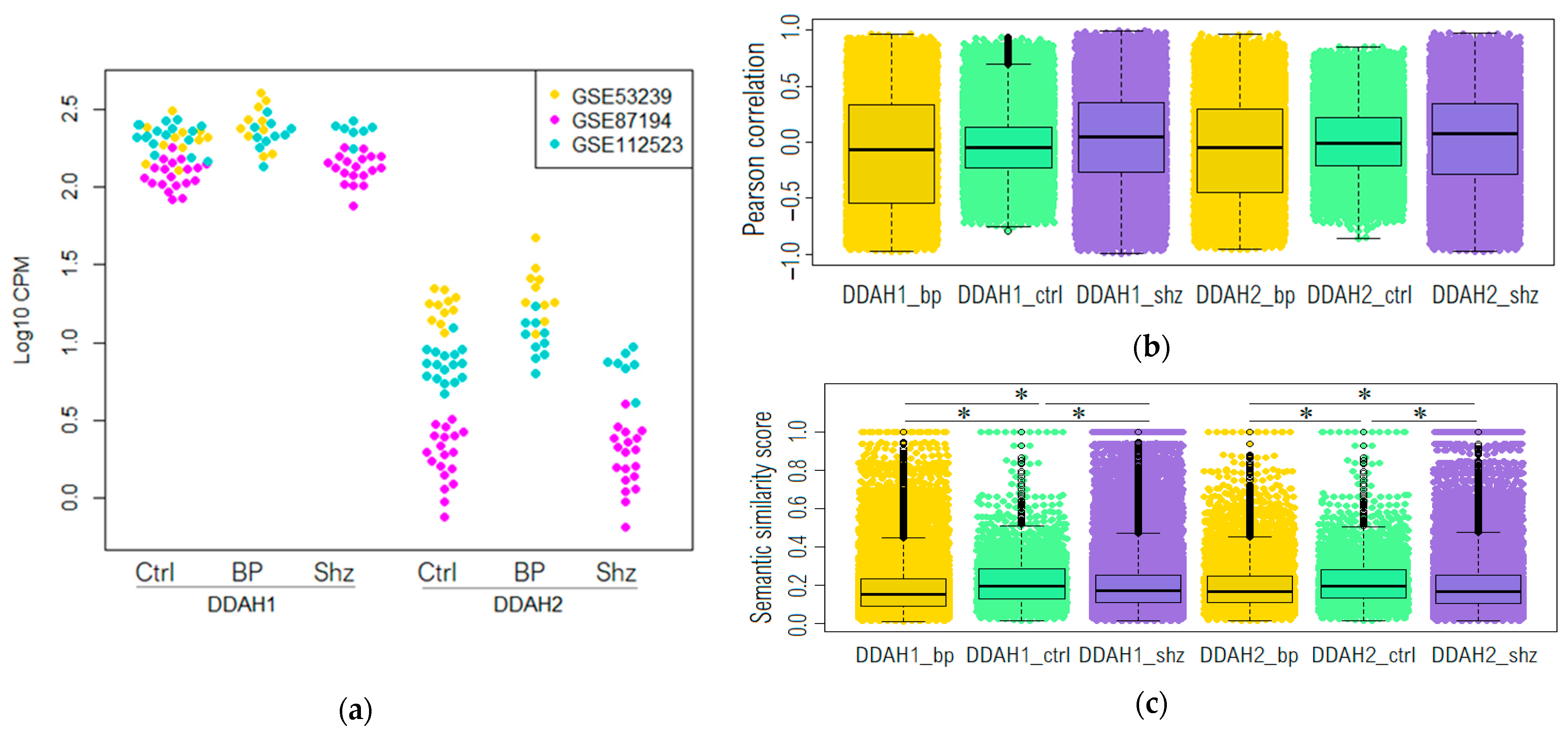
Figure 1. (a) Expression levels of DDAH1 and DDAH2 in the dorsolateral prefrontal cortex in non-psychiatric controls (Ctrl) and patients with bipolar affective disorder (BP) or schizophrenia (Shz) presented in three datasets: GSE53239 (yellow), GSE87194 (violet) and GSE112523 (blue). Beeswarm plot representing the distribution of correlation coefficients between DAAH1 or DAAH2 to all other genes expression levels in Brodmann area 46 in all groups (b) and the distribution of semantic similarity scores between the genes co-expressed (r > 0.8, p > 0.05) with DDAH1 or DDAH2 in all groups (c). * Games–Howell post hoc test p > 0.0001.
Ijms 23 11902 g001
Figure 2. Expression of DDAH1 (a) and DDAH2 (b) interacting genes in the study groups. Venn diagram illustrating an overlay of genes co-expressed (r > 0.3, p > 0.05) with DDAH1 or DDAH2 in non-psychiatric controls (Ctrl), patients with bipolar disorder (BP), and schizophrenia (Shz) and the sets of genes involved in the functional interaction with DDAH1 and DDAH2 according to the public resources’ data. Gene ontology (GO) enrichment analysis of genes involved in the functional interaction with DDAH1 and DDAH2 according to the public resources’ data, which are co-expressed with DDAH1, top five of enriched GO groups are represented in controls (a’), patients with schizophrenia (a”), and DDAH2 in the controls (b’), patients with schizophrenia (b”), and patients with bipolar disorder (b’”). GO biological process terms showed no appropriate terms enrichment in the DDAH1 co-expressed gene cluster in patients with bipolar disorder.
Ijms 23 11902 g002aIjms 23 11902 g002b
Figure 3. The genes involved in the developmental disorder of mental health co-expressed with DDAH1 and DDAH2 in non-psychiatric subjects. The enrichment plot for the representation of “DOID:0060037: developmental disorder of mental health” Disease Ontology (DO) term in the set of genes ranked based on their co-expression with DDAH1 in non-psychiatric subjects (a) and top 10 of biological processes in which enrichment core genes are implicated (a’). The enrichment plot for the representation of “DOID:0060037: developmental disorder of mental health” DO term (b) and “DOID:0060037: developmental disorder of mental health” DO term (b”). “DOID:0060041: autism spectrum disorder”/“DOID:12849: autistic disorder” in genes ranked based on their co-expression with DDAH2 in non-psychiatric subjects; (b’,b’”) top 10 of biological processes in which enrichment core genes for “DOID:0060037: developmental disorder of mental health” and “DOID:0060041:autism spectrum disorder”/“DOID:12849: autistic disorder” are implicated respectively.
Ijms 23 11902 g003aIjms 23 11902 g003b
Table 1. Human transcriptomic GEO datasets meeting the inclusion criteria and included in the analysis.
Accession NumberTitleDiagnosisNon-Psychiatric Controls (N)Patients (N)
GSE53239RNA-sequencing of the brain transcriptome implicates dysregulation of neuroplasticity, circadian rhythms, and GTPase binding in bipolar disorderBipolar affective disorder1110
GSE87194Schizophrenia: post-mortem dorsolateral prefrontal cortexSchizophrenia1919
GSE112523DNA methylation in neurons from post-mortem brains in schizophrenia and bipolar disorderBipolar affective disorder1710
Schizophrenia7
Table 2. GSE112523 study groups’ demographic and clinical characteristics, data from [41].
CharacteristicsBipolar DisorderSchizophreniaNon-Psychiatric Controls
n = 10n = 7n = 17
GenderMale7612
Female315
AgeMedian47.745.145.8
Range29–7729–5531–68
Smoker statusYes825
No119
Previous001
Unknown142
Antipsychotic therapyYes604
No4173
Mood stabilizer therapyYes600
No4717
Table 3. Enriched promoter motifs in genes co-expressed with DDAH1 or DDAH2 in the dorsolateral prefrontal cortex of non-psychiatric control subjects, patients with bipolar disorder or schizophrenia.
Co-Expressed GenesDDAH1DDAH2
Number of MotivesProtein Families Which Bind the Enriched MotivesNumber of MotivesProtein Families Which Bind the Enriched Motives
Non-psychiatric controls1CxxC1C2H2 ZF
Bipolar affective disorderNo enrichment29AP-2, bHLH, C2H2 ZF, CxxC, E2F, GCM, Nuclear receptor, Paired box
SchizophreniaNo enrichmentNo enrichment
Publisher’s Note: MDPI stays neutral with regard to jurisdictional claims in published maps and institutional affiliations.