Article

Acidovorax citrulli Effector AopV Suppresses Plant Immunity and Interacts with Aromatic Dehydratase ADT6 in Watermelon

1
State Key Laboratory for Biology of Plant Diseases and Insect Pests, Institute of Plant Protection, Chinese Academy of Agricultural Sciences, Beijing 100193, China
2
Department of Plant Pathology, College of Plant Protection, China Agricultural University, Beijing 100193, China
3
Department of Plant Pathology, University of Georgia, Athens, GA 30602, USA
*
Authors to whom correspondence should be addressed.
These authors contributed equally to this work.
Int. J. Mol. Sci. 2022, 23(19), 11719; https://doi.org/10.3390/ijms231911719
Submission received: 5 September 2022 / Revised: 25 September 2022 / Accepted: 26 September 2022 / Published: 3 October 2022
(This article belongs to the Collection Feature Papers in Molecular Plant Sciences)

Abstract

:
Bacterial fruit blotch (BFB) is a disease of cucurbit plants caused by Acidovorax citrulli. Although A. citrulli has great destructive potential, the molecular mechanisms of pathogenicity of A. citrulli are not clear, particularly with regard to its type III secreted effectors. In this study, we characterized the type III secreted effector protein, AopV, from A. citrulli strain Aac5. We show that AopV significantly inhibits reactive oxygen species and the expression of PTI marker genes, and helps the growth of Pseudomonas syringae D36E in Nicotiana benthamiana. In addition, we found that the aromatic dehydratase ADT6 from watermelon was a target of AopV. AopV interacts with ADT6 in vivo and in vitro. Subcellular localization indicated ADT6 and AopV were co-located at the cell membrane. Together, our results reveal that AopV suppresses plant immunity and targets ADT6 in the cell membrane. These findings provide an new characterization of the molecular interaction of A. citrulli effector protein AopV with host cells.

1. Introduction

Plants have an innate immune system to defend themselves against invasion and colonization by plant pathogens [1]. On the plant cell surface, receptor kinases and receptor-like proteins act as pattern recognition receptors (PRRs) to sense foreign microbial molecules, called pathogen-associated molecular patterns (PAMPs) or microbial-associated molecular patterns (MAMPs). When pathogens infect plants, they may trigger two layers of defense responses. The first layer is the PAMPs/MAMPs-triggered immunity (PTI) [2,3]. The PTI responses are mainly manifested as the expression of defense-related genes, reactive oxygen species (ROS) burst, callose deposition, etc. The second layer is the effector-triggered immunity (ETI). The ETI includes accelerated and amplified PTI responses, which are related to hypersensitive response (HR) and systemic acquired resistance (SAR) [1].
Many plant pathogenic bacteria use the type III secretion system (T3SS) to secrete effector proteins into plant cells; these effectors help bacterial cells proliferate in the host [4]. Type III secreted effectors (T3Es) suppress plant immune responses using various strategies, which include interference with signal transduction of immune pathways, blocking RNA pathways and vesicle transport, changing organelle functions, and inhibition of plant PTI responses [5,6]. For example, Pseudomonas syringae T3E HopAI1 inhibits Arabidopsis thaliana mitogen-activated protein kinases (MAPKs) activated by exposure to PAMPs [7]. The Xanthomonas oryzae T3E XopL activates ROS accumulation in the non-host plant Nicotiana benthamiana by mediating the degradation of ferredoxin [8]. Effectors AvrPto and AvrPtoB, from P. syringae, target BIK1 in A. thaliana to prevent its signal transduction [9]. However, T3Es also trigger the ETI responses during incompatible plant-pathogen interactions. For example, XopQ from Xanthomonas oryzae pv. oryzae triggers ETI in the non-host plant N. benthamiana [10] and interacts with the resistance protein Roq1 in N. benthamiana, resulting in an HR [11]. The immune responses during PTI and ETI are interconnected, but different [12]. ETI and PTI are not two distinct lines of defense [13]. The occurrence of the ETI responses in A. thaliana also depends on various elements in the PTI pathways [14].
After the pathogen attack, salicylic acid (SA), jasmonic acid (JA), ethylene, and other plant hormones play an important role in the activation of plant defense responses [15,16]. SA mainly plays a role in the expression of defense-related genes and SAR [17]. For example, transgenic tobacco and A. thaliana that express bacterial salicylate hydroxylase cannot accumulate SA. This not only prevents plants from inducing SAR, but also increases their susceptibility to pathogens, such as viruses, fungi, and bacteria [18,19]. A recent study showed that A. citrulli T3E AopP targets the watermelon transcription factor ClWRKY6 and suppresses SA signaling [20]. With regards to JA, it mainly protects the host from necrotrophic pathogens by inducing systemic resistance [21]. Therefore, JA synthesis-related genes are also targets of pathogenic microorganisms. For example, the P. syringae T3E HopZ1a interacts with the transcription repressor ZIM domain protein to activate JA signaling [22]. Pseudomonas syringae T3E HopBB1 interacts with A. thaliana transcription factor TCP14 to de-repress JA response genes [23]. However, no studies have reported A. citrulli T3Es affecting JA.
Bacterial fruit blotch (BFB) of cucurbits has occurred worldwide, possibly via seed transmission [24]. Acidovorax citrulli, the causal agent, can infect a variety of Cucurbitaceae crops, especially watermelon and cantaloupe, and cause severe economic losses. Despite this, the molecular interactions between A. citrulli and host plants are not well-defined, and effective BFB management methods, including commercial sources of resistance, are lacking. At present, the research on A. citrulli mainly focuses on type II, type III, and type VI secretion systems, among which the research on the T3SS has made progress [25,26,27,28]. The T3SS is a key pathogenicity factor of A. citrulli [26]. The T3SS apparatus gene hrpE encodes for the Hrp pilus and serves as a conduit to secrete effector proteins into host cells [29]. HrpG and hrpX are key regulatory genes of T3SS in A. citrulli [30]. In addition, N. benthamiana can be used to study A. citrulli T3Es [31]. Comparing the differences between the A. citrulli wild-type strain M6 and the hrpX mutant strain, some T3Es were successfully identified [32]. However, research on A. citrulli T3Es is still relatively limited. Detailed A. citrulli T3E characterization showed that AopP suppressed SA signaling and AopN caused programmed cell death in plants. Both AopP and AopN interfered with the plant immune responses [20,33]. However, other effector proteins interfering with plant immunity in A. citrulli have not been reported yet. Exactly, how A. citrulli T3Es interfere with plant immunity is still largely unknown.
In this study, we identified AopV as a T3E of A. citrulli, and found it was localized in the plant host cell nucleus and cell membrane. Importantly, the effector significantly inhibited the PTI responses in N. benthamiana. In addition, AopV activated the JA pathways. This study provides new insights into the pathogenicity mechanism of A. citrulli.

2. Results

2.1. Sequence Analysis of AopV in A. Citrulli Strain Aac5

The DNA sequence of aopV from Aac5 is 100% identical to the homologous genes from A. citrulli group I strain M6 (genome accession number CP029373.1) and AAC00-1 (genome accession number CP000512.1). The putative effector AopV in A. citrulli strain Aac5 encodes 342 amino acids with a predicted molecular size of 37 kDa. The homologous protein of AopV was found in Acidovorax, Xanthomonas, Ralstonia, and Pseudomonas genomes. The amino acid sequence of AopV shows 31% similarity to XopV from Xanthomonas oryzae pv. oryzicola (NCBI protein accession AEQ94858). AopV is most similar to XopV from Xanthomonas, compared with other homologous sequences from Ralstonia and Pseudomonas. The pairwise alignment of AopV and XopV is shown in Figure 1a. In addition, we found that representative AopV homologs from all four species contained nine consecutive identical amino acid sequences, which showed that these amino acid sequences are very conserved (Figure 1b).

2.2. ApoV Is a Type III Secreted Effector in A. citrulli

To determine if A. citrulli aopV was regulated by its T3SS, aopV gene expression levels were measured. The expression of aopV in the hrpX mutant [30] was significantly lower than that of the wild-type Aac5 (WT), which indicated the expression of aopV was regulated by the T3SS (Figure 2a). In addition, in order to test whether AopV was secreted during A. citrulli infection, secretion and translocation were tested with the adenylate cyclase (CyaA) translocation reporter assay. Eight hours after inoculation, the cAMP content in the host watermelon was significantly greater in the treatment inoculated with the strain WT-AopV-CyaA compared to the treatment inoculated with the strain ΔhrcJ-AopV-CyaA (Figure 2b). Together, these results showed that AopV is a T3E in A. citrulli strain Aac5.

2.3. AopV Is Expressed in the Nucleus and Cell Membrane of N. benthamiana

In order to determine the localization of effector protein AopV in plant cells, we transiently expressed AopV tagged with GFP (green fluorescent protein) in N. benthamiana, and observed it under laser confocal microscopy 48 h after transient expression. AopV showed green fluorescent signals in the cell nucleus and cell membrane (Figure 3). A cell nucleus marker carrying a red fluorescent protein (RFP) tag was used as the control for nucleus localization.

2.4. AopV Inhibits the ROS Burst and Expression Levels of PTI Marker Genes in N. Benthamiana

PTI is the plant’s first step in defense against pathogens, and an important PTI response is the ROS burst [1]. The transient expression of AopV significantly inhibited flg22-induced ROS compared to the empty vector (EV) control (Figure 4a). In addition, the effect of AopV on the expression of PTI marker genes was analyzed using qPCR. Compared with the EV control, AopV significantly inhibited the expression of PTI marker genes NbPti5 and NbAcre3 (Figure 4b), which suggested that the expression of AopV significantly inhibited the PTI pathway in N. benthamiana.

2.5. AopV Helps the Growth of D36E in N. benthamiana

Pseudomonas syringae pv. tomato (Pst) D36E colonized N. benthamiana expressing AopV at a significantly higher population level than N. benthamiana expressing EV (Figure 5). This indicated that AopV inhibited the PTI response induced by Pst D36E and enhanced Pst D36E’s colonization of N. benthamiana.

2.6. AopV Stimulates JA Production

JA is a main plant hormone involved in disease resistance. Some virulent pathogens have evolved various strategies to manipulate JA signaling to facilitate their exploitation of plant hosts [34,35]. To determine if AopV affects JA production, AopV and EV (control) were transiently expressed in N. benthamiana. After 48 h, the treated leaves were collected and JA production was measured. JA production in N. benthamiana was significantly increased in N. benthamiana expressing AopV (Figure 6).

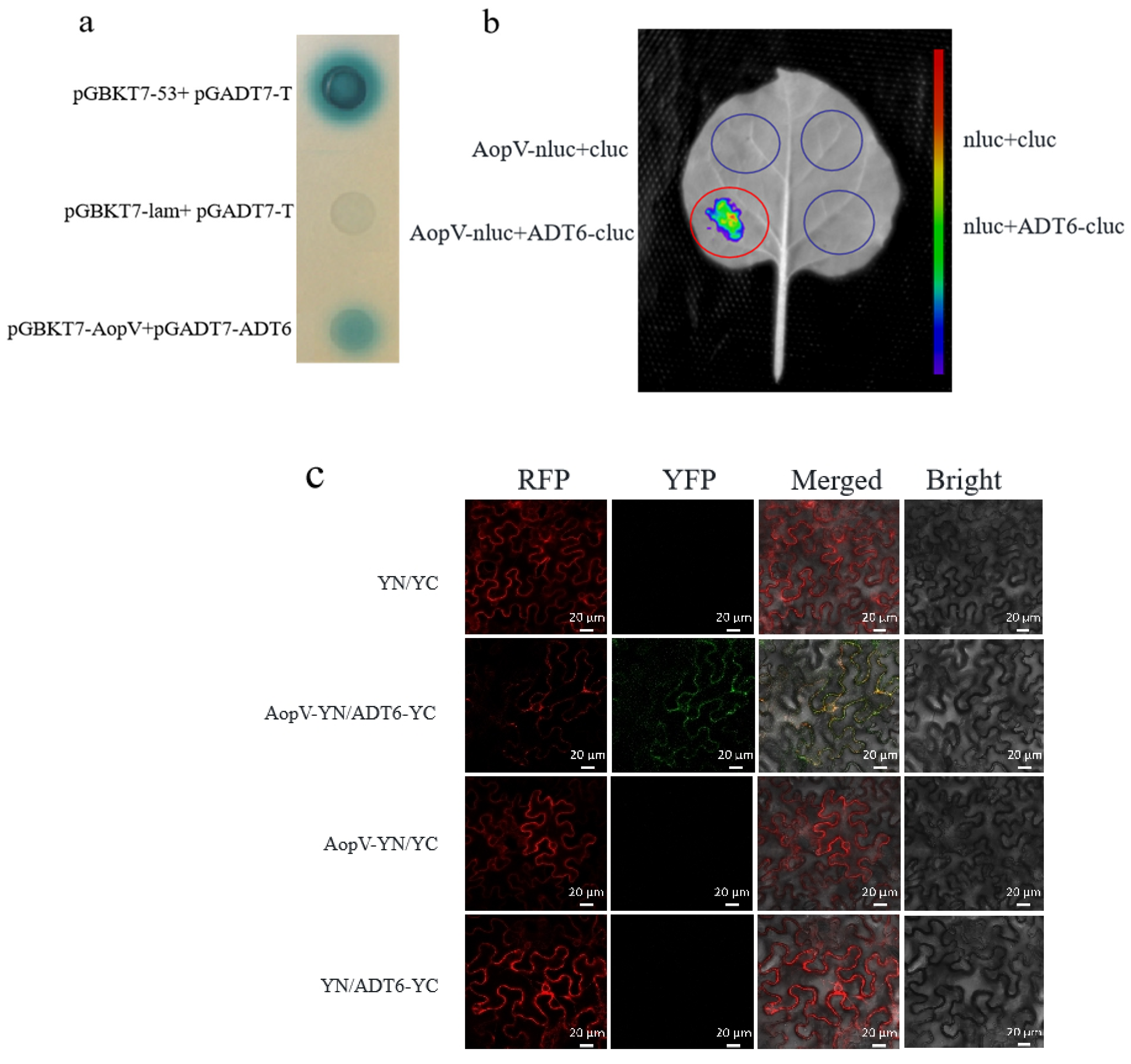
2.7. AopV Interacted with ADT6

To further analyze the AopV interaction protein in the host, we screened an A. citrulli-induced yeast library of watermelon cDNA. The results showed an interaction between the aromatic dehydratase ADT6 from watermelon and the T3E AopV from A. citrulli. Aromatic dehydratase is involved in the biosynthesis of phenylalanine in A. thaliana [36]. The yeast two hybrid (Y2H) assay was conducted to verify this interaction. The Y2H assay showed that the test colony (pGBKT7-AopV and pGADT7-ADT6) and the positive control turned blue, but the negative control did not turn blue. We confirmed a direct interaction between AopV and ADT6 using this system (Figure 7a).
AopV-nluc and ADT6-cluc recombinant proteins were simultaneously expressed in N. benthamiana. Luciferase complementation imaging (LCI) analysis showed that the tissues expressing AopV and ADT6 produced fluorescence (Figure 7b). The test strains interacted to produce luciferase, which acted on the luciferin substrate to produce fluorescence. The negative control group did not produce fluorescence (Figure 7b). This further demonstrated the interaction between AopV and ADT6.
BiFC was conducted to provide more evidence for the interaction between AopV and ADT6. The pSPYNE®173-AopV and pSPYCE(M)-ADT6 recombinant proteins exhibited strong green fluorescence signals [37]. AopV and ADT6 were co-localized in the cell membrane of N. benthamiana, while the control group showed the red fluorescence marker, but no green fluorescence signal (Figure 7c). This indicated that there was a direct interaction between the effector protein AopV and ADT6.

3. Discussion

BFB is a devastating disease caused by A. citrulli, severely impacting the cucurbit industry worldwide. However, little is known about the molecular interaction between A. citrulli and host plants. Acidovorax citrulli secretes T3Es as a major pathogenicity strategy [28], however, only a few effectors have been studied in A. citrulli thus far. In 2020, Jimenez-Guerrero successfully identified some A. citrulli T3E proteins by machine learning and by comparing the transcriptomic differences between the A. citrulli wild-type strain M6 and the hrpX mutant strain [32]. In addition, two effectors AopP and AopN were identified and characterized in A. citrulli [20,33]. In this study, we found a putative T3E AopV in A. citrulli group II strain Aac5. The gene sequence from strain Aac5 is identical to that of Aave_3085 in AAC00-1. BLAST analysis showed that AopV has homology with the Xanthomonas oryzae pv. oryzicola effector XopV, with 31% similarity (Figure 1a). The homologous protein of AopV was found in Acidovorax, Xanthomonas, Ralstonia, and Pseudomonas genomes. We found that nine amino acids in the homologous proteins of AopV in these strains are very conserved, and we speculate that these nine amino acids NDSGRFSEY from the AopV effector protein family may be a core motif performing important functions (Figure 1b). The importance and functions of these amino acids should be investigated in the future.
Screening key effector proteins is important. Our study showed that the aopV expression was down-regulated in A. citrulli hrpX mutant strain compared to the wild-type strain Aac5 (Figure 2a), indicating that the expression of aopV was regulated by T3SS. The regulatory gene hrpX is a key T3SS regulatory gene, and has been used for T3E screening in A. citrulli [30]. Additionally, the exocrine function is important for T3E identification. We used the CyaA translocation reporter assay to confirm that AopV in strain Aac5 had an exocrine function and was regulated by the T3SS (Figure 2b), which indicated that AopV was a T3E.
ROS plays a key role in both PTI and ETI responses. Transient ROS bursts can be part of early signaling, or ultimately leads to hypersensitivity, a type of programmed cell death that limits the systemic spread of pathogens [38]. We showed that AopV inhibited ROS production induced by flg22 in N. benthamiana (Figure 4a). Previous reports showed that T3Es AopP and AopN in A. citrulli also significantly inhibited ROS production [20,33]. ROS is an important signal molecule, and the burst of ROS is one of the early responses of PTI [39]. Another piece of evidence supporting AopV inhibiting PTI responses was provided by its ability to promote D36E colonization in N. benthamiana (Figure 5). D36E is a derivative strain of Pst DC3000 with all 36 T3Es deleted, but with intact T3SS and bacterial flagella that can stimulate PTI [40]. Strain D36E induced PTI responses in N. benthamiana, which were counter-inhibited by AopV. These results were consistent with previous findings of the effector XopV from Xanthomonas oryzae pv. oryzicola, which can inhibit the immune responses of plants [41]. In addition, AopV was predicted to have a nuclear localization signal at positions 206–234 (http://www.moseslab.csb.utoronto.ca/NLStradamus/, accessed on 25 September 2022), and our experiments demonstrated its localization in the nucleus. As opposed to AopV, although the homologous protein RipAD in R. solanacearum also has a nuclear localization signal, it is only detected in the cytoplasm and chloroplast [42].
Additionally, plant defense hormones are important immune responses in plants. ELISA and high-performance liquid chromatography (HPLC) can be used to detect phytohormones [20]. ELISA was used to detect JA in this study. Our study showed that after transiently expressing AopV in N. benthamiana, AopV significantly stimulated JA production (Figure 6). Our study is the first report on the effect of A. citrulli T3Es on JA. However, how AopV affects the JA pathway needs to be further explored.
Gram-negative plant pathogenic bacteria deliver effector proteins into plant cells through the T3SS to manipulate the host cell environment [43]. However, in many cases, the targets of T3Es are unknown, including XopV from X. oryzae pv. oryzicola [44] and RipAD from R. solanacearum [42]. In our study, we found that AopV inhibited plant immune responses in N. benthamiana. To further study the interaction of A. citrulli with its natural host, watermelon, AopV was used as a bait to screen a yeast library of watermelon cDNA induced by A. citrulli, and a candidate target protein, ADT6, was identified. The interaction between AopV and ADT6 was confirmed by Y2H in vitro (Figure 7a) and bimolecular fluorescence complementation in vivo (Figure 7c).
ADT6 is an aromatic dehydratase with 35% homology with ADT6 (NC_003070.9) in A. thaliana. In A. thaliana, ADT6 is involved in the synthesis of phenylalanine and participates in photosynthesis [36]. However, the role of ADT6 in watermelon is unknown. The interaction between AopV and ADT6 is different from the previously reported interaction between AopP and CLWRKY6 [20]. The interaction between AopV and ADT6 was in the cell membrane of N. benthamiana (Figure 7c), which indicated that ADT6 likely participated in different pathways than CLWRKY6, which occurred in the nucleus. The interaction between AopV and ADT6 was verified by LCI (Figure 7b). The experiment further confirmed that AopV from A. citrulli and ADT6 from watermelon directly interact. Through three verification methods, we determined that ADT6 from watermelon was an interaction target of A. citrulli T3E AopV. We speculated that ADT6 might be involved in the immune responses of watermelon. The specific immune pathways that ADT6 participates in are currently unknown and should be explored.
In summary, we found that AopV, a T3E protein of A. citrulli, was located in the cell membrane and nucleus of cells of N. benthamiana, and inhibited the PTI pathway by inhibiting the production of ROS in N. benthamiana. In addition, AopV affected the host immune responses by stimulating JA. Moreover, AopV interacted with ADT6 from watermelon. Our findings provide important insights into the immune responses of watermelon when interacting with A. citrulli.

4. Materials and Methods

4.1. Bacterial Strains, Plasmids, and Plant Materials

The strains and plasmids used in this study are listed in Table S1. Watermelon (cv. ‘Ruixin’) and N. benthamiana seedlings were cultivated at 24 °C and 30~50% relative humidity. Acidovorax citrulli strains were cultured overnight at 28 °C in King’s B (KB) or T3SS induction medium [45] (Bacto peptone: 10 g, NaCl: 5 g, Yeast extract: 5 g, MgCl2·6H2O: 10 mmol/L, water: 1 L, pH = 5.8). Escherichia coli DH5α was cultured in Luria-Bertani (LB) medium at 37 °C for about 7 h, and Agrobacterium tumefaciens GV3101 was cultured in LB medium at 28 °C overnight. Antibiotics were used at the following concentrations: ampicillin 100 μg/mL, kanamycin 100 μg/mL, rifampicin 100 μg/mL, and chloramphenicol 50 μg/mL.

4.2. AopV Sequence Analysis

The amino acid sequences of AopV from A. citrulli and other homologous effector proteins were searched in NCBI, and the amino acids were aligned using ClustalW by DNAMAN version 5.2.2 (Lynnon Biosoft, Quebec, QC, Canada).

4.3. CyaA Translocation Assay

The full-length cDNA of aopV (Aave_3085 from A. citrulli AAC00-1, GenBank accession number CP000512.1) was cloned into the pBBRNolac-CyaA vector using seamless ligase (Vazyme), and then the constructed vector was transformed into the A. citrulli wild-type strain Aac5 and its T3SS-deficient hrcJ mutant by tri-parental conjugation [30]. The CyaA translocation reporter assay was used for effector identification analysis. All primers used are listed in Table S2. Acidovorax citrulli strains were grown in KB medium to the logarithmic phase, resuspended in 10 mM MgCl2, and adjusted to 3 × 10⁸ CFU/mL. The leaves of 3-week-old watermelon seedlings were inoculated by syringe-infiltration. Two leaf discs (9 mm in diameter) from three leaves of each plant were taken 8 h after inoculation, and three different seedlings were sampled. An ELISA cAMP immunoassay kit (Enzo Life Sciences, Farmingdale, NY, USA) was used to detect cAMP levels.

4.4. Agrobacterium Infiltration

Agrobacterium tumefaciens GV3101 was infiltrated into leaves of 3- to 5-week-old N. benthamiana seedlings using a 1 mL syringe. The A. tumefaciens strains were cultured overnight at 28 °C in LB amended with kanamycin and rifampicin. The overnight cultures were centrifuged, resuspended in infiltration buffer (10 mM MES, 0.5 mM As, and 10 mM MgCl2), and adjusted to a final concentration equivalent to an optical density (OD600) = 0.5. After incubating in the dark for 1 h, the bacterial suspension was directly infiltrated into the N. benthamiana leaves.

4.5. ROS Burst Measurement

The full-length cDNA sequence of aopV was cloned and fused with pBI121-eGFP vector [46]. All primers used for PCR are listed in Table S2. Agrobacterium tumefaciens GV3101 was used to transiently express EV (pBI121-eGFP empty vector) and AopV at an optical density (OD600) of 0.5. A Flg22-induced ROS assay was conducted with N. benthamiana leaves 36 h after inoculation. Twelve leaf discs (4 mm in diameter) were collected from each inoculation area and floated on 100 μL sterilized distilled water in a 96-well plate. The water was then removed, and 100 μL of a solution containing 100 nM flg22, 20 μg/mL horseradish peroxidase, and 100 μM luminol was added to each well. The luminescence was recorded immediately for 1 h using Tecan Infinite F200 luminometer (Tecan, Männedorf, Switzerland) [47].

4.6. RNA Extraction

To determine whether aopV was regulated by hrpX, RNA extraction and gene expression analysis were performed. The A. citrulli wild-type strain Aac5 and the hrpX mutant strain were cultured to the logarithmic phase, and then the cells were collected by centrifugation, resuspended in the T3SS induction medium and incubated for 3 h with shaking. RNA was extracted and reverse transcribed into cDNA using HiScript II Q RT SuperMix for qPCR kit (TIANGEN). qPCR was conducted to detect the expression of aopV in different strains. rpoB was used as a reference gene. The experiment was conducted three times.
To analyze the effect of AopV on the expression of PTI marker genes, RNA extraction and gene expression analysis were performed. RNA was extracted from N. benthamiana leaves 24 h after Agrobacterium infiltration, and reverse transcribed into cDNA using a kit (Zymo Research, Irvine, CA, United States; cat. no. R2024). The expression levels of the PTI-related genes NbPti5 and NbAcre31 were measured, and the N. benthamiana gene EF1a was used as the internal reference gene. The primers used in this experiment are shown in Table S2. SYBR Green Real-Time PCR Master Mix (TOYOBO) was used for real-time quantitative PCR (qPCR). The experiment was conducted three times.

4.7. JA Detection

The full-length cDNA sequence of aopV was cloned into the PBI121-3FLAG vector, and the vector was transformed into A. tumefaciens GV3101. The A. tumefaciens GV3101 strains were infiltrated into three-week-old N. benthamiana leaves. A. tumefaciens GV3101 transformed with EV (pBI121-3FLAG) was used as a control. Four leaves were placed in a 15 mL centrifuge tube, and flash-frozen with liquid nitrogen 48 h after infiltration. JA content was measured using the ELISA method [48]. The experiment was conducted three times.

4.8. Subcellular Localization

The full-length cDNA sequence of aopV was cloned into the pBI121-eGFP vector. The A. tumefaciens GV3101 carrying AopV was expressed in three-week-old N. benthamiana seedlings. A. tumefaciens GV3101 carrying the EV was used as a control. RFP (red fluorescent protein) was used as a nuclear localization marker. The subcellular localization of the effector protein AopV was observed using a fluorescence confocal microscope (Zeiss LSM 880, Oberkochen, Germany) [49].

4.9. Impact of AopV on Pseudomonas Syringae pv. Tomato D36E Growth

The recombinant vector pBI121-AopV-3FLAG and EV were transiently expressed in N. benthamiana at a concentration of OD600 = 0.5. After four days, a bacterial suspension of Pseudomonas syringae pv. tomato (Pst) strain D36E (OD600 = 0.1) was infiltrated into N. benthamiana leaves. D36E is a derivative strain of Pst DC3000, in which all 36 T3Es were deleted, but the T3SS and the bacterial flagella that stimulate PTI were retained [40]. Six leaf discs were sampled (12 mm in diameter) 24 h after inoculation and plated on KB medium to measure D36E population levels in N. benthamiana leaves.

4.10. Yeast Two-Hybrid Assay

To analyze AopV and ADT6 interactions, a yeast two-hybrid assay was conducted. The full-length cDNA of AopV was cloned into the pGBKT7 vector, and the full-length cDNA of ADT6 (a putative ADT family protein encoded by CM018018.1 from the watermelon 97103 genome; The ADT6 coding sequence is gene ID Cla97C01G011870 according to 97103) was cloned into the pGADT7 vector. Subsequently, AopV and ADT6 were co-transformed into the Y2H Gold strain. A positive control (Y2H Gold strain co-transformed with pGBKT7-53 and pGADT7-T) and negative control (Y2H Gold strain co-transformed with pGBKT7-lam and pGADT7-T) were used. The Y2H Gold strain was transformed with the above vectors and cultured on SD/-Leu-Trp medium [50]. Then, the transformants were cultured in YPDA medium (Peptone: 0.2 g, Yeast extract: 10 g, Glucose: 2 g, 2% Adenine solution: 15 mL) with shaking for 48 h, they were transferred to SD/-Leu-Trp-His-Ade agar medium containing 20 mg/mL X-α-gal. The blue colonies were considered to be potential positive interactions.

4.11. Luciferase (LUC) Complementary Imaging (LCI) Assay

LCI analysis was conducted as previously described with slight modifications. ADT6 was inserted into the pCAMBIA-cLUC vector and transformed into A. tumefaciens GV3101 [51,52]. AopV was inserted into the pCAMBIA-nLUC vector and transformed into A. tumefaciens GV3101. The two constructed A. tumefaciens GV3101 strains were equally mixed at a concentration of OD600 = 1.0, and co-expressed in N. benthamiana. After 48 h, leaves were collected and observed using a charge-coupled device (CCD) imaging apparatus (NightSHADE LB985; Berthold). The experiment was conducted three times independently.

4.12. BiFC Assays

The full-length cDNA sequence of aopV was cloned and fused with the pSPYNE®173 vector, and the full-length cDNA sequence of cloned ADT6 was fused with the pSPYCE(M) vector [37]. The proteins tested for interaction were constructed into A. tumefaciens GV3101 strains. They (AopV-nYFP and ADT6-cYFP) were equally mixed at a concentration of OD600 = 1.0, and co-expressed in N. benthamiana. The A. tumefaciens strains carrying either single empty or double empty vectors were used as controls. After 48 h, the fluorescence signal was observed under the excitation wavelength of 488 nm using laser confocal fluorescence microscopy (Zeiss LSM 880, Oberkochen, Germany). The experiment was conducted three times independently.

4.13. Statistical Analysis

Data were analyzed by one-way analysis of variance and Tukey’s honest significant difference tests. For the data analysis, the internal reference gene expression was set to 1, and the 2−ΔΔCt method [53] was used. The independent-samples t-tests were used to analyze the significance of the differences between treatments. A p-value less than 0.05 was considered statistically significant. The statistical analysis was performed using GraphPad PRISM 5.0 software (GraphPad Software Inc., La Jolla, CA, USA).

5. Conclusions

By studying the function, expression, and secretion of AopV, we found AopV was a type III secreted effector protein that inhibited PTI responses. AopV was localized in the nucleus and cell membrane of N. benthamiana, and it inhibited ROS bursts in plants. More importantly, the effector protein AopV interacted with ADT6 from watermelon. The findings provide novel insights into how A. citrulli type III effector AopV interacts with its natural host watermelon, as well as with N. benthamian. How AopV interacts with ADT6 at the molecular level and its structure−function relationship are questions to be further investigated in the future.

Supplementary Materials

The following supporting information can be downloaded at: https://www.mdpi.com/article/10.3390/ijms231911719/s1, Table S1: Strains and plasmids used in the study. Table S2: Primers used in the study.

Author Contributions

T.Z., J.J., M.Z. and X.Z. designed the study. J.J., X.Z., L.Y., N.F. and W.J. performed the experiments. J.J., Y.Y. and W.G. performed data analyses. J.J. and M.Z. wrote the manuscript. T.Z. and R.W. critically reviewed the manuscript. All authors have read and agreed to the published version of the manuscript.

Funding

This study was supported by the China Earmarked Fund for Modern Agro-industry Technology Research System (CARS-25), the National Key Research and Development Program of China (2018YFD0201300), Central Public-Interest Scientific Institution Basal Research Fund (S2022XM25), the CAAS Science and Technology Innovation Program (CAAS-ASTIP), and the Xinjiang Production and Construction Corps’ Scientific and Technological Research Plan Project in Agricultural (2022AB015).

Institutional Review Board Statement

Not applicable.

Informed Consent Statement

Not applicable.

Data Availability Statement

Not applicable.

Conflicts of Interest

The authors declare no conflict of interest.

References

  1. Jones, J.; Dangl, J.L. The plant immune system. Nature 2006, 444, 323–329. [Google Scholar] [CrossRef] [PubMed] [Green Version]
  2. Zipfel, C. Plant pattern-recognition receptors. Trends Immunol. 2014, 35, 345–351. [Google Scholar] [CrossRef]
  3. Zipfel, C. Early molecular events in PAMP-triggered immunity. Curr. Opin. Plant Biol. 2009, 12, 414–420. [Google Scholar] [CrossRef]
  4. Lee, A.H.; Middleton, M.A.; Guttman, D.S.; Desveaux, D. Phytopathogen type III effectors as probes of biological systems. Microb. Biotechnol. 2013, 6, 230–240. [Google Scholar] [CrossRef] [PubMed]
  5. Chisholm, S.T.; Coaker, G.; Day, B.; Staskawicz, B.J. Host-Microbe interactions: Shaping the evolution of the plant immune response. Cell 2006, 124, 803–814. [Google Scholar] [CrossRef] [PubMed] [Green Version]
  6. Block, A.; Alfano, J.R. Plant targets for Pseudomonas syringae type III effectors: Virulence targets or guarded decoys? Curr. Opin. Microbiol. 2011, 14, 39–46. [Google Scholar] [CrossRef] [Green Version]
  7. Zhang, J.; Shao, F.; Li, Y.; Cui, H.T.; Chen, L.J.; Li, H.T.; Zou, Y.; Long, C.Z.; Lan, L.F.; Chai, J.J.; et al. A Pseudomonas syringae effector inactivates MAPKs to suppress PAMP-induced immunity in plants. Cell Host Microbe 2007, 1, 175–185. [Google Scholar] [CrossRef] [Green Version]
  8. Ma, W.; Xu, X.; Cai, L.; Cao, Y.; Chen, G.A. Xanthomonas oryzae type III effector XopL causes cell death through mediating ferredoxin degradation in N. benthamiana. Phytopathol. Res. 2020, 2, 1–12. [Google Scholar] [CrossRef]
  9. Shan, L.B.; He, P.; Li, J.M.; Heese, A.; Peck, S.C.; Nürnberger, T.; Martin, G.B.; Sheen, J. Bacterial effectors target the common signaling partner BAK1 to disrupt multiple MAMP receptor-signaling complexes and impede plant immunity. Cell Host Microbe 2008, 4, 17–27. [Google Scholar] [CrossRef] [Green Version]
  10. Gupta, M.K.; Nathawat, R.; Sinha, D.; Haque, A.S.; Sankaranarayanan, R.; Sont, R.V. Mutations in the Predicted Active Site of Xanthomonas oryzae pv. oryzae XopQ Differentially Affect Virulence, Suppression of Host Innate Immunity, and Induction of the HR in a Nonhost Plant. Mol. Plant-Microbe Interact. 2015, 28, 195. [Google Scholar] [CrossRef]
  11. Schultink, A.; Qi, T.; Lee, A.; Steinbrenner, A.D.; Staskawicz, B. Roq1 mediates recognition of the Xanthomonas and Pseudomonas effector proteins XopQ and HopQ1. Plant J. 2017, 92, 787–795. [Google Scholar] [CrossRef] [PubMed] [Green Version]
  12. Yuan, M.; Jiang, Z.; Bi, G.; Nomura, K.; Xin, X.F. Pattern-recognition receptors are required for NLR-mediated plant immunity. Nature 2021, 592, 105–109. [Google Scholar] [CrossRef] [PubMed]
  13. Thomma, B.P.H.J.; Nürnberger, T.; Joosten, M.H.A.J. Of PAMPs and effectors: The blurred PTI-ETI dichotomy. Plant Cell 2011, 23, 4–15. [Google Scholar] [CrossRef] [PubMed] [Green Version]
  14. Pieterse, C.M.; Leon-Reyes, A.; Van der Ent, S.; Van Wees, S. Networking by small-molecule hormones in plant immunity. Nat. Chem. Biol. 2009, 5, 308–316. [Google Scholar] [CrossRef] [Green Version]
  15. Klessig, D.F.; Durner, J.; Noad, R.; Navarre, D.A.; Wendehenne, D.; Kumar, D.; Zhou, J.M.; Shah, J.; Zhang, S.; Kachroo, P.; et al. Nitric oxide and salicylic acid signaling in plant defense. Proc. Natl. Acad. Sci. USA 2000, 97, 8849–8855. [Google Scholar] [CrossRef] [PubMed] [Green Version]
  16. Rao, M.V.; Lee, H.I.; Creelman, R.A.; Davis, M.K.R. Jasmonic acid signaling modulates ozone-induced hypersensitive cell death. Plant Cell 2000, 12, 1633–1646. [Google Scholar] [CrossRef] [PubMed] [Green Version]
  17. Wildermuth, M.C.; Dewdney, J.; Wu, G.; Ausubel, F.M. Isochorismate synthase is required to synthesize salicylic acid for plant defence. Nature 2001, 414, 562–565. [Google Scholar] [CrossRef]
  18. Vernooij, B.; Friedrich, L.; Weymann, K.; Negrotto, D.; Gaffney, T.; Gut-Rella, M.; Kessmann, H.; Ward, E.; Delaney, T.P.; Uknes, S. A central role of salicylic acid in plant disease resistance. Science 1994, 266, 1247–1250. [Google Scholar]
  19. Chaturvedi, R.; Shah, J. Salicylic Acid in Plant Disease Resistance. Springer: Heidelberg, The Netherlands, 2007. [Google Scholar]
  20. Zhang, X.; Yang, Y.; Zhao, M.; Yang, L.; Jiang, J.; Walcott, R.; Yang, S.; Zhao, T. Acidovorax citrulli type III effector AopP suppresses plant immunity by targeting the watermelon transcription factor WRKY6. Front. Plant Sci. 2020, 11, 579218. [Google Scholar] [CrossRef]
  21. Hu, Z.J.; Shao, S.J.; Zheng, C.F.; Sun, Z.H.; Shi, J.Y.; Yu, J.Q.; Qi, Z.Y.; Shi, K. Induction of systemic resistance in tomato against Botrytis cinerea by N-decanoyl-homoserine lactone via jasmonic acid signaling. Planta 2018, 247, 1217–1227. [Google Scholar] [CrossRef]
  22. Jiang, S.S.; Yao, J.; Ma, K.Y.; Zhou, H.B.; Song, J.K. Bacterial effector activates jasmonate signaling by directly targeting JAZ transcriptional repressors. PLoS Pathog. 2013, 9, e1003715. [Google Scholar] [CrossRef] [PubMed]
  23. Li, Y.; Teixeira, P.J.P.L.; Biswas, S.; Finkel, O.M.; He, Y.J.; Salas-González, I.; English, M.E.; Epple, P.; Mieczkowski, P.; Dangl, J. Pseudomonas syringae type III effector HopBB1 promotes host transcriptional repressor degradation to regulate phytohormone responses and virulence. Cell Host Microbe 2017, 21, 156–168. [Google Scholar] [CrossRef] [Green Version]
  24. Burdman, S.; Walcott, R. Acidovorax citrulli: Generating basic and applied knowledge to tackle a global threat to the cucurbit industry. Mol. Plant Pathol. 2012, 13, 805–815. [Google Scholar] [CrossRef] [PubMed]
  25. Rosenberg, T.; Jiménez-Guerrero, I.; Tamir-Ariel, D.; Yarnitzky, T.; Burdman, S. The GDSL-lipolytic enzyme Lip1 is required for full virulence of the cucurbit pathogenic bacterium Acidovorax citrulli. Microorganisms 2022, 5, 1016. [Google Scholar] [CrossRef] [PubMed]
  26. Johnson, K.L.; Minsavage, G.V.; Le, T.; Jones, J.B.; Walcott, R.R. Efficacy of a nonpathogenic Acidovorax citrulli strain as a biocontrol seed treatment for bacterial fruit blotch of cucurbits. Plant Dis. 2011, 95, 697–704. [Google Scholar] [CrossRef] [Green Version]
  27. Bahar, O.; Goffer, T.; Burdman, S. Type IV pili are required for virulence, twitching motility, and biofilm formation of Acidovorax avenae subsp. citrulli. Mol. Plant-Microbe Interact. 2009, 22, 909–920. [Google Scholar] [CrossRef] [Green Version]
  28. Fei, N.Y.; Ji, W.Q.; Yang, L.L.; Yu, C.Y.; Qiao, P.; Yan, J.P.; Guan, W.; Yang, Y.W.; Zhao, T.C. Hcp of the type VI secretion system (T6SS) in Acidovorax citrulli group II strain Aac5 has a dual role as a core structural protein and an effector protein in colonization, growth ability, competition, biofilm formation, and ferric iron absorption. Int. J. Mol. Sci. 2022, 23, 9632. [Google Scholar] [CrossRef]
  29. Ji, W.Q.; Zhao, M.; Fei, N.Y.; Yang, L.L.; Qiao, P.; Walcott, R.; Yang, Y.Y.; Zhao, T.C. Essential Acidovorax citrulli virulence gene hrpE activates host immune response against pathogen. Int. J. Mol. Sci. 2022, 23, 9144. [Google Scholar] [CrossRef]
  30. Zhang, X.; Zhao, M.; Yan, J.; Yang, L.; Yang, Y.; Guan, W.; Walcott, R.; Zhao, T. Involvement of hrpX and hrpG in the virulence of Acidovorax citrulli strain Aac5, Causal Agent of Bacterial Fruit Blotch in Cucurbits. Front. Microbiol. 2018, 9, 507. [Google Scholar] [CrossRef]
  31. Traore, S.M.; Eckshtain-Levi, N.; Miao, J.; Sparks, C.; Wang, Z.; Wang, K.; Li, Q.; Burdman, S.; Walcott, R.; Welbaum, G.E. Nicotiana species as surrogate host for studying the pathogenicity of Acidovorax citrulli, the causal agent of bacterial fruit blotch of cucurbits. Mol. Plant Pathol. 2019, 20, 800–814. [Google Scholar] [CrossRef] [Green Version]
  32. Jiménez-Guerrero, I.; Pérez-Montaño, F.; Da Silva, G.M.; Wagner, N.; Shkedy, D.; Zhao, M.; Pizarro, L.; Bar, M.; Walcott, R.; Sessa, G.; et al. Show me your secret(ed) weapons: A multifaceted approach reveals a wide arsenal of type III-secreted effectors in the cucurbit pathogenic bacterium Acidovorax citrulli and novel effectors in the Acidovorax genus. Mol. Plant Pathol. 2020, 21, 12877. [Google Scholar] [CrossRef] [PubMed]
  33. Zhang, X.; Zhao, M.; Jiang, J.; Yang, L.; Yang, Y.W.; Yang, S.S.; Walcott, R.; Qiu, D.W.; Zhao, T. Identification and functional analysis of AopN, an Acidovorax citrulli effector that induces programmed cell death in plants. Int. J. Mol. Sci. 2020, 21, 6050. [Google Scholar] [CrossRef] [PubMed]
  34. Turne, J.G.; Ellis, C.; Devoto, A. The jasmonate signal pathway. Plant Cell 2002, 14, S153. [Google Scholar] [CrossRef] [PubMed] [Green Version]
  35. Zhang, L.; Zhang, F.; Melotto, M.; Yao, J.; He, S.Y. Jasmonate signaling and manipulation by pathogens and insects. J. Exp. Bot. 2017, 68, 1371–1385. [Google Scholar] [CrossRef] [PubMed] [Green Version]
  36. Corea, O.R.A. Arabidopsis Arogenate Dehydratases: Influence on Aromatic Amino Acid and Downstream Phenylpropanoid Biosynthesis. Ph.D. Thesis, Gradworks. Washington State University, Pullman, WA, USA, 2011. Available online: http://hdl.handle.net/2376/3526 (accessed on 25 September 2022).
  37. Waadt, R.; Schmidt, L.K.; Lohse, M.; Hashimoto, K.; Bock, R.; Kudla, J. Multicolor bimolecular fluorescence complementation reveals simultaneous formation of alternative CBL/CIPK complexes in planta. Plant J. 2008, 56, 505–516. [Google Scholar] [CrossRef]
  38. Jwa, N.S.; Hwang, B.K. Convergent evolution of pathogen effectors toward reactive oxygen species signaling networks in plants. Front. Plant Sci. 2017, 9, 1687. [Google Scholar] [CrossRef] [Green Version]
  39. Wojtaszek, P. Oxidative burst: An early plant response to pathogen infection. Biochem. J. 1997, 322, 681–692. [Google Scholar] [CrossRef] [Green Version]
  40. Wei, H.L.; Zhang, W.; Collmer, A. Modular study of the type III effector repertoire in Pseudomonas syringae pv. tomato DC3000 reveals a matrix of effector interplay in pathogenesis. Cell Rep. 2018, 23, 1630–1638. [Google Scholar] [CrossRef] [Green Version]
  41. Deb, S.; Ghosh, P.; Patel, H.K.; Sonti, R.V. Interaction of the Xanthomonas effectors XopQ and XopX results in induction of rice immune responses. Plant J. 2020, 104, 332–350. [Google Scholar] [CrossRef]
  42. Jeon, H.; Kim, W.; Kim, B.; Lee, S.; Segonzac, C. Ralstonia solanacearum type III effectors with predicted nuclear localization signal localize to various cell compartments and modulate immune responses in Nicotiana spp. Plant Pathol. J. 2020, 36, 43–53. [Google Scholar] [CrossRef]
  43. Martinez-Argudo, I.; Blocker, A.J. The Shigella T3SS needle transmits a signal for MxiC release, which controls secretion of effectors. Mol. Microbiol. 2010, 78, 1365–1378. [Google Scholar] [CrossRef] [PubMed]
  44. Long, J.; Song, C.; Fang, Y.; Zhou, J.; Bing, Y. Non-TAL effectors from Xanthomonas oryzae pv. oryzae suppress peptidoglycan-triggered MAPK activation in rice. Front. Plant Sci. 2018, 9, 1857. [Google Scholar] [CrossRef] [PubMed] [Green Version]
  45. Ma, Y.N.; Chen, L.; Si, N.G.; Jiang, W.J.; Zhou, Z.G.; Liu, J.L.; Zhang, L.Q. Identification of benzyloxy carbonimidoyl dicyanide derivatives as novel type III secretion system inhibitors via high-throughput screening. Front. Plant Sci. 2019, 10, 1059. [Google Scholar] [CrossRef] [PubMed] [Green Version]
  46. Chen, P.Y.; Wang, C.K.; Soong, S.C.; To, K.Y. Complete sequence of the binary vector pBI121 and its application in cloning T-DNA insertion from transgenic plants. Mol. Breed. 2003, 11, 287–293. [Google Scholar] [CrossRef]
  47. Priller, J.P.; Reid, S.; Konein, P.; Dietrich, P.; Sonnewald, S. The Xanthomonas campestris pv. vesicatoria type-3 effector XopB inhibits plant defence responses by interfering with ROS production. PLoS ONE 2016, 11, e0159107. [Google Scholar] [CrossRef] [Green Version]
  48. Wang, S.; Xu, L.; Li, G.; Chen, P.; Kai, X.; Xie, Z. An ELISA for the determination of salicylic acid in plants using a monoclonal antibody. Plant Sci. 2002, 162, 529–535. [Google Scholar] [CrossRef]
  49. Nelson, B.K.; Cai, X.; Andreas, N. A multicolored set of in vivo organelle markers for co-localization studies in Arabidopsis and other plants. Plant J. 2010, 51, 1126–1136. [Google Scholar] [CrossRef]
  50. Yang, F.; Tian, F.; Li, X.; Fan, S.; Chen, H.; Wu, M.; Yang, C.H.; He, C. The degenerate EAL-GGDEF domain protein filp functions as a cyclic di-GMP receptor and specifically interacts with the PilZ-domain protein PXO_02715 to regulate virulence in Xanthomonas oryzae pv. oryzae. Mol. Plant-Microbe Interact. 2014, 27, 578. [Google Scholar] [CrossRef] [Green Version]
  51. Chen, H.M.; Zou, Y.; Shang, Y.L.; Lin, H.Q.; Wang, Y.J.; Cai, R.; Tang, X.Y.; Zhou, J.M. Firefly luciferase complementation imaging assay for protein-protein interactions in plants. Plant Physiol. 2008, 146, 368–376. [Google Scholar] [CrossRef] [Green Version]
  52. Yang, S.; Pan, L.; Chen, Y.; Yang, D.; Liu, Q.; Jian, H. Heterodera avenae GLAND5 effector interacts with pyruvate dehydrogenase subunit of plant to promote nematode parasitism. Front. Plant Sci. 2019, 10, 1241. [Google Scholar] [CrossRef]
  53. Livak, K.J.; Schmittgen, T.D. Analysis of relative gene expression data using real-time quantitative PCR and the 2(-Delta Delta C(T)) Method. Methods 2001, 25, 402–408. [Google Scholar] [CrossRef] [PubMed]
Figure 1. Amino acid sequence homology analysis. (a) Alignment of Acidovorax citrulli AopV and Xanthomonas oryzae pv. oryzicola XopV (NCBI protein accession AEQ94858). Acidovorax citrulli AopV shares 31% similarity with the sequence of the effector protein XopV of Xanthomonas oryzae pv. oryzicola. Sequences in red represent similar amino acids and gray denotes gaps. (b) Alignment of A. citrulli AopV and orthologs from Xanthomonas oryzae pv. oryzicola (AEQ94858), Ralstonia solanacearum (WP_003263919.1), and Pseudomonas avellanae (OZI86990.1). Amino acids highlighted in dark blue, cyan, and pink denote amino acids identical in four, three, and two species, respectively. Conserved amino acids among all four species are shown at the bottom in lowercase. The red box marks the nine consecutive identical amino acids.
Ijms 23 11719 g001
Figure 2. Identification of AopV as a type III secreted effector. (a) Quantitative real-time PCR was performed to determine whether aopV was regulated by hrpX. RpoB was used as an internal reference gene [30]. The experiment was conducted three times with similar results. The data represent the mean ± SD (standard deviation, n = 3). The ** above the bar indicates a significant difference determined by the t-test, p < 0.01; (b) Detection of cAMP content in watermelon host. Acidovorax citrulli wild-type strain Aac5 and ΔhrcJ carrying AopV-CyaA were inoculated into watermelon leaves and the cAMP contents were measured 8 h after inoculation. The experiment was conducted three times with similar results. The data represent the mean ± SD (n = 3). The ** above the bar indicates a significant difference determined by the t-test, p < 0.01.
Ijms 23 11719 g002
Figure 3. Subcellular localization of AopV in Nicotiana benthamiana. Leaves were injected with Agrobacterium tumefaciens GV3101 strain containing 35S::AopV-GFP (AopV) and observed at room temperature 48 h after infiltration using a confocal microscope (20×). Leaves injected with A. tumefaciens GV3101 containing 35S::EV-GFP were used as a control (EV). Red fluorescent protein (RFP) was used as a nuclear localization marker. The experiment was conducted three times with similar results. Scale bar, 20 µm.
Ijms 23 11719 g003
Figure 4. AopV inhibited PTI responses. (a) AopV inhibited flg22-induced ROS burst in Nicotiana benthamiana. Compared with Agrobacterium tumefaciens carrying an empty vector (EV), AopV-eGFP reduced ROS production in N. benthamiana induced by flg22. The experiment was carried out three times, and similar results were obtained each time. The data represent the mean ± SD (n = 10). (b) Expression of PTI related genes. Compared with A. tumefaciens carrying EV, AopV significantly inhibited the expression of PTI related genes NbPti5 and NbAcre3. EF1α was used as an internal reference gene. The experiment was carried out three times each with similar results. The data represent the mean ± SD (n = 3). The * above bars indicate significant differences as determined by the t-test, p < 0.05. The ** means p < 0.01.
Ijms 23 11719 g004
Figure 5. AopV helps the growth of Pseudomonas syringae pv. tomato (Pst) strain D36E in Nicotiana benthamiana. AopV was transiently expressed in N. benthamiana, and 4 days later, the bacterial suspension of Pst strain D36E was syringe-infiltrated. The empty vector (EV) was used as a control. After 24 h, the number of D36E colonies was counted. The experiment was carried out three times, and similar results were obtained each time. The data represent the mean ± SD (n = 3). * indicates statistically significant differences, as determined by the Student’s t-test, p < 0.05.
Ijms 23 11719 g005
Figure 6. The effects of AopV on JA production in Nicotiana benthamiana. ELISA detection of JA content in plants. Nicotiana benthamiana leaves were injected with the Agrobacterium tumefaciens strain expressing AopV or empty vector (EV) at OD600 = 0.5. After 48 h, the leaves were collected and JA was quantified by ELISA. The experiment was carried out three times, and similar results were obtained each time. The data represent the mean ± SD (n = 3). ** indicates statistically significant differences, as determined by Student’s t-test, p < 0.01.
Ijms 23 11719 g006
Figure 7. Interaction of Acidovorax citrulli effector AopV and watermelon aromatic dehydratase ADT6. (a) Yeast two-hybrid assay showed that AopV and ADT6 turned blue on SD/-Leu-Trp-His-Ade agar medium with 20 mg/mL X-α-gal. pGBKT7-53 and pGADT7-T are positive controls. pGBKT7-lam and pGADT7-T are negative controls. (b) Luc system verified protein interaction. Agrobacterium tumefaciens GV3101 carrying the AopV and ADT6 recombinant vector were expressed in Nicotiana benthamiana. After 48 h, the leaves were observed using a CCD imaging apparatus (NightSHADE LB985; Berthold). The experimental group produced fluorescence, and the control group showed no fluorescence. (c) Bimolecular Fluorescence Complementation (BiFC) verified the interaction between AopV and ADT6. AopV was fused with the nYFP tag, and ADT6 was fused with the cYFP tag. The subcellular localization of AopV and ADT6 in N. benthamiana leaves at 48 h after infiltration was observed under confocal microscopy. YN denotes pSPYNE®173 vector and YC denotes pSPYCE(M) vector. Red fluorescent protein was used as a cell membrane localization marker. Bars represent 20 µm. Each experiment was conducted three times, and similar results were obtained each time.
Ijms 23 11719 g007
Publisher’s Note: MDPI stays neutral with regard to jurisdictional claims in published maps and institutional affiliations.

