Chemonastic Stalked Glands in the Carnivorous Rainbow Plant Byblis gigantea LINDL. (Byblidaceae, Lamiales)
Abstract
1. Introduction
2. Results
2.1. Lengths and Densities of Stalked Glands
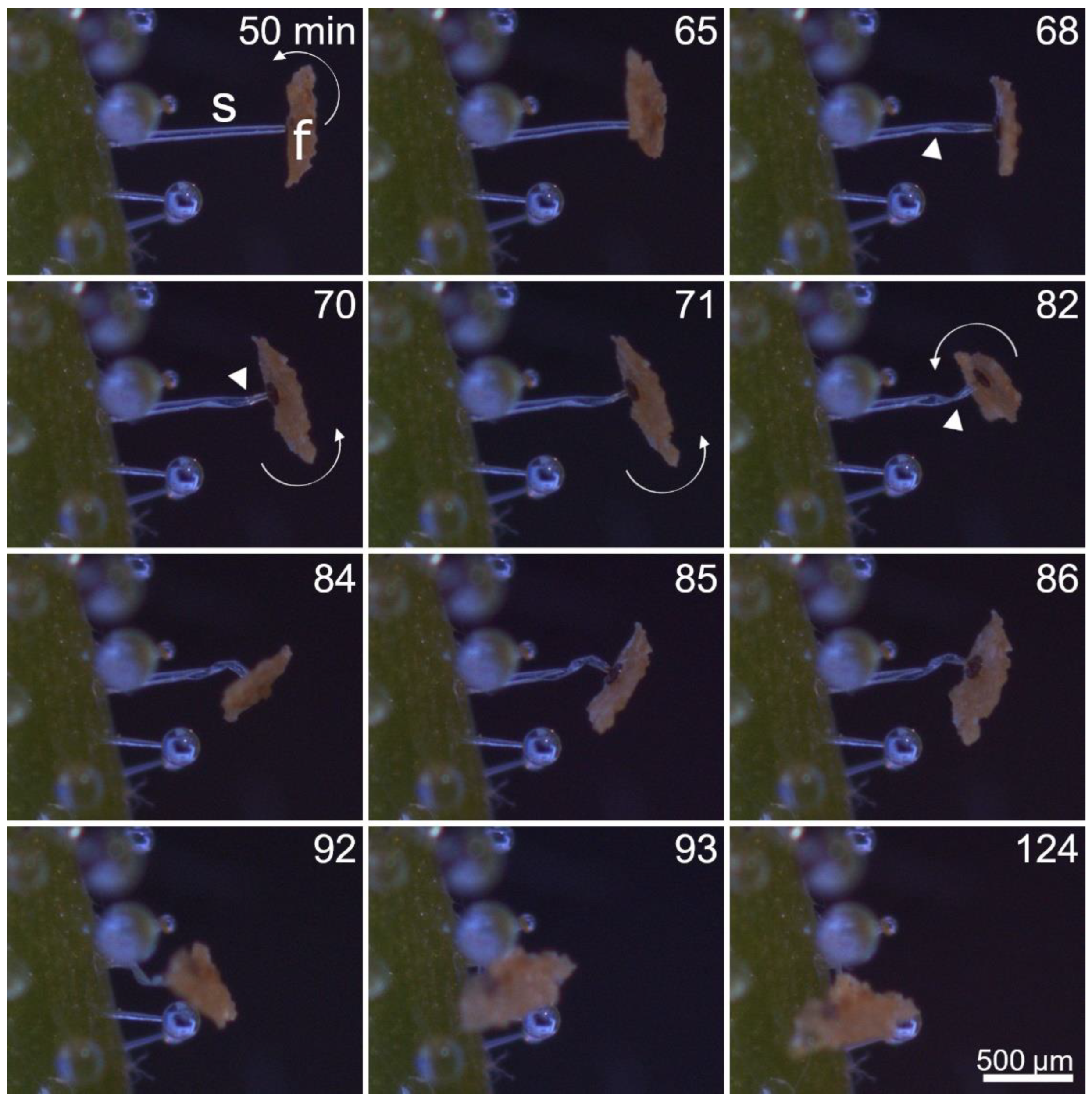
2.2. Stalked Gland Stimulation, Reaction Time, Movement Duration, and Temperature Dependency
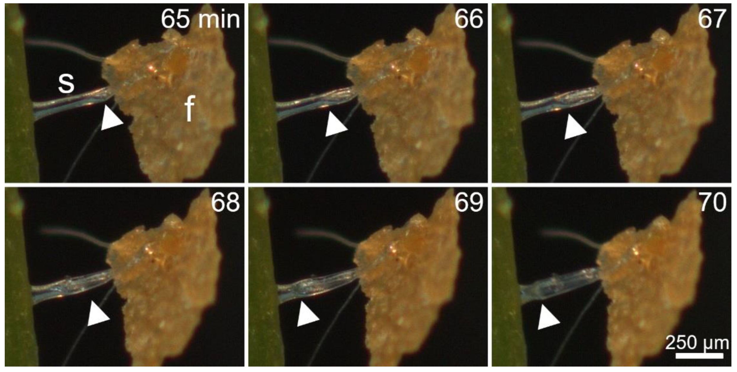
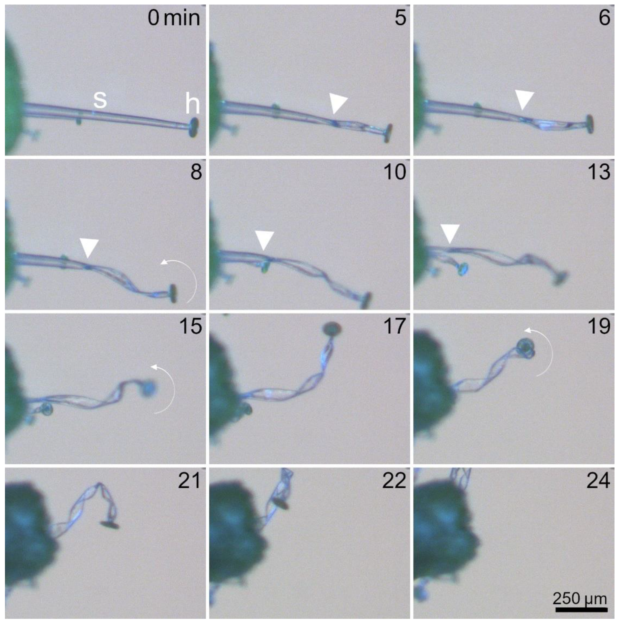
2.3. Kinematics of Stalked Glands
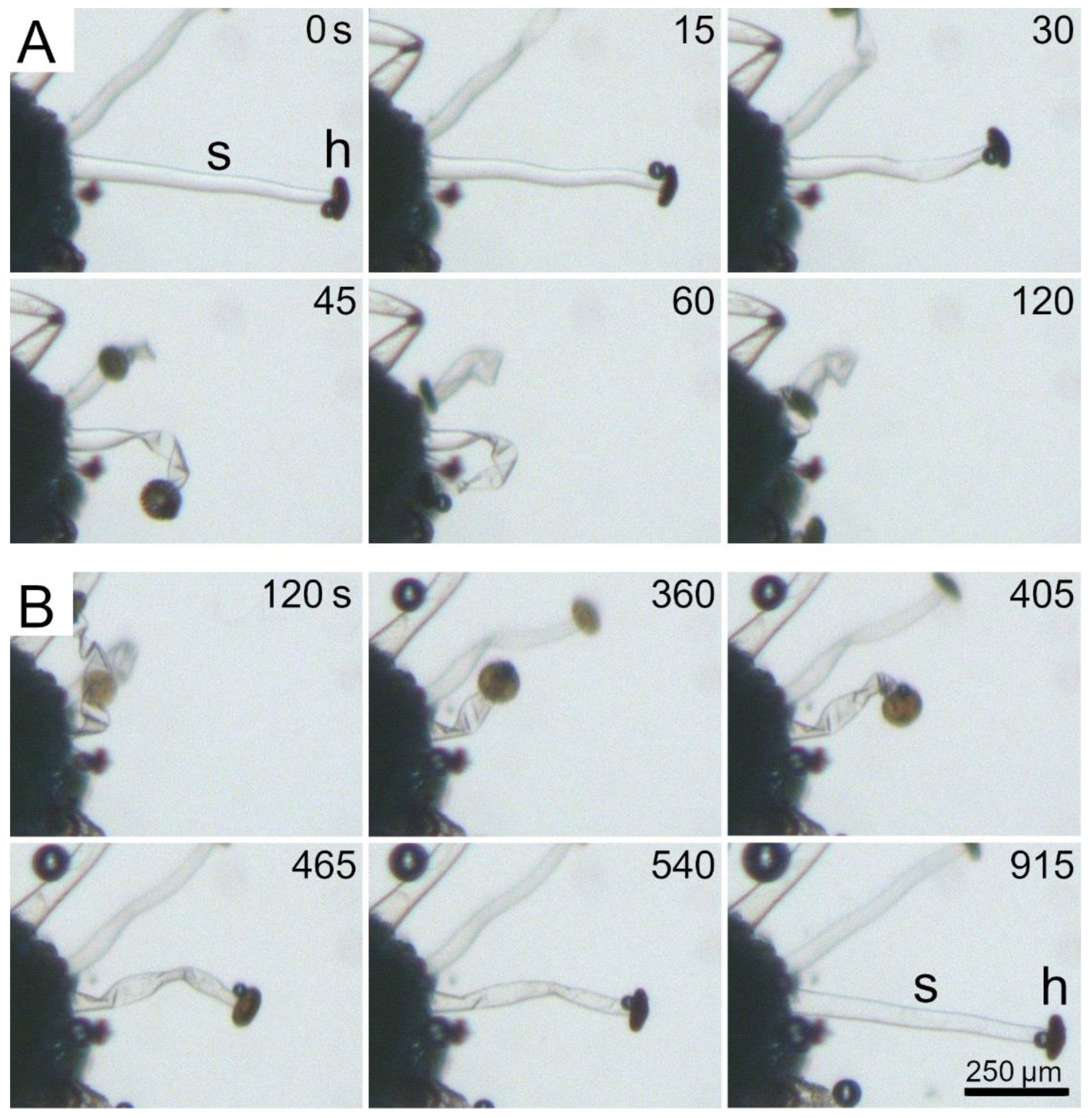
2.4. Dehydration and Rehydration of Stalked Glands
2.5. Functional Morphology of Stalked Glands
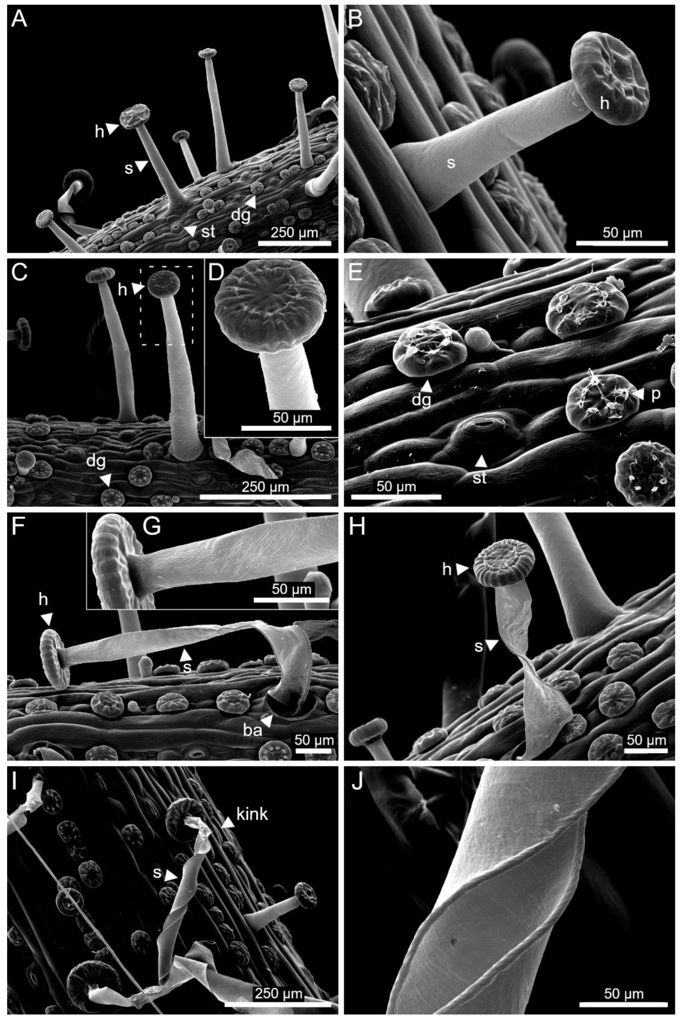
2.5.1. General Results
2.5.2. Cellulose Microfibril Angles in Stalk Cells
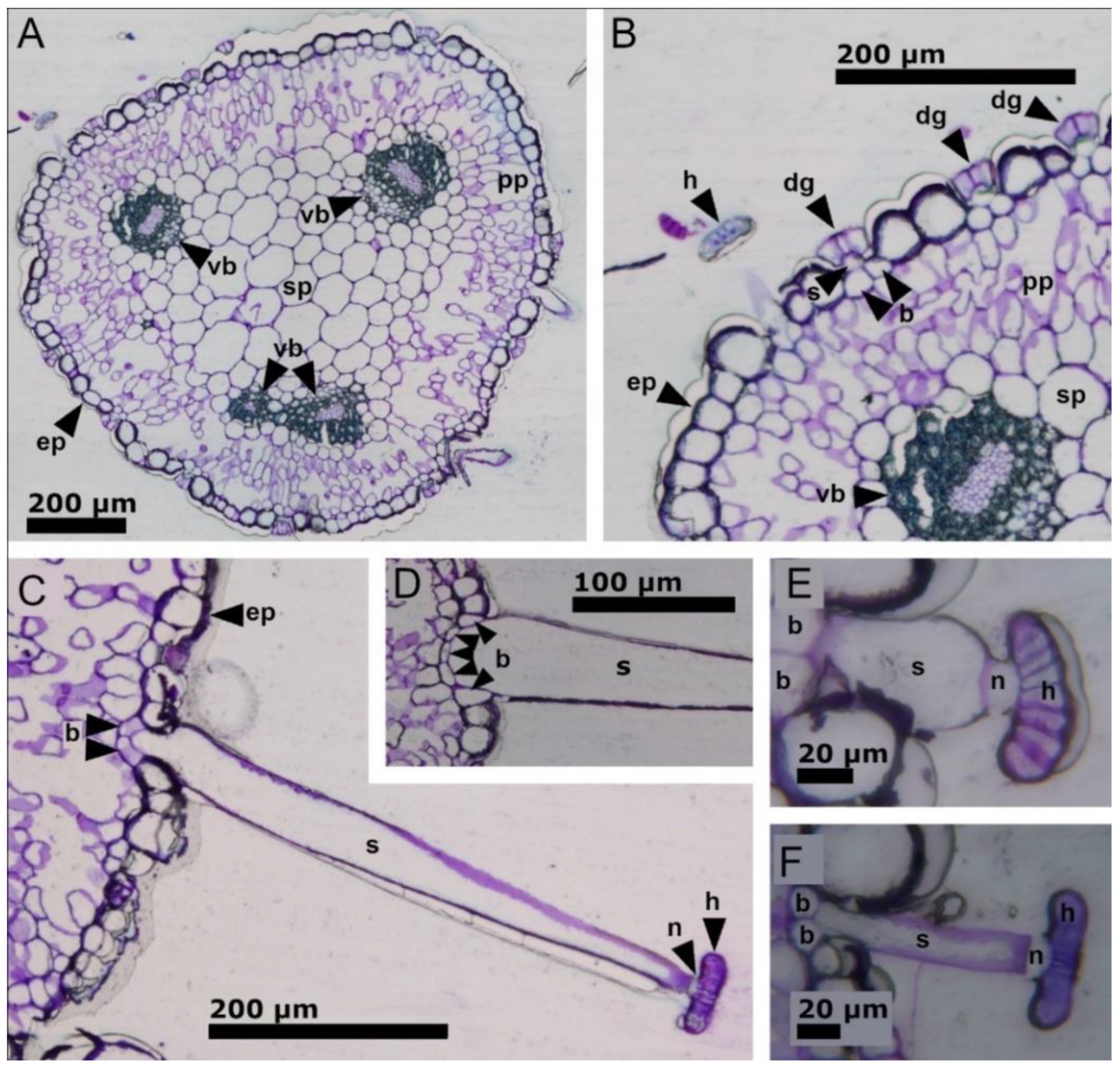
2.5.3. Histological Staining of Stalk Cells
3. Discussion
3.1. Stalked Gland Lengths and Densities
3.2. Stalked Gland Stimulation, Kinematics, and Actuation
3.3. Functional Morphology of Stalked Glands
3.4. Conclusion and Outlook
4. Materials and Methods
4.1. Plant Material
4.2. Lengths and Densities of Stalked Glands
4.2.1. Length Measurements
4.2.2. Density Measurements
4.3. Stimulation Experiments and Kinematical Analyses
4.3.1. Stimulation Experiments
- Pure chemical stimulation: Small fish flake food fragments (a few mm in diameter) (TetraMin Flakes, Tetra GmbH, Melle, Germany) were carefully placed on the glue drops of the stalked gland heads using forceps (cf. [11]). Care was taken to deposit the flakes only on the glue drops and to not exert mechanical stimuli on the stalked glands‘ heads and stalk cells. For the experiments, 10 stalked glands from two leaves of Byblis-1 and 10 stalked glands from 7 leaves of Byblis-2 were used.
- Pure mechanical stimulation: Stalked glands were stimulated with bursts of five quick consecutive touches with an eyelash (diameter: 80 μm) either on their heads or bases, which took place at intervals of 5 min during a 60 min period each. For the experiments, 10 stalked glands from four leaves of Byblis-1 and 10 stalked glands from four leaves of Byblis-2 were used.
- Combined chemical and mechanical stimulation: Fish flake food fragments were placed on the glue drops of stalked gland heads according to the protocol described for pure chemical stimulation. The stalked glands were then tapped following the protocol for pure mechanical stimulation. For the experiments, 10 stalked glands from two leaves of Byblis-1 and 10 stalked glands from two leaves of Byblis-2 were used.
4.3.2. Experiments with Stalked Glands on Detached Leaf Pieces
4.3.3. Temperature Dependencies of Reaction Time and Movement Duration
4.3.4. Analysis of Water Displacement in the Stalk Cell
4.4. Dehydration and Rehydration Experiments
4.4.1. Dehydration and Rehydration of Non-Stimulated Stalked Glands
- Drying in air: A small leaf piece was clamped between two microscope slides. A single, protruding stalked gland was recorded (recording speed: 1 frame per 15 s) with a SZX7 stereo microscope (Olympus GmbH, Hamburg, Germany) equipped with a ColorViewII camera and by using the cell^D software during drying at ambient conditions of ~60% relative humidity and ~26 °C in the microscopy lab of the Plant Biomechanics Group Freiburg;
- “Drying” in a hypertonic aqueous solution: A small piece of leaf was submerged in saturated NaCl solution (Carl Roth GmbH + Co. KG, Karlsruhe, Germany) in a small watch glass dish. Stalked glands were recorded with the same setup and recording speed as described for the experiment “Drying in air”;
- Rehydration with distilled water: At the end of the experiment “Drying in a hypertonic aqueous solution”, the NaCl-solution was aspirated by a paper towel and distilled water was carefully added with a pipette. Stalked glands were recorded with the same setup and recording speed as described for the experiment “Drying in air”.
4.4.2. Rehydration of a Chemically Stimulated Stalked Gland
4.4.3. Test for Glue Production in Rehydrated Stalked Glands
4.5. Functional-Morphological Analyses
4.5.1. General Light Microscopy Analyses
4.5.2. Scanning Electron Microscopy Analyses
4.5.3. Determination of the Cellulose Microfibril Angles in Stalk Cells
4.5.4. Histological Staining of Stalk Cells
- Cellulose staining: The sample was placed in a 0.01% Calcofluor white solution for 10 min and then rinsed with distilled water. Separated trichomes were placed in distilled water on a slide and viewed and photographed under UV light using the microscope setup described in Section 4.5.1;
- Cutin staining: The sample was placed in Sudan IV solution (1 g Sudan IV, 100 mL 80% ethanol, 10 mL glycerol) for 30 min and then briefly rinsed with 80% ethanol and thoroughly rinsed with distilled water. The trichomes were fixed in glycerol gelatin on the slide and investigated using the microscope setup described in Section 4.5.1;
- Pectin staining: A 0.1% ruthenium red solution was applied to the leaf-piece for five minutes and then rinsed twice with distilled water. The stained trichomes were placed in glycerol gelatin on the slide and investigated using the microscope setup described in Section 4.5.1;
- Lignin staining: The sample was placed in a phloroglucinol solution (10 g phloroglucinol, 100 mL 95% ethanol) for 20 min. Afterwards, the trichomes were placed in 25% hydrochloric acid on the slide and analyzed with a Primo Star microscope equipped with an Axiocam ERc 5s (Zeiss, Wetzlar, Germany).
Supplementary Materials
Author Contributions
Funding
Data Availability Statement
Acknowledgments
Conflicts of Interest
References
- Darwin, C. Insectivorous Plants; John Murray: London, UK, 1875. [Google Scholar]
- Fleischmann, A.; Schlauer, J.; Smith, S.A.; Givnish, T.J. Evolution of carnivory in angiosperms. In Carnivorous Plants: Physiology, Ecology, and Evolution; Ellison, A.M., Adamec, L., Eds.; Oxford University Press: Oxford, UK, 2018; pp. 22–41. [Google Scholar] [CrossRef]
- Lloyd, F.E. The Carnivorous Plants; Chronica Botanica: Waltham, MA, USA, 1942. [Google Scholar]
- Juniper, B.E.; Robins, R.J.; Joel, D.M. The Carnivorous Plants; Academic Press Ltd.: London, UK, 1989. [Google Scholar]
- Bauer, U.; Jetter, R.; Poppinga, S. Non-motile traps. In Carnivorous Plants: Physiology, Ecology, and Evolution; Ellison, A.M., Adamec, L., Eds.; Oxford University Press: Oxford, UK, 2018; pp. 194–206. [Google Scholar] [CrossRef]
- Poppinga, S.; Bauer, U.; Speck, T.; Volkov, A.G. Motile traps. In Carnivorous Plants: Physiology, Ecology, and Evolution; Ellison, A.M., Adamec, L., Eds.; Oxford University Press: Oxford, UK, 2018; pp. 180–193. [Google Scholar] [CrossRef]
- Bauer, U.; Müller, U.K.; Poppinga, S. Complexity and diversity of motion amplification and control strategies in motile carnivorous plant traps. Proc. R. Soc. B 2021, 288, 20210771. [Google Scholar] [CrossRef]
- Poppinga, S.; Hartmeyer, S.; Seidel, R.; Masselter, T.; Hartmeyer, I.; Speck, T. Catapulting tentacles in a sticky carnivorous plant. PLoS ONE 2012, 7, e45735. [Google Scholar] [CrossRef]
- Nakamura, Y.; Reichelt, M.; Mayer, V.E.; Mithöfer, A. Jasmonates trigger prey-induced formation of “outer stomach” in carnivorous sundew plants. Proc. R. Soc. B 2013, 280, 20130228. [Google Scholar] [CrossRef]
- Krausko, M.; Perutka, Z.; Šebela, M.; Šamajová, O.; Šamaj, J.; Novák, O.; Pavlovič, A. The role of electrical and jasmonate signalling in the recognition of captured prey in the carnivorous sundew plant Drosera capensis. New Phytol. 2017, 213, 1818–1835. [Google Scholar] [CrossRef]
- Allan, G. Evidence of motile traps in Byblis. Carniv. Plant Newsl. 2019, 48, 51–63. [Google Scholar] [CrossRef]
- Cross, A.T.; Paniw, M.; Scatigna, A.V.; Kalfas, N.; Anderson, B.; Givnish, T.J.; Fleischmann, A. Systematics and evolution of small genera of carnivorous plants. In Carnivorous Plants: Physiology, Ecology, and Evolution; Ellison, A.M., Adamec, L., Eds.; Oxford University Press: Oxford, UK, 2018; pp. 120–134. [Google Scholar] [CrossRef]
- Krueger, T. Field observations of Byblis in Australia. Carniv. Plant Newsl. 2019, 48, 44–50. [Google Scholar] [CrossRef]
- Steenis, C.G.G.J. van Byblidaceae. In Flora Malesiana Series 1, Spermatophyta 7.1; Wolters-Nordhoff Publishing: Groningen, The Netherlands, 1972; pp. 135–137. [Google Scholar]
- Lowrie, A. Carnivorous Plants of Australia Magnum Opus, Volume 1; Redfern Natural History Production: Poole, UK, 2013. [Google Scholar]
- Conran, J.G.; Carolin, R. Byblidaceae. In Flowering Plants-Dicotyledons: Lamiales (except Acanthaceae including Avicenniaceae); Kadereit, J.W., Ed.; Springer: Berlin, Germany, 2004; pp. 45–49. [Google Scholar]
- Płachno, B.J.; Adamec, L.; Lichtscheidl, I.K.; Peroutka, M.; Adlassnig, W.; Vrba, J. Fluorescence labelling of phosphatase activity in digestive glands of carnivorous plants. Plant Biol. 2006, 8, 813–820. [Google Scholar] [CrossRef]
- Adlassnig, W.; Lendl, T.; Peroutka, M.; Lang, I. Deadly glue-adhesive traps of carnivorous plants. In Biological Adhesive Systems: From Nature to Technical and Medical Application; Byern, J., Grunwald, I., Eds.; Springer: Vienna, Austria, 2010; pp. 15–28. [Google Scholar] [CrossRef]
- Lang, F.X. Untersuchungen über Morphologie, Anatomie und Samenentwicklung von Polypompholyx und Byblis gigantea. Flora 1901, 88, 149–206. [Google Scholar]
- Fenner, F.A. Beiträge zur Kenntnis der Anatomie, Entwicklungsgeschichte und Biologie der Laubblätter und Drüsen einiger Insektivoren. Flora 1904, 93, 335–434. [Google Scholar]
- McPherson, S. Carnivorous Plants and Their Habitats Volume 2; Redfern Natural History Productions: Poole, UK, 2010. [Google Scholar]
- Voigt, D.; Gorb, E.; Gorb, S. Hierarchical organisation of the trap in the protocarnivorous plant Roridula gorgonias (Roridulaceae). J. Exp. Biol. 2009, 212, 3184–3191. [Google Scholar] [CrossRef][Green Version]
- Cross, A.T.; Davies, A.R.; Fleischmann, A.; Horner, J.D.; Jürgens, A.; Merritt, D.J.; Murza, G.L.; Turner, S.R. Reproductive biology and pollinator-prey conflicts. In Carnivorous Plants: Physiology, Ecology, and Evolution; Ellison, A.M., Adamec, L., Eds.; Oxford University Press: Oxford, UK, 2018; pp. 294–313. [Google Scholar] [CrossRef]
- Krueger, T.; Cross, A.T.; Fleischmann, A. Size matters: Trap size primarily determines prey spectra differences among sympatric species of carnivorous sundews. Ecosphere 2020, 11, e03179. [Google Scholar] [CrossRef]
- Slack, A. Carnivorous Plants; Marston House: Somerset, UK, 1979. [Google Scholar]
- Gaume, L.; Forterre, Y. A viscoelastic deadly fluid in carnivorous pitcher plants. PLoS ONE 2007, 11, e1185. [Google Scholar] [CrossRef]
- Erni, P.; Varagnat, M.; Clasen, C.; Crest, J.; McKinley, G.H. Microrheometry of sub-nanolitre biopolymer samples: Non-Newtonian flow phenomena of carnivorous plant mucilage. Soft Matter 2011, 7, 10889–10898. [Google Scholar] [CrossRef]
- Williams, S.E.; Pickard, B.G. 1972. Properties of action potentials in Drosera tentacles. Planta 1972, 103, 222–240. [Google Scholar] [CrossRef]
- Procko, C.; Radin, I.; Hou, C.; Chory, J. Dynamic calcium signals mediate the feeding response of the carnivorous sundew plant. Proc. Natl. Acad. Sci. USA 2022, 119, e2206433119. [Google Scholar] [CrossRef]
- Kocáb, O.; Jakšová, J.; Novák, O.; Petřík, I.; Lenobel, R.; Chamrád, I.; Pavlovič, A. Jasmonate-independent regulation of digestive enzyme activity in the carnivorous butterwort Pinguicula × Tina. J. Exp. Bot. 2020, 71, 3749–3758. [Google Scholar] [CrossRef]
- Skotheim, J.M.; Mahadevan, L. Physical limits and design principles for plant and fungal movements. Science 2005, 308, 1308–1310. [Google Scholar] [CrossRef]
- Abraham, Y.; Tamburu, C.; Klein, E.; Dunlop, J.W.C.; Fratzl, P.; Raviv, U.; Elbaum, R. Tilted cellulose arrangement as a novel mechanism for hygroscopic coiling in the stork’s bill awn. J. R. Soc. Interface 2012, 9, 9640–9647. [Google Scholar] [CrossRef]
- Heslop-Harrison, Y.; Knox, R.B. A cytochemical study of the leaf-gland enzymes of insectivorous plants of the genus Pinguicula. Planta 1971, 96, 183–211. [Google Scholar] [CrossRef]
- Wagner, G.J. Secreting glandular trichomes: More than just hairs. Plant Physiol. 1991, 96, 675–679. [Google Scholar] [CrossRef]
- Muravnik, L.E. The structural peculiarities of the leaf glandular trichomes: A review. In Plant Cell and Tissue Differentiation and Secondary Metabolites. Reference Series in Phytochemistry; Ramawat, K., Ekiert, H., Goyal, S., Eds.; Springer: Cham, Germany, 2020. [Google Scholar] [CrossRef]
- Løe, G.; Toräng, P.; Gaudeul, M.; Ågren, J. Trichome production and spatiotemporal variation in herbivory in the perennial herb Arabidopsis lyrata. Oikos 2007, 116, 134–142. [Google Scholar] [CrossRef]
- Skaltsa, H.; Verykokidou, E.; Harvala, C.; Karabourniotis, G.; Manetas, Y. UV-B protective potential and flavonoid content of leaf hairs of Quercus ilex. Phytochemistry 1994, 37, 987–990. [Google Scholar] [CrossRef]
- Bosabalidis, A.M. Glandular hairs, non-glandular hairs, and essential oils, in the winter and summer leaves of the seasonally dimorphic Thymus sibthorpii (Lamiaceae). J. Plant Dev. 2013, 20, 3–12. [Google Scholar]
- Sletvold, N.; Ågren, J. Variation in tolerance to drought among Scandinavian populations of Arabidopsis lyrata. Evol. Ecol. 2011, 26, 559–577. [Google Scholar] [CrossRef]
- Płachno, B.J.; Muravnik, E. Functional anatomy of carnivorous traps. In Carnivorous Plants: Physiology, Ecology, and Evolution; Ellison, A.M., Adamec, L., Eds.; Oxford University Press: Oxford, UK, 2018; pp. 167–179. [Google Scholar] [CrossRef]
- Williams, S.E.; Pickard, B.G. Connections and barriers between cells of Drosera tentacles in relation to their electrophysiology. Planta 1974, 116, 1–16. [Google Scholar] [CrossRef]
- Melzer, B.; Steinbrecher, T.; Seidel, R.; Kraft, O.; Schwaiger, R.; Speck, T. The attachment strategy of English ivy: A complex mechanism acting on several hierarchical levels. J. R. Soc. Interface 2010, 7, 1383–1389. [Google Scholar] [CrossRef]
- Kolattukudy, P.E. Biopolyester membranes of plants: Cutin and suberin. Science 1980, 208, 990–1000. [Google Scholar] [CrossRef]
- Vanholme, R.; Demedts, B.; Morreel, K.; Ralph, J.; Boerjan, W. Lignin biosynthesis and structure. Plant Physiol. 2010, 153, 895–905. [Google Scholar] [CrossRef]
- Mohnen, D. Pectin structure and biosynthesis. Curr. Opin. Plant Biol. 2008, 11, 266–277. [Google Scholar] [CrossRef]
- Schreel, J.D.M.; Leroux, O.; Goossens, W.; Brodersen, C.; Rubinstein, A.; Steppe, K. Identifying the pathways for foliar water uptake in beech (Fagus sylvatica L.): A major role for trichomes. Plant J. 2020, 103, 769–780. [Google Scholar] [CrossRef]
- Schindelin, J.; Arganda-Carreras, I.; Frise, E.; Kaynig, V.; Longair, M.; Pietzsch, T.; Preibisch, S.; Rueden, C.; Saalfeld, S.; Schmid, B.; et al. Fiji: An open-source platform for biological-image analysis. Nat. Methods 2012, 9, 676–682. [Google Scholar] [CrossRef] [PubMed]
- R Core Team. R: A Language and Environment for Statistical Computing; R Foundation for Statistical Computing: Vienna, Austria, 2020. [Google Scholar]
- Demarco, D. Histochemical analysis of plant secretory structures. Methods Mol. Biol. 2017, 1560, 313–330. [Google Scholar] [CrossRef] [PubMed]


| Plant No. | Leaf No. | Ratio | Mean ± SD |
|---|---|---|---|
| Byblis-1 | 1 | 0.67 | 0.55 ± 0.15 |
| Byblis-1 | 2 | 0.38 | |
| Byblis-1 | 3 | 0.60 | |
| Byblis-2 | 1 | 0.53 | 0.45 ± 0.07 |
| Byblis-2 | 2 | 0.43 | |
| Byblis-2 | 3 | 0.40 | |
| Byblis-3 | 1 | 0.81 | 0.51 ± 0.20 |
| Byblis-3 | 2 | 0.42 | |
| Byblis-3 | 3 | 0.43 | |
| Byblis-3 | 4 | 0.39 | |
| Byblis 1–3 (total) | 0.51 ± 0.14 | ||
Publisher’s Note: MDPI stays neutral with regard to jurisdictional claims in published maps and institutional affiliations. |
© 2022 by the authors. Licensee MDPI, Basel, Switzerland. This article is an open access article distributed under the terms and conditions of the Creative Commons Attribution (CC BY) license (https://creativecommons.org/licenses/by/4.0/).
Share and Cite
Poppinga, S.; Knorr, N.; Ruppert, S.; Speck, T. Chemonastic Stalked Glands in the Carnivorous Rainbow Plant Byblis gigantea LINDL. (Byblidaceae, Lamiales). Int. J. Mol. Sci. 2022, 23, 11514. https://doi.org/10.3390/ijms231911514
Poppinga S, Knorr N, Ruppert S, Speck T. Chemonastic Stalked Glands in the Carnivorous Rainbow Plant Byblis gigantea LINDL. (Byblidaceae, Lamiales). International Journal of Molecular Sciences. 2022; 23(19):11514. https://doi.org/10.3390/ijms231911514
Chicago/Turabian StylePoppinga, Simon, Noah Knorr, Sebastian Ruppert, and Thomas Speck. 2022. "Chemonastic Stalked Glands in the Carnivorous Rainbow Plant Byblis gigantea LINDL. (Byblidaceae, Lamiales)" International Journal of Molecular Sciences 23, no. 19: 11514. https://doi.org/10.3390/ijms231911514
APA StylePoppinga, S., Knorr, N., Ruppert, S., & Speck, T. (2022). Chemonastic Stalked Glands in the Carnivorous Rainbow Plant Byblis gigantea LINDL. (Byblidaceae, Lamiales). International Journal of Molecular Sciences, 23(19), 11514. https://doi.org/10.3390/ijms231911514


