Article

Targeted Sequencing of Cytokine-Induced PI3K-Related Genes in Ulcerative Colitis, Colorectal Cancer and Colitis-Associated Cancer

by
Nurul Nadirah Razali
1,
Raja Affendi Raja Ali
2,3,
Khairul Najmi Muhammad Nawawi
2,3,
Azyani Yahaya
4 and
Norfilza M. Mokhtar
1,3,*
1
Department of Physiology, Faculty of Medicine, Universiti Kebangsaan Malaysia, Cheras, Kuala Lumpur 56000, Malaysia
2
Gastroenterology Unit, Department of Medicine, Faculty of Medicine, Universiti Kebangsaan Malaysia, Cheras, Kuala Lumpur 56000, Malaysia
3
GUT Research Group, Faculty of Medicine, Universiti Kebangsaan Malaysia, Cheras, Kuala Lumpur 56000, Malaysia
4
Department of Pathology, Faculty of Medicine, Universiti Kebangsaan Malaysia, Cheras, Kuala Lumpur 56000, Malaysia
*
Author to whom correspondence should be addressed.
Int. J. Mol. Sci. 2022, 23(19), 11472; https://doi.org/10.3390/ijms231911472
Submission received: 16 August 2022 / Revised: 15 September 2022 / Accepted: 20 September 2022 / Published: 29 September 2022
(This article belongs to the Special Issue Molecular Mechanisms and Therapies of Colorectal Cancer)

Abstract

:
Chronic relapsing inflammatory bowel disease is strongly linked to an increased risk of colitis-associated cancer (CAC). One of the well-known inflammatory carcinogenesis pathways, phosphatidylinositol 3-kinase (PI3K), was identified to be a crucial mechanism in long-standing ulcerative colitis (UC). The goal of this study was to identify somatic variants in the cytokine-induced PI3K-related genes in UC, colorectal cancer (CRC) and CAC. Thirty biopsies (n = 8 long-standing UC, n = 11 CRC, n = 8 paired normal colorectal mucosa and n = 3 CAC) were subjected to targeted sequencing on 13 PI3K-related genes using Illumina sequencing and the SureSelectXT Target Enrichment System. The Genome Analysis Toolkit was used to analyze variants, while ANNOVAR was employed to detect annotations. There were 5116 intronic, 355 exonic, 172 untranslated region (UTR) and 59 noncoding intronic variations detected across all samples. Apart from a very small number of frameshifts, the distribution of missense and synonymous variants was almost equal. We discovered changed levels of IL23R, IL12Rß1, IL12Rß2, TYK2, JAK2 and OSMR in more than 50% of the samples. The IL23R variant in the UTR region, rs10889677, was identified to be a possible variant that might potentially connect CAC with UC and CRC. Additional secondary structure prediction using RNAfold revealed that mutant structures were more unstable than wildtype structures. Further functional research on the potential variants is, therefore, highly recommended since it may provide insight on the relationship between inflammation and cancer risk in the cytokine-induced PI3K pathway.

1. Introduction

Carcinogenesis is the most severe complication that could arise from prolonged inflammatory bowel disease (IBD). Colitis-associated cancer (CAC) is a type of colorectal cancer (CRC) that develops as a consequence of IBD due to the presence of chronic inflammation in the gastrointestinal tract [1,2]. The likelihood of CAC developing from ulcerative colitis (UC) and Crohn’s disease (CD), the two major subtypes of IBD, is 1.4% and 0.8%, respectively [3,4]. Additionally, the prevalence rate of developing CAC is expected to range from 0.6 to 17% in Western nations and from 0.3 to 1.8% in Asia Pacific regions [5]. Moreover, in Malaysia, the mean incidence of IBD doubled to 1.46 per 100,000 person/year in between 2010 and 2018 [6]. The rising urbanization of societies, which includes dietary changes, the use of antibiotics, personal cleanliness standards, microbiological exposures, and pollution, may be the cause of the rising trend in IBD over the past decades [7]. As a result, this increasing trend may ultimately contribute to the dynamic shift of CAC among Asians. Moreover, CAC has contributed to 10 to 15% of IBD fatality cases in Western countries [4,8].
Generally, CAC only accounts for 1 to 2% of CRC cases [8]. There are several characteristics that may distinguish CAC from sporadic CRC. Their clinicopathological characteristics are comparable, but CAC has a higher proportion of numerous cancer lesions, an increased percentage of superficial and invasive type lesions, and a higher proportion of mucinous or signet ring cell carcinomas [9]. Contrary to CRC, CAC develops through the inflammation–dysplasia–carcinoma sequence, where the change from low to high grade dysplasia is triggered by field precursor cells that are present in or close to the dysplastic mucosa [10]. According to a study by Choi et al. (2015), 20% of UC patients with low-grade dysplasia may have developed high-grade dysplasia or CRC within 53 months of their first diagnosis [11].
Interestingly, there are many similarities between the molecular pathogenesis of CAC and CRC. K-Ras, p53, APC and COX2 are some of the common genes that are altered as CAC and CRC progressed [8]. As p53 is widely distributed in the inflamed mucosal area, this suggests that chronic inflammation has a propensity to become mutagenic [12]. In fact, a study by Claessen et al. (2010) indicated that p53 staining was found moderately in non-dysplastic tissue and exhibition of stronger expression was seen in the low- and high-grade dysplastic lesion in more than 60% of IBD patients [13].
Immune cells, epithelial cells, stromal cells, cytokines, and chemokines are among the diverse cell types that make up the inflammatory process in CAC development and are similar to those found in the microenvironment of the malignancy [14]. Cytokines have a tendency to control the pro-tumorigenic response in chronic inflammatory conditions by causing cell malignancies and transformation [15,16]. Inflammatory mediators such as tumor necrosis factor alpha (TNF-α), interleukin-6 (IL6) and STAT3 played significant roles in pre-neoplastic growth regulation during CAC tumorigenesis as demonstrated in an animal model study [17].
The release of those mediators is more likely to target several signaling pathways that play major roles in carcinogenesis such as NF-κB, PI3K, JAK/STAT and Wnt/B-catenin [18]. Phosphatidylinositol 3-kinases (PI3K) were recognized in promoting cancer progression as they play a key role in the regulation of survival, differentiation, and proliferation of cancer cells. PI3K enzymatic activity was found to be involved in the pathogenesis of various diseases, ranging from chronic inflammation to cancer, for instance CRC [19,20]. A recent microarray study on UC patients with two different durations discovered PI3K as one of the important pathways in the long-duration UC compared to short duration [21]. Nevertheless, there is still a paucity of knowledge about the role of the PI3K signaling pathway in the carcinogenesis progression of colitis-associated cancer. Thus, in the present study, we have performed targeted sequencing on 13 genes that are related to the cytokines-induced PI3K signaling pathway for the identification of driver gene mutations in colitis-associated cancer, long-standing ulcerative colitis, and sporadic colorectal cancer patients.

2. Results

2.1. Information on Clinical Samples

Table 1 displays demographic data for all samples. The median age of all samples was 69 years old (IQR:8.75). Malays made up the majority of the samples (70%) and were followed by Chinese (17%) and Indians (13%). Females had a somewhat larger gender distribution (60%) than men (40%). Most patients’ smoking status was non-smoker (93%) as opposed to ex-smoker (7%). The mean disease duration for all long-standing UC was 28.5 ± 6.61 years. Most UC patients had a diagnosis of left-sided colitis or pancolitis, with a Mayo index score of 1 to 3 and a Geboes score of Grade 2A.1 to 2A.2 of. Meanwhile, patients with CAC had a chronic active history of colitis for 22 ± 13 years. The majority of CRC and CAC patients were at stages 1 to 3, and the rectosigmoid and distal colon were the sites of the malignancies. Histologically, the majority of CRC were moderately differentiated, whereas tumors from CAC patients were categorized as poorly and well differentiated. None of the patients had a history of CRC in their families.

2.2. The Technical Performance of the Target Enrichment System Panel

The average percentage of clean reads across all raw reads produced by the SureSelectXT Target Enrichment System was 96.4% (range 80.9 to 98.8%). The range of the total reads was 1,200,800 to 6,106,992 reads. For each sample, the percentage of the target base covered by at least of 100× for each sample had reached 100%.

2.3. Summary of Identified Variants in PI3K-Related Genes

Targeted sequencing was performed on 13 genes that are related to PI3K, namely IL12Rß1, IL12Rß2, IL23R, IL31, OSMR, JAK2, TYK2, STAT1, STAT3, STAT4, STAT6, PDK1 and SGK2. Long-standing ulcerative colitis (n = 8), colitis-associated cancer (n = 3), colorectal cancer (n = 11) and paired normal colorectal mucosa (n = 8) made up the total 30 samples. In total, we found 5702 variants across all samples in the cytokine-induced PI3K-related genes. Ninety percent (5116) of such variants were intronic mutations, followed by 355 exonic mutations (6%), 172 mutations in the 3′ and 5′ untranslated region (UTR) region (3%) and 59 mutations on the noncoding intronic region (1%) (Figure 1A).
Single-nucleotide polymorphisms (SNPs) made up most of the discovered variants (75% = 4256 variants), followed by 25% (1446 variants) of insertion–deletion (InDel) variants. The top three genes with the most SNPs were OSMR (640 variants), IL12Rß1 (549 variants) and IL23R (515 variants). Meanwhile, IL12Rß2 (202 variants), IL12Rß1 (174 variants) and STAT6 (163 variants) showed a markedly high frequency of number of InDel variants (Figure 1B).
The number of missense and synonymous mutations from a total of 355 exonic variants had somewhat similar distribution, with 179 variants (50.4%) and 171 variants (48.2%), respectively, followed by 5 variants (1.4%) with frameshift mutations (insertion and deletion). Only 5.6% (n = 10) of missense mutations in the four genes IL12Rß1, OSMR, JAK2 and STAT1 were predicted to be damaging or potentially damaging, whereas the remaining 174 variants (94.4%) were predicted to have a benign or neutral function (Figure 1C).
More than 50% of the patients had changed exonic sequences in 6 out of the 13 cytokine-induced PI3K-related genes. IL23R missense mutations appeared in all samples including the normal colonic mucosa, which is the paired sample of CRC patients (30/30 samples). This was followed by IL12Rß2, where all paired normal samples had the same mutations as CRC (28/30 samples). In contrast, missense mutations TYK2 and OSMR were found in 27 and 21 out of 30 samples, respectively, even though 6 of those samples had paired normal colonic tissues. A total of 7 out of the 30 samples were matched normal samples where 23 of them contained missense mutations in the JAK2. Meanwhile, IL12Rß1 missense mutations were present in 19 out of 30 samples, including four matched normal tissues. Apart from IL12Rß2 and JAK2, where synonymous mutations were frequently observed, practically all top changed genes showed significant amount of missense mutation. Frameshift mutations, however, were infrequently observed and were only found in OSMR (2/21 samples) and TYK2 (1/27 samples) (Figure 2).

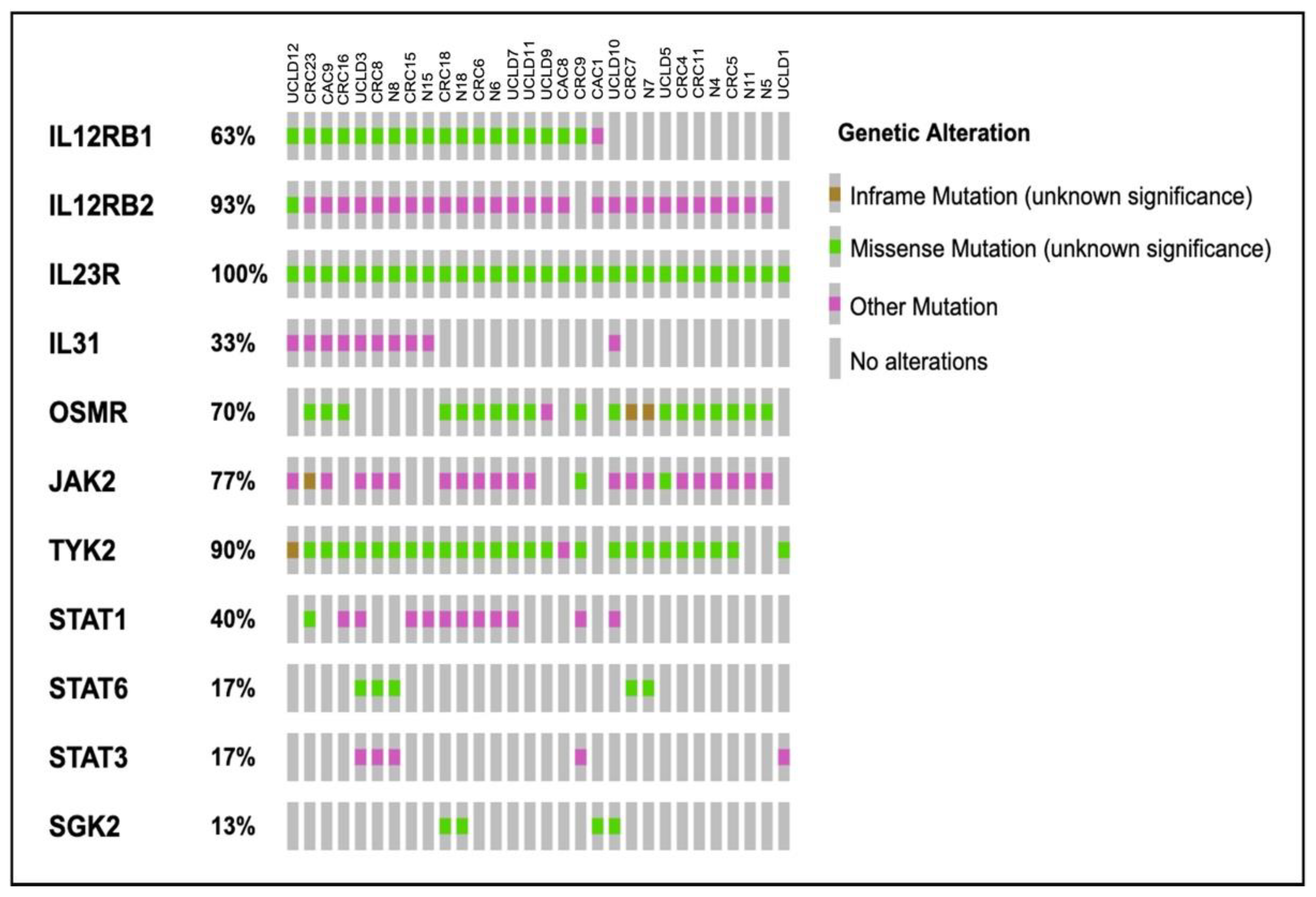
2.4. Somatic Variants Distribution in PI3K-Related Genes among All Samples

A total of 634 mutations were found as recurrent mutations, in the most frequently mutated gene, IL23R. Almost half of the 314 overall mutations were discovered in two or more samples. Of the 634 total mutations, 57 were recurrent missense mutations, 3 were synonymous mutations, 551 were intronic and 23 were UTR. These mutations were discovered sporadically in exon 2 to 10 (Figure 3A).
Only 453 alterations with 102 recurrent mutations were found in the second-most frequently occurring gene, TYK2, which had a 90% incidence of mutations. A total of 362 intronic and 3′-5′ flanking regions, 31 missense, and 6 synonymous were discovered throughout exon 1 to 23. TYK2 had the most splice mutations, 50 in total (Figure 3B).
Meanwhile, IL12Rß2 reported 489 alterations with recurring 163 mutations. Among all groups studied, 60 recurrent synonymous mutations were distributed across exons 2 to 15, followed by 416 intronic and 12 splice areas. In addition, exon 10 only contained one missense mutation (Figure 3C).
IL12Rß1 had the highest number of alterations, with 723 mutations, including 299 recurrences while being affected in just 60% of all cases. Between exon 7 to 15, 55 recurrence missense mutations were discovered, while 28 synonymous mutations were widely distributed around exon 1 to 16, followed by 623 intronic and 8 UTR (Figure 3D).
Another gene with a lot of modification numbers is OSMR, with 699 alterations, 281 of which were recurrent mutations. Exon 2 to 18 had 641 intronic mutations, 28 UTR, 20 missense mutations, and 8 synonymous mutations. Two samples contained a single frameshift insertion in exon 10, resulting in the protein’s function being truncated (Figure 3E).
On the other hand, JAK2 has 120 recurring mutations with 461 overall alterations. Throughout exonic region 3 to 25, 34 synonymous, 20 3′UTR, 400 intronic and 2 splice mutations were found in exonic region 3 to 25. Additionally, at exon 9 and 17, only two missenses were discovered. Exon 8 and 13 both had two frameshift deletion mutations that led to truncated proteins (Figure 3F).
However, no mutations were identified in the cancer hotspot locations of IL23R, IL12Rß1, IL12Rß2, OSMR, TYK2, and JAK2 across all mutant distributions.

2.5. Somatic Variants Distribution of PI3K-Related Genes per Group

The three main groups in this study are ulcerative colitis (UC), colorectal cancer (CRC) and colitis-associated cancer (CAC). As a result, each group’s distribution of variants in PI3K-related genes was also examined.
A total 1625 variants, including 51 missense, 46 synonymous, 1 frameshift, 1461 intronic, 48 UTR and 18 non-coding intronic, were observed in PI3K-related genes in the UC group. The top five altered genes were IL12Rß2, IL23R, OSMR, STAT1 and STAT3 (Figure 4A). Additionally, at least two UC samples were the only ones to contain all 26 recurrent mutations in IL12Rß1, IL12Rß2, IL23R, SGK2, OSMR, STAT4 and STAT6. All recurrences, however, were only discovered in the intronic region (Table 2).
In the CRC group, 2029 variants were found, including 68 missense, 61 synonymous, 3 frameshift, 1809 intronic, 68 UTR and 20 non-coding intronic variants. The top five altered genes in the CRC group comprised IL12Rß1, IL23R, OSMR, STAT1 and STAT3 (Figure 4B). Furthermore, at least two samples from the CRC group only had 19 recurrent mutations in the exonic, intronic, and UTR regions of IL12Rß1, IL12Rß2, OSMR, JAK2, IL23R, STAT4 and STAT6. Both missense mutations that affected the exonic region were predicted to be benign or neutral (Table 2).
In all, 614 variants were found in the CAC group, comprising 16 missense, 20 synonymous, 561 intronic, 13 UTR and 4 non-coding intronic mutations. Most alterations were found in IL12Rß1, IL12Rß2, IL23R, OSMR and TYK2 (Figure 4C). Only two recurrence frameshift mutations in IL12Rß2 and TYK2 were discovered in at least two samples of CAC, in contrast to the UC and CRC groups (Table 2).
We were able to identify variants that co-occurred in the CAC group with the UC and CRC, respectively. Data filtration was applied on the selection of only CAC group, paired with either the UC or CRC group which have yielded a total of 27 intronic variants. For CAC with UC, these co-existing variants were scattered from IL12Rß1, IL12Rß2, IL23R, OSMR, JAK2, TYK2, STAT1, STAT3 and STAT6, but for CAC with CRC, only two genes were involved (JAK2 and STAT4) (Table 3).

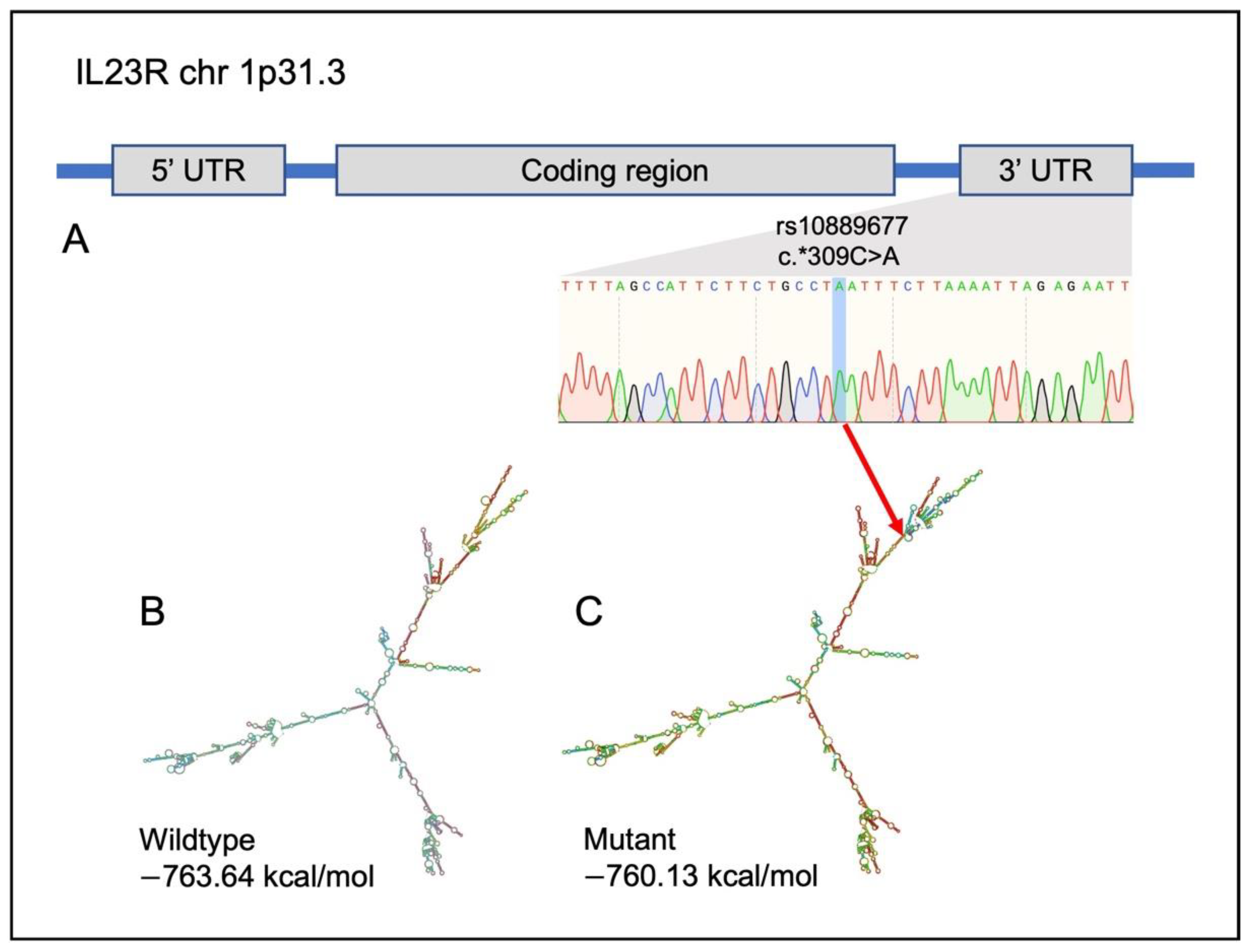
2.6. Identification of Potential Variants That Link UC, CRC and CAC

Additional research was also carried out to identify possible polymorphisms that link CAC with UC and CRC. In particular, we were looking for variants that should be present in the majority of CAC samples. Data were filtered with a high CAC number (minimum n = 2), and low paired normal number (maximum is three out of eight) for better analysis. Data on UC and CRC numbers, however, were not filtered. In addition, because the analysis’s objective was to detect in the coding region, data on the variants’ function were also sorted by ‘exonic’ and ‘UTR’ exclusively.
With the help of data filtering, we identified six potential variants that were present in at least two CAC samples, three in matched normal samples and half of the CRC and UC samples. In IL12Rß1, IL12Rß2 and IL23R, one missense, four synonymous and one UTR variant were found. Sanger sequencing was used to confirm these possible variants further. Only the variant rs10889677 (c.*309C>A), located at the UTR region of IL23R, was validated, and found in practically all CAC samples.
Other than missense mutations, it was not possible to apply protein function prediction methods such as SIFT and PolyPhen-2. Nevertheless, based on their mRNA secondary structure, predictions about the effects of those synonymous and UTR variations are still possible. In terms of the enhanced presence of hairpins, stem-loops, bulge loops, multi-branch loops, and stacking, changes in the secondary structure of the mRNA could be seen.
In silico prediction analysis showed obvious changes in the secondary structure IL23R variant (rs10889677) (Figure 5). In comparison to the wildtype, the presence of variant rs10889677 significantly altered the structure close to the terminal branch by adding additional branches and loops. The minimum free energy (MFE) value predicted from the created structure was also affected because of the changing of the color coding on the base structure.

3. Discussion

The risk of contracting colitis-related cancer rose in UC patients who had chronic inflammation that persisted for an extended period of time. The development of CAC was recently studied in relation to p53, APC and K-Ras [25]. However, the underlying role of the inflammation–carcinogenesis pathway in the pathogenesis of CAC is still poorly understood. We are interested in investigating how the PI3K signaling pathway contributes to the pathophysiology of CAC. In this study, we used the targeted sequencing approach via the SureSelectXT Target Enrichment System to test for somatic mutations in 13 cytokine-induced PI3K-related genes in long-standing UC, CAC and CRC patients, instead of choosing the common cancer-related genes.
Screening on the somatic alterations in our group samples showed that the majority of the variants were found on the intron, not the exonic region. In fact, silent mutations and neutral predicted variants predominated in the coding region. Few insertions and deletions that could result in frameshift mutations were seen. Nevertheless, the SureSelectXT Target Enrichment System is professed as an accurate genome analysis method from small-scale research to large sample cohorts. It has demonstrated high performance, as measured by capture efficiency, sensitivity, reproducibility, and SNP detection [26]. In this study, this application has successfully sequenced more than 95% of the clean reads coverage with an average error rate of less than 0.15% across all bases. In fact, the high precision of genome sequencing was explained by the fact that there are at least 100× as many reads per given nucleotides in the genome.
In each of our sample populations, interleukin-23 receptor (IL23R) was found to be the gene that was most frequently altered. A previous study has reported IL23R as a gene associated with inflammatory bowel disease (IBD), whereby nine SNPs in IL23R at various locations such as intronic, exonic and UTR have shown significant associations with Crohn’s disease [27]. Moreover, it has been shown that IL23R variants in IBD may operate as a protective variant or contribute to the development of inflammation [28,29]. It has been demonstrated that IL23R polymorphisms also may increase the risk of CRC [30].
There are several IL23R variants that we have discovered, but just two of them, rs7530511 (c.929T>C: p.L310P) and rs1884444 (c.9G>T: p.Q3H), have been linked with colorectal-related diseases. In contrast, both variants were found in intracerebral hemorrhage and cancer-related disease (bladder and esophageal) [31,32,33]. Despite this, one interesting variant in IL23R, rs10889677 (c.*309C>A), was primarily discovered in UC, CRC and CAC. This is in line with recent studies that found this polymorphism to be a strong predictor of CRC in Asians and a risk factor for IBD [34,35]. In fact, according to a prior genome-wide association study, rs10889677 demonstrated a favorable link with IBD and might subsequently make the condition worse clinically [27]. Additionally, IL23R has just recently been discussed as a potential IBD treatment [36,37]. The activity of the IL23 signaling pathway could be inhibited by specific binding of the oral peptide (PTG-200) to the IL23R, which would subsequently influence JAK2 and TYK2 activation and perhaps result in abnormal STAT3 and STAT4 expression.
The next frequently altered gene in our study was interleukin-12 receptor beta 1 (IL12Rß1). Twenty percent of our samples included the IL12Rß1 variant (rs11575935; c.1573G>A: p.A525T) that may be harmful. This variant has not yet been connected to any illnesses. Only 1.7% and 0.4% of the population in Asian and European regions, respectively, had this variant [38]. The next frequently altered gene in our study was tyrosine kinase-2 (TYK2). TYK2 has lately gained attention as a potential therapeutic for IBD; its connection to gastrointestinal disorders has long been established [39,40]. TYK2 mutation rs2304256 (c.1084G>T: p.V362F), which was discovered in almost 80% of our samples, was an intriguing finding. This variant has been linked to autoimmune and inflammatory diseases, including IBD [41,42]. The mutation rs2304256 was initially thought to be benign, but subsequent studies showed that it might encourage exon 8 inclusion and subtly boost TYK2 expression in whole blood [43]. In fact, the Genotype-Tissue Expression (GTEx) database shows that rs2304256 is indeed linked to a slight increase in TYK2 in several tissues, including colonic tissue. The Oncostatin M receptor (OSMR) is a different gene that has drawn attention. Our finding on the OSMR variant, rs2278329 (c.1657G>A: p.D553N), which predominately affects CRC and UC, is analogous to prior studies on OSMR that looked at the role of OSMR in inflammation and its potential link with other cancers such as bladder and thyroid cancer [44,45,46].
Apart from that, there was only one variant in the untranslated region (UTR) region of IL23R, rs10889677, which was found in 85% of CAC samples, and validated as the potential variant that correlates CAC with UC and CRC. Although, the selected potential variant was not located in the coding region and predicted as damaging, yet the effect of that variant on the translation efficacy still can be predicted via the construction of the mRNA secondary structure. The terminal branch of the IL23R variant rs10889677’s mRNA secondary structure was clearly altered. Compared to the wildtype structure, prominent additional branches and loops had impacted the minimum free energy (MFE) value. MFE demonstrates the stability of a structure. In a stable structure, there should be more negative values. Stability is essential in the mRNA secondary structure because stable complexes may increase translation efficiency [47]. This finding was also supported by another studies that showed the presence of a non-damaging intronic variant had caused alteration in the mRNA secondary structure and stability, thus affecting the mRNA expression level in the brain tissue [48]. Moreover, results of additional studies also corroborated this assumption, where the mutant rs10889677 variant that had aberrant translation efficiency showed lower rates of T-cell proliferation, subsequently increasing susceptibility of the IBD and elevating the risk for developing cancers, such as breast, lung and nasopharyngeal [49,50].
There are a few limitations of this study. This study only involved a single center. Most likely, it was due to our stringent inclusion and exclusion criteria. Moreover, we were cautious in selecting the potential CAC variants, as data filtering was conducted based only exonic and UTR variants. Therefore, further studies with a larger cohort would be highly recommended to corroborate the results. In addition, exploring the underlying mechanism of more variety of potential variants in CAC via in vitro functional study would be highly beneficial in discovering the linkage of those gene variants with tumorigenesis.
Through our findings, we successfully identified somatic variants in cytokine-induced PI3K-related genes in long-standing UC, CAC and CRC samples. Targeted therapies for CRC now focus on a few common pathways, including EGFR (cetuximab and panitumumab) and VEGF (bevacizumab), which can stimulate a number of downstream intracellular signaling pathways including the PI3K signaling pathway [51]. So far, therapeutics interventions based on cytokine-induced PI3K-related genes such as IL23R have primarily been studied in inflammatory illnesses [37,52]. Hence, introducing IL23R as the next cancer small-molecule inhibitor may be advantageous for future therapies.

4. Materials and Methods

4.1. Sample Collection

A total of 30 fresh frozen and archive samples from long-standing UC, CAC, CRC and the corresponding adjacent normal colorectal mucosa tissue were collected from patients that were attending the Endoscopy Unit, Universiti Kebangsaan Malaysia Medical Centre (UKMMC), Kuala Lumpur, Malaysia. Upon admission, informed consent was obtained from each patient. Basic clinical and demographic data were gathered and analyzed while reviewing the patient’s medical records. Prior to further processing, the tissues were collected in RNAlater (Sigma Aldrich, St. Louis, MO, USA) and kept frozen at −80 °C. An experienced pathologist examined the confirmation of the diagnosis, the level of inflammation, and the presence of metastases based on the hematoxylin and eosin (H&E)-stained sections. Only cancer tissues with more than 80% tumor cell content were used in this study for cancer samples. The normal samples were confirmed to be free from tumor or inflammatory cells. The Universiti Kebangsaan Malaysia Research Ethics Committee (UKM/PPI/111/8/JEP-2019-572) granted approval for this study.

4.2. Nucleic Acid Extraction and Quality Assessment

DNA extraction from the fresh frozen tissues was performed using the AllPrep DNA/RNA/miRNA Universal Kit (Qiagen, Valencia, CA, USA) in accordance with the manufacturer’s protocol. Meanwhile, using GENEREAD DNA FFPE Kit (Qiagen, Valencia, CA, USA), DNA was extracted from formalin-fixed paraffin-embedded (FFPE) blocks of archival samples. DNA concentration was measured using DeNovix DS11+ Spectrophotometer (DeNovix Inc., Wilmington, DE, USA) and Qubit® DNA Assay Kit in Qubit® 2.0 Fluorometer (Life Technologies, Carlsbad, CA, USA). The agarose gel electrophoresis was used to evaluate the extracted DNA’s purity. Targeted sequencing was performed on the genomic DNA that was largely undamaged and free of RNA.

4.3. Targeted Sequencing

Library preparation was conducted using DNA random fragmentation by sonication (Covaris, MA, USA) to the size of 180–280 bp fragments, followed by PCR enrichment and purification with AMPure XP system (Beckman Coulter, Beverly, USA). The library was then quantified using the high-sensitivity DNA assay on the Agilent Bioanalyzer 2100 (Agilent Technologies Inc, Santa Clara, CA, USA). Targeted sequencing was carried out using Illumina sequencing and SureSelectXT Target Enrichment System (Agilent Technologies Inc., Santa Clara, CA, USA).

4.4. Sequence Alignment and Variant Annotation

Burrows–Wheeler Aligner (BWA) was utilized to map the paired-end clean reads to the human reference genome (hg19). After the discovery of the genomic variant, the program ANNOVAR [53] was used to annotate the variants in a variety of ways, including the genomic regions impacted by the variants (RefSeq and Genecode), protein-coding changes and deleteriousness prediction (SIFT [54], PolyPhen [55] and MutationAssessor [56]), mRNA secondary structure (RNAfold) [24], allele frequency (1000 Human Genome) [57], disease associations (dbSNP [58], COSMIC (cancer.sanger.ac.uk) [59], OMIM [60], GWAS Catalog [61] and HGMD [62]) and pathway annotation (Gene Ontology [63], KEGG [64] and Reactome [65]).

4.5. Validation of Genomic Variants

The discovered somatic variants were then validated using the Sanger sequencing method. Primers were designed using the NCBI Primer Tool (National Center for Biotechnology Information, Bethesda MD, USA) and Primer3Plus [66]. The sequencing results were analyzed using the SnapGene Viewer 5.3.2. The primers used for validation were IL12Rß2 rs2229546 5′-GCTGAGAGCAGACAACTGGT-3′ (forward), 5′-CCATCATGGGTGGGAAGGTC-3′ (reverse), rs2228420 5′-GGGCGCATACACCAATCAG-3′ (forward), 5′-TTTCCCTGACCCATGGCAG-3′, IL23R rs10889677 5′-TCTGTGCTCCTACCATCACC-3′ (forward), 5′-TGTGCCTGTATGTGTGACCA-3′ (reverse) and JAK2 rs2230722 5′-GAGATCTTGCCATGTTGCCC-3′ (forward), and 5′-ACACTGCCATCCCAAGACAT-3′ (reverse).

4.6. Statistical Analysis

Normally distributed variables are presented as the mean ± standard deviation, and non-normally distributed variables as the median (25th and 75th percentiles). Statistical analyses were performed using SPSS 26 software (SPSS Inc., Chicago, IL, USA).

5. Conclusions

We were able to identify somatic variants in the PI3K-related genes among UC, CRC and CAC, and it was discovered that most of these variants were found in the IL23R, IL12Rß1 and IL12Rß2 genes, followed by TYK2, JAK2 and OSMR. The discovery of IL23R variant rs10889677 as a possible mutation may help to give an insight on how the cytokine-induced PI3K pathway links inflammation with a higher risk of developing cancer, opening the door to improved care for CAC patients in the near future.

Author Contributions

Conceptualization, N.M.M. and R.A.R.A.; methodology and participant recruitments, N.M.M., R.A.R.A., K.N.M.N. and N.N.R.; validation, N.N.R., A.Y. and N.M.M.; data analysis, N.N.R. and N.M.M.; writing—original draft preparation, N.N.R.; writing—review and editing, N.M.M., R.A.R.A. and K.N.M.N.; supervision, N.M.M., R.A.R.A. and K.N.M.N. All authors have read and agreed to the published version of the manuscript.

Funding

This work was supported by the Fundamental Research Grant Scheme, Ministry of Higher Education, Malaysia (FRGS/1/2018/SKK06/UKM/02/4).

Institutional Review Board Statement

The study was conducted in accordance with the Declaration of Helsinki and approved by the Universiti Kebangsaan Malaysia Research Ethics Committee (UKM/PPI/111/8/JEP-2019-572).

Informed Consent Statement

Informed consent was obtained from all subjects involved in the study.

Data Availability Statement

The raw data in this study are available upon request.

Acknowledgments

We would like to thank the staff from the Gastroenterology Unit, UKMMC and the Department of Physiology, UKM for their assistance and coordination with biospecimen collection.

Conflicts of Interest

The authors declare no conflict of interest.

References

  1. Grivennikov, S.I.; Cominelli, F. Colitis-Associated and Sporadic Colon Cancers: Different Diseases, Different Mutations? Gastroenterology 2016, 150, 808–810. [Google Scholar] [CrossRef]
  2. Liverani, E.; Scaioli, E.; John Digby, R.; Bellanova, M.; Belluzi, A. How to predict clinical relapse in inflammatory bowel disease patients. World J. Gastroenterol. 2016, 22, 1017–1033. [Google Scholar] [CrossRef] [PubMed]
  3. Laukoetter, M.G.; Mennigen, R.; Hannig, C.M.; Osada, N.; Rijcken, E.; Vowinkel, T.; Krieglstein, C.F.; Senniger, N.; Anthoni, C.; Bruewer, M. Intestinal Cancer Risk in Crohn’s Disease: A Meta-Analysis. J. Gastrointest. Surg. 2011, 15, 576–583. [Google Scholar] [CrossRef] [PubMed]
  4. Zhou, Q.; Shen, Z.F.; Wu, B.S.; Xu, C.B.; He, Z.Q.; Chen, T.; Shang, H.T.; Xie, C.F.; Huang, S.Y.; Chen, Y.G.; et al. Risk of Colorectal Cancer in Ulcerative Colitis Patients: A Systematic Review and Meta-Analysis. Gastroenterol. Res. Pract. 2019, 2019, 5363261. [Google Scholar] [CrossRef] [PubMed]
  5. Zhiqin, W.; Palaniappan, S.; Raja Ali, R.A. Inflammatory Bowel Disease-related Colorectal Cancer in the Asia-Pacific Region: Past, Present, and Future. Intest. Res. 2014, 12, 194. [Google Scholar] [CrossRef]
  6. Mokhtar, N.M.; Nawawi, K.N.M.; Verasingam, J.; Zhiqin, W.; Sagap, I.; Azman, Z.A.M.; Mazlan, L.; Hamid, H.A.; Yaacob, N.Y.; Rose, I.M.; et al. A four-decade analysis of the incidence trends, sociodemographic and clinical characteristics of inflammatory bowel disease patients at single tertiary centre, Kuala Lumpur, Malaysia. BMC Public Health 2019, 19, 1–10. [Google Scholar] [CrossRef]
  7. Ananthakrishnan, A.N.; Bernstein, C.N.; Iliopoulos, D.; Macpherson, A.; Neurath, M.F.; Raja Ali, R.A.; Vavricka, S.R.; Fiocchi, C. Environmental triggers in IBD: A review of progress and evidence. Nat. Rev. Gastroenterol. Hepatol. 2018, 15, 39–49. [Google Scholar] [CrossRef]
  8. Kameyama, H.; Nagahashi, M.; Shimada, Y.; Tajima, Y.; Ichikawa, H.; Nakano, M.; Sakata, J.; Kobayashi, T.; Narayanan, S.; Takabe, K.; et al. Genomic characterization of colitis-associated colorectal cancer. World J. Surg. Oncol. 2018, 16, 4–9. [Google Scholar] [CrossRef]
  9. Watanabe, T.; Konishi, T.; Kishimoto, J.; Kotake, K.; Muto, T.; Sugihara, K. Ulcerative Colitis-associated Colorectal Cancer Shows a Poorer Survival than sporadic colorectal cancer: A Nationwide Japanese Study. Inflamm. Bowel Dis. 2011, 17, 1–7. [Google Scholar] [CrossRef]
  10. Hartnett, L.; Egan, L.J. Inflammation, DNA methylation and colitis-associated cancer. Carcinogenesis 2012, 33, 723–731. [Google Scholar] [CrossRef]
  11. Choi, C.R.; Ignjatovic-Wilson, A.; Askari, A.; Lee, G.H.; Warusavitarne, J.; Moorghen, M.; Thomas-Gibson, S.; Saunders, B.P.; Rutter, M.D.; Graham, T.A.; et al. Low-Grade Dysplasia in Ulcerative Colitis: Risk Factors for Developing High-Grade Dysplasia or Colorectal Cancer. Am. J. Gastroenterol. 2015, 110, 1461–1471. [Google Scholar] [CrossRef] [PubMed]
  12. Ullman, T.A.; Itzkowitz, S.H. Intestinal inflammation and cancer. Gastroenterology 2011, 140, 1807–1816.e1. [Google Scholar] [CrossRef] [PubMed]
  13. Claessen, M.M.H.; Schipper, M.E.I.; Oldenburg, B.; Siersema, P.D.; Offerhaus, G.J.A.; Vleggaar, F.P. WNT-pathway activation in IBD-associated colorectal carcinogenesis: Potential biomarkers for colonic surveillance. Cell Oncol. 2010, 32, 303–310. [Google Scholar] [CrossRef] [PubMed]
  14. Axelrad, J.E.; Lichtiger, S.; Yajnik, V. Inflammatory Bowel Disease: Global view Inflammatory bowel disease and cancer: The role of inflammation, immunosuppression, and cancer treatment. World J. Gastroenterol. 2016, 22, 4794–4801. [Google Scholar] [CrossRef]
  15. Shrihari, T.G. Dual role of inflammatory mediators in cancer. Ecancermedicalscience 2017, 11, 1–9. [Google Scholar] [CrossRef]
  16. Galdiero, M.R.; Marone, G.; Mantovani, A. Cancer inflammation and cytokines. Cold Spring Harb. Perspect. Biol. 2018, 10, a028662. [Google Scholar] [CrossRef]
  17. Grivennikov, S.I.; Cominello, F. IL-6 and Stat3 Are Required for Survival of Intestinal Epithelial Cells and Development of Colitis-Associated Cancer. Cancer Cell 2009, 15, 103–113. [Google Scholar] [CrossRef]
  18. Qu, X.; Tang, Y.; Hua, S. Immunological approaches towards cancer and inflammation: A cross talk. Front. Immunol. 2018, 9, 699. [Google Scholar] [CrossRef]
  19. Ciraolo, E.; Gulluni, F.; Hirsch, E. Methods to measure the enzymatic activity of PI3Ks. Methods Enzym. 2014, 543, 115–140. [Google Scholar]
  20. Abdul, S.N.; Ab Mutalib, N.S.; Sean, K.S.; Syafruddin, S.E.; Ishak, M.; Sagap, I.; Mazlan, L.; Rose, I.M.; Abu, N.; Mokhtar, N.M.; et al. Molecular characterization of somatic alterations in Dukes’ B and C colorectal cancers by targeted sequencing. Front. Pharm. 2017, 8, 465. [Google Scholar] [CrossRef]
  21. Low, E.N.D.; Mokhtar, N.M.; Wong, Z.; Raja Ali, R.A. Colonic mucosal transcriptomic changes in patients with long-duration ulcerative colitis revealed colitis-associated cancer pathways. J. Crohns Colitis 2017, 13, 755–763. [Google Scholar] [CrossRef] [PubMed]
  22. Gao, J.; Aksoy, B.A.; Dogrusoz, U.; Dresdner, G.; Gross, B.; Sumer, S.O.; Sun, Y.; Jacobsen, A.; Sinha, R.; Larsson, E.; et al. Integrative Analysis of Complex Cancer Genomics and Clinical Profiles Using the cBioPortal Complementary Data Sources and Analysis Options. Sci. Signal. 2014, 6, 1–20. [Google Scholar] [CrossRef]
  23. Cerami, E.; Gao, J.; Dogrusoz, U.; Gross, B.E.; Sumer, S.O.; Aksoy, B.A.; Jacobsen, A.; Byrne, C.J.; Heuer, M.L.; Larsson, E.; et al. The cBio Cancer Genomics Portal: An Open Platform for Exploring Multidimensional Cancer Genomics Data. Cancer Discov. 2012, 2, 401–404. [Google Scholar] [CrossRef] [PubMed]
  24. Gruber, A.R.; Lorenz, R.; Bernhart, S.H.; Neubock, R.; Hofacker, I.L. The Vienna RNA websuite. Nucleic Acids Res. 2008, 36, W70–W74. [Google Scholar] [CrossRef]
  25. Du, L.; Kim, J.J.; Shen, J.; Chen, B.; Dai, N. KRAS and TP53 mutations in inflammatory bowel disease associated colorectal cancer: A meta-analysis. Oncotarget 2017, 8, 22175–22186. [Google Scholar] [CrossRef]
  26. Ong, J.; Giuffre, A.; Joshi, S.; Ravi, H.; Pabon-Pena, C.; Novak, B.; Visitacion, M.; Hamady, M.; Useche, F.; Arezi, B.; et al. Overview of the Agilent Technologies SureSelectTM Target Enrichment System. J. Mol. Tech. 2011, 22, S30–S31. [Google Scholar]
  27. Duerr, R.H.; Taylor, K.D.; Brant, S.R.; Rioux, J.D.; Silverberg, M.S.; Daly, M.J.; Steinhart, A.H.; Abraham, C.; Regueiro, M.; Griffiths, A.; et al. A genome-wide association study identifies IL23R as an inflammatory bowel disease gene. Science 2006, 314, 1461–1463. [Google Scholar] [CrossRef]
  28. Sivanesan, D.; Beauchamp, C.; Quinou, C.; Lee, J.; Lesage, S.; Chemtob, S.; Rioux, J.D.; Michnick, S.W. IL23R (Interleukin 23 Receptor) variants protective against inflammatory bowel diseases (IBD) display loss of function due to impaired protein stability and intracellular trafficking. J. Biol. Chem. 2016, 291, 8673–8685. [Google Scholar] [CrossRef]
  29. Zhu, Y.; Jiang, H.G.; Chen, Z.H.; Lu, B.H.; Li, J.; Shen, X.N. Genetic association between IL23R rs11209026 and rs10889677 polymorphisms and risk of Crohn’s disease and ulcerative colitis: Evidence from 41 studies. Inflamm. Res. 2020, 69, 87–103. [Google Scholar] [CrossRef]
  30. Poole, E.M.; Curtin, K.; Hsu, L.; Duggan, D.J.; Makar, K.W.; Xiao, L.; Carlson, C.S.; Caan, B.J.; Potter, J.D.; Slattery, M.L.; et al. Genetic variability in IL23R and risk of colorectal adenoma and colorectal cancer. Cancer Epidemiol. 2012, 36, e104–e110. [Google Scholar] [CrossRef]
  31. Park, H.J.; Kim, S.K.; Park, H.K.; Chung, J.H. Association of IL23R polymorphism (rs7530511) with intracerebral hemorrhage in Korean population. Neurol. Sci. 2016, 37, 983–985. [Google Scholar] [CrossRef] [PubMed]
  32. El-Gedamy, M.; El-khayat, Z.; Abol-Enein, H.; El-said, A. Rs-1884444 G/T variant in IL-23 receptor is likely to modify risk of bladder urothelial carcinoma by regulating IL-23/IL-17 inflammatory pathway. Cytokine 2021, 138, 155355. [Google Scholar] [CrossRef] [PubMed]
  33. Li, M.; Yue, C.; Jin, G.; Guo, H.; Ma, H.; Wang, G.; Huangm, S.; Wu, F.; Zhao, X. Rs1884444 variant in IL23R gene is associated with a decreased risk in esophageal cancer in Chinese population. Mol. Carcinog. 2019, 58, 1822–1831. [Google Scholar] [CrossRef]
  34. Peng, L.L.; Wang, Y.; Zhu, F.L.; Xu, W.D.; Ji, X.L.; Ni, J. IL-23R mutation is associated with ulcerative colitis: A systemic review and meta-analysis. Oncotarget 2017, 8, 4849–4863. [Google Scholar] [CrossRef] [PubMed]
  35. Mosallaei, M.; Simonian, M.; Esmaeilzadeh, E.; Bagheri, H.; Miraghajani, M.; Salehi, A.R.; Mehrzad, V.; Salehi, R. Single nucleotide polymorphism rs10889677 in miRNAs Let-7e and Let-7f binding site of IL23R gene is a strong colorectal cancer determinant: Report and meta-analysis. Cancer Genet. 2019, 239, 46–53. [Google Scholar] [CrossRef]
  36. Cheng, X.; Taranath, R.; Mattheakis, L.; Bhandari, A.; Liu, D. The biomarker profile of PTG-200, an oral peptide antagonist of IL-23 receptor, tracks with efficacy in a preclinical model of IBD. J. Crohn’s Colitis 2017, 11, S502–S540. [Google Scholar] [CrossRef]
  37. Cheng, X.; Lee, T.Y.; Ledet, G.; Zemade, G.; Tovera, M.; Campbell, R.; Purro, N.; Annamali, T.; Masjedizadeh, M.; Liu, D.; et al. Safety, Tolerability, and Pharmacokinetics of PTG-200, an Oral GI-Restricted Peptide Antagonist of IL-23 Receptor, in Normal Healthy Volunteers. Am. J. Gastroenterol. 2019, 114, S439–S440. [Google Scholar] [CrossRef]
  38. van de Vosse, E.; Haverkamp, M.H.; Ramirez-Alejo, N.; Martinez-Gallo, M.; Blancas-Galicia, L.; Metin, A.; Garty, B.Z.; Sun-Tan, C.; Broides, A.; de Paus, R.A.; et al. IL-12Rβ1 deficiency: Mutation update and description of the IL12RB1 variation database. Hum. Mutat. 2013, 34, 1329–1339. [Google Scholar] [CrossRef]
  39. Danese, S.; Peyrin-Biroulet, L. Selective tyrosine kinase 2 inhibition for treatment of inflammatory bowel disease: New hope on the rise. Inflamm. Bowel Dis. 2021, 27, 2023–2030. [Google Scholar] [CrossRef]
  40. Villanueva, M.T. TYK2 inhibition shows promise. Nature 2019, 18, 668–669. [Google Scholar] [CrossRef]
  41. Tao, J.H.; Zou, Y.F.; Feng, X.L.; Li, J.; Wang, F.; Pan, F.M.; Ye, D.Q. Meta-analysis of TYK2 gene polymorphisms association with susceptibility to autoimmune and inflammatory diseases. Mol. Biol. Rep. 2010, 38, 4663–4672. [Google Scholar] [CrossRef] [PubMed]
  42. Can, G.; Tezel, A.; Gürkan, H.; Can, H.; Yilmaz, B.; Unsal, G.; Soylu, A.R.; Ummit, H.C. Tyrosine kinase-2 gene polymorphisms are associated with ulcerative colitis and Crohn’s disease in Turkish Population. Clin. Res. Hepatol. Gastroenterol. 2015, 39, 489–498. [Google Scholar] [CrossRef] [PubMed]
  43. Li, Z.; Rotival, M.; Patin, E.; Michel, F.; Pellegrini, S. Two common disease-associated TYK2 variants impact exon splicing and TYK2 dosage. PLoS ONE 2020, 15, e0225289. [Google Scholar] [CrossRef] [PubMed]
  44. West, N.R.; Hegazy, A.N.; Owens, B.M.J.; Bullers, S.J.; Linggi, B.; Buonocore, S.; Coccia, M.; Gortz, D.; This, S.; Stockenhuber, K.; et al. Oncostatin M drives intestinal inflammation and predicts response to tumor necrosis factor–neutralizing therapy in patients with inflammatory bowel disease. Nat. Med. 2017, 23, 579–589. [Google Scholar] [CrossRef] [PubMed]
  45. Hong, I.K.; Eun, Y.G.; Chung, D.H.; Kwon, K.H.; Kim, D.Y. Association of the oncostatin M receptor gene polymorphisms with papillary thyroid cancer in the Korean population. Clin. Exp. Otorhinolaryngol. 2011, 4, 193–198. [Google Scholar] [CrossRef]
  46. Deng, S.; He, S.Y.; Zhao, P.; Zhang, P. The role of oncostatin M receptor gene polymorphisms in bladder cancer. World J. Surg. Oncol 2019, 17, 1–9. [Google Scholar] [CrossRef]
  47. Gaspar, P.; Moura, G.; Santos, M.A.S.; Oliveira, J.L. mRNA secondary structure optimization using a correlated stem-loop prediction. Nucleic Acids Res. 2012, 41, e73. [Google Scholar] [CrossRef]
  48. Chen, M.H.; Fang, C.; Wu, N.Y.; Xia, Y.H.; Zeng, Y.J.; Ouyang, W. Genetic variation of rs12918566 affects GRIN2A expression and is associated with spontaneous movement response during sevoflurane anesthesia induction. Brain Behav. 2021, 11, 1–9. [Google Scholar] [CrossRef]
  49. Zwiers, A.; Kraal, L.; van de Pouw Kraan, T.C.; Wurdinger, T.; Bouma, G.; Kraal, G. Cutting edge: A variant of the IL-23R gene associated with inflammatory bowel disease induces loss of microRNA regulation and enhanced protein production. J. Immunol. 2012, 188, 1573–1577. [Google Scholar] [CrossRef]
  50. Zheng, J.; Jiang, L.; Zhang, L.; Yang, L.; Deng, J.; You, Y.; Li, N.; Wu, H.; Li, W.; Lu, J.; et al. Functional genetic variations in the IL-23 receptor gene are associated with risk of breast, lung and nasopharyngeal cancer in Chinese populations. Carcinogenesis 2012, 33, 2409–2416. [Google Scholar] [CrossRef]
  51. Xie, Y.H.; Chen, Y.X.; Fang, J.Y. Comprehensive review of targeted therapy for colorectal cancer. Signal. Transduct. Target 2020, 5, 22. [Google Scholar] [CrossRef] [PubMed]
  52. Quiniou, C.; Domínguez-Punaro, M.; Cloutier, F.; Erfani, A.; Ennaciri, J.; Sivanesan, D.; Sanchez, M.; Chognard, G.; Hou, X.; Rivera, J.C.; et al. Specific targeting of the IL-23 receptor, using a novel small peptide noncompetitive antagonist, decreases the inflammatory response. Am. J. Physiol. Regul. Integr. Comp. Physiol. 2014, 307, R1216–R1230. [Google Scholar] [CrossRef] [PubMed] [Green Version]
  53. Wang, K.; Li, M.; Hakonarson, H. ANNOVAR: Functional annotation of genetic variants from high-throughput sequencing data. Nucleic Acids Res. 2010, 38, e164. [Google Scholar] [CrossRef]
  54. Ng, P.C.; Henikoff, S. SIFT: Predicting amino acid changes that affect protein function. Nucleic Acids Res. 2003, 31, 3812–3814. [Google Scholar] [CrossRef] [PubMed]
  55. Adzhubei, I.A.; Jordan, D.M.; Sunyaev, S.R. Predicting functional effect of human missense mutations using polyphen-2. In Current Protocols in Human Genetics; John Wiley & Sons: Hoboken, NJ, USA, 2013. [Google Scholar] [CrossRef]
  56. Reva, B.; Antipin, Y.; Sander, C. Predicting the functional impact of protein mutations: Application to cancer genomics. Nucleic Acids Res. 2011, 39, 37–43. [Google Scholar] [CrossRef]
  57. Auton, A.; Brooks, L.D.; Durbin, R.M.; Garrison, E.P.; Kang, H.M.; Korbel, J.O.; Marchini, J.L.; McCarthy, S.; McVean, G.A.; Abecasis, G.R.; et al. A global reference for human genetic variation. Nature 2015, 526, 68–74. [Google Scholar] [CrossRef]
  58. Sherry, S.T.; Ward, M.; Sirotkin, K. dbSNP—Database for single nucleotide polymorphisms and other classes of minor genetic variation. Genome Res. 1999, 9, 677–679. [Google Scholar] [CrossRef]
  59. Bamford, S.; Dawson, E.; Forbes, S.; Clements, J.; Pettett, R.; Dogan, A.; Flanagan, A.; Teague, J.; Futreal, P.A.; Stratton, M.R.; et al. The COSMIC (Catalogue of Somatic Mutations in Cancer) database and website. Br. J. Cancer 2004, 91, 355–358. [Google Scholar] [CrossRef]
  60. McKusick, V.A. Mendelian Inheritance in Man: A catalog of Human Genes and Genetic Disorders; JHU Press: Baltimor, MA, USA, 1998. [Google Scholar]
  61. Buniello, A.; MacArthur, J.A.L.; Cerezo, M.; Harris, L.W.; Hayhurst, J.; Malangone, C.; McMahon, A.; Morales, J.; Mountjoy, E.; Sollis, E.; et al. The NHGRI-EBI GWAS Catalog of published genome-wide association studies, targeted arrays and summary statistics 2019. Nucleic Acids Res. 2019, 47, D1005–D1012. [Google Scholar] [CrossRef]
  62. Stenson, P.D.; Ball, E.V.; Mort, M.; Phillips, A.D.; Shiel, J.A.; Thomas, N.S.; Abeysinghe, S.; Krawczak, M.; Cooper, D.N. Human Gene Mutation Database (HGMD): 2003 update. Hum. Mutat. 2003, 21, 577–581. [Google Scholar] [CrossRef]
  63. Gene Ontology Consortium. The Gene Ontology resource: Enriching a GOld mine. Nucleic Acids Res. 2021, 49, D325–D334. [Google Scholar] [CrossRef] [PubMed]
  64. Kanehisa, M.; Goto, S. KEGG: Kyoto Encyclopedia of Genes and Genomes. Nucleic Acids Res. 2000, 28, 27–30. [Google Scholar] [CrossRef] [PubMed]
  65. Wu, G.; Haw, R. Functional Interaction Network Construction and Analysis for Disease Discovery. Methods Mol. Biol. 2017, 1558, 235–253. [Google Scholar] [CrossRef] [PubMed]
  66. Untergasser, A.; Nijveen, H.; Rao, X.; Bisseling, T.; Geurts, R.; Leunissen, J.A.M. Primer3Plus, an enhanced web interface to Primer3. Nucleic Acids Res. 2007, 35, 71–74. [Google Scholar] [CrossRef] [Green Version]
Figure 1. (A) Pie chart displaying the overall distribution of the variants identified in PI3K-related genes. (B) A bar column showing the total number of variants discovered in each PI3K-related gene and fractionated into Indel and SNPs. (C) Bar charts displaying the different somatic alterations found in the exonic region across all samples.
Ijms 23 11472 g001
Figure 2. Oncoprint diagram showing the genetic alterations found in the exonic region of PI3K-related genes. Each bar represents the patient’s number [22,23].
Ijms 23 11472 g002
Figure 3. Distribution of somatic mutations within the functional domain of each gene. Circle and the hues green (missense), black (truncating mutation), and purple symbolize the mutations of other genes. The number with asterisk displays the location of protein change. The number of mutations identified in the coding area is shown by the length of the line. (A) IL23R, (B) TYK2, (C) IL12Rß2, (D) IL12Rß1, (E) OSMR, and (F) JAK2 changes were identified.
Ijms 23 11472 g003
Figure 4. The top five altered genes for each group are shown in a bar graph. (A) Ulcerative colitis (B) Colorectal cancer, and (C) Colitis-associated cancer groups.
Ijms 23 11472 g004
Figure 5. Details on IL23R variant rs10889677 and the in silico prediction of the mRNA secondary structure by RNAfold. (A) Location of the variant in the IL23R. (B) Wildtype (C) Mutant. Red arrow showing the location of nucleotide change [24].
Ijms 23 11472 g005
Table 1. Clinical and demographic details of the recruited patients. All data are expressed as n except where indicated in the table. UC, ulcerative colitis; CRC, colorectal cancer; CAC, colitis-associated cancer; n, number.
UC (n = 8)CRC (n = 11)Normal (n = 8)CAC (n = 3)
Median age (range)65.5 (60–69)60 (36–74)64 (51–74)59 (20–69)
Race
Malay31071
Chinese311-
Indian2--2
Gender
Male453-
Female4653
Smoking status
Ex-smoker-11-
Non-smoker81073
StageNot applicable Not applicable
I4-
II3-
III43
Adenocarcinoma typesNot applicable Not applicable
Poorly differentiated11
Moderately differentiated9-
Well differentiated12
Mayo score (range)1–3Not applicableNot applicableData unavailable
Geboes score (range)2A.1–2A.2Not applicableNot applicableData unavailable
Table 2. List of recurrence somatic variants in two samples or more in each UC, CRC and CAC group. UC, ulcerative colitis; CRC, colorectal cancer; CAC, colitis-associated cancer.
GroupGene LocationdbSNPChangesPrediction
UCIL12Rß1Intronicrs201422056g.18174947_18174948delNot applicable
IL12Rß2Intronicrs17838042g.67792801G>CNot applicable
Intronicrs17129778g.67787691A>TNot applicable
Intronicrs17129794g.67794918A>CNot applicable
Intronicrs147756804g.67796641_67796646delNot applicable
IL23RIntronicrs41313260g.67706309C>TNot applicable
SGK2Intronicrs73620603g.42195665C>TNot applicable
OSMRIntronicrs367864552g.38881296_38881299delNot applicable
Intronicrs55964556g.38931022_38931024delNot applicable
Intronicrs757333768g.38881299insNot applicable
STAT4Intronicrs370820216g.191898876T>CNot applicable
STAT6Intronic-g.57494483_57494499delNot applicable
Intronic-g.57494421insNot applicable
CRCIL12Rß1Exonicrs370238890c.1781G>A; p.G594EBenign
IL12Rß2Intronic-g.67860953_67860954delNot applicable
IL23RIntronicrs767258696g.67699612_67699616delNot applicable
OSMRIntronicrs113727379g.38885647C>TNot applicable
Exonicrs34675408c.561T>G; p.H187QBenign
JAK2Intronicrs3780378g.5112288C>TNot applicable
STAT4Intronicrs35593987g.191916526_191916527delNot applicable
Intronicrs11272763g.191992821insNot applicable
STAT6UTR5rs71802646g.57505072_57505076delNot applicable
CACIL12Rß2IntronicNot availableg.67795960insNot applicable
TYK2IntronicNot availableg.10477409_10477412delNot applicable
Table 3. List of somatic variants from UC and CRC that co-exist with CAC group. UC, ulcerative colitis; CRC, colorectal cancer; CAC, colitis-associated cancer.
GroupGeneLocationdbSNPChange
CAC with UCIL12Rß1Intronicrs372889g.18173603T>C
Intronicrs439409g.18193613A>G
Intronicrs382634g.18187562G>A
Intronicrs17878594g.18173513C>T
IntronicNot availableg.18179560_18179562del
IL12Rß2Intronicrs12410480g.67803994G>T
Intronicrs145598332g.67833145ins
Intronicrs66726768g.67795956_67795960del
IL23RIntronicNot availableg.67672567_67672569del
OSMRIntronicrs79215370g.38882285C>T
Intronicrs137968159g.38919267_38919270del
JAK2Intronicrs10283730g.5073289G>A
Intronicrs7865719g.5082333A>G
Intronicrs138377711g.5111358_5111359del
TYK2Intronicrs12720294g.10469699A>G
Intronicrs12720293g.10470293A>G
Intronicrs143429818g.10469743ins
STAT1Intronicrs2066803g.191839459C>A
Intronicrs41371944g.191844745T>C
Intronicrs376961322g.191844269_191844270
STAT3Intronicrs9909659g.40473835G>A
Intronicrs8081037g.40499158C>T
STAT6IntronicNot availableg.57494483ins
IntronicNot availableg.57494421ins
Intronicrs398019756g.57494880_57494881del
CAC with CRCJAK2Intronicrs9987451g.5113452C>T
STAT4IntronicNot availableg.191940749_191940750del
Publisher’s Note: MDPI stays neutral with regard to jurisdictional claims in published maps and institutional affiliations.

