Article

Genome-Wide Identification and Expression Analysis of Cytokinin Response Regulator (RR) Genes in the Woody Plant Jatropha curcas and Functional Analysis of JcRR12 in Arabidopsis

1
State Key Laboratory for Conservation and Utilization of Subtropical Agro-Bioresources, College of Forestry, Guangxi University, Nanning 530004, China
2
Key Laboratory of National Forestry and Grassland Administration for Fast-Growing Tree Breeding and Cultivation in Central and Southern China, College of Forestry, Guangxi University, Nanning 530004, China
*
Author to whom correspondence should be addressed.
Int. J. Mol. Sci. 2022, 23(19), 11388; https://doi.org/10.3390/ijms231911388
Submission received: 22 August 2022 / Revised: 20 September 2022 / Accepted: 22 September 2022 / Published: 27 September 2022
(This article belongs to the Section Molecular Plant Sciences)

Abstract

:
The cytokinin (CK) response regulator (RR) gene family plays a pivotal role in regulating the developmental and environmental responses of plants. Axillary bud outgrowth in the perennial woody plant Jatropha curcas is regulated by the crosstalk between CK and gibberellins (GA). In this study, we first analyzed the effects of gibberellin A3 (GA3), lovastatin (a CK synthesis inhibitor), decapitation, and their interaction, on the outgrowth of axillary buds. The results indicate that lovastatin completely inhibited GA-promoted axillary bud outgrowth and partially weakened the decapitation-promoted axillary bud outgrowth. To further characterize and understand the role of CK signaling in promoting the development of female flowers and branches, we performed bioinformatics and expression analyses to characterize the CK RR gene (JcRR) family in J. curcas. A total of 14 members of the JcRR family were identified; these genes were distributed on 10 chromosomes. Phylogenetic analysis indicated that the corresponding RR proteins are evolutionarily conserved across different plant species, and the Myb-like DNA-binding domain divides the 14 members of the JcRR family into type-A and type-B proteins. Further analysis of cis-acting elements in the promoter regions of JcRRs suggests that JcRRs are expressed in response to phytohormones, light, and abiotic stress factors; thus, JcRRs may be involved in some plant development processes. Genomic sequence comparison revealed that segmental duplication may have played crucial roles in the expansion of the JcRR gene family, and five pairs of duplicated genes were all subjected to purifying selection. By analyzing RNA sequencing (RNA-seq) and quantitative reverse transcription-polymerase chain reaction (qRT–PCR) data, we characterized that the temporospatial expression patterns of JcRRs during the development of various tissues and the response of these genes to phytohormones and abiotic stress. The JcRRs were mainly expressed in the roots, while they also exhibited differential expression patterns in other tissues. The expression levels of all six type-A and one type-B JcRRs increased in response to 6-benzylaminopurine (6-BA), while the four type-B JcRRs levels decreased. The expression levels of two type-B JcRRs increased in response to exogenous GA3 treatment, while those of three type-A and three type-B JcRRs decreased. We found that type-A JcRRs may play a positive role in the continuous growth of axillary buds, while the role of type-B JcRRs might be the opposite. In response to abiotic stress, the expression levels of two type-A and three type-B JcRRs strongly increased. The overexpression of JcRR12 in Arabidopsis thaliana slightly increased the numbers of rosette branches after decapitation, but not under normal conditions. In conclusion, our results provide detailed knowledge of JcRRs for further analysis of CK signaling and JcRR functions in J. curcas.

1. Introduction

Cytokinins (CKs) are kinds of phytohormones that participate in various growth and response processes during plant development. Many studies have found that CKs are beneficial for cell division [1,2,3], development of plant organs [4,5,6,7] and responses to environment stimuli [8,9,10]. CK signals are transmitted through a complex two-component system (TCS), in which histidine kinases (HKs) are auto-phosphorylated after combining with CKs and then transfer phosphate groups from histidine in the kinase region to aspartate in the signal-receiving region. Next, the phosphate groups on aspartate are delivered to histidine-phosphotransferase proteins (HPs) in the cytoplasm, and the phosphorylated HPs enter the nucleus and transfer the phosphate groups to downstream response regulators (RRs) [11]. Functional RRs mainly include type-A and type-B RRs; the former is involved in negative regulation to balance CK signals, while the latter has a positive effect on activating downstream CK-responsive genes [12]. Type-B RRs contain a conserved receiver domain at the N-terminus and a variable-length fragment at the C-terminus, which contains a conversed Myb-like DNA-binding domain that functions in DNA binding and the activation of downstream gene transcription [13]. The (A/G) GAT core sequence corresponds to a critical site for the binding of type-B RRs, which is sufficient for the interaction with other phytohormones and transcription factors [14]. Type-A RRs lack this DNA-binding domain, so they do not affect the transcription of CK response genes directly. However, they negatively regulate CK signaling via competition with type-B RRs for phosphorylation signaling to weaken CK transcriptional activity [15], which is vital for optimal plant growth and development.
RR family members have been identified and functionally characterized in different plant species [16,17,18,19,20]. Studies have found that there are crucial roles of RRs in plant development and growth. In Arabidopsis, type-B ARR12 promotes regeneration, while ARR1 inhibits regeneration in an ARR12-dependent manner [21]. ARR1 inhibits axillary bud outgrowth in Arabidopsis [22], whereas SlRR10, a homolog of ARR1, promotes axillary bud outgrowth in tomato [23]. This means that the functions of different homologous RRs vary in diverse species. In rice, a monocot, the type-B OsRR22-overexpressing lines have smaller panicles and reduced branching [24]. Overexpression of the type-B OsORR2 gene has been shown to decrease plant height [25]. In Populus, the type-B PtRR13 gene negatively regulates adventitious root development [26]. The type-A ARR7 and ARR15 genes alter embryonic root patterns via interactions with auxin in Arabidopsis [27]. The function of type-B members is largely redundant [28], and arr1 arr10 arr12 triple mutants display most hallmarks of CK signaling deficiency symptoms and enhanced tolerance to drought stress [29]. A recent study found that the overexpression of the type-A ZmRR1 gene confers chilling tolerance to maize [30]. Taken together, the results of the above studies imply that RR genes also play a role in abiotic stress resistance, and studies of their regulatory networks are also important for plant breeding.
Jatropha curcas is a perennial woody plant species whose seeds contain oil, and this species was once considered a promising raw material for biofuels [31,32,33]. Low yields are a major concern for the popularization and application of this species [34,35,36,37], whereas reasonably increasing the number of female flowers and branches could solve this problem. In previous studies, we found that CK plays important roles in flower differentiation and axillary bud outgrowth [38,39,40]. This study will help us to further illustrate how CK signaling affects these processes. Moreover, interactions among plant hormone signaling pathways constitute effective means for hormones to regulate the activity of each other, and the study of JcRRs is highly important for in-depth exploration of the interactions between CK and other plant hormones. A recent de novo genome assembly and Hi-C analysis have significantly improved our abilities to understand the structures and functions of the JcRR gene family members involved in CK signal transduction [41]. To identify potential RR members and better understand the evolution and function of RR genes in J. curcas, genome-wide identification and analysis of JcRRs were conducted in this study. We identified 14 RR genes in J. curcas, namely, 6 type-A and 8 type-B JcRRs. Using transcriptome data and quantitative reverse transcription-polymerase chain reaction (qRT–PCR) analysis, we determined that the expression profiles of these genes in different tissues and organs, and evaluated the expression of these genes in response to exogenous 6-benzylaminopurine (6-BA) treatment [42].

2. Results

2.1. CK Is Required for GA- and Decapitation-Promoted Axillary Bud Outgrowth in J. curcas

To further analyze the crosstalk between CK and other factors (gibberellins (GA), auxin, and sugar) in J. curcas seedlings during the outgrowth of axillary buds, we treated the seedlings with gibberellin A3 (GA3), decapitation and lovastatin (Lov) (an inhibitor of cytokinin biosynthesis). The length of the axillary buds were measured and calculated at 14 d after treatment. As shown in Figure 1, GA3 and decapitation treatment promoted the outgrowth of axillary buds, while the latter had a stronger effect. We then investigated whether the inhibition of endogenous CK biosynthesis could affect this process. Accordingly, we found that the elongation of the stimulated axillary bud was significantly reduced by Lov treatment. In addition, the GA3 + Lov treatment had no effect on the outgrowth of axillary buds, showing no significant difference compared with the mock treatment, which indicates that CK played a critical effect on GA-promoted axillary bud outgrowth. In addition, Lov treatment partially reduced the length of axillary bud outgrowth, which indicated that CK also had an important impact on the release of apical dominance.

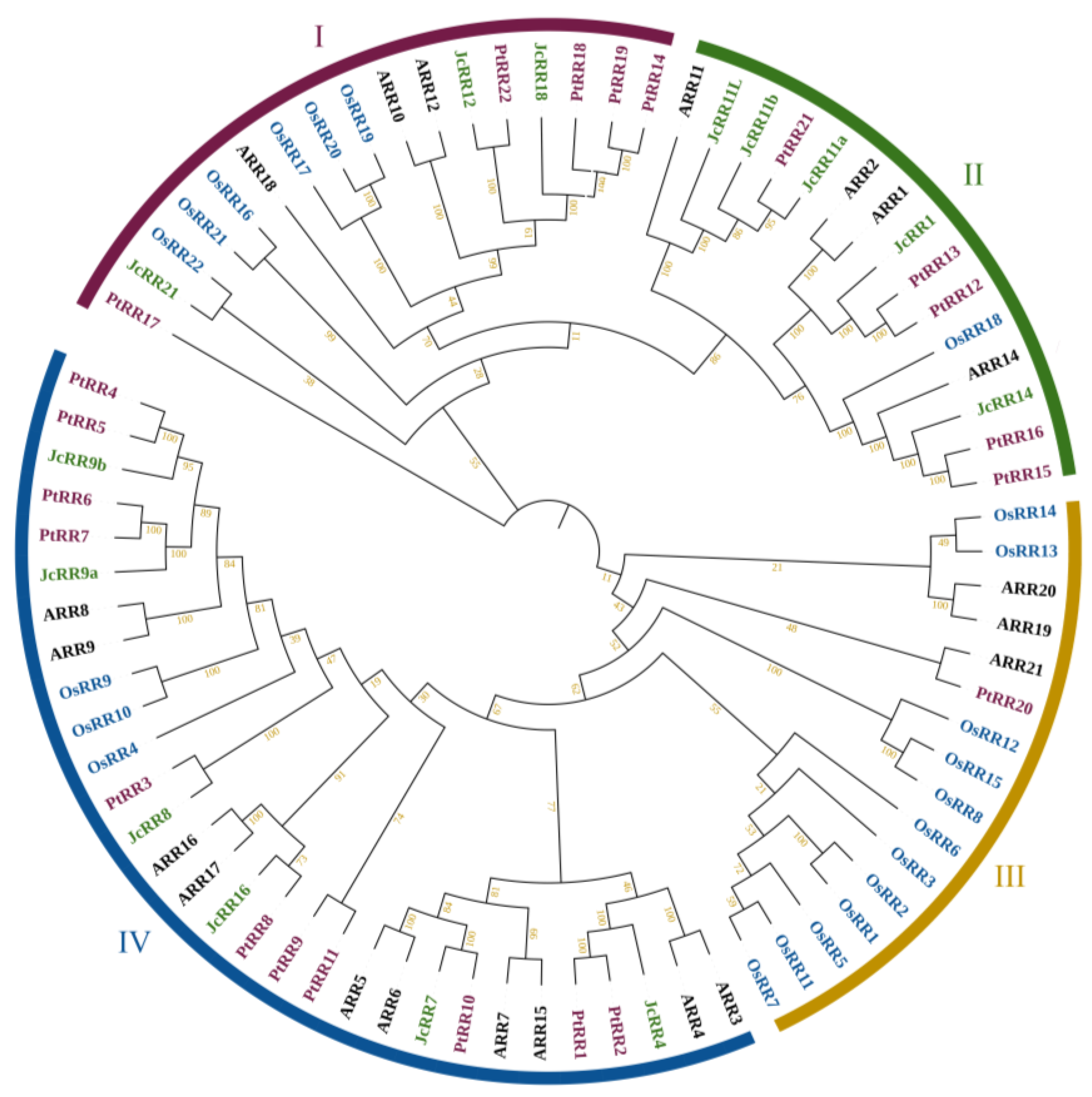
2.2. Identification of the RR Gene Family in J. curcas

Because all the RR members in Arabidopsis, Populus and rice had been identified, and their functions had been characterized, their sequences were selected to query the J. curcas genome, and we first obtained 40 candidates after querying. To identify the RR proteins encoded in the genome of J. curcas, local BLAST searches via the hidden Markov models (HMMs) of the SMART and Pfam databases were conducted. Then we performed a structural domain screening based on the Pfam database. All of the predicted JcRRs shared the same conserved D-D-K receiver domain; furthermore, the type-B JcRRs contained an additional Myb-like DNA-binding domain. Finally, 14 JcRRs members were retained according to the conserved domain of RR within the J. curcas genome (Table 1). There were significantly fewer JcRRs than Arabidopsis RR homologs (21 ARR members), which indicated that the regulatory network and function of RRs in J. curcas may be more flexible and complex. To better compare the functional differences between JcRRs and ARRs, we named these JcRRs according to their homologs in Arabidopsis.
We constructed a phylogenetic tree on the basis of the results of an alignment of RR protein sequences from J. curcas, the dicotyledonous model plant Arabidopsis thaliana, the woody plant species Populus trichocarpa, and the monocotyledonous plant Oryza sativa (Figure 2 and Text S1–S4). The RR proteins were clustered into four monophyletic groups. The results showed that the RR proteins of J. curcas are more closely related to those of the dicotyledonous species Populus and Arabidopsis than to those of the monocotyledonous species rice.
We then predicted the basic characteristics of the JcRR members, including the encoding gene ID, encoding gene type, protein length, molecular weight (MW), theoretical isoelectric point (pI) and subcellular localization (Table 1). The lengths of the JcRR proteins ranged from 158 (JcRR16) to 741 (JcRR14) amino acids (aa), the MW ranged from 17.77 (JcRR16) to 80.28 (JcRR14) kDa, and the theoretical pI values ranged from 4.86 (JcRR8) to 8.28 (JcRR7). Subcellular localization prediction analysis showed that all JcRR proteins were located in the nucleus; therefore, they can interact with various transcription factors. The grand average of hydropathicity (GRAVY) value ranged from −0.817 (JcRR8) to −0.248 (JcRR7), indicating that all the JcRR proteins were hydrophilic.

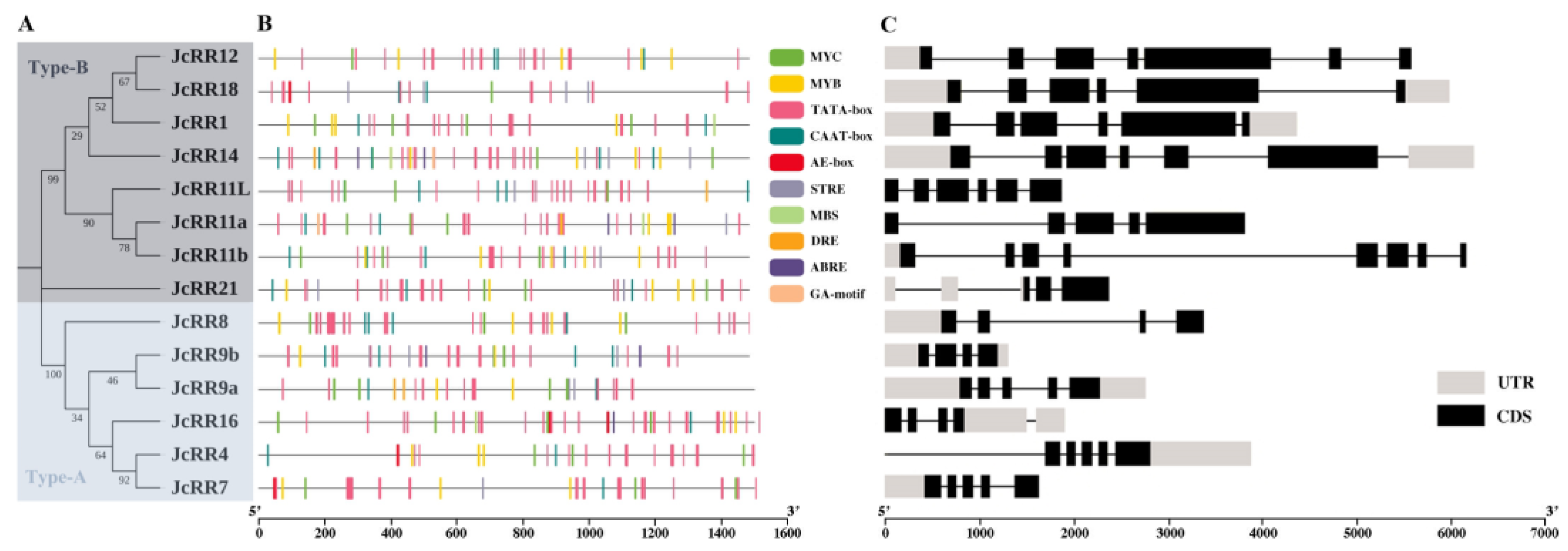
2.3. Gene Structure Analysis and Chromosome Locations

To further explore the structure and sequence characteristics of JcRRs in J. curcas, a simpler neighbor-joining (NJ) phylogenetic tree was constructed on the basis of the results of a classification analysis in which full-length amino acid sequences were used (Text S1). The results of the phylogenetic analysis suggested that the 14 JcRR proteins of J. curcas could be divided into two types: there were 6 type-A and 8 type-B members (Figure 3A).
Cis-acting elements within 1500 base pairs (bp) potential promoter sequences upstream of the start codon were analyzed, and the results were visualized (Figure 3B). The promoter region of the JcRRs contained multiple cis-acting elements, including phytohormone-responsive (abscisic acid (ABA)-responsive and GA motif), light-responsive (AE-box and MYC), plant development-related (MYB, TATA-box and CAAT-box), and stress-responsive elements (STRE, DRE and MBS). These results reflected the potential function of JcRRs during plant development as well as in response to stress. The structures of the JcRRs were relatively different, showing different numbers and positions of exons and introns (Figure 3C). The numbers of introns of JcRRs ranged from 3 (JcRR8 and JcRR9) to 7 (JcRR11b), the numbers of exons of JcRRs ranged from 4 (JcRR8 and JcRR9) to 8 (JcRR11b) and the full genomic length of JcRRs ranged from 1297 (JcRR9b) to 6229 (JcRR14) bp.

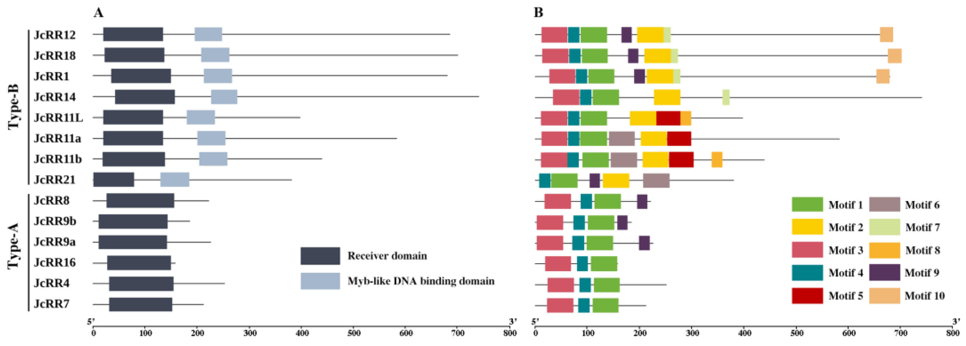
2.4. Conserved Domain and Motif Analysis

To further reveal the diversification among JcRRs, the conserved domains and motifs of the JcRR proteins were analyzed. All the JcRRs shared a conserved typical D-D-K receiver domain (CDD: cd17584), and the type-B JcRRs had an additional Myb-like DNA-binding domain (CDD: pfam00249), which plays a key role in regulating the expression of CK response genes (Figure 4A). The Multiple Em for Motif Elicitation (MEME) online tool was used to predict the conserved motif composition of the JcRRs; the number of motifs varied from 3 to 7. Motifs 1 and 4 were found in all JcRRs, and they may correspond to the receiver domain. Motif 2 was present only in the type-B JcRRs and may correspond to the Myb-like DNA-binding domain. In addition, other motifs were present in different JcRRs, indicating that motifs 5–10 may vary or may have been lost throughout evolution (Figure 4B and Figure S1, and Table S2).

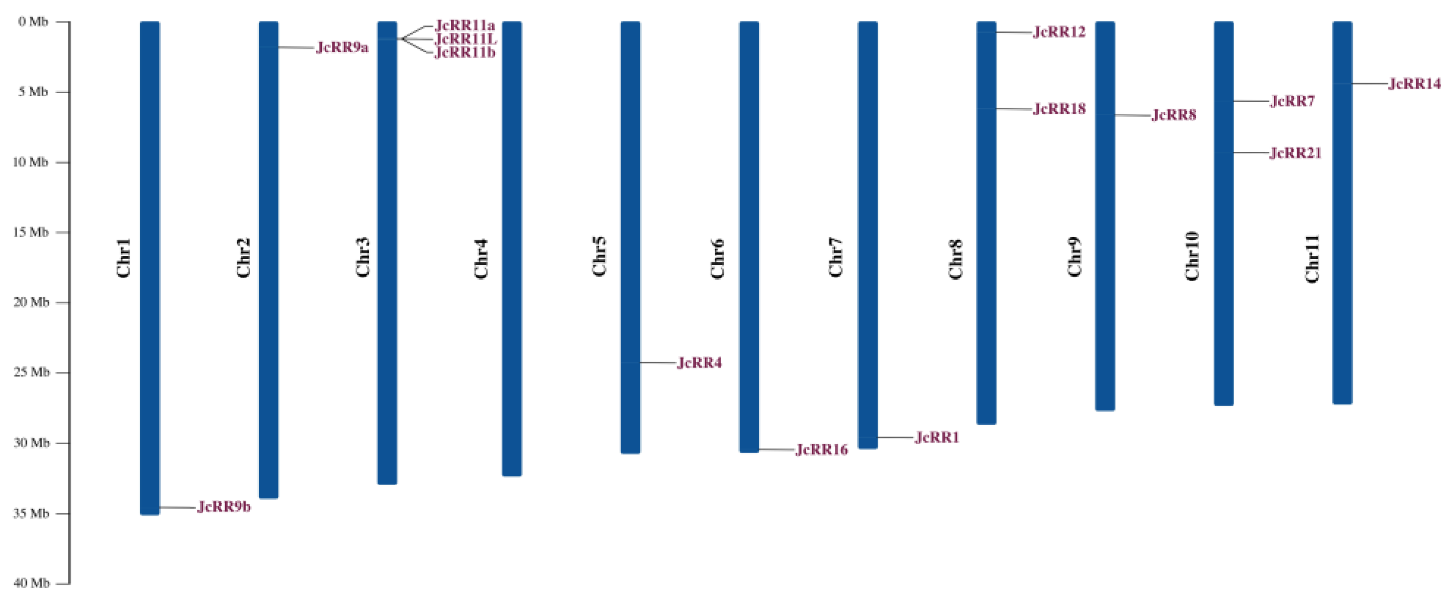
2.5. Chromosomal Distribution and Synteny Analysis

The distribution of JcRRs on the chromosomes was analyzed according to their physical locations in the J. curcas genome. All 14 genes were distributed on 10 chromosomes in J. curcas, and 8 of them were at the proximate or distal ends of the chromosomes. Among these genes, JcRR11a, JcRR11b, and JcRR11L were adjacent to each other on chromosome 3, the phenomenon of which may have been caused by a tandem duplication event (Figure 5).
Then we analyzed the gene duplication events of JcRRs to elucidate the evolutionary relationships of JcRRs. In total, five pairs of duplicated JcRR genes were found, including three pairs of segmental duplication (60% of all duplicated JcRR genes) and two pairs of tandem duplications (40% of all duplicated JcRR genes) (Figure 6A and Table S3). To further understand the evolutionary constraints acting on duplicated JcRRs, the non-synonymous substitution rate (Ka), synonymous substitution rate (Ks), and Ka/Ks ratio of 5 pairs of duplicated JcRRs were calculated (Figure 6B and Table S3). The Ka/Ks ratio of 5 pairs was less than one, which implied that they tended to be subjected to purifying selections. To establish the orthologous relationships of JcRRs, we performed a collinearity analysis between J. curcas and three other species (Arabidopsis, Populus, and rice) (Figure 6C). The results indicated that RRs in J. curcas and Populus (27 pairs) showed a closer evolutionary relationship, followed by Arabidopsis (19 pairs) and rice (7 pairs).

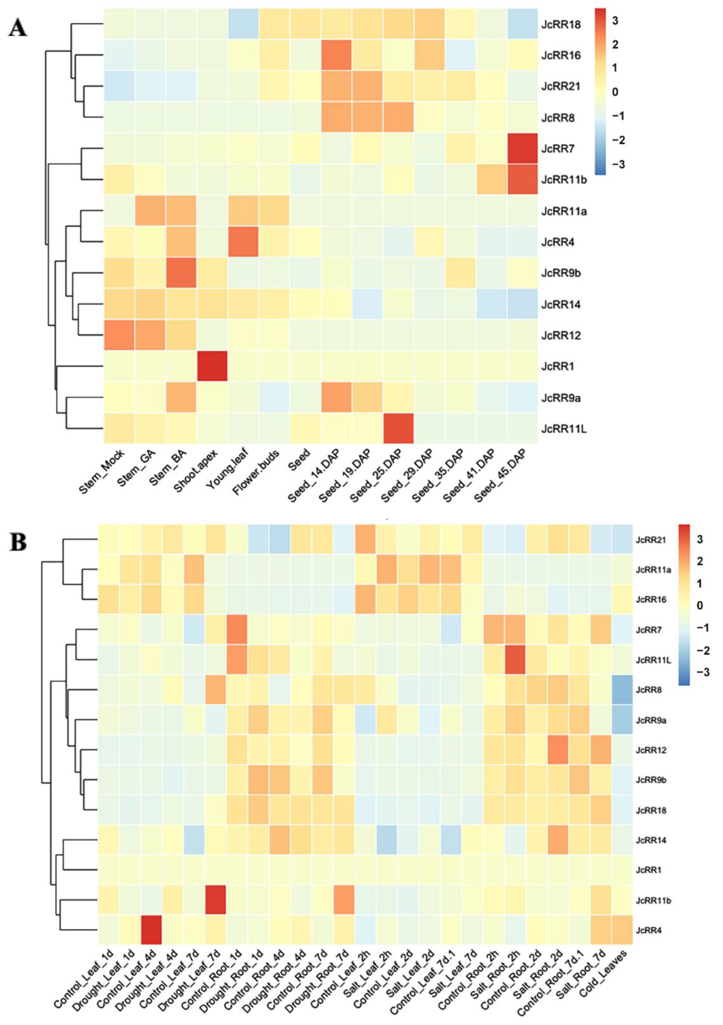
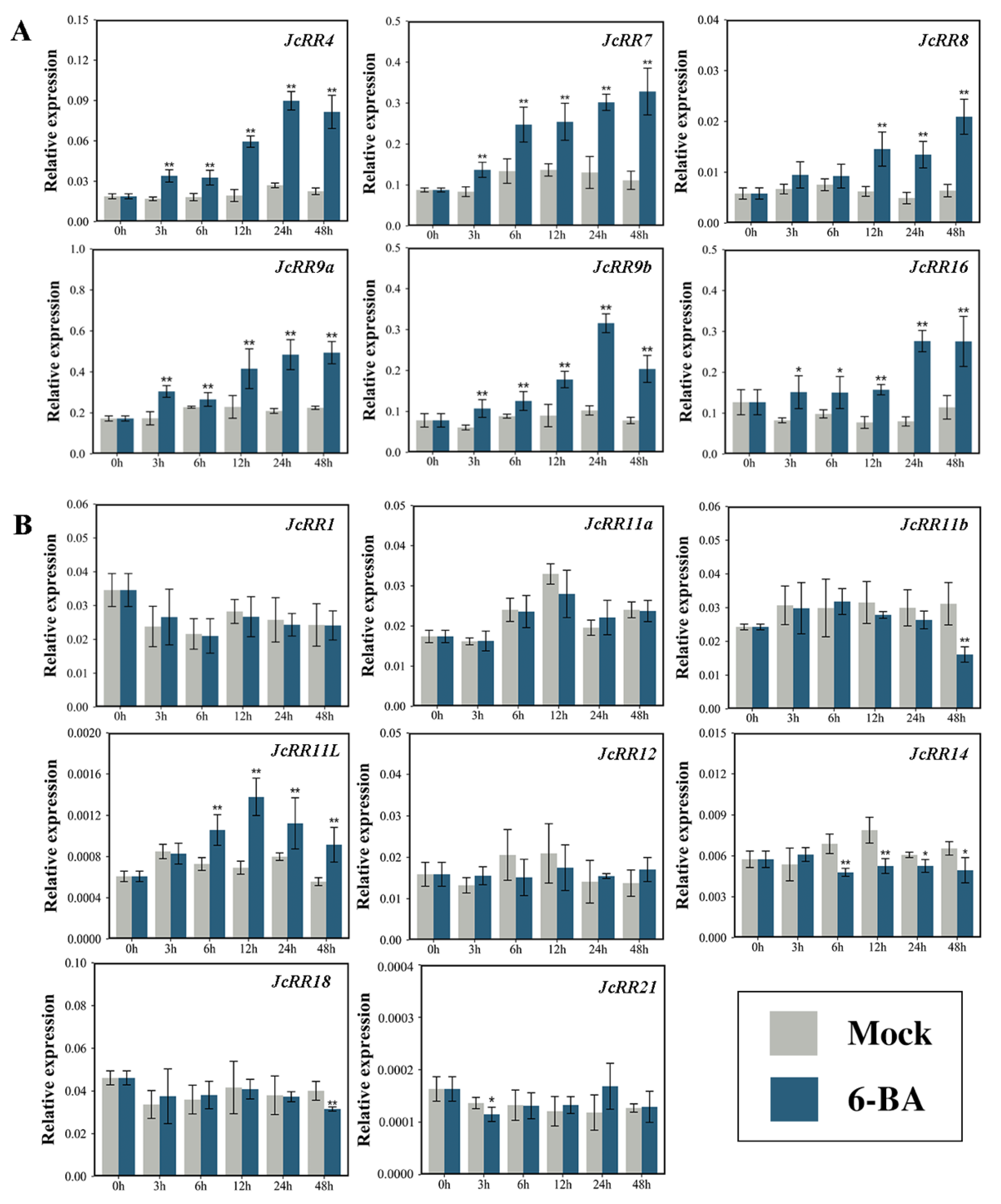
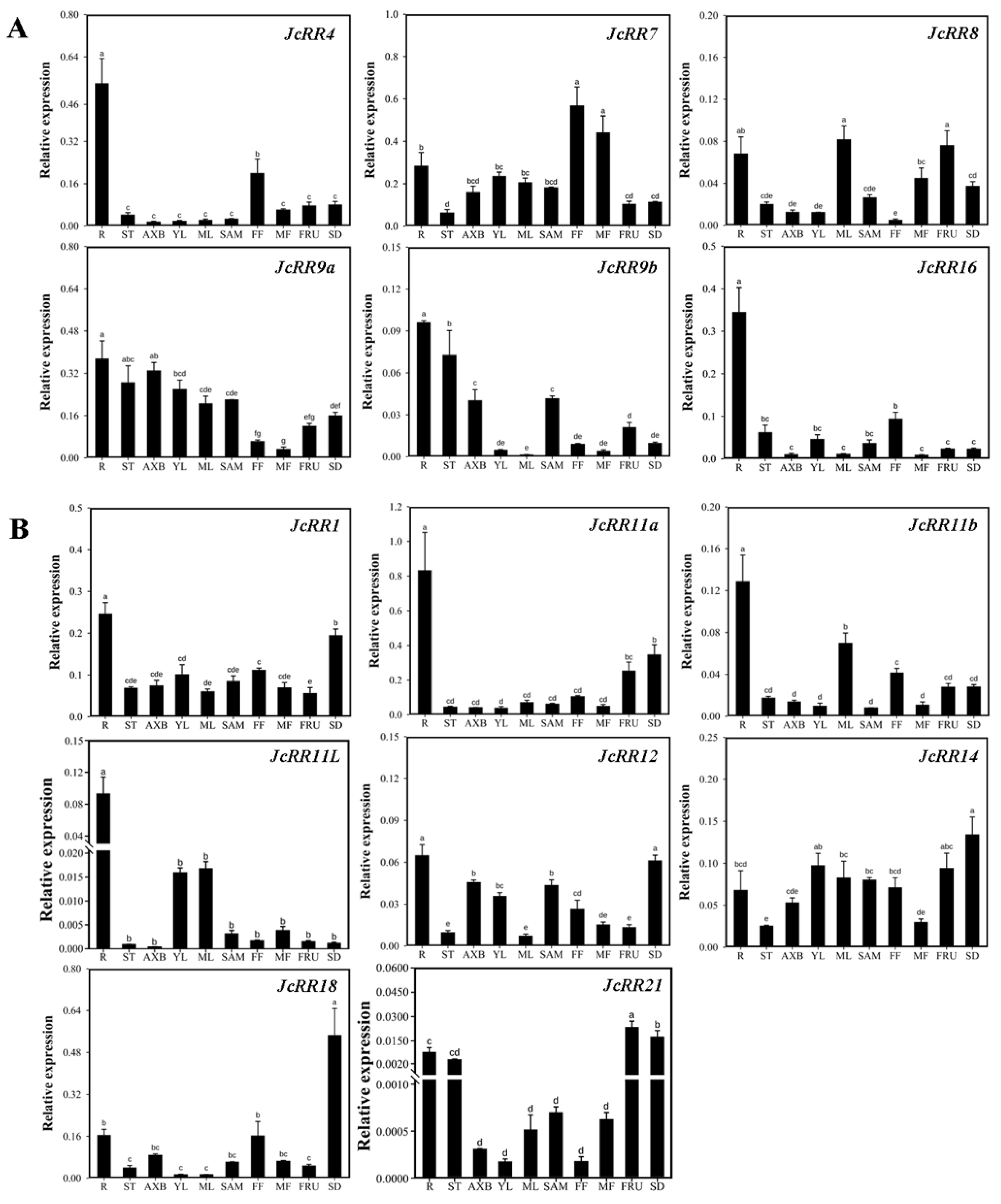
2.6. Expression Profiles of JcRRs

According to the results of the analysis of the cis-acting elements within the promoters (Figure 3B), the expression level of the JcRRs might be affected by hormones and abiotic stresses. To further understand the functions of JcRRs, various transcriptome data were used to generate heatmaps, such as transcriptomic data in response to phytohormones [39], tissues [43,44,45], and abiotic stress [46,47,48] (Table S5). JcRRs showed diverse responses under various treatments and conditions (Figure 7). For hormone responses (Figure 7A), the expression level of JcRR11a strongly increased in response to both GA3 and 6-BA, while the expression level of JcRR21 slightly increased. The expression levels of JcRR4, JcRR9a, and JcRR9b increased in response to 6-BA but decreased in response to GA3. The expression levels of JcRR11b, JcRR11L, and JcRR12 significantly decreased in response to both GA3 and 6-BA. During the development of seeds after pollination, the expression levels of JcRR8, JcRR9a, JcRR11L, JcRR16, and JcRR21 first increased and later decreased. JcRR7 and JcRR11b showed the highest expression levels in the mature seeds. Under drought stress (Figure 7B), the expression of JcRR8 and JcRR11b increased in the leaves, while JcRR11a and JcRR16 were downregulated. The expression level of JcRR11b increased in the roots, while JcRR9a, JcRR9b, and JcRR11L were downregulated. Under salt stress (Figure 7B), the expression levels of JcRR7 and JcRR14 increased in the leaves, while JcRR4, JcRR11a, and JcRR16 were downregulated. The expression levels of JcRR4 and JcRR18 increased in the roots, while JcRR9a and JcRR11L were downregulated. JcRR4 expression was highest in leaves under cold stress, indicating that this gene may play an important role in cold tolerance (Figure 7B). We found that the expression levels of both JcRR11a and JcRR16 decreased in the leaves under drought and salt stresses, and both JcRR9a and JcRR11L were downregulated in the roots. Taken together, these results implied that these genes play potential roles in the abiotic stress response, and further study of their functions is important.
Our previous studies showed that exogenous CK treatment promoted the outgrowth of axillary buds in J. curcas [39,49]. To better understand the regulatory mechanism, we measured the expression level of JcRRs via qRT–PCR analysis after 6-BA treatment (Figure 8). The expression levels of five JcRRs (JcRR4, JcRR7, JcRR9a, JcRR9b, and JcRR16) notably increased at 3 h, while the expression of JcRR8 began to increase at 12 h after 6-BA treatment. The expression of most type-B JcRRs was not induced by CK, a phenomenon that was consistent with that reported in a previous study of Populus [16]. However, the expression of JcRR11L began to increase at 6 h after treatment (Figure 8B), and this gene may be involved in a specific regulatory pathway. The expression levels of JcRR11b, JcRR14, JcRR18, and JcRR21 slightly decreased after treatment. Taken together, these results indicated that type-A JcRRs may play a positive role in the continuous growth of axillary buds, while the roles of type-B JcRRs might be the opposite.
To determine the tissue preferences of the JcRR gene family members, qRT–PCR analysis was conducted to further confirm the expression levels of JcRRs in different tissues (Figure 9). JcRR4 was mainly expressed in the roots, whereas JcRR7 was mainly expressed in the roots, female flowers, and male flowers. JcRR8 was mainly expressed in the roots, male flowers, and fruits, whereas JcRR9a was expressed in all tissues except the female flowers and male flowers. JcRR9b was mainly expressed in the roots, shoot apical meristems, axillary buds, stems, and fruits. JcRR16 showed a high expression level only in the roots. While JcRR1 was expressed the highest in the roots, its expression was relatively consistent in the other tissues. JcRR11a was mainly expressed in the roots, fruits, and seeds, whereas JcRR11b was mainly expressed in the roots, mature leaves, female flowers, and fruits. JcRR11L exhibited high expression in the young leaves and mature leaves and extremely high expression levels in the roots. JcRR12 and JcRR14 were expressed in all the tested tissues. JcRR18 showed the highest expression in the seeds and relatively high levels in the roots and female flowers. Compared to the other JcRRs, JcRR21 showed lower expression and was mainly expressed in the roots, stems, fruits, and seeds. In general, the expression patterns of these 14 JcRRs were consistent with the RNA sequencing (RNA-seq) data. In the future, researchers should consider how JcRRs transmit CK signals and activate downstream genes.

2.7. Overexpression of J. curcas JcRR12 in Arabidopsis Led to Slightly Increased Numbers of Rosette Branches after Decapitation Treatment

To investigate the functions of JcRR12 during axillary bud outgrowth, JcRR12 was overexpressed in Arabidopsis under the control of a CaMV (Capsicum Mottle Virus) 35S promoter. We obtained ten independent 35S:JcRR12 transgenic Arabidopsis lines (Figure 10A). After examining the expression level of each line by qRT-PCR, three lines (OE-3, OE-4 and OE-9) that expressed the highest were retained for subsequent studies (Figure 10B). During plant growth and development, we did not find any changes in the number of rosette branches between wild-type (WT) and transgenic plants under normal growth conditions. To further study the function of JcRR12 in axillary bud outgrowth, we generated a more-branches phenotype by decapitation treatment after the onset of flowering (Figure 10C,D). We found that transgenic plants produced more rosette branches than WT plants after decapitation treatment. These results indicated that JcRR12 was able to affect decapitation-induced axillary bud outgrowth in Arabidopsis.

3. Discussion

Decreased CK completely inhibited GA3-stimulated axillary bud outgrowth and weakened the release of apical dominance. This result indicates that GA-promoted axillary bud outgrowth is dependent on CK and that GA may interact with CK signaling to indirectly influence the expression of downstream related genes. In addition, other factors may regulate axillary bud outgrowth by interacting with CK. Therefore, we performed the identification and analysis to study the function of RR genes in J. curcas, which are key components of cytokinin signaling.
In this study, we identified 14 RR genes in J. curcas, and there are 21, 22 and 22 RR genes in Arabidopsis [50], Populus [16] and rice [51], respectively. We named JcRR members according to their corresponding homologs in Arabidopsis, which facilitated the study of their functions. There were fewer RRs in J. curcas than in other species, which may be due to the loss of some homologous genes during chromosome replication.
Cis-acting elements play an important role in gene expressions regulatory networks, such as those involved in plant growth and development [52], stress responses [53], hormone responses [54,55], and signal transduction [56]. We analyzed the promoter regions of JcRRs and found that many important cis-acting elements are present in the promoters (Figure 3B), such as various plant hormone (abscisic acid (ABA)-responsive and GA motif)-related, light (AE-box and MYC)-related and stress response (STRE, DRE, and MBS) elements. Although previous studies have partially illustrated their role in stress resistance, further studies on the interaction between CK signaling and stress stimuli are needed. Studying the crosstalk between JcRRs and different phytohormones is also valuable. In Arabidopsis, ARR1 recruits DELLA proteins, which are transcriptional coactivators that regulate the expression of CK-responsive genes [57]. We found that CK and GA synergistically regulated the axillary bud outgrowth of J. curcas, and CK could regulate the expression levels of GA synthetase (JcGA20oxs, JcGA3oxs) and degradase (JcGA2oxs) enzyme genes to maintain GA homeostasis [39]. Functional analysis of JcRRs may help to elucidate the regulatory network underlying the interaction between CK and GA signaling during axillary bud outgrowth. An analysis of conserved motifs was performed on the RR gene family proteins, and 10 motifs were identified (Figure 4B). Two conserved motifs corresponding to the D-D-K receiver domain were present in all the proteins, so they were all RR gene family members. Eight type-B members shared the same motif—motif 2 (which corresponds to a Myb-like DNA-binding domain), which was vital for activating downstream genes, while the 6 type-A members did not have this motif. In addition, other motifs were present in different JcRRs, indicating that they may vary or have been lost during evolution (Figure 4B). The numbers and distributions in gene families were determined by duplication events, which helped to explore the differences in functions among gene family members [58]. Among the 14 JcRRs, three gene-pairs from segmental duplication (JcRR9b/JcRR9a, JcRR7/JcRR4, and JcRR12/JcRR18) and two from tandem duplication (JcRR11a/JcRR11L, and JcRR11L/JcRR11b) were found in J. curcas. We found that all the duplicated gene pairs had Ka/Ks ratios of less than one, suggesting shorter divergence of time and more limited functional divergence. There are more collinear blocks of JcRR and PtRR genes, indicating a conserved evolutionary history before the divergence between J. curcas and Populus.
JcRRs showed diverse responses to various treatments and developmental conditions. We generated heat maps to study the response patterns of members of the JcRR gene family (Figure 7). The expression level of JcRR12 decreased in response to GA3 treatment (Figure 7A), the findings of which are consistent with those of a previous study in Arabidopsis showing that GA repressed the expression of ARR1, which was mediated by the DELLA protein RGA [59]. The expression of JcRR11a was strongly upregulated in response to GA and CK (Figure 7A), suggesting that JcRR11a may be involved in processes regulated synergistically by GA and CK, such as axillary bud outgrowth of J. curcas [39,49], although CK and GA are often antagonistic regulators of multiple developmental processes [60,61]. During the development of seeds after pollination, the expression levels of JcRR8, JcRR9a, JcRR11L, JcRR16, and JcRR21 increased at early stages and then decreased, while JcRR7 and JcRR11b showed the highest expression in the mature seeds (at 45 days after pollination (DAP)) (Figure 7A); together, these findings indicated that these JcRRs could play roles in seed development.
To verify the reliability of the transcriptome data shown in Figure 7A, using qRT–PCR analysis, we analyzed the expression patterns of JcRRs in response to 6-BA. Overall, the results of qRT–PCR analysis for the type-A JcRRs were generally consistent with those of the transcriptome data, although this was less so for the type-B JcRRs. All 6 type-A and 1 type-B (JcRR11L) JcRRs were upregulated in response to 6-BA treatment, while 4 type-B JcRRs were downregulated (Figure 8). The expression of most type-B JcRRs was not induced by exogenous 6-BA treatment, which was consistent with the findings of a previous study in Populus [16]. Moreover, our previous study found that overexpressing AtIPT4 under the control of the flower-specific JcTM6 promoter increased the number of inflorescences and led to a high expression level of JcRR3 (named JcRR4 in this article) in the inflorescences [38]. In rice, OsRR5 (a homolog of JcRR9b) specifically participates in strigolactone (SL)-controlled CK responses in shoot bases [62]. Since type-A JcRRs were continuously expressed upon 6-BA treatment, they may promote axillary bud growth and inflorescence formation, while the functions of type-B JcRRs might be opposite.
Under abiotic stress, 2 type-A and 2 type-B JcRRs were consistently downregulated (Figure 7B). Their expression changes may be related to stress resistance. JcRR4 was hardly expressed in the leaves, and the highest expression level occurring in the leaves after cold treatment indicated that this may be vital for cold tolerance. Arabidopsis plants overexpressing ARR5 were drought resistant [63], and accordingly, the expression level of JcRR7 increased after drought stress. OsRR6 (a homolog of JcRR9a) plays an important role in the abiotic stress response and CK signaling [64], while JcRR9a exhibits the same response patterns. Our results imply that these JcRRs may be important to the abiotic stress response. However, further studies are needed to elucidate their functions.
In addition, we performed a qRT–PCR analysis to further explore the expression profiles of JcRRs in different tissues (Figure 9). In general, CKs are mainly produced in the roots and later transported to the shoots [65], but many CKs are also produced in other tissues and organs [66]. In Arabidopsis, the spatial expression pattern of ARR1, ARR2, ARR10, and ARR12 revealed a comprehensive distribution in different tissues, while those of the remaining members varied [67], and the functions of these genes in CK signaling in Arabidopsis were essential but redundant [28]. According to the comprehensive expression profiles, JcRR1 and JcRR12 may be involved in regulatory networks and have functions similar to those of ARR1, ARR2, ARR10, and ARR12 in Arabidopsis. Although most JcRRs showed the highest expression in the roots, spatial expression pattern analysis showed that JcRRs may play various functions among different tissues, excluding their key effects on root development. JcRR7 may regulate the development of flowers, and JcRR8 may be involved in the development of male flowers and fruits. JcRR9a and JcRR9b may participate in cell division, therefore regulating the growth of shoot apical meristems as well as axillary buds. JcRR11a and JcRR11b may have a similar function in terms of regulating fruit development. JcRR11L may regulate the development of leaves, and JcRR18 and JcRR21 may mainly regulate the development of seeds. Taken together, the above results indicated that different JcRRs might be involved in the development of different tissues and organs, laying a foundation for future functional analysis of JcRRs.
The outgrowth of axillary buds was regulated by multiple interactions of hormones, nutritional conditions, and environmental factors. Type-B RRs, the critical component of CK signaling [68], played a vital role in the promotion of axillary bud outgrowth. Type-B ARR1, ARR2, ARR10, and ARR12 synergistically affected shoot regeneration and axillary meristem development in Arabidopsis [69]. Their homologs were respectively identified as JcRR1 and JcRR12 in J. curcas, and we considered that these two genes may play critical roles in CK signaling. However, the regulatory mechanism of type-B RRs is complex, and a recent study found that ARR1 has contrary function to ARR10 in some cases [21]. We found that overexpressing JcRR12 promoted decapitation-induced rosette branch outgrowth in Arabidopsis, which is consistent with the arr1 mutant [22]. However, the regulatory network may be largely different between herbaceous and woody plants. Therefore, further studies of JcRR12 in J. curcas are needed to elucidate the molecular mechanism underlying axillary bud outgrowth.

4. Materials and Methods

4.1. Plant Materials and Growth Conditions

Two-year-old J. curcas trees were planted in the field at Guangxi University, Nanning, Guangxi, China (22°82′ N, 108°32′ E; 100 m above sea level). Seedlings to be used for the 6-BA treatment were grown in the greenhouse (28 °C, 12 h light/12 h dark photoperiod, 70% humidity). Flowers, fruits, and seeds at different developmental stages were collected in May-June 2022 for qRT–PCR analysis. All other samples used for qRT–PCR experiments were collected at the same time in May 2022. Various plant tissue samples, including lateral roots 1–2 mm in diameter with fine roots and root tips, shoot apices 0.3 cm in length from the top of shoots, stems 1.5 cm in diameter, young leaf blades 2 cm in length, mature leaf blades 15 cm in length, the first axillary bud closest to the shoot apical meristem, recently opened female and male flowers, fruits at 15 days after pollination, and seeds 30 days after pollination, were harvested for qRT–PCR analysis. All the tissues were immediately frozen in liquid nitrogen and stored at −80 °C until needed.
Wild-type Arabidopsis thaliana ecotype Columbia (Col-0) and the transgenic lines were grown in an environmentally controlled room (22 °C, 16 h light/8 h dark photoperiod, 70% humidity).

4.2. Sequence Identification and Phylogenetic Tree Construction

The genome sequences and annotations were downloaded from the J. curcas database (JCDB) (http://jcdb.liu-lab.com/, accessed on 13 March 2022) [42], and we further downloaded the genome and annotations of the other three species for comparison in EnsemblPlants (http://EnsemblPlants.org/, accessed on 13 March 2022). The selection of RR members in A. thaliana [50], P. trichocarpa [16], and O. sativa [51] was obtained from published papers, and the sequences of RR members in each species are listed in Text S1–S4. Local BLASTP was used with the default parameters to search the genome sequence of J. curcas; the known Arabidopsis RR sequences were used as queries. The obtained candidate sequences were submitted to the Pfam database (http://pfam.xfam.org, accessed on 15 March 2022) for domain prediction. The sequences of RR genes in J. curcas containing the receiver domain (conserved domain of RR proteins) were ultimately retained. Phylogenetic trees of RR members in four species (Text S1–S4) were constructed by using MEGA 7.0 software (http://www.megasoftware.net (accessed on 15 March 2022)) with the NJ method and 1000 bootstrap replicates and the output was visualized using the online software tool iTOL (https://itol.embl.de/tree/, accessed on 4 June 2022).

4.3. Analysis of Protein Structure and Physicochemical Properties

Protein length, MW, theoretical pI, instability index, and GRAVY values of the JcRR proteins were calculated by using the ProtParam tool (https://web.exp-asy.org/protparam/, accessed on 13 June 2022) [70]. The prediction of subcellular localizations of JcRRs was analyzed by using Plant-mPLoc (http://www.csbio.sjtu.edu.cn/bioinf/plant-multi/, accessed on 13 June 2022) [71]. The results were later visualized by using TBtools (https://github.com/CJ-Chen/TBtools/releases (accessed on 13 March 2022)) [72].

4.4. Cis-Acting Elements in the Promoter Region and Gene Structure Analysis

The 1500 bp upstream coding sequence of JcRRs was obtained. The sequences were submitted to the PlantCARE website (http://bioinformat-ics.psb.ugent.be/web-t-ools/plantcare/html/, accessed on 12 June 2022) to analyze the cis-acting elements.

4.5. Conserved Motif and Domain Prediction

The conserved motifs of the JcRR proteins were analyzed using MEME (https://meme-suite.org/meme/tools/meme, accessed on 4 June 2022). The maximum number of motifs to be identified was set to 10. The conserved domains of JcRR family members were predicted by using the Batch Web CD-Search Tool (https://www.ncbi.nlm.nih.gov/Structure/bwrpsb/bwrpsb.cgi, accessed on 4 June 2022).

4.6. Chromosomal Distribution and Synteny Analysis

Chromosomal localization and synteny analysis between J. curcas and the other three species (Arabidopsis, Populus, and rice) were performed using TBtools (program “Advanced circos” and “One StepMC ScanX”). Then we used the “Simple Ka/Ks Calculator (NG)” program of TBtools to calculate the rates of synonymous substitutions (Ks), nonsynonymous substitutions (Ka), and evolutionary rates (Ka/Ks ratio) of the J. curcas RR gene family. Scatter plot for the Ka/Ks value of duplicated JcRR gene pairs was constructed using R (https://www.r-project.org/, accessed on 11 October 2020, version 4.0.3).

4.7. Hormone, Stress Response, and Spatiotemporal Expression Analysis of JcRRs According to Transcriptome Data

Transcriptome data of J. curcas were downloaded from the JCDB (accessed on 13 March 2022). The raw data were retrieved from the NCBI Sequence Read Archive (SRA) database (https://www.ncbi.nlm.nih.gov/ (accessed on 15 March 2022)), including those in response to hormones [39], from different tissues [43,44,45], from roots and leaves under drought stress [46], under salt stress [47] and from leaves after cold treatment [48] (Table S4). The data were collated, and a matrix was constructed by Microsoft Office Excel (Table S5). Heatmaps for the expression profiles of JcRRs were constructed using R. Data in heatmaps were normalized for each row. Hierarchical clustering was performed, and the default parameters were used.

4.8. Exogenous CK Treatment in J. curcas

Lovastatin (SL8280, Solarbio, Beijing, China), GA3 (G7645, Sigma, Saint Louis, USA), and 6-BA (B3408, Sigma) were first dissolved in 1 M NaOH, anhydrous ethanol, and dimethyl sulfoxide (DMSO) to form a stock solution (20 mM). The lovastatin stock solution was later diluted to 100 µM working solutions, while GA3 and 6-BA were diluted to 200 µM working solutions. For hormone treatments, 20 μL of the working solutions were directly applied to the leaf axil at node 1 of three-week-old J. curcas seedlings [39]. For GA3, lovastatin, and decapitation treatment, the control group was plants treated with the same concentration of ethanol or DMSO, or intact plants, respectively. The length of each axillary bud was measured with a vernier caliper at 14 d after treatment. For the 6-BA treatment, the seedlings were treated at 0 h, 3 h, 6 h, 12 h, 24 h, and 48 h before collection, and the control group was treated with the same concentration of NaOH at the same time. All the samples were collected at one time, and the stems of axillary buds (approximately 10–20 mg, 3–4 mm in length) were separately sliced and immediately frozen in liquid nitrogen for subsequent qRT–PCR analysis.

4.9. mRNA Extraction and qRT–PCR

Total RNA was extracted with a Plant RNA Kit (R6827, OMEGA, Norcross, USA), and cDNA was synthesized using HiScript III All-in-one RT SuperMix Perfect for qPCR (R333, Vazyme, Nanjing, China). Quantitative real-time PCR (qPCR) was performed using ChamQ Universal SYBR qPCR Master Mix (Q311, Vazyme) on a Roche Light Cycler 96 Real-Time PCR Detection System (Roche Diagnostics, Mannheim, Germany), and then the threshold cycle (Ct) values of each gene were collected. The combination of three marker genes JcActin, JcGAPDH, and JcEF1ɑ was used for the normalization of gene expression profiles for all developmental stages [73]. The primers used for qRT–PCR are listed in Table S1. qRT–PCR was performed for three independent biological replicates (tissue samples were harvested from different plants) and three technical replicates per sample. The data were analyzed using the 2ΔΔCT method as described by Livak [74]. The calculation formula is shown below, where y is the expression level of JcRRs, and Ctx1 is the value of each JcRR used for the study. CtxA, CtxG, and CtxE are the Ct values of JcActin, JcGAPDH, and JcEF1ɑ, respectively.
y = 2 [ C t x 1 1 3 ( C t x A + C t x G + C t x E ) ]

4.10. Construction of the JcRR12 Overexpression Vector and Arabidopsis Transformation

The CDS of JcRR12 was amplified with the primers listed in Table S1 (JcRR12-OE-F/R). The product was cloned into the pEASY®-Blunt simple cloning vector (CB111, TransGen, Beijing, China). To generate the 35S:JcRR12 overexpression vector, the plant transformation vector pOCA30 [75] and the pEASY®-Blunt simple cloning vector containing the CDS of JcRR12 were digested by XbaI/SalI, and the resulting fragments were ligated by using T4 DNA ligase (EL0014, Thermo Scientific, Waltham, MA, USA). The generated 35S:JcRR12 plasmid was transformed into Arabidopsis plants using Agrobacterium tumefaciens EHA105. Transformation of Arabidopsis was performed using the floral dip method [76]. Transgenic plants were grown on 1/2 MS medium (PM1061, Coolabor, Beijing, China) containing 50 μg/mL kanamycin (CK6731, Coolabor). After identification, T3 generation homozygous transgenic plants were used for the statistical analysis of phenotypes.

4.11. Decapitation Assays and Branch Counts in Arabidopsis

For decapitation treatment, primary bolts were removed when the plants reached a height of 1 cm after the onset of flowering, and the control group was treated with intact plants. The number of rosette branches was investigated 10 days after treatment. For each group, at least 10 plants were investigated.

4.12. Statistical Analysis

The error bars in all figures indicate the standard deviation. SPSS 26.0 (SPSS, Inc., Chicago, IL, USA) was used to assess statistical significance by one-way ANOVA and Student’s t-test.

5. Conclusions

Exogenous treatment showed that CK was indispensable in GA-promoted axillary bud outgrowth, which indicates that GA may regulate axillary bud outgrowth by interacting with CK signaling. In addition, it also played a vital role in the release of apical dominance. To further elucidate the interaction mechanism, in this study, we identified 14 members of the RR gene family in J. curcas. The physicochemical characteristics, phylogenetic relationships, subcellular localization, and conserved structural domains of JcRR proteins and the chromosomal localization and gene structure of JcRRs were analyzed. Several vital cis-acting elements in the promoter region of the JcRRs were identified, and these elements are related to multiple hormones and stress responses.
The temporospatial expression patterns of JcRRs during the development of various tissues and their response to phytohormones and abiotic stress were further characterized via analysis of RNA-seq and qRT–PCR data. The results suggested that JcRRs are widely involved in phytohormone crosstalk and abiotic stress responses. JcRRs were mainly expressed in the roots, while they also showed differential expression patterns in other tissues, suggesting that JcRRs may also play vital roles in the development of other tissues in addition to roots. The preliminary functional analysis of JcRR12 in Arabidopsis indicates its role in the decapitation-induced axillary bud outgrowth, which deserves further investigation in J. curcas. In conclusion, our study provides information for further functional studies of JcRRs, especially those that may be involved in axillary bud outgrowth, flower sex differentiation, and stress tolerance.

Supplementary Materials

The supporting information can be downloaded at: https://www.mdpi.com/article/10.3390/ijms231911388/s1.

Author Contributions

X.G. and Z.-F.X. planned and designed the study. X.G., C.Z. and L.W. performed the experiments. K.L. implemented the software. X.G. and C.Z. collected the sequences of genes in this work. X.G. and Z.-F.X. wrote the manuscript. All authors have read and agreed to the published version of the manuscript.

Funding

This work was supported by the Guangxi Postgraduate Innovation Project Education Plan (no. YCSW2021022) from the education department of Guangxi province, the funding from the Natural Science Foundation of China (no. 31771605), and the start-up research funds from the Guangxi University.

Institutional Review Board Statement

This study did not involve humans or animals.

Informed Consent Statement

Informed consent was obtained from all subjects involved in the study.

Data Availability Statement

Not applicable.

Acknowledgments

We thank Maosheng Chen from Xishuangbanna Tropical Botanical Garden, Chinese Academy of Sciences for his help in bioinformatic analysis. And we thank Jun Ni from Guangxi University for his help in the design of experiments.

Conflicts of Interest

The authors declare no conflict of interest.

References

  1. Lindsay, D.L.; Sawhney, V.K.; Bonham-Smith, P.C. Cytokinin-induced changes in CLAVATA1 and WUSCHEL expression temporally coincide with altered floral development in Arabidopsis. Plant Sci. 2006, 170, 1111–1117. [Google Scholar] [CrossRef]
  2. Werner, T.; Motyka, V.; Laucou, V.; Smets, R.; Van Onckelen, H.; Schmulling, T. Cytokinin-deficient transgenic Arabidopsis plants show multiple developmental alterations indicating opposite functions of cytokinins in the regulation of shoot and root meristem activity. Plant Cell 2003, 15, 2532–2550. [Google Scholar] [CrossRef] [PubMed]
  3. Gordon, S.P.; Chickarmane, V.S.; Ohno, C.; Meyerowitz, E.M. Multiple feedback loops through cytokinin signaling control stem cell number within the Arabidopsis shoot meristem. Proc. Natl. Acad. Sci. USA 2009, 106, 16529–16534. [Google Scholar] [CrossRef]
  4. Aloni, R. Role of cytokinin in differentiation of secondary xylem fibers. Plant Phyisol. 1982, 70, 1631–1633. [Google Scholar] [CrossRef]
  5. Lara, M.E.B.; Garcia, M.C.G.; Fatima, T.; Ehness, R.; Lee, T.K.; Proels, R.; Tanner, W.; Roitsch, T. Extracellular invertase is an essential component of cytokinin-mediated delay of senescence. Plant Cell 2004, 16, 1276–1287. [Google Scholar] [CrossRef] [PubMed]
  6. Bishopp, A.; Help, H.; El-Showk, S.; Weijers, D.; Scheres, B.; Friml, J.; Benkova, E.; Mahonen, A.P.; Helariutta, Y. A mutually inhibitory interaction between auxin and cytokinin specifies vascular pattern in roots. Curr. Biol. 2011, 21, 917–926. [Google Scholar] [CrossRef] [PubMed]
  7. Jameson, P.E.; Song, J. Cytokinin: A key driver of seed yield. J. Exp. Bot. 2016, 67, 593–606. [Google Scholar] [CrossRef] [PubMed]
  8. Peleg, Z.; Reguera, M.; Tumimbang, E.; Walia, H.; Blumwald, E. Cytokinin-mediated source/sink modifications improve drought tolerance and increase grain yield in rice under water-stress. Plant Biotechnol. J. 2011, 9, 747–758. [Google Scholar] [CrossRef] [PubMed]
  9. Ramireddy, E.; Chang, L.; Schmulling, T. Cytokinin as a mediator for regulating root system architecture in response to environmental cues. Plant Signal. Behav. 2014, 9, e27771. [Google Scholar] [CrossRef]
  10. Roman, H.; Girault, T.; Barbier, F.; Péron, T.; Brouard, N.; Pěnčík, A.; Novák, O.; Vian, A.; Sakr, S.; Lothier, J.; et al. Cytokinins are initial targets of light in the control of bud outgrowth. Plant Physiol. 2016, 172, 489–509. [Google Scholar] [CrossRef] [Green Version]
  11. Werner, T.; Schmülling, T. Cytokinin action in plant development. Curr. Opin. Plant Biol. 2009, 12, 527–538. [Google Scholar] [CrossRef] [PubMed]
  12. Hutchison, C.E.; Kieber, J.J. Cytokinin signaling in Arabidopsis. Plant Cell 2002, 14, S47–S59. [Google Scholar] [CrossRef]
  13. Imamura, A.; Hanaki, N.; Nakamura, A.; Suzuki, T.; Taniguchi, M.; Kiba, T.; Ueguchi, C.; Sugiyama, T.; Mizuno, T. Compilation and characterization of Arabiopsis thaliana response regulators implicated in His-Asp phosphorelay signal transduction. Plant Cell Physiol. 1999, 40, 733–742. [Google Scholar] [CrossRef] [PubMed]
  14. Zubo, Y.O.; Blakley, I.C.; Yamburenko, M.V.; Worthen, J.M.; Street, I.H.; Franco-Zorrilla, J.M.; Zhang, W.; Hill, K.; Raines, T.; Solano, R.; et al. Cytokinin induces genome-wide binding of the type-B response regulator ARR10 to regulate growth and development in Arabidopsis. Proc. Natl. Acad. Sci. USA 2017, 114, E5995–E6004. [Google Scholar] [CrossRef] [PubMed]
  15. To, J.P.C.; Deruere, J.; Maxwell, B.B.; Morris, V.F.; Hutchison, C.E.; Ferreira, F.J.; Schaller, G.E.; Kieber, J.J. Cytokinin regulates type-A Arabidopsis response regulator activity and protein stability via two-component phosphorelay. Plant Cell 2007, 19, 3901–3914. [Google Scholar] [CrossRef] [PubMed]
  16. Ramírez-Carvajal, G.A.; Morse, A.M.; Davis, J.M. Transcript profiles of the cytokinin response regulator gene family in Populus imply diverse roles in plant development. New Phytol. 2008, 177, 77–89. [Google Scholar] [CrossRef]
  17. Zeng, J.; Zhu, X.; Haider, M.S.; Wang, X.; Zhang, C.; Wang, C. Genome-wide identification and analysis of the type-B authentic response regulator gene family in peach (Prunus persica). Cytogenet. Genome Res. 2017, 151, 41–49. [Google Scholar] [CrossRef]
  18. Li, H.; Chen, R.; Chen, Z.; Lin, J.; Jin, X.; Ren, C.; Chen, Q.; Chen, F.; Yu, G.; Zhang, Y. The molecular characteristics of soybean ARR-B transcription factors. Biocell 2022, 46, 1575–1592. [Google Scholar] [CrossRef]
  19. Chen, C.; Liu, A.; Ren, H.; Yu, Y.; Duanmu, H.; Duan, X.; Sun, X.; Liu, B.; Zhu, Y. Genome-wide analysis of Glycine soja response regulator GsRR genes under alkali and salt stresses. Front. Plant Sci. 2018, 9, 1306. [Google Scholar] [CrossRef]
  20. D’Agostino, I.B.; Deruere, J.; Kieber, J.J. Characterization of the response of the Arabidopsis response regulator gene family to cytokinin. Plant Physiol. 2000, 124, 1706–1717. [Google Scholar] [CrossRef] [Green Version]
  21. Liu, Z.; Dai, X.; Li, J.; Liu, N.; Liu, X.; Li, S.; Xiang, F. The type-B cytokinin response regulator ARR1 inhibits shoot regeneration in an ARR12-dependent manner in Arabidopsis. Plant Cell 2020, 32, 2271–2291. [Google Scholar] [CrossRef]
  22. Waldie, T.; Leyser, O. Cytokinin targets auxin transport to promote shoot branching. Plant Phyisol. 2018, 177, 803–818. [Google Scholar] [CrossRef]
  23. Xia, X.; Dong, H.; Yin, Y.; Song, X.; Gu, X.; Sang, K.; Zhou, J.; Shi, K.; Zhou, Y.; Foyer, C.H.; et al. Brassinosteroid signaling integrates multiple pathways to release apical dominance in tomato. Proc. Natl. Acad. Sci. USA 2021, 118, e2004384118. [Google Scholar] [CrossRef]
  24. Yamburenko, M.V.; Worthen, J.M.; Zeenat, A.; Azhar, B.J.; Swain, S.; Couitt, A.R.; Shakeel, S.N.; Kieber, J.J.; Schaller, G.E. Functional analysis of the rice type-B response regulator RR22. Front. Plant Sci. 2020, 11, 577676. [Google Scholar] [CrossRef] [PubMed]
  25. Shi, F.; Wang, M.; An, Y. Overexpression of a B-type cytokinin response regulator (OsORR2) reduces plant height in rice. Plant Signal. Behav. 2020, 15, 1780405. [Google Scholar] [CrossRef] [PubMed]
  26. Ramirez-Carvajal, G.A.; Morse, A.M.; Dervinis, C.; Davis, J.M. The cytokinin type-B response regulator PtRR13 Is a negative regulator of adventitious root development in Populus. Plant Physiol. 2009, 150, 759–771. [Google Scholar] [CrossRef] [PubMed]
  27. Müller, B.; Sheen, J. Cytokinin and auxin interaction in root stem-cell specification during early embryogenesis. Nature 2008, 453, 1094–1097. [Google Scholar] [CrossRef] [PubMed]
  28. Ishida, K.; Yamashino, T.; Yokoyama, A.; Mizuno, T. Three type-B response regulators, ARR1, ARR10 and ARR12, play essential but redundant roles in cytokinin signal transduction throughout the life cycle of Arabidopsis thaliana. Plant Cell Physiol. 2008, 49, 47–57. [Google Scholar] [CrossRef]
  29. Nguyen, K.H.; Van Ha, C.; Nishiyama, R.; Watanabe, Y.; Leyva-González, M.A.; Fujita, Y.; Tran, U.T.; Li, W.; Tanaka, M.; Seki, M.; et al. Arabidopsis type B cytokinin response regulators ARR1, ARR10, and ARR12 negatively regulate plant responses to drought. Proc. Natl. Acad. Sci. USA 2016, 113, 3090–3095. [Google Scholar] [CrossRef]
  30. Zeng, R.; Li, Z.; Shi, Y.; Fu, D.; Yin, P.; Cheng, J.; Jiang, C.; Yang, S. Natural variation in a type-A response regulator confers maize chilling tolerance. Nat.Commun. 2021, 12, 4713. [Google Scholar] [CrossRef] [PubMed]
  31. Pandey, V.C.; Singh, K.; Singh, J.S.; Kumar, A.; Singh, B.; Singh, R.P. Jatropha curcas: A potential biofuel plant for sustainable environmental development. Renew. Sustain. Energy Rev. 2012, 16, 2870–2883. [Google Scholar] [CrossRef]
  32. Makkar, H.P.S.; Becker, K. Jatropha curcas, a promising crop for the generation of biodiesel and value-added coproducts. Eur. J. Lipid. Sci. Technol. 2009, 111, 773–787. [Google Scholar] [CrossRef]
  33. Singh, D.; Sharma, D.; Soni, S.L.; Inda, C.S.; Sharma, S.; Sharma, P.K.; Jhalani, A. A comprehensive review of physicochemical properties, production process, performance and emissions characteristics of 2nd generation biodiesel feedstock: Jatropha curcas. Fuel 2021, 285, 119110. [Google Scholar] [CrossRef]
  34. Divakara, B.N.; Upadhyaya, H.D.; Wani, S.P.; Gowda, C.L.L. Biology and genetic improvement of Jatropha curcas L.: A review. Appl. Energy 2010, 87, 732–742. [Google Scholar] [CrossRef]
  35. Mazumdar, P.; Singh, P.; Babu, S.; Siva, R.; Harikrishna, J.A. An update on biological advancement of Jatropha curcas L.: New insight and challenges. Renew. Sustain. Energy Rev. 2018, 91, 903–917. [Google Scholar] [CrossRef]
  36. Ewunie, G.A.; Morken, J.; Lekang, O.I.; Yigezu, Z.D. Factors affecting the potential of Jatropha curcas for sustainable biodiesel production: A critical review. Renew. Sustain. Energy Rev. 2021, 137, 110500. [Google Scholar] [CrossRef]
  37. Lama, A.D.; Klemola, T.; Saloniemi, I.; Niemelä, P.; Vuorisalo, T. Factors affecting genetic and seed yield variability of Jatropha curcas (L.) across the globe: A review. Energy Sustain. Dev. 2018, 42, 170–182. [Google Scholar] [CrossRef]
  38. Ming, X.; Tao, Y.-B.; Fu, Q.; Tang, M.; He, H.; Chen, M.-S.; Pan, B.-Z.; Xu, Z.-F. Flower-specific overproduction of cytokinins altered flower development and sex expression in the perennial woody plant Jatropha curcas L. Int. J. Mol. Sci. 2020, 21, 640. [Google Scholar] [CrossRef]
  39. Ni, J.; Zhao, M.-L.; Chen, M.-S.; Pan, B.-Z.; Tao, Y.-B.; Xu, Z.-F. Comparative transcriptome analysis of axillary buds in response to the shoot branching regulators gibberellin A3 and 6-benzyladenine in Jatropha curcas. Sci. Rep. 2017, 7, 11417. [Google Scholar] [CrossRef]
  40. Chen, M.-S.; Zhao, M.-L.; Wang, G.-J.; He, H.-Y.; Bai, X.; Pan, B.-Z.; Fu, Q.-T.; Tao, Y.-B.; Tang, M.-Y.; Martínez-Herrera, J.; et al. Transcriptome analysis of two inflorescence branching mutants reveals cytokinin is an important regulator in controlling inflorescence architecture in the woody plant Jatropha curcas. BMC Plant Biol. 2019, 19, 468. [Google Scholar] [CrossRef]
  41. Chen, M.-S.; Niu, L.; Zhao, M.-L.; Xu, C.; Pan, B.-Z.; Fu, Q.; Tao, Y.-B.; He, H.; Hou, C.; Xu, Z.-F. De novo genome assembly and Hi-C analysis reveal an association between chromatin architecture alterations and sex differentiation in the woody plant Jatropha curcas. GigaScience 2020, 9, giaa009. [Google Scholar] [CrossRef] [PubMed]
  42. Zhang, X.; Pan, B.-Z.; Chen, M.; Chen, W.; Li, J.; Xu, Z.-F.; Liu, C. JCDB: A comprehensive knowledge base for Jatropha curcas, an emerging model for woody energy plants. BMC Genom. 2019, 20, 958. [Google Scholar] [CrossRef]
  43. Niu, L.; Tao, Y.-B.; Chen, M.-S.; Fu, Q.; Dong, Y.; He, H.; Xu, Z.-F. Identification and characterization of tetraploid and octoploid Jatropha curcas induced by colchicine. Caryologia 2016, 69, 58–66. [Google Scholar] [CrossRef]
  44. Xu, G.; Huang, J.; Yang, Y.; Yao, Y.-A. Transcriptome analysis of flower sex differentiation in Jatropha curcas L. using RNA sequencing. PLoS ONE 2016, 11, e0145613. [Google Scholar] [CrossRef] [PubMed]
  45. Jiang, H.; Wu, P.; Zhang, S.; Song, C.; Chen, Y.; Li, M.; Jia, Y.; Fang, X.; Chen, F.; Wu, G. Global analysis of gene expression profiles in developing physic nut (Jatropha curcas L.) seeds. PLoS ONE 2012, 7, e36522. [Google Scholar] [CrossRef]
  46. Juntawong, P.; Sirikhachornkit, A.; Pimjan, R.; Sonthirod, C.; Sangsrakru, D.; Yoocha, T.; Tangphatsornruang, S.; Srinives, P. Elucidation of the molecular responses to waterlogging in Jatropha roots by transcriptome profiling. Front. Plant Sci. 2014, 5, 658. [Google Scholar] [CrossRef]
  47. Zhang, L.; Zhang, C.; Wu, P.; Chen, Y.; Li, M.; Jiang, H.; Wu, G. Global analysis of gene expression profiles in physic nut (Jatropha curcas L.) seedlings exposed to salt stress. PLoS ONE 2014, 9, e97878. [Google Scholar]
  48. Wang, H.; Zou, Z.; Wang, S.; Gong, M. Global analysis of transcriptome responses and gene expression profiles to cold stress of Jatropha curcas L. PLoS ONE 2013, 8, e82817. [Google Scholar] [CrossRef]
  49. Ni, J.; Gao, C.; Chen, M.-S.; Pan, B.-Z.; Ye, K.; Xu, Z.-F. Gibberellin promotes shoot branching in the perennial woody plant Jatropha curcas. Plant Cell Physiol. 2015, 56, 1655–1666. [Google Scholar] [CrossRef]
  50. Hwang, I.; Sheen, J.; Müller, B. Cytokinin signaling networks. Annu. Rev. 2012, 63, 353–380. [Google Scholar] [CrossRef]
  51. Du, L.; Jiao, F.; Chu, J.; Jin, G.; Chen, M.; Wu, P. The two-component signal system in rice (Oryza sativa L.): A genome-wide study of cytokinin signal perception and transduction. Genomics 2007, 89, 697–707. [Google Scholar] [CrossRef] [PubMed] [Green Version]
  52. Manevski, A.; Bertoni, G.; Bardet, C.; Tremousaygue, D.; Lescure, B. In synergy with various cis-acting elements, plant insterstitial telomere motifs regulate gene expression in Arabidopsis root meristems. FEBS Lett. 2000, 483, 43–46. [Google Scholar] [CrossRef]
  53. Yamaguchi-Shinozaki, K.; Shinozaki, K. Organization of cis-acting regulatory elements in osmotic- and cold-stress-responsive promoters. Trends Plant Sci. 2005, 10, 88–94. [Google Scholar] [CrossRef] [PubMed]
  54. Narusaka, Y.; Nakashima, K.; Shinwari, Z.K.; Sakuma, Y.; Furihata, T.; Abe, H.; Narusaka, M.; Shinozaki, K.; Yamaguchi-Shinozaki, K. Interaction between two cis-acting elements, ABRE and DRE, in ABA-dependent expression of Arabidopsis rd29A gene in response to dehydration and high-salinity stresses. Plant J. 2003, 34, 137–148. [Google Scholar] [CrossRef] [PubMed]
  55. Sutoh, K.; Yamauchi, D. Two cis-acting elements necessary and sufficient for gibberellin-upregulated proteinase expression in rice seeds. Plant J. 2003, 34, 635–645. [Google Scholar] [CrossRef]
  56. Sleckman, B.; Gorman, J.; Alt, F. Accessibility control of antigen-receptor variable-region gene assembly: Role of cis-acting elements. Annu. Rev. Immunol. 1996, 14, 459–481. [Google Scholar] [CrossRef] [PubMed]
  57. Marin-de la Rosa, N.; Pfeiffer, A.; Hill, K.; Locascio, A.; Bhalerao, R.P.; Miskolczi, P.; Gronlund, A.L.; Wanchoo-Kohli, A.; Thomas, S.G.; Bennett, M.J.; et al. Genome wide binding site analysis reveals transcriptional coactivation of cytokinin-responsive genes by DELLA proteins. PLoS Genet. 2015, 11, e1005337. [Google Scholar] [CrossRef] [PubMed]
  58. Cannon, S.B.; Mitra, A.; Baumgarten, A.; Young, N.D.; May, G. The roles of segmental and tandem gene duplication in the evolution of large gene families in Arabidopsis thaliana. BMC Plant Biol. 2004, 4, 10. [Google Scholar] [CrossRef]
  59. Moubayidin, L.; Perilli, S.; Dello Ioio, R.; Di Mambro, R.; Costantino, P.; Sabatini, S. The rate of cell differentiation controls the Arabidopsis root meristem growth phase. Curr. Biol. 2010, 20, 1138–1143. [Google Scholar] [CrossRef]
  60. Fleishon, S.; Shani, E.; Ori, N.; Weiss, D. Negative reciprocal interactions between gibberellin and cytokinin in tomato. New Phytol. 2011, 190, 609–617. [Google Scholar] [CrossRef]
  61. Weiss, D.; Ori, N. Mechanisms of cross talk between gibberellin and other hormones. Plant Physiol. 2007, 144, 1240–1246. [Google Scholar] [CrossRef] [PubMed] [Green Version]
  62. Duan, J.; Yu, H.; Yuan, K.; Liao, Z.; Meng, X.; Jing, Y.; Liu, G.; Chu, J.; Li, J. Strigolactone promotes cytokinin degradation through transcriptional activation of CYTOKININ OXIDASE/DEHYDROGENASE 9 in rice. Proc. Natl. Acad. Sci. USA 2019, 116, 14319–14324. [Google Scholar] [CrossRef]
  63. Huang, X.; Hou, L.; Meng, J.; You, H.; Li, Z.; Gong, Z.; Yang, S.; Shi, Y. The antagonistic action of abscisic acid and cytokinin signaling mediates drought stress response in Arabidopsis. Mol. Plant 2018, 11, 970–982. [Google Scholar] [CrossRef] [PubMed]
  64. Jain, M.; Tyagi, A.K.; Khurana, J.P. Molecular characterization and differential expression of cytokinin-responsive type-A response regulators in rice (Oryza sativa). BMC Plant Biol. 2006, 6, 1. [Google Scholar] [CrossRef] [PubMed]
  65. Müller, D.; Leyser, O. Auxin, cytokinin and the control of shoot branching. Ann. Bot. 2011, 107, 1203–1212. [Google Scholar] [CrossRef]
  66. Miyawaki, K.; Matsumoto-Kitano, M.; Kakimoto, T. Expression of cytokinin biosynthetic isopentenyltransferase genes in Arabidopsis: Tissue specificity and regulation by auxin, cytokinin, and nitrate. Plant J. 2004, 37, 128–138. [Google Scholar] [CrossRef]
  67. Jameson, P.E. Cytokinins. In Encyclopedia of Applied Plant Sciences, 2nd ed.; Thomas, B., Murray, B.G., Murphy, D.J., Eds.; Academic Press: Oxford, UK, 2017; pp. 391–402. [Google Scholar]
  68. Worthen, J.M.; Yamburenko, M.V.; Lim, J.; Nimchuk, Z.L.; Kieber, J.J.; Schaller, G.E. Type-B response regulators of rice play key roles in growth, development and cytokinin signaling. Development 2019, 146, dev174870. [Google Scholar] [CrossRef]
  69. Zhang, F.; May, A.; Irish, V.F. Type-B ARABIDOPSIS resonse regulators directly activate WUSCHEL. Trends Plant Sci. 2017, 22, 815–817. [Google Scholar] [CrossRef]
  70. Gasteiger, E.; Hoogland, C.; Gattiker, A.; Duvaud, S.; Wilkins, M.R.; Appel, R.D.; Bairoch, A. Protein identification and analysis tools on the ExPASy server. In The Proteomics Protocols Handbook; Springer: Berlin/Heidelberg, Germany, 2005; pp. 571–607. [Google Scholar]
  71. Chou, K.-C.; Shen, H.-B. Plant-mPLoc: A top-down strategy to augment the power for predicting plant protein subcellular localization. PLoS ONE 2010, 5, e11335. [Google Scholar] [CrossRef]
  72. Chen, C.; Chen, H.; Zhang, Y.; Thomas, H.R.; Frank, M.H.; He, Y.; Xia, R. TBtools: An integrative toolkit developed for interactive analyses of big biological data. Mol. Plant 2020, 13, 1194–1202. [Google Scholar] [CrossRef]
  73. Zhang, L.; He, L.-L.; Fu, Q.-T.; Xu, Z.-F. Selection of reliable reference genes for gene expression studies in the biofuel plant Jatropha curcas using real-time quantitative PCR. Int. J. Mol. Sci. 2013, 14, 24338–24354. [Google Scholar] [CrossRef] [PubMed] [Green Version]
  74. Livak, K.J.; Schmittgen, T.D. Analysis of relative gene expression data using real-time quantitative PCR and the 2−ΔΔCT method. Methods 2001, 25, 402–408. [Google Scholar] [CrossRef] [PubMed]
  75. Chen, C.H.; Chen, Z.X. Potentiation of developmentally regulated plant defense response by AtWRKY18, a pathogen-induced Arabidopsis transcription factor. Plant Phyisol. 2002, 129, 706–716. [Google Scholar] [CrossRef] [PubMed]
  76. Clough, S.J.; Bent, A.F. Floral dip: A simplified method for Agrobacterium-mediated transformation of Arabidopsis thaliana. Plant J. 1998, 16, 735–743. [Google Scholar] [CrossRef] [PubMed] [Green Version]
Figure 1. Lovastatin (Lov) inhibited the gibberellin A3 (GA3)- and decapitation-promoted outgrowth of axillary buds in 3-week-old J. curcas seedlings. (A) Treatment with 200 µM GA3 and decapitation promoted the outgrowth of axillary buds, while 100 µM Lov inhibited these processes. (B) The elongation of axillary buds under different treatments. AXB, axillary bud. Scale bars = 1 cm. All data and photos were taken at 2 weeks after treatment. Different letters indicate significant differences (p < 0.01, n = 10, one-way ANOVA, Tukey’s test).
Ijms 23 11388 g001
Figure 2. Phylogenetic tree of RR proteins among J. curcas (Jc), Arabidopsis, Populus, and rice. The unrooted phylogenetic tree was constructed via MEGA 7.0 by the neighbor-joining method with 1000 bootstrap replicates. The RRs were divided into four major subfamilies. The different subfamilies are indicated with different colors. The green, black, purple and blue words represent J. curcas, Arabidopsis, Populus and rice, respectively.
Ijms 23 11388 g002
Figure 3. Evolutionary analysis and genomic organization of JcRRs. (A) Phylogenetic tree of type-A and type-B JcRRs. (B) Analysis of cis-acting elements in the promoter region (1500 base pairs (bp) upstream of the start codon); each motif is represented by a specific color. (C) Genomic organization of JcRRs. Untranslated regions (UTRs) and coding DNA sequence (CDS) regions are represented by solid gray and black boxes, respectively.
Ijms 23 11388 g003
Figure 4. Analysis of the conserved domain and motif elements of the RR gene family members in J. curcas. (A) Domain structure of RRs; (B) Motif composition of JcRRs. Each motif is represented by a specific color.
Ijms 23 11388 g004
Figure 5. Locations and duplications of putative RRs on J. curcas chromosomes. Shown is the distribution of RR genes across the chromosomes of J. curcas plants. The genetic distance of seven chromosomes is represented by the scale in megabases (Mb) on the left. The JcRRs are displayed on the basis of the nomenclature used for the high-quality reference genome of J. curcas. The black lines represent the location of the genes on each chromosome.
Ijms 23 11388 g005
Figure 6. Evolutionary analysis of RRs. (A) Gene density and duplications of putative RRs on J. curcas chromosomes. The heatmap represents the density of genes at corresponding positions on the chromosome. Collinear blocks in the whole J. curcas, and red lines indicate duplicated JcRR gene pairs. The dark red rectangle represents tandem duplications. (B) Ka/Ks values of duplicated JcRR gene pairs. Five pairs of duplicated JcRR genes with Ka/Ks less than one. The detailed Ka/Ks information of duplication pairs is described in Table S3. (C) Collinearity analysis of J. curcas with three other species (A. thaliana, P. trichocarpa and O. sativa). Chr, the serial number of each chromosome. The gray lines in the background indicate the collinear blocks between J. curcas and other plant genomes, while the red lines highlight the syntenic RR gene pairs.
Ijms 23 11388 g006
Figure 7. Expression patterns of JcRRs, as revealed by RNA sequencing analysis. (A) Expression in response to 6-benzylaminopurine (6-BA) or GA3 treatment, in different tissues, and in seeds at different developmental stages after pollination. (B) Leaves and roots under different stresses. The genome-wide expression profiles of JcRRs are shown on a heatmap using the log2 (reads per kilobase per million mapped reads (RPKM)) value, and −3.00 to 3.00 were artificially set with the color scale limits according to the normalized value. The color scale shows increasing expression levels from blue to red.
Ijms 23 11388 g007
Figure 8. Expression profiles of JcRRs in response to exogenous 6-BA treatment, as revealed by qRT–PCR analysis. (A) Expression profiles of 6 type-A JcRRs. (B) Expression profiles of 8 type-B JcRRs. Samples were collected at 0 h, 3 h, 6 h, 12 h, 24 h, and 48 h after 6-BA (200 µM) treatment. The mock group served as a control. Student’s t-test was used to determine significant differences between the experimental group and control group. Significance levels: *, p < 0.05; **, p < 0.01.
Ijms 23 11388 g008
Figure 9. Expression profiles of JcRRs in various tissues, as revealed by qRT–PCR analysis. (A) Expression profiles of 6 type-A JcRRs. (B) Expression profiles of 8 type-B JcRRs. qRT–PCR analysis was performed on ten tissues: R, roots; ST, stems; AXB, axillary buds; YL, young leaves; ML, mature leaves; SAM, shoot apical meristems; FF, female flowers; MF, male flowers; FRU, fruits; SD, seeds. The qRT–PCR results were obtained from three independent biological replicates and three technical replicates per sample. The error bars represent the standard deviations (SDs). Different letters indicate significant differences (p < 0.05, n = 3, one-way ANOVA, Tukey’s test).
Ijms 23 11388 g009
Figure 10. Phenotypic changes in 35S:JcRR12 transgenic Arabidopsis. (A) PCR identification of 35S:JcRR12 transgenic Arabidopsis. WT, wild type negative control; +, plasmid positive control; −, ddH2O negative control. (B) qRT-PCR analysis of JcRR12 expression in WT and transgenic Arabidopsis (OE-3, OE-4 and OE-9). The qRT-PCR results were obtained from three biological replicates and three technical replicates. The values were normalized to the expression of AtActin2. The error bars represent the standard deviations (SDs). Student’s t-test was used to determine significant differences between transgenic and WT Arabidopsis. Significance levels: **, p < 0.01. (C) Phenotypes of WT and transgenic Arabidopsis after decapitation (Dec) treatment. Scale bar = 1 cm. (D) Number of rosette branches of WT and transgenic Arabidopsis after mock, 6-BA and decapitation treatments. The values are presented as the means ± standard deviations. Different letters indicate significant differences (p < 0.05, n = 10, one-way ANOVA, Tukey’s test).
Ijms 23 11388 g010
Table 1. Features of cytokinin response regulators in J. curcas. GRAVY, grand average of hydropathicity. kDa, kilodaltons.
NameGene IDTypeProtein
Length
(aa)
MW
(kDa)
pISubcellular
Localization
Instability
Index
GRAVY
JcRR4105649825A25227.798.18Nucleus87.84−0.392
JcRR7105640448A21223.608.28Nucleus66.37−0.248
JcRR8105638952A22225.064.86Nucleus69.70−0.817
JcRR9a105634414A22625.525.80Nucleus74.07−0.733
JcRR9b110009224A18520.905.19Nucleus53.56−0.523
JcRR16105649171A15817.777.58Nucleus31.31−0.313
JcRR1105643496B68074.155.64Nucleus44.46−0.459
JcRR11a105644735B58365.665.65Nucleus45.23−0.618
JcRR11b105644733B43949.375.76Nucleus48.08−0.608
JcRR11L105644734B39844.866.52Nucleus36.01−0.387
JcRR12105637574B68575.295.79Nucleus32.63−0.488
JcRR14105634128B74180.286.27Nucleus44.31−0.272
JcRR18105633481B70176.855.47Nucleus43.80−0.566
JcRR21110009834B38143.115.55Nucleus45.77−0.671
Publisher’s Note: MDPI stays neutral with regard to jurisdictional claims in published maps and institutional affiliations.

