

Immuno-Mediated Inflammation in Hypertensive Patients with 1-h Post-Load Hyperglycemia
 , ,
, ,  ,
, 
 , and
, and
Abstract
1. Introduction
2. Results
2.1. Study Population
2.2. Correlational Analysis
3. Discussion
4. Materials and Methods
4.1. Study Population
4.2. Blood Pressure Measurements
4.3. Laboratory Determinations
4.4. Oral Glucose Tolerance Tests
4.5. Evaluation of TLR-2 and TLR-4 Expression
4.6. NF-kβ (p65) Activity Assay
4.7. Pro-Inflammatory Cytokines Measurement
4.8. Statistical Analysis
Author Contributions
Funding
Institutional Review Board Statement
Informed Consent Statement
Data Availability Statement
Acknowledgments
Conflicts of Interest
References
- Geovanini, G.R.; Libby, P. Atherosclerosis and inflammation: Overview and updates. Clin. Sci. 2018, 132, 1243–1252. [Google Scholar] [CrossRef] [PubMed]
- Raggi, P.; Genest, J.; Giles, J.T.; Rayner, K.J.; Dwivedi, G.; Beanlands, R.S.; Gupta, M. Role of inflammation in the pathogenesis of atherosclerosis and therapeutic interventions. Atherosclerosis 2018, 276, 98–108. [Google Scholar] [CrossRef] [PubMed]
- Libby, P. The changing landscape of atherosclerosis. Nature 2021, 592, 524–533. [Google Scholar] [CrossRef] [PubMed]
- Ruparelia, N.; Choudhury, R. Inflammation and atherosclerosis: What is on the horizon? Heart 2020, 106, 80–85. [Google Scholar] [CrossRef] [PubMed]
- Roubille, C.; Richer, V.; Starnino, T.; McCourt, C.; McFarlane, A.; Fleming, P.; Siu, S.; Kraft, J.; Lynde, C.; Pope, J.; et al. The effects of tumour necrosis factor inhibitors, methotrexate, non-steroidal anti-inflammatory drugs and corticosteroids on cardiovascular events in rheumatoid arthritis, psoriasis and psoriatic arthritis: A systematic review and meta-analysis. Ann. Rheum. Dis. 2015, 74, 480–489. [Google Scholar] [CrossRef] [PubMed]
- Zhang, J.; Xie, F.; Yun, H.; Chen, L.; Muntner, P.; Levitan, E.B.; Safford, M.M.; Kent, S.T.; Osterman, M.T.; Lewis, J.D.; et al. Comparative effects of biologics on cardiovascular risk among older patients with rheumatoid arthritis. Ann. Rheum. Dis. 2016, 75, 1813–1818. [Google Scholar] [CrossRef] [PubMed]
- Ross, R. Atherosclerosis—An inflammatory disease. N. Engl. J. Med. 1999, 340, 115–126. [Google Scholar] [CrossRef]
- Libby, P.; Ridker, P.M.; Hansson, G.K. Progress and challenges in translating the biology of atherosclerosis. Nature 2011, 473, 317–325. [Google Scholar] [CrossRef]
- Frostegård, J. Immunity, atherosclerosis and cardiovascular disease. BMC Med. 2013, 11, 117. [Google Scholar] [CrossRef]
- Tedgui, A.; Mallat, Z. Cytokines in atherosclerosis: Pathogenic and regulatory pathways. Physiol. Rev. 2006, 86, 515–581. [Google Scholar] [CrossRef]
- Perticone, M.; Zito, R.; Miceli, S.; Pinto, A.; Suraci, E.; Greco, M.; Gigliotti, S.; Hribal, M.L.; Corrao, S.; Sesti, G.; et al. Immunity, inflammation and heart failure: Their role on cardiac function and iron status. Front. Immunol. 2019, 10, 2315. [Google Scholar] [CrossRef] [PubMed]
- Wolf, D.; Ley, K. Immunity and inflammation in atherosclerosis. Circ. Res. 2019, 124, 315–327. [Google Scholar] [CrossRef] [PubMed]
- Libby, P.; Loscalzo, J.; Ridker, P.M.; Farkouh, M.E.; Hsue, P.Y.; Fuster, V.; Hasan, A.A.; Amar, S. Inflammation, immunity, and infection in atherothrombosis: JACC review topic of the week. J. Am. Coll. Cardiol. 2018, 72, 2071–2081. [Google Scholar] [CrossRef]
- Perticone, F.; Sciacqua, A.; Perticone, M.; Miceli, S.; Maio, R.; Tassone, J.E.; Arturi, F.; Sesti, G. Phenotypic characterization of normotolerant hypertensive patients. Int. J. Cardiol. 2013, 165, 322–326. [Google Scholar] [CrossRef]
- Abdul-Ghani, M.A.; Abdul-Ghani, T.; Ali, N.; Defronzo, R.A. One-hour plasma glucose concentration and the metabolic syndrome identify subjects at high risk for future type 2 diabetes. Diabetes Care 2008, 31, 1650–16555. [Google Scholar] [CrossRef] [PubMed]
- Marcovecchio, M.L.; Bagordo, M.; Marisi, E.; de Giorgis, T.; Chiavaroli, V.; Chiarelli, F.; Mohn, A. One-hour post-load plasma glucose levels associated with decreased insulin sensitivity and secretion and early makers of cardiometabolic risk. J. Endocrinol. Investig. 2017, 40, 771–778. [Google Scholar] [CrossRef]
- Bergman, M.; Jagannathan, R.; Buysschaert, M.; Pareek, M.; Olsen, M.H.; Nilsson, P.M.; Medina, J.L.; Roth, J.; Chetrit, A.; Groop, L.; et al. Lessons learned from the 1-hour post-load glucose level during OGTT: Current screening recommendations for dysglycaemia should be revised. Diabetes Metab. Res. Rev. 2018, 34, e2992. [Google Scholar] [CrossRef]
- Sciacqua, A.; Miceli, S.; Carullo, G.; Greco, L.; Succurro, E.; Arturi, F.; Sesti, G.; Perticone, F. One-hour postload plasma glucose and left ventricular mass in hypertensive patients. Diabetes Care 2011, 34, 1406–1411. [Google Scholar] [CrossRef]
- Sciacqua, A.; Miceli, S.; Greco, L.; Arturi, F.; Naccarato, P.; Mazzaferro, D.; Tassone, E.J.; Turano, L.; Martino, F.; Sesti, G.; et al. One-hour postload plasma levels levels and diastolic function in hypertensive patients. Diabetes Care 2011, 34, 2291–2296. [Google Scholar] [CrossRef]
- Sciacqua, A.; Maio, R.; Miceli, S.; Pascale, A.; Carullo, G.; Grillo, N.; Arturi, F.; Sesti, G.; Perticone, F. Association between one-hour post-load plasma glucose levels and vascular stiffness in essential hypertension. PLoS ONE 2012, 7, e44470. [Google Scholar] [CrossRef]
- Succurro, E.; Arturi, F.; Lugarà, M.; Grembiale, A.; Fiorentino, T.V.; Caruso, V.; Andreozzi, F.; Sciacqua, A.; Hribal, M.L.; Perticone, F.; et al. One-hour postload plasma glucose levels are associated with kidney dysfunction. Clin. J. Am. Soc. Nephrol. 2010, 5, 1922–1927. [Google Scholar] [CrossRef] [PubMed]
- Fiorentino, T.V.; Marini, M.A.; Andreozzi, F.; Arturi, F.; Succurro, E.; Perticone, M.; Sciacqua, A.; Hribal, M.L.; Perticone, F.; Sesti, G. One-hour postload hyperglycemia is a stronger predictor of type 2 diabetes than impaired fasting glucose. J. Clin. Endocrinol. Metab. 2015, 100, 3744–3751. [Google Scholar] [CrossRef] [PubMed]
- Akira, S.; Uematsu, S.; Takeuchi, O. Pathogen recognition and innate immunity. Cell 2006, 124, 783–801. [Google Scholar] [CrossRef]
- Sasai, M.; Yamamoto, M. Pathogen recognition receptors: Ligands and signaling pathways by Toll-like receptors. Int. Rev. Immunol. 2013, 32, 116–133. [Google Scholar] [CrossRef] [PubMed]
- Matsuda, M.; DeFronzo, R.A. Insulin sensitivity indices obtained from oral glucose tolerance testing: Comparison with the euglycemic insulin clamp. Diabetes Care 1999, 22, 1462–1470. [Google Scholar] [CrossRef]
- Perticone, F.; Ceravolo, R.; Candigliota, M.; Ventura, G.; Iacopino, S.; Sinopoli, F.; Mattioli, P.L. Obesity and body fat distribution induces endothelial dysfunction by oxidative stress. Protective effect of vitamin C. Diabetes 2001, 501, 159–165. [Google Scholar] [CrossRef]
- Sciacqua, A.; Ventura, E.; Tripepi, G.; Cassano, V.; D’Arrigo, G.; Roumeliotis, S.; Maio, R.; Miceli, S.; Perticone, M.; Andreozzi, F.; et al. Ferritin modifies the relationship between inflammation and arterial stiffness in hypertensive patients with different glucose tolerance. Cardiovasc. Diabetol. 2020, 19, 123. [Google Scholar] [CrossRef]
- Vallance, P.; Leone, A.; Calver, A.; Collier, J.; Moncada, S. Accumulation of an endogenous inhibitor of nitric oxide synthesis in chronic renal failure. Lancet 1992, 339, 572–575. [Google Scholar] [CrossRef]
- Zoccali, C.; Maio, R.; Tripepi, G.; Mallamaci, F.; Perticone, F. Inflammation as a mediator of the link between mild to moderate renal insufficiency and endothelial dysfunction in essential hypertension. J. Am. Soc. Nephrol. 2006, 17, S64–S68. [Google Scholar] [CrossRef]
- Perticone, F.; Ceravolo, R.; Iacopino, S.; Cloro, C.; Ventura, G.; Maio, R.; Gulletta, E.; Perrotti, N.; Mattioli, P.L. Relationship between angiotensin-converting enzyme gene polymorphism and insulin resistance in never-treated hypertensive patients. J. Clin. Endocrinol. Metab. 2001, 86, 172–178. [Google Scholar] [CrossRef][Green Version]
- Perticone, F.; Maio, R.; Di Paola, R.; Sciacqua, A.; Marucci, A.; De Cosmo, S.; Perticone, M.; Sesti, G.; Trischitta, V. Role of PC-1 and ACE genes on insulin resistance and cardiac mass in never-treated hypertensive patients. Suggestive evidence for a digenic additive modulation. Nutr. Metab. Cardiovasc. Dis. 2007, 17, 181–187. [Google Scholar] [CrossRef] [PubMed]
- Cosentino, F.; Grant, P.J.; Aboyans, V.; Bailey, C.J.; Ceriello, A.; Delgado, V.; Federici, M.; Filippatos, G.; Grobbee, D.E.; Hansen, T.B.; et al. 2019 ESC Guidelines on diabetes, pre-diabetes, and cardiovascular diseases developed in collaboration with the EASD. Eur. Heart J. 2020, 41, 255–323. [Google Scholar] [CrossRef] [PubMed]
- Mitchell, S.; Vargas, J.; Hoffmann, A. Signaling via the NFκ system. Wiley Interdiscip. Rev. Syst. Biol. Med. 2016, 8, 227–241. [Google Scholar] [CrossRef]
- Lingappan, K. NF-kβ in oxidative stress. Curr. Opin. Toxicol. 2017, 7, 81–86. [Google Scholar] [CrossRef] [PubMed]
- Perticone, F.; Ceravolo, R.; Pujia, A.; Ventura, G.; Iacopino, S.; Scozzafava, A.; Ferraro, A.; Chello, M.; Mastroroberto, P.; Verdecchia, P.; et al. Prognostic significance of endothelial dysfunction in hypertensive patients. Circulation 2001, 104, 191–196. [Google Scholar] [CrossRef]
- Incalza, M.A.; D’Oria, R.; Natalicchio, A.; Perrini, S.; Laviola, L.; Giorgino, F. Oxidative stress and reactive oxygen species in endothelial dysfunction associated with cardiovascular and metabolic diseases. Vascul. Pharmacol. 2018, 100, 1–19. [Google Scholar] [CrossRef] [PubMed]
- Lacy, P.; Stow, J.L. Cytokine release from innate immune cells: Association with diverse membrane trafficking pathways. Blood 2011, 118, 9–18. [Google Scholar] [CrossRef] [PubMed]
- Rehman, K.; Hamid Akash, M.S. Mechanisms of inflammatory responses and development of insulin resistance: How are they interlinked? J. Biomed. Sci. 2016, 23, 87. [Google Scholar] [CrossRef]
- Andreozzi, F.; Laratta, E.; Procopio, C.; Hribal, M.L.; Sciacqua, A.; Perticone, M.; Miele, C.; Perticone, F.; Sesti, G. Interleukin-6 impairs the insulin signaling pathway, promoting production of nitric oxide in human umbilical vein endothelial cell. Mol. Cell. Biol. 2007, 27, 2372–2383. [Google Scholar] [CrossRef]
- Reaven, G.M.; Lithell, H.; Landsberg, L. Hypertension and associated metabolic abnormalities: The role of insulin resistance and the sympathoadrenal system. N. Engl. J. Med. 1996, 334, 374–381. [Google Scholar] [CrossRef]
- Fiorentino, T.V.; Succurro, E.; Andreozzi, F.; Sciacqua, A.; Perticone, F.; Sesti, G. One-hour post-load hyperglycemia combined with HbA1c identifies individuals with higher risk of cardiovascular diseases: Cross-sectional data from the CATAMERI study. Diabetes Metab. Res. Rev. 2019, 35, e3096. [Google Scholar] [CrossRef] [PubMed]
- Williams, B.; Mancia, G.; Spiering, W.; Agabiti Rosei, E.; Azizi, M.; Burnier, M.; Clement, D.L.; Coca, A.; De Simone, G.; Dominiczak, A.; et al. Authors/Task Force Members. 2018 ESC/ESH Guidelines for the management of arterial hypertension: The Task Force for the management of arterial hypertension of the European Society of Cardiology and the European Society of Hypertension. J. Hypertens. 2018, 36, 1953–2041. [Google Scholar] [CrossRef] [PubMed]
- Levey, A.S.; Stevens, L.A.; Schmid, C.H.; Zhang, Y.L.; Castro, A.F., 3rd; Feldman, H.I.; Kusek, J.W.; Eggers, P.; Van Lente, F.; Greene, T.; et al. A new equation to estimate glomerular filtration rate. Ann. Intern. Med. 2009, 150, 604–612. [Google Scholar] [CrossRef] [PubMed]


| Variables | Controls (n = 25) | HT (n = 50) | p | NGT 1-h < 155 (n = 25) | NGT 1-h ≥ 155 (n = 25) | p |
|---|---|---|---|---|---|---|
| Gender, M/F | 12/13 | 20/30 | 0.324 | 16/9 | 14/11 | 0.564 |
| age, years | 39.4 ± 11.3 | 56.6 ± 7.1 | 0.000 | 56.0 ± 7.3 | 57.3 ± 7.1 | 0.506 |
| BMI, Kg/m2 | 23.9 ± 2.4 | 27.6 ± 2.1 | 0.000 | 27.3 ± 1.5 | 27.9 ± 2.6 | 0.323 |
| SBP, mmHg | 117.0 ± 8.7 | 146.0 ± 5.4 | 0.000 | 146.0 ± 4.5 | 146.0 ± 6.3 | 0.979 |
| DBP, mmHg | 71.7 ± 6.7 | 91.8 ± 8.6 | 0.000 | 91.0 ± 11.5 | 92.5 ± 11.5 | 0.517 |
| Glucose, mg/dL | 86.6 ± 11.4 | 98.8 ± 10.5 | 0.000 | 95.6 ± 8.6 | 102.0 ± 11.4 | 0.029 |
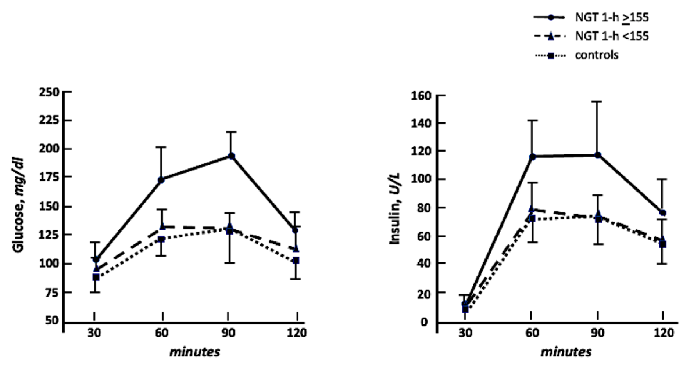
| Glucose T60, mg/dL | 130.4 ± 31.4 | 162.0 ± 36.7 | 0.000 | 130.0 ± 7.6 | 193.9 ± 23.5 | 0.000 |
| Glucose T120, mg/dL | 102.0 ± 17.9 | 121.0 ± 16.9 | 0.000 | 114.0 ± 15.6 | 128.0 ± 15.4 | 0.003 |
| Insulin, U/L | 9.9 ± 2.1 | 10.9 ± 3.9 | 0.241 | 10.2 ± 2.5 | 11.7 ± 4.9 | 0.187 |
| Insulin T60, U/L | 74.9 ± 19.9 | 96.3 ± 39.8 | 0.014 | 74.4 ± 14.9 | 118.2 ± 44.9 | 0.000 |
| Insulin T120, U/L | 54.5 ± 14.8 | 67.5 ± 26.0 | 0.023 | 57.4 ± 15.4 | 77.6 ± 30.5 | 0.005 |
| HOMA | 2.1 ± 0.6 | 2.7 ± 1.0 | 0.014 | 2.4 ± 0.7 | 2.9 ± 1.1 | 0.060 |
| Matsuda index | 86.9 ± 19.6 | 65.7 ± 20.0 | 0.000 | 76.7 ± 15.5 | 54.7 ± 18 | 0.000 |
| Creatinine, mg/dL | 0.77 ± 0.09 | 0.84 ± 0.09 | 0.006 | 0.84 ± 0.09 | 0.85 ± 0.10 | 0.518 |
| e-GFR, mL/min/1.73 m2 | 112.1 ± 17.7 | 96.3 ± 16.9 | 0.003 | 106.4 ± 16.6 | 91.5 ± 14.8 | 0.002 |
| Uric acid, mg/dL | 4.3 ± 0.9 | 5.1 + 1.0 | 0.001 | 4.6 ± 0.5 | 5.7 ± 1.0 | 0.000 |
| Cholesterol, mg/dL | 190.5 ± 34.8 | 212.0 ± 14.0 | 0.003 | 188.1 ± 28.5 | 220.2 ± 17.8 | 0.000 |
| LDL-Chol, mg/dL | 107.5 ± 34.1 | 138.4 ± 17.2 | 0.000 | 105.1 ± 26.5 | 146.0 ± 17.1 | 0.001 |
| HDL-Chol, mg/dL | 63.6 + 9.5 | 49.2 ± 9.5 | 0.000 | 46.6 ± 8.5 | 47.8 ± 6.6 | 0.245 |
| Triglyceride, mg/dL | 99.8 ± 40.9 | 122.3 ± 34.7 | 0.015 | 112.4 ± 30.4 | 132.3 ± 36.2 | 0.041 |
| Variables | Controls (n = 25) | HT (n = 50) | p | NGT 1-h < 155 (n = 25) | NGT 1-h ≥ 155 (n = 25) | p |
|---|---|---|---|---|---|---|
| TLR2, MFI | 4.6 ± 2.4 | 7.7 ± 3.9 | 0.000 | 5.9 ± 2.6 | 9.4 ± 4.2 | 0.002 |
| TLR4, MFI | 7.1 ± 4.3 | 10.4 ± 4.1 | 0.002 | 7.8 ± 2.3 | 13.1 ± 3.9 | 0.000 |
| NF-kβ | 0.07 ± 0.05 | 0.18 ± 0.06 | 0.000 | 0.14 ± 0.04 | 0.21 ± 0.07 | 0.000 |
| IL-1β, pg/mL | 2.8 ± 1.7 | 5.9 ± 2.8 | 0.000 | 3.2 ± 2.1 | 6.9 + 3.4 | 0.000 |
| IL-6, pg/mL | 3.1 ± 2.3 | 7.5 ± 3.9 | 0.000 | 4.1 ± 1.6 | 10.8 + 2.6 | 0.000 |
| IL-8, pg/mL | 9.1 ± 6.0 | 19.7 ± 10.3 | 0.000 | 13.3 ± 5.6 | 27.6 + 9.3 | 0.000 |
| IL-10, pg/mL | 1.6 ± 0.9 | 4.5 ± 2.4 | 0.000 | 2.8 ± 1.3 | 6.8 + 2.5 | 0.000 |
| TNF-α, pg/mL | 2.5 ± 1.2 | 5.0 ± 2.6 | 0.000 | 3.3 ± 1.4 | 6.4 ± 2.9 | 0.000 |
| hs-CRP, mg/dL | 1.4 ± 0.9 | 3.7 ± 1.6 | 0.000 | 2.7 ± 1.0 | 4.8 ± 1.5 | 0.000 |
| Fibrinogen, mg/dL | 263.6 ± 35.8 | 301.4 ± 49.2 | 0.001 | 301.4 ± 38.3 | 301.6 ± 59.1 | 0.111 |
| Variables | TLR2 | TLR4 | NF-kβ | |||
|---|---|---|---|---|---|---|
| R | P | R | P | R | P | |
| Whole study population | ||||||
| Matsuda index | −0.686 | 0.0001 | −0.684 | 0.0001 | −0.676 | 0.0001 |
| 1-h post-load glycemia | 0.583 | 0.0001 | 0.615 | 0.0001 | 0.632 | 0.0001 |
| BMI | 0.501 | 0.0001 | 0.430 | 0.0001 | 0.552 | 0.0001 |
| SBP | 0.480 | 0.0001 | 0.427 | 0.0001 | 0.643 | 0.0001 |
| Age | 0.453 | 0.0001 | 0.456 | 0.0001 | 0.558 | 0.0001 |
| HT vs. NT status | 0.413 | 0.0001 | 0.356 | 0.001 | 0.664 | 0.0001 |
| e-GFR | −0.425 | 0.0001 | −0.416 | 0.0001 | −0.570 | 0.0001 |
| LDL-cholesterol | 0.414 | 0.0001 | 0.543 | 0.0001 | 0.547 | 0.0001 |
| Triglyceride | 0.284 | 0.007 | 0.359 | 0.001 | 0.245 | 0.017 |
| HDL-cholesterol | −0.260 | 0.012 | −0.287 | 0.0006 | −0.474 | 0.0001 |
| Gender, M vs. F | −0.155 | 0.092 | −0.079 | 0.250 | −0.074 | 0.265 |
| Creatinine | 0.078 | 0.254 | 0.048 | 0.340 | 0.318 | 0.003 |
| Hypertensives NGT-1h < 155 | ||||||
| Matsuda index | −0.593 | 0.001 | −0.528 | 0.003 | −0.610 | 0.001 |
| LDL-cholesterol | 0.363 | 0.037 | 0.062 | 0.385 | 0.302 | 0.071 |
| Age | 0.319 | 0.060 | 0.009 | 0.483 | 0.322 | 0.058 |
| HDL-cholesterol | −0.232 | 0.132 | −0.035 | 0.433 | −0.338 | 0.049 |
| SBP | 0.209 | 0.158 | 0.325 | 0.056 | 0.502 | 0.005 |
| Triglyceride | 0.202 | 0.166 | 0.487 | 0.007 | 0.069 | 0.372 |
| Creatinine | 0.185 | 0.187 | 0.331 | 0.053 | 0.081 | 0.351 |
| 1-h post-load glycemia | 0.170 | 0.208 | 0.517 | 0.004 | 0.214 | 0.152 |
| Gender, M vs. F | −0.163 | 0.219 | 0.105 | 0.309 | 0.176 | 0.200 |
| BMI | 0.102 | 0.313 | −0.207 | 0.161 | 0.271 | 0.095 |
| e-GFR | −0.008 | 0.485 | −0.257 | 0.108 | −0.063 | 0.382 |
| Hypertensives NGT 1-h ≥ 155 | ||||||
| Matsuda index | −0.709 | 0.0001 | −0.630 | 0.0001 | −0.327 | 0.055 |
| LDL-cholesterol | 0.142 | 0.250 | 0.055 | 0.397 | 0.124 | 0.278 |
| Age | 0.076 | 0.360 | 0.106 | 0.308 | −0.009 | 0.483 |
| HDL-cholesterol | −0.028 | 0.447 | 0.001 | 0.497 | 0.068 | 0.374 |
| SBP | 0.345 | 0.045 | 0.283 | 0.085 | 0.052 | 0.403 |
| Triglyceride | 0.200 | 0.169 | 0.072 | 0.366 | 0.276 | 0.091 |
| Creatinine | 0.339 | 0.048 | 0.368 | 0.035 | 0.229 | 0.136 |
| 1-h post-load glycemia | 0.441 | 0.014 | 0.381 | 0.030 | 0.118 | 0.287 |
| Gender, M vs. F | 0.532 | 0.003 | −0.417 | 0.019 | −0.391 | 0.027 |
| BMI | 0.498 | 0.006 | 0.322 | 0.058 | 0.132 | 0.265 |
| e-GFR | −0.236 | 0.128 | −0.157 | 0.227 | −0.524 | 0.004 |
| Whole Study Population | |||
| TLR2 | r2 Partial | r2 Total | P |
| Matsuda index | 47.0% | 47.0% | 0.0001 |
| 1-h post-load glycemia | 4.6% | 51.6% | 0.015 |
| Age | 3.6% | 55.2% | 0.020 |
| TLR4 | |||
| Matsuda index | 46.8% | 46.8% | 0.0001 |
| LDL-cholesterol | 7.9% | 53.4% | 0.002 |
| 1-h post-load glycemia | 4.2% | 57.6% | 0.006 |
| NF-kβ | |||
| Matsuda index | 45.7% | 45.7% | 0.0001 |
| HT vs. NT status | 16.1% | 61.8% | 0.0001 |
| e-GFR | 8.0% | 69.8% | 0.0001 |
| Triglyceride | 3.3% | 73.1% | 0.003 |
| 1-h post-load glycemia | 2.1% | 75.2% | 0.020 |
| Hypertensives NGT-1h < 155 | |||
| TLR2 | |||
| Matsuda index | 35.2% | 35.2% | 0.002 |
| TLR4 | |||
| Matsuda index | 27.9% | 27.9% | 0.007 |
| 1-h post-load glycemia | 12.5% | 40.4% | 0.042 |
| Triglyceride | 11.0% | 51.5% | 0.041 |
| NF-kβ | |||
| Matsuda index | 37.2% | 37.2% | 0.001 |
| Hypertensives NGT-1h ≥ 155 | |||
| TLR2 | |||
| Matsuda index | 50.2% | 50.2% | 0.0001 |
| 1-h post-load glycemia | 27.6% | 77.8% | 0.0001 |
| BMI | 5.0% | 82.8% | 0.0001 |
| TLR4 | |||
| Matsuda index | 39.7% | 39.7% | 0.0001 |
| 1-h glycemia | 28.5% | 682% | 0.0001 |
| Triglyceride | 9.3% | 77.5% | 0.008 |
| NF-kβ | |||
| e-GFR | 27.5% | 27.5% | 0.007 |
Publisher’s Note: MDPI stays neutral with regard to jurisdictional claims in published maps and institutional affiliations. |
© 2022 by the authors. Licensee MDPI, Basel, Switzerland. This article is an open access article distributed under the terms and conditions of the Creative Commons Attribution (CC BY) license (https://creativecommons.org/licenses/by/4.0/).
Share and Cite
Perticone, M.; Maio, R.; Gigliotti, S.; Arturi, F.; Succurro, E.; Sciacqua, A.; Andreozzi, F.; Sesti, G.; Perticone, F. Immuno-Mediated Inflammation in Hypertensive Patients with 1-h Post-Load Hyperglycemia. Int. J. Mol. Sci. 2022, 23, 10891. https://doi.org/10.3390/ijms231810891
Perticone M, Maio R, Gigliotti S, Arturi F, Succurro E, Sciacqua A, Andreozzi F, Sesti G, Perticone F. Immuno-Mediated Inflammation in Hypertensive Patients with 1-h Post-Load Hyperglycemia. International Journal of Molecular Sciences. 2022; 23(18):10891. https://doi.org/10.3390/ijms231810891
Chicago/Turabian StylePerticone, Maria, Raffaele Maio, Simona Gigliotti, Franco Arturi, Elena Succurro, Angela Sciacqua, Francesco Andreozzi, Giorgio Sesti, and Francesco Perticone. 2022. "Immuno-Mediated Inflammation in Hypertensive Patients with 1-h Post-Load Hyperglycemia" International Journal of Molecular Sciences 23, no. 18: 10891. https://doi.org/10.3390/ijms231810891
APA StylePerticone, M., Maio, R., Gigliotti, S., Arturi, F., Succurro, E., Sciacqua, A., Andreozzi, F., Sesti, G., & Perticone, F. (2022). Immuno-Mediated Inflammation in Hypertensive Patients with 1-h Post-Load Hyperglycemia. International Journal of Molecular Sciences, 23(18), 10891. https://doi.org/10.3390/ijms231810891