
Vitamin D as a Possible COVID-19 Prevention Strategy


Abstract
:1. Introduction
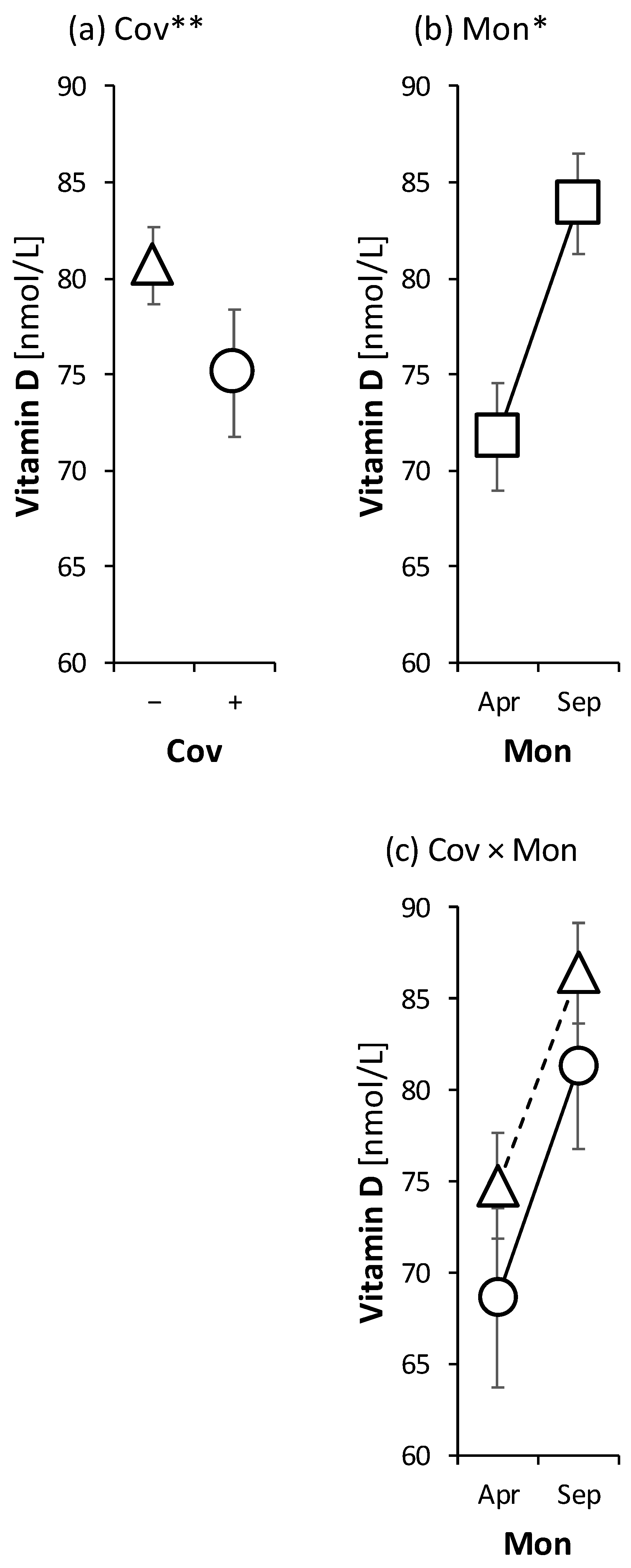
2. Results
3. Discussion
4. Materials and Methods
4.1. Probands
4.2. Chemicals and Materials
4.3. Instrumentation
4.4. Blood Sample Collection, Handling, and Storage
4.5. Sample Processing
4.6. LC-MS/MS Conditions
4.7. Method Validation
4.8. Statistical Evaluation
5. Conclusions
Author Contributions
Funding
Institutional Review Board Statement
Informed Consent Statement
Data Availability Statement
Conflicts of Interest
References
- Bičíková, M.; Máčová, L.; Jandová, D.; Třískala, Z.; Hill, M. Movement as a Positive Modulator of Aging. Int. J. Mol. Sci. 2021, 22, 6278. [Google Scholar] [CrossRef] [PubMed]
- Vieth, R. Vitamin D supplementation: Cholecalciferol, calcifediol, and calcitriol. Eur. J. Clin. Nutr. 2020, 74, 1493–1497. [Google Scholar] [CrossRef] [PubMed]
- Xu, J.; Zhou, Y.; Yan, C.; Wang, X.; Lou, J.; Luo, Y.; Gao, S.; Wang, J.; Wu, L.; Gao, X.; et al. Neurosteroids: A novel promise for the treatment of stroke and post-stroke complications. J. Neurochem. 2021, 160, 113–127. [Google Scholar] [CrossRef]
- BiČíková, M.; Duskova, M.; Vítků, J.; Kalvachová, B.; Řípová, D.; Mohr, P.; Stárka, L. Vitamin D in Anxiety and Affective Disorders. Physiol. Res. 2015, 64, S101–S103. [Google Scholar] [CrossRef]
- Nithila, M.R.; Roy, N.M.; Al-Harthi, L.; Sampat, N.; Al-Mujaini, R.; Mahadevan, S.; Al Adawi, S.; Essa, M.M.; Al Subhi, L.; Al-Balushi, B.; et al. Impact of vitamin D on neurocognitive function in dementia, depression, schizophrenia and ADHD. Front. Biosci. 2021, 26, 566–611. [Google Scholar] [CrossRef]
- Carlberg, C. Nutrigenomics of Vitamin D. Nutrients 2019, 11, 676. [Google Scholar] [CrossRef]
- Grant, W.B. Review of Recent Advances in Understanding the Role of Vitamin D in Reducing Cancer Risk: Breast, Colorectal, Prostate, and Overall Cancer. Anticancer Res. 2019, 40, 491–499. [Google Scholar] [CrossRef] [PubMed]
- Abdi, F.; Ozgoli, G.; Rahnemaie, F.S. A systematic review of the role of vitamin D and calcium in premenstrual syndrome. Obstet. Gynecol. Sci. 2019, 62, 73–86. [Google Scholar] [CrossRef]
- Ciavattini, A.; Serri, M.; Carpini, G.D.; Morini, S.; Clemente, N. Ovarian endometriosis and vitamin D serum levels. Gynecol. Endocrinol. 2016, 33, 164–167. [Google Scholar] [CrossRef]
- Kalaitzopoulos, D.R.; Lempesis, I.G.; Athanasaki, F.; Schizas, D.; Samartzis, E.P.; Kolibianakis, E.M.; Goulis, D.G. Association between vitamin D and endometriosis: A systematic review. Hormones 2019, 19, 109–121. [Google Scholar] [CrossRef]
- Giampaolino, P.; Corte, L.D.; Foreste, V.; Bifulco, G. Is there a Relationship Between Vitamin D and Endometriosis? An Overview of the Literature. Curr. Pharm. Des. 2019, 25, 2421–2427. [Google Scholar] [CrossRef] [PubMed]
- Ao, T.; Kikuta, J.; Ishii, M. The Effects of Vitamin D on Immune System and Inflammatory Diseases. Biomolecules 2021, 11, 1624. [Google Scholar] [CrossRef] [PubMed]
- Charoenngam, N.; Holick, M.F. Immunologic Effects of Vitamin D on Human Health and Disease. Nutrients 2020, 12, 2097. [Google Scholar] [CrossRef] [PubMed]
- Dissanayake, H.A.; de Silva, N.L.; Sumanatilleke, M.; de Silva, S.D.N.; Gamage, K.K.K.; Dematapitiya, C.; Kuruppu, D.C.; Ranasinghe, P.; Pathmanathan, S.; Katulanda, P. Prognostic and Therapeutic Role of Vitamin D in COVID-19: Systematic Review and Meta-analysis. J. Clin. Endocrinol. Metab. 2021, 107, 1484–1502. [Google Scholar] [CrossRef] [PubMed]
- Ghasemian, R.; Shamshirian, A.; Heydari, K.; Malekan, M.; Alizadeh-Navaei, R.; Ebrahimzadeh, M.A.; Warkiani, M.E.; Jafarpour, H.; Bazaz, S.R.; Shahmirzadi, A.R.; et al. The role of vitamin D in the age of COVID-19: A systematic review and meta-analysis. Int. J. Clin. Pract. 2021, 75, e14675. [Google Scholar] [CrossRef] [PubMed]
- Pereira, M.; Dantas Damascena, A.D.; Galvão Azevedo, L.M.G.; de Almeida Oliveira, T.D.A.; da Mota Santana, J.D.M. Vitamin D deficiency aggravates COVID-19: Systematic review and meta-analysis. Crit. Rev. Food Sci. Nutr. 2022, 62, 1308–1316. [Google Scholar] [CrossRef]
- Zelzer, S.; Prüller, F.; Curcic, P.; Sloup, Z.; Holter, M.; Herrmann, M.; Mangge, H. Vitamin D Metabolites and Clinical Outcome in Hospitalized COVID-19 Patients. Nutrients 2021, 13, 2129. [Google Scholar] [CrossRef]
- Hadizadeh, F. Supplementation with vitamin D in the COVID-19 pandemic? Nutr. Rev. 2020, 79, 200–208. [Google Scholar] [CrossRef]
- Povaliaeva, A.; Bogdanov, V.; Pigarova, E.; Dzeranova, L.; Katamadze, N.; Malysheva, N.; Ioutsi, V.; Nikankina, L.; Rozhinskaya, L.; Mokrysheva, N. Impaired Vitamin D Metabolism in Hospitalized COVID-19 Patients. Pharmaceuticals 2022, 15, 906. [Google Scholar] [CrossRef]
- De Smet, D.; De Smet, K.; Herroelen, P.; Gryspeerdt, S.; A Martens, G. Serum 25(OH)D Level on Hospital Admission Associated With COVID-19 Stage and Mortality. Am. J. Clin. Pathol. 2020, 155, 381–388. [Google Scholar] [CrossRef]
- Diaz-Curiel, M.; Cabello, A.; Arboiro-Pinel, R.; Mansur, J.L.; Heili-Frades, S.; Mahillo-Fernandez, I.; Herrero-González, A.; Andrade-Poveda, M. The relationship between 25(OH) vitamin D levels and COVID-19 onset and disease course in Spanish patients. J. Steroid Biochem. Mol. Biol. 2021, 212, 105928. [Google Scholar] [CrossRef] [PubMed]
- Reis, B.Z.; Fernandes, A.L.; Sales, L.P.; Santos, M.D.; dos Santos, C.C.; Pinto, A.J.; Goessler, K.F.; Franco, A.S.; Duran, C.S.C.; Silva, C.B.R.; et al. Influence of vitamin D status on hospital length of stay and prognosis in hospitalized patients with moderate to severe COVID-19: A multicenter prospective cohort study. Am. J. Clin. Nutr. 2021, 114, 598–604. [Google Scholar] [CrossRef] [PubMed]
- Grant, W.B.; Lahore, H.; McDonnell, S.L.; Baggerly, C.A.; French, C.B.; Aliano, J.L.; Bhattoa, H.P. Evidence that Vitamin D Supplementation Could Reduce Risk of Influenza and COVID-19 Infections and Deaths. Nutrients 2020, 12, 988. [Google Scholar] [CrossRef]
- Yasui, T.; Miyatani, Y.; Tomita, J.; Yamada, M.; Uemura, H.; Miura, M.; Irahara, M. Effect of vitamin K2 treatment on carboxylation of osteocalcin in early postmenopausal women. Gynecol. Endocrinol. 2006, 22, 455–459. [Google Scholar] [CrossRef]
- Pizzini, A.; Aichner, M.; Sahanic, S.; Böhm, A.; Egger, A.; Hoermann, G.; Kurz, K.; Widmann, G.; Bellmann-Weiler, R.; Weiss, G.; et al. Impact of Vitamin D Deficiency on COVID-19—A Prospective Analysis from the CovILD Registry. Nutrients 2020, 12, 2775. [Google Scholar] [CrossRef] [PubMed]
- Carlberg, C.; Haq, A. The concept of the personal vitamin D response index. J. Steroid Biochem. Mol. Biol. 2018, 175, 12–17. [Google Scholar] [CrossRef] [PubMed]
- DeLuca, H.F. Overview of general physiologic features and functions of vitamin D. Am. J. Clin. Nutr. 2004, 80, 1689S–1696S. [Google Scholar] [CrossRef] [PubMed]
- Elamir, Y.M.; Amir, H.; Lim, S.; Rana, Y.P.; Lopez, C.G.; Feliciano, N.V.; Omar, A.; Grist, W.P.; Via, M.A. A randomized pilot study using calcitriol in hospitalized COVID-19 patients. Bone 2021, 154, 116175. [Google Scholar] [CrossRef]
- Amraei, R.; Rahimi, N. COVID-19, Renin-Angiotensin System and Endothelial Dysfunction. Cells 2020, 9, 1652. [Google Scholar] [CrossRef]
- Shukla, A.K.; Banerjee, M. Angiotensin-Converting-Enzyme 2 and Renin-Angiotensin System Inhibitors in COVID-19: An Update. High Blood Press. Cardiovasc. Prev. 2021, 28, 129–139. [Google Scholar] [CrossRef]
- Goddek, S. Vitamin D3 and K2 and their potential contribution to reducing the COVID-19 mortality rate. Int. J. Infect. Dis. 2020, 99, 286–290. [Google Scholar] [CrossRef] [PubMed]
- Myneni, V.D.; Mezey, E. Regulation of bone remodeling by vitamin K2. Oral Dis. 2016, 23, 1021–1028. [Google Scholar] [CrossRef] [PubMed]
- Bioanalytical Method Validation Guidance for Industry; Center for Drug Evaluation and Research Center for Veterinary Medicine: Rockville, MD, USA, 2018.
- Meloun, M.; Hill, M.; Militky, J.; Kupka, K. Transformation in the PC-Aided Biochemical Data Analysis. Clin. Chem. Lab. Med. 2000, 38, 553–559. [Google Scholar] [CrossRef] [PubMed]
- Meloun, M.; Militký, J.; Hill, M.; Brereton, R.G. Crucial problems in regression modelling and their solutions. Analyst 2002, 127, 433–450. [Google Scholar] [CrossRef] [PubMed]

| Time (min) | %B |
|---|---|
| 0.00 | 40 |
| 0.20 | 40 |
| 0.30 | 75 |
| 4.50 | 84 |
| 4.60 | 97.5 |
| 5.20 | 97.5 |
| 5.25 | 40 |
| 6.10 | 40 |
| 7.00 | 40 |
| Analyte | RT | Quantification Q1/Q3 | Confirmation Q1/Q2 |
|---|---|---|---|
| 25-hydroxyvitamin D3 | 3.74 | 383.2/257.2 | 383.2/365.3 |
| 25-hydroxyvitamin D2 | 3.92 | 395.1/209.1 | 395.1/377.1 |
| 3-epi-25-hydroxyvitamin D3 | 3.90 | 383.2/257.2 | 383.2/365.3 |
| 3-epi-25-hydroxyvitamin D2 | 4.06 | 395.1/209.1 | 395.1/377.1 |
| 24,25-dihydroxyvitamin D3 | 2.41 | 399.1/381.3 | 399.1/215.1 |
| d6-25-hydroxyvitamin D3 | 3.72 | 389.2/371.2 | - |
Publisher’s Note: MDPI stays neutral with regard to jurisdictional claims in published maps and institutional affiliations. |
© 2022 by the authors. Licensee MDPI, Basel, Switzerland. This article is an open access article distributed under the terms and conditions of the Creative Commons Attribution (CC BY) license (https://creativecommons.org/licenses/by/4.0/).
Share and Cite
Bičíková, M.; Máčová, L.; Hill, M. Vitamin D as a Possible COVID-19 Prevention Strategy. Int. J. Mol. Sci. 2022, 23, 10532. https://doi.org/10.3390/ijms231810532
Bičíková M, Máčová L, Hill M. Vitamin D as a Possible COVID-19 Prevention Strategy. International Journal of Molecular Sciences. 2022; 23(18):10532. https://doi.org/10.3390/ijms231810532
Chicago/Turabian StyleBičíková, Marie, Ludmila Máčová, and Martin Hill. 2022. "Vitamin D as a Possible COVID-19 Prevention Strategy" International Journal of Molecular Sciences 23, no. 18: 10532. https://doi.org/10.3390/ijms231810532
APA StyleBičíková, M., Máčová, L., & Hill, M. (2022). Vitamin D as a Possible COVID-19 Prevention Strategy. International Journal of Molecular Sciences, 23(18), 10532. https://doi.org/10.3390/ijms231810532