Review

Role of Circular RNAs in Pulmonary Fibrosis

Laboratory of Anesthesia and Critical Care Medicine, Department of Anesthesiology, West China Hospital, Sichuan University, Chengdu 610017, China
*
Authors to whom correspondence should be addressed.
Int. J. Mol. Sci. 2022, 23(18), 10493; https://doi.org/10.3390/ijms231810493
Submission received: 18 August 2022 / Revised: 4 September 2022 / Accepted: 6 September 2022 / Published: 10 September 2022
(This article belongs to the Section Molecular Genetics and Genomics)

Abstract

:
Pulmonary fibrosis is a chronic progressive form of interstitial lung disease, characterized by the histopathological pattern of usual interstitial pneumonia. Apart from aberrant alterations of protein-coding genes, dysregulation of non-coding RNAs, including microRNAs, long non-coding RNAs, and circular RNAs (circRNAs), is crucial to the initiation and progression of pulmonary fibrosis. CircRNAs are single-stranded RNAs that form covalently closed loops without 5′ caps and 3′ tails. Different from canonical splicing of mRNA, they are produced from the back-splicing of precursor mRNAs and have unique biological functions, as well as potential biomedical implications. They function as important gene regulators through multiple actions, including sponging microRNAs and proteins, regulating transcription, and splicing, as well as protein-coding and translation in a cap-independent manner. This review comprehensively summarizes the alteration and functional role of circRNAs in pulmonary fibrosis, with a focus on the involvement of the circRNA in the context of cell-specific pathophysiology. In addition, we discuss the diagnostic and therapeutic potential of targeting circRNA and their regulatory pathway mediators, which may facilitate the translation of recent advances from bench to bedside in the future.

1. Introduction

Pulmonary fibrosis (PF) is a chronic progressive form of interstitial lung disease (ILD), characterized by the histopathological pattern of usual interstitial pneumonia (UIP) [1]. Clinical manifestations of PF include increasing cough, progressively exertional dyspnea with or without dry cough, hypoxemia, and respiratory failure [2,3]. Genetic factors, aging, environmental exposures (such as smoking, silica, or radiation), and certain comorbidities (such as autoimmune/connective tissue diseases) are risk factors that may cause PF [4]. The underlying pathophysiology of PF involves dysregulated reparative response, wherein repetitive epithelial injury leads to epithelial-to-mesenchymal transition (EMT), macrophage activation (MA), and polarization, as well as persistent fibroblast activation and fibroblast-to-myofibroblast transition (FMT) and the subsequent excessive deposition of extracellular matrix (ECM) (see Figure 1) [5]. PF incurs substantial socioeconomic costs and a considerable public health burden, with a conservatively estimated prevalence of 3–9 per 100,000 individuals [6]. In addition, PF exerts significant effects on quality of life, and the median survival after diagnosis is only 3–5 years [7]. Treatment of PF is still challenging, due to limited treatment options. The only two FDA-approved drugs for idiopathic pulmonary fibrosis (IPF), pirfenidone, and nintedanib showed limited effect on decline of disease progression or mortality [5,8]. As lung transplantation is the only possible way to cure PF, it is important to identify new therapeutic targets for developing mechanism-driven treatments to tackle this refractory disease.
Circular RNAs (circRNAs) are single-stranded RNAs that form covalently closed loops without 5′ N7-methylguanosine (m7G) caps and 3′ polyadenylated tails [9]. Back in 1976, circRNAs were first discovered in pathogenic plant viroid [10]. However, they were considered to be functionless by-products of abnormal RNA splicing and did not draw much scientific attention until decades later. A timeline of significant discoveries in circRNA research is depicted in Figure 2 [10,11,12,13,14,15,16,17,18,19,20,21,22,23,24,25]. Different from canonical splicing of mRNA, they are produced from the back-splicing of precursor mRNAs (pre-mRNA) and have unique biological functions [26]. They function as important gene regulators through multiple actions, including miRNA or protein sponges or decoys, templates for translation, protein scaffolding, and recruitment or enhancer of protein function [27]. CircRNA has been reported to be implicated in many different pathophysiological processes through diverse mechanisms of action. A bulk of studies demonstrated that their aberrant expression or function are involved in the initiation and development of various diseases, such as central nervous system diseases, aging and longevity, cardiovascular diseases, kidney disease, and cancer [28,29,30,31,32]. In particular, the enrichment of circRNA was observed in the EMT [33], and accumulating evidence indicated that circRNA may play a regulatory role in PF.
It has now been realized that dysregulation of non-coding RNAs (ncRNAs) is critical to the development and maintenance of PF. Although there has been much research on mi-RNAs and lncRNAs, few researchers have taken circRNAs into consideration. With the rapid development of high-throughput sequencing technologies and bioinformatics analysis, a growing body of original articles demonstrated that circRNA expression is dysregulated in many types of PF, including silicosis (silica-induced pulmonary fibrosis), idiopathic PF (pulmonary fibrosis of unknown cause), cigarette smoke-induced PF, in which circRNA dysregulation gives rise to a complex interplay of cell types and signaling pathways, such as endothelial–mesenchymal transition (EndMT), EMT, MA and polarization, and FMT. In this state-of-art review, we comprehensively summarize the alteration and functional role of circRNAs in PF, followed by in-depth discussion of the regulatory mechanisms of circRNAs in PF in the context of cell-specific pathophysiology. In addition, we discuss the diagnostic and therapeutic potential of targeting circRNA and their regulatory pathway mediators, which may facilitate translation of recent advances from bench to bedside in the future.

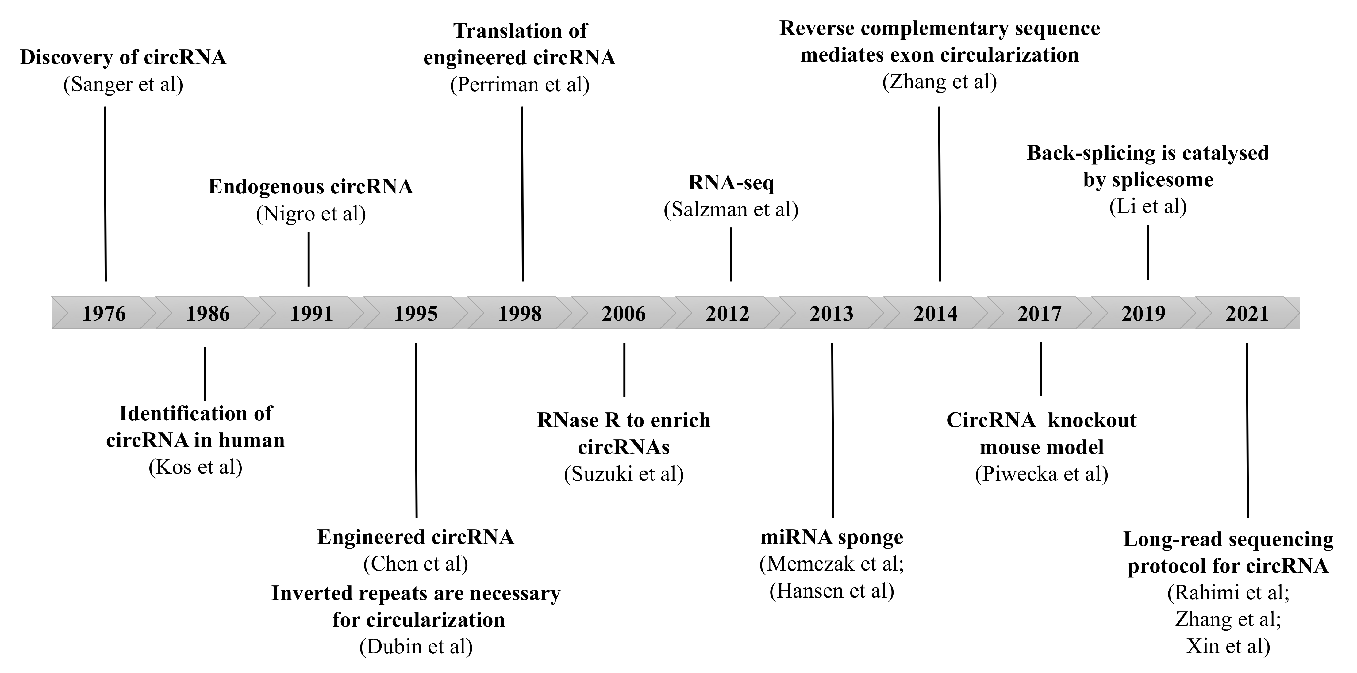
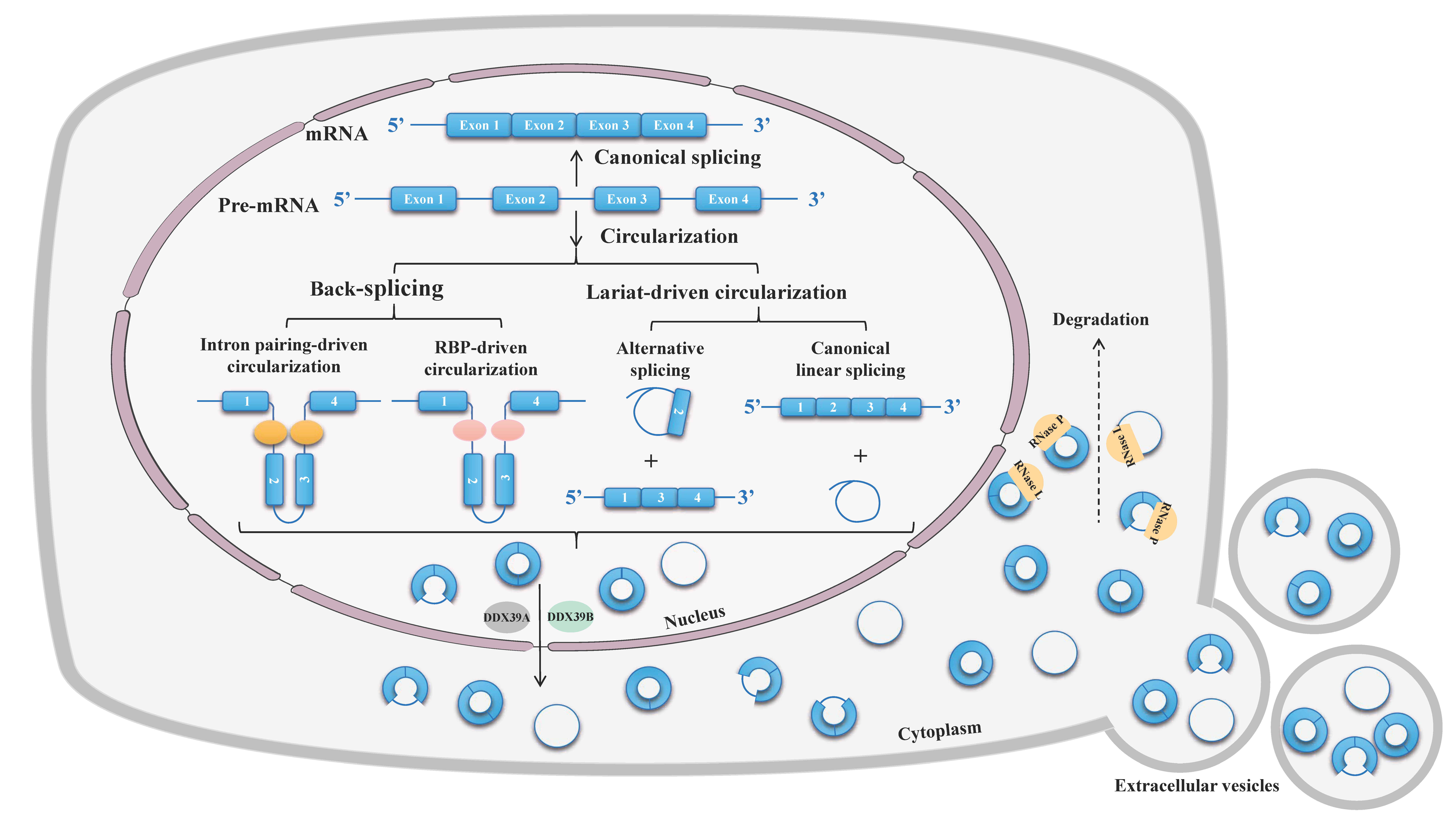
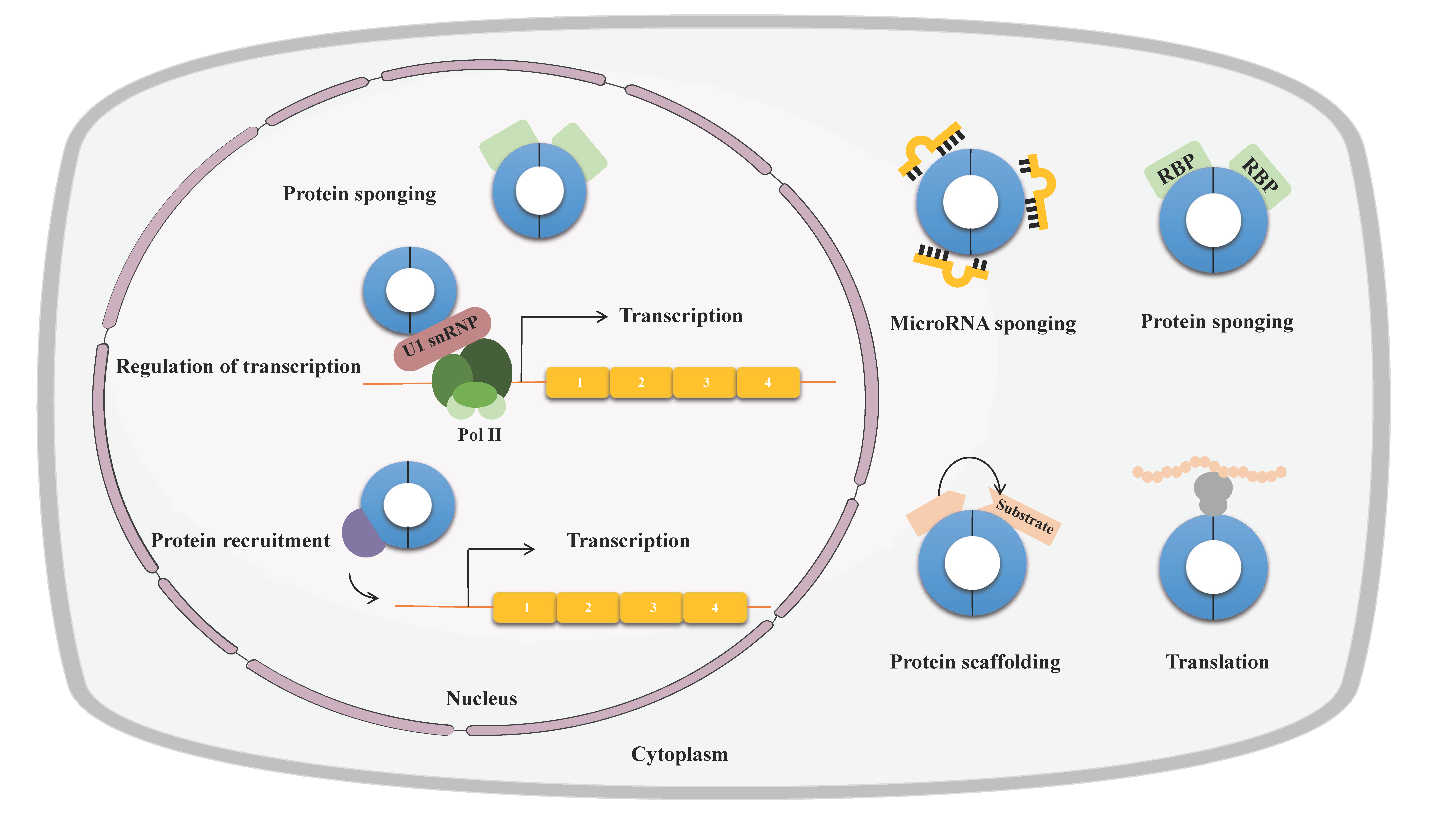
2. Properties of circRNAs

The majority of circRNAs are formed by back-splicing of pre-mRNAs or long noncoding RNAs, in which a downstream 5′ splice donor site is joined to an upstream 3′ splice acceptor site in reverse order with a 3′,5′-phosphodiester bond at the back-splicing junction sites (BSJ) [26,34]. Back-splicing relies on the canonical spliceosomal machinery but is much less efficient than cognate linear splicing [35,36]. The looping structure that brings BSJ into close proximity is critical to back-splicing, which can be facilitated by base-paring between inverted intronic complementary sequences or dimerization of RNA-binding proteins (RBP) [9]. More rarely, if the intron lariats produced during conventional splicing avoid debranching and preserve a circular form with a 2′,5′-phosphodiester bond between the back-spliced exons, they are referred to as circular intronic RNA (ciRNA) [37] (see Figure 3).
In addition to the regulation of circRNA biogenesis, the abundance of circRNA also depends on the trafficking and turnover of circRNA. With the exception of ciRNA and exon-intron circRNAs (ElciRNA), which are limited to the nucleus, most circRNAs are predominantly located in the cytoplasm [18,38,39,40]. Nuclear export of circRNAs to the cytoplasm occurs in a length-dependent manner and requires multiple proteins, such as spliceosome RNA helicase DDX39B (for long circRNAs) and ATP-dependent RNA helicase DDX39A (for short circRNAs) [41]. After binding to transcription–export complexes, they are further recruited by the NTF2-related export protein 1-nuclear RNA export factor 1 heterodimeric export receptor and transferred into the cytoplasm through the nuclear pore complex. Moreover, circRNA shuttling also can be modulated by RNA modification or further exported to extracellular vesicles [42,43,44]. Owing to their circular structure, circRNAs are resistant to degradation by linear RNA decay machineries, making them exceptionally stable once formed (a median half-life of 18.8–23.7 h of circRNAs, compared with a median half-life of 4.0–7.4 h of their linear counterparts) [45]. So far the mechanisms of circRNAs degradation are not fully elucidated. Recent studies have shown that both miRNA and some endonucleases participate in circRNA decay [46,47,48] (see Figure 3).
Many circRNAs exert important biological functions through diverse mechanisms of action. Firstly, miRNA sponging is the most frequently proposed and extensively studied function of circRNAs. By binding and sequestration of miRNAs and preventing miRNAs from repressing their target mRNAs, circRNAs can participate in gene regulation in a post-transcriptional way [49,50]. Secondly, some circRNAs containing RBP-binding motifs can interact with the RBP and function as protein sponges or decoys, thus indirectly regulating their functions [51,52]. Thirdly, circRNA may act as scaffolds to bring enzymes and their substrates into proximity, influencing the reaction kinetics [53,54,55]. Fourthly, a few circRNA have been shown to recruit certain proteins to specific loci or subcellular compartments [56]. Fifthly, some nuclear circRNAs can also regulate gene expression by enhancing the RNA polymerase II [37,38]. Last but not least, circRNAs that contain internal ribosome entry sites can serve as templates for translation in a cap-independent manner or m6A-dependent manner with low efficiency [57,58,59] (see Figure 4).

3. The circRNA Expression Profiling and Integrative Analysis in Pulmonary Fibrosis

Currently, microarray or RNA sequencing (RNA-seq) analysis and reverse transcription-quantitative PCR are the most commonly used methods for the identification and validation of expression profilings of deregulated circRNAs in various disease models. Several studies have reported that many circRNAs are deregulated in various models of PF (Table 1 and Table 2; Figure 5). By Arraystar Human circRNA Microarray, 67 differentially expressed circRNAs (DECs) were identified, among which 38 and 29 were upregulated and downregulated, respectively, in the plasma of patients with IPF, in comparison with matched healthy controls [60]. Based on RNA sequencing analysis in the murine model of bleomycin-induced pulmonary fibrosis (BIPF), Li and colleagues found 74 DECs between PF and corresponding control tissues [61]. Similarly, a total of 10 circRNAs were detected by Hiseq 2000 from a rat model of BIPF, and there were 2 circRNAs upregulated and 8 downregulated according to the criteria p < 0.05 and fold change > 2.0 [62]. To elucidate the interaction networks of differentially expressed mRNAs and ncRNAs during PF process, Liu and colleagues analyzed the circRNA expression profiling from BIPF rats and control rats. The results of whole transcriptome sequencing analysis showed that 287 DECs were upregulated and 318 DECs were downregulated [63]. Aside from IPF-specific circRNAs, efforts have been made to identify circRNAs pertinent to silicosis. Cheng and colleagues performed RNA-seq screening to identify plasmatic circRNAs as potential biomarkers for diagnosing silicosis. Overall, 243 DECs were identified, of which 139 were upregulated DECs and 104 were downregulated [64]. Additionally, another study on the murine model of silicon dioxide (SiO2)-induced silicosis, using microarray analysis, identified 120 DECs (73 upregulated and 47 downregulated) between the SiO2 and control group [65]. Interestingly, further Kyoto Encyclopedia of Genes and Genomes (KEGG) analysis revealed that these DECs were enriched in pathways of adherens junction [60,63], purine metabolism [66], lysine degradation [63], phosphoinositide 3 kinase (PI3K)-serine/threonine Akt signaling [62], and wingless/integrated (Wnt) signaling [63].

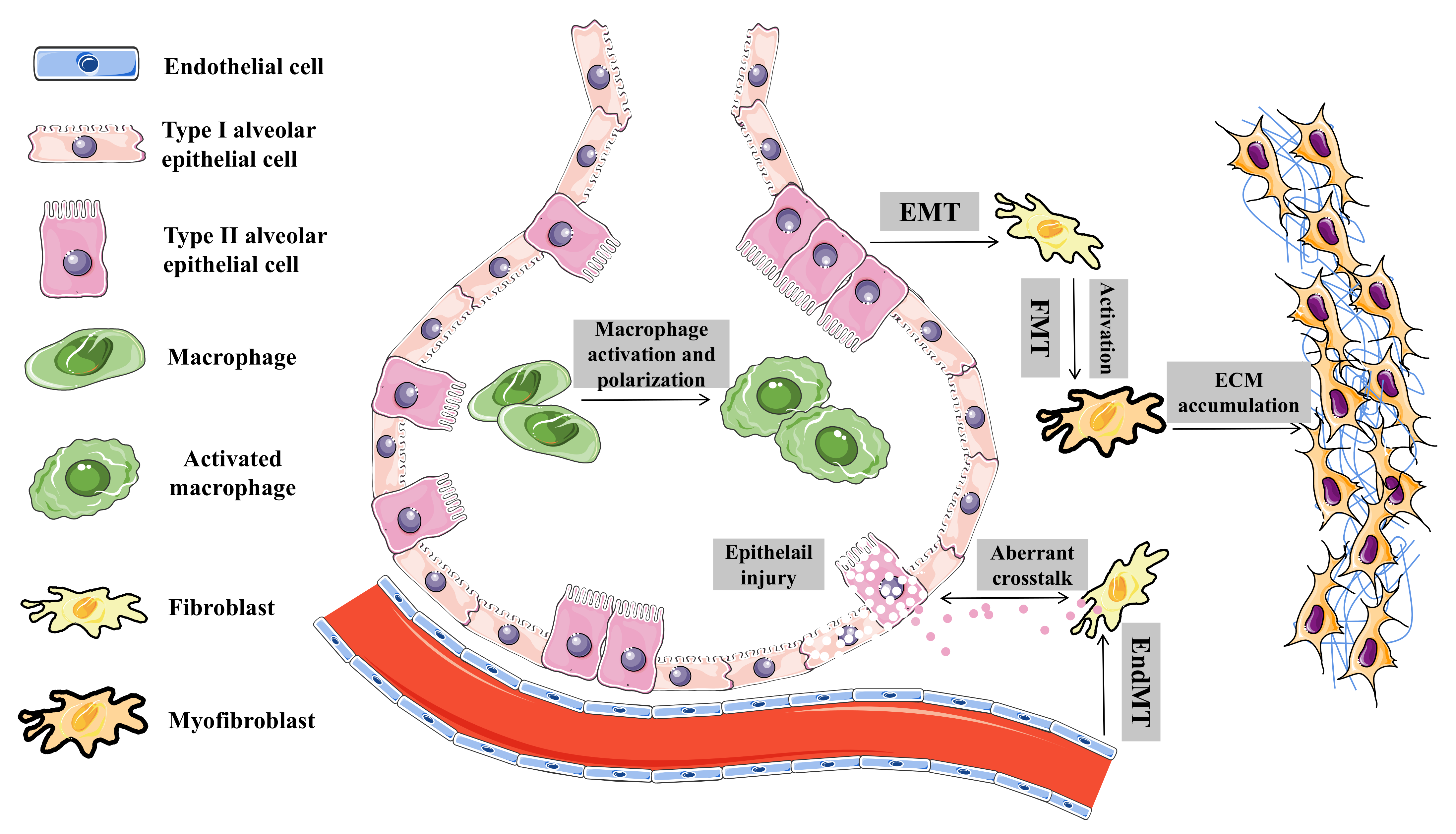
4. Biological Roles and Regulatory Mechanisms of circRNAs in Pulmonary Fibrosis

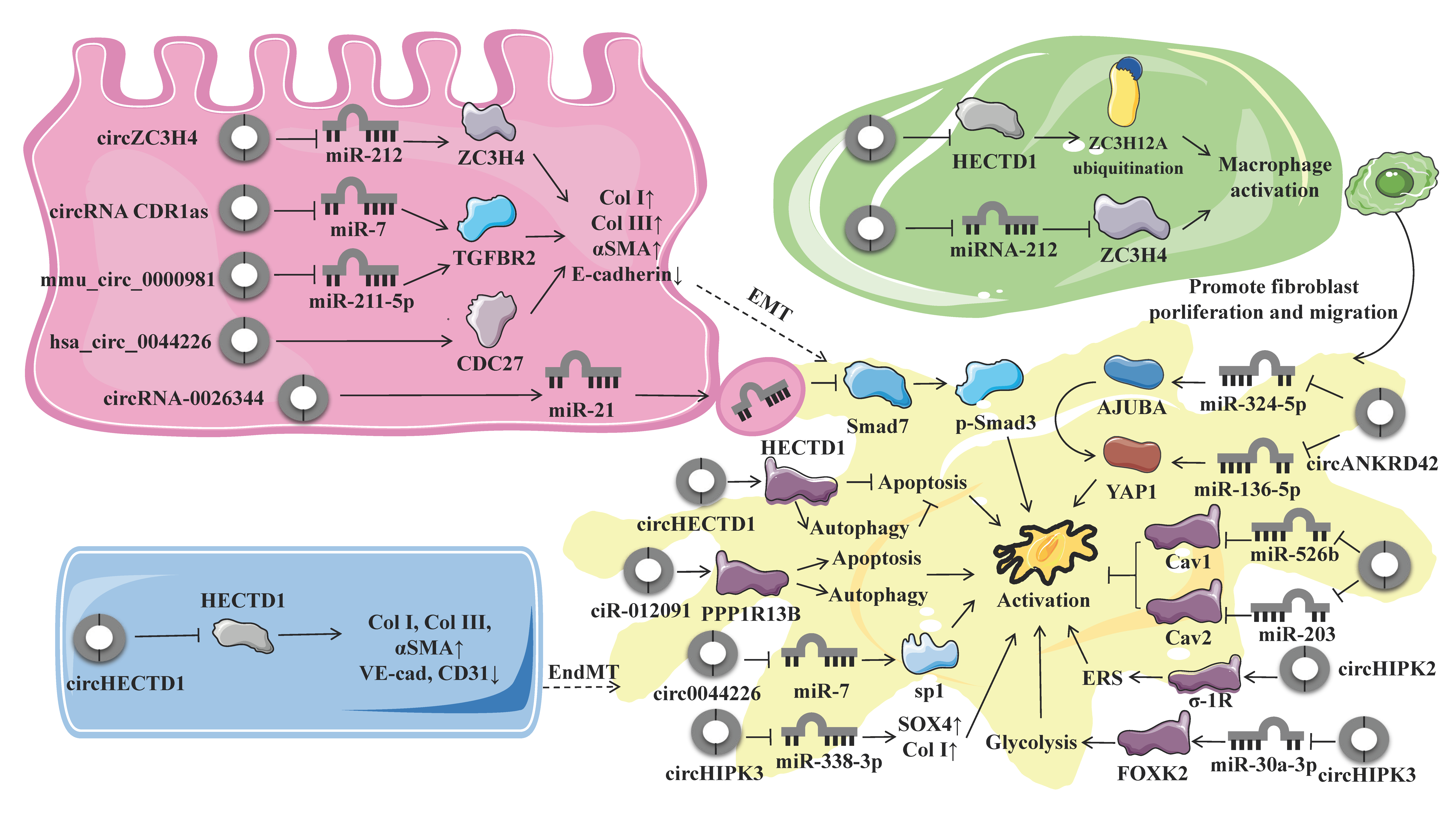
4.1. Endothelial–Mesenchymal Transition

Apart from EMT, endothelial cells can also be the origins of the mesenchymal cells responsible for dysregulated ECM accumulation in the PF; the process through which endothelial cells transdifferentiate into motile mesenchymal cells is termed EndMT. EndMT is a cellular and molecular process through which cells lose their endothelial markers (downregulation of VE-Cadherin) and acquire the mesenchymal or myofibroblast phenotype (upregulation of α-smooth muscle actin [α-SMA] and type I collagen [Col I/COL1A1]) [67]. Fang and colleagues reported that the level of circular RNA HECT domain E3 ubiquitin protein ligase 1 (circHECTD1) was rapidly increased in SiO2-induced EndMT in both mouse endothelial cell line MML1 and human endothelial cell line HUVECs. In turn, the elevated circHECTD1 downregulated the expression of HECT domain E3 ubiquitin protein ligase 1 (HECTD1) protein. Targeting circHECTD1 via small interfering RNA (siRNA) or overexpression of HECTD1 by the CRISPR/Cas9 system prevented SiO2-induced EndMT. Moreover, consistent downward tendency of HECTD1 expression was also found in tissue samples from the mice model of SiO2-induced silicosis and silicosis patients, demonstrating the involvement of the circHECTD1/HECTD1 pathway in the pathogenesis of silicosis. Further functional experiments revealed that circHECTD1-mediated downregulation of HECTD1 is involved in the SiO2-induced EndMT by promoting endothelial cell migration and activation [82].

4.2. Epithelial-To-Mesenchymal Transition

During fibrogenesis, the critical myofibroblasts responsible for ECM synthesis can be derived from resident fibroblasts, circulating fibrocytes, epithelial cells, endothelial cells, and other specialized cell types. Apart from activation of tissue-resident fibroblasts and differentiation of circulating fibrocytes, EMT is an important mechanism that increases the net pool of myofibroblasts [83]. EMT is characterized by the loss of epithelial identity (apical–basal polarity and stable intercellular junctions) and acquisition of the mesenchymal phenotype (cytoskeletal and morphological rearrangements, fibroblast-like gene expression profile, migratory capacity, and ability to produce the ECM) [84]. As an embryonic process, EMT is not only involved in the development of wound healing but also contributes to pathological conditions, such as cancer and fibrosis [85]. Historically, the functional role of EMT in pathological conditions has been classified into type Ι EMT (embryogenesis), type II EMT (pathological fibrosis), and type III EMT (tumorigenesis) [67,86]. However, subsequent studies using genetically engineered knock-out mouse models coupled with lineage tracing strategies rebutted that instead of transdifferentiation into myofibroblasts; the epithelial cells still reside within the epithelial basement membrane in a partial EMT state during EMT but with impaired epithelial function and regenerative potential [84]. Hsa_circ_0044226 was found to be significantly upregulated in pulmonary samples from IPF patients, compared with healthy controls. Functional experiments suggested that hsa_circ_0044226 plays a regulatory role in EMT of PF in vitro and in vivo, and the downregulation of hsa_circ_0044226 using a targeted shRNA alleviated both the transforming growth factor-β1 (TGF-β1)-induced fibrosis in RLE-6TN cells and bleomycin-induced mouse model of IPF through the inhibition of EMT. This protective effect was mediated by decreased expression of CDC27, the overexpression of which can reverse the antifibrotic effect of hsa_circ_0044226 knockdown through activation of EMT. Taken together, downregulation of hsa_circ_0044226 exerted antifibrotic effects in PF by suppressing EMT through inhibiting CDC27 [66]. Another study identified that the circular CDR1as/miR-7/TGFBR2 axis exerts a moderating effect on EMT in silica-induced PF. CircRNA CDR1as was upregulated in silica-treated pulmonary epithelial cells. Acting as miRNA sponges, circRNA CDR1as could sponge miR-7 to release its target gene transforming growth factor beta receptor 2 (TGFBR2), thereby promoting the EMT process during PF [72]. Similarly, circular ZC3H4 RNA (circZC3H4) serves as a miR-212 sponge to regulate the expression of Zinc finger CCCH-type containing 4 protein (ZC3H4), which was found to be involved in silica-induced EMT, as well as its accompanying migratory characteristics. Jiang et al. reported that the expression of circZC3H4 was increased in epithelial cells exposed to SiO2. Subsequent CRISPR/Cas9 system and siRNAs techniques revealed that the circZC3H4/miR-212/ZC3H4 axis regulated the EMT process via endoplasmic reticulum stress (ERS) but not autophagy during PF [71]. A recent study investigated the antifibrotic effects of atractylon on EMT of pulmonary epithelial cells in asthma-induced PF. Mechanistic experiments suggested that atractylon treatment downregulated ovalbumin (OVA)-induced circRNA-0000981 and TGFBR2 expression but upregulated the expression of miR-211-5p. Luciferase reporter assay validated that the downregulation of circRNA-0000981 expression suppressed TGFBR2 by sponging miR-211-5p, suggesting the protective effect of atractylon may be mediated by the mmu_circ_0000981/miR-211-5p/TGFBR2 axis. In vivo studies further demonstrated that atractylon treatment suppressed PF in OVA-induced asthma by attenuating EMT via circRNA-0000981/miR-211-5p/TGFBR2 [73]. Taken together, these studies demonstrated that the interaction between circRNA and miRNA may exert important functions and provide potential therapeutic targets in pulmonary fibrosis.

4.3. Macrophage Activation and Polarization

CircRNAs have also been studied in the context of MA and polarization. Given the critical role of the immune dysregulation in the pathophysiology of PF, multiple studies of individuals with PF have focused on monocyte-derived alveolar macrophages and M2 macrophages, which contribute to the fibrotic niche [4]. Following tissue injury, monocytes are recruited and differentiated into M1 phenotype (classical activation, proinflammatory) and M2 phenotype (alternative activation, profibrotic), with a predominance of M2 macrophages [87]. The activation and polarization of macrophages promote fibrosis through multiple mechanisms, including fibroblast activation, myofibroblast differentiation, and ECM remodeling [87]. To determine whether circRNAs are involved in the pathophysiologic process of silicosis, Zhou et al. investigated the molecular mechanisms and functional effects of circRNAs on MA. CircRNA microarray analysis in a murine model of SiO2-induced silicosis showed that the expression level of circHECTD1 was decreased in lung tissues from SiO2-treated mice, which was further verified in macrophages exposed to SiO2 by qRT-PCR analysis and fluorescence in situ hybridization (FISH) assay. Transfection with the circHECTD1 lentivirus mitigated SiO2-induced phenotypic transformation of the macrophages, as well as cell migration, confirming the involvement of circHECTD1 in SiO2-induced MA. Results from RNA-binding protein immunoprecipitation assay demonstrated that circHECTD1 interacts with HECTD1 and negatively regulated the protein expression of HECTD1, possibly by competing with the pre-mRNA splicing machinery to inhibit host gene expression. Moreover, tissue samples from silicosis patients also confirmed the upregulation of HECTD1. Further gain- and loss-of-function experiments revealed that HECTD1 mediates MA via ZC3H12A ubiquitination, and HECTD1 in macrophages is also involved in fibroblast activation and migration. Collectively, SiO2-activated macrophages promoted fibroblast proliferation and migration via the circHECTD1/HECTD1 pathway [65]. Apart from EMT, the circular RNA ZC3H4 (circZC3H4)/miR-212/ZC3H4 pathway was also reported to be involved in MA in the murine model of silicosis and the primary cultures of alveolar macrophages from patients, as well as the RAW264.7 macrophage cell line. On the other hand, SiO2 concurrently induces MA and the upregulation of ZC3H4 protein, while the knockdown of ZC3H4 by the CRISPR/Cas9 system rescued the phenotypic transformation and decreased cell viability of macrophages induced by SiO2; on the other hand, results from the high-throughput screening demonstrated that the expression of circZC3H4 was increased in mice subjected to SiO2 treatment, which was further confirmed by in situ hybridization. The regulatory pathway of circZC3H4/miR-212/ZC3H4 was further predicted using bioinformatics analysis and confirmed by RNA in situ hybridization assays, RNA pulldown assay, and base pair mutations of circZC3H4 [70]. Together, these data support the idea that targeting the circRNA/miRNA/protein axis may provide novel therapeutic strategies for MA and polarization in PF.

4.4. Fibroblast Activation and Fibroblast-To-Myofibroblast Transition

During PF, fibroblasts are activated and become myofibroblasts, being the major source of ECM [88]. Many individual circRNAs have been shown to function by activating fibroblast [61,68,74,76,77,78,79,89,90]. One example is provided by circHECTD1, which was downregulated in HPF-α cells after exposure to silica, as well as in fibroblasts derived from patients with silicosis. The downregulation of circHECTD1 was associated with an upregulation of HECTD1, which mediates autophagy in fibroblasts exposed to silica, thereby regulating the activation, proliferation, and migration of fibroblast [76]. Similarly, ciR-012091, which is also decreased in murine model of silicosis, promotes the proliferation and migration of fibroblasts and thereby leads to PF. Functionally, SiO2-induced downregulated ciR-012091 increases its downstream target PPP1R13B, thereby mediating fibroblast proliferation and migration via ERS and autophagy [77]. In addition, Li et al. reported that both circ949 and circ057 share a common direct targeted miRNA miR-29b-2-5p. Further gain- and loss-of-function experiments showed that miR-29b-2-5p blocked the proliferation and migration of fibroblasts [61]. Circular RNA TADA2A (circTADA2A) is another example of a circRNA that was downregulated in both IPF primary human lung fibroblasts and human IPF fibroblastic cell lines. Mechanistically, circTADA2A repressed the activation and proliferation of fibroblasts induced by several fibrogenic growth factors. Results of FISH combined with luciferase reporter assays and RNA pull-down confirmed that circTADA2A function as sponges of miR-526b and miR-203, thereby releasing the expression of Caveolin (Cav)-1 and Cav-2. Specifically, circTADA2A inhibits the excessive deposition of ECM and attenuates PF by suppressing lung fibroblasts activation via miR-526b/Cav1 and reducing lung fibroblasts proliferation via miR-203/Cav2 [78]. On the contrary, the expression of circular RNA HIPK3 (circHIPK3) was increased in vitro experiment of TGF-β1-induced lung fibroblasts and in vivo experiment of fibrotic murine lung tissues induced by silica, while inhibition of circHIPK3 attenuated the activation, proliferation, and glycolysis of fibroblasts. Functioning as a sponge of miR-30a-3p, circHIPK3 negatively regulates its expression and enhances the expression of FOXK2, which is the direct target of miR-30a-3p and a driver transcription factor of glycolysis. Accordingly, miR-30a-3p overexpression or FOXK2 knockdown blocked fibroblast activation induced by TGF-β1 and abrogated the profibrotic effects of circHIPK3. Adeno-associated virus (AAV)-mediated circHIPK3 silence or miR-30a-3p overexpression alleviated silica-induced PF in vivo. Therefore, circHIPK3 could enhance the expression of FOXK2 via sponging miR-30a-3p, thereby facilitating fibroblast glycolysis and activation in PF [74]. Circular RNA HIPK2 (circHIPK2) was also upregulated in human pulmonary fibroblasts exposed to SiO2. Functional experiments and specific knockdown of circHIPK2 with siRNA confirmed that circHIPK2 takes part in SiO2-induced ERS via promoting the expression of sigma-1 receptor, which is an endoplasmic reticulum chaperone. As ERS further enhances the activation, proliferation, and migration of fibroblasts, circHIPK2 may play a profibrotic role in silicosis [79]. In addition to chemical stimulation and metabolic alteration, changes in mechanical properties play a substantial role in regulating lung fibrogenesis [89,90]. A recent study reported that a novel circRNA ANKRD42 (circANKRD42), which is from peripheral blood of patients with IPF, participated in PF through mediating the crosstalk between mechanical stiffness and biochemical signals and facilitating FMT. Mechanistic studies revealed that the circANKRD42 biogenesis was activated by the upstream hnRNP L via promoting RNA reverse splicing. As circANKRD42 simultaneously served as a sponge of miR-324-5p and miR-136-5p, two signal pathways mediating the profibrotic effect of circANKRD42 have been uncovered. First, the circANKRD42 sponged miR-324-5p to promote the AJUBA expression, which blocked the binding between phosphorylated yes-associated protein 1 (YAP1) and large tumor suppressor kinase 1/2, leading to increased YAP1 entering the nucleus. Second, circANKRD42 also sponged miR-136-5p to promote the YAP1 translation. Then the accumulating YAP1 in nucleus bound to TEAD, which initiated the transcription of genes related to mechanical stiffness, such as F-actin and Myo1c, thus worsening PF [68].
Myofibroblasts are the dominant effector cells in IPF, which deposit collagen and localize to fibrotic foci. Among multiple cellular sources from which that myofibroblast could originate, resident fibroblasts via FMT are considered to be the major contributors to myofibroblasts in the lung [4]. A few circRNAs have been proposed to influence the process of FMT, including circHIPK3 and circ0044226 [75,91]. CircHIPK3 was reported to be upregulated both in the murine model of BIPF, FMT-derived myofibroblasts, and clinical samples of patients with idiopathic PF. Notably, silencing circHIPK3 by AAV shRNAs markedly ameliorates FMT and collagen deposition both in vivo and in vitro. Mechanistically, circHIPK3 functions as an endogenous miR-338-3p sponge and inhibits miR-338-3p activity, whose inhibition increased the expression of SOX4 and COL1A1, thereby promoting FMT and PF [91]. Circ0044226 (named circRNA_102100) is another circRNA that was found to be increased in murine model of BIPF and FMT-derived myofibroblasts, which was distributed mostly in the cytoplasm. In vivo combined with in vitro experiments demonstrated that silencing circ0044226 by siRNA transfection inhibits fibroblast proliferation and differentiation, suggesting the regulatory role of circ0044226 in FMT. Dual-luciferase reporter assay and FISH assay further confirmed that circ0044226 function as an miR-7 sponge and miR-7 represses sp1 mRNA via pairing to its 3′UTR. Thereby, intervention of the circ0044226/miR-7/sp1 network may represent a promising therapy for PF [75].
In addition, aberrant cross-talk of epithelium-fibroblasts is also involved in FMT and PF [92,93]. Recently, Bai and colleagues reported that circRNA_0026344 mediates the CS-induced aberrant crosstalk of epithelium-fibroblasts in smoking-induced PF. By using microarray analysis and subsequent qRT-PCR, they verified that circRNA_0026344 was substantially decreased following cigarette smoke extract (CSE) exposure in human bronchial epithelial cells (HBE cells). Then the regulatory relationship between circRNA_0026344 and miR-21 was speculated by competing endogenous RNA (ceRNA) network analysis and verified by RNA pull down assays. Functional analysis by qRT-PCR combined with the transfection of high expression plasmid revealed that the downregulated circRNA_0026344 by CSE increased miR-21 levels by acting as a miRNA sponge for miR-21 and reducing its adsorption in airway epithelial cells. The exosomal miR-21 from CSE-treated epithelial cells bound to the 3′UTR region of Smad7 mRNA decreases Smad7 and subsequently activates the TGF-β1/Smad3 pathway in fibroblasts, which further promotes fibroblasts to differentiate into myofibroblasts [80].

4.5. Other Biological Processes

Several lines of evidence indicated that injury and dysfunction of the lung epithelium is central to initiating the pathogenic process of PF [93,94]. Airway inflammation is the pathological basis of many fine particulate matter (PM2.5)-induced pulmonary diseases, including PF [95]. Circular RNA 406961 (circ_406961) has been shown to be decreased in human bronchial epithelial cells (BEAS-2B) exposed to PM2.5. This circRNA was proposed to inhibit PM2.5-induced inflammatory reaction. Functional experiments further confirmed that circ_406961 interacted with interleukin enhancer-binding factor 2 (ILF2), leading to the inhibition of signal transducer and activator of transcription 3 (STAT3) and mitogen-activated protein kinase 8 (MAPK8, JNK) pathways and subsequent release of the inflammatory factors IL-6 and IL-8 [81].

5. Diagnostic and Therapeutic Potential of Targeting circRNAs in Pulmonary Fibrosis

Accurate diagnosis and treatment of specific forms of ILD from IPF are frequently challenging [96]. To establish a definitive diagnosis with prognostic significance, clinical and radiological data combined with invasive examinations, such as bronchoalveolar lavage, lung biopsy, and transbronchial lung cryobiopsy, are required [97,98]. In addition, progressive fibrosis is not exclusive to the UIP histological pattern of high-resolution CT [99]. Given that (1) circRNAs are stable and resistant to RNA exonuclease or RNase R, (2) the expression pattern of circRNAs is tissue- and cell-type-specific, and (3) the abundance of circ-RNAs in body fluids are detectable for non-invasive detection, circRNAs may potentially serve as diagnostic biomarkers and therapeutic targets of PF. For example, hsa_circ_0058493 from peripheral blood lymphocyte was highly expressed in both silicosis and IPF cases, which was identified by whole transcriptome RNA-seq strategy and verified by multi-stage validation [64]. In addition, the results of RNA FISH assay and qRT-PCR demonstrated that circHIPK3 is upregulated in the lung tissues of IPF patients, compared with healthy people [91]. Furthermore, serum levels of circANKRD42 were higher in patients with IPF (n = 25) [68]. However, these potential biomarkers have not been rigorously tested for the correlation with clinicopathological manifestations, as well as severity or prognosis of PF. Taken together, these data suggest that aberrant circRNAs from blood samples are strongly associated with PF. The investigation of the circulating circRNA profile in PF for non-invasive approaches for diagnosis and prognosis would be critical areas of future research.
Given that circRNAs are critical in many cellular processes of PF through diverse modes of action, therapy targeting circRNAs may be feasible and promising. Although still in its infancy, recent attempts to engineer the circRNA-based therapy in different contexts have provided new insights into RNA therapy [100]. By silencing or overexpression of circRNA, it is conceivable that they could modulate the pathophysiological processes of PF, thereby alleviating or reversing the disease progression. For antifibrotic circRNAs, the overexpression of circRNAs could be performed by the delivery system of lentivirus or AAV vectors and extracellular vesicles or plasmid conjugated with colloidal gold nanoparticles [101]. On the contrary, for profibrotic circRNAs, the silencing of the circRNAs is typically achieved using RNA interference approach or the CRISPR-Cas genome editing system [102,103,104]. However, challenges, such as off-target effects and immunogenicity, still remain before translation from bench to bedside. So far, circRNA-based therapeutic approaches have not yet been shown to outperform the current treatment of PF, and further investigation of their potential for RNA therapy is warranted.

6. Conclusions and Future Perspectives

Over the last decades, the advent of high-throughput RNA-seq technologies, combined with dedicated bioinformatics algorithms for circRNA annotation have enabled a better understanding of the characterization, biological functions, and pre-clinical applications of circRNAs. As a relatively young field of research, circRNA holds great translational promise. In this regard, well-designed cohort studies with large samples and internal and external validation are critical in the future. However, several questions and challenges in the field need to be addressed. Firstly, the majority of currently available evidence linking circRNAs and PF is derived from in vitro experiments or pre-clinical models. Indeed, the important roles of circRNA should also be verified during human pathology, particularly in the relation to clinicopathological and survival parameters, which is of paramount importance for the clinical translation of promising biomarkers. Secondly, studies elaborating the expression pattern at the single-cell level are still lacking. As the expression pattern of certain circRNA may be cell-dependent, for example, circHECTD1 [65,76,82], future endeavors should, therefore, be made to characterize the spatial resolution and temporal expression pattern in specific cell types. Thirdly, apart from miRNA sponge mechanisms, other mechanisms of circRNAs in PF have rarely been studied. Similarly, research on epigenetic modifications, such as circRNA methylation in PF are understudied [105,106]. Future work is needed to fill these knowledge gaps, as well as to translate from bench to bedside.

Author Contributions

J.Z. and M.H. collected the related paper. J.Z. and Y.C. drafted and wrote the manuscript. X.L. and R.W. revised the manuscript. All authors have read and agreed to the published version of the manuscript.

Funding

This research was funded by the National Natural Science Foundation of China, grant number 81900064 and 82170079.

Institutional Review Board Statement

Not applicable.

Informed Consent Statement

Not applicable.

Data Availability Statement

Not applicable.

Acknowledgments

We acknowledge all staff who helped perform this study.

Conflicts of Interest

The authors declare no conflict of interest.

References

  1. Wijsenbeek, M.; Cottin, V. Spectrum of Fibrotic Lung Diseases. N. Engl. J. Med. 2020, 383, 958–968. [Google Scholar] [CrossRef] [PubMed]
  2. Richeldi, L.; Collard, H.R.; Jones, M.G. Idiopathic pulmonary fibrosis. Lancet 2017, 389, 1941–1952. [Google Scholar] [CrossRef]
  3. Lederer, D.J.; Martinez, F.J. Idiopathic Pulmonary Fibrosis. N. Engl. J. Med. 2018, 378, 1811–1823. [Google Scholar] [CrossRef]
  4. Moss, B.J.; Ryter, S.W.; Rosas, I.O. Pathogenic Mechanisms Underlying Idiopathic Pulmonary Fibrosis. Annu. Rev. Pathol. Mech. Dis. 2022, 17, 515–546. [Google Scholar] [CrossRef] [PubMed]
  5. Spagnolo, P.; Kropski, J.A.; Jones, M.G.; Lee, J.S.; Rossi, G.; Karampitsakos, T.; Maher, T.M.; Tzouvelekis, A.; Ryerson, C.J. Idiopathic pulmonary fibrosis: Disease mechanisms and drug development. Pharmacol. Ther. 2021, 222, 107798. [Google Scholar] [CrossRef] [PubMed]
  6. Chanda, D.; Otoupalova, E.; Smith, S.R.; Volckaert, T.; De Langhe, S.P.; Thannickal, V.J. Developmental pathways in the pathogenesis of lung fibrosis. Mol. Asp. Med. 2019, 65, 56–69. [Google Scholar] [CrossRef]
  7. Martinez, F.J.; Collard, H.R.; Pardo, A.; Raghu, G.; Richeldi, L.; Selman, M.; Swigris, J.J.; Taniguchi, H.; Wells, A.U. Idiopathic pulmonary fibrosis. Nat. Rev. Dis. Primers 2017, 3, 17074. [Google Scholar] [CrossRef]
  8. Johannson, K.A.; Chaudhuri, N.; Adegunsoye, A.; Wolters, P.J. Treatment of fibrotic interstitial lung disease: Current approaches and future directions. Lancet 2021, 398, 1450–1460. [Google Scholar] [CrossRef]
  9. Liu, C.-X.; Chen, L.-L. Circular RNAs: Characterization, cellular roles, and applications. Cell 2022, 185, 2016–2034. [Google Scholar] [CrossRef]
  10. Sanger, H.L.; Klotz, G.; Riesner, D.; Gross, H.J.; Kleinschmidt, A.K. Viroids are single-stranded covalently closed circular RNA molecules existing as highly base-paired rod-like structures. Proc. Natl. Acad. Sci. USA 1976, 73, 3852–3856. [Google Scholar] [CrossRef] [Green Version]
  11. Kos, A.; Dijkema, R.; Arnberg, A.C.; Van Der Meide, P.H.; Schellekens, H. The hepatitis delta (δ) virus possesses a circular RNA. Nature 1986, 323, 558–560. [Google Scholar] [CrossRef] [PubMed]
  12. Nigro, J.M.; Cho, K.R.; Fearon, E.R.; Kern, S.E.; Ruppert, J.M.; Oliner, J.D.; Kinzler, K.W.; Vogelstein, B. Scrambled exons. Cell 1991, 64, 607–613. [Google Scholar] [CrossRef]
  13. Chen, C.-Y.; Sarnow, P. Initiation of Protein Synthesis by the Eukaryotic Translational Apparatus on Circular RNAs. Science 1995, 268, 415–417. [Google Scholar] [CrossRef] [PubMed]
  14. Dubin, R.A.; Kazmi, M.A.; Ostrer, H. Inverted repeats are necessary for circularization of the mouse testis Sry transcript. Gene 1995, 167, 245–248. [Google Scholar] [CrossRef]
  15. Perriman, R.; Ares, M., Jr. Circular mRNA can direct translation of extremely long repeating-sequence proteins in vivo. RNA 1998, 4, 1047–1054. [Google Scholar] [CrossRef]
  16. Suzuki, H.; Zuo, Y.; Wang, J.; Zhang, M.Q.; Malhotra, A.; Mayeda, A. Characterization of RNase R-digested cellular RNA source that consists of lariat and circular RNAs from pre-mRNA splicing. Nucleic Acids Res. 2006, 34, e63. [Google Scholar] [CrossRef] [PubMed]
  17. Salzman, J.; Gawad, C.; Wang, P.L.; Lacayo, N.; Brown, P.O. Circular RNAs Are the Predominant Transcript Isoform from Hundreds of Human Genes in Diverse Cell Types. PLoS ONE 2012, 7, e30733. [Google Scholar] [CrossRef]
  18. Memczak, S.; Jens, M.; Elefsinioti, A.; Torti, F.; Krueger, J.; Rybak, A.; Maier, L.; Mackowiak, S.D.; Gregersen, L.H.; Munschauer, M.; et al. Circular RNAs are a large class of animal RNAs with regulatory potency. Nature 2013, 495, 333–338. [Google Scholar] [CrossRef] [PubMed]
  19. Hansen, T.B.; Jensen, T.I.; Clausen, B.H.; Bramsen, J.B.; Finsen, B.; Damgaard, C.K.; Kjems, J. Natural RNA circles function as efficient microRNA sponges. Nature 2013, 495, 384–388. [Google Scholar] [CrossRef]
  20. Zhang, X.O.; Wang, H.B.; Zhang, Y.; Lu, X.; Chen, L.L.; Yang, L. Complementary Sequence-Mediated Exon Circularization. Cell 2014, 159, 134–147. [Google Scholar] [CrossRef] [Green Version]
  21. Piwecka, M.; Glažar, P.; Hernandez-Miranda, L.R.; Memczak, S.; Wolf, S.A.; Rybak-Wolf, A.; Filipchyk, A.; Klironomos, F.; Cerda Jara, C.A.; Fenske, P.; et al. Loss of a mammalian circular RNA locus causes miRNA deregulation and affects brain function. Science 2017, 357, eaam8526. [Google Scholar] [CrossRef]
  22. Li, X.; Liu, S.; Zhang, L.; Issaian, A.; Hill, R.C.; Espinosa, S.; Shi, S.; Cui, Y.; Kappel, K.; Das, R.; et al. A unified mechanism for intron and exon definition and back-splicing. Nature 2019, 573, 375–380. [Google Scholar] [CrossRef] [PubMed]
  23. Rahimi, K.; Venø, M.T.; Dupont, D.M.; Kjems, J. Nanopore sequencing of brain-derived full-length circRNAs reveals circRNA-specific exon usage, intron retention and microexons. Nat. Commun. 2021, 12, 4825. [Google Scholar] [CrossRef] [PubMed]
  24. Zhang, J.; Hou, L.; Zuo, Z.; Ji, P.; Zhang, X.; Xue, Y.; Zhao, F. Comprehensive profiling of circular RNAs with nanopore sequencing and CIRI-long. Nat. Biotechnol. 2021, 39, 836–845. [Google Scholar] [CrossRef] [PubMed]
  25. Xin, R.; Gao, Y.; Gao, Y.; Wang, R.; Kadash-Edmondson, K.E.; Liu, B.; Wang, Y.; Lin, L.; Xing, Y. isoCirc catalogs full-length circular RNA isoforms in human transcriptomes. Nat. Commun. 2021, 12, 266. [Google Scholar] [CrossRef] [PubMed]
  26. Kristensen, L.S.; Andersen, M.S.; Stagsted, L.V.W.; Ebbesen, K.K.; Hansen, T.B.; Kjems, J. The biogenesis, biology and characterization of circular RNAs. Nat. Rev. Genet. 2019, 20, 675–691. [Google Scholar] [CrossRef]
  27. Chen, L.-L. The expanding regulatory mechanisms and cellular functions of circular RNAs. Nat. Rev. Mol. Cell Biol. 2020, 21, 475–490. [Google Scholar] [CrossRef]
  28. Mehta, S.L.; Dempsey, R.J.; Vemuganti, R. Role of circular RNAs in brain development and CNS diseases. Prog. Neurobiol. 2020, 186, 101746. [Google Scholar] [CrossRef]
  29. Kim, E.; Kim, Y.K.; Lee, S.-J.V. Emerging functions of circular RNA in aging. Trends Genet. 2021, 37, 819–829. [Google Scholar] [CrossRef]
  30. Mei, X.; Chen, S.-Y. Circular RNAs in cardiovascular diseases. Pharmacol. Ther. 2022, 232, 107991. [Google Scholar] [CrossRef]
  31. van Zonneveld, A.J.; Kölling, M.; Bijkerk, R.; Lorenzen, J.M. Circular RNAs in kidney disease and cancer. Nat. Rev. Nephrol. 2021, 17, 814–826. [Google Scholar] [CrossRef] [PubMed]
  32. Kristensen, L.S.; Jakobsen, T.; Hager, H.; Kjems, J. The emerging roles of circRNAs in cancer and oncology. Nat. Rev. Clin. Oncol. 2022, 19, 188–206. [Google Scholar] [CrossRef] [PubMed]
  33. Conn, S.J.; Pillman, K.A.; Toubia, J.; Conn, V.M.; Salmanidis, M.; Phillips, C.A.; Roslan, S.; Schreiber, A.W.; Gregory, P.A.; Goodall, G.J. The RNA Binding Protein Quaking Regulates Formation of circRNAs. Cell 2015, 160, 1125–1134. [Google Scholar] [CrossRef] [PubMed]
  34. Xiao, M.S.; Ai, Y.; Wilusz, J.E. Biogenesis and Functions of Circular RNAs Come into Focus. Trends Cell Biol. 2020, 30, 226–240. [Google Scholar] [CrossRef] [PubMed]
  35. Ashwal-Fluss, R.; Meyer, M.; Pamudurti, N.R.; Ivanov, A.; Bartok, O.; Hanan, M.; Evantal, N.; Memczak, S.; Rajewsky, N.; Kadener, S. circRNA biogenesis competes with pre-mRNA splicing. Mol. Cell 2014, 56, 55–66. [Google Scholar] [CrossRef] [PubMed]
  36. Zhang, Y.; Xue, W.; Li, X.; Zhang, J.; Chen, S.; Zhang, J.-L.; Yang, L.; Chen, L.-L. The Biogenesis of Nascent Circular RNAs. Cell Rep. 2016, 15, 611–624. [Google Scholar] [CrossRef] [PubMed]
  37. Zhang, Y.; Zhang, X.-O.; Chen, T.; Xiang, J.-F.; Yin, Q.-F.; Xing, Y.-H.; Zhu, S.; Yang, L.; Chen, L.-L. Circular Intronic Long Noncoding RNAs. Mol. Cell 2013, 51, 792–806. [Google Scholar] [CrossRef]
  38. Li, Z.; Huang, C.; Bao, C.; Chen, L.; Lin, M.; Wang, X.; Zhong, G.; Yu, B.; Hu, W.; Dai, L.; et al. Exon-intron circular RNAs regulate transcription in the nucleus. Nat. Struct. Mol. Biol. 2015, 22, 256–264. [Google Scholar] [CrossRef]
  39. Hsu, M.-T.; Coca-Prados, M. Electron microscopic evidence for the circular form of RNA in the cytoplasm of eukaryotic cells. Nature 1979, 280, 339–340. [Google Scholar] [CrossRef]
  40. Jeck, W.R.; Sorrentino, J.A.; Wang, K.; Slevin, M.K.; Burd, C.E.; Liu, J.; Marzluff, W.F.; Sharpless, N.E. Circular RNAs are abundant, conserved, and associated with ALU repeats. RNA 2013, 19, 141–157. [Google Scholar] [CrossRef] [Green Version]
  41. Huang, C.; Liang, D.; Tatomer, D.C.; Wilusz, J.E. A length-dependent evolutionarily conserved pathway controls nuclear export of circular RNAs. Genes Dev. 2018, 32, 639–644. [Google Scholar] [CrossRef] [PubMed]
  42. Zhou, C.; Molinie, B.; Daneshvar, K.; Pondick, J.V.; Wang, J.; Van Wittenberghe, N.; Xing, Y.; Giallourakis, C.C.; Mullen, A.C. Genome-Wide Maps of m6A circRNAs Identify Widespread and Cell-Type-Specific Methylation Patterns that Are Distinct from mRNAs. Cell Rep. 2017, 20, 2262–2276. [Google Scholar] [CrossRef] [PubMed]
  43. Chen, R.-X.; Chen, X.; Xia, L.-P.; Zhang, J.-X.; Pan, Z.-Z.; Ma, X.-D.; Han, K.; Chen, J.-W.; Judde, J.-G.; Deas, O.; et al. N6-methyladenosine modification of circNSUN2 facilitates cytoplasmic export and stabilizes HMGA2 to promote colorectal liver metastasis. Nat. Commun. 2019, 10, 4695. [Google Scholar] [CrossRef]
  44. Li, Y.; Zheng, Q.; Bao, C.; Li, S.; Guo, W.; Zhao, J.; Chen, D.; Gu, J.; He, X.; Huang, S. Circular RNA is enriched and stable in exosomes: A promising biomarker for cancer diagnosis. Cell Res. 2015, 25, 981–984. [Google Scholar] [CrossRef] [PubMed]
  45. Enuka, Y.; Lauriola, M.; Feldman, M.E.; Sas-Chen, A.; Ulitsky, I.; Yarden, Y. Circular RNAs are long-lived and display only minimal early alterations in response to a growth factor. Nucleic Acids Res. 2016, 44, 1370–1383. [Google Scholar] [CrossRef] [PubMed]
  46. Hansen, T.B.; Wiklund, E.D.; Bramsen, J.B.; Villadsen, S.B.; Statham, A.L.; Clark, S.J.; Kjems, J. miRNA-dependent gene silencing involving Ago2-mediated cleavage of a circular antisense RNA. EMBO J. 2011, 30, 4414–4422. [Google Scholar] [CrossRef]
  47. Park, O.H.; Ha, H.; Lee, Y.; Boo, S.H.; Kwon, D.H.; Song, H.K.; Kim, Y.K. Endoribonucleolytic Cleavage of m6A-Containing RNAs by RNase P/MRP Complex. Mol. Cell 2019, 74, 494–507.e8. [Google Scholar] [CrossRef]
  48. Liu, C.-X.; Li, X.; Nan, F.; Jiang, S.; Gao, X.; Guo, S.-K.; Xue, W.; Cui, Y.; Dong, K.; Ding, H.; et al. Structure and Degradation of Circular RNAs Regulate PKR Activation in Innate Immunity. Cell 2019, 177, 865–880.e21. [Google Scholar] [CrossRef]
  49. Denzler, R.; Agarwal, V.; Stefano, J.; Bartel, D.P.; Stoffel, M. Assessing the ceRNA Hypothesis with Quantitative Measurements of miRNA and Target Abundance. Mol. Cell 2014, 54, 766–776. [Google Scholar] [CrossRef]
  50. Thomson, D.W.; Dinger, M.E. Endogenous microRNA sponges: Evidence and controversy. Nat. Rev. Genet. 2016, 17, 272–283. [Google Scholar] [CrossRef]
  51. Das Mahapatra, K.; Pasquali, L.; Søndergaard, J.N.; Lapins, J.; Nemeth, I.B.; Baltás, E.; Kemény, L.; Homey, B.; Moldovan, L.-I.; Kjems, J.; et al. A comprehensive analysis of coding and non-coding transcriptomic changes in cutaneous squamous cell carcinoma. Sci. Rep. 2020, 10, 3637. [Google Scholar] [CrossRef]
  52. Xia, P.; Wang, S.; Ye, B.; Du, Y.; Li, C.; Xiong, Z.; Qu, Y.; Fan, Z. A Circular RNA Protects Dormant Hematopoietic Stem Cells from DNA Sensor cGAS-Mediated Exhaustion. Immunity 2018, 48, 688–701.e7. [Google Scholar] [CrossRef] [PubMed]
  53. Essers, M.A.; Offner, S.; Blanco-Bose, W.E.; Waibler, Z.; Kalinke, U.; Duchosal, M.A.; Trumpp, A. IFNalpha activates dormant haematopoietic stem cells in vivo. Nature 2009, 458, 904–908. [Google Scholar] [CrossRef] [PubMed]
  54. Suenkel, C.; Cavalli, D.; Massalini, S.; Calegari, F.; Rajewsky, N. A Highly Conserved Circular RNA Is Required to Keep Neural Cells in a Progenitor State in the Mammalian Brain. Cell Rep. 2020, 30, 2170–2179.e5. [Google Scholar] [CrossRef] [PubMed]
  55. Hollensen, A.K.; Thomsen, H.S.; Lloret-Llinares, M.; Kamstrup, A.B.; Jensen, J.M.; Luckmann, M.; Birkmose, N.; Palmfeldt, J.; Jensen, T.H.; Hansen, T.B.; et al. circZNF827 nucleates a transcription inhibitory complex to balance neuronal differentiation. eLife 2020, 9, e58478. [Google Scholar] [CrossRef] [PubMed]
  56. Jakobsen, T.; Dahl, M.; Dimopoulos, K.; Grønbæk, K.; Kjems, J.; Kristensen, L.S. Genome-Wide Circular RNA Expression Patterns Reflect Resistance to Immunomodulatory Drugs in Multiple Myeloma Cells. Cancers 2021, 13, 365. [Google Scholar] [CrossRef] [PubMed]
  57. Pamudurti, N.R.; Bartok, O.; Jens, M.; Ashwal-Fluss, R.; Stottmeister, C.; Ruhe, L.; Hanan, M.; Wyler, E.; Perez-Hernandez, D.; Ramberger, E.; et al. Translation of CircRNAs. Mol. Cell 2017, 66, 9–21.e7. [Google Scholar] [CrossRef]
  58. Legnini, I.; Di Timoteo, G.; Rossi, F.; Morlando, M.; Briganti, F.; Sthandier, O.; Fatica, A.; Santini, T.; Andronache, A.; Wade, M.; et al. Circ-ZNF609 Is a Circular RNA that Can Be Translated and Functions in Myogenesis. Mol. Cell 2017, 66, 22–37.e9. [Google Scholar] [CrossRef]
  59. Yang, Y.; Fan, X.; Mao, M.; Song, X.; Wu, P.; Zhang, Y.; Jin, Y.; Yang, Y.; Chen, L.-L.; Wang, Y.; et al. Extensive translation of circular RNAs driven by N6-methyladenosine. Cell Res. 2017, 27, 626–641. [Google Scholar] [CrossRef]
  60. Li, R.; Wang, Y.; Song, X.; Sun, W.; Zhang, J.; Liu, Y.; Li, H.; Meng, C.; Zhang, J.; Zheng, Q.; et al. Potential regulatory role of circular RNA in idiopathic pulmonary fibrosis. Int. J. Mol. Med. 2018, 42, 3256–3268. [Google Scholar] [CrossRef] [Green Version]
  61. Li, C.; Wang, Z.; Zhang, J.; Zhao, X.; Xu, P.; Liu, X.; Li, M.; Lv, C.; Song, X. Crosstalk of mRNA, miRNA, lncRNA, and circRNA and Their Regulatory Pattern in Pulmonary Fibrosis. Mol. Ther. Nucleic Acids 2019, 18, 204–218. [Google Scholar] [CrossRef]
  62. Yang, L.; Liu, X.; Zhang, N.; Chen, L.; Xu, J.; Tang, W. Investigation of circular RNAs and related genes in pulmonary fibrosis based on bioinformatics analysis. J. Cell. Biochem. 2019, 120, 11022–11032. [Google Scholar] [CrossRef] [PubMed]
  63. Liu, X.; Liu, H.; Jia, X.; He, R.; Zhang, X.; Zhang, W. Changing Expression Profiles of Messenger RNA, MicroRNA, Long Non-coding RNA, and Circular RNA Reveal the Key Regulators and Interaction Networks of Competing Endogenous RNA in Pulmonary Fibrosis. Front. Genet. 2020, 11, 558095. [Google Scholar] [CrossRef] [PubMed]
  64. Cheng, Z.; Zhang, Y.; Wu, S.; Zhao, R.; Yu, Y.; Zhou, Y.; Zhou, Z.; Dong, Y.; Qiu, A.; Xu, H.; et al. Peripheral blood circular RNA hsa_circ_0058493 as a potential novel biomarker for silicosis and idiopathic pulmonary fibrosis. Ecotoxicol. Environ. Saf. 2022, 236, 113451. [Google Scholar] [CrossRef] [PubMed]
  65. Zhou, Z.; Jiang, R.; Yang, X.; Guo, H.; Fang, S.; Zhang, Y.; Cheng, Y.; Wang, J.; Yao, H.; Chao, J. circRNA Mediates Silica-Induced Macrophage Activation Via HECTD1/ZC3H12A-Dependent Ubiquitination. Theranostics 2018, 8, 575–592. [Google Scholar] [CrossRef] [PubMed]
  66. Piera-Velazquez, S.; Li, Z.; Jimenez, S.A. Role of Endothelial-Mesenchymal Transition (EndoMT) in the Pathogenesis of Fibrotic Disorders. Am. J. Pathol. 2011, 179, 1074–1080. [Google Scholar] [CrossRef]
  67. Qi, F.; Li, Y.; Yang, X.; Wu, Y.; Lin, L.; Liu, X. Hsa_circ_0044226 knockdown attenuates progression of pulmonary fibrosis by inhibiting CDC27. Aging 2020, 12, 14808–14818. [Google Scholar] [CrossRef]
  68. Xu, P.; Zhang, J.; Wang, M.; Liu, B.; Li, R.; Li, H.; Zhai, N.; Liu, W.; Lv, C.; Song, X. hnRNPL-activated circANKRD42 back-splicing and circANKRD42-mediated crosstalk of mechanical stiffness and biochemical signal in lung fibrosis. Mol. Ther. 2022, 30, 2370–2387. [Google Scholar] [CrossRef]
  69. Fang, S.; Guo, H.; Cheng, Y.; Zhou, Z.; Zhang, W.; Han, B.; Luo, W.; Wang, J.; Xie, W.; Chao, J. circHECTD1 promotes the silica-induced pulmonary endothelial–mesenchymal transition via HECTD1. Cell Death Dis. 2018, 9, 396. [Google Scholar] [CrossRef]
  70. Yang, X.; Wang, J.; Zhou, Z.; Jiang, R.; Huang, J.; Chen, L.; Cao, Z.; Chu, H.; Han, B.; Cheng, Y.; et al. Silica-induced initiation of circular ZC3H4 RNA/ZC3H4 pathway promotes the pulmonary macrophage activation. FASEB J. 2018, 32, 3264–3277. [Google Scholar] [CrossRef] [Green Version]
  71. Jiang, R.; Zhou, Z.; Liao, Y.; Yang, F.; Cheng, Y.; Huang, J.; Wang, J.; Chen, H.; Zhu, T.; Chao, J. The emerging roles of a novel CCCH-type zinc finger protein, ZC3H4, in silica-induced epithelial to mesenchymal transition. Toxicol. Lett. 2019, 307, 26–40. [Google Scholar] [CrossRef] [PubMed]
  72. Yao, W.; Li, Y.; Han, L.; Ji, X.; Pan, H.; Liu, Y.; Yuan, J.; Yan, W.; Ni, C. The CDR1as/miR-7/TGFBR2 Axis Modulates EMT in Silica-Induced Pulmonary Fibrosis. Toxicol. Sci. 2018, 166, 465–478. [Google Scholar] [CrossRef] [PubMed]
  73. Zeng, H.; Gao, H.; Zhang, M.; Wang, J.; Gu, Y.; Wang, Y.; Zhang, H.; Liu, P.; Zhang, X.; Zhao, L. Atractylon Treatment Attenuates Pulmonary Fibrosis via Regulation of the mmu_circ_0000981/miR-211-5p/TGFBR2 Axis in an Ovalbumin-Induced Asthma Mouse Model. Inflammation 2021, 44, 1856–1864. [Google Scholar] [CrossRef]
  74. Xu, Q.; Cheng, D.; Li, G.; Liu, Y.; Li, P.; Sun, W.; Ma, D.; Ni, C. CircHIPK3 regulates pulmonary fibrosis by facilitating glycolysis in miR-30a-3p/FOXK2-dependent manner. Int. J. Biol. Sci. 2021, 17, 2294–2307. [Google Scholar] [CrossRef] [PubMed]
  75. Zhang, L.; Chi, X.; Luo, W.; Yu, S.; Zhang, J.; Guo, Y.; Ren, Q.; Zhang, W. Lung myofibroblast transition and fibrosis is regulated by circ0044226. Int. J. Biochem. Cell Biol. 2020, 118, 105660. [Google Scholar] [CrossRef]
  76. Chu, H.; Wang, W.; Luo, W.; Zhang, W.; Cheng, Y.; Huang, J.; Wang, J.; Dai, X.; Fang, S.; Chao, J. CircHECTD1 mediates pulmonary fibroblast activation via HECTD1. Ther. Adv. Chronic Dis. 2019, 10, 2040622319891558. [Google Scholar] [CrossRef]
  77. Cheng, Y.; Luo, W.; Li, Z.; Cao, M.; Zhu, Z.; Han, C.; Dai, X.; Zhang, W.; Wang, J.; Yao, H.; et al. CircRNA-012091/PPP1R13B-mediated Lung Fibrotic Response in Silicosis via Endoplasmic Reticulum Stress and Autophagy. Am. J. Respir. Cell Mol. Biol. 2019, 61, 380–391. [Google Scholar] [CrossRef]
  78. Li, J.; Li, P.; Zhang, G.; Qin, P.; Zhang, D.; Zhao, W. CircRNA TADA2A relieves idiopathic pulmonary fibrosis by inhibiting proliferation and activation of fibroblasts. Cell Death Dis. 2020, 11, 553. [Google Scholar] [CrossRef]
  79. Cao, Z.; Xiao, Q.; Dai, X.; Zhou, Z.; Jiang, R.; Cheng, Y.; Yang, X.; Guo, H.; Wang, J.; Xi, Z.; et al. circHIPK2-mediated sigma-1R promotes endoplasmic reticulum stress in human pulmonary fibroblasts exposed to silica. Cell Death Dis. 2017, 8, 3212. [Google Scholar] [CrossRef]
  80. Bai, J.; Deng, J.; Han, Z.; Cui, Y.; He, R.; Gu, Y.; Zhang, Q. CircRNA_0026344 via exosomal miR-21 regulation of Smad7 is involved in aberrant cross-talk of epithelium-fibroblasts during cigarette smoke-induced pulmonary fibrosis. Toxicol. Lett. 2021, 347, 58–66. [Google Scholar] [CrossRef]
  81. Jia, Y.; Li, X.; Nan, A.; Zhang, N.; Chen, L.; Zhou, H.; Zhang, H.; Qiu, M.; Zhu, J.; Ling, Y.; et al. Circular RNA 406961 interacts with ILF2 to regulate PM2.5-induced inflammatory responses in human bronchial epithelial cells via activation of STAT3/JNK pathways. Environ. Int. 2020, 141, 105755. [Google Scholar] [CrossRef]
  82. Weiskirchen, R.; Weiskirchen, S.; Tacke, F. Organ and tissue fibrosis: Molecular signals, cellular mechanisms and translational implications. Mol. Asp. Med. 2018, 65, 2–15. [Google Scholar] [CrossRef] [PubMed]
  83. Lovisa, S. Epithelial-to-Mesenchymal Transition in Fibrosis: Concepts and Targeting Strategies. Front. Pharmacol. 2021, 12, 737570. [Google Scholar] [CrossRef] [PubMed]
  84. Lamouille, S.; Xu, J.; Derynck, R. Molecular mechanisms of epithelial–mesenchymal transition. Nat. Rev. Mol. Cell Biol. 2014, 15, 178–196. [Google Scholar] [CrossRef] [PubMed]
  85. Kalluri, R.; Weinberg, R.A. The basics of epithelial-mesenchymal transition. J. Clin. Investig. 2009, 119, 1420–1428. [Google Scholar] [CrossRef] [PubMed]
  86. Zeisberg, M.; Neilson, E.G. Biomarkers for epithelial-mesenchymal transitions. J. Clin. Investig. 2009, 119, 1429–1437. [Google Scholar] [CrossRef]
  87. Kishore, A.; Petrek, M. Roles of Macrophage Polarization and Macrophage-Derived miRNAs in Pulmonary Fibrosis. Front. Immunol. 2021, 12, 678457. [Google Scholar] [CrossRef]
  88. Liu, G.; Philp, A.M.; Corte, T.; Travis, M.A.; Schilter, H.; Hansbro, N.G.; Burns, C.J.; Eapen, M.S.; Sohal, S.S.; Burgess, J.K.; et al. Therapeutic targets in lung tissue remodelling and fibrosis. Pharmacol. Ther. 2021, 225, 107839. [Google Scholar] [CrossRef]
  89. Tschumperlin, D.J.; Lagares, D. Mechano-therapeutics: Targeting Mechanical Signaling in Fibrosis and Tumor Stroma. Pharmacol. Ther. 2020, 212, 107575. [Google Scholar] [CrossRef]
  90. Jaffar, J.; Yang, S.-H.; Kim, S.Y.; Kim, H.-W.; Faiz, A.; Chrzanowski, W.; Burgess, J.K. Greater cellular stiffness in fibroblasts from patients with idiopathic pulmonary fibrosis. Am. J. Physiol. Lung Cell. Mol. Physiol. 2018, 315, L59–L65. [Google Scholar] [CrossRef] [Green Version]
  91. Zhang, J.-X.; Lu, J.; Xie, H.; Wang, D.-P.; Ni, H.E.; Zhu, Y.; Ren, L.-H.; Meng, X.-X.; Wang, R.-L. circHIPK3 regulates lung fibroblast-to-myofibroblast transition by functioning as a competing endogenous RNA. Cell Death Dis. 2019, 10, 182. [Google Scholar] [CrossRef]
  92. Xie, L.; Zeng, Y. Therapeutic Potential of Exosomes in Pulmonary Fibrosis. Front. Pharmacol. 2020, 11, 590972. [Google Scholar] [CrossRef] [PubMed]
  93. Hewlett, J.C.; Kropski, J.A.; Blackwell, T.S. Idiopathic pulmonary fibrosis: Epithelial-mesenchymal interactions and emerging therapeutic targets. Matrix Biol. 2018, 71–72, 112–127. [Google Scholar] [CrossRef] [PubMed]
  94. Katzen, J.; Beers, M.F. Contributions of alveolar epithelial cell quality control to pulmonary fibrosis. J. Clin. Investig. 2020, 130, 5088–5099. [Google Scholar] [CrossRef] [PubMed]
  95. Sesé, L.; Nunes, H.; Cottin, V.; Sanyal, S.; Didier, M.; Carton, Z.; Israel-Biet, D.; Crestani, B.; Cadranel, J.; Wallaert, B.; et al. Role of atmospheric pollution on the natural history of idiopathic pulmonary fibrosis. Thorax 2017, 73, 145–150. [Google Scholar] [CrossRef]
  96. Spagnolo, P.; Ryerson, C.J.; Putman, R.; Oldham, J.; Salisbury, M.; Sverzellati, N.; Valenzuela, C.; Guler, S.; Jones, S.; Wijsenbeek, M.; et al. Early diagnosis of fibrotic interstitial lung disease: Challenges and opportunities. Lancet Respir. Med. 2021, 9, 1065–1076. [Google Scholar] [CrossRef]
  97. Molyneaux, P.L.; Smith, J.J.; Saunders, P.; Chua, F.; Wells, A.U.; Renzoni, E.A.; Nicholson, A.G.; Fahy, W.A.; Jenkins, R.G.; Maher, T.M. BAL Is Safe and Well Tolerated in Individuals with Idiopathic Pulmonary Fibrosis: An Analysis of the PROFILE Study. Am. J. Respir. Crit. Care Med. 2021, 203, 136–139. [Google Scholar] [CrossRef]
  98. Tomassetti, S.; Ravaglia, C.; Wells, A.U.; Cavazza, A.; Colby, T.V.; Rossi, G.; Ley, B.; Ryu, J.H.; Puglisi, S.; Arcadu, A.; et al. Prognostic value of transbronchial lung cryobiopsy for the multidisciplinary diagnosis of idiopathic pulmonary fibrosis: A retrospective validation study. Lancet Respir. Med. 2020, 8, 786–794. [Google Scholar] [CrossRef]
  99. Renzoni, E.A.; Poletti, V.; Mackintosh, J.A. Disease pathology in fibrotic interstitial lung disease: Is it all about usual interstitial pneumonia? Lancet 2021, 398, 1437–1449. [Google Scholar] [CrossRef]
  100. He, A.T.; Liu, J.; Li, F.; Yang, B.B. Targeting circular RNAs as a therapeutic approach: Current strategies and challenges. Signal Transduct. Target. Ther. 2021, 6, 185. [Google Scholar] [CrossRef]
  101. Meganck, R.M.; Borchardt, E.K.; Rivera, R.M.; Scalabrino, M.L.; Wilusz, J.E.; Marzluff, W.F.; Asokan, A. Tissue-Dependent Expression and Translation of Circular RNAs with Recombinant AAV Vectors In Vivo. Mol. Ther.-Nucleic Acids 2018, 13, 89–98. [Google Scholar] [CrossRef]
  102. Zheng, Q.; Bao, C.; Guo, W.; Li, S.; Chen, J.; Chen, B.; Luo, Y.; Lyu, D.; Li, Y.; Shi, G.; et al. Circular RNA profiling reveals an abundant circHIPK3 that regulates cell growth by sponging multiple miRNAs. Nat. Commun. 2016, 7, 11215. [Google Scholar] [CrossRef]
  103. Li, S.; Li, X.; Xue, W.; Zhang, L.; Yang, L.-Z.; Cao, S.-M.; Lei, Y.-N.; Liu, C.-X.; Guo, S.-K.; Shan, L.; et al. Screening for functional circular RNAs using the CRISPR–Cas13 system. Nat. Methods 2021, 18, 51–59. [Google Scholar] [CrossRef] [PubMed]
  104. Zhang, Y.; Nguyen, T.M.; Zhang, X.-O.; Wang, L.; Phan, T.; Clohessy, J.G.; Pandolfi, P.P. Optimized RNA-targeting CRISPR/Cas13d technology outperforms shRNA in identifying functional circRNAs. Genome Biol. 2021, 22, 41. [Google Scholar] [CrossRef] [PubMed]
  105. Ligresti, G.; Pham, T.X.; Sanders, Y.Y. Circular RNA Methylation: A New Twist in Lung Fibrosis. Am. J. Respir. Cell Mol. Biol. 2022, 66, 471–472. [Google Scholar] [CrossRef]
  106. Wang, S.; Luo, W.; Huang, J.; Chen, M.; Ding, J.; Cheng, Y.; Zhang, W.; Fang, S.; Wang, J.; Chao, J. The Combined Effects of Circular RNA Methylation Promote Pulmonary Fibrosis. Am. J. Respir. Cell Mol. Biol. 2022, 66, 510–523. [Google Scholar] [CrossRef] [PubMed]
Figure 1. Pathophysiology of pulmonary fibrosis. Underlying pathophysiology of pulmonary fibrosis involves repetitive epithelial injury, epithelial-to-mesenchymal transition, macrophage activation, polarization, persistent fibroblast activation, and fibroblast-to-myofibroblast transition and subsequent excessive deposition of extracellular matrix.
Ijms 23 10493 g001
Figure 2. Overview of research history of circRNA. The discovery of circRNA starts with identification of viroids, which was published in 1976. Subsequent investigations and novel technologies revealed the biogenesis and properties of circRNA. (see Refs. [10,11,12,13,14,15,16,17,18,19,20,21,22,23,24,25]).
Ijms 23 10493 g002
Figure 3. Biogenesis, trafficking, and degradation of circRNA. Different from the canonical splicing that joins an upstream 5′ splice site with a downstream 3′ splice site; circRNA are produced from back-splicing of precursor mRNA, in which a downstream 5′ splice site joins an upstream 3′ splice site in reverse order. In addition, intron lariats from conventional splicing can retain a circular form when escape debranching. Apart from biogenesis, trafficking and degradation also regulate the abundance of circRNA. Nuclear export of circRNAs to the cytoplasm occurs in a length-dependent manner and requires multiple proteins, such as spliceosome RNA helicase DDX39B (for long circRNAs) and ATP-dependent RNA helicase DDX39A (for short circRNAs). CircRNA also can be degraded by endonucleases or further exported to extracellular vesicles.
Ijms 23 10493 g003
Figure 4. Biological functions of circRNA. CircRNAs function as important gene regulators through multiple actions, including miRNA or protein sponges or decoys, templates for translation, protein scaffolding, and recruitment or enhancer of protein function.
Ijms 23 10493 g004
Figure 5. circRNAs in pulmonary fibrosis and their influence on biological processes. CircRNA dysregulation intrigues a complex interplay of signaling pathways in endothelial-to-mesenchymal transition, epithelial-to-mesenchymal transition, macrophage activation, and fibroblast-to-myofibroblast transition.
Ijms 23 10493 g005
Table 1. circRNAs expression profiles in pulmonary fibrosis.
SpeciesModelMethodCriteriaTotal circRNAUpregulatedDownregulatedReference
MiceMouse model of SiO2-induced silicosisMicroarray analysisFold change > 2.0; p < 0.051207347[65]
MiceMice model of BIPFRNA sequencingFold-change ≥ 3.074--[61]
HumanIPF Patients (GEO database [GSE102660])Bioinformatics analysis|Fold change| > 1.5; −log10 p-value > 1.3452223[67]
HumanSilicosis patientsRNA sequencingFold change > 1.5; p < 0.05243139104[64]
HumanIPF Patients (GEO database [GSE102660])Bioinformatics analysisp < 0.05316200116
HumanPeripheral blood samples of IPF patientsMicroarray hybridization|Fold change| ≥ 1.5; p ≤ 0.05673829[60,68]
RatsRat model of BIPFRNA sequencingFold change > 2.0; p < 0.051028[62]
RatsRat model of BIPFWhole transcriptome sequencing|Fold change| > 2; p ≤ 0.05605287318[63]
Abbreviations: SiO2, silicon dioxide; IPF, idiopathic pulmonary fibrosis; BIPF, bleomycin-induced pulmonary fibrosis.
Table 2. Dysregulated circRNAs in pulmonary fibrosis.
CircRNADysregulationRole in PFTarget Gene; Related MolecularFunctionModelCellReference
Endothelial–mesenchymal transition
circHECTD1UpProHECTD1EndMTMurine model of silicosisMML1[69]
Macrophage activation
circHECTD1DownAntiHECTD1; ZC3H12AMAMurine model of silicosisRAW264.7[65]
circZC3H4UpProZC3H4MASilicosis patientsRAW264.7[70]
Epithelial–mesenchymal transition
hsa_circ_0044226UpProCDC27EMTMurine model of BIPFRLE-6TN[67]
circZC3H4UpPromiR-212; ZC3H4EMTMurine model of silicosisMLE-12, A549, BEAS-2B[71]
ciRS-7UpPromiR-7; TGFBR2EMTMurine model of silicosisHBE, A549; MRC-5, NIH/3T3[72]
mmu_circ_0000981UpPromiR-211-5p; TGFBR2EMTMurine model of asthmaTC-1[73]
Fibroblast-to-myofibroblast transition
circHIPK3UpPromiR-338-3p; SOX4 and COL1A1FMTMurine model of BIPFWI-38[74]
circ0044226UpPromiR-7; sp1FMTMurine model of BIPFWI-38[75]
circANKRD42UpPromiR-324-5p, miR-136-5p; AJUBA, YAP1FMTMurine model of BIPF and IPF patientsMRC-5[68]
Fibroblast activation
circHECTD1DownAntiHECTD1FAMurine model of silicosisHPF-α[76]
ciR-012091DownAntiPPP1R13BFAMurine model of silicosisL929 and HPF-α[77]
circ949 and circ057UpPromiR-29b-2-5p; STAT3 phosphorylationFAMurine model of BIPFL929[61]
circTADA2ADownAntimiR-526b, miR-203; Caveolin-1, Caveolin-2FAMurine model of BIPFFibroblasts[78]
circHIPK3UpPromiR-30a-3p; FOXK2FAMurine model of silicosisMRC-5[74]
circHIPK2UpProσ-1RFANAHPF-α[79]
circRNA 0026344DownAntimiR-21; Smad7FAMurine model of cigarette smoke-induced PFHBE, MRC-5[80]
circ_406961DownAntiILF2; STAT3, MAPK8, JNKAirway inflammationNABEAS-2B[81]
Abbreviations: PF, pulmonary fibrosis; EndMT, endothelial–mesenchymal transition; MA, macrophage activation; EMT, epithelial-to-mesenchymal transition; FMT, fibroblast-to-myofibroblast transition; FA, fibroblast activation; BIPF, bleomycin-induced pulmonary fibrosis; IPF, idiopathic pulmonary fibrosis; NA, not applicable.
Publisher’s Note: MDPI stays neutral with regard to jurisdictional claims in published maps and institutional affiliations.

