

The Levels of Ghrelin, Glucagon, Visfatin and Glp-1 Are Decreased in the Peritoneal Fluid of Women with Endometriosis along with the Increased Expression of the CD10 Protease by the Macrophages

 ,
,
Abstract
:1. Introduction
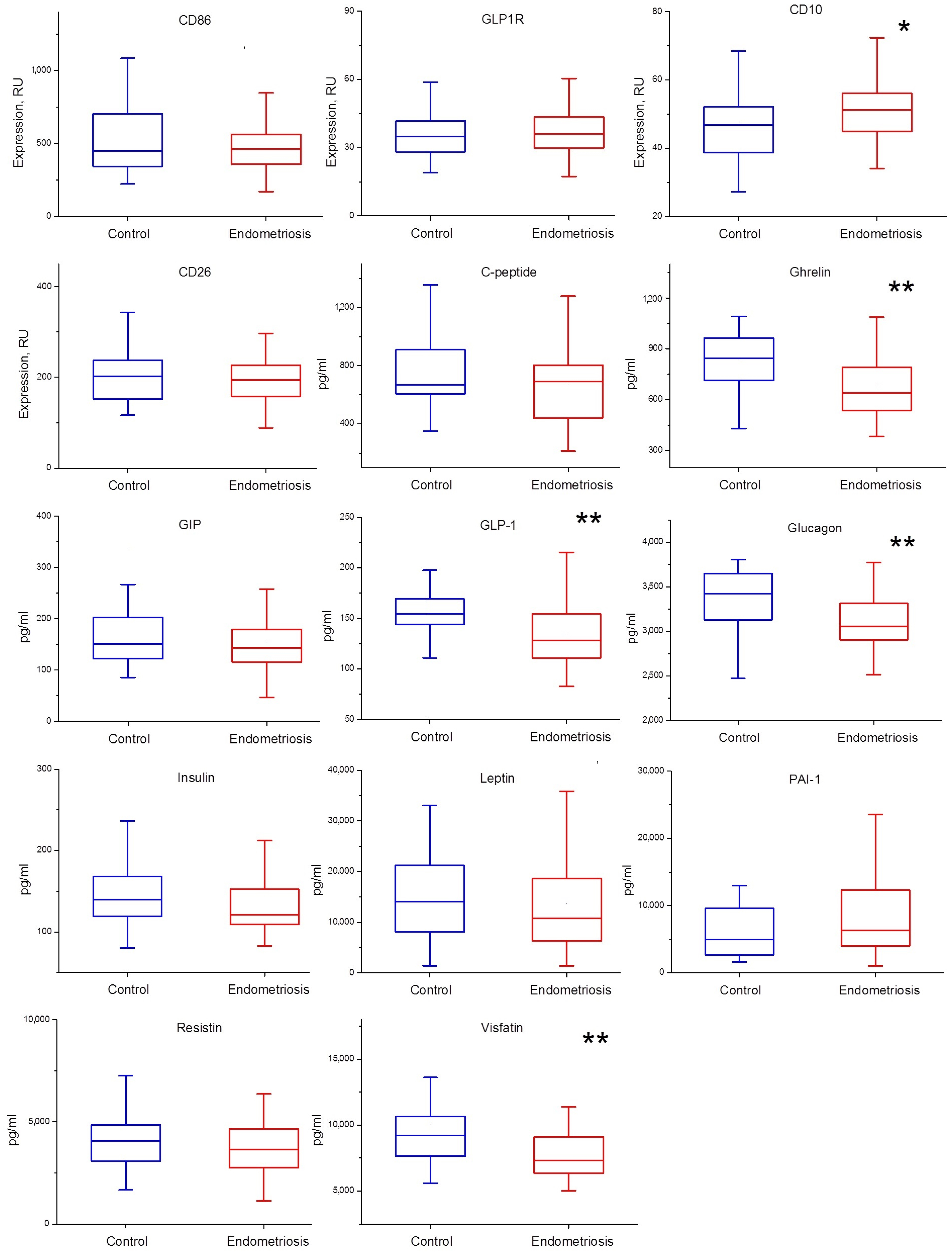
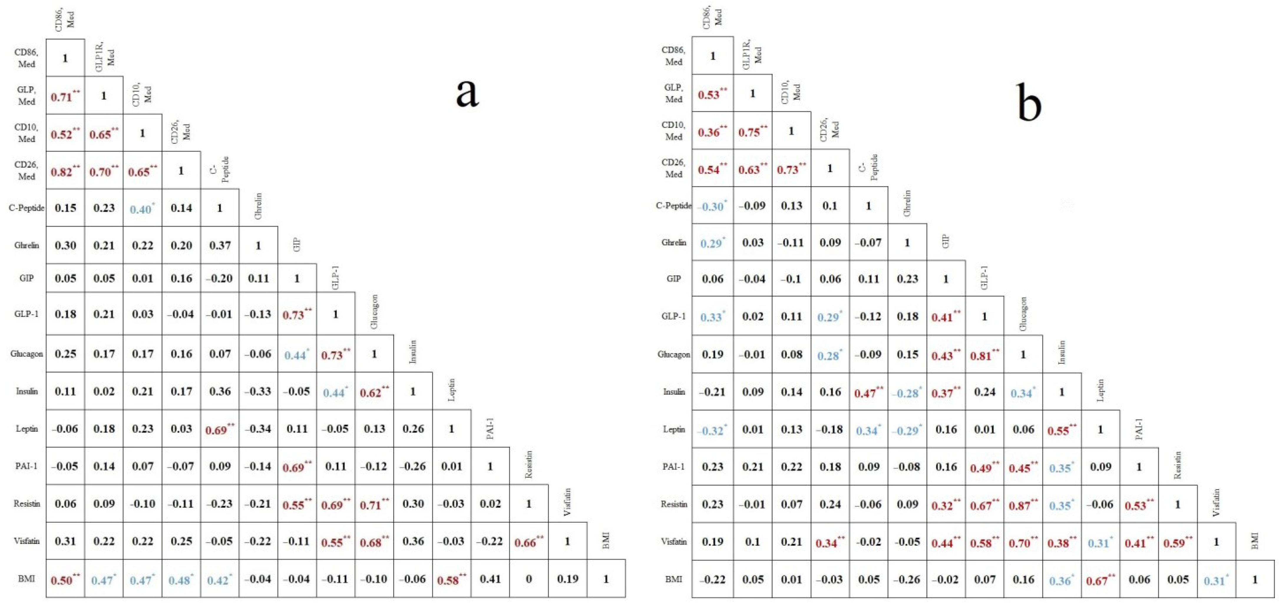
2. Results
3. Discussion
4. Materials and Methods
4.1. Ethics and Sample Collection
4.2. Concentration of Energy Metabolism Markers
4.3. Flow Cytometric Analysis of Expression of CD Antigens
4.4. Statistical Analysis
5. Conclusions
Author Contributions
Funding
Institutional Review Board Statement
Informed Consent Statement
Data Availability Statement
Acknowledgments
Conflicts of Interest
References
- Giudice, L.C.; Kao, L.C. Endometriosis. Lancet 2004, 364, 1789–1799. [Google Scholar] [CrossRef]
- Acién, P.; Velasco, I. Endometriosis: A disease that remains enigmatic. ISRN Obstet. Gynecol. 2013, 2013, 242149. [Google Scholar] [CrossRef]
- Backonja, U.; Louis, G.M.B.; Lauver, D.R. Overall adiposity, adipose tissue distribution, and endometriosis: A systematic review. Nur. Res. 2016, 65, 151–166. [Google Scholar] [CrossRef] [PubMed]
- Hong, J.; Yi, K.W. What is the link between endometriosis and adiposity? Obstet. Gynecol. Sci. 2022, 65, 227–233. [Google Scholar] [CrossRef] [PubMed]
- Viganò, P.; Somigliana, E.; Panina, P.; Rabellotti, E.; Vercellini, P.; Candiani, M. Principles of phenomics in endometriosis. Hum. Reprod. Update 2012, 18, 248–259. [Google Scholar] [CrossRef]
- Kobayashi, H.; Shigetomi, H.; Imanaka, S. Nonhormonal therapy for endometriosis based on energy metabolism regulation. Reprod. Fertil. 2021, 2, C42–C57. [Google Scholar] [CrossRef]
- Kimber-Trojnar, Z.; Dłuski, D.F.; Wierzchowska-Opoka, M.; Ruszała, M.; Leszczyńska-Gorzelak, B. Metformin as a Potential Treatment Option for Endometriosis. Cancers 2022, 14, 577. [Google Scholar] [CrossRef]
- Tian, Z.; Wang, Y.; Zhao, Y.; Chang, X.H.; Zhu, H.L. Serum and peritoneal fluid leptin levels in endometriosis: A systematic review and meta-analysis. Gynecol. Endocrinol. 2021, 37, 689–693. [Google Scholar] [CrossRef]
- Rathore, N.; Kriplani, A.; Yadav, R.K.; Jaiswal, U.; Netam, R. Distinct peritoneal fluid ghrelin and leptin in infertile women with endometriosis and their correlation with interleukin-6 and vascular endothelial growth factor. Gynecol. Endocrinol. 2014, 30, 671–675. [Google Scholar] [CrossRef]
- Dziunycz, P.; Milewski, Ł.; Radomski, D.; Barcz, E.; Kamiński, P.; Roszkowski, P.I.; Malejczyk, J. Elevated ghrelin levels in the peritoneal fluid of patients with endometriosis: Associations with vascular endothelial growth factor (VEGF) and inflammatory cytokines. Fertil. Steril. 2009, 92, 1844–1849. [Google Scholar] [CrossRef] [PubMed]
- Lumeng, C.N.; Bodzin, J.L.; Saltiel, A.R. Obesity induces a phenotypic switch in adipose tissue macrophage polarization. J. Clin. Investig. 2007, 117, 175–184. [Google Scholar] [CrossRef] [PubMed]
- Bacci, M.; Capobianco, A.; Monno, A.; Cottone, L.; Di Puppo, F.; Camisa, B.; Mariani, M.; Brignole, C.; Ponzoni, M.; Ferrari, S.; et al. Macrophages are alternatively activated in patients with endometriosis and required for growth and vascularization of lesions in a mouse model of disease. Am. J. Pathol. 2009, 175, 547–556. [Google Scholar] [CrossRef]
- Krasnyi, A.M.; Sadekova, A.A.; Sefihanov, T.G.; Vtorushina, V.V.; Krechetova, L.V.; Khilkevich, E.G.; Arakelyan, A.S.; Pavlovich, S.V. The content of cytokines IL-6, IL-8, TNF-α, IL-4 and the level of CD86 and CD163 expression in peritoneal fluid macrophages has a reverse correlation with the degree of severity of external genital endometriosis. Biomed. Chem. 2020, 14, 52–56. [Google Scholar] [CrossRef]
- Flatow, E.A.; Komegae, E.N.; Fonseca, M.T.; Brito, C.F.; Musteata, F.M.; Antunes-Rodrigues, J.; Steiner, A.A. Elucidating the role of leptin in systemic inflammation: A study targeting physiological leptin levels in rats and their macrophages. Am. J. Physiol. Regul. Integr. Comp. Physiol. 2017, 313, R572–R582. [Google Scholar] [CrossRef]
- Shiraishi, D.; Fujiwara, Y.; Komohara, Y.; Mizuta, H.; Takeya, M. Glucagon-like peptide-1 (GLP-1) induces M2 polarization of human macrophages via STAT3 activation. Biochem. Biophys. Res. Commun. 2012, 425, 304–308. [Google Scholar] [CrossRef] [PubMed]
- Corrêa da Silva, F.; Aguiar, C.; Pereira, J.A.S.; de Brito Monteiro, L.; Davanzo, G.G.; Codo, A.C.; Pimentel de Freitas, L.; Berti, A.S.; Lopes Ferrucci, D.; Castelucci, B.G.; et al. Ghrelin effects on mitochondrial fitness modulates macrophage function. Free Radic. Biol. Med. 2019, 145, 61–66. [Google Scholar] [CrossRef]
- Wu, H.; Ballantyne, C.M. Metabolic inflammation and insulin resistance in obesity. Circ. Res. 2020, 126, 1549–1564. [Google Scholar] [CrossRef] [PubMed]
- Lee, Y.S.; Wollam, J.; Olefsky, J.M. An integrated view of immunometabolism. Cell 2018, 172, 22–40. [Google Scholar] [CrossRef]
- Kalaitzopoulos, D.R.; Lempesis, I.G.; Samartzis, N.; Kolovos, G.; Dedes, I.; Daniilidis, A.; Nirgianakis, K.; Leeners, B.; Goulis, D.G.; Samartzis, E.P. Leptin concentrations in endometriosis: A systematic review and meta-analysis. J. Reprod. Immunol. 2021, 146, 103338. [Google Scholar] [CrossRef]
- Pantelis, A.; Machairiotis, N.; Lapatsanis, D.P. The formidable yet unresolved interplay between endometriosis and obesity. Sci. World J. 2021, 2021, 6653677. [Google Scholar] [CrossRef]
- van der Stouwe, J.G.; Aeschbacher, S.; Krisai, P.; Schoen, T.; Meyre, P.; Todd, J.; Estis, J.; Risch, M.; Risch, L.; Conen, D. Plasma levels of glucagon-like peptide 1 and markers of obesity among young and healthy adults. Clin. Endocrinol. 2015, 83, 636–642. [Google Scholar] [CrossRef]
- Cappellari, G.G.; Semolic, A.; Kharrat, F.; Caporale, R.; Cremasco, G.; Ius, M.; Zanetti, M.; Barazzoni, R. Plasma glucagon levels are associated with obesity, insulin resistance and metabolic syndrome in a general population cohort. Clin. Nutr. ESPEN 2020, 40, 431. [Google Scholar] [CrossRef]
- Fukuhara, A.; Matsuda, M.; Nishizawa, M.; Segawa, K.; Tanaka, M.; Kishimoto, K.; Matsuki, Y.; Murakami, M.; Ichisaka, T.; Murakami, H.; et al. Visfatin: A protein secreted by visceral fat that mimics the effects of insulin. Science 2005, 307, 426–430. [Google Scholar] [CrossRef] [PubMed]
- Castorina, S.; Barresi, V.; Luca, T.; Privitera, G.; De Geronimo, V.; Lezoche, G.; Cosentini, I.; Di Vincenzo, A.; Barbatelli, G.; Giordano, A.; et al. Gastric ghrelin cells in obese patients are hyperactive. Int. J. Obes. 2021, 45, 184–194. [Google Scholar] [CrossRef] [PubMed]
- Luddi, A.; Marrocco, C.; Governini, L.; Semplici, B.; Pavone, V.; Luisi, S.; Petraglia, F.; Piomboni, P. Expression of matrix metalloproteinases and their inhibitors in endometrium: High levels in endometriotic lesions. Int. J. Mol. Sci. 2020, 21, 2840. [Google Scholar] [CrossRef] [PubMed]
- Canis, M.; Donnez, J.G.; Guzick, D.S.; Halme, J.K.; Rock, J.A.; Schenken, R.S.; Vernon, M.W. Revised American Society for Reproductive Medicine classification of endometriosis: 1996. Fertil Steril. 1997, 67, 817–821. [Google Scholar] [CrossRef]
- Tuttlies, F.; Keckstein, J.; Ulrich, U.; Possover, M.; Schweppe, K.W.; Wustlich, M.; Buchweitz, O.; Greb, R.; Kandolf, O.; Mangold, R.; et al. ENZIAN-score, a classification of deep infiltrating endometriosis. Zentralbl. Gynakol. 2005, 127, 275–281. [Google Scholar] [CrossRef]


| Endometriosis n = 54 | Control n = 30 | p-Value | ||
|---|---|---|---|---|
| Age (year) | 35 (31; 37.75) | 37 (31.75; 40) | 0.11 | |
| BMI (kg/m2) | 21.3 (20.5; 24.2) | 24.9 (22.6; 27.8) | 0.05 | |
| Menstrual cycle phase | Proliferative | 18 (33.3%) | 11(36.7%) | 0.81 |
| Secretory | 23 (42.6%) | 14 (46.7%) | 0.82 | |
| Menstrual cycle disorder | 13 (24.1%) | 5 (13.3%) | 0.58 | |
| Stage of endometriosis | Stage I–II | 20 (37.03%) | ||
| Stage III–IV | 34 (62.97%) | |||
| Type of myomas | Intramural-subserous myoma | 19 (63.3%) | ||
| intramural-submucous myoma | 11 (36.7%) | |||
Publisher’s Note: MDPI stays neutral with regard to jurisdictional claims in published maps and institutional affiliations. |
© 2022 by the authors. Licensee MDPI, Basel, Switzerland. This article is an open access article distributed under the terms and conditions of the Creative Commons Attribution (CC BY) license (https://creativecommons.org/licenses/by/4.0/).
Share and Cite
Krasnyi, A.M.; Sadekova, A.A.; Smolnova, T.Y.; Chursin, V.V.; Buralkina, N.A.; Chuprynin, V.D.; Yarotskaya, E.; Pavlovich, S.V.; Sukhikh, G.T. The Levels of Ghrelin, Glucagon, Visfatin and Glp-1 Are Decreased in the Peritoneal Fluid of Women with Endometriosis along with the Increased Expression of the CD10 Protease by the Macrophages. Int. J. Mol. Sci. 2022, 23, 10361. https://doi.org/10.3390/ijms231810361
Krasnyi AM, Sadekova AA, Smolnova TY, Chursin VV, Buralkina NA, Chuprynin VD, Yarotskaya E, Pavlovich SV, Sukhikh GT. The Levels of Ghrelin, Glucagon, Visfatin and Glp-1 Are Decreased in the Peritoneal Fluid of Women with Endometriosis along with the Increased Expression of the CD10 Protease by the Macrophages. International Journal of Molecular Sciences. 2022; 23(18):10361. https://doi.org/10.3390/ijms231810361
Chicago/Turabian StyleKrasnyi, Aleksey M., Alsu A. Sadekova, Tatyana Y. Smolnova, Vyacheslav V. Chursin, Natalya A. Buralkina, Vladimir D. Chuprynin, Ekaterina Yarotskaya, Stanislav V. Pavlovich, and Gennadiy T. Sukhikh. 2022. "The Levels of Ghrelin, Glucagon, Visfatin and Glp-1 Are Decreased in the Peritoneal Fluid of Women with Endometriosis along with the Increased Expression of the CD10 Protease by the Macrophages" International Journal of Molecular Sciences 23, no. 18: 10361. https://doi.org/10.3390/ijms231810361
APA StyleKrasnyi, A. M., Sadekova, A. A., Smolnova, T. Y., Chursin, V. V., Buralkina, N. A., Chuprynin, V. D., Yarotskaya, E., Pavlovich, S. V., & Sukhikh, G. T. (2022). The Levels of Ghrelin, Glucagon, Visfatin and Glp-1 Are Decreased in the Peritoneal Fluid of Women with Endometriosis along with the Increased Expression of the CD10 Protease by the Macrophages. International Journal of Molecular Sciences, 23(18), 10361. https://doi.org/10.3390/ijms231810361