Review

Variable Expression of GABAA Receptor Subunit Gamma 2 Mutation in a Nuclear Family Displaying Developmental and Encephalopathic Phenotype

by
Gerald Nwosu
1,2,3,
Shilpa B. Reddy
2,4,
Heather Rose Mead Riordan
2,4 and
Jing-Qiong Kang
2,3,5,*
1
Department of Neuroscience and Pharmacology, Meharry Medical College, Nashville, TN 37208, USA
2
Department of Neurology, Vanderbilt University Medical Center, Nashville, TN 37232, USA
3
Brain Institute, Vanderbilt University, Nashville, TN 37232, USA
4
Department of Pediatrics, Vanderbilt University Medical Center, Nashville, TN 37212, USA
5
Department of Pharmacology, Vanderbilt University, Nashville, TN 37233, USA
*
Author to whom correspondence should be addressed.
Int. J. Mol. Sci. 2022, 23(17), 9683; https://doi.org/10.3390/ijms23179683
Submission received: 21 July 2022 / Revised: 13 August 2022 / Accepted: 23 August 2022 / Published: 26 August 2022
(This article belongs to the Section Molecular Neurobiology)

Abstract

:
Mutations in GABAA receptor subunit genes (GABRs) are a major etiology for developmental and epileptic encephalopathies (DEEs). This article reports a case of a genetic abnormality in GABRG2 and updates the pathophysiology and treatment development for mutations in DEEs based on recent advances. Mutations in GABRs, especially in GABRA1, GABRB2, GABRB3, and GABRG2, impair GABAergic signaling and are frequently associated with DEEs such as Dravet syndrome and Lennox–Gastaut syndrome, as GABAergic signaling is critical for early brain development. We here present a novel association of a microdeletion of GABRG2 with a diagnosed DEE phenotype. We characterized the clinical phenotype and underlying mechanisms, including molecular genetics, EEGs, and MRI. We then compiled an update of molecular mechanisms of GABR mutations, especially the mutations in GABRB3 and GABRG2 attributed to DEEs. Genetic therapy is also discussed as a new avenue for treatment of DEEs through employing antisense oligonucleotide techniques. There is an urgent need to define treatment targets and explore new treatment paradigms for the DEEs, as early deployment could alleviate long-term disabilities and improve quality of life for patients. This study highlights biomolecular targets for future therapeutic interventions, including via both pharmacological and genetic approaches.

1. Introduction

Developmental and epileptic encephalopathies (DEEs) are a collection of disorders and syndromes defined by an underlying genetic cause, which results in both a neurodevelopmental and epileptic phenotype. Due to early onset, the progression of epileptic encephalopathy conjoins to dampen normal maturation of the brain throughout development and continues to impact brain function [1,2]. The resultant phenotype manifests as unremitting and often pharmaco-resistant seizures, behavioral dysfunction, neurodevelopmental delay, and lowered quality of life for the patient. There are a host of genes that have been identified and, when mutated, are causative for the developmental and epileptic encephalopathic phenotype. These gene mutations affect brain function through altering GABAergic signaling, GABA transport, ion channel function, and cortical formation among other mechanisms [3,4,5,6,7,8,9].
GABAA receptor genes (GABR) are a group of genes associated with DEE, although previous studies have been focused on relatively mild epileptic syndromes, such as childhood absence epilepsy (CAE), febrile seizures (FS), and generalized tonic-clonic seizures with febrile seizure plus (GEFS+). DEEs such as Dravet syndrome (DS), Lennox–Gastaut syndrome (LGS), West syndrome (WS), or infantile spasm (IS) commonly show epileptic activity comorbid with intellectual disability or autism spectrum disorder and have been frequently associated with mutations in GABRs. Some patients with GABR variants present with neurodevelopmental delay or impaired cognition without seizures, indicating that seizure is just one part of disease phenotype.
GABA signaling is fundamental for normal brain development and function. The GABAA receptors are responsible for both phasic and tonic inhibitory postsynaptic potentials that dictate neuronal communication in the central nervous system [10,11]. The GABAA receptors are members of a super family of Cys-loop, ligand-gated ion channels possessing a characteristic loop formed by a disulfide bond between two cysteine residues [12]. GABAA receptors are composed of three of nineteen subunits in the family, present at the cell surface and synapse as a hetero-pentamer. There have been at least ten GABAA receptor subunit genes (GABR) associated with various genetic epilepsy syndromes. Such genes include GABRA1-3, GABRA5-6, GABRB1-3, GABRG2, and GABRD [13,14,15]. The clinical diagnoses associated with GABR mutations include CAE, FS, juvenile myoclonic epilepsy (JME), GEFS+, DS, and LGS [16,17,18,19,20,21,22]. We have identified multiple mechanisms affecting receptor biogenesis and trafficking, ranging from reducing subunit mRNA transcription or stability; impairing subunit folding, stability, or oligomerization; or inhibiting receptor trafficking [14,18,23,24]. Thus, there are various mechanisms underlying GABR mutations associated with DEEs.
Here, we report a novel case of DEE associated with a GABRG2 mutation and compiled the mechanisms of recent studies with a focus on both GABRG2 mutations and GABRB3 mutations. We focus on these two subunits because mutations in GABRG2 and GABRB3 are more likely to display common phenotypical characteristics. We thus compiled the perturbed biomolecular mechanisms of GABRB3 and GABRG2 mutations and grouped them based on affected function for design and application of precise therapeutic intervention.

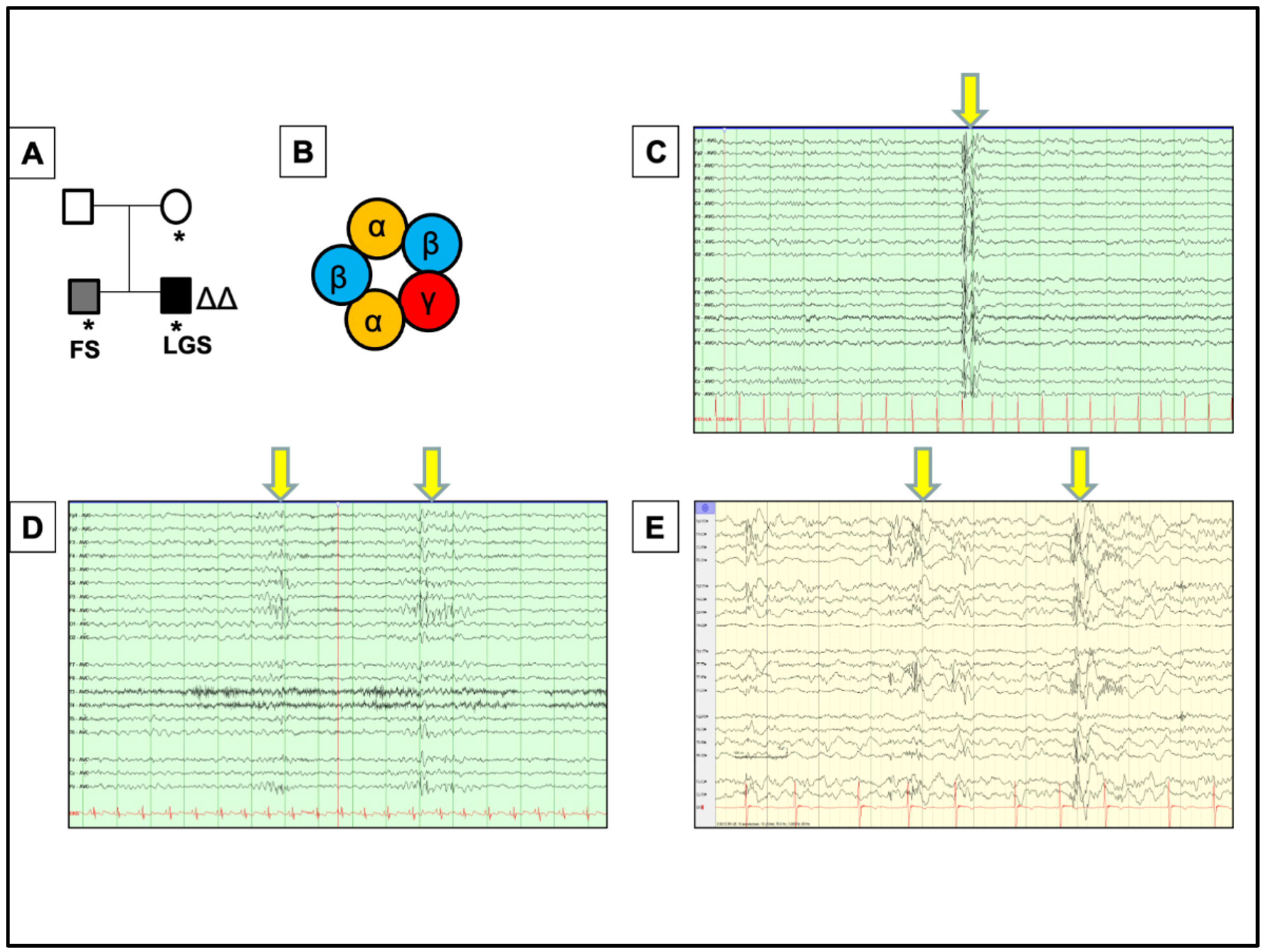
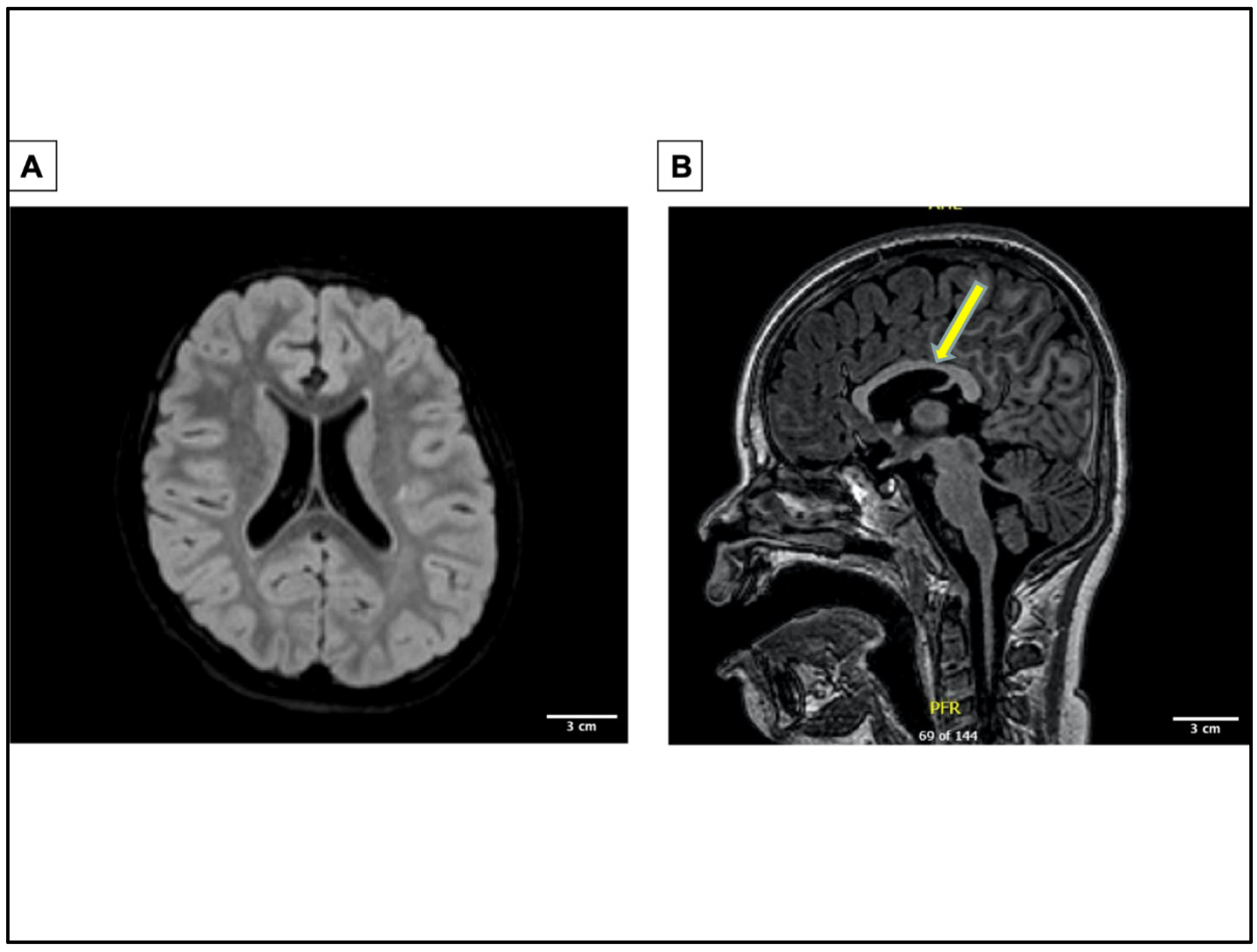
2. A Novel Case of GABRG2 Mutation Associated with DEE, Expanding the Disease Phenotypes Associated with GABRG2 in DEEs

Recently, a nine-year-old male child was found to have a GABRG2 mutation. Sequence analysis and deletion/duplication testing of the 151 genes in the Invitae Epilepsy Panel were performed. This showed there was a heterozygous deletion of the entire coding sequence for the GABRG2 gene, which was also found in his asymptomatic mother and twin brother, who only has a history of febrile seizures. In this case, it will be useful to obtain a genotype of GABRG2 in the proband and twin brother to further define the phenotypic spectrum of GABRG2 mutations. This could delineate the difference in symptom severity. The child has a history of moderate global developmental delay in addition to intractable symptomatic generalized epilepsy. It is intriguing that the mother, who is carrier of the mutation, is asymptomatic, while the monochorionic-monoamniotic twin brother only has febrile seizures (Figure 1A). This is in corroboration with many reports of a male predominance to the DEE phenotype associated with mutations in genes such as CDKL5 and SCN1A. It is also possible that the mother is only a carrier, with carrying males expressing the phenotype [3,25]. GABRG2 encodes the γ2 subunit of GABAA receptor, one of the most abundant subunits critical for clustering the pentameric receptors at the synapse (Figure 1B). The patient showed generalized-appearing spike and polyspike wave discharges (Figure 1C) and multifocal epileptiform discharges, most prominent in the right mesial parietal (Figure 1D) and left posterior regions (Figure 1E). Similar intrafamilial phenotypic heterogeneity has been observed in other GABRG2 mutations. Of note, the proband had a history of hepatoblastoma treated with chemotherapy and resection; thus, one possible explanation for the phenotypic difference could be changes in brain structure and function related to the chemotherapy, resulting in the more severe seizure semiology [26]. This suggests that an underlying condition could modify disease phenotype presentation during development.
The proband’s first seizure was reported at the age of four, presenting as an unprovoked focal tonic seizure with shifting gaze deviation. At this time, the EEG showed interictal sharp waves in the right-frontal region. Magnetic resonance imaging showed multifocal abnormality with abnormal configuration of lateral and third ventricle (Figure 2A,B). This suggests minimal infra- and supratentorial volume loss, microcephaly, abnormal folia architecture within the cerebellum, and abnormal appearance of the globes. These findings are nonspecific and may be suggestive of the underlying genetic mutation. Possible causes would include variable penetrance, modifier genes, or epigenetic/environmental factors. The etiology of phenotype can ultimately determine the treatment paradigm for patients.
He first walked at the age of four and continues to walk on his toes with a wide-based gait. The proband was diagnosed with autism spectrum disorder (ASD) at the age of six. He continues to have global delays. Seizures occurred both during wakefulness and sleep, with all EEG recordings being obtained around nine years of age. At the time of his last visit when he was at age ten and no longer experienced myoclonic or atonic seizures but continued to have non-epileptic myoclonus multiple times per day and generalized seizures (characterized by a slow head drop and behavioral arrest) three times per week. He has frequent complex motor stereotypies. His exam is otherwise notable for subtle ataxia and mild contractures in his gastrocnemius muscles, felt to be related to idiopathic toe walking as opposed to underlying spasticity. The patient was started on levetiracetam, but at age five, seizures became more frequent with multiple semiologies. The background activity was consistent with multifocal and generalized potential epileptogenicity and generalized cerebral dysfunction, including myoclonic and atonic seizures (Figure 1C–E, Supplementary Figures S1–S3) as well as decreased responsiveness associated with quasi-rhythmic generalized discharges lasting ten seconds to twenty minutes. Seizures continued despite escalation of antiepileptic medications, with lacosamide and levetiracetam proving ineffective. Topiramate and valproic acid were discontinued due to side effects, including decreased clearance of clobazam, leading to toxicity. At the age of nine, he developed multifocal, non-epileptic myoclonus. This corresponded with the cessation of topiramate, and he did not respond to benzodiazepines. His current antiepileptic regimen consists of rufinamide, clobazam, and epidiolex.

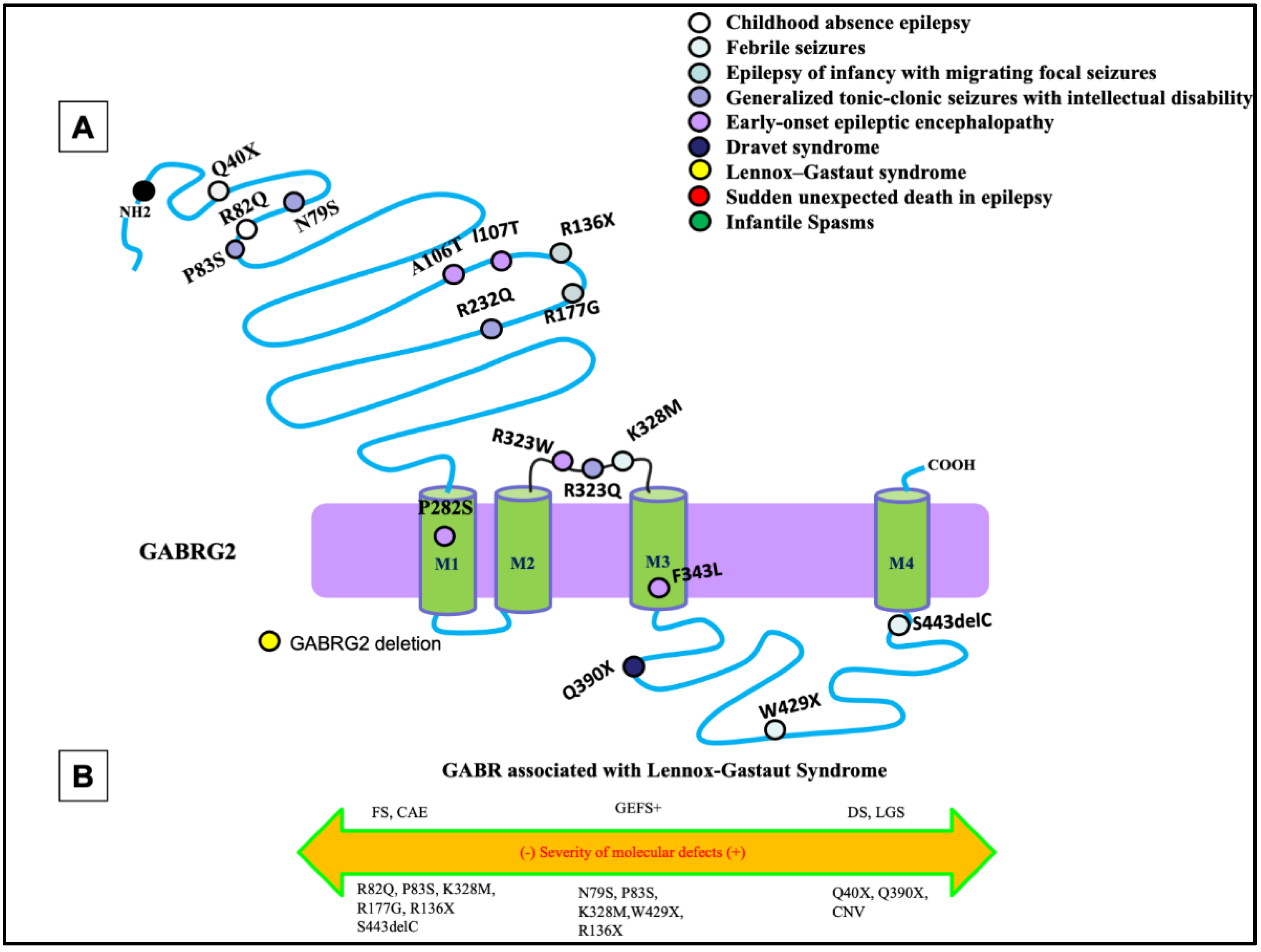
3. Unique Functional Properties of GABRG2-Encoded γ2 Subunit

As mentioned above, the GABRG2-encoded γ2 subunit is a member of the GABAA receptor subunit family that plays a unique role in postsynaptic clustering of the receptor during synaptogenesis [27]. The majority of GABAA receptors found at postsynaptic sites contain the γ2 subunit and are essential for the clustering of α1 and α2 receptors at such sites via the scaffold protein gephyrin [28]. It has been demonstrated that the γ2 subunit is responsible for the accumulation of gephyrin, which in turn is responsible for the synaptic clustering of major subtypes of the GABAA receptor [29,30]. Thus, the γ2 subunit is required for normal channel function and for postsynaptic clustering of the receptor during synaptogenesis [30]. Regulation of proper synaptic localization is a contributing factor of functional plasticity at excitatory and inhibitory synapses. The γ2 subunit containing ternary receptors confers larger channel conductance than the binary αβ receptors [31]. It has been previously reported that mice deficient in the γ2 subunit show a reduction in channel conductance, open probability, and open duration [32,33]. The γ2 subunit is also essential for the formation of a benzodiazepine binding site, which, when disrupted, can result in pharmaco-resistance to certain drugs, as seen in the patient detailed above [33]. In summary, there are at least two unique features for the γ2 subunit. First, it is essential for channel opening with a larger conductance compared with other GABAA receptor subunits. Second, the γ2 subunit is a requirement for synaptic localization and clustering of GABAA receptors during development and maturation, thereby being essential for the proper facilitation of inhibitory postsynaptic signaling. Considering the critical role of the γ2 subunit in synaptic GABAA receptor function, mutations of this gene can lead to altered GABA signaling and GABAergic neurotransmission, a critical pathomechanism underlying the clinical DEE phenotype.

4. Pathomechanisms for GABRG2 Mutations in DEEs

The most common phenotypes associated with GABRG2 mutations are febrile seizures, generalized tonic-clonic seizures with febrile plus (GEFS+), and DS, with febrile seizures being the core phenotype (Figure 3A). Extensive work from previous studies has shown that GABRG2 mutations are associated with a wide spectrum of seizure disorders (Figure 3B). There has been much investigation into the impact of GABRG2 mutations in vitro and in vivo using knock-in mouse models, which would allow us to infer the pathophysiology of this mutation in the patient.
Mutations of GABRG2 are frequently associated with developmental and epileptic disorders through both missense and nonsense mutations. Nonsense mutations in GABRG2 have been identified and are associated with FS, GEFS+, or DEEs. A GABRG2 (R43Q) mutation had reduced cell surface expression of the subunit and reduced cortical inhibition in knock-in mice [34,35]. However, in some cases those mutant receptors, which traffic to the cell surface, have normal function [36]. It was also identified that a subset of mutations, especially the nonsense mutations GABRG2 (Q351X), cannot traffic to the cell surface and synapse due to severe misfolding [37,38]. The mutant protein was retained in the endoplasmic reticulum and reduced trafficking of wildtype partnering subunits, implicating its dominant-negative suppression causing GEFS+. The mutant GABRG2 (Q351X) has slow degradation and can impose a dominant-negative effect on the wildtype GABRG2 allele and the partnering subunits [37]. These findings have been validated in vivo with Gabrg2+/Q390X mice, showing that the mutation GABRG2 (Q390X) is proven to cause chronic subunit accumulation and neurodegeneration [38]. In older mice, the mutated subunit was shown to form protein aggregates due to chronic accumulation of the mutant protein in the endoplasmic reticulum [38]. Mice expressing the GABRG2 (Q390X) mutation showed a severe epileptic phenotype, which included spontaneous generalized tonic-clonic seizures in a seizure-resistant C57BL/6J mouse background. The phenotype is much more severe than the Gabrg2+/− heterozygous knockout mice without generation of the mutant protein [39].
As mentioned above, the γ2 subunit plays an essential role in GABAergic neurotransmission and the overall homeostasis of the central nervous system, while impairment in the subunit results in seizures and neurodevelopmental abnormalities. Some mutants of this protein display a dominant-negative effect that suppresses expression of both wildtype subunit and its binding partners in the hetero-pentamer. Of note, the proband displays an uncharacteristic phenotype for a mutation of this subunit, suggesting further investigation into the interplay of associated proteins with the receptor and how their function is affected by the mutation. The GABAA receptor exerts channel function as a pentamer, and the γ2 subunits oligomerize and colocalize with partnering subunits. There is the possibility that the deficiency due to mutant γ2 protein can result in suppression of proper function of the β3 subunit whose perturbation is often attributed to the LGS phenotype.

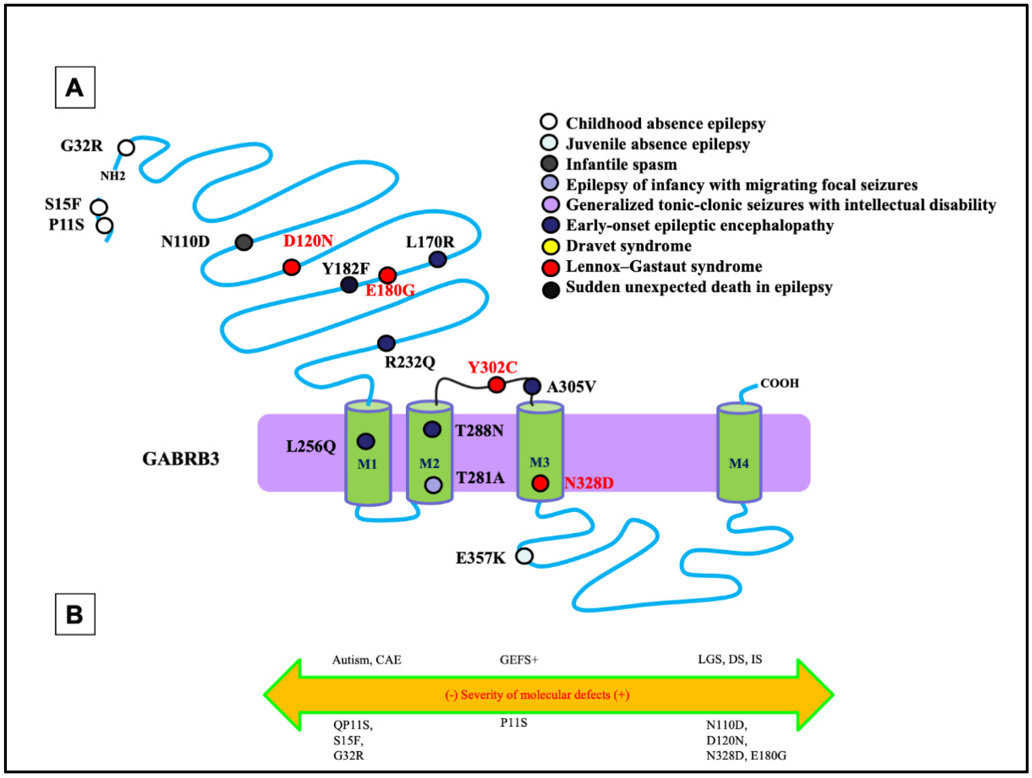
5. Unique Functional Properties of GABRB3 Encoded β3 Subunit

GABRB3 has been identified as an emerging cause for early infantile developmental and epileptic encephalopathies. This subunit is highly expressed in the embryonic brain suggestive of its role in neuronal migration, synaptogenesis, and brain development and maintains expression in the hippocampus exclusively during development [40]. It has come to consensus in the field that the β3 subunit is a major subunit seen in the majority of synaptic GABAA receptors. Like other GABAA receptor subunits, the β3 subunit is composed of a large extracellular NH2 terminal region, four hydrophobic transmembrane domains, an M3-M4 linker, and a short carboxyl terminal. The M2 segment is exposed to the ion channel pore along with the other four subunits of the receptor [41,42]. This is important, as the position of the mutation can affect ion channel capabilities for the whole receptor. GABRB3 has a regulatory role in membrane insertion, as it contains specific endocytosis motifs within the intracellular domains for the clathrin adaptor protein AP2. It is worth noting that the β3 subunit protein contains two phosphorylation sites in its TM3-TM4 intracellular loop. Specifically, phosphorylation of the β3 subunit at S408/S409 is significant for regulating GABAA receptor endocytosis [43]. GABRB3 can itself impose direct impact on channel function, and genetic abnormalities in other genes can also alter GABRB3 expression and contribute to disease pathophysiology. For example, reduced GABRB3 gene expression has been hypothesized as the pathogenesis of Rett syndrome (RS), Angelman syndrome (AS), ASD, and LGS [44,45]. There is an important structural role for GABRB3 akin to GABRG2, so the pathophysiology and phenotypes associated with GABRG2 and GABRB3 mutation may overlap.

6. Pathomechanisms for GABRB3 Mutations in DEEs

Like the phenotypic heterogeneity associated with mutations in GABRG2, mutations in GABRB3 have also been associated with a spectrum of disease phenotypes, including ASD, IS, and DS [46,47] (Figure 4A,B). Among all GABRs, only mutations in GABRB3 have been previously reported to be associated with LGS [20]. In addition to LGS, the β3 subunit encoded by GABRB3 has also been frequently associated with CAE [32,48]. In addition to the defect in the mutant β3 subunit, a mutant β3 subunit can also compromise the assembly and trafficking of partnering subunits α1 or γ2. This suggests there may exist overlapping phenotypes at the clinical level among mutations in the partnering subunits of the receptor through a dominant-negative effect of one, suppressing the maturation and trafficking of the others, as previously reported [39].
Mutations in GABRB3 have been frequently related to DEEs (Figure 4). A previous study evaluated the mutations identified by the Epilepsy Phenome/Genome Project (D120N, E180G, and Y302C) [46]. Follow-up functional assays identified the alterations of GABA-activated channel function, including reduced GABA-evoked current amplitudes, slowed activation, and accelerated deactivation [49]. This was inferred to be due to a reduction in the GABA potency, representing the concentration of GABA, and efficacy, representing the potential of GABA to bind, caused by the mutations. Specifically, the D120N mutation reduced GABA potency, whereas the E180G and Y302C mutations reduced GABA efficacy [49]. This implicates that different mutations can have separate effects on the inhibitory neurotransmission mechanisms. It is important to note that each mutation was in either loop A or loop B of the GABA-binding pocket or the M2-M3 loop, which is involved in the ligand-binding channel, gating-coupling mechanism. The specific pathomechanism could be the likely cause of the mutations disrupting coupling of GABA binding to channel gating.
The differential pathomechanisms in GABRB3 mutations associated with epilepsy with variable severities have been identified [50]. There was a thorough comparison of the GABRB3 (N328D) mutation associated with LGS with GABRB3 (E357K), a mutation associated with a less severe phenotype: juvenile absence epilepsy [50]. In the patient carrying the N328D mutation, there was presence of generalized tonic-clonic seizures and myoclonic seizures. The initial EEG examination showed ictal 2–2.5 Hz generalized polyspike-wave discharges during myoclonic seizures, interictal spike, and slow wave complexes, and irregular fast rhythms with slow waves were recorded during wakefulness [50]. It was identified that the LGS-associated GABRB3 (N328D) mutation caused more reduced cell-surface expression and synaptic presentation of α1β3γ2 than GABRB3(E357K) mutation associated with the juvenile absence epilepsy. However, both mutations caused trafficking defects due to endoplasmic reticulum retention of the mutant protein. Through use of a high-throughput flow cytometry assay, the surface expression of β3 and γ2 subunits for several GABRB3 mutations was evaluated. There was a significant reduction in the expression of the β3 and γ2 subunits at the cell surface for both mutations [50]. This implies that a mutation of this subunit not only prevents proper trafficking of the individual subunit itself but for the partnering wildtype subunits such as γ2 and α1 subunit expression, suggesting loss of whole receptor function instead of subunit alone. By comparing mutations in GABRB3 associated with different phenotypes or different mutations with the same phenotype such as LGS, it is likely that different mutations can have separate effects on the inhibitory neurotransmission mechanisms.
The impact of the GABRB3 mutation associated with LGS has also been investigated in mutant, knock-in Gabrb3+/D120N mice [20]. Like seizures observed in patients carrying the mutation, multiple types of spontaneous seizures including absence, myoclonic, tonic, and generalized tonic-clonic seizures were observed in mice of approximately four months of age. More specifically, the mutant mice showed a seizure frequency of 445 absence, 99 myoclonic, and 4 tonic seizures [20]. As there is a neuropsychiatric comorbidity in LGS, the disease-relevant behavioral abnormalities commonly observed in LGS such as ID and ASD have been evaluated with a battery of behavioral tests conducted in Gabrb3+/D120N mice. The Gabrb3+/D120N mice displayed impaired sociability and cognition evaluated with the three-chamber socialization test. In the Barnes maze test, the Gabrb3+/D120N mice showed a longer latency to find the target hole and increased number of errors in the learning trials. In the memory trial, there was also a significant increase in the number of errors to find the target hole, which both suggests learning and memory deficits in the mice [20].

7. Non-GABR Mutations Associated with DEEs and Therapeutic Interventions

Alongside other GABR mutants that contribute to DEE such as GABRA1 [24,51], many other non-GABR ion channel or non-ion channel genes also contribute to DEEs. These genes are involved in kinase activity (cyclin-dependent kinase-like 5) [52], ion channel function (sodium voltage-gated channel alpha subunits 1, 2, and 8; potassium voltage-gated channel subfamily Q member 2) [53,54,55,56], cell adhesion (protocadherin 19) [57], and general cell homeostasis (tuberous sclerosis complex) [58]. There have also been mutations of the GABA transporter (SLC6A1) and previously mentioned NMDA receptors, which attribute to this phenotype [7,9,59,60]. Many genes have been identified as causation for the DEE phenotype, illustrating a need for genetic therapy that can be constructed precisely for each patient (Figure 5) [61,62]. The DEEs are caused by a defective nonfunctional protein due to a genetic mutation; a logical treatment would be therapeutic options through genetic approaches. Antisense oligonucleotide (ASO) therapy is promising, as it provides different methods for treating neurological disorders. For example, the STK-001 ASO encourages exon skipping to increase the amount of productive protein transcript and is currently in phase 1 clinical trial (NCT04442295) for treatment of Dravet syndrome. ASO technology can be employed to treat exonic frameshift or nonsense variants, toxic gain-of-function mutations, and loss-of-function mutations. Although the ethical guidelines for research, development, and use need to be rationed out, this proves to be a promising treatment paradigm moving further into the 21st century [63,64,65].

8. Conclusions

In summary, we have reported a case of DEE associated with a deletion in GABRG2, which has been previously associated with febrile seizures, CAE, GEFS+, and Dravet syndrome. This is a report of complex movement abnormalities associated with GABRG2 variants. The seizure phenotype among GABR mutations shows overlapping and differential clinical features and, with the genetic mutation being the source of pathophysiology, are defined as developmental and epileptic encephalopathies.
Previous studies indicate mutations in GABRB3 and GABRG2 can both give rise to CAE, GEFS+, and DS. This report of DEE associated with GABRG2 indicates both GABRB3 and GABRG2 can give rise to the DEE phenotype. This is consistent with the fact that β3 and γ2 subunits are partnered in the pentameric receptors at synapses. This is also consistent with the dominant-negative effect displayed by separate mutations of GABRG2, which suppress partnering subunits such as GABRB3. The deficiency of γ2 subunits may compromise the function of partnering subunits such as β3 subunits and thus impair the pentameric receptor function. This expands the clinical phenotype spectrum of GABRG2 mutations in epilepsy and further strengthens the notion of overlapping clinical phenotypes between GABRG2, GABRB3, and possibly other GABR mutations. Alongside other more studied DEE mutations, there is promise in treatment development for DEEs, especially via genetic interventions such as ASOs or gene therapy.

Supplementary Materials

The following supporting information can be downloaded at: https://www.mdpi.com/article/10.3390/ijms23179683/s1.

Author Contributions

Conceptualization, J.-Q.K. and G.N.; case report, S.B.R. and H.R.M.R.; writing—original draft preparation, G.N., J.-Q.K., S.B.R. and H.R.M.R.; writing—review and editing, G.N. and J.-Q.K. All authors have read and agreed to the published version of the manuscript.

Funding

Research reported in this publication was supported by the National Institute of General Medical Sciences of the National Institutes of Health under Award Numbers R25 GM059994 and R01 NS128727.

Institutional Review Board Statement

Not applicable.

Informed Consent Statement

Not applicable.

Data Availability Statement

Not applicable.

Conflicts of Interest

The authors declare no conflict of interest.

References

  1. Scheffer, I.E.; Liao, J. Deciphering the concepts behind “Epileptic encephalopathy” and “Developmental and epileptic encephalopathy”. Eur. J. Paediatr. Neurol. 2020, 24, 11–14. [Google Scholar] [CrossRef] [PubMed]
  2. Scheffer, I.E.; Berkovic, S.; Capovilla, G.; Connolly, M.B.; French, J.; Guilhoto, L.; Hirsch, E.; Jain, S.; Mathern, G.W.; Moshe, S.; et al. ILAE classification of the epilepsies: Position paper of the ILAE Commission for Classification and Terminology. Epilepsia 2017, 58, 512–521. [Google Scholar] [CrossRef] [PubMed]
  3. Specchio, N.; Curatolo, P. Developmental and epileptic encephalopathies: What we do and do not know. Brain 2020, 144, 32–43. [Google Scholar] [CrossRef] [PubMed]
  4. Han, Z.; Chen, C.; Christiansen, A.; Ji, S.; Lin, Q.; Anumonwo, C.; Liu, C.; Leiser, S.C.; Meena; Aznarez, I.; et al. Antisense oligonucleotides increase Scn1a expression and reduce seizures and SUDEP incidence in a mouse model of Dravet syndrome. Sci. Transl. Med. 2020, 12, eaaz6100. [Google Scholar] [CrossRef] [PubMed]
  5. Lopez-Santiago, L.; Isom, L.L. Dravet Syndrome: A Developmental and Epileptic Encephalopathy. Epilepsy Curr. 2019, 19, 51–53. [Google Scholar] [CrossRef] [PubMed]
  6. Gorman, K.M.; Peters, C.H.; Lynch, B.; Jones, L.; Bassett, D.S.; King, M.D.; Ruben, P.C.; Rosch, R.E. Persistent sodium currents in SCN1A developmental and degenerative epileptic dyskinetic encephalopathy. Brain Commun. 2021, 3, fcab235. [Google Scholar] [CrossRef]
  7. Camp, C.R.; Yuan, H. GRIN2D/GluN2D NMDA receptor: Unique features and its contribution to pediatric developmental and epileptic encephalopathy. Eur. J. Paediatr. Neurol. 2020, 24, 89–99. [Google Scholar] [CrossRef]
  8. Amador, A.; Bostick, C.D.; Olson, H.; Peters, J.; Camp, C.R.; Krizay, D.; Chen, W.; Han, W.; Tang, W.; Kanber, A.; et al. Modelling and treating GRIN2A developmental and epileptic encephalopathy in mice. Brain 2020, 143, 2039–2057. [Google Scholar] [CrossRef]
  9. Tsuchida, N.; Hamada, K.; Shiina, M.; Kato, M.; Kobayashi, Y.; Tohyama, J.; Kimura, K.; Hoshino, K.; Ganesan, V.; Teik, K.W.; et al. GRIN2D variants in three cases of developmental and epileptic encephalopathy. Clin. Genet. 2018, 94, 538–547. [Google Scholar] [CrossRef]
  10. Cherubini, E. Phasic GABAA-Mediated Inhibition. In Jasper’s Basic Mechanisms of the Epilepsies, 4th ed.; Noebels, J.L., Avoli, M., Rogawski, M.A., Olsen, R.W., Delgado-Escueta, A.V., Eds.; National Center for Biotechnology Information (US): Bethesda, MD, USA, 2012. [Google Scholar]
  11. Lee, V.; Maguire, J. The impact of tonic GABAA receptor-mediated inhibition on neuronal excitability varies across brain region and cell type. Front. Neural Circuits 2014, 8, 3. [Google Scholar] [CrossRef] [Green Version]
  12. Zhu, S.; Noviello, C.M.; Teng, J.; Walsh, R.M., Jr.; Kim, J.J.; Hibbs, R.E. Structure of a human synaptic GABAA receptor. Nature 2018, 559, 67–72. [Google Scholar] [CrossRef] [PubMed]
  13. Kumari, R.; Lakhan, R.; Kalita, J.; Garg, R.; Misra, U.; Mittal, B. Potential role of GABAA receptor subunit; GABRA6, GABRB2 and GABRR2 gene polymorphisms in epilepsy susceptibility and pharmacotherapy in North Indian population. Clin. Chim. Acta Int. J. Clin. Chem. 2011, 412, 1244–1248. [Google Scholar] [CrossRef] [PubMed]
  14. Hernandez, C.C.; Macdonald, R.L. A structural look at GABAA receptor mutations linked to epilepsy syndromes. Brain Res. 2019, 1714, 234–247. [Google Scholar] [CrossRef] [PubMed]
  15. Macdonald, R.L.; Kang, J.Q.; Gallagher, M.J. GABAA Receptor Subunit Mutations and Genetic Epilepsies. In Jasper’s Basic Mechanisms of the Epilepsies, 4th ed.; Noebels, J.L., Avoli, M., Rogawski, M.A., Olsen, R.W., Delgado-Escueta, A.V., Eds.; National Center for Biotechnology Information (US): Bethesda, MD, USA, 2012. [Google Scholar]
  16. Wallace, R.H.; Marini, C.; Petrou, S.; Harkin, L.A.; Bowser, D.N.; Panchal, R.G.; Williams, D.A.; Sutherland, G.R.; Mulley, J.C.; Scheffer, I.E.; et al. Mutant GABAA receptor γ2-subunit in childhood absence epilepsy and febrile seizures. Nat. Genet. 2001, 28, 49–52. [Google Scholar] [CrossRef] [PubMed]
  17. Gallagher, M.J.; Song, L.; Arain, F.; Macdonald, R.L. The Juvenile Myoclonic Epilepsy GABAA Receptor 1 Subunit Mutation A322D Produces Asymmetrical, Subunit Position-Dependent Reduction of Heterozygous Receptor Currents and 1 Subunit Protein Expression. J. Neurosci. Off. J. Soc. Neurosci. 2004, 24, 5570–5578. [Google Scholar] [CrossRef] [PubMed]
  18. Harkin, L.A.; Bowser, D.N.; Dibbens, L.M.; Singh, R.; Phillips, F.; Wallace, R.H.; Richards, M.C.; Williams, D.A.; Mulley, J.C.; Berkovic, S.F.; et al. Truncation of the GABAA-Receptor γ2 Subunit in a Family with Generalized Epilepsy with Febrile Seizures Plus. Am. J. Hum. Genet. 2002, 70, 530–536. [Google Scholar] [CrossRef] [PubMed]
  19. Hernandez, C.C.; Tian, X.; Hu, N.; Shen, W.; Catron, M.A.; Yang, Y.; Chen, J.; Jiang, Y.; Zhang, Y.; Macdonald, R.L. Dravet syndrome-associated mutations in GABRA1, GABRB2 and GABRG2 define the genetic landscape of defects of GABAA receptors. Brain Commun. 2021, 3, fcab033. [Google Scholar] [CrossRef]
  20. Qu, S.; Catron, M.; Zhou, C.; Janve, V.; Shen, W.; Howe, R.K.; Macdonald, R.L. GABAA receptor β3 subunit mutation D120N causes Lennox–Gastaut syndrome in knock-in mice. Brain Commun. 2020, 2, fcaa028. [Google Scholar] [CrossRef]
  21. Tanaka, M.; Olsen, R.W.; Medina, M.T.; Schwartz, E.; Alonso, M.E.; Duron, R.M.; Castro-Ortega, R.; Martinez-Juarez, I.E.; Pascual-Castroviejo, I.; Machado-Salas, J.; et al. Hyperglycosylation and Reduced GABA Currents of Mutated GABRB3 Polypeptide in Remitting Childhood Absence Epilepsy. Am. J. Hum. Genet. 2008, 82, 1249–1261. [Google Scholar] [CrossRef]
  22. Huang, X.; Tian, M.; Hernandez, C.C.; Hu, N.; Macdonald, R.L. The GABRG2 nonsense mutation, Q40X, associated with Dravet syndrome activated NMD and generated a truncated subunit that was partially rescued by aminoglycoside-induced stop codon read-through. Neurobiol. Dis. 2012, 48, 115–123. [Google Scholar] [CrossRef] [Green Version]
  23. Macdonald, R.L.; Kang, J.-Q.; Gallagher, M.J. Mutations in GABAA receptor subunits associated with genetic epilepsies. J. Physiol. 2010, 588 Pt 11, 1861–1869. [Google Scholar] [CrossRef] [PubMed]
  24. Cossette, P.; Liu, L.; Brisebois, K.; Dong, H.; Lortie, A.; Vanasse, M.; Saint-Hilaire, J.-M.; Carmant, L.; Verner, A.; Lu, W.-Y.; et al. Mutation of GABRA1 in an autosomal dominant form of juvenile myoclonic epilepsy. Nat. Genet. 2002, 31, 184–189. [Google Scholar] [CrossRef] [PubMed]
  25. Depienne, C.; Bouteiller, D.; Keren, B.; Cheuret, E.; Poirier, K.; Trouillard, O.; Benyahia, B.; Quelin, C.; Carpentier, W.; Julia, S.; et al. Sporadic infantile epileptic encephalopathy caused by mutations in PCDH19 resembles Dravet syndrome but mainly affects females. PLoS Genet. 2009, 5, e1000381. [Google Scholar] [CrossRef]
  26. Feng, Y.; Zhang, X.D.; Zheng, G.; Zhang, L.J. Chemotherapy-induced brain changes in breast cancer survivors: Evaluation with multimodality magnetic resonance imaging. Brain Imaging Behav. 2019, 13, 1799–1814. [Google Scholar] [CrossRef] [PubMed]
  27. Schweizer, C. The γ2 subunit of GABAA receptors is required for maintenance of receptors at mature synapses. Mol. Cell. Neurosci. 2003, 24, 442–450. [Google Scholar] [CrossRef]
  28. Essrich, C.; Lorez, M.; Benson, J.A.; Fritschy, J.-M.; Luscher, B. Postsynaptic clustering of major GABAA receptor subtypes requires the γ2 subunit and gephyrin. Nat. Neurosci. 1998, 1, 563–571. [Google Scholar] [CrossRef] [PubMed]
  29. Alldred, M.J.; Mulder-Rosi, J.; Lingenfelter, S.E.; Chen, G.; Lüscher, B. Distinct 2 Subunit Domains Mediate Clustering and Synaptic Function of Postsynaptic GABAA Receptors and Gephyrin. J. Neurosci. Off. J. Soc. Neurosci. 2005, 25, 594–603. [Google Scholar] [CrossRef]
  30. Angelotti, T.P.; Macdonald, R.L. Assembly of GABAA receptor subunits: Alpha 1 beta 1 and alpha 1 beta 1 gamma 2S subunits produce unique ion channels with dissimilar single- channel properties. J. Neurosci. Off. J. Soc. Neurosci. 1993, 13, 1429–1440. [Google Scholar] [CrossRef]
  31. Lorez, M.; Benke, D.; Lüscher, B.; Möhler, H.; Benson, J.A. Single-channel properties of neuronal GABA A receptors from mice lacking the γ2 subunit. J. Physiol. 2000, 527, 11–31. [Google Scholar] [CrossRef]
  32. Gurba, K.N.; Hernandez, C.; Hu, N.; Macdonald, R.L. GABRB3 Mutation, G32R, Associated with Childhood Absence Epilepsy Alters α1β3γ2L γ-Aminobutyric Acid Type A (GABAA) Receptor Expression and Channel Gating. J. Biol. Chem. 2012, 287, 12083–12097. [Google Scholar] [CrossRef] [Green Version]
  33. Günther, U.; Benson, J.; Benke, D.; Fritschy, J.M.; Reyes, G.; Knoflach, F.; Crestani, F.; Aguzzi, A.; Arigoni, M.; Lang, Y.; et al. Benzodiazepine-insensitive mice generated by targeted disruption of the gamma 2 subunit gene of gamma-aminobutyric acid type A receptors. Proc. Natl. Acad. Sci. USA 1995, 92, 7749–7753. [Google Scholar] [CrossRef] [PubMed]
  34. Kang, J.-Q. The GABAA Receptor 2 Subunit R43Q Mutation Linked to Childhood Absence Epilepsy and Febrile Seizures Causes Retention of 1 2 2S Receptors in the Endoplasmic Reticulum. J. Neurosci. Off. J. Soc. Neurosci. 2004, 24, 8672–8677. [Google Scholar] [CrossRef] [PubMed]
  35. Huang, X.; Hernandez, C.C.; Hu, N.; Macdonald, R.L. Three epilepsy-associated GABRG2 missense mutations at the γ+/β− interface disrupt GABAA receptor assembly and trafficking by similar mechanisms but to different extents. Neurobiol. Dis. 2014, 68, 167–179. [Google Scholar] [CrossRef] [PubMed]
  36. Kang, J.-Q.; Shen, W.; Zhou, C.; Xu, D.; Macdonald, R.L. The human epilepsy mutation GABRG2(Q390X) causes chronic subunit accumulation and neurodegeneration. Nat. Neurosci. 2015, 18, 988–996. [Google Scholar] [CrossRef] [PubMed]
  37. Kang, J.-Q.; Shen, W.; Lee, M.; Gallagher, M.J.; Macdonald, R.L. Slow Degradation and Aggregation In Vitro of Mutant GABAA Receptor 2(Q351X) Subunits Associated with Epilepsy. J. Neurosci. Off. J. Soc. Neurosci. 2010, 30, 13895–13905. [Google Scholar] [CrossRef]
  38. Kang, J.-Q.; Shen, W.; Macdonald, R.L. The GABRG2 Mutation, Q351X, Associated with Generalized Epilepsy with Febrile Seizures Plus, Has Both Loss of Function and Dominant-Negative Suppression. J. Neurosci. Off. J. Soc. Neurosci. 2009, 29, 2845–2856. [Google Scholar] [CrossRef]
  39. Warner, T.A.; Shen, W.; Huang, X.; Liu, Z.; Macdonald, R.L.; Kang, J.-Q. Differential molecular and behavioural alterations in mouse models of GABRG2haploinsufficiency versus dominant negative mutations associated with human epilepsy. Hum. Mol. Genet. 2016, 25, 3192–3207. [Google Scholar] [CrossRef]
  40. Tanaka, M.; DeLorey, T.M.; Delgado-Escueta, A.; Olsen, R.W. GABRB3, Epilepsy, and Neurodevelopment. In Jasper’s Basic Mechanisms of the Epilepsies, 4th ed.; Noebels, J.L., Avoli, M., Rogawski, M.A., Olsen, R.W., Delgado-Escueta, A.V., Eds.; National Center for Biotechnology Information (US): Bethesda, MD, USA, 2012. Available online: https://www.ncbi.nlm.nih.gov/books/NBK98178/ (accessed on 23 March 2021).
  41. Carvill, G.L.; Weckhuysen, S.; McMahon, J.M.; Hartmann, C.; Møller, R.S.; Hjalgrim, H.; Cook, J.; Geraghty, E.; O’Roak, B.J.; Petrou, S.; et al. GABRA1 and STXBP1: Novel genetic causes of Dravet syndrome. Neurology 2014, 82, 1245–1253. [Google Scholar] [CrossRef]
  42. Le, S.V.; Le, P.H.T.; Van Le, T.K.; Huynh, T.T.K.; Do, T.T.H. A mutation in GABRB3 associated with Dravet syndrome. Am. J. Med Genet. Part A 2017, 173, 2126–2131. [Google Scholar] [CrossRef]
  43. Vithlani, M.; Moss, S.J. The role of GABAAR phosphorylation in the construction of inhibitory synapses and the efficacy of neuronal inhibition. Biochem. Soc. Trans. 2009, 37, 1355–1358. [Google Scholar] [CrossRef]
  44. Hamdan, F.F.; Srour, M.; Capo-Chichi, J.-M.; Daoud, H.; Nassif, C.; Patry, L.; Massicotte, C.; Ambalavanan, A.; Spiegelman, D.; Diallo, O.; et al. De Novo Mutations in Moderate or Severe Intellectual Disability. PLoS Genet. 2014, 10, e1004772. [Google Scholar] [CrossRef] [PubMed]
  45. Urak, L.; Feucht, M.; Fathi, N.; Hornik, K.; Fuchs, K. A GABRB3 promoter haplotype associated with childhood absence epilepsy impairs transcriptional activity. Hum. Mol. Genet. 2006, 15, 2533–2541. [Google Scholar] [CrossRef] [PubMed]
  46. Epi4K Consortium, Epilepsy Phenome/Genome Project; Allen, A.S.; Berkovic, S.F.; Cossette, P.; Delanty, N.; Dlugos, D.; Eichler, E.E.; Epstein, M.P.; Glauser, T.; Goldstein, D.B.; et al. De novo mutations in epileptic encephalopathies. Nature 2013, 501, 217–221. [Google Scholar] [CrossRef] [PubMed]
  47. Adak, P.; Sinha, S.; Banerjee, N. An Association Study of Gamma-Aminobutyric Acid Type A Receptor Variants and Susceptibility to Autism Spectrum Disorders. J. Autism Dev. Disord. 2021, 51, 4043–4053. [Google Scholar] [CrossRef] [PubMed]
  48. Feucht, M.; Fuchs, K.; Pichlbauer, E.; Hornik, K.; Scharfetter, J.; Goessler, R.; Füreder, T.; Cvetkovic, N.; Sieghart, W.; Kasper, S.; et al. Possible association between childhood absence epilepsy and the gene encoding GABRB3. Biol. Psychiatry 1999, 46, 997–1002. [Google Scholar] [CrossRef]
  49. Ms, V.S.J.; Hernandez, C.C.; Bs, K.M.V.; Hu, N.; Macdonald, R.L. Epileptic encephalopathy de novoGABRBmutations impair γ-aminobutyric acid type A receptor function. Ann. Neurol. 2016, 79, 806–825. [Google Scholar] [CrossRef]
  50. Shi, Y.-W.; Zhang, Q.; Cai, K.; Poliquin, S.; Shen, W.; Winters, N.; Yi, Y.-H.; Wang, J.; Hu, N.; Macdonald, R.L.; et al. Synaptic clustering differences due to different GABRB3 mutations cause variable epilepsy syndromes. Brain 2019, 142, 3028–3044. [Google Scholar] [CrossRef]
  51. Chen, W.; Ge, Y.; Lu, J.; Melo, J.; So, Y.W.; Juneja, R.; Liu, L.; Wang, Y.T. Distinct Functional Alterations and Therapeutic Options of Two Pathological De Novo Variants of the T292 Residue of GABRA1 Identified in Children with Epileptic Encephalopathy and Neurodevelopmental Disorders. Int. J. Mol. Sci. 2022, 23, 2723. [Google Scholar] [CrossRef]
  52. Demarest, S.T.; Olson, H.E.; Moss, A.; Pestana-Knight, E.; Zhang, X.; Parikh, S.; Swanson, L.C.; Riley, K.D.; Bazin, G.A.; Angione, K.; et al. CDKL5 deficiency disorder: Relationship between genotype, epilepsy, cortical visual impairment, and development. Epilepsia 2019, 60, 1733–1742. [Google Scholar] [CrossRef]
  53. Mantegazza, M.; Gambardella, A.; Rusconi, R.; Schiavon, E.; Annesi, F.; Cassulini, R.R.; Labate, A.; Carrideo, S.; Chifari, R.; Canevini, M.P.; et al. Identification of an Nav 1.1 sodium channel (SCN1A) loss-of-function mutation associated with familial simple febrile seizures. Proc. Natl. Acad. Sci. USA 2005, 102, 18177–18182. [Google Scholar] [CrossRef] [Green Version]
  54. Wolff, M.; Johannesen, K.M.; Hedrich, U.; Masnada, S.; Rubboli, G.; Gardella, E.; Lesca, G.; Ville, D.; Milh, M.; Villard, L.; et al. Genetic and phenotypic heterogeneity suggest therapeutic implications in SCN2A-related disorders. Brain 2017, 140, 1316–1336. [Google Scholar] [CrossRef] [PubMed]
  55. Hammer, M.F.; Wagnon, J.L.; Mefford, H.C.; Meisler, M.H. SCN8A-Related Epilepsy with Encephalopathy. In GeneReviews®; Adam, M.P., Everman, D.B., Mirzaa, G.M., et al., Eds.; University of Washington: Seattle, WA, USA, 2016. [Google Scholar]
  56. Goto, A.; Ishii, A.; Shibata, M.; Ihara, Y.; Cooper, E.C.; Hirose, S. Characteristics of KCNQ 2 variants causing either benign neonatal epilepsy or developmental and epileptic encephalopathy. Epilepsia 2019, 60, 1870–1880. [Google Scholar] [CrossRef] [PubMed]
  57. Smith, L.; Singhal, N.; El Achkar, C.M.; Truglio, G.; Sheidley, B.R.; Sullivan, J.; Poduri, A. PCDH19 -related epilepsy is associated with a broad neurodevelopmental spectrum. Epilepsia 2018, 59, 679–689. [Google Scholar] [CrossRef]
  58. Tye, C.; Thomas, L.; Sampson, J.R.; Lewis, J.; O’Callaghan, F.; Yates, J.R.W.; Bolton, P.F. Secular changes in severity of intellectual disability in tuberous sclerosis complex: A reflection of improved identification and treatment of epileptic spasms? Epilepsia Open 2018, 3, 276–280. [Google Scholar] [CrossRef] [PubMed]
  59. Hamdan, F.F.; Myers, C.T.; Cossette, P.; Lemay, P.; Spiegelman, D.; Laporte, A.D.; Nassif, C.; Diallo, O.; Monlong, J.; Cadieux-Dion, M.; et al. High Rate of Recurrent De Novo Mutations in Developmental and Epileptic Encephalopathies. Am. J. Hum. Genet. 2017, 101, 664–685. [Google Scholar] [CrossRef]
  60. Cai, K.; Wang, J.; Eissman, J.; Wang, J.; Nwosu, G.; Shen, W.; Liang, H.-C.; Li, X.-J.; Zhu, H.-X.; Yi, Y.-H.; et al. A missense mutation in SLC6A1 associated with Lennox-Gastaut syndrome impairs GABA transporter 1 protein trafficking and function. Exp. Neurol. 2019, 320, 112973. [Google Scholar] [CrossRef] [PubMed]
  61. Synofzik, M.; van Roon-Mom, W.M.; Marckmann, G.; van Duyvenvoorde, H.A.; Graessner, H.; Schüle, R.; Aartsma-Rus, A. Preparing n-of-1 Antisense Oligonucleotide Treatments for Rare Neurological Diseases in Europe: Genetic, Regulatory, and Ethical Perspectives. Nucleic Acid Ther. 2022, 32, 83–94. [Google Scholar] [CrossRef]
  62. Mirza, N.; Stevelink, R.; Taweel, B.; Koeleman, B.P.C.; Marson, A.G.; Abou-Khalil, B.; Auce, P.; Avbersek, A.; Bahlo, M.; Balding, D.J.; et al. Using common genetic variants to find drugs for common epilepsies. Brain Commun. 2021, 3, fcab287. [Google Scholar] [CrossRef]
  63. Symonds, J.; Zuberi, S.M.; Johnson, M.R. Advances in epilepsy gene discovery and implications for epilepsy diagnosis and treatment. Curr. Opin. Neurol. 2017, 30, 193–199. [Google Scholar] [CrossRef]
  64. McTague, A.; Howell, K.B.; Cross, J.H.; Kurian, M.A.; Scheffer, I.E. The genetic landscape of the epileptic en-cephalopathies of infancy and childhood. Lancet Neurol. 2016, 15, 304–316. [Google Scholar] [CrossRef]
  65. Thakran, S.; Guin, D.; Singh, P.; Singh, P.; Kukal, S.; Rawat, C.; Yadav, S.; Kushwaha, S.S.; Srivastava, A.K.; Hasija, Y.; et al. Genetic Landscape of Common Epilepsies: Advancing towards Precision in Treatment. Int. J. Mol. Sci. 2020, 21, 7784. [Google Scholar] [CrossRef] [PubMed]
Figure 1. A novel case of Developmental Epileptic Encephalopathy (DEE) associated with GABRG2 mutation and the patient EEGs. (A) A pedigree of seizure disorder identified to be heterozygous deletion of GABRG2. * symbolizes GABRG2 deletion. ΔΔ symbolizes proband, the patient of Developmental Epileptic Encephalopathy (DEE). FS stands for febrile seizures. (B) Schematic representation of the major isoform of pentameric GABAA receptor composed of two α, two β, and one γ2 subunit encoded by GABRG2. (CE) The proband carrying GABRG2 mutation showed generalized-appearing spike and polyspike wave discharges (C); multifocal epileptiform discharges, most prominent in the right mesial parietal (D); and left posterior regions (E), both during wakefulness and sleep, all obtained around 9 years of age (Supplementary Figure S1). The image of EEG graph (C) is enlarged (Supplementary Figure S2). The image of EEG graph (D) is enlarged (Supplementary Figure S3). The image of EEG graph (E) is enlarged.
Ijms 23 09683 g001
Figure 2. Brain magnetic resonance image of patient showing multifocal abnormality. Multifocal abnormality with abnormal configuration of lateral and third ventricle, suggestion of minimal infra and supratentorial volume loss, microcephaly, abnormal foliar architecture within the cerebellum, and abnormal appearance of the globes. (A) MRI image of transverse plane showing abnormal lateral ventricles. (B) MRI image of sagittal plane showing abnormal third ventricle indicated by the yellow arrow.
Ijms 23 09683 g002
Figure 3. Epilepsy phenotypes associated with GABRG2 mutations. (A) Two-dimensional protein topology of the γ2 subunit showing locations of mutations in human GABRG2 associated with various epilepsy syndromes and neurodevelopmental disorders. Each mutation is color coded to indicate associated diagnosis, and each dot represents the relative location on the γ2 subunit protein peptide. (B) Diagram dictating the severity of each mutation on a continuum of epilepsy phenotypes. The left side of the diagram infers a less severe epileptic phenotype, the middle dictates a moderate, and the right side of the diagram infers a more severe epileptic phenotype.
Ijms 23 09683 g003
Figure 4. Epilepsy phenotype associated GABRB3 mutations. (A) Two-dimensional protein topology of the β3 subunit showing relative locations of mutations in human GABRB3 associated with various epilepsy syndromes and neurodevelopmental disorders. Each mutation is color coded to indicate associated diagnosis, and each dot represents the β3 subunit protein peptide. (B) Diagram dictating the severity of each mutation on a continuum of epilepsy phenotypes. The left side of the diagram infers a less severe epileptic phenotype, the middle dictates a moderate, and the right side of the diagram infers a more severe epileptic phenotype.
Ijms 23 09683 g004
Figure 5. Other genes in addition to GABAA receptor subunit genes associated with developmental epileptic encephalopathies. Protein interaction network displaying genes of varied function discovered to contribute to a diagnosis of DEE. In addition to GABAA receptor subunit genes (GABR), there are other genes unrelated to the GABAergic signaling network, implicating the wide range of pathways contributing to an DEE phenotype. All nodes are grouped by attribute circle layout based on name. Edges are based on publications depicting protein–network interactions. Analysis was generated through String version 11.9 and visualized with Cytoscape version 3.19.
Ijms 23 09683 g005
Publisher’s Note: MDPI stays neutral with regard to jurisdictional claims in published maps and institutional affiliations.

