Review

Viruses Hijack ERAD to Regulate Their Replication and Propagation

1
College of Veterinary Medicine, South China Agricultural University, No. 483, Wushan Road, Tianhe District, Guangzhou 510642, China
2
Guangdong Laboratory for Lingnan Modern Agriculture, College of Veterinary Medicine, South China Agricultural University, Guangzhou 510642, China
3
Key Laboratory of Zoonosis Prevention and Control of Guangdong Province, Guangzhou 510642, China
*
Authors to whom correspondence should be addressed.
Int. J. Mol. Sci. 2022, 23(16), 9398; https://doi.org/10.3390/ijms23169398
Submission received: 13 July 2022 / Revised: 17 August 2022 / Accepted: 18 August 2022 / Published: 20 August 2022
(This article belongs to the Special Issue ERAD and Ubiquitination 2.0)

Abstract

:
Endoplasmic reticulum-associated degradation (ERAD) is highly conserved in yeast. Recent studies have shown that ERAD is also ubiquitous and highly conserved in eukaryotic cells, where it plays an essential role in maintaining endoplasmic reticulum (ER) homeostasis. Misfolded or unfolded proteins undergo ERAD. They are recognized in the ER, retrotranslocated into the cytoplasm, and degraded by proteasomes after polyubiquitin. This may consist of several main steps: recognition of ERAD substrates, retrotranslocation, and proteasome degradation. Replication and transmission of the virus in the host is a process of a “game” with the host. It can be assumed that the virus has evolved various mechanisms to use the host’s functions for its replication and transmission, including ERAD. However, until now, it is still unclear how the host uses ERAD to deal with virus infection and how the viruses hijack the function of ERAD to obtain a favorable niche or evade the immune clearance of the host. Recent studies have shown that viruses have also evolved mechanisms to use various processes of ERAD to promote their transmission. This review describes the occurrence of ERAD and how the viruses hijack the function of ERAD to spread by affecting the homeostasis and immune response of the host, and we will focus on the role of E3 ubiquitin ligase.

1. Introduction

The ER, the principal location for protein synthesis and maturation in eukaryotic cells, is rich in various molecular chaperones and enzymes that help with protein folding and modification. The host uses multiple methods to help proteins fold correctly [1]. Although all resources are devoted to protein folding, many of the nascent proteins entering the ER fail to obtain their natural conformation [2]. Eukaryotic cells regulate ER pressure mainly through the unfolded protein response (UPR) and ERAD [3,4]. UPR enhances protein folding ability by upregulating the expression of molecular chaperones and folding enzymes through a series of intracellular signal transduction responses [5], while ERAD transports the proteins that cannot be folded correctly out of the ER and further degrades them by the cytoplasmic ubiquitin-proteasome system [6,7]. Disruption of ERAD can lead to many diseases, such as Parkinson’s disease, Alzheimer’s disease, cancer, and infection, confirming its importance in correct cell functioning [8]. The genetic ablation of many ERAD components leads to the death of mouse embryos [9,10,11], which also highlights the importance of ERAD in cell and organism homeostasis. ERAD is exploited by viruses with two main features: one is that secretory proteins or membrane proteins (including many immune proteins) can be degraded by ERAD so that the virus can escape the surveillance of the immune system [12]; second, the process of ERAD transport from the ER to the cytoplasm is utilized by the virus [13], which is conducive to virus invasion. At present, many studies have revealed how viruses use ERAD. In this review, we examined the occurrence and development of ERAD and the mechanism by which different viruses use ERAD to promote its transmission.

2. The Occurrence and Development of ERAD

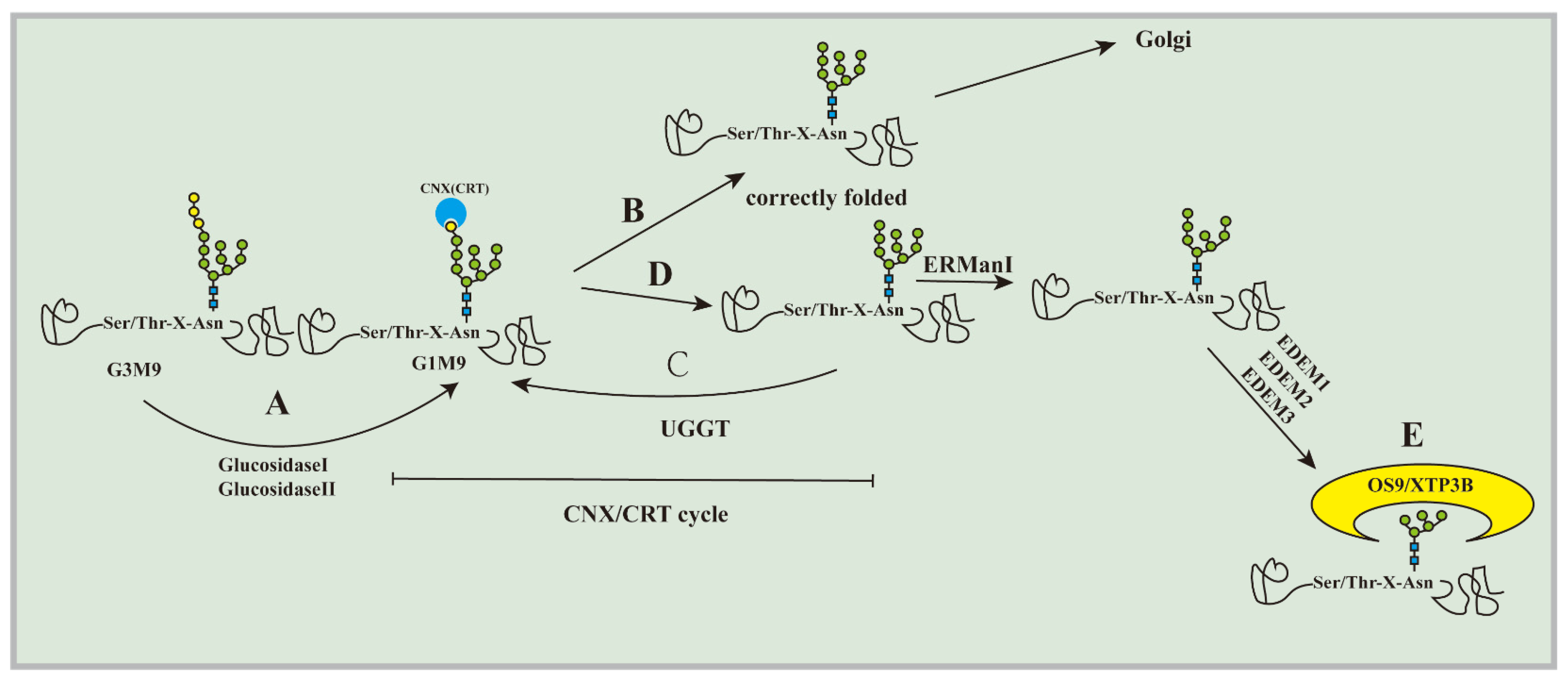
2.1. Substrate Recognition

Substrate recognition is the initial step of ERAD. How can the substrate of ERAD be selected from the proteins that are folded correctly or are being folded? The current understanding is still minimal. Most glycoproteins transported to the ER may cotranslate and modify a high mannose core oligosaccharide containing GlcNAc2-Man9-Glc3 (Glc: glucose, Man: mannose, GlcNAc: N-acetylglucosamine) sequence on the aspartic acid residue containing Asn-X-Ser/Thr (X represents any amino acid) at its N-terminus [14]. Glycosylation of core glycans under the action of glucosidases leads to the entry of new glycoproteins into the calnexin (CNX)/calreticulin (CRT) cycle (Figure 1A). The ‘‘CNX/CRTcycle’’ refers to the nascent N-linked glycoproteins that can bind to CNX and CRT in the ER through glucose residues. When binding to CNX and CRT, the glycoprotein starts to fold, and the folded protein with the correct structure will be released from CNX and CRT, while the protein without the correct structure will recycle and repeatedly fold until it can fold correctly [15]. For correctly folded proteins, further deglycosylation removes N-glycans to produce glycoproteins containing GlcNAc2-Man9, which prevents them from binding to CNX or CRT again, causing glycoproteins to flow out (Figure 1B). Proteins that do not have the correct conformation will be recognized by UDP-glucose: glycoprotein glucosyltransferase (UGGT). Under the action of UGGT, a glucose molecule is added to N-glycans to be glycosylated so that they can preferentially return to the CNX/CRT cycle for further folding (Figure 1C) [16,17]. Glycoproteins that cannot fold correctly must be transferred from the CNX/CRT cycle to ERAD for degradation, and mannosidases accomplish this detachment. Mannosidase removes terminal mannose residues from core polysaccharides so that they can interact with other mannose-specific lectins to combine them with ERAD [18]. Under the action of ER mannosidase I (ERManI) [19], ER degradation-enhancing α-mannosidase like protein 1 (EDEM1) [20,21], EDEM2, and EDEM3 [22,23], glycoproteins are incompatible with UGGT-mediated glycosylation, and misfolded glycoproteins are separated from CNX/CRT to produce glycan signals for ERAD system identification (Figure 1D). Soluble ER-resident proteins, OS-9 and XTP3B, play a vital role in recognizing mammalian ERAD substrates [24]. The possible mechanism for this is that OS-9 and XTP3B can recognize oligosaccharides modified by ERMan I and EDEM1-3 through the mannose-6-phosphate homologous receptor domain (MRH). The pruned oligosaccharides expose their terminal α-1 mannose and are recognized by the MRH regions of OS-9 and XTP3B. OS-9 and XTP3B bind to SEL1L through their MRH domain, and the misfolded protein is guided to the next step of reverse translocation (Figure 1E) [25,26,27]. There are many molecular chaperones involved in this process. GRP94, a metazoan-specific Hsp90 in the ER lumen, acts upstream of OS-9 to further recognize misfolded α1 subunits in a glycan-dependent manner [28].
The recognition of nonglycoproteins may be facilitated by chaperones. Folding of nonglycoproteins in the ER is assisted by the “chaperone cycle” (cycle of substrate binding and release). The ER-localized Hsp70 chaperone Bip, which is composed of the ATP-bound form and ADP-bound form, plays an important role in protein folding homeostasis [29]. Similar to Hsp70s in other compartments, Bip does so by the reversible binding and releasing of unfolded client proteins. A substrate containing a hydrophobic region exposed on the surface binds to the ATP-bound form, which is converted to the ADP-bound form by cochaperone-mediated stimulation of intrinsic ATPase activity to grab the substrate. The ADP-bound form is converted to the ATP-bound form by the action of nucleotide exchange factor (NEF) Lhs1/Grp170 to release the substrate [30]. This cycle continues until the folding is completed. Disulfide bond formation catalyzed by protein disulfide isomerase Pdi1/PDI family proteins also helps productive folding [31,32]. At present, it is known that several nonglycoproteins can be degraded through the ERAD pathway [33,34,35,36,37], and Bip plays an important role. However, how Bip plays a role in this process and how nonglycoproteins are transported to the retrotranslocation channel are still unclear.

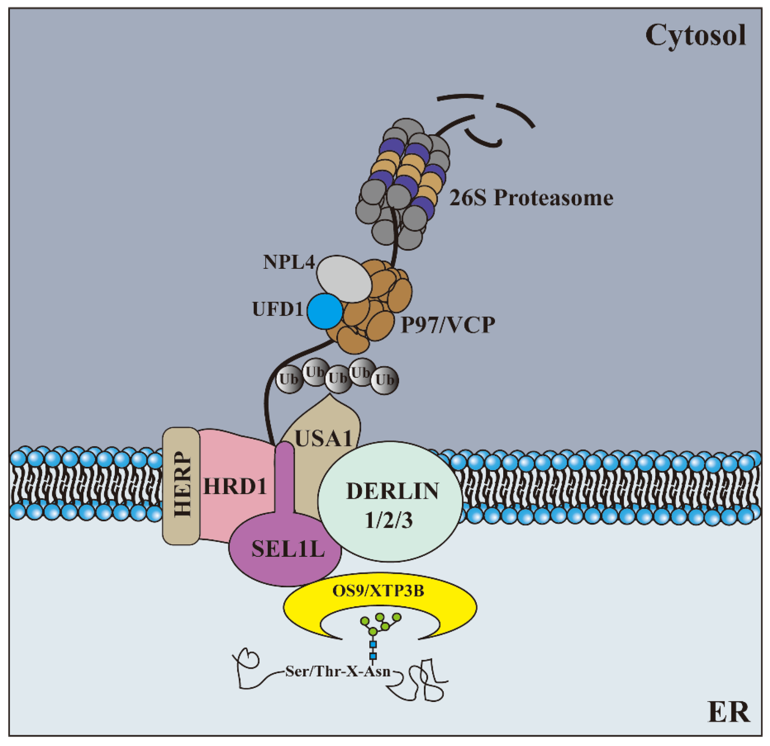
2.2. Retrotranslocation and Ubiquitination

After recognition, the substrate retrotranslocates into the cytoplasm according to its specific position in the ER, which is closely combined with the ubiquitination of the substrate. In most cases, the classic E3 ligase enzyme involved in ERAD is itself a multitransmembrane protein, where the RING domain responsible for ligase activity is located in the cytoplasm, and the substrate is ubiquitinated at the same time as its reverse translocation. Subsequently, the ubiquitin substrate is extracted from the membrane in an ATP-dependent manner and released into the cytoplasm to be degraded by proteasomes. For many years, the identity of the retrotranslocation channel has been controversial. With the development of technology and the deepening of research, mainly in budding yeast but also in mammalian cells, an increasing number of components of retrotranslocation channels have been identified, and most are E3 ligase enzymes. Based on the analysis of some model substrates, the specificity of the E3 ligase enzyme complex seems to depend on the location of the misfolded lesion on the substrate relative to the ER membrane: proteins with lumen (ERAD-L substrate) or intramembrane (ERAD-M substrate) misfolded domains are targeted to HRD1 complexes; proteins with misfolded domains on the cytoplasmic side of the membrane (ERAD-C) are degraded by Doa10 complexes [38,39,40].
Based on the mutation, pull-down experiments, and interaction with the proteasome, it is speculated that the Sec61 channel, which usually allows peptides to enter the ER cavity from the cytoplasm, can work in reverse [41]. However, subsequent studies have shown that there is only a weak interaction between the ERAD substrate and Sec61, and its role as a retrotranslocation channel of ERAD has been rejected [42]. With the deepening of study, the voice of the HRD1 complex as a candidate for the retrotranslocation channel is getting louder [43], and the fact that the HRD1 complex can be used as a retrotranslocation channel of the ERAD substrate has also been proven by photocrosslinking [44]. HRD1 is a multitransmembrane protein with E3 ubiquitin ligase activity and a cytoplasmic ring finger domain that can mediate the ubiquitination of reverse transcriptional translocation substrates and act as a retrotranslocation channel. The HRD1 complex consists of the E3 ubiquitin ligase HRD1 and four other proteins (SEL1L, Der1, Usa1, and OS-9) [26,45]. SEL1L plays an important role in the HRD1 complex, and SEL1L is an important link to coordinating ERAD substrate recruitment, translocation, and ubiquitination [46]. SEL1L combines with OS-9/XTP3B [25,26], EDEM1 [47], and EDEM3 [48]. SEL1L is necessary to transfer the substrate from ER lectin to HRD1 [18,26,49]. One of the ERAD ubiquitin ligases, SEL1L, forms a nucleation complex with intact membrane ERAD components, including Derlin-1, Derlin-2, AUP1, UBXD8, VIMP, and Herp [26,27,50,51]. This, in turn, recruits the VCP/p97 complex necessary to drive substrate dislocation. The UBXD family is a diverse group of UBX (ubiquitin-regulatory X) domain-containing proteins in mammalian cells. The UBX domain enables all members of the UBXD family to bind to the multifunctional AAA-ATPase p97/VCP protein [52] via the amino terminal domain of p97 [53]. Several analyses in yeast show that to a large extent, the Hrd1-SEL1L complex itself is the retrotranslocation channel through which the substrate is brought back from the ER to the cytoplasm [54]. The self-ubiquitination of HRD1 is considered the trigger of substrate retrotranslocation, which is similar to the “door” of the substrate retrotranslocation channel [45,55,56,57,58]. Recently, the structural analysis of the HRD1 complex by cryo-EM from Saccharomyces cerevisiae showed that Hrd1 and Der1 were connected on the cytoplasmic side of the membrane through Usa1. Both Hrd1 and Der1 have a lateral door that regulates substrate entry, and these also face each other in the membrane; at the same time, Hrd1 and Der1 each have a hydrophilic groove on one side of the cytoplasm and one side of the ER, respectively. Both proteins distort the lipid membrane structure near the lateral door, making it thinner than a normal phospholipid bilayer [59].
Doa10 is located throughout the ER, including the inner and outer nuclear membranes. Localization of the Doa10 inner nuclear membrane is necessary for the degradation of its nucleoprotein substrate [60]. A recent study has shown that ubiquitin ligase Doa10 (Teb-4/MARCH6 in mammals) is a reverse transcriptase that promotes the extraction of membrane proteins. It helps to overcome the energy barrier of membrane protein extraction [61]. A recent study in yeast showed that Derlin paralog Dfm1 might also act as a core mechanism in the degradation of ERAD-M (ERAD substrates located in the membrane) [62].
In addition to the channel, the retrotranslocation of the ERAD substrate requires a force to pull them into the cytoplasm, which p97/VCP provides [63]. P97/VCP is a member of the family of ATP enzymes (AAA+ATP enzymes) related to various cellular activities, which consists of an N-terminal domain (N-domain) and two ATPase domains (D1 and D2) and contains a central pore. The ubiquitin substrate must be pulled out of the ER by the p97/VCP complex before entering the 26S proteasome for degradation [64,65]. At present, one of the generally accepted models of ERAD substrates using the p97/VCP complex is that the ubiquitin substrates are bound to the N-terminal Ufd1-Npl4 cofactor complex of p97/VCP through the ubiquitin chain and pulled into the central hole formed by the D1 domain [52], which may be exited through the sequential interaction with the monomer and the central hole of D2 [66,67,68,69]. In contrast, the C-terminal domain interacts with its adaptor proteins through the P97/VCP-binding region, P97/VCP-interacting motif, or SHP box [70]. The mechanism of this process may be that VCP provides energy for binding and hydrolysis by extracting or “shifting” the ubiquitinated substrate from the ER membrane. Ufd1, through the recognition of the K48 Ub chain, combined with the ubiquitin substrate Npl4, stabilized UFD1L [71].

2.3. Degradation

Once misfolded proteins are identified, they must be transported to the cytoplasm where the ubiquitin-proteasome system (UPS) is located, and this process must be closely coupled with the degradation process because most of the ERAD substrates are hydrophobic and can easily accumulate in an aqueous environment. Proteins with ubiquitin- and proteasome-binding domains, such as hHR2 (Rad23 in yeast), may be used as shuttle factors to transfer the substrate from p97/VCP to the proteasome [72]. Cellular solute chaperone proteins, such as Hsc70 [73] and Bag6 [74], can interact with ERAD clients in the cytoplasm after p97/VCP-mediated extraction from the ER by exposing hydrophobic regions on these deployment clients. They may help maintain the substrate’s solubility and prevent accumulation in the aqueous cellular solute environment, but they also help the substrate guide the degradation mechanism [66]. Once transferred to the 26S proteasome, the ERAD substrate is degraded in the same manner as described by all proteins. (All the stages are shown in Figure 2, and most of the components involved in ERAD are listed in Table 1).

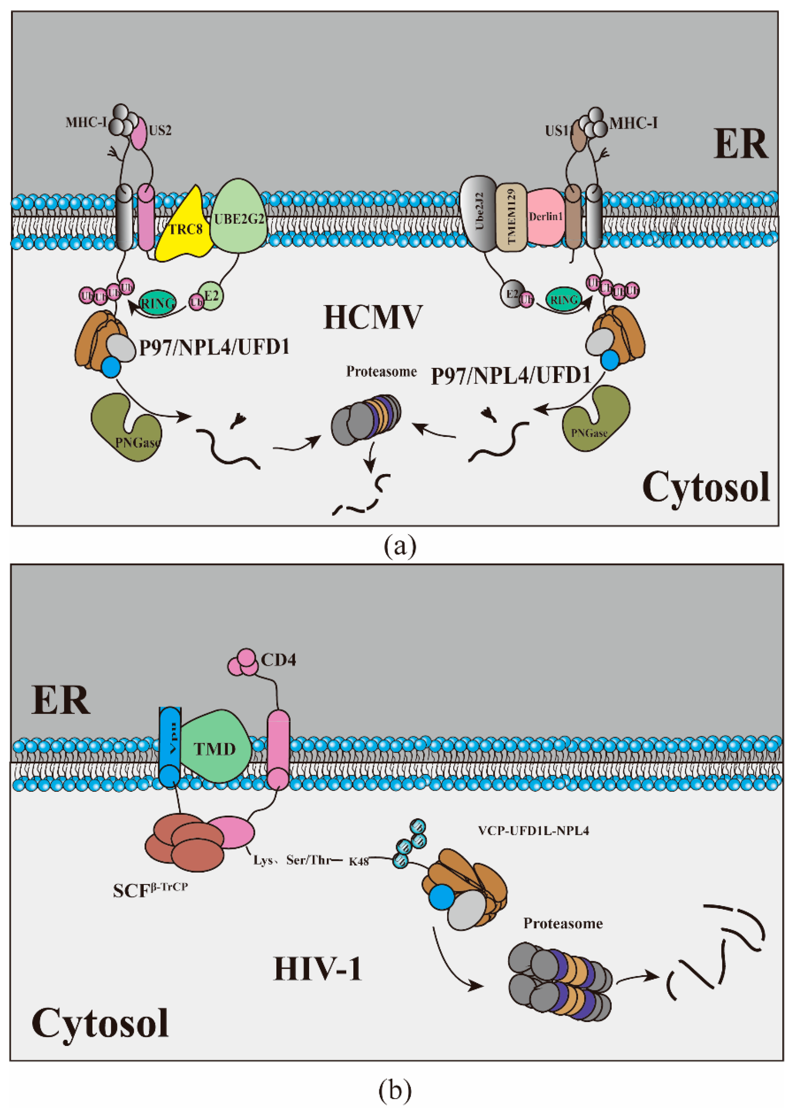
3. Viruses Hijack ERAD to Manipulate the Host Immune Response

A variety of viruses have evolved the ability to suppress the body’s immune response to promote persistent infection. Currently, the most in-depth examples of virus-induced ERAD that are involved in regulating the immune response are herpesviruses and human immunodeficiency virus (HIV). Human cytomegalovirus (HCMV) induces ERAD to accelerate the degradation of MHC-I to prevent CD8+ T lymphocytes from recognizing virus-infected cells and promoting a persistent infection. US2 and US11 are ER-resident type I membrane virus glycoproteins expressed in the early stage of HCMV infection. After cotranslation and insertion into the ER, the newly synthesized MHC-I immediately binds to US2 or US11, and US2 and US11 transport them from the ER to the cytoplasm for degradation [76]. Although both US2 and US11 can degrade MHC-I, their mechanisms do not seem to be precisely the same. US11 recruits TMEM129, and TMEM129 recruits Ube2J2 for US11-induced MHC-I ubiquitin through Derlin1; thus, driving MHC-I reverse translocation from the ER back to the cytoplasm, where MHC-I is deglycosylated by protein N-glycanase (PNGase) and then degraded by the proteasome. The E3 ubiquitin ligase TMEM129 plays a central role in US11-mediated MHC-I decomposition [77,78,79,80]. Unlike US11, which depends on Derlin-1, US2 usurps the E3 ubiquitin ligase TRC8, which plays a central role in US2-mediated MHC-I decomposition. TRC8 binds to the cytoplasmic tail of US2, resulting in rapid polyubiquitin of MHC-I, which triggers MHC-I to completely reverse into the cytoplasm [81,82]. The degradation of MHC-I was inhibited in UBE2G2-depleted cells (UBE2G2 is a kind of E2 ubiquitin binding enzyme), indicating that UBE2G2 plays a critical role in the ubiquitination of US2, possibly because of the cooperation of TRC8 and UBE2G2 [83]. Although they use different E3 ubiquitin ligases during reverse translocation, both rely on p97 to translocate MHC-I to the proteasome for degradation. In addition to MHC-I, US2 induces the downregulation of multiple immunoreceptors to modulate cellular migration and immune signaling, whereas US11-mediated degradation is restricted to MHC-I [84]. Recently, genome-wide CRISPR/Cas9 library screening found that the ubiquitin-fold modifier 1 (UFM1) pathway is also involved in the degradation of MHC-I, and interference with UFM1 specifically inhibits the reverse transport of MHC-I molecules from the ER to the cytoplasm, but the effect of UFM1 on the degradation of MHC-I molecules may be indirect [85] (For a schematic view of the mechanism, see Figure 3a). Like herpesviruses, retroviruses can also manipulate the host’s immune system through the ERAD pathway. HIV-Ⅰ Vpu targets newly synthesized CD4 receptors and rapidly degrades CD4 through a process similar to ERAD [86,87]. Magadan uses siRNA to interfere with 18 proteins related to each process of ERAD, and the results showed that the interference of VCP, UFD1L, and NPL4 had the best protection on the degradation of CD4 [71] (for a schematic view of the mechanism, see Figure 3b). Virus-infected cells are specifically eliminated through the synergistic effect of helper T cells and cytotoxic T lymphocytes. This elimination is mainly dependent on MHC-I and MHC-II presenting virus-derived peptide antigens on the surface of virus-infected cells. HCMV has evolved the mechanism to use the ERAD system to eliminate MHC-I so that the virus circumvents the monitoring of the host immune system artfully. They hijack alternative ubiquitin ligases instead of HRD1, suggesting the necessity for a specialized ubiquitin ligation system in pathogenic ERAD.

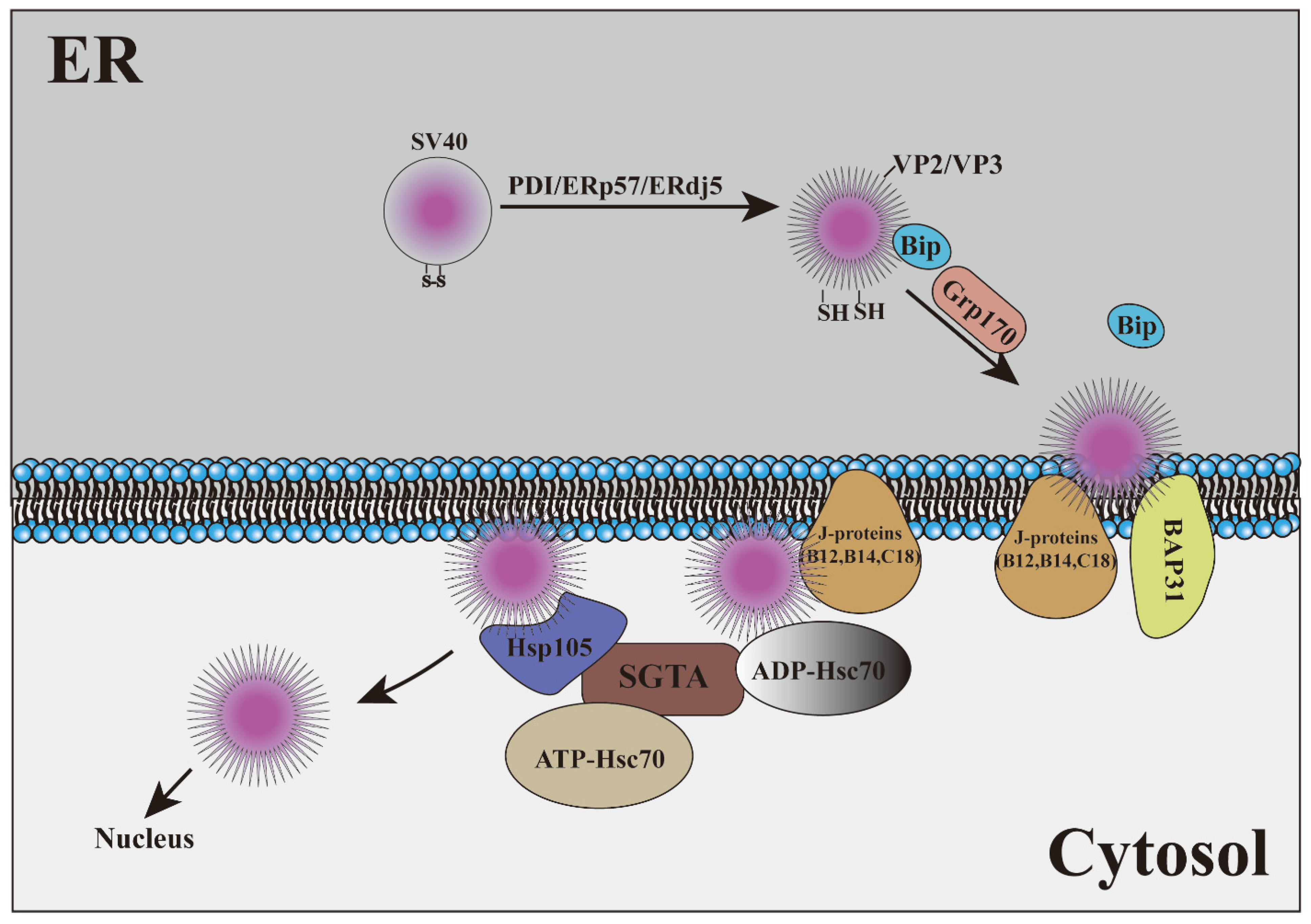
4. Viruses Hijack ERAD as a Transport Mechanism

Studies on the use of ERAD as a transport mechanism have been reported in cholera toxins (CTx), pertussis toxins, Shiga and Shiga-like toxins (STx), heat-labile enterotoxins, Pseudomonas exotoxins (PEx), cytolethal distending toxins (CDT), and the plant toxin ricin and other toxins [88,89,90]. Some viruses have also evolved the mechanism of using ERAD to penetrate the membrane to reach the cytoplasm. Polyomaviruses (PyVs) are a kind of nonenveloped DNA tumor virus. To infect cells, PyV is transported from the cell surface to the ER, where it hijacks the elements of the ERAD mechanism to penetrate the ER and reach the cytoplasm. The virus is transported from the cytosol to the nucleus, resulting in lytic infection or cell transformation. How PyV is transported from the ER to the cytoplasm is a key issue in the process of viral infection. Many studies have shown that ERAD takes advantage of this process [91,92,93,94,95]. SV40 is the best-studied virus that utilizes ERAD as a transport mechanism. After SV40 reaches the ER, under the action of PDI, ERp57, and ERdj5, hydrophobic VP2 and VP3 proteins are exposed, and hydrophobic virus particles are produced, which are disguised as misfolded proteins [91,96,97]. To prevent the aggregation of hydrophobic virus particles, the ERAD molecular chaperone BiP was recruited by SV40 [93,98]. When the SV40-BiP complex approaches the lumen surface of the ER membrane, the virus must be released from BiP to initiate ER-membrane transport. Glucose-regulated protein, 170 kDa (Grp170), plays an important role in this process [99,100]. When hydrophobic SV40 is inserted into the ER membrane, the amino terminal region of the VP2 protein binds to the ER membrane protein BAP31 to stabilize the membrane-embedded virus and then participates in virus transport from the ER to the cytoplasm under the action of ER transmembrane J-proteins (B12, B14, and C18) [93,101,102]. Some studies have also shown that SV40 can form a specific region on the ER, which can be used as a selective membrane penetration site for viruses. The forms of VP2/VP3 exposure and membrane penetration of SV40 mainly exist in these specific regions, and SV40 mutants that cannot be transferred across the ER membrane to the cytoplasm cannot trigger the formation of lesions [92,103]. The transfer of SV40 from the ER to the cytoplasm also requires a “force”. In general, the driving force of the ERAD process is usually provided by p97, but studies have shown that this “force” may be provided by Hsc70-Hsp105-SGTA rather than p97 in SV40 [92]. Under the action of membrane-bound J proteins, Hsc70 was transformed into high-affinity ADP-Hsc70. This enables Hsc70 to first bind to membrane-embedded SV40, and Hsp105 then induces the nucleotide exchange of Hsc70 to produce ATP-Hsc70 that releases viral particles. Once the virus is detached from Hsc70 and Hsp105, it can capture SV40 and release it from Hsp105, with Hsc70 recombining the virus. The iterative cycles of Hsc70–Hsp105 binding and releasing SV40 are considered to provide the main driving energy for extracting viral particles from the ER membrane [104,105,106,107]. In short, the unenveloped virus SV40 transports the virus from the ER to the cytoplasm through the host ERAD pathway and inserts the virion into the ER by changing the structure of the virion (Figure 4). The formation of a unique membrane penetration site in the local cell membrane so that the virus can successfully enter the cytoplasm is a new discovery of the virus in the process of using ERAD.

5. Viruses Hijack ERAD to Regulate Viral Protein Expression

ERAD plays an essential role in the regulation of viral protein expression. JEV and DENV degrade NS4B protein in convoluted membranes (CM) through the Derlin2-mediated ERAD pathway to avoid the excessive accumulation of nonstructural proteins from damaging virus replication. In addition, the researchers also found that JEV and DENV NS4B proteins can interact with p97/VCP. The inhibition of VCP activity by VCP-specific chemical inhibitors not only inhibited the degradation of nonstructural proteins but also inhibited the production of infectious JEV and DENV virus particles [108]. A recent study showed that Bardoxolonemethyl (CDDO-me), an inhibitor of protein translocation mediated by HRD1, can bind to grp94, a vital component of the HRD1 pathway, thus inhibiting the replication of DENV and ZIKV. The knockdown of grp94 can also significantly inhibit the replication of DENV and the synthesis of viral membrane proteins [109]. Ruan found that CP26, another small molecular inhibitor that targets the HRD1 complex and inhibits the process of protein dislocation from the ER cavity to the cytoplasm, can also inhibit DENV and ZIKV infection in cells [110]. HCV, a member of Flaviviridae, can also activate ERAD after.
Infection studies have shown that HCV infection induces the expression of EDEM1, EDEM2, and EDEM3. When IRE1 was knocked out, the increase in XBP1 splicing and EDEM induced by HCV was reversed, indicating that HCV-induced ERAD is caused by IRE1. Subsequent studies showed that EDEMs could interact with HCV E1 and E2 proteins, which significantly increased the ubiquitination of HCV glycoproteins and significantly improved the stability of HCV E2 proteins after EDEM1 knockout, and EDEM knockout can significantly promote the production of HCV infectious particles but does not affect the replication of the HCV genome [48], which may play a role by controlling the posttranslational modification of HCV glycoproteins. Similar to HCV, the HBV envelope protein can also be degraded through ERAD. HBV infection upregulates the level of EDEM, especially EDEM1, which is the result of viral protein expression, independent of viral replication and nucleocapsid protein expression, and EDEM1 can interact with the HBV envelope protein. To study whether EDEM1 can affect the stability of envelope proteins, wild-type S, M, and L envelope proteins were cotransfected with the EDEM1 plasmid or siRNA in 293T cells. The results showed that the overexpression of EDEM1 promoted the degradation of the HBV envelope protein, and the interference of EDEM1 promoted the stability of the viral envelope protein, thus controlling the number of natural peptides available for SVPs assembly and virion envelope [111]. Wang found that the level of SEL1L in the liver of HBV carriers was significantly higher than that within immune tolerance. Overexpression of SEL1L in cells leads to a significant decrease in the levels of HBV, RNA, DNA, core, and envelope proteins, and the silencing of SEL1L leads to the opposite result. However, the decrease in RNA mediated by SEL1L is not due to transcriptional inhibition but may be related to a posttranscriptional mechanism. Treatment with inhibitors of ERAD significantly increased the level of intracellular S protein but did not increase the level of envelope protein, indicating that the decrease in the core protein caused by SEL1L overexpression was not through the ERAD pathway [112].
UL148 is a viral ER (ER)-resident glycoprotein that contributes to the cellular tropism of human cytomegalovirus (HCMV). The effect of UL148 on tropism is related to its potential to promote the expression of glycoprotein O (gO). To study whether UL148 regulates gO abundance by affecting ERAD, small interfering RNA (siRNA) silencing was performed on SEL1L or its partner HRD1 in the case of an infection. The knockout of these ERAD factors will significantly increase the level of gO but will not increase the level of glycoproteins from other viruses, and the effect is amplified in the presence of UL148. Further studies have shown that UL148 can interact with the ERAD adapter SEL1L in the infected state. In addition, the pharmacological inhibition of ERAD showed similar results. The silencing of SEL1L during infection also stabilized the interaction between gO and ER lectin OS-9, which also indicated that gO was the substrate of ERAD [113]. The posttranslational instability of viral glycoproteins provides a basis for the mechanism of viral regulation of tropism and transmission.
Env is a necessary protein for HIV-Ⅰ to enter cells, and HIV-Ⅰ Vpr stabilizes HIV-Ⅰ Env glycoprotein by preventing the degradation of Env through the lysosomal pathway [114]. The mechanism may be that the Vpr protein enhances the redox state in the ER and promotes the folding of Env [115]. The replication of HIV-Ⅰ was severely inhibited in the human CD4+ T-cell line CEM. NKR (NKR). Zhou suggested that this phenomenon may be because mitochondrial translocator protein (TSPO) can induce the rapid degradation of the HIV-Ⅰ envelope (Env) through the ERAD pathway in NKR cells [116]. ERManI inhibited the expression of HIV-Ⅰ Env in a dose-dependent manner. The knockout of ERManI interferes with TSPO-mediated Env degradation. Further studies show that ERManI can interact with Env, and its catalytic domain and enzyme activity play a key role in the degradation of Env [117].
When the Flaviviridae virus replicates, it first synthesizes polypeptide-containing structural proteins and nonstructural proteins and then cleaves these into single structural proteins and nonstructural proteins under the action of an enzyme. The infected cells produce the same number of structural and nonstructural proteins, but the virions are mainly composed of structural proteins, and the excessive accumulation of nonstructural proteins will damage the replication of the virus genome. Viral structural proteins and nonstructural proteins need to be in a state of balance, and the homeostasis of this viral protein level is considered necessary for the stable transmission of the virus [118]. Most viruses can control the amount of each viral protein at the transcriptional level; however, viruses of the Flaviviridae and Picornaviridae families do not have such properties. Recent studies have proposed a model suggesting that the cellular ERAD pathway plays a central role in maintaining viral protein homeostasis. That is, Flaviviridae may have evolved to usurp the ERAD system to discard extra NS proteins in virus-infected cells.

6. Viruses Utilize EDEMosomes as an Enclosed Safe Scaffold for Their Replication

Under normal growth conditions, the activity of the ERAD mechanism must be kept at a low level to avoid the premature interruption of folding procedures and to obtain natural structures rather than degrade immature peptides [119]. The ERAD process may be regulated or tuned through the disposal of its own regulating factors through proteasomes or autophagosomes/vesicular trafficking to lysosomes [120]. It relies on the selective sorting of EDEM1 and some other short-lived ER chaperones that are carried by 200–800 nm vesicles called EDEMosomes. Some evidence shows that there is LC3/Atg8 at the EDEMosome limiting membrane [121]. LC3 plays a crucial regulatory role in autophagy; when autophagy occurs, LC3-I is converted into LC3-II and located in autophagosomes. Interestingly, unlike autophagosomes, LC3-positive EDEMosomes do not contain LC3-II; rather, LC3-I is noncovalently associated with its limiting membrane.
Some positive-strand RNA viruses can hijack ERAD as well. Coronavirus induces the formation of double-membrane vesicles (DMVs), CMs, and open double-membrane spherules (DMSs) in the process of virus replication, providing sites for virus replication, transcription, and translation and avoiding recognition by the host immune system [122,123]. EDEMosomes provide an appropriate enclosed structure. Recent studies have found that EDEM1, OS-9, and SEL1L colocalize with mouse hepatitis virus (MHV) double-stranded RNA and viral protein nsp2/3 (nonstructural proteins 2 and 3), which are part of replication and transcription complexes [121]. The siRNA-mediated knockdown of SEL1L receptor cells can reduce the acceleration of MHV transmission [124]. This suggests that MHV hijacks ERAD to regulate vesicle replication. Subsequently, it was reported that equine arteritis virus (EAV) hijacked ERAD regulatory vesicles like MHV, indicating that this mechanism may be conservative in different viral strains of Nidovirales [125]. Cells infected with SARS-CoV also showed a significant accumulation of EDEM1 [121]. Electron tomography showed that NSP-3 and NSP-4 were coexpressed in SARS-CoV to induce DMV vesicles. In addition, many DMVs and CMs were found near the nuclei of Huh7 cells infected by MERS-CoV [124,125]. The consequence of coronavirus infection in the ERAD pathway induces the formation of an encapsulated bilayer membrane covered by LC3-I; although autophagy is not necessary for MHV infection, the loss of LC3 will prevent the formation of DMV. Some studies have shown that the deletion of the autophagy key genes Atg5 and Atg7 will not effectively respond to virus replication; even in the case of blocking autophagy, MHV can maintain its transmission ability [125]. However, the depletion of LC3-I leads to a significant decrease in viral load. The same is true in JEV. The inhibition of JEV replication has been observed by inhibiting the expression of SEL1L and EDEM1. In fact, JEV cannot restore its proliferation without releasing EDEMosomes [126]. However, it is still unclear how DMVs are shaped from single-membrane EDEMosome vesicles.

7. Conclusions

As a molecular mechanism for the degradation of misfolded proteins, ERAD is highly conserved in eukaryotes and controls the disposal of misfolded or misassembled proteins. Deregulation of this process can lead to pathogenic conditions. Because ERAD can be used as a method for protein degradation, during the game between the virus and the host, the virus has evolved a mechanism to degrade MHC-I or virus proteins through ERAD; it helps the virus escape immune surveillance to achieve sustained infection or balance the number of viral proteins to promote viral replication. As this degradative pathway contains a portal for a protein to reach the cytosol from the ER, it can be co-opted by viruses to gain access to the host’s cytosol during infection. Our review analyzed how well-characterized viruses hijack elements of the ERAD pathway to accomplish this feat (see Table 2) and focused on the role of E3 ubiquitin ligase. As a vital site for protein biosynthesis, the ER plays a basic role in maintaining cellular integrity, but ironically, the virus can target the organelle to provide favorable conditions for virus replication.
Several questions remain to be answered. First, although there is evidence of a crucial role for ERAD within virus development, the precise mechanism and identification of ERAD substrates in this process are still limited. Second, ERAD is an important measure to maintain the homeostasis of ER, but current studies have shown that the ERAD process can be used by viruses to promote viral replication, and the mechanism of how viruses manipulate ERAD is still limited. Third, can the ERAD pathway be modulated pharmacologically to prevent viral infection? The role of ubiquitin in virus infection is an intriguing new topic with great potential to provide new insights into the host machinery involved in regulating virus infection. We presented examples of viruses belonging to a broad number of virus genera that interact with the E3 ubiquitin ligase to either regulate its own replication cycle or evade host defenses. The discovery of more E3 ubiquitin ligases involved in ERAD will deepen our understanding of the ERAD process to screen out suitable antiviral drugs. As an aggregate, this work shows that viruses can manipulate the ERAD machinery to suppress host defenses, modulate virus replication, and regulate viral protein turnover.

Author Contributions

Writing—original draft preparation, L.Z.; Writing—review and editing, X.W. and F.Z.; Conceptualization, supervision and project administration. K.W., X.L. (Xiaowen Li) and L.Z.; Y.L., W.C., Z.L., S.Z., X.L. (Xiaodi Liu), L.Y. and M.Z. Contributed to the conception and design of the work; S.F. and J.C. Contributed to the topic selection, intention, grammar editing and funding acquisition. All authors have read and agreed to the published version of the manuscript.

Funding

This work was funded by Guangdong Major Project of Basic and Applied Basic Research (No. 2020B0301030007), The Program of National Natural Science Foundation of China (No. 32172824 and No. 32102643), The Key Research Projects of Universities in Guangdong Province (No. 2019KZDXM026), The Key Realm R&D Program of Guangdong Province, China (No. 2019B020211003), and The Science and Technology Program of Guangzhou, China (No. 202206010161).

Institutional Review Board Statement

Not applicable.

Informed Consent Statement

Not applicable.

Data Availability Statement

Not applicable.

Conflicts of Interest

The authors declare no conflict of interest.

References

  1. Dobson, C.M. Protein folding and misfolding. Nature 2003, 426, 884–890. [Google Scholar] [CrossRef] [PubMed]
  2. Hartl, F.U.; Hayer-Hartl, M. Converging concepts of protein folding in vitro and in vivo. Nat. Struct. Mol. Biol. 2009, 16, 574–581. [Google Scholar] [CrossRef] [PubMed]
  3. Wiseman, R.L.; Mesgarzadeh, J.S.; Hendershot, L.M. Reshaping endoplasmic reticulum quality control through the unfolded protein response. Mol. Cell 2022, 82, 1477–1491. [Google Scholar] [CrossRef] [PubMed]
  4. Nam, S.M.; Jeon, Y.J. Proteostasis In the Endoplasmic Reticulum: Road to Cure. Cancers (Basel) 2019, 11, 1793. [Google Scholar] [CrossRef] [PubMed] [Green Version]
  5. Wu, J.; Kaufman, R.J. From acute ER stress to physiological roles of the Unfolded Protein Response. Cell Death Differ. 2006, 13, 374–384. [Google Scholar] [CrossRef] [PubMed]
  6. Lemberg, M.K.; Strisovsky, K. Maintenance of organellar protein homeostasis by ER-associated degradation and related mechanisms. Mol. Cell 2021, 81, 2507–2519. [Google Scholar] [CrossRef]
  7. Mccracken, A.A.; Brodsky, J.L. Recognition and delivery of ERAD substrates to the proteasome and alternative paths for cell survival. Curr. Top. Microbiol. Immunol. 2005, 300, 17–40. [Google Scholar] [CrossRef] [PubMed]
  8. Guerriero, C.J.; Brodsky, J.L. The delicate balance between secreted protein folding and endoplasmic reticulum-associated degradation in human physiology. Physiol. Rev. 2012, 92, 537–576. [Google Scholar] [CrossRef]
  9. Yagishita, N.; Ohneda, K.; Amano, T.; Yamasaki, S.; Sugiura, A.; Tsuchimochi, K.; Shin, H.; Kawahara, K.; Ohneda, O.; Ohta, T.; et al. Essential role of synoviolin in embryogenesis. J. Biol. Chem. 2005, 280, 7909–7916. [Google Scholar] [CrossRef] [Green Version]
  10. Francisco, A.B.; Singh, R.; Li, S.; Vani, A.K.; Yang, L.; Munroe, R.J.; Diaferia, G.; Cardano, M.; Biunno, I.; Qi, L.; et al. Deficiency of suppressor enhancer Lin12 1 like (SEL1L) in mice leads to systemic endoplasmic reticulum stress and embryonic lethality. J. Biol. Chem. 2010, 285, 13694–13703. [Google Scholar] [CrossRef] [PubMed] [Green Version]
  11. Eura, Y.; Yanamoto, H.; Arai, Y.; Okuda, T.; Miyata, T.; Kokame, K. Derlin-1 deficiency is embryonic lethal, Derlin-3 deficiency appears normal, and Herp deficiency is intolerant to glucose load and ischemia in mice. PLoS ONE 2012, 7, e34298. [Google Scholar] [CrossRef] [PubMed]
  12. Morito, D.; Nagata, K. Pathogenic Hijacking of ER-Associated Degradation: Is ERAD Flexible? Mol. Cell 2015, 59, 335–344. [Google Scholar] [CrossRef] [PubMed] [Green Version]
  13. Dupzyk, A.; Tsai, B. How Polyomaviruses Exploit the ERAD Machinery to Cause Infection. Viruses 2016, 8, 242. [Google Scholar] [CrossRef] [PubMed] [Green Version]
  14. Helenius, A.; Aebi, M. Roles of N-linked glycans in the endoplasmic reticulum. Annu. Rev. Biochem. 2004, 73, 1019–1049. [Google Scholar] [CrossRef]
  15. Lamriben, L.; Graham, J.B.; Adams, B.M.; Hebert, D.N. N-Glycan-based ER Molecular Chaperone and Protein Quality Control System: The Calnexin Binding Cycle. Traffic 2016, 17, 308–326. [Google Scholar] [CrossRef] [PubMed] [Green Version]
  16. Caramelo, J.J.; Castro, O.A.; Alonso, L.G.; De Prat-Gay, G.; Parodi, A.J. UDP-Glc:glycoprotein glucosyltransferase recognizes structured and solvent accessible hydrophobic patches in molten globule-like folding intermediates. Proc. Natl. Acad. Sci. USA 2003, 100, 86–91. [Google Scholar] [CrossRef] [Green Version]
  17. Taylor, S.C.; Thibault, P.; Tessier, D.C.; Bergeron, J.J.; Thomas, D.Y. Glycopeptide specificity of the secretory protein folding sensor UDP-glucose glycoprotein:glucosyltransferase. EMBO Rep. 2003, 4, 405–411. [Google Scholar] [CrossRef] [Green Version]
  18. Caramelo, J.J.; Parodi, A.J. Getting in and out from calnexin/calreticulin cycles. J. Biol. Chem. 2008, 283, 10221–10225. [Google Scholar] [CrossRef] [Green Version]
  19. Gonzalez, D.S.; Karaveg, K.; Vandersall-Nairn, A.S.; Lal, A.; Moremen, K.W. Identification, expression, and characterization of a cDNA encoding human endoplasmic reticulum mannosidase I, the enzyme that catalyzes the first mannose trimming step in mammalian Asn-linked oligosaccharide biosynthesis. J. Biol. Chem. 1999, 274, 21375–21386. [Google Scholar] [CrossRef] [Green Version]
  20. Olivari, S.; Cali, T.; Salo, K.E.; Paganetti, P.; Ruddock, L.W.; Molinari, M. EDEM1 regulates ER-associated degradation by accelerating de-mannosylation of folding-defective polypeptides and by inhibiting their covalent aggregation. Biochem. Biophys. Res. Commun. 2006, 349, 1278–1284. [Google Scholar] [CrossRef]
  21. Hosokawa, N.; Tremblay, L.O.; Sleno, B.; Kamiya, Y.; Wada, I.; Nagata, K.; Kato, K.; Herscovics, A. EDEM1 accelerates the trimming of alpha1,2-linked mannose on the C branch of N-glycans. Glycobiology 2010, 20, 567–575. [Google Scholar] [CrossRef] [PubMed] [Green Version]
  22. Hirao, K.; Natsuka, Y.; Tamura, T.; Wada, I.; Morito, D.; Natsuka, S.; Romero, P.; Sleno, B.; Tremblay, L.O.; Herscovics, A.; et al. EDEM3, a soluble EDEM homolog, enhances glycoprotein endoplasmic reticulum-associated degradation and mannose trimming. J. Biol. Chem. 2006, 281, 9650–9658. [Google Scholar] [CrossRef] [Green Version]
  23. Hosokawa, N.; You, Z.; Tremblay, L.O.; Nagata, K.; Herscovics, A. Stimulation of ERAD of misfolded null Hong Kong alpha1-antitrypsin by Golgi alpha1,2-mannosidases. Biochem. Biophys. Res. Commun. 2007, 362, 626–632. [Google Scholar] [CrossRef] [PubMed]
  24. Bhamidipati, A.; Denic, V.; Quan, E.M.; Weissman, J.S. Exploration of the topological requirements of ERAD identifies Yos9p as a lectin sensor of misfolded glycoproteins in the ER lumen. Mol. Cell 2005, 19, 741–751. [Google Scholar] [CrossRef] [PubMed]
  25. Bernasconi, R.; Pertel, T.; Luban, J.; Molinari, M. A dual task for the Xbp1-responsive OS-9 variants in the mammalian endoplasmic reticulum: Inhibiting secretion of misfolded protein conformers and enhancing their disposal. J. Biol. Chem. 2008, 283, 16446–16454. [Google Scholar] [CrossRef] [Green Version]
  26. Christianson, J.C.; Shaler, T.A.; Tyler, R.E.; Kopito, R.R. OS-9 and GRP94 deliver mutant alpha1-antitrypsin to the Hrd1-SEL1L ubiquitin ligase complex for ERAD. Nat. Cell Biol. 2008, 10, 272–282. [Google Scholar] [CrossRef] [Green Version]
  27. Hosokawa, N.; Wada, I.; Nagasawa, K.; Moriyama, T.; Okawa, K.; Nagata, K. Human XTP3-B forms an endoplasmic reticulum quality control scaffold with the HRD1-SEL1L ubiquitin ligase complex and BiP. J. Biol. Chem. 2008, 283, 20914–20924. [Google Scholar] [CrossRef] [PubMed] [Green Version]
  28. Di, X.J.; Wang, Y.J.; Han, D.Y.; Fu, Y.L.; Duerfeldt, A.S.; Blagg, B.S.; Mu, T.W. Grp94 Protein Delivers gamma-Aminobutyric Acid Type A (GABAA) Receptors to Hrd1 Protein-mediated Endoplasmic Reticulum-associated Degradation. J. Biol. Chem. 2016, 291, 9526–9539. [Google Scholar] [CrossRef] [PubMed] [Green Version]
  29. Simons, J.F.; Ferro-Novick, S.; Rose, M.D.; Helenius, A. BiP/Kar2p serves as a molecular chaperone during carboxypeptidase Y folding in yeast. J. Cell Biol. 1995, 130, 41–49. [Google Scholar] [CrossRef]
  30. Li, Z.; Hartl, F.U.; Bracher, A. Structure and function of Hip, an attenuator of the Hsp70 chaperone cycle. Nat. Struct. Mol. Biol. 2013, 20, 929–935. [Google Scholar] [CrossRef] [Green Version]
  31. Sato, Y.; Kojima, R.; Okumura, M.; Hagiwara, M.; Masui, S.; Maegawa, K.; Saiki, M.; Horibe, T.; Suzuki, M.; Inaba, K. Synergistic cooperation of PDI family members in peroxiredoxin 4-driven oxidative protein folding. Sci. Rep. 2013, 3, 2456. [Google Scholar] [CrossRef] [PubMed]
  32. Okumura, M.; Noi, K.; Kanemura, S.; Kinoshita, M.; Saio, T.; Inoue, Y.; Hikima, T.; Akiyama, S.; Ogura, T.; Inaba, K. Dynamic assembly of protein disulfide isomerase in catalysis of oxidative folding. Nat. Chem. Biol. 2019, 15, 499–509. [Google Scholar] [CrossRef]
  33. Okuda-Shimizu, Y.; Hendershot, L.M. Characterization of an ERAD pathway for nonglycosylated BiP substrates, which require Herp. Mol. Cell 2007, 28, 544–554. [Google Scholar] [CrossRef] [PubMed] [Green Version]
  34. Ushioda, R.; Hoseki, J.; Nagata, K. Glycosylation-independent ERAD pathway serves as a backup system under ER stress. Mol. Biol. Cell 2013, 24, 3155–3163. [Google Scholar] [CrossRef] [PubMed]
  35. Dong, M.; Bridges, J.P.; Apsley, K.; Xu, Y.; Weaver, T.E. ERdj4 and ERdj5 are required for endoplasmic reticulum-associated protein degradation of misfolded surfactant protein C. Mol. Biol. Cell 2008, 19, 2620–2630. [Google Scholar] [CrossRef] [PubMed] [Green Version]
  36. Inoue, T.; Tsai, B. The Grp170 nucleotide exchange factor executes a key role during ERAD of cellular misfolded clients. Mol. Biol. Cell 2016, 27, 1650–1662. [Google Scholar] [CrossRef] [Green Version]
  37. Cunningham, C.N.; He, K.; Arunagiri, A.; Paton, A.W.; Paton, J.C.; Arvan, P.; Tsai, B. Chaperone-Driven Degradation of a Misfolded Proinsulin Mutant in Parallel With Restoration of Wild-Type Insulin Secretion. Diabetes 2017, 66, 741–753. [Google Scholar] [CrossRef] [Green Version]
  38. Taxis, C.; Hitt, R.; Park, S.H.; Deak, P.M.; Kostova, Z.; Wolf, D.H. Use of modular substrates demonstrates mechanistic diversity and reveals differences in chaperone requirement of ERAD. J. Biol. Chem. 2003, 278, 35903–35913. [Google Scholar] [CrossRef] [Green Version]
  39. Vabulas, R.M.; Hartl, F.U. Protein synthesis upon acute nutrient restriction relies on proteasome function. Science 2005, 310, 1960–1963. [Google Scholar] [CrossRef] [Green Version]
  40. Carvalho, P.; Goder, V.; Rapoport, T.A. Distinct ubiquitin-ligase complexes define convergent pathways for the degradation of ER proteins. Cell 2006, 126, 361–373. [Google Scholar] [CrossRef] [Green Version]
  41. Pilon, M.; Schekman, R.; Romisch, K. Sec61p mediates export of a misfolded secretory protein from the endoplasmic reticulum to the cytosol for degradation. EMBO J. 1997, 16, 4540–4548. [Google Scholar] [CrossRef] [Green Version]
  42. Romisch, K. A Case for Sec61 Channel Involvement in ERAD. Trends Biochem. Sci. 2017, 42, 171–179. [Google Scholar] [CrossRef] [PubMed]
  43. Nakatsukasa, K.; Wigge, S.; Takano, Y.; Kawarasaki, T.; Kamura, T.; Brodsky, J.L. A positive genetic selection for transmembrane domain mutations in HRD1 underscores the importance of Hrd1 complex integrity during ERAD. Curr. Genet. 2022, 68, 227–242. [Google Scholar] [CrossRef] [PubMed]
  44. Carvalho, P.; Stanley, A.M.; Rapoport, T.A. Retrotranslocation of a misfolded luminal ER protein by the ubiquitin-ligase Hrd1p. Cell 2010, 143, 579–591. [Google Scholar] [CrossRef] [PubMed] [Green Version]
  45. Gardner, R.G.; Swarbrick, G.M.; Bays, N.W.; Cronin, S.R.; Wilhovsky, S.; Seelig, L.; Kim, C.; Hampton, R.Y. Endoplasmic reticulum degradation requires lumen to cytosol signaling. Transmembrane control of Hrd1p by Hrd3p. J. Cell Biol. 2000, 151, 69–82. [Google Scholar] [CrossRef] [PubMed] [Green Version]
  46. Sun, S.; Shi, G.; Han, X.; Francisco, A.B.; Ji, Y.; Mendonca, N.; Liu, X.; Locasale, J.W.; Simpson, K.W.; Duhamel, G.E.; et al. Sel1L is indispensable for mammalian endoplasmic reticulum-associated degradation, endoplasmic reticulum homeostasis, and survival. Proc. Natl. Acad. Sci. USA 2014, 111, E582–E591. [Google Scholar] [CrossRef] [Green Version]
  47. Cormier, J.H.; Tamura, T.; Sunryd, J.C.; Hebert, D.N. EDEM1 recognition and delivery of misfolded proteins to the SEL1L-containing ERAD complex. Mol. Cell 2009, 34, 627–633. [Google Scholar] [CrossRef] [Green Version]
  48. Saeed, M.; Suzuki, R.; Watanabe, N.; Masaki, T.; Tomonaga, M.; Muhammad, A.; Kato, T.; Matsuura, Y.; Watanabe, H.; Wakita, T.; et al. Role of the endoplasmic reticulum-associated degradation (ERAD) pathway in degradation of hepatitis C virus envelope proteins and production of virus particles. J. Biol. Chem. 2011, 286, 37264–37273. [Google Scholar] [CrossRef] [Green Version]
  49. Gauss, R.; Jarosch, E.; Sommer, T.; Hirsch, C. A complex of Yos9p and the HRD ligase integrates endoplasmic reticulum quality control into the degradation machinery. Nat. Cell Biol. 2006, 8, 849–854. [Google Scholar] [CrossRef]
  50. Mueller, B.; Klemm, E.J.; Spooner, E.; Claessen, J.H.; Ploegh, H.L. SEL1L nucleates a protein complex required for dislocation of misfolded glycoproteins. Proc. Natl. Acad. Sci. USA 2008, 105, 12325–12330. [Google Scholar] [CrossRef] [Green Version]
  51. Christianson, J.C.; Olzmann, J.A.; Shaler, T.A.; Sowa, M.E.; Bennett, E.J.; Richter, C.M.; Tyler, R.E.; Greenblatt, E.J.; Harper, J.W.; Kopito, R.R. Defining human ERAD networks through an integrative mapping strategy. Nat. Cell Biol. 2011, 14, 93–105. [Google Scholar] [CrossRef] [PubMed] [Green Version]
  52. Ye, Y.; Meyer, H.H.; Rapoport, T.A. The AAA ATPase Cdc48/p97 and its partners transport proteins from the ER into the cytosol. Nature 2001, 414, 652–656. [Google Scholar] [CrossRef] [PubMed]
  53. Alberts, S.M.; Sonntag, C.; Schafer, A.; Wolf, D.H. Ubx4 modulates cdc48 activity and influences degradation of misfolded proteins of the endoplasmic reticulum. J. Biol. Chem. 2009, 284, 16082–16089. [Google Scholar] [CrossRef] [Green Version]
  54. Vasic, V.; Denkert, N.; Schmidt, C.C.; Riedel, D.; Stein, A.; Meinecke, M. Hrd1 forms the retrotranslocation pore regulated by auto-ubiquitination and binding of misfolded proteins. Nat. Cell Biol. 2020, 22, 274–281. [Google Scholar] [CrossRef] [PubMed]
  55. Plemper, R.K.; Bordallo, J.; Deak, P.M.; Taxis, C.; Hitt, R.; Wolf, D.H. Genetic interactions of Hrd3p and Der3p/Hrd1p with Sec61p suggest a retro-translocation complex mediating protein transport for ER degradation. J. Cell Sci. 1999, 112, 4123–4134. [Google Scholar] [CrossRef]
  56. Mueller, B.; Lilley, B.N.; Ploegh, H.L. SEL1L, the homologue of yeast Hrd3p, is involved in protein dislocation from the mammalian ER. J. Cell Biol. 2006, 175, 261–270. [Google Scholar] [CrossRef] [Green Version]
  57. Hirsch, C.; Gauss, R.; Horn, S.C.; Neuber, O.; Sommer, T. The ubiquitylation machinery of the endoplasmic reticulum. Nature 2009, 458, 453–460. [Google Scholar] [CrossRef]
  58. Zattas, D.; Hochstrasser, M. Ubiquitin-dependent protein degradation at the yeast endoplasmic reticulum and nuclear envelope. Crit. Rev. Biochem. Mol. Biol. 2015, 50, 1–17. [Google Scholar] [CrossRef] [PubMed] [Green Version]
  59. Wu, X.; Siggel, M.; Ovchinnikov, S.; Mi, W.; Svetlov, V.; Nudler, E.; Liao, M.; Hummer, G.; Rapoport, T.A. Structural basis of ER-associated protein degradation mediated by the Hrd1 ubiquitin ligase complex. Science 2020, 368, eaaz2449. [Google Scholar] [CrossRef]
  60. Deng, M.; Hochstrasser, M. Spatially regulated ubiquitin ligation by an ER/nuclear membrane ligase. Nature 2006, 443, 827–831. [Google Scholar] [CrossRef]
  61. Schmidt, C.C.; Vasic, V.; Stein, A. Doa10 is a membrane protein retrotranslocase in ER-associated protein degradation. eLife 2020, 9, e56945. [Google Scholar] [CrossRef] [PubMed]
  62. Neal, S.; Jaeger, P.A.; Duttke, S.H.; Benner, C.; Glass, C.K.; Ideker, T.; Hampton, R.Y. The Dfm1 Derlin Is Required for ERAD Retrotranslocation of Integral Membrane Proteins. Mol. Cell 2018, 69, 306–320. [Google Scholar] [CrossRef] [PubMed] [Green Version]
  63. Richly, H.; Rape, M.; Braun, S.; Rumpf, S.; Hoege, C.; Jentsch, S. A series of ubiquitin binding factors connects CDC48/p97 to substrate multiubiquitylation and proteasomal targeting. Cell 2005, 120, 73–84. [Google Scholar] [CrossRef] [Green Version]
  64. Bays, N.W.; Wilhovsky, S.K.; Goradia, A.; Hodgkiss-Harlow, K.; Hampton, R.Y. HRD4/NPL4 is required for the proteasomal processing of ubiquitinated ER proteins. Mol. Biol. Cell 2001, 12, 4114–4128. [Google Scholar] [CrossRef] [Green Version]
  65. Rabinovich, E.; Kerem, A.; Frohlich, K.U.; Diamant, N.; Bar-Nun, S. AAA-ATPase p97/Cdc48p, a cytosolic chaperone required for endoplasmic reticulum-associated protein degradation. Mol. Cell Biol. 2002, 22, 626–634. [Google Scholar] [CrossRef] [PubMed] [Green Version]
  66. Bodnar, N.O.; Rapoport, T.A. Molecular Mechanism of Substrate Processing by the Cdc48 ATPase Complex. Cell 2017, 169, 722–735. [Google Scholar] [CrossRef] [PubMed] [Green Version]
  67. Twomey, E.C.; Ji, Z.; Wales, T.E.; Bodnar, N.O.; Ficarro, S.B.; Marto, J.A.; Engen, J.R.; Rapoport, T.A. Substrate processing by the Cdc48 ATPase complex is initiated by ubiquitin unfolding. Science 2019, 365, eaax1033. [Google Scholar] [CrossRef] [PubMed]
  68. Cooney, I.; Han, H.; Stewart, M.G.; Carson, R.H.; Hansen, D.T.; Iwasa, J.H.; Price, J.C.; Hill, C.P.; Shen, P.S. Structure of the Cdc48 segregase in the act of unfolding an authentic substrate. Science 2019, 365, 502–505. [Google Scholar] [CrossRef]
  69. Olszewski, M.M.; Williams, C.; Dong, K.C.; Martin, A. The Cdc48 unfoldase prepares well-folded protein substrates for degradation by the 26S proteasome. Commun. Biol. 2019, 2, 29. [Google Scholar] [CrossRef] [Green Version]
  70. Meyer, H. p97 complexes as signal integration hubs. BMC Biol. 2012, 10, 48. [Google Scholar] [CrossRef] [Green Version]
  71. Magadan, J.G.; Perez-Victoria, F.J.; Sougrat, R.; Ye, Y.; Strebel, K.; Bonifacino, J.S. Multilayered mechanism of CD4 downregulation by HIV-1 Vpu involving distinct ER retention and ERAD targeting steps. PLoS Pathog. 2010, 6, e1000869. [Google Scholar] [CrossRef] [Green Version]
  72. Christianson, J.C.; Ye, Y. Cleaning up in the endoplasmic reticulum: Ubiquitin in charge. Nat. Struct. Mol. Biol. 2014, 21, 325–335. [Google Scholar] [CrossRef]
  73. Matsumura, Y.; David, L.L.; Skach, W.R. Role of Hsc70 binding cycle in CFTR folding and endoplasmic reticulum-associated degradation. Mol. Biol. Cell 2011, 22, 2797–2809. [Google Scholar] [CrossRef]
  74. Claessen, J.H.; Sanyal, S.; Ploegh, H.L. The chaperone BAG6 captures dislocated glycoproteins in the cytosol. PLoS ONE 2014, 9, e90204. [Google Scholar] [CrossRef] [Green Version]
  75. Hebert, D.N.; Simons, J.F.; Peterson, J.R.; Helenius, A. Calnexin, calreticulin, and Bip/Kar2p in protein folding. Cold Spring Harb. Symp. Quant. Biol. 1995, 60, 405–415. [Google Scholar] [CrossRef] [PubMed]
  76. van der Wal, F.J.; Kikkert, M.; Wiertz, E. The HCMV gene products US2 and US11 target MHC class I molecules for degradation in the cytosol. Curr. Top. Microbiol. Immunol. 2002, 269, 37–55. [Google Scholar] [CrossRef]
  77. Lilley, B.N.; Ploegh, H.L. A membrane protein required for dislocation of misfolded proteins from the ER. Nature 2004, 429, 834–840. [Google Scholar] [CrossRef]
  78. van de Weijer, M.L.; Luteijn, R.D.; Wiertz, E.J. Viral immune evasion: Lessons in MHC class I antigen presentation. Semin. Immunol. 2015, 27, 125–137. [Google Scholar] [CrossRef]
  79. van den Boomen, D.J.; Timms, R.T.; Grice, G.L.; Stagg, H.R.; Skodt, K.; Dougan, G.; Nathan, J.A.; Lehner, P.J. TMEM129 is a Derlin-1 associated ERAD E3 ligase essential for virus-induced degradation of MHC-I. Proc. Natl. Acad. Sci. USA 2014, 111, 11425–11430. [Google Scholar] [CrossRef] [Green Version]
  80. van de Weijer, M.L.; Bassik, M.C.; Luteijn, R.D.; Voorburg, C.M.; Lohuis, M.A.; Kremmer, E.; Hoeben, R.C.; Leproust, E.M.; Chen, S.; Hoelen, H.; et al. A high-coverage shRNA screen identifies TMEM129 as an E3 ligase involved in ER-associated protein degradation. Nat. Commun. 2014, 5, 3832. [Google Scholar] [CrossRef] [Green Version]
  81. Wiertz, E.J.; Jones, T.R.; Sun, L.; Bogyo, M.; Geuze, H.J.; Ploegh, H.L. The human cytomegalovirus US11 gene product dislocates MHC class I heavy chains from the endoplasmic reticulum to the cytosol. Cell 1996, 84, 769–779. [Google Scholar] [CrossRef] [Green Version]
  82. Stagg, H.R.; Thomas, M.; van den Boomen, D.; Wiertz, E.J.; Drabkin, H.A.; Gemmill, R.M.; Lehner, P.J. The TRC8 E3 ligase ubiquitinates MHC class I molecules before dislocation from the ER. J. Cell Biol. 2009, 186, 685–692. [Google Scholar] [CrossRef] [Green Version]
  83. van de Weijer, M.L.; Schuren, A.; van den Boomen, D.; Mulder, A.; Claas, F.; Lehner, P.J.; Lebbink, R.J.; Wiertz, E. Multiple E2 ubiquitin-conjugating enzymes regulate human cytomegalovirus US2-mediated immunoreceptor downregulation. J. Cell Sci. 2017, 130, 2883–2892. [Google Scholar] [CrossRef] [Green Version]
  84. Hsu, J.L.; van den Boomen, D.J.; Tomasec, P.; Weekes, M.P.; Antrobus, R.; Stanton, R.J.; Ruckova, E.; Sugrue, D.; Wilkie, G.S.; Davison, A.J.; et al. Plasma membrane profiling defines an expanded class of cell surface proteins selectively targeted for degradation by HCMV US2 in cooperation with UL141. PLoS Pathog. 2015, 11, e1004811. [Google Scholar] [CrossRef] [Green Version]
  85. Schuren, A.; Boer, I.; Bouma, E.M.; Van de Weijer, M.L.; Costa, A.I.; Hubel, P.; Pichlmair, A.; Lebbink, R.J.; Wiertz, E. The UFM1 Pathway Impacts HCMV US2-Mediated Degradation of HLA Class I. Molecules 2021, 26, 287. [Google Scholar] [CrossRef]
  86. Willey, R.L.; Maldarelli, F.; Martin, M.A.; Strebel, K. Human immunodeficiency virus type 1 Vpu protein induces rapid degradation of CD4. J. Virol. 1992, 66, 7193–7200. [Google Scholar] [CrossRef] [Green Version]
  87. Schubert, U.; Anton, L.C.; Bacik, I.; Cox, J.H.; Bour, S.; Bennink, J.R.; Orlowski, M.; Strebel, K.; Yewdell, J.W. CD4 glycoprotein degradation induced by human immunodeficiency virus type 1 Vpu protein requires the function of proteasomes and the ubiquitin-conjugating pathway. J. Virol. 1998, 72, 2280–2288. [Google Scholar] [CrossRef] [Green Version]
  88. Hazes, B.; Read, R.J. Accumulating evidence suggests that several AB-toxins subvert the endoplasmic reticulum-associated protein degradation pathway to enter target cells. Biochemistry 1997, 36, 11051–11054. [Google Scholar] [CrossRef]
  89. Nowakowska-Golacka, J.; Sominka, H.; Sowa-Rogozinska, N.; Slominska-Wojewodzka, M. Toxins Utilize the Endoplasmic Reticulum-Associated Protein Degradation Pathway in Their Intoxication Process. Int. J. Mol. Sci. 2019, 20, 1307. [Google Scholar] [CrossRef] [Green Version]
  90. Inoue, T.; Moore, P.; Tsai, B. How viruses and toxins disassemble to enter host cells. Annu. Rev. Microbiol. 2011, 65, 287–305. [Google Scholar] [CrossRef]
  91. Schelhaas, M.; Malmstrom, J.; Pelkmans, L.; Haugstetter, J.; Ellgaard, L.; Grunewald, K.; Helenius, A. Simian Virus 40 depends on ER protein folding and quality control factors for entry into host cells. Cell 2007, 131, 516–529. [Google Scholar] [CrossRef] [PubMed] [Green Version]
  92. Geiger, R.; Andritschke, D.; Friebe, S.; Herzog, F.; Luisoni, S.; Heger, T.; Helenius, A. BAP31 and BiP are essential for dislocation of SV40 from the endoplasmic reticulum to the cytosol. Nat. Cell Biol. 2011, 13, 1305–1314. [Google Scholar] [CrossRef] [PubMed]
  93. Goodwin, E.C.; Lipovsky, A.; Inoue, T.; Magaldi, T.G.; Edwards, A.P.; Van Goor, K.E.; Paton, A.W.; Paton, J.C.; Atwood, W.J.; Tsai, B.; et al. BiP and multiple DNAJ molecular chaperones in the endoplasmic reticulum are required for efficient simian virus 40 infection. mBio 2011, 2, e101–e111. [Google Scholar] [CrossRef] [Green Version]
  94. Lilley, B.N.; Gilbert, J.M.; Ploegh, H.L.; Benjamin, T.L. Murine polyomavirus requires the endoplasmic reticulum protein Derlin-2 to initiate infection. J. Virol. 2006, 80, 8739–8744. [Google Scholar] [CrossRef] [PubMed] [Green Version]
  95. Bennett, S.M.; Jiang, M.; Imperiale, M.J. Role of cell-type-specific endoplasmic reticulum-associated degradation in polyomavirus trafficking. J. Virol. 2013, 87, 8843–8852. [Google Scholar] [CrossRef] [Green Version]
  96. Walczak, C.P.; Tsai, B. A PDI family network acts distinctly and coordinately with ERp29 to facilitate polyomavirus infection. J. Virol. 2011, 85, 2386–2396. [Google Scholar] [CrossRef] [Green Version]
  97. Inoue, T.; Dosey, A.; Herbstman, J.F.; Ravindran, M.S.; Skiniotis, G.; Tsai, B. ERdj5 Reductase Cooperates with Protein Disulfide Isomerase To Promote Simian Virus 40 Endoplasmic Reticulum Membrane Translocation. J. Virol. 2015, 89, 8897–8908. [Google Scholar] [CrossRef] [Green Version]
  98. Nishikawa, S.I.; Fewell, S.W.; Kato, Y.; Brodsky, J.L.; Endo, T. Molecular chaperones in the yeast endoplasmic reticulum maintain the solubility of proteins for retrotranslocation and degradation. J. Cell Biol. 2001, 153, 1061–1070. [Google Scholar] [CrossRef] [Green Version]
  99. Daniels, R.; Rusan, N.M.; Wadsworth, P.; Hebert, D.N. SV40 VP2 and VP3 insertion into ER membranes is controlled by the capsid protein VP1: Implications for DNA translocation out of the ER. Mol. Cell 2006, 24, 955–966. [Google Scholar] [CrossRef]
  100. Kuksin, D.; Norkin, L.C. Disassembly of simian virus 40 during passage through the endoplasmic reticulum and in the cytoplasm. J. Virol. 2012, 86, 1555–1562. [Google Scholar] [CrossRef] [Green Version]
  101. Walczak, C.P.; Ravindran, M.S.; Inoue, T.; Tsai, B. A cytosolic chaperone complexes with dynamic membrane J-proteins and mobilizes a nonenveloped virus out of the endoplasmic reticulum. PLoS Pathog. 2014, 10, e1004007. [Google Scholar] [CrossRef] [Green Version]
  102. Bagchi, P.; Walczak, C.P.; Tsai, B. The endoplasmic reticulum membrane J protein C18 executes a distinct role in promoting simian virus 40 membrane penetration. J. Virol. 2015, 89, 4058–4068. [Google Scholar] [CrossRef] [Green Version]
  103. Ravindran, M.S.; Bagchi, P.; Inoue, T.; Tsai, B. A Non-enveloped Virus Hijacks Host Disaggregation Machinery to Translocate across the Endoplasmic Reticulum Membrane. PLoS Pathog. 2015, 11, e1005086. [Google Scholar] [CrossRef] [Green Version]
  104. Polier, S.; Dragovic, Z.; Hartl, F.U.; Bracher, A. Structural basis for the cooperation of Hsp70 and Hsp110 chaperones in protein folding. Cell 2008, 133, 1068–1079. [Google Scholar] [CrossRef] [Green Version]
  105. Bracher, A.; Verghese, J. The nucleotide exchange factors of Hsp70 molecular chaperones. Front. Mol. Biosci. 2015, 2, 10. [Google Scholar] [CrossRef]
  106. Bracher, A.; Verghese, J. GrpE, Hsp110/Grp170, HspBP1/Sil1 and BAG domain proteins: Nucleotide exchange factors for Hsp70 molecular chaperones. Subcell Biochem. 2015, 78, 1–33. [Google Scholar] [CrossRef]
  107. Nillegoda, N.B.; Kirstein, J.; Szlachcic, A.; Berynskyy, M.; Stank, A.; Stengel, F.; Arnsburg, K.; Gao, X.; Scior, A.; Aebersold, R.; et al. Crucial HSP70 co-chaperone complex unlocks metazoan protein disaggregation. Nature 2015, 524, 247–251. [Google Scholar] [CrossRef] [Green Version]
  108. Tabata, K.; Arakawa, M.; Ishida, K.; Kobayashi, M.; Nara, A.; Sugimoto, T.; Okada, T.; Mori, K.; Morita, E. Endoplasmic Reticulum-Associated Degradation Controls Virus Protein Homeostasis, Which Is Required for Flavivirus Propagation. J. Virol. 2021, 15, e02234-20. [Google Scholar] [CrossRef]
  109. Rothan, H.A.; Zhong, Y.; Sanborn, M.A.; Teoh, T.C.; Ruan, J.; Yusof, R.; Hang, J.; Henderson, M.J.; Fang, S. Small molecule grp94 inhibitors block dengue and Zika virus replication. Antivir. Res. 2019, 171, 104590. [Google Scholar] [CrossRef]
  110. Ruan, J.; Rothan, H.A.; Zhong, Y.; Yan, W.; Henderson, M.J.; Chen, F.; Fang, S. A small molecule inhibitor of ER-to-cytosol protein dislocation exhibits anti-dengue and anti-Zika virus activity. Sci. Rep. 2019, 9, 10901. [Google Scholar] [CrossRef] [Green Version]
  111. Lazar, C.; Macovei, A.; Petrescu, S.; Branza-Nichita, N. Activation of ERAD pathway by human hepatitis B virus modulates viral and subviral particle production. PLoS ONE 2012, 7, e34169. [Google Scholar] [CrossRef] [PubMed]
  112. Wang, J.; Li, J.; Wu, J.; Dong, M.; Shen, Z.; Lin, Y.; Li, F.; Zhang, Y.; Mao, R.; Lu, M.; et al. Host Gene SEL1L Involved in Endoplasmic Reticulum-Associated Degradation Pathway Could Inhibit Hepatitis B Virus at RNA, DNA, and Protein Levels. Front. Microbiol. 2019, 10, 2869. [Google Scholar] [CrossRef] [Green Version]
  113. Nguyen, C.C.; Siddiquey, M.; Zhang, H.; Li, G.; Kamil, J.P. Human Cytomegalovirus Tropism Modulator UL148 Interacts with SEL1L, a Cellular Factor That Governs Endoplasmic Reticulum-Associated Degradation of the Viral Envelope Glycoprotein gO. J. Virol. 2018, 92, e00688-18. [Google Scholar] [CrossRef] [PubMed] [Green Version]
  114. Mashiba, M.; Collins, D.R.; Terry, V.H.; Collins, K.L. Vpr Overcomes Macrophage-Specific Restriction of HIV-1 Env Expression and Virion Production. Cell Host Microbe 2015, 17, 414. [Google Scholar] [CrossRef] [PubMed] [Green Version]
  115. Zhang, X.; Zhou, T.; Frabutt, D.A.; Zheng, Y.H. HIV-1 Vpr increases Env expression by preventing Env from endoplasmic reticulum-associated protein degradation (ERAD). Virology 2016, 496, 194–202. [Google Scholar] [CrossRef]
  116. Zhou, T.; Dang, Y.; Zheng, Y.H. The mitochondrial translocator protein, TSPO, inhibits HIV-1 envelope glycoprotein biosynthesis via the endoplasmic reticulum-associated protein degradation pathway. J. Virol. 2014, 88, 3474–3484. [Google Scholar] [CrossRef] [Green Version]
  117. Zhou, T.; Frabutt, D.A.; Moremen, K.W.; Zheng, Y.H. ERManI (Endoplasmic Reticulum Class I alpha-Mannosidase) Is Required for HIV-1 Envelope Glycoprotein Degradation via Endoplasmic Reticulum-associated Protein Degradation Pathway. J. Biol. Chem. 2015, 290, 22184–22192. [Google Scholar] [CrossRef] [Green Version]
  118. Murray, C.L.; Jones, C.T.; Rice, C.M. Architects of assembly: Roles of Flaviviridae non-structural proteins in virion morphogenesis. Nat. Rev. Microbiol. 2008, 6, 699–708. [Google Scholar] [CrossRef] [Green Version]
  119. Cali, T.; Galli, C.; Olivari, S.; Molinari, M. Segregation and rapid turnover of EDEM1 by an autophagy-like mechanism modulates standard ERAD and folding activities. Biochem. Biophys. Res. Commun. 2008, 371, 405–410. [Google Scholar] [CrossRef]
  120. Merulla, J.; Fasana, E.; Solda, T.; Molinari, M. Specificity and regulation of the endoplasmic reticulum-associated degradation machinery. Traffic 2013, 14, 767–777. [Google Scholar] [CrossRef]
  121. Reggiori, F.; Monastyrska, I.; Verheije, M.H.; Cali, T.; Ulasli, M.; Bianchi, S.; Bernasconi, R.; de Haan, C.A.; Molinari, M. Coronaviruses Hijack the LC3-I-positive EDEMosomes, ER-derived vesicles exporting short-lived ERAD regulators, for replication. Cell Host Microbe 2010, 7, 500–508. [Google Scholar] [CrossRef] [PubMed] [Green Version]
  122. Salonen, A.; Ahola, T.; Kaariainen, L. Viral RNA replication in association with cellular membranes. Curr. Top. Microbiol. Immunol. 2005, 285, 139–173. [Google Scholar] [CrossRef]
  123. Neuman, B.W.; Angelini, M.M.; Buchmeier, M.J. Does form meet function in the coronavirus replicative organelle? Trends Microbiol. 2014, 22, 642–647. [Google Scholar] [CrossRef] [PubMed] [Green Version]
  124. Bernasconi, R.; Noack, J.; Molinari, M. Unconventional roles of nonlipidated LC3 in ERAD tuning and coronavirus infection. Autophagy 2012, 8, 1534–1536. [Google Scholar] [CrossRef] [PubMed] [Green Version]
  125. Monastyrska, I.; Ulasli, M.; Rottier, P.J.; Guan, J.L.; Reggiori, F.; de Haan, C.A. An autophagy-independent role for LC3 in equine arteritis virus replication. Autophagy 2013, 9, 164–174. [Google Scholar] [CrossRef] [PubMed] [Green Version]
  126. Sharma, M.; Bhattacharyya, S.; Nain, M.; Kaur, M.; Sood, V.; Gupta, V.; Khasa, R.; Abdin, M.Z.; Vrati, S.; Kalia, M. Japanese encephalitis virus replication is negatively regulated by autophagy and occurs on LC3-I- and EDEM1-containing membranes. Autophagy 2014, 10, 1637–1651. [Google Scholar] [CrossRef] [Green Version]
Figure 1. The recognition process of glycoproteins. (A) Glycosylation of core glycans under the action of glucosidases leads to the entry of new glycoproteins into the calnexin (CNX)/calreticulin (CRT) cycle. (B) The protein folded correctly and exudes to the Golgi. (C) Under the act of UGGT, misfolded proteins run back to interact with CNX/CRT for further folding. (D) With the act of ERMan\EDEM1\EDEM2\EDEM3, misfolded glycoproteins are separated from CNX/CRT to produce glycan signals for ERAD system identification. (E) OS-9/XTP3B recognizes misfolded glycoproteins and submits them to the next step.
Ijms 23 09398 g001
Figure 2. The processes of ERAD. Misfolded proteins are recognized in the ER by different quality control mechanisms, which escort terminally misfolded polypeptides to a putative channel. Cytoplasm-exposed lysine residues are ubiquitinated by ubiquitin ligases. Dislocation is completed with the help of the Cdc48p/p97 complex, and membrane-extracted substrates are conveyed to the proteasome by accessory factors.
Ijms 23 09398 g002
Figure 3. How viruses interfere with the immune system. (a) US11 recruits TMEM129, and TMEM129 recruits Ube2J2 for US11-induced MHC-I ubiquitin through Derlin1. US2 recruits TRC8 for US2-induced MHC-I ubiquitin. (b) HIV-Ⅰ targets newly synthesized CD4 receptors and rapidly degrades CD4 through ERAD.
Ijms 23 09398 g003
Figure 4. SV40 hijacks ERAD as a transport mechanism.
Ijms 23 09398 g004
Table 1. List of components involved in ERAD.
Component (Yeast)Component (Mammals)FunctionReferences
Kar2BipSubstrate recognition and
recruitment
[75]
Cne1Calnexin (CNX)Lectin chaperone[14,15]
Calreticulin (CRT)
-UGGT1Glycoprotein glucosyltransferase[17]
UGGT2
Mns1Man1B1 (ERMan Ⅰ)N-glycan trimming from M9[19]
Htm1EDEM1N-glycan trimming fromM8 to M7[20,21]
EDEM2N-glycan trimming fromM9 to M8[23]
EDEM3N-glycan trimming fromM8 to M7[22,23]
Yos9OS-9Recognize a terminal α1,6-linked mannosyl residue[24]
XTP3
Hrd1HRD1Retrotranslocation channel[44,59]
gp78
Hrd3SEL1LSubstrate recognition and
recruitment
[46]
Der1Derlin1Retrotranslocation channel[59]
Derlin2
Derlin3
Doa10Teb-4/MARCH6Retrotranslocation channel[60]
Cdc48p97/VCPSubstrates dislocation[63]
Ufd1UFD1Cofactor of p97[52]
Npl4NPL4Cofactor of p97[52]
Table 2. Mechanism by which viruses hijack ERAD.
VirusERAD ComponentMechanismReferences
HCMVTMEM129, Derlin1,
Ube2j2
US11 recruits TMEM129, and TMEM129 recruit Ube2J2 driving MHC-I to cytoplasm, then deglycosylated by PNGase[77,78,79,80]
TRC8, Ube2g2TRC8 bind to US2, resulting in polyubiquitin of MHC-I,[83]
HIVVCP, UFD1 L, NPL4Vpu targets CD4 receptors and rapidly degrades CD4[86,87]
SV40PDI, ERp57, ERdj5SV40 VP2 binds to BAP31 to stabilize the membrane-embedded virus and then SV40 is transport to the cytoplasm under the action of ER transmembrane J-proteins[98,101,102]
DENVDerlin2, grp94, VCPavoid excessive accumulation of nonstructural protein[108,109,110]
ZIKVHRD1avoid excessive accumulation of nonstructural protein[109,110]
JEVVCPavoid excessive accumulation of nonstructural protein[108]
HCVEDEM1, EDEM2, EDEM3IRE1 induces ERAD to degrade nonstructural protein[48]
HBVEDEM1, SEL1Ldegrade nonstructural protein[111,112]
MHVEDEMosomensp2 and nsp3 make RTC near to ER and induces EDEMosome, DMV, CM and DMS[121,124]
EAVEDEMosomeutilize EDEMosome as replication sites[125]
SARS-CoVEDEMosomensp3/4 induces DMV construct[124,125]
Publisher’s Note: MDPI stays neutral with regard to jurisdictional claims in published maps and institutional affiliations.

