Article

Novel Antibiofilm Inhibitor Ginkgetin as an Antibacterial Synergist against Escherichia coli

1
Key Laboratory of New Animal Drug Project of Gansu Province, Lanzhou 730050, China
2
Key Laboratory of Veterinary Pharmaceutical Development, Ministry of Agriculture, Lanzhou 730050, China
3
Lanzhou Institute of Husbandry and Pharmaceutical Sciences, Chinese Academy of Agricultural Sciences, Lanzhou 730050, China
*
Author to whom correspondence should be addressed.
Int. J. Mol. Sci. 2022, 23(15), 8809; https://doi.org/10.3390/ijms23158809
Submission received: 26 July 2022 / Revised: 4 August 2022 / Accepted: 6 August 2022 / Published: 8 August 2022
(This article belongs to the Special Issue Antivirulence Strategies to Overcome Antimicrobial Resistance)

Abstract

:
As an opportunistic pathogen, Escherichia coli (E. coli) forms biofilm that increases the virulence of bacteria and antibiotic resistance, posing a serious threat to human and animal health. Recently, ginkgetin (Gin) has been discovered to have antiinflammatory, antioxidant, and antitumor properties. In the present study, we evaluated the antibiofilm and antibacterial synergist of Gin against E. coli. Additionally, Alamar Blue assay combined with confocal laser scanning microscope (CLSM) and crystal violet (CV) staining was used to evaluate the effect of antibiofilm and antibacterial synergist against E. coli. Results showed that Gin reduces biofilm formation, exopolysaccharide (EPS) production, and motility against E. coli without limiting its growth and metabolic activity. Furthermore, we identified the inhibitory effect of Gin on AI-2 signaling molecule production, which showed apparent anti-quorum sensing (QS) properties. The qRT-PCR also indicated that Gin reduced the transcription of curli-related genes (csgA, csgD), flagella-formation genes (flhC, flhD, fliC, fliM), and QS-related genes (luxS, lsrB, lsrK, lsrR). Moreover, Gin showed obvious antibacterial synergism to overcome antibiotic resistance in E. coli with marketed antibiotics, including gentamicin, colistin B, and colistin E. These results suggested the potent antibiofilm and novel antibacterial synergist effect of Gin for treating E. coli infections.

1. Introduction

Due to the overuse of antibiotics, and bacterial mutations, multidrug-resistant bacteria have emerged as one of the world’s most significant health problems today [1,2]. The National Institutes of Health (NIH) estimates that more than 16 million people die annually because of bacterial infections worldwide [3,4]. In addition, about 80% of them are linked to bacterial biofilm formation-mediated resistance [4]. The biofilm is an aggregate of microorganisms where microorganisms are adhered to the substrate and encapsulated within a self-produced matrix of EPS, proteins, and extracellular DNA (eDNA) [5,6,7]. It can enhance bacterial tolerance to antibiotics and innate immune response, ensuring survival and reproduction when antibiotic therapy stops [8]. In addition, biofilms contribute to adaptive mutations that may lead to antibiotic resistance [9]. Finally, the process associated with horizontal gene transfer increases in biofilm, increasing the range of plasmid stability and mobile genetic elements [10,11]. Biofilm-forming bacteria are less sensitive to antibiotics than planktonic cells once formed [12,13]. In turn, this has increased biofilm-producing bacteria’s resistance to antimicrobial agents and reduced their effectiveness in treating biofilm-associated infections [14]. A study of biofilm bacteria found that they could tolerate more than 1000 times as many antibiotics as planktonic bacteria [15]. Therefore, the biofilm has been considered a potential target for treating and managing clinical microbial infections, which has become an obvious drug target [16,17,18]. Combined with marketed antibiotics or alone, biofilm inhibitors reduce bacterial adhesion, thus reducing the persistence of infection, eliminating biofilm infection [19], and reducing the chance of drug-resistance development [20]. For example, combining L-arginine with gentamicin can effectively enhance the sensitivity of planktonic and biofilm bacteria to gentamicin [21]. Cinnamaldehyde was a biofilm inhibitor of Pseudomonas aeruginosa, and it appears to work synergistically with colistin. Resveratrol is a stilbene phytoantigen widely distributed in grapes, which has the characteristics of anti-QS and antibiofilm against E. coli. It can increase the effectiveness of different antibiotics against bacteria [22,23,24].
QS is a way for bacteria to coordinate population communication. It can regulate biofilm formation, virulence factor production, motility, and antibiotic resistance [25,26,27,28]. In Gram-negative and Gram-positive bacteria, AI-2 is considered to be a “universal” QS signaling molecule involved in inter- and intra-species communication. For most bacteria, the AI-2 QS system can regulate the virulence production, biofilm formation, and motility [29,30,31,32]. In E. coli, pfs and luxS are responsible for synthesizing AI-2 through the activated methyl cycle [33]. Together with other components of the lsr transport system, lsrB acts as a substrate-binding protein in the ABC transport system and plays a role in AI-2 internalization. Lsrk is a putative kinase that phosphorylates DPD to phospho-DPD. Moreover, phospho-AI-2 binds to transcriptional repressor lsrR to induce transcription of the lsr gene [34].
Biosynthesis of curli fimbriae and cellulose, two factors important for bacterial adhesion and biofilm formation, is controlled by csgD, a positive regulator of csgAB [35,36,37,38]. For instance, inhibition of curli-dependent biofilm formation by myricetin derivatives was shown by decreases in the transcription of csgA and csgD [39]. Bacterial flagellum functions as a rotary motor dependent on electrochemical differences between specific ions across the cytoplasmic membrane, which is closely related to motility [40,41]. flhDC is an activator of the flagella regulatory cascade, the second set of transcription factors for flagellar, and is related to the synthesis of early gene products of flagella [42,43]. fliC is the major subunit of flagella filaments responsible for E. coli motility [44]. fliM is the flagellar components of the C ring, components of flagellar switch, enabling rotation and determining its direction [45].
E. coli, a common opportunistic Gram-negative intestinal pathogenic bacterium for humans and animals, often causes diarrhea, acute enteritis, sepsis, urinary tract infection, and a series of diseases, and is still a significant cause of intestinal illness in humans and animals [46,47]. At the same time, E. coli is also a common pathogen of biofilm infection [6]. It can not only adhere to medical devices (e.g., indwelling catheter, endotracheal intubation, etc.) to form chronic infection but also can form biofilms of the bladder and intestine, leading to persistent and chronic infections, causing a significant threat to livestock and poultry breeding and human health [46,48,49].
In response to these challenges caused by E. coli, the current study aims to use antivirulence therapy that reduces pathogenicity alone without any harmful impact on bacteria [50,51,52,53]. In addition, compounds with non-bactericidal properties have effects on reducing the likelihood of developing drug resistance and mutants [54].
In this context, it is reported that plant-derived compounds with a huge structural variety offer a wide range of benefits in handling infections [55,56,57,58]. Gin, a flavonoid isolated from the leaves of Ginkgo biloba, had antiinflammatory, antioxidant, antitumor, and other biological activities [59]. Despite many pharmacologic investigations, there have been no reports on the antibiofilm activity of Gin. Herein, this study unraveled the antibiofilm and antivirulence properties of Gin against E. coli, investigated the possible mechanism of Gin, and evaluated the synergistic effects of combining Gin with antibiotics.

2. Results

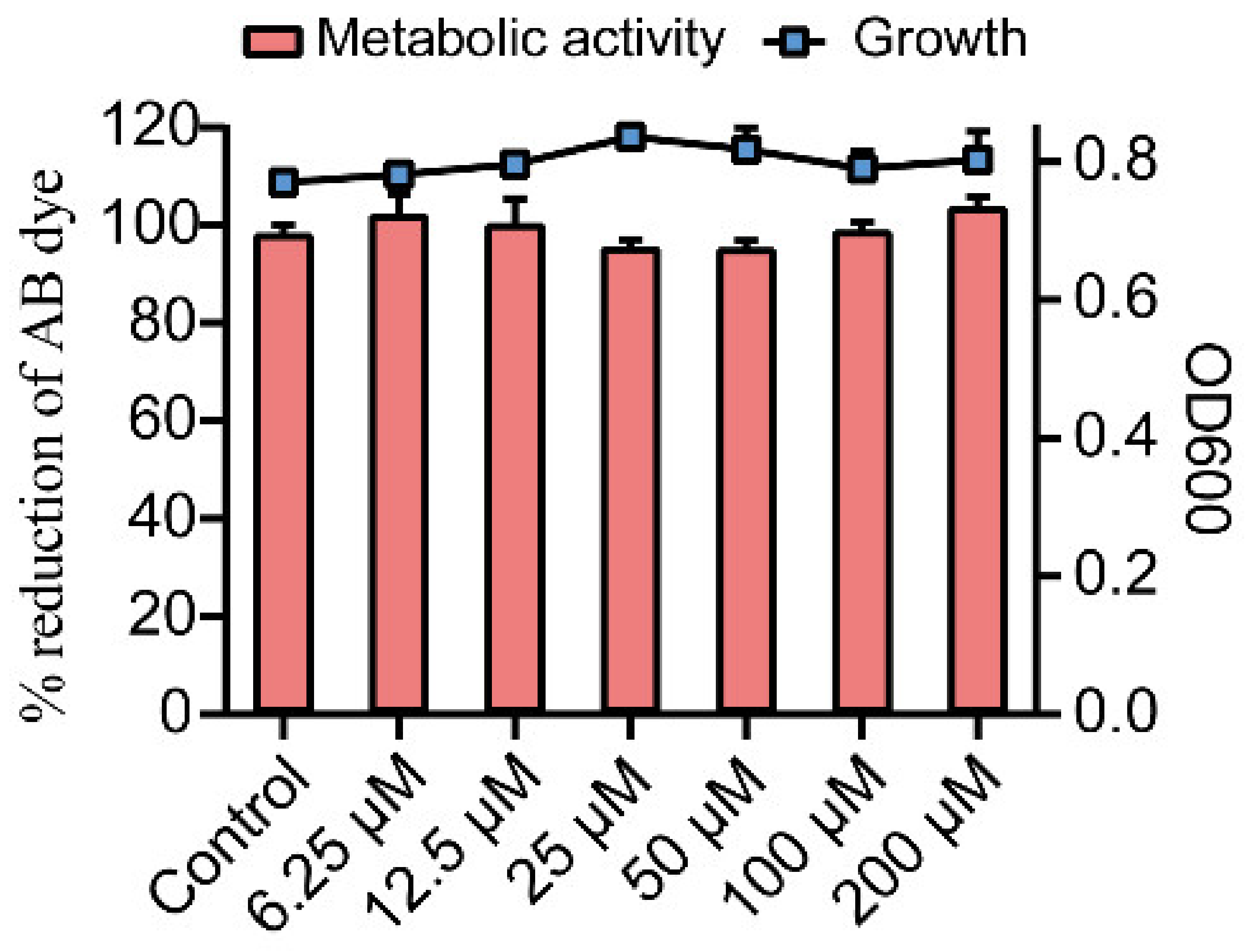
2.1. Effects of Gin on Growth and Metabolic Activity of E. coli

The effect of Gin on growth and metabolic activity of E. coli was measured by microbroth dilution and Alamar Blue (AB) assay. Gin did not significantly adversely affect the growth and metabolic activity of cells in the range of 6.25~100 μM compared to the control group (Figure 1). These results revealed the non-bactericidal nature and unaltered metabolic activity of Gin on E. coli.

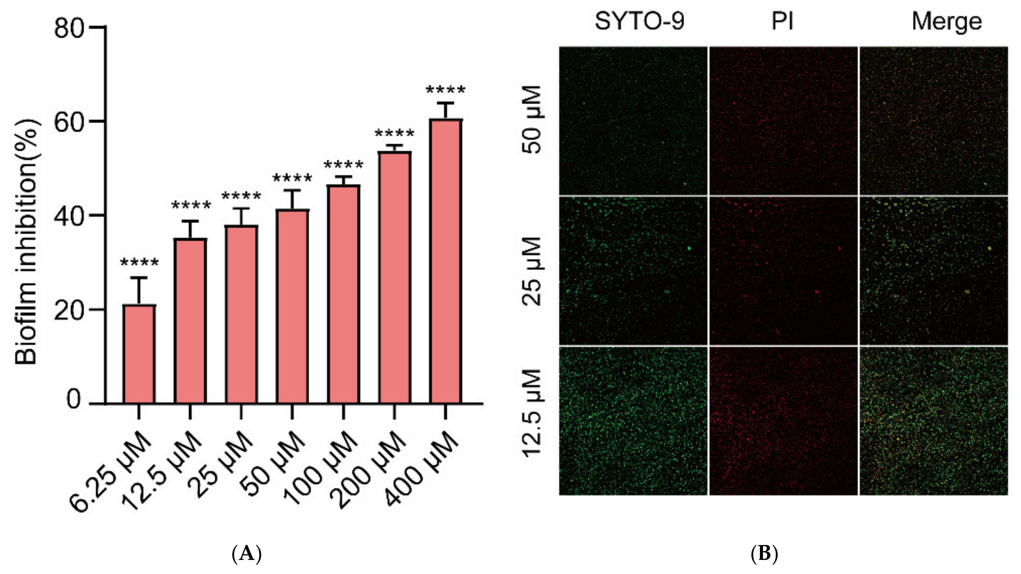
2.2. Inhibition of Biofilm Formation in E. coli by Gin

Different concentrations of Gin (6.25, 12.5, 25, 50, 100, 200, and 400 μM) were measured for antibiofilm activity by CV staining. This study showed that Gin treatment significantly inhibited the formation of E. coli ATCC 25922 in a dose-dependent manner (Figure 2A, p < 0.0001). As low as 6.25 μM of Gin was also found to be effective against the formation of biofilm (inhibition rate > 20%). To further observe the biofilm inhibitory effect of Gin, CLSM imaging analysis was conducted using fluorescent dye SYTO9 and propidium iodide (PI). The result showed that Gin significantly inhibited biofilm formation, as demonstrated by a negative gradient in green fluorescence in E. coli ATCC 25922 strains (Figure 2B). In short, the results showed that Gin inhibited biofilm formation.

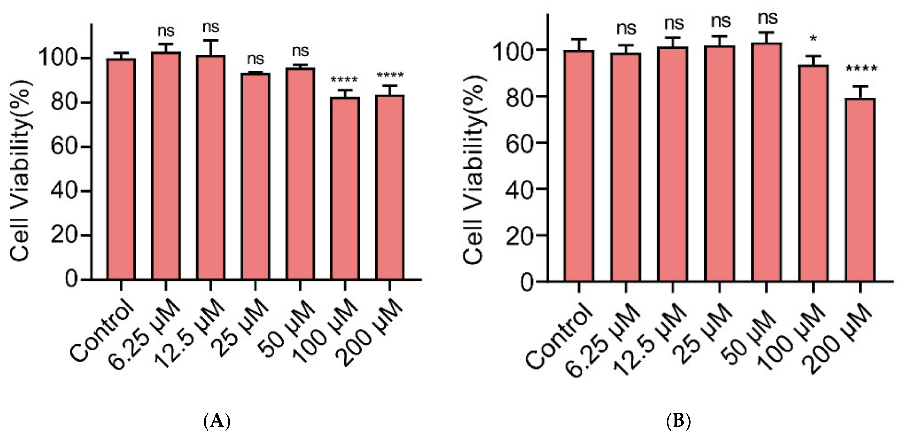
2.3. Cytotoxicity of Gin on Caco-2 and IPEC-J2 Cells

Next, we examined whether Gin showed cytotoxicity toward Caco-2 and IPEC-J2 cells (Figure 3). Gin showed no cytotoxic effect at 50 µM or lower concentrations, and cytotoxicity appeared at 100 µM and 200 µM (p < 0.05 and p < 0.0001, respectively). These results indicate that Gin was not cytotoxic at concentrations effective in inhibiting biofilm formation.

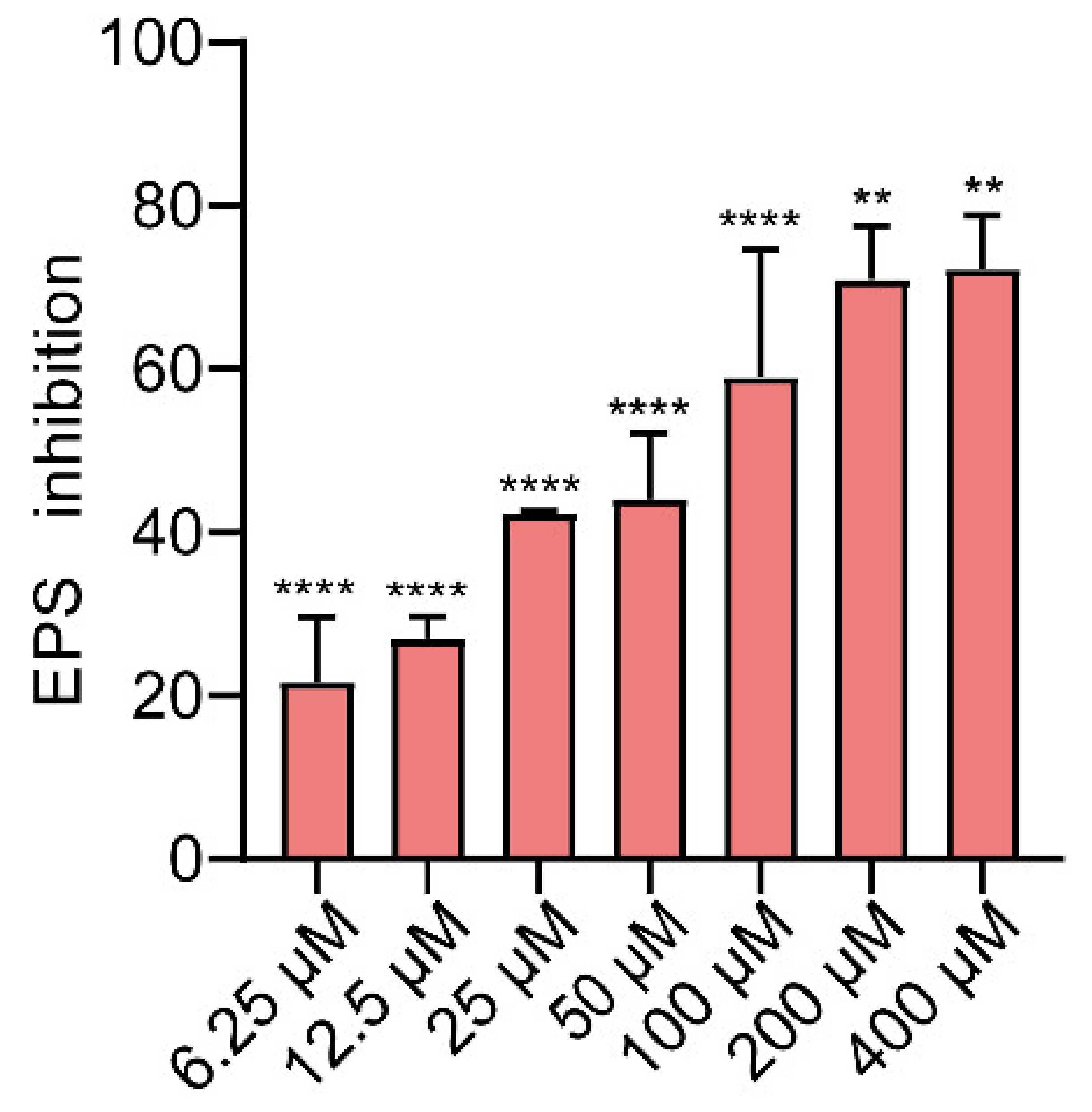
2.4. Effects of Gin on EPS Production of E. coli

In biofilms, bacteria produce EPS that help entrap nutrients and serve as defense mechanisms [60]. EPS production in biofilms was studied using Ruthenium Red staining. Gin inhibited EPS production in a dose-dependent manner (Figure 4, p < 0.01, and p < 0.0001). As low as 6.25 μM of Gin was also found to be effective against the production of EPS (inhibition rate > 20%). This result was consistent with the inhibitory effect of Gin on E. coli biofilm. The results showed that Gin reduces the EPS production of E. coli.

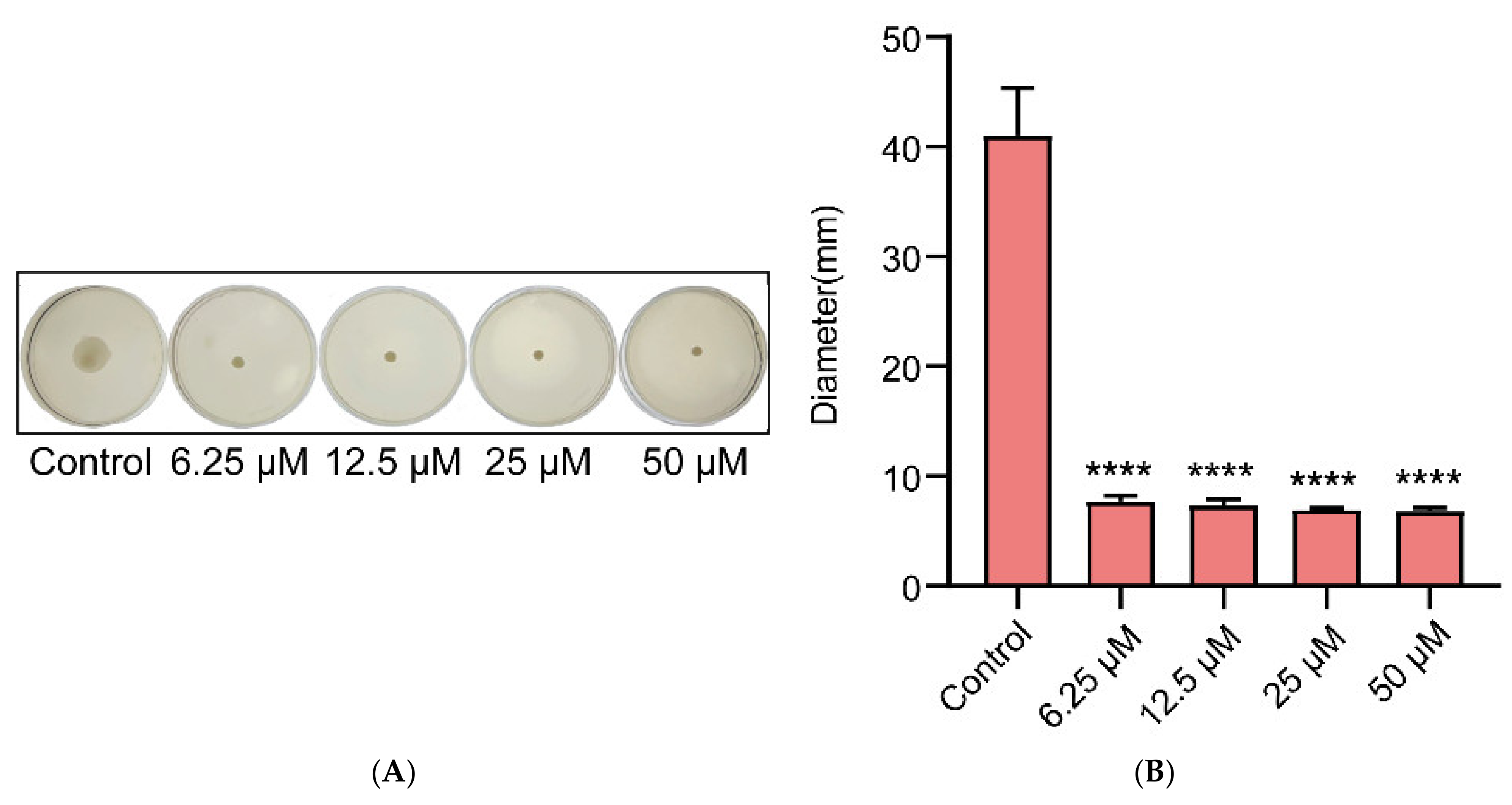
2.5. Effects of Gin on the Motility of E. coli

Due to the closely related relationship between biofilm formation and bacterial motility, Gin was evaluated for its effect on motility and antibiofilm activity. As seen in Figure 5A, Gin at different concentrations significantly inhibited E. coli motility compared with a control group (p < 0.0001). Further, the size of the halo zone was quantitatively determined (Figure 5B). The halo zone of Gin with different concentrations was significantly smaller compared with the control group (p < 0.0001). This result proved that Gin could inhibit the production of biofilm by inhibiting the motility of E. coli.

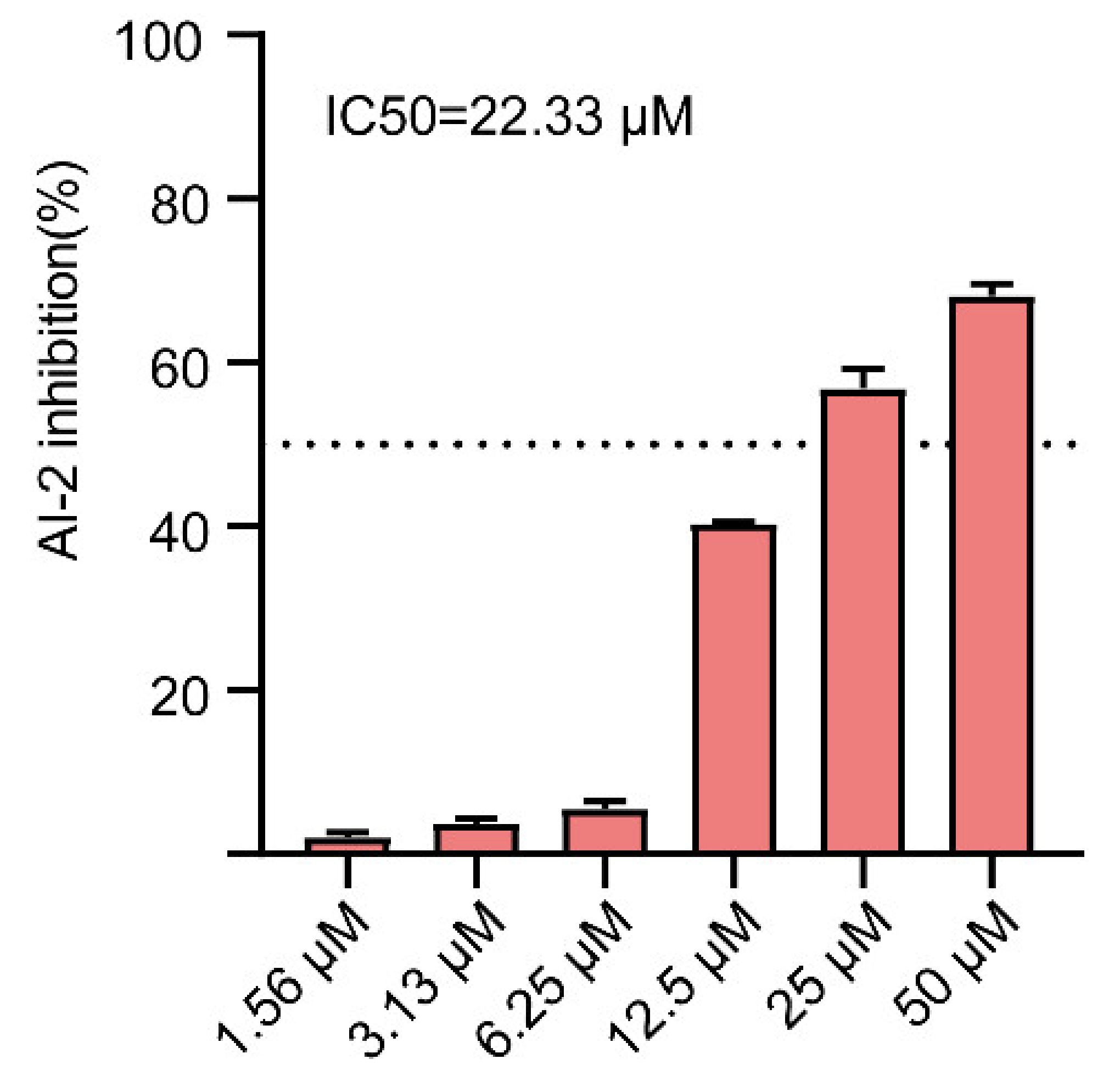
2.6. Effect of Gin on AI-2 Production in E. coli

Biofilm formation by bacteria depends on QS [61]. Further, the bioluminescence assay of V. harveyi BB170 was used to determine whether Gin could affect AI-2 production of QS induced by E. coli. As shown in Figure 6, the inhibitory effect of Gin on bioluminescence was dose-dependent, with an IC50 value of 22.33 μM. Our data indicated that Gin inhibited the formation of biofilm by inhibiting the production of AI-2.

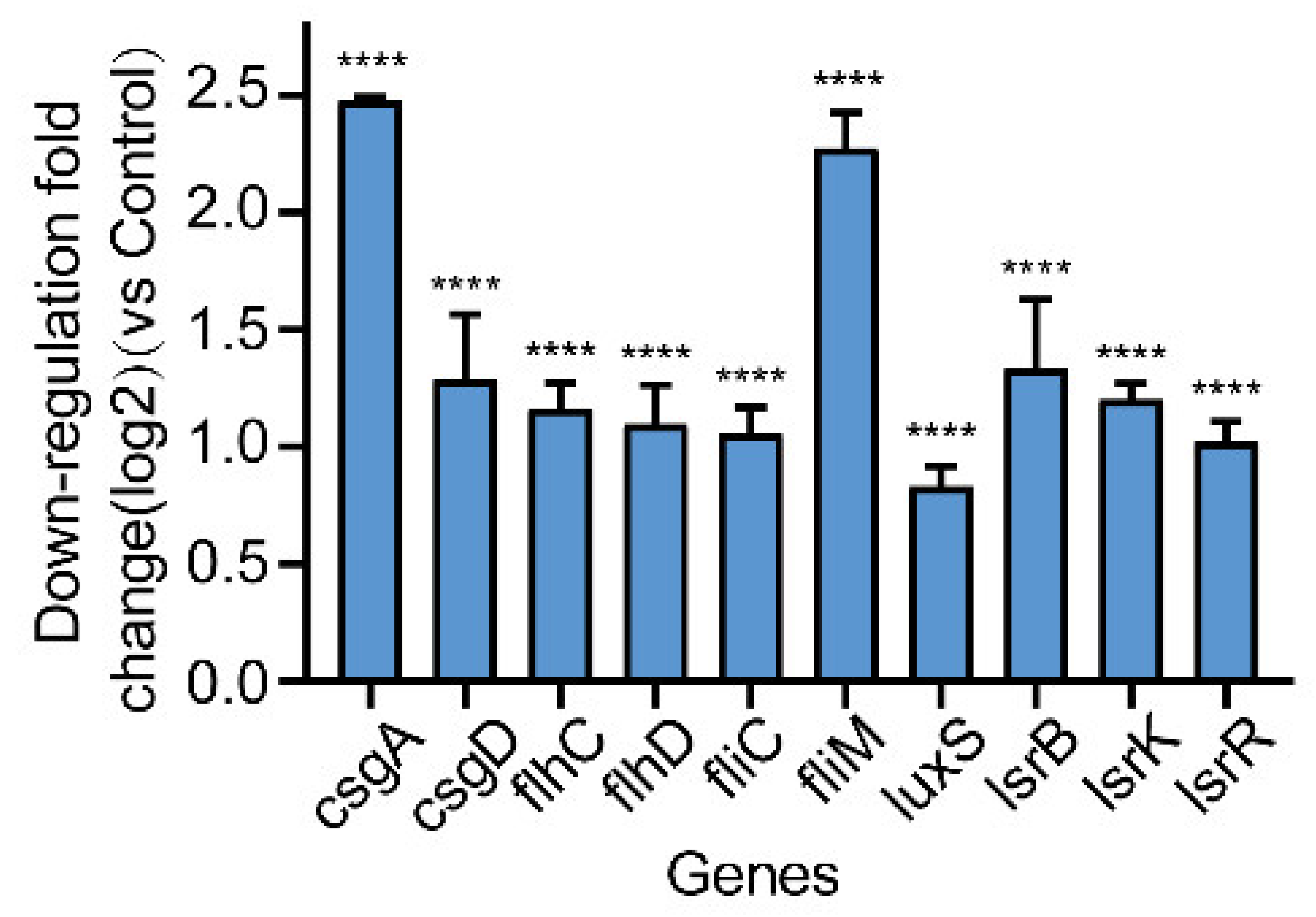
2.7. Effect of Gin on the Transcription of Biofilm-Regulated Genes of E. coli

To elucidate how Gin might inhibit the formation of biofilms, we assessed the transcription of genes such as curli-regulated genes (csgA, csgD) and flagella-formation genes (flhC, flhD, fliC, fliM), and QS-related genes (luxS, lsrB, lsrK, lsrR) by qRT-PCR. As shown in Figure 7, Gin significantly inhibited curli-regulated genes csgA, and csgD (82%, 58%, respectively), flagella-formation genes flhC, flhD, fliC, and fliM (55%, 53%, 52%, and 79%, respectively) and QS-related genes luxS, lsrB, lsrK, and lsrR (44%, 60%, 56%, and 51%, respectively). These results demonstrated that Gin inhibited the formation of biofilm by inhibiting the transcription of the curli genes, flagella genes, and QS genes of E. coli.

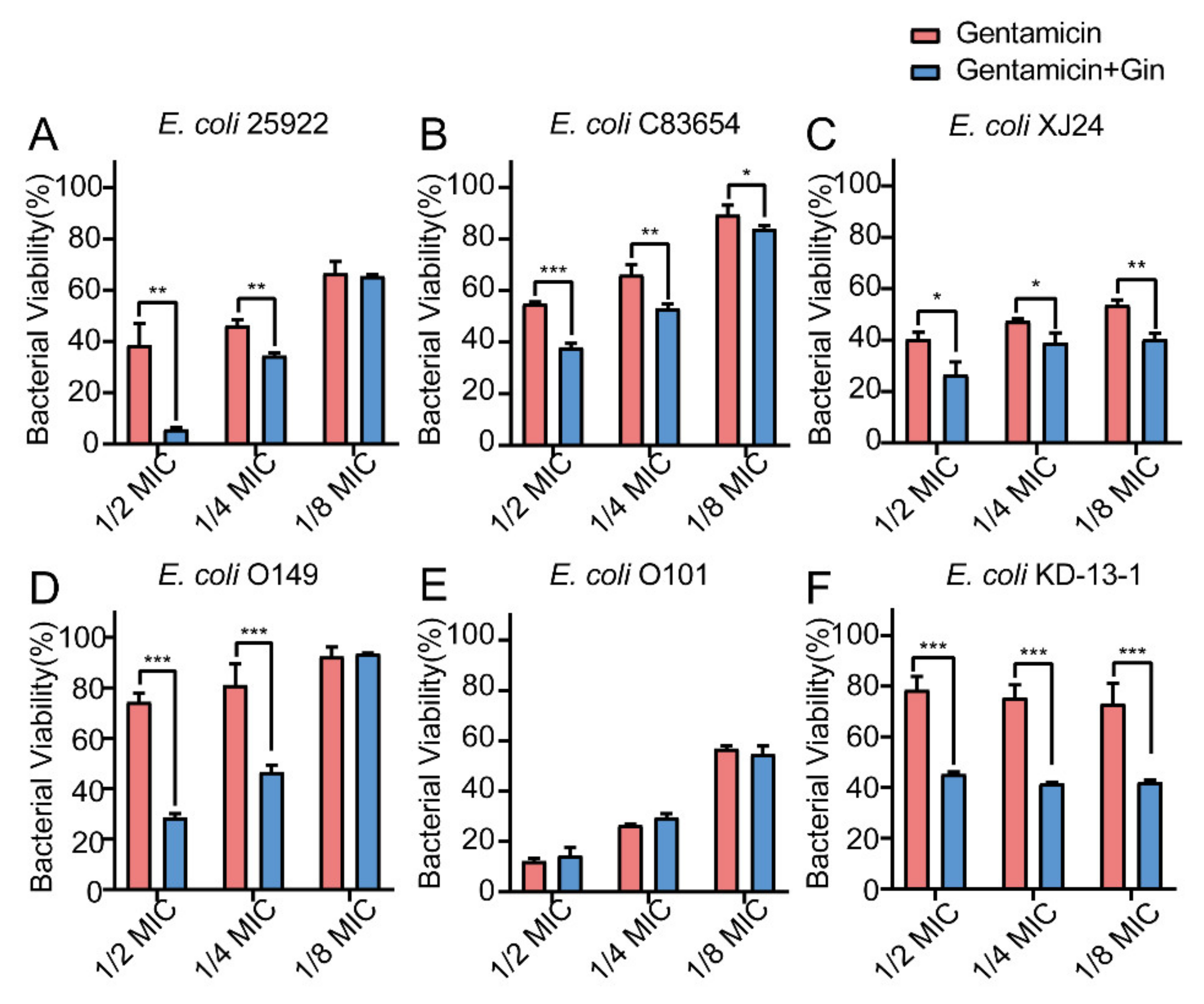
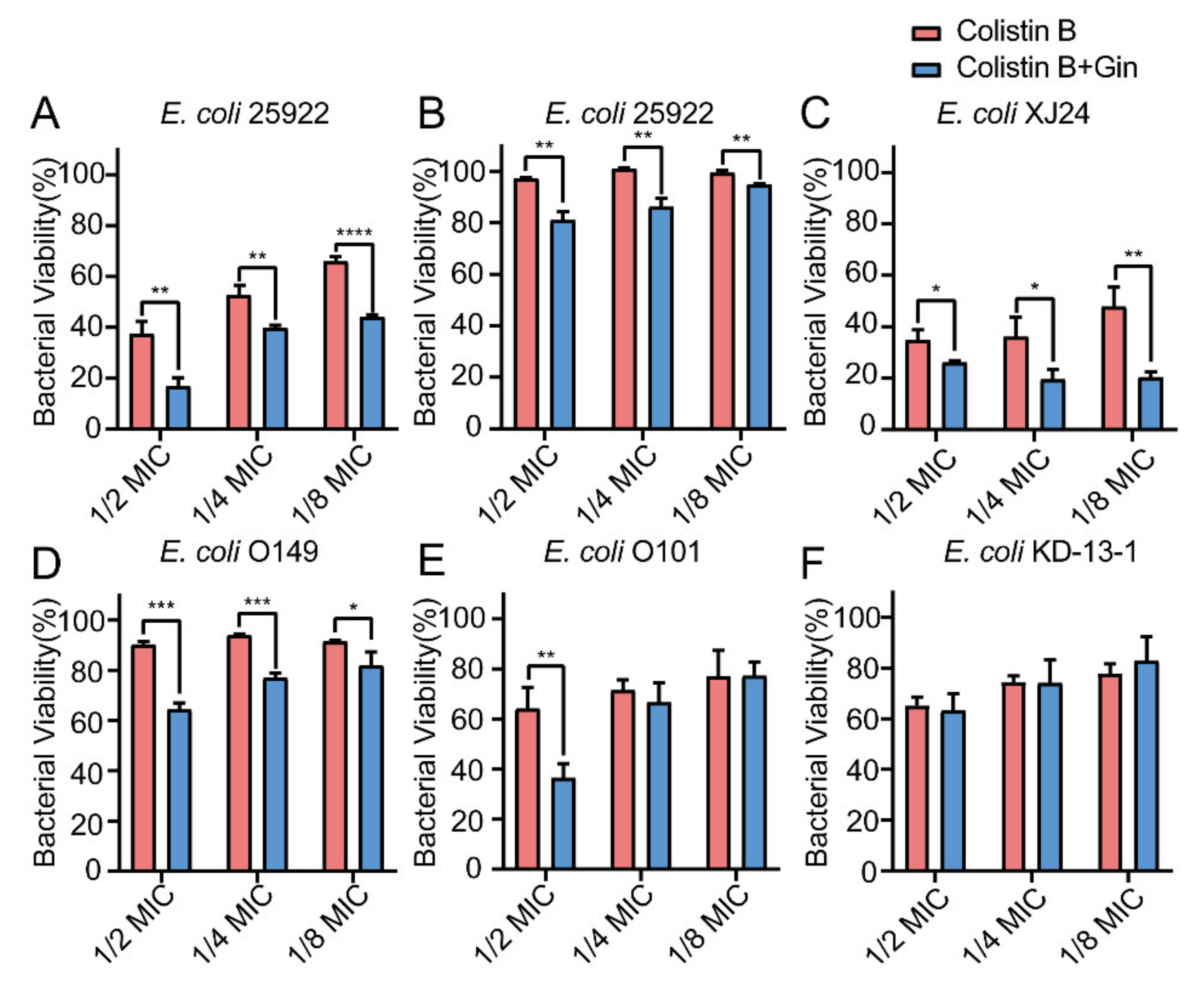
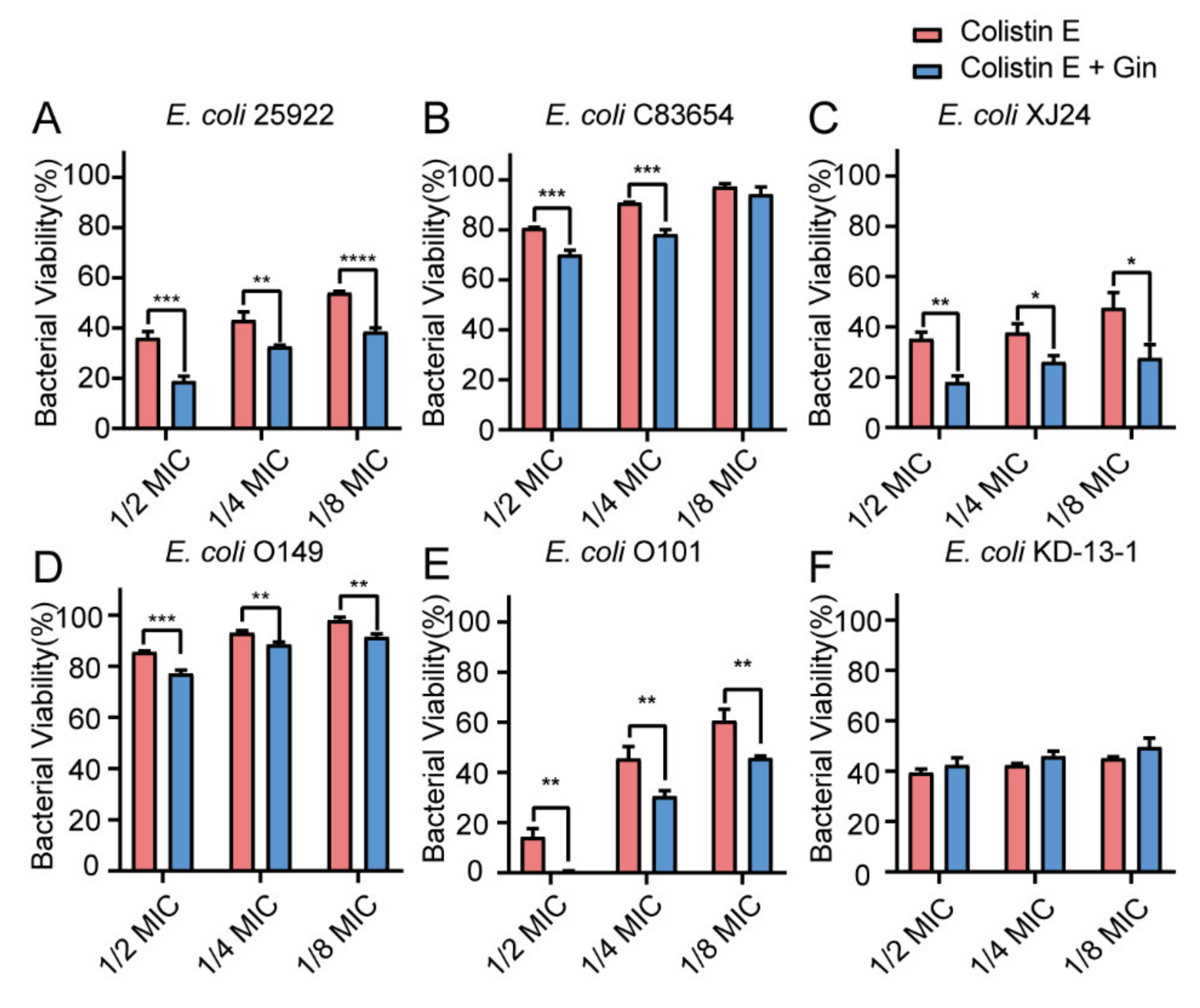
2.8. Synergistic Effects of Gin in Combination with Antibiotics against Six E. coli Strains

Biofilm bacteria are known to be able to increase their antibiotic resistance. Therefore, it is urgently important to find a way to reduce biofilm formation by overcoming these increasingly serious infections. Here, by combining marketed antibiotics, including gentamicin and colistin, with the antibiofilm compound Gin, we can combat E. coli biofilms. The synergistic effects of Gin (50 µM) combined with antibiotics on E. coli ATCC 25922 and five isolated clinical strains (E. coli XJ24, E. coli O101, E. coli O149, E. coli KD-13-1, and E. coli C83654) were investigated. The MIC of gentamicin, colistin B, and colistin E against E. coli ATCC 25922, XJ24, O101, O149, and KD-13-1 were tested (Table S1). As depicted in Figure 8, Figure 9 and Figure 10, antibacterial activity revealed that combining gentamicin, colistin B, and colistin E (1/2 MIC, 1/4 MIC, 1/8 MIC) with antibiofilm inhibitor Gin on E. coli ATCC 25922 and five isolated clinical strains has a synergistic effect. Gin showed biofilm inhibition properties and significantly increased the antibacterial activities of gentamicin, colistin B, and colistin E. These results suggested that the antibiofilm compound Gin, in conjunction with conventional antibiotics, could be an efficient therapeutic strategy for tackling pathogens such as E. coli.

3. Discussion

Antivirulence therapy is an important defense mechanism for the host against pathogenic bacteria infection [62,63]. Here, the present study was the first to report the antibiofilm and antivirulence activity of Gin against E. coli without affecting its growth. Furthermore, Gin did not exhibit cytotoxicity within the effective concentration range in IPEC-J2 and Caco-2 cells, resulting in a non-toxic effect in humans. We also demonstrated that Gin exhibited synergistic effects when combined with existing antibiotics (e.g., gentamicin, colistin B, and colistin E). Thus, we concluded that Gin presents antibiofilm and synergistic effects against E. coli.
EPS forms pathways for transporting metabolites and nutrients, maintaining biofilm structure, and protecting biofilms from external pressure. In addition, the secretion of EPS promotes bacteria’s adhesion and colonization on nonbiological surfaces [64]. In our study, the effects of Gin on EPS production were dose-dependent, resulting in decreased bacterial colonization and biofilm formation. Additionally, it is reported that Gin effectively inhibited biomass of biofilm adhered to the cover slip surface by CLSM. These results showed that Gin could reduce biofilm formation by inhibiting the production of adhesive molecules [65,66].
The possible mechanism of Gin’s antibiofilm activity was identified using gene transcription analysis. The qRT-PCR showed that Gin significantly down-regulated the transcription of flagella-formation genes (flhC, flhD, fliC, fliM), curli-related genes (csgA, csgD), and QS-related genes (luxS, lsrB, lsrK, lsrR) (Figure 7). The curli, the main component of a complex extracellular matrix produced by E. coli, is regulated by csgA, and csgD [67]. Results showed that Gin significant down-regulated the transcription of csgA, and csgD, thus inhibiting the curli production, which corresponded with the results of EPS production (Figure 4). Additionally, results also showed a significant reduction in the transcription of flhC, flhD, fliC, and fliM, indicating that Gin significantly inhibited the transcription of flagella-regulated genes in E. coli, thus reducing the motility of E. coli, which corresponded with the results obtained in semi-solid agar medium for the motility assay (Figure 5). Furthermore, to assess whether Gin affects QS, we verified the effect of Gin on the transcription of luxS, lsrB, lsrK, and lsrR. AI-2 was a type II QS signal molecule in E. coli. It can affect phenotypes of E. coli, including biofilm formation, motility, virulence, bacterial adhesion, and biofilm matrix production [68,69,70,71,72]. Results showed that Gin reduces the transcription of luxS, lsrB, lsrK, and lsrR, which was consistent with the results obtained in the AI-2 bioluminescence assay (Figure 6). Combined with all these findings above, biofilm inhibition by Gin may be due to inhibiting QS and then regulating motility and the EPS production of E. coli. However, further research is needed to determine the exact mechanism of action.
Biofilm is a structured bacterial community, which poses a significant challenge to traditional antibiotics’ effectiveness and is considered a breeding ground for antibiotic resistance. Combined with marketed antibiotics, biofilm inhibitors can enhance bacterial sensitivities to antibiotics [73]. The present study also proved that antibiofilm inhibitors could enhance the sensitivity of traditional antibiotics to resistant strains (Figure 8, Figure 9 and Figure 10). The results suggested that combining antibiotics with antibiofilm compounds appears to be an effective therapeutic strategy for treating pathogen infections.

4. Materials and Methods

4.1. Materials, Bacterial Strains, and Cells

Gin used in this study was obtained from Shanghai Yuanye Bio-Technology Co., Ltd. (Shanghai, China). Based on the water solubility of Gin, it was dissolved in dimethyl sulfoxide (DMSO) at a final concentration was 40 mM, and the concentration of DMSO in LB medium was not more than 1%.
E. coli ATCC 25922 was obtained from American Type Culture Collection (ATCC). Clinical isolate strains E. coli O101, E. coli O149, E. coli C83654, E. coli XJ24, and E. coli KD-13-1 were isolated and maintained in our laboratory. Luria-Bertani (LB, HuanKai Microbial, Guangdong, China) and Luria-Bertani agar (LA, HuanKai Microbial, Guangdong, China) medium were used for the growth of all E. coli strains. Vibrio harveyi BB170 (V. harveyi BB170) and V. harveyi BB152 were gifts from Researcher Han Xiangan (Shanghai Veterinary Research Institute, Chinese Academy of Agricultural Sciences), and were cultured under conditions containing AB medium supplemented with 1 mM L-arginine, 1% phosphate buffer (pH = 7.2), and 1% glycerol.
Caco-2 cell lines were obtained from ATCC and cultured under standard conditions using MEM medium with 20% FBS, 1 mM sodium pyruvate, 1 mM L-glutamine, 10 mM HEPES, and 1% non-essential amino acids. IPEC-J2 cell lines were obtained from Beina Chuanglian Biology Research Institute and cultured under standard conditions using DMEM medium with 10% FBS, 1 mM sodium pyruvate, and 1 mM L-glutamine, 10 mM HEPES, and 1% non-essential amino acids.

4.2. Growth and Metabolic Activity

A microbroth dilution test was conducted to determine the growth effects of Gin by the Clinical & Laboratory Standards Institute (CLSI) [74]. In brief, cell suspensions of E. coli (106 CFU/mL) and different concentrations of Gin were incubated for 24 h at 3 °C. A Multiskan Go Reader (Thermo Fisher Scientific, USA) was used to measure the absorbance at 600 nm following incubation.
In continuation, E. coli metabolic activity was analyzed by the Alamar Blue (AB) assay according to the previous method [75]. Briefly, cells were collected from each well using fresh 2 mL tubes followed by centrifugation for 10 min at 5000 rpm, washed twice with PBS (pH = 7.2), and then resuspended in 1 mL of PBS (pH = 7.2). Next, the tubes were incubated in dark conditions for 1 h at 37 °C, with 10 µL of AB dye (Invitrogen™, Thermo Fisher Scientific, USA) added. The blank control was PBS (pH = 7.2) containing only AB dye. The metabolic activity was calculated based on the absorbance at 570 nm and 600 nm using the following formula:
M e t a b o l i c   a c t i v i t y ( % ) = E o x i O D 570 × T O D 570 E o x i O D 600 × T O D 600 E r e d O D 570 × B O D 570 E r e d O D 600 × B O D 600 × 100 %
  • Eoxi (OD570)—extinction coefficient of AB in its oxidized form at 570 nm = 80,586;
  • Ered (OD570)—extinction coefficient of AB in its reduced form at 570 nm = 155,677;
  • Eoxi (OD600)—extinction coefficient of AB in its oxidized form at 600 nm = 117,216;
  • Ered (OD600)—extinction coefficient of AB in its reduced form at 570 nm = 14,652;
  • B—blank; T—samples.

4.3. Biofilm Assay

4.3.1. Crystal Violet (CV) Staining

The previous method was slightly modified to detect the formation of E. coli biofilm using CV staining [76]. Briefly, E. coli ATCC 25922 was statically grown for 24 h at 37 °C. Next, the cells were resuspended at OD600 of 1.0 and diluted about 100-fold. Subsequently, the diluted bacterial solution was mixed with Gin in a white 96-well plate (Corning Costar® 3599, Corning, Kennebunk, ME, USA). Cells underwent static incubation for 24 h at 37 °C, were washed three times with PBS (pH = 7.2) to eliminate the nonadherent cells, and fixed for 1 h at 60 °C. After fixation with methanol and staining with 0.1% CV for 30 min, the stained biofilm was rinsed with tap water to remove the dye that was not bound. The CV contained in biofilm was dissolved in 150 µL of 95% ethanol, and its absorbance at 570 nm was measured.

4.3.2. Confocal Laser Scanning Microscopy

According to a previous report, we analyzed the E. coli biofilms using confocal laser scanning microscopy (CLSM) [77]. In brief, E. coli (106 CFU/mL) was inoculated in LB broth with or without Gin (0, 12.5, 25, and 50 µM) to form static biofilms on the cover slips for 24 h at 37℃ in the 12-well plates. The biofilms were washed twice with PBS (pH = 7.2) before removing loosely bound cells, and stained using a BacLight Live/Dead viability kit (L7012, Invitrogen™, Thermo Fisher Scientific, Eugene, OR, USA) according to the procedure. Afterward, excess staining was removed by washing twice with PBS (pH = 7.2), and then the biofilm was imaged by CLSM (LSM800, Zeiss, Jena, Germany).

4.4. Cytotoxicity

Gin cytotoxicity was assessed in Caco-2 and IPEC-J2 cells using the CCK-8 assay. Caco-2 and IPEC-J2 cells at a density of 1 × 10⁵ cells/mL were seeded into 96-well plates and cultured until the density of the cells reached approximately 80%. Then, the cells were treated for 24 h with 100 μL of Gin (6.25, 12.5, 25, 50, 100, 200 µM). Further, the cells with 10 μL of CCK-8 (MCE, Shanghai, China) were incubated for 1 h. After incubation, a Multiskan Go Reader (Thermo Fisher Scientific, Waltham, MA, USA) was used to measure absorbance at 450 nm.

4.5. EPS Production

The EPS production was quantitatively estimated by Ruthenium Red Staining [78]. Cell suspensions (100 µL) of E. coli (106 CFU/mL) and different concentrations of Gin were cultured for 24 h at 37 °C. Afterward, the cells were washed with PBS (pH = 7.2). In each well, biofilm cells were stained with 0.01% ruthenium red (Yuanye, Shanghai, China) by incubation at 37 °C for 60 min. Ruthenium red was served as a blank. Following that, the solution containing the remaining stain was retransferred onto a new 96-well plate, and the absorbance at 450 nm was measured. EPS inhibition was calculated as:
E P S   inhibition % = AS AP AB AP × 100
where:
  • AB = absorbance of blank
  • AS = absorbance of sample
  • AP = absorbance of positive control

4.6. Motility Assay

Gin was evaluated for its effects on E. coli motility as described earlier [79]. In brief, E. coli cultures overnight were adjusted to an OD600 of 0.01. A semisolid agar media (0.3% LB agar) containing 12.5, 25, and 50 µM of Gin was used for the motility assay. A 1 µL measure of the diluted bacterial solution was inserted into the middle of the plate and then incubated for 24 h at 37 °C. The size of the halo zone compared to the control was used to evaluate the motility.

4.7. AI-2 Bioluminescence Assay

According to previous reports, AI-2 bioluminescence assays were performed [80,81,82]. In brief, incubation of E. coli (106 CFU/mL) with 12.5, 25, or 50 µM of Gin for 16 h and centrifugation for 5 min at 12,000× g was performed. The supernatant was filtered with a 0.22 µm filter. The bioluminescence reporter V. harveyi BB170 grew in AB medium to OD600 of 1.0~1.1 and was then diluted with fresh medium at 1:2500. A 20 µL measure of cell-free supernatant was mixed with 180 µL of the V. harveyi BB170 dilution in black 96-well plates (Jingan, Shanghai, China) and incubated for 3.5 h at 37 °C. Measurement of bioluminescence was conducted using a multi-purpose microplate reader (Enspire, PerkinElmer, Waltham, MA, USA). Controls were cellular-free supernatants of overnight V. harveyi BB152 cultures.

4.8. qRT-PCR

qRT-PCR was used to study the effect of Gin on the transcription of biofilm-regulated genes of E. coli. E. coli was incubated with and without Gin for 24 h at 37 °C. Bacterial RNA Kit (Omega, Norcross, GA, USA) was used to extract total RNA. RNA quantity was measured by NanoDrop OneC spectrophotometer (Thermo Scientific, USA) and RNA quality was determined via agarose gel electrophoresis. Further, the RNA was reverse-transcribed into cDNA with a PrimeScript™ RT reagent Kit with gDNA Eraser (TAKARA Corporation, Kusatsu, Japan). The TB Green® Premix Ex TaqTM II (Tli RNaseH Plus) (TAKARA Corporation, Kusatsu, Japan) was used for qRT-PCR. The 2−∆∆Ct method was used to assess relative changes in gene transcription levels. The gapA gene was used as an internal control [83]. In Table S2, the primers used in the current study are listed.

4.9. Antibacterial Activity

Based on the previous method [75,84], we modified some procedures to evaluate the antibacterial effects of Gin. Briefly, after overnight culture, bacteria were diluted to an OD600 of 0.01. The diluted bacterial solution was mixed with antibiotics (1/2 MIC, 1/4 MIC, 1/8 MIC) with or without Gin (50 µM). The metabolic activity of mixed suspensions was analyzed with the Alamar Blue (AB) assay after incubation for 16–18 h at 37 °C. Tests were conducted in triplicate.

4.10. Statistical Analysis

Comparison between two groups was performed using multiple t-tests, while multiple comparisons were made using non-parametric one-way ANOVA in GraphPad Prism (GraphPad Prism 8.0.1, GraphPad, San Diego, CA, USA). Results from all experiments were presented as the mean ± SD of three replicates.

5. Conclusions

In summary, this study demonstrated that Gin has an antibiofilm and synergistic antibacterial effect against E. coli. These results showed that Gin could inhibit biofilm without affecting bacterial growth and metabolic activity. Besides, Gin inhibited the bacteria motility, EPS production, and QS of E. coli. More importantly, the antibacterial effects of combining gentamicin and colistin antibiotics with Gin were studied. The combination showed significantly synergistic effort against E. coli ATCC 25922 and five clinical isolates strains. In summary, these results indicate that Gin was an effective antibiofilm compound which can combine with marketed antibiotics to treat drug resistance caused by biofilms to treat E. coli infections in the future.

Supplementary Materials

The following supporting information can be downloaded at: https://www.mdpi.com/article/10.3390/ijms23158809/s1.

Author Contributions

Conceptualization, Y.B. and J.Z.; methodology, Y.B. and M.S.; validation, W.W., X.W. and M.S.; formal analysis, Y.B.; investigation, Y.B.; resources, J.Z. and W.W.; data curation, Y.B. and W.W; writing—original draft preparation, Y.B.; writing—review and editing, W.W., X.Z. and B.L.; visualization, Y.B.; supervision, W.W. and J.Z.; project administration, J.Z.; funding acquisition, J.Z. and W.W. All authors have read and agreed to the published version of the manuscript.

Funding

This research was funded by the research on the National Natural Science Foundation of China, funding number 32102727, Drug Development and Clinical Drug Use Posts of the National Beef Yak Industry Technical System, funding number CARS-37, and Innovation Project of Chinese Academy of Agricultural Sciences, funding number 25-LZIHPS-05.

Institutional Review Board Statement

Not applicable.

Informed Consent Statement

Not applicable.

Data Availability Statement

Not applicable.

Acknowledgments

We thank Researcher Xiangan Han for providing the strains V. harveyi BB170 and V. harveyi BB152. Additionally, we are grateful for the suggestions made by anonymous reviewers.

Conflicts of Interest

The authors declare no conflict of interest.

References

  1. Larsson, D.G.J.; Flach, C.F. Antibiotic resistance in the environment. Nat. Rev. Microbiol. 2022, 20, 257–269. [Google Scholar] [CrossRef] [PubMed]
  2. Fernández, L.; Hancock, R.E. Adaptive and mutational resistance: Role of porins and efflux pumps in drug resistance. Clin. Microbiol. Rev. 2012, 25, 661–681. [Google Scholar] [CrossRef] [PubMed] [Green Version]
  3. Lewis, K. Persister cells, dormancy and infectious disease. Nat. Rev. Microbiol. 2007, 5, 48–56. [Google Scholar] [CrossRef] [PubMed]
  4. Fong, J.; Yuan, M.; Jakobsen, T.H.; Mortensen, K.T.; Delos Santos, M.M.; Chua, S.L.; Yang, L.; Tan, C.H.; Nielsen, T.E.; Givskov, M. Disulfide bond-containing ajoene analogues as novel quorum sensing inhibitors of Pseudomonas aeruginosa. J. Med. Chem. 2017, 60, 215–227. [Google Scholar] [CrossRef]
  5. Karygianni, L.; Ren, Z.; Koo, H.; Thurnheer, T. Biofilm matrixome: Extracellular components in structured microbial communities. Trends Microbiol. 2020, 28, 668–681. [Google Scholar] [CrossRef]
  6. Hobley, L.; Harkins, C.; MacPhee, C.E.; Stanley-Wall, N.R. Giving structure to the biofilm matrix: An overview of individual strategies and emerging common themes. FEMS Microbiol. Rev. 2015, 39, 649–669. [Google Scholar] [CrossRef] [Green Version]
  7. Flemming, H.C.; Wingender, J.; Szewzyk, U.; Steinberg, P.; Rice, S.A.; Kjelleberg, S. Biofilms: An emergent form of bacterial life. Nat. Rev. Microbiol. 2016, 14, 563–575. [Google Scholar] [CrossRef]
  8. Ciofu, O.; Moser, C.; Jensen, P.; Høiby, N. Tolerance and resistance of microbial biofilms. Nat. Rev. Microbiol. 2022, 35, 322–332. [Google Scholar] [CrossRef]
  9. Cohen, N.R.; Lobritz, M.A.; Collins, J.J. Microbial persistence and the road to drug resistance. Cell Host Microbe 2013, 13, 632–642. [Google Scholar] [CrossRef] [Green Version]
  10. Lebeaux, D.; Ghigo, J.M.; Beloin, C. Biofilm-related infections: Bridging the gap between clinical management and fundamental aspects of recalcitrance toward antibiotics. Microbiol. Mol. Biol. Rev. 2014, 78, 510–543. [Google Scholar] [CrossRef] [Green Version]
  11. Bakkeren, E.; Huisman, J.S.; Fattinger, S.A.; Hausmann, A.; Furter, M.; Egli, A.; Slack, E.; Sellin, M.E.; Bonhoeffer, S.; Regoes, R.R.; et al. Salmonella persisters promote the spread of antibiotic resistance plasmids in the gut. Nature 2019, 573, 276–280. [Google Scholar] [CrossRef]
  12. Adnan, M.; Morton, G.; Singh, J.; Hadi, S. Contribution of rpoS and bolA genes in biofilm formation in Escherichia coli K-12 MG1655. Mol. Cell Biochem. 2010, 342, 207–213. [Google Scholar] [CrossRef]
  13. Adnan, M.; Sousa, A.M.; Machado, I.; Pereira, M.O.; Khan, S.; Morton, G.; Hadi, S. Role of bolA and rpoS genes in biofilm formation and adherence pattern by Escherichia coli K-12 MG1655 on polypropylene, stainless steel, and silicone surfaces. Acta Microbiol. Immunol. Hung. 2017, 64, 179–189. [Google Scholar] [CrossRef] [Green Version]
  14. Meesilp, N.; Mesil, N. Effect of microbial sanitizers for reducing biofilm formation of Staphylococcus aureus and Pseudomonas aeruginosa on stainless steel by cultivation with UHT milk. Food Sci. Biotechnol. 2019, 28, 289–296. [Google Scholar] [CrossRef]
  15. Sharma, D.; Misba, L.; Khan, A.U. Antibiotics versus biofilm: An emerging battleground in microbial communities. Antimicrob Resist. Infect Control. 2019, 8, 76. [Google Scholar] [CrossRef]
  16. Vuotto, C.; Donelli, G. Novel treatment strategies for biofilm-based infections. Drugs 2019, 79, 1635–1655. [Google Scholar] [CrossRef]
  17. Jiang, Y.; Geng, M.; Bai, L. Targeting biofilms therapy: Current research strategies and development hurdles. Microorganisms 2020, 8, 1222. [Google Scholar] [CrossRef]
  18. Grandclement, C.; Tannieres, M.; Morera, S.; Dessaux, Y.; Faure, D. Quorum quenching: Role in nature and applied developments. FEMS Microbiol. Rev. 2016, 40, 86–116. [Google Scholar] [CrossRef]
  19. Beloin, C.; Renard, S.; Ghigo, J.M.; Lebeaux, D. Novel approaches to combat bacterial biofilms. Curr. Opin. Pharmacol. 2014, 18, 61–68. [Google Scholar] [CrossRef] [Green Version]
  20. Chen, X.; Zhang, M.; Zhou, C.; Kallenbach, N.R.; Ren, D. Control of bacterial persister cells by Trp/Arg-containing antimicrobial peptides. Appl. Environ. Microbiol. 2011, 77, 4878–4885. [Google Scholar] [CrossRef] [Green Version]
  21. Lebeaux, D.; Chauhan, A.; Letoffe, S.; Fischer, F.; de Reuse, H.; Beloin, C.; Ghigo, J.M. PH-mediated potentiation of aminoglycosides kills bacterial persisters and eradicates in vivo biofilms. J. Infect. Dis. 2014, 210, 1357–1366. [Google Scholar] [CrossRef] [Green Version]
  22. Ma, D.S.L.; Tan, L.T.H.; Chan, K.G.; Yap, W.H.; Pusparajah, P.; Chuah, L.H.; Ming, L.C.; Khan, T.M.; Lee, L.H.; Goh, B.H. Resveratrol-potential antibacterial agent against foodborne pathogens. Front Pharmacol. 2018, 9, 102. [Google Scholar] [CrossRef] [Green Version]
  23. Zhou, J.W.; Chen, T.T.; Tan, X.J.; Sheng, J.Y.; Jia, A.Q. Can the quorum sensing inhibitor resveratrol function as an aminoglycoside antibiotic accelerant against Pseudomonas aeruginosa? Int. J. Antimicrob Agents 2018, 52, 35–41. [Google Scholar] [CrossRef]
  24. Vestergaard, M.; Ingmer, H. Antibacterial and antifungal properties of resveratrol. Int. J. Antimicrob Agents 2019, 53, 716–723. [Google Scholar] [CrossRef] [PubMed]
  25. Papenfort, K.; Bassler, B.L. Quorum sensing signal-response systems in Gram-negative bacteria. Nat. Rev. Microbiol. 2016, 14, 576–588. [Google Scholar] [CrossRef] [Green Version]
  26. Wang, W.; Huang, X.; Yang, H.; Niu, X.; Li, D.; Yang, C.; Li, L.; Zou, L.; Qiu, Z.; Wu, S.; et al. Antibacterial activity and anti-quorum sensing mediated phenotype in response to essential oil from Melaleuca bracteata leaves. Int. J. Mol. Sci. 2019, 20, 5696. [Google Scholar] [CrossRef] [Green Version]
  27. Minich, A.; Levarski, Z.; Mikulášová, M.; Straka, M.; Liptáková, A.; Stuchlík, S. Complex analysis of vanillin and syringic acid as natural antimicrobial agents against Staphylococcus epidermidis biofilms. Int. J. Mol. Sci. 2022, 23, 1816. [Google Scholar] [CrossRef]
  28. Manner, S.; Fallarero, A. Screening of natural product derivatives identifies two structurally related flavonoids as potent quorum sensing inhibitors against Gram-negative bacteria. Int. J. Mol. Sci. 2018, 19, 1346. [Google Scholar] [CrossRef] [Green Version]
  29. Bassler, B.L. Small talk. cell-to-cell communication in bacteria. Cell 2002, 109, 421–424. [Google Scholar] [CrossRef] [Green Version]
  30. Choudhary, S.; Schmidt-Dannert, C. Applications of quorum sensing in biotechnology. Appl. Microbiol. Biotechnol. 2010, 86, 1267–1279. [Google Scholar] [CrossRef] [PubMed]
  31. Reading, N.C.; Sperandio, V. Quorum sensing: The many languages of bacteria. FEMS Microbiol. Lett. 2006, 254, 1–11. [Google Scholar] [CrossRef] [PubMed]
  32. Sturme, M.H.; Kleerebezem, M.; Nakayama, J.; Akkermans, A.D.; Vaugha, E.E.; de Vos, W.M. Cell to cell communication by autoinducing peptides in gram-positive bacteria. Antonie Van Leeuwenhoek 2002, 81, 233–243. [Google Scholar] [CrossRef] [PubMed]
  33. Peng, L.Y.; Yuan, M.; Wu, Z.M.; Song, K.; Zhang, C.L.; An, Q.; Xia, F.; Yu, J.L.; Yi, P.F.; Fu, B.D.; et al. Anti-bacterial activity of baicalin against APEC through inhibition of quorum sensing and inflammatory responses. Sci. Rep. 2019, 9, 4063. [Google Scholar] [CrossRef] [PubMed]
  34. Roy, V.; Fernandes, R.; Tsao, C.Y.; Bentley, W.E. Cross species quorum quenching using a native AI-2 processing enzyme. ACS Chem. Biol. 2010, 5, 223–232. [Google Scholar] [CrossRef]
  35. Römling, U.; Rohde, M.; Olsén, A.; Normark, S.; Reinköster, J. AgfD, the checkpoint of multicellular and aggregative behaviour in Salmonella typhimurium regulates at least two independent pathways. Mol. Microbiol. 2000, 36, 10–23. [Google Scholar] [CrossRef]
  36. Ogasawara, H.; Yamamoto, K.; Ishihama, A. Role of the biofilm master regulator csgD in cross-regulation between biofilm formation and flagellar synthesis. J. Bacteriol. 2011, 193, 2587–2597. [Google Scholar] [CrossRef] [Green Version]
  37. Barnhart, M.M.; Chapman, M.R. Curli biogenesis and function. Ann. Rev. Microbiol. 2006, 60, 131–147. [Google Scholar] [CrossRef] [Green Version]
  38. Blanco, L.P.; Evans, M.L.; Smith, D.R.; Badtke, M.P.; Chapman, M.R. Diversity, biogenesis and function of microbial amyloids. Trends Microbiol. 2012, 20, 66–73. [Google Scholar] [CrossRef] [Green Version]
  39. Arita-Morioka, K.I.; Yamanaka, K.; Mizunoe, Y.; Tanaka, Y.; Ogura, T.; Sugimoto, S. Inhibitory effects of myricetin derivatives on curli-dependent biofilm formation in Escherichia coli. Sci. Rep. 2018, 8, 8452. [Google Scholar] [CrossRef] [Green Version]
  40. Guttenplan, S.B.; Kearns, D.B. Regulation of flagellar motility during biofilm formation. FEMS Microbiol. Rev. 2013, 37, 849–871. [Google Scholar] [CrossRef] [Green Version]
  41. Wood, T.K.; González Barrios, A.F.; Herzberg, M.; Lee, J. Motility influences biofilm architecture in Escherichia coli. Appl. Microbiol. Biotechnol. 2006, 72, 361–367. [Google Scholar] [CrossRef]
  42. Zhao, K.; Liu, M.; Burgess, R.R. Adaptation in bacterial flagellar and motility systems: From regulon members to ‘foraging’-like behavior in E. coli. Nucleic Acids Res. 2007, 35, 4441–4452. [Google Scholar] [CrossRef] [Green Version]
  43. Guan, G.H.; Wang, Y.; Chen, W.F.; Li, Y. Microbial physiology teaching exploration—Taking the teaching of the structure and function of bacterial flagella as an example. Microbiol. China 2016, 43, 756–761. [Google Scholar]
  44. Chakraborty, S.; Randall, A.; Vickers, T.J.; Molina, D.; Harro, C.D.; DeNearing, B.; Brubaker, J.; Sack, D.A.; Bourgeois, A.L.; Felgner, P.L.; et al. Human experimental challenge with enterotoxigenic Escherichia coli elicits immune responses to canonical and novel antigens relevant to vaccine development. J. Infect. Dis. 2018, 218, 1436–1446. [Google Scholar] [CrossRef]
  45. Henderson, L.D.; Matthews-Palmer, T.R.S.; Gulbronson, C.J.; Ribardo, D.A.; Beeby, M.; Hendrixson, D.R. Diversification of Campylobacter jejuni flagellar C-ring composition impacts its structure and function in motility, flagellar assembly, and cellular processes. mBio 2020, 11, e02286-19. [Google Scholar] [CrossRef] [Green Version]
  46. Nataro, J.P.; Kaper, J.B. Diarrheagenic Escherichia coli. Clin. Microbiol. Rev. 1998, 11, 142–201. [Google Scholar] [CrossRef] [Green Version]
  47. Braz, V.S.; Melchior, K.; Moreira, C.G. Escherichia coli as a multifaceted pathogenic and versatile bacterium. Front Cell Infect. Microbiol. 2020, 10, 548492. [Google Scholar] [CrossRef]
  48. Anderson, G.G.; Palermo, J.J.; Schilling, J.D.; Roth, R.; Heuser, J.; Hultgren, S.J. Intracellular bacterial biofilm-like pods in urinary tract infections. Science 2003, 301, 105–107. [Google Scholar] [CrossRef] [Green Version]
  49. Ageorges, V.; Monteiro, R.; Leroy, S.; Burgess, C.M.; Pizza, M.; Chaucheyras-Durand, F.; Desvaux, M. Molecular determinants of surface colonisation in diarrhoeagenic Escherichia coli (DEC): From bacterial adhesion to biofilm formation. FEMS Microbiol. Rev. 2020, 44, 314–350. [Google Scholar] [CrossRef]
  50. Silva, L.N.; Zimmer, K.R.; Macedo, A.J.; Trentin, D.S. Plant natural products targeting bacterial virulence factors. Chem. Rev. 2016, 116, 9162–9236. [Google Scholar] [CrossRef]
  51. Escaich, S. Novel agents to inhibit microbial virulence and pathogenicity. Expert Opin. Ther. Pat. 2010, 20, 1401–1418. [Google Scholar] [CrossRef] [PubMed]
  52. Cegelski, L.; Marshall, G.R.; Eldridge, G.R.; Hultgren, S.J. The biology and future prospects of antivirulence therapies. Nat. Rev. Microbiol. 2008, 6, 17–27. [Google Scholar] [CrossRef] [PubMed]
  53. Buroni, S.; Chiarelli, L.R. Antivirulence compounds: A future direction to overcome antibiotic resistance? Future Microbiol. 2020, 15, 299–301. [Google Scholar] [CrossRef] [PubMed]
  54. Patsilinakos, A.; Artini, M.; Papa, R.; Sabatino, M.; Božović, M.; Garzoli, S.; Vrenna, G.; Buzzi, R.; Manfredini, S.; Selan, L.; et al. Machine learning analyses on data including essential oil chemical composition and in vitro experimental antibiofilm activities against Staphylococcus species. Molecules 2019, 24, 890. [Google Scholar] [CrossRef] [Green Version]
  55. Abdullah, A.; Asghar, A.; Algburi, A.; Huang, Q.; Ahmad, T.; Zhong, H.; Javed, H.U.; Ermakov, A.M.; Chikindas, M.L. Anti-biofilm potential of Elletaria cardamomum essential oil against Escherichia coli O157:H7 and Salmonella Typhimurium JSG 1748. Front Microbiol. 2021, 12, 620227. [Google Scholar] [CrossRef]
  56. Townsley, L.; Shank, E.A. Natural-product antibiotics: Cues for modulating bacterial biofilm formation. Trends Microbiol. 2017, 25, 1016–1026. [Google Scholar] [CrossRef]
  57. Ruan, X.; Deng, X.; Tan, M.; Wang, Y.; Hu, J.; Sun, Y.; Yu, C.; Zhang, M.; Jiang, N.; Jiang, R. Effect of resveratrol on the biofilm formation and physiological properties of avian pathogenic Escherichia coli. J. Proteom. 2021, 249, 104357. [Google Scholar] [CrossRef]
  58. Kim, U.; Kim, J.H.; Oh, S.W. Review of multi-species biofilm formation from foodborne pathogens: Multi-species biofilms and removal methodology. Crit. Rev. Food Sci. Nutr. 2021, 62, 5783–5793. [Google Scholar] [CrossRef]
  59. Adnan, M.; Rasul, A.; Hussain, G.; Shah, M.A.; Zahoor, M.K.; Anwar, H.; Sarfraz, I.; Riaz, A.; Manzoor, M.; Adem, Ş.; et al. Ginkgetin: A natural biflavone with versatile pharmacological activities. Food Chem. Toxicol. 2020, 145, 111642. [Google Scholar] [CrossRef]
  60. Jayathilake, P.G.; Jana, S.; Rushton, S.; Swailes, D.; Bridgens, B.; Curtis, T.; Chen, J. Extracellular polymeric substance production and aggregated bacteria colonization influence the competition of microbes in biofilms. Front. Microbiol. 2017, 8, 1865. [Google Scholar] [CrossRef]
  61. Miller, M.B.; Bassler, B.L. Quorum sensing in bacteria. Ann. Rev. Microbiol. 2001, 55, 165–199. [Google Scholar] [CrossRef] [Green Version]
  62. Spellberg, B.; Bartlett, J.G.; Gilbert, D.N. The future of antibiotics and resistance. N. Engl. J. Med. 2013, 368, 299–302. [Google Scholar] [CrossRef] [Green Version]
  63. Laxminarayan, R.; Duse, A.; Wattal, C.; Zaidi, A.K.; Wertheim, H.F.; Sumpradit, N.; Vlieghe, E.; Hara, G.L.; Gould, I.M.; Goossens, H.; et al. Antibiotic resistance-the need for global solutions. Lancet Infect. Dis. 2013, 13, 1057–1098. [Google Scholar] [CrossRef] [Green Version]
  64. Vu, B.; Chen, M.; Crawford, R.J.; Ivanova, E.P. Bacterial extracellular polysaccharides involved in biofilm formation. Molecules 2009, 14, 2535–2554. [Google Scholar] [CrossRef]
  65. Christner, M.; Franke, G.C.; Schommer, N.N.; Wendt, U.; Wegert, K.; Pehle, P.; Kroll, G.; Schulze, C.; Buck, F.; Mack, D.; et al. The giant extracellular matrix-binding protein of Staphylococcus epidermidis mediates biofilm accumulation and attachment to fibronectin. Mol. Microbiol. 2010, 75, 187–207. [Google Scholar] [CrossRef]
  66. Hussain, M.; Herrmann, M.; von Eiff, C.; Perdreau-Remington, F.; Peters, G. A 140-kilodalton extracellular protein is essential for the accumulation of Staphylococcus epidermidis strains on surfaces. Infect Immun. 1997, 65, 519–524. [Google Scholar] [CrossRef] [Green Version]
  67. Miller, A.L.; Bessho, S.; Grando, K.; Tükel, Ç. Microbiome or infections: Amyloid-containing biofilms as a trigger for complex human diseases. Front Immunol. 2021, 12, 638867. [Google Scholar] [CrossRef]
  68. Gonzalez Barrios, A.F.; Zuo, R.; Hashimoto, Y.; Yang, L.; Bentley, W.E.; Wood, T.K. Autoinducer 2 controls biofilm formation in Escherichia coli through a novel motility quorum-sensing regulator (mqsR, B3022). J. Bacteriol. 2006, 188, 305–316. [Google Scholar] [CrossRef] [Green Version]
  69. Bansal, T.; Jesudhasan, P.; Pillai, S.; Wood, T.K.; Jayaraman, A. Temporal regulation of enterohemorrhagic Escherichia coli virulence mediated by autoinducer-2. Appl. Microbiol. Biotechnol. 2008, 78, 811–819. [Google Scholar] [CrossRef]
  70. Hardie, K.R.; Cooksley, C.; Green, A.D.; Winzer, K. Autoinducer 2 activity in Escherichia coli culture supernatants can be actively reduced despite maintenance of an active synthase, luxS. Microbiology 2003, 149 Pt 3, 715–728. [Google Scholar] [CrossRef] [Green Version]
  71. Drees, S.L.; Li, C.; Prasetya, F.; Saleem, M.; Dreveny, I.; Williams, P.; Hennecke, U.; Emsley, J.; Fetzner, S. PqsBC, a condensing enzyme in the biosynthesis of the Pseudomonas aeruginosa quinolone signal: Crystal structure, inhibition, and reaction mechanism. J. Biol. Chem. 2016, 291, 6610–6624. [Google Scholar] [CrossRef] [Green Version]
  72. Ma, R.; Qiu, S.; Jiang, Q.; Sun, H.; Xue, T.; Cai, G.; Sun, B. AI-2 quorum sensing negatively regulates rbf expression and biofilm formation in Staphylococcus aureus. Int. J. Med. Microbiol. 2017, 307, 257–267. [Google Scholar] [CrossRef]
  73. Brackman, G.; Cos, P.; Maes, L.; Nelis, H.J.; Coenye, T. Quorum sensing inhibitors increase the susceptibility of bacterial biofilms to antibiotics in vitro and in vivo. Antimicrob Agents Chemother. 2011, 55, 2655–2661. [Google Scholar] [CrossRef] [Green Version]
  74. CLSI. Methods for Dilution Antimicrobial Susceptibility Tests for Bacteria That Grow Aerobically, 11th ed.; Clinical and Laboratory Standards Institute: Wayne, PA, USA, 2018. [Google Scholar]
  75. Swetha, T.K.; Pooranachithra, M.; Subramenium, G.A.; Divya, V.; Balamurugan, K.; Pandian, S.K. Umbelliferone impedes biofilm formation and virulence of methicillin-resistant Staphylococcus epidermidis via impairment of initial attachment and intercellular adhesion. Front. Cell Infect Microbiol. 2019, 9, 357. [Google Scholar] [CrossRef] [Green Version]
  76. Zuo, J.; Yin, H.; Hu, J.; Miao, J.; Chen, Z.; Qi, K.; Wang, Z.; Gong, J.; Phouthapane, V.; Jiang, W.; et al. Lsr operon is associated with AI-2 transfer and pathogenicity in avian pathogenic Escherichia coli. Vet. Res. 2019, 50, 109. [Google Scholar] [CrossRef] [Green Version]
  77. Zhang, P.; Xu, C.; Zhou, X.; Qi, R.; Liu, L.; Lv, F.; Li, Z.; Wang, S. Cationic conjugated polymers for enhancing beneficial bacteria adhesion and biofilm formation in gut microbiota. Colloids Surf. B Biointerfaces 2020, 188, 110815. [Google Scholar] [CrossRef]
  78. Adnan, M.; Patel, M.; Deshpande, S.; Alreshidi, M.; Siddiqui, A.J.; Reddy, M.N.; Emira, N.; De Feo, V. Effect of adiantum philippense extract on biofilm formation, adhesion with its antibacterial activities against foodborne pathogens, and characterization of bioactive metabolites: An in vitro-in silico approach. Front. Microbiol. 2020, 11, 823. [Google Scholar] [CrossRef] [PubMed]
  79. Vikram, A.; Jayaprakasha, G.K.; Uckoo, R.M.; Patil, B.S. Inhibition of Escherichia coli O157:H7 motility and biofilm by β-sitosterol glucoside. Biochim. Biophys. Acta 2013, 1830, 5219–5228. [Google Scholar] [CrossRef] [PubMed]
  80. Taga, M.E.; Xavier, K.B. Methods for analysis of bacterial autoinducer-2 production. Curr. Protoc. Microbiol. 2011, 23, 1C-1. [Google Scholar] [CrossRef] [PubMed]
  81. Bassler, B.L.; Wright, M.; Silverman, M.R. Multiple signalling systems controlling expression of luminescence in Vibrio harveyi: Sequence and function of genes encoding a second sensory pathway. Mol. Microbiol. 1994, 13, 273–286. [Google Scholar] [CrossRef] [PubMed]
  82. Surette, M.G.; Miller, M.B.; Bassler, B.L. Quorum sensing in Escherichia coli, Salmonella typhimurium, and Vibrio harveyi: A new family of genes responsible for autoinducer production. Proc. Natl. Acad. Sci. USA 1999, 96, 1639–1644. [Google Scholar] [CrossRef] [Green Version]
  83. Hu, J.; Wang, B.; Fang, X.; Means, W.J.; McCormick, R.J.; Gomelsky, M.; Zhu, M.J. C-di-GMP signaling regulates E. coli O157:H7 adhesion to colonic epithelium. Vet. Microbiol. 2013, 164, 344–351. [Google Scholar] [CrossRef]
  84. Liu, J.; Hou, J.S.; Chang, Y.Q.; Peng, L.J.; Zhang, X.Y.; Miao, Z.Y.; Sun, P.H.; Lin, J.; Chen, W.M. New pqs quorum sensing system inhibitor as an antibacterial synergist against multidrug-resistant Pseudomonas aeruginosa. J. Med. Chem. 2021, 65, 688–709. [Google Scholar] [CrossRef]
Figure 1. Growth and metabolic activity of E. coli in the presence of Gin. The line graph shows the growth of E. coli with Gin using a microbroth dilution assay. The bar graph shows the metabolic activity of E. coli based on the AB assay. n = 3. Results from all experiments are presented as the mean ± SD of three replicates.
Ijms 23 08809 g001
Figure 2. Gin’s effects on the growth of biofilms in E. coli. (A) Biofilm formation inhibition at various concentrations of Gin (6.25, 12.5, 25, 50, 100 200, and 400 μM) for 24 h. **** = p < 0.0001. (B) CLSM image of E. coli biofilm at different concentrations of Gin (12.5, 5, and 50 μM) for 24 h. n = 3. Results from all experiments are presented as the mean ± SD of three replicates.
Ijms 23 08809 g002
Figure 3. The cytotoxicity of Gin in (A) Caco-2 cell and (B) IPEC-J2 cell. Gin was applied to cells at various concentrations (0, 6.25, 12.5, 25, 50, 100, and 200 µM) for 24 h. n = 3. Results from all experiments are presented as the mean ± SD of three replicates. ns: no significant, * = p < 0.05, **** = p < 0.0001.
Ijms 23 08809 g003
Figure 4. Results of EPS inhibition (%) at various concentrations of Gin (0, 6.25, 12.5, 25, 50, 100, 200, and 400 µM) for 24 h. n = 3. Results from all experiments are presented as the mean ± SD of three replicates. ** = p < 0.01, **** = p < 0.0001.
Ijms 23 08809 g004
Figure 5. Motility inhibition of E. coli with Gin. (A) Images of motility following incubation with E. coli at various concentrations of Gin (0, 6.25, 12.5, 25, and 50 µM). (B) Quantitative estimation of motility based on the diameter of the halo zone. n = 3. Results from all experiments are presented as the mean ± SD of three replicates. **** = p < 0.0001.
Ijms 23 08809 g005
Figure 6. Inhibition of AI-2 activity treated with different concentrations of Gin (1.56, 3.13, 6.25, 12.5, 25, and 50 µM). n = 3. Results from all experiments are presented as the mean ± SD of three replicates.
Ijms 23 08809 g006
Figure 7. Effects of Gin on transcription of biofilm-regulated genes. The qRT-PCR results revealed significant difference in ten genes (csgA, csgD, flhC, flhD, fliC, fliM, luxS, lsrB, lsrK, and lsrR) as compared with the control. Results from all experiments are presented as the mean ± SD of three replicates. **** = p < 0.0001.
Ijms 23 08809 g007
Figure 8. Effects of Gin (50 μM) and Gentamicin on bacterial viability in six different E. coli strains. (A) ATCC 25922, (B) C83654, (C) XJ24, (D) O149, (E) O101, (F) KD-13-1. n = 3. Results from all experiments are presented as the mean ± SD of three replicates. * = p < 0.05, ** = p < 0.01, *** = p < 0.001.
Ijms 23 08809 g008
Figure 9. Effects of Gin (50 μM) and Colistin B on bacterial viability in six different E. coli strains. (A) ATCC 25922, (B) C83654, (C) XJ24, (D) O149, (E) O101, (F) KD-13-1. n = 3. Results from all experiments are presented as the mean ± SD of three replicates. * = p < 0.05, ** = p < 0.01, *** = p < 0.001, **** = p < 0.0001.
Ijms 23 08809 g009
Figure 10. Effects of Gin (50 μM) and Colistin E on bacterial viability in six different E. coli strains. (A) ATCC 25922, (B) C83654, (C) XJ24, (D) O149, (E) O101, (F) KD-13-1. n = 3. Results from all experiments are presented as the mean ± SD of three replicates. * = p < 0.05, ** = p < 0.01, *** = p < 0.001, **** = p < 0.0001.
Ijms 23 08809 g010
Publisher’s Note: MDPI stays neutral with regard to jurisdictional claims in published maps and institutional affiliations.

