
Synthesis of Four Steroidal Carbamates with Antitumor Activity against Mouse Colon Carcinoma CT26WT Cells: In Vitro and In Silico Evidence

 , , , and
Abstract
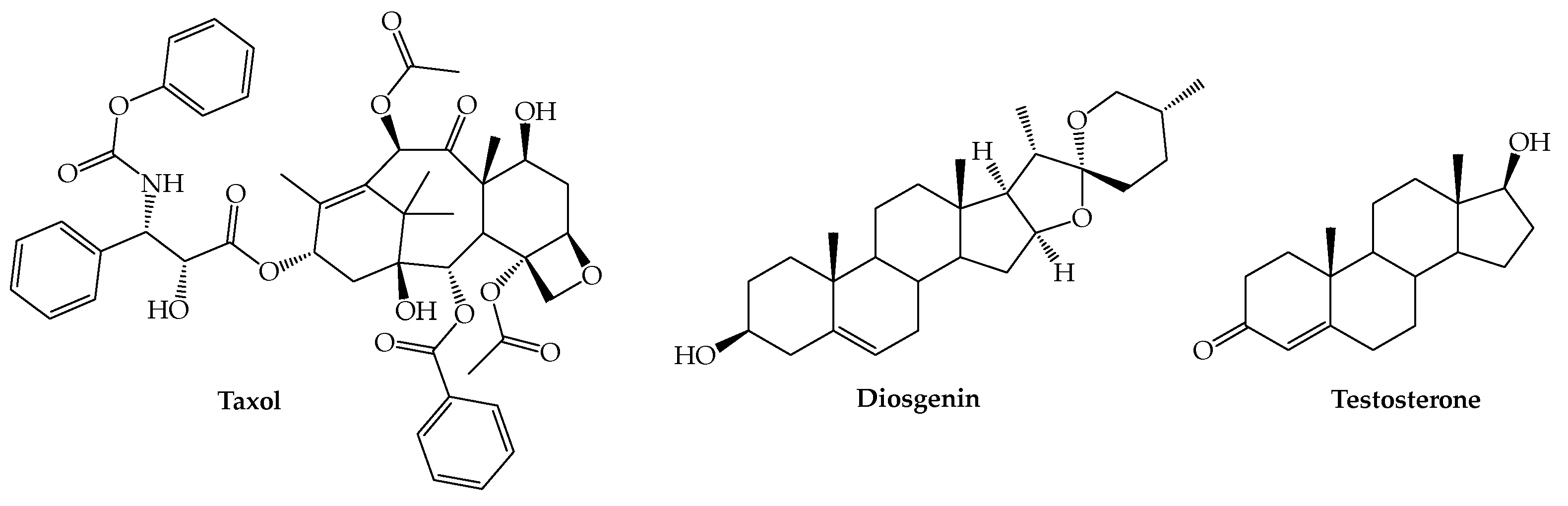
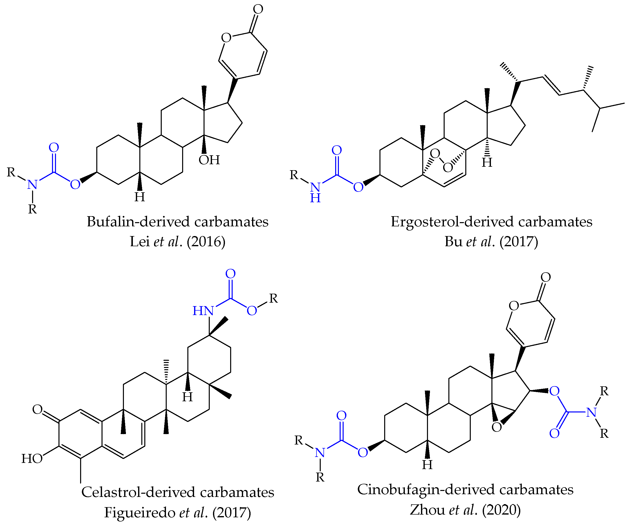
:1. Introduction
2. Results and Discussion
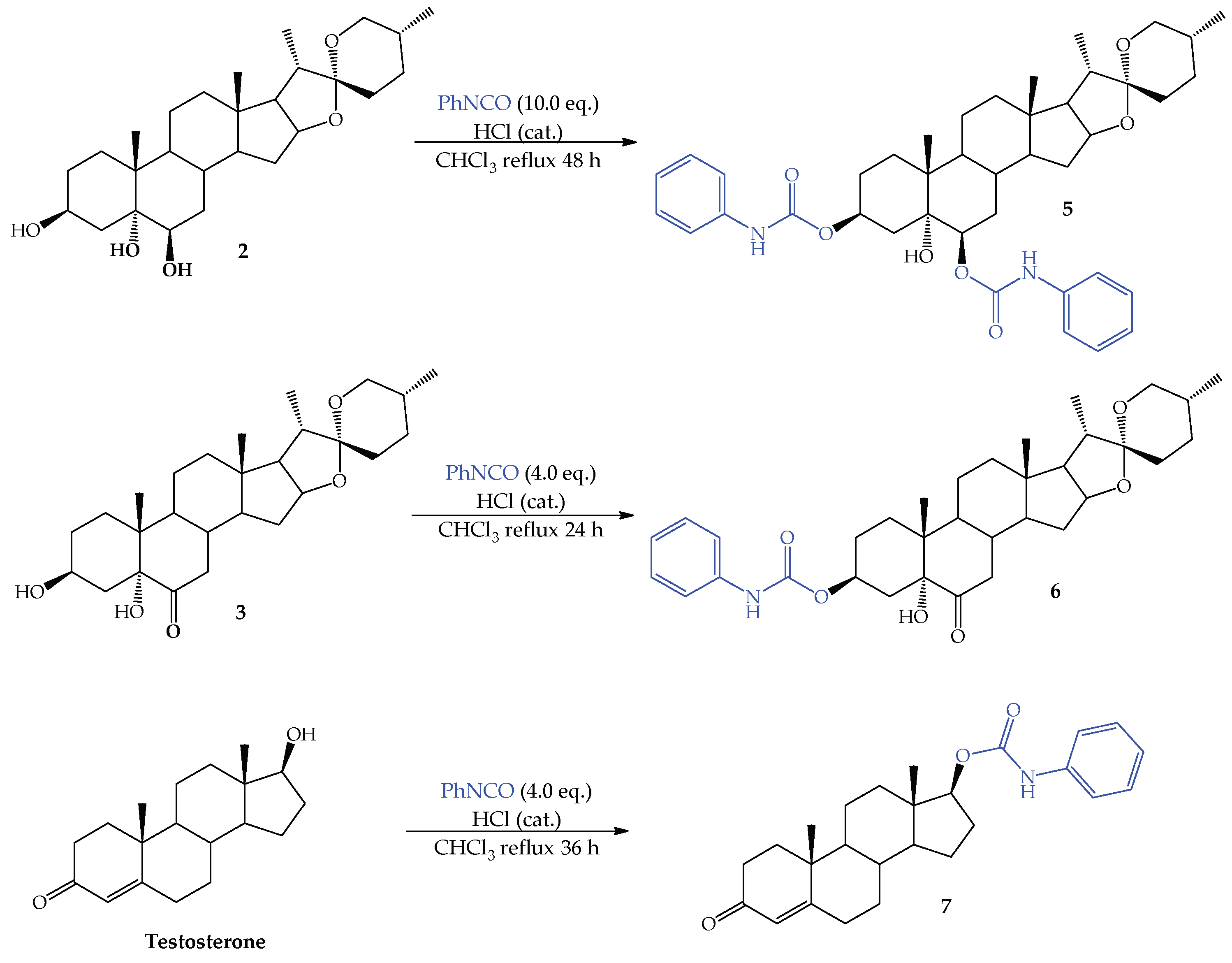
2.1. Synthesis of Steroidal Carbamates
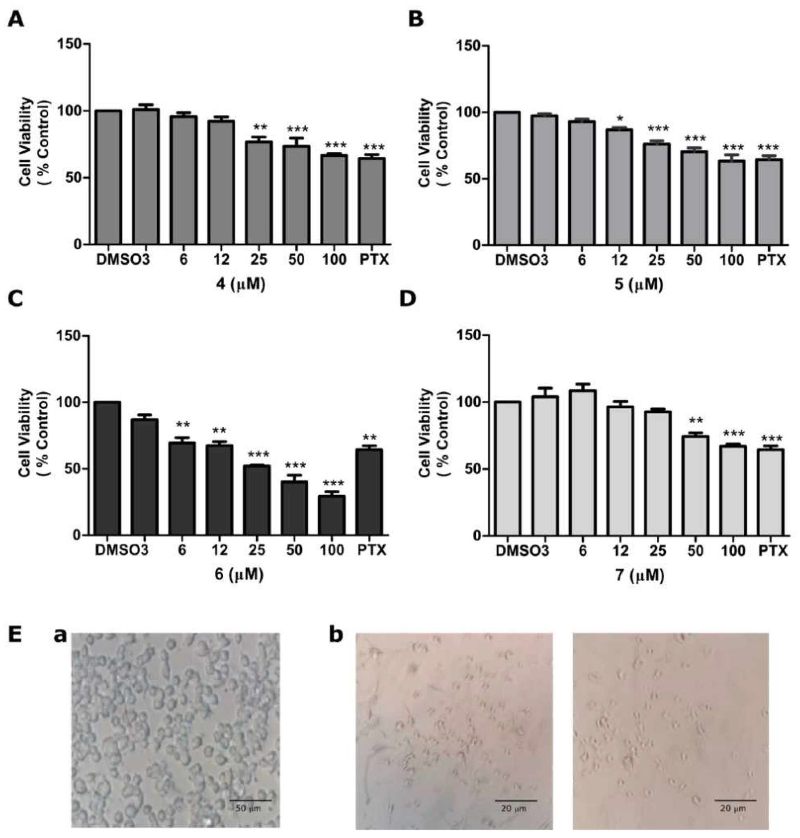
2.2. Antiproliferative Activity of Compounds 4, 5, 6, and 7 on CT26WT Cells
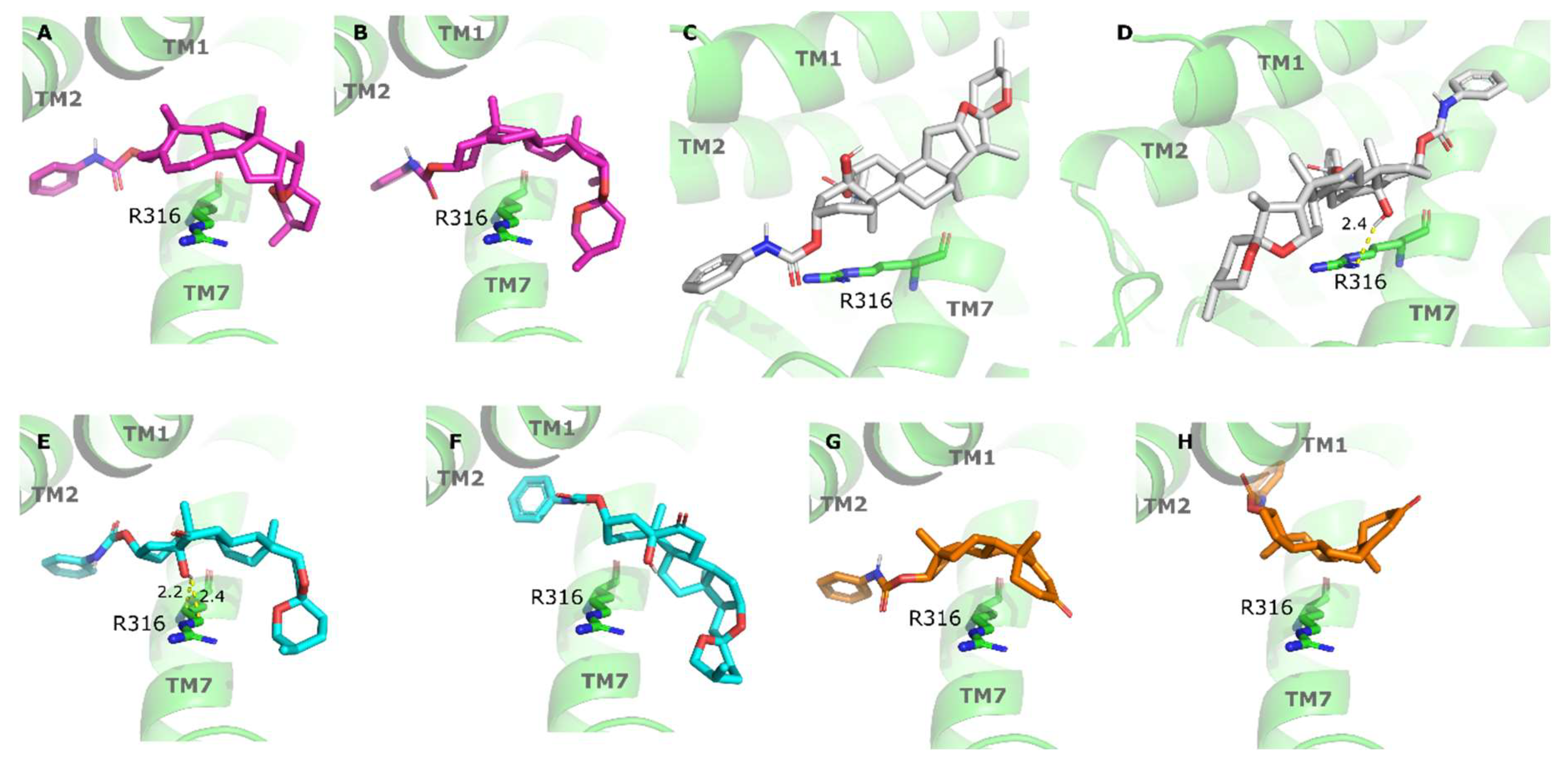
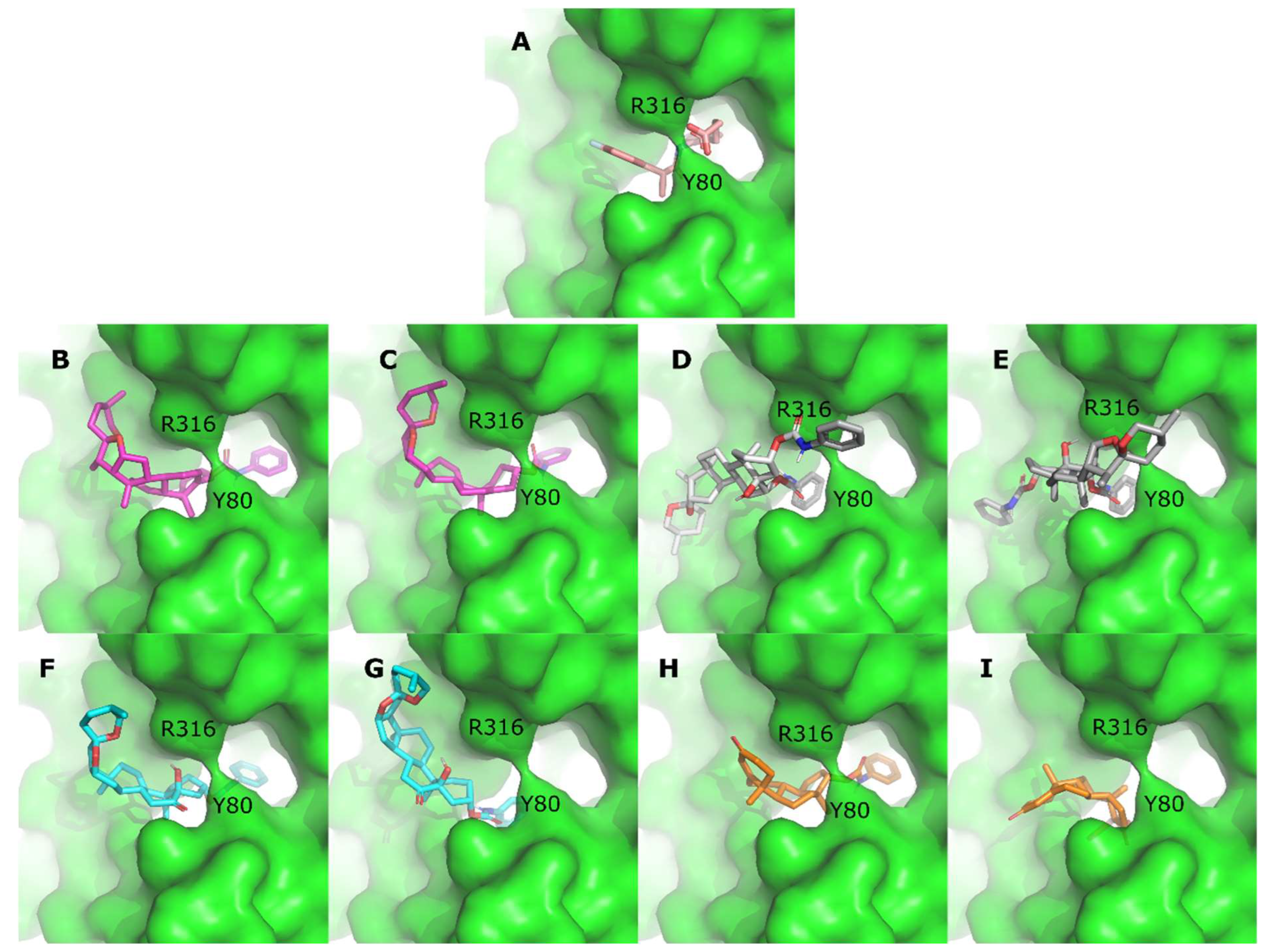
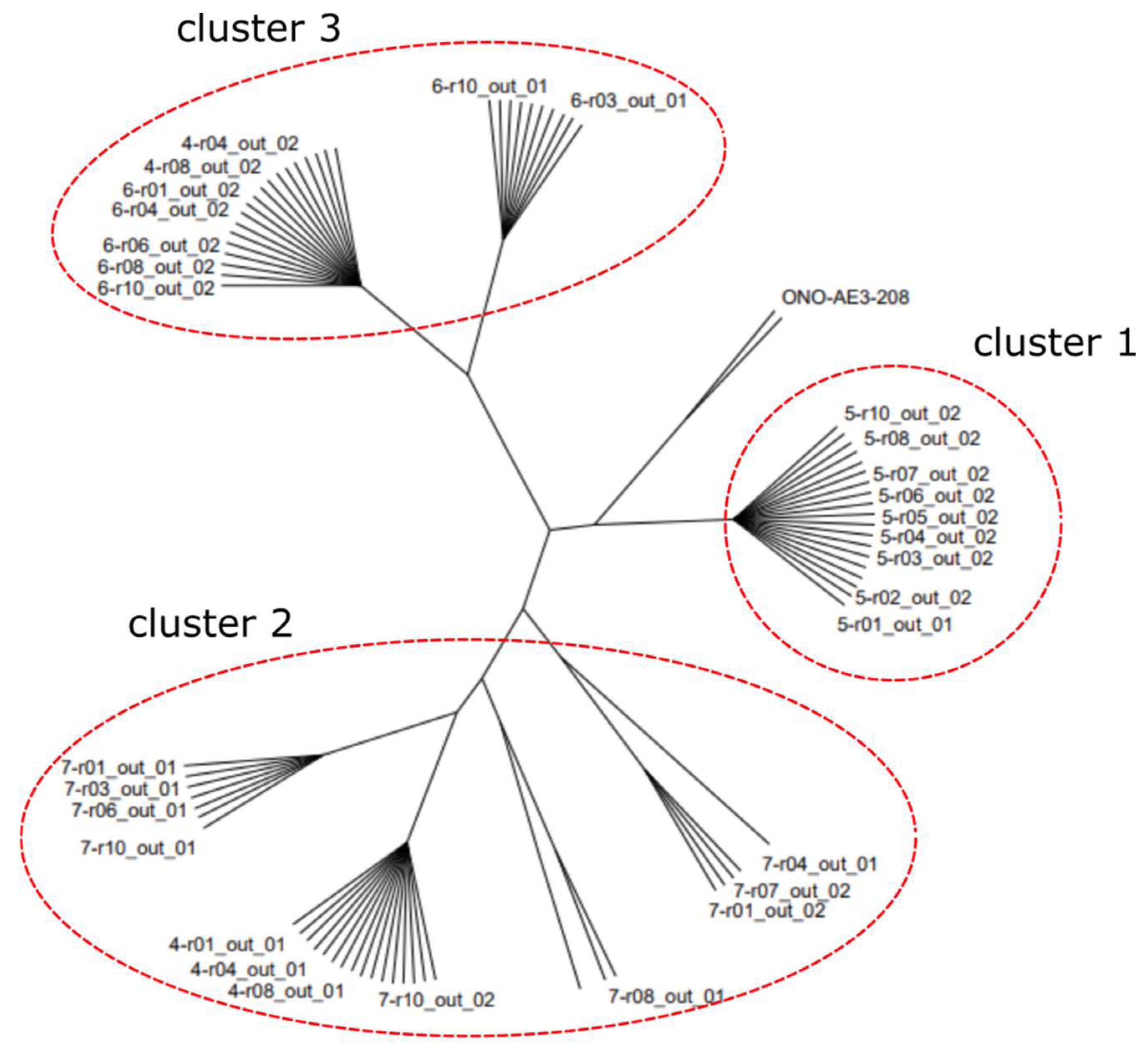
2.3. Molecular Docking Study
3. Materials and Methods
3.1. General
3.2. Methods of Synthesis
3.2.1. Synthesis of (25R)-5α-Hydroxy-spirostan-3β,6β-yl Phenylcarbamate (5)
3.2.2. Synthesis of (25R)-5α-Hydroxy-6-oxo-spirostan-3β-yl Phenylcarbamate (6)
3.2.3. Synthesis of 4-En-androst-17β-yl Phenylcarbamate (7)
3.3. Anticancer Studies
3.3.1. Cell Line and Culture Condition
3.3.2. Cell Viability Analysis
3.4. Molecular Docking
4. Conclusions
Supplementary Materials
Author Contributions
Funding
Institutional Review Board Statement
Informed Consent Statement
Data Availability Statement
Acknowledgments
Conflicts of Interest
References
- Siegel, R.L.; Miller, K.D.; Jemal, A. Cancer statistics, 2017. CA Cancer J. Clin. 2017, 67, 7–30. [Google Scholar] [CrossRef] [PubMed] [Green Version]
- Bequet-Romero, M.; Morera, Y.; Ayala-Ávila, M.; Ancizar, J.; Soria, Y.; Blanco, A.; Suárez-Alba, J.; Gavilondo, J.V. CIGB-247: A VEGF-based therapeutic vaccine that reduces experimental and spontaneous lung metastasis of C57Bl/6 and BALB/c mouse tumors. Vaccine 2012, 30, 1790–1799. [Google Scholar] [CrossRef] [PubMed]
- Na, H.S.; Lim, Y.K.; Jeong, Y.-I.; Lee, H.S.; Lim, Y.J.; Kang, M.S.; Cho, C.-S.; Lee, H.C. Combination antitumor effects of micelle-loaded anticancer drugs in a CT-26 murine colorectal carcinoma model. Int. J. Pharm. 2010, 383, 192–200. [Google Scholar] [CrossRef] [PubMed]
- Kim, H.Y.; Jung, S.K.; Byun, S.; Son, J.E.; Oh, M.H.; Lee, J.; Kang, M.J.; Heo, Y.-S.; Lee, K.W.; Lee, H.J. Raf and PI3K are the molecular targets for the anti-metastatic effect of luteolin. Phytother. Res. 2013, 27, 1481–1488. [Google Scholar] [CrossRef] [PubMed]
- Castle, J.C.; Loewer, M.; Boegel, S.; De Graaf, J.; Bender, C.; Tadmor, A.D.; Boisguerin, V.; Bukur, T.; Sorn, P.; Paret, C.; et al. Immunomic, genomic and transcriptomic characterization of CT26 colorectal carcinoma. BMC Genom. 2014, 15, 190. [Google Scholar] [CrossRef] [PubMed] [Green Version]
- Livshits, Z.; Rao, R.B.; Smith, S.W. An approach to chemotherapy-associated toxicity. Emerg. Med. Clin. N. Am. 2014, 32, 167–203. [Google Scholar] [CrossRef]
- Holohan, C.; Van Schaeybroeck, S.; Longley, D.B.; Johnston, P.G. Cancer drug resistance: An evolving paradigm. Nat. Rev. Cancer 2013, 13, 714–726. [Google Scholar] [CrossRef]
- Atanasov, A.G.; Zotchev, S.B.; Dirsch, V.M.; Orhan, I.E.; Banach, M.; Rollinger, J.M.; Barreca, D.; Weckwerth, W.; Bauer, R.; Bayer, E.A.; et al. Natural products in drug discovery: Advances and opportunities. Nat. Rev. Drug Discov. 2021, 20, 200–216. [Google Scholar] [CrossRef]
- Newman, D.J.; Cragg, G.M. Natural products as sources of new drugs over the nearly four decades from 01/1981 to 09/2019. J. Nat. Prod. 2020, 83, 770–803. [Google Scholar] [CrossRef]
- Mann, J. Natural products in cancer chemotherapy: Past, present and future. Nat. Rev. Cancer 2002, 2, 143–148. [Google Scholar] [CrossRef]
- Meher, C.P.; Sethy, S.P.; Pochaiah, B. Structure and biological activities: Steroid moieties. Res. J. Pharm. Biol. Chem. SCI 2013, 4, 253–272. [Google Scholar]
- Jastrzebska, I. Synthesis and application of steroidal 22, 16β-carbolactones: A review. J. Steroid Biochem. Mol. Biol. 2020, 199, 105592. [Google Scholar] [CrossRef] [PubMed]
- Singla, P.; Salunke, D.B. Recent advances in steroid amino acid conjugates: Old scaffolds with new dimensions. Eur. J. Med. Chem. 2020, 187, 111909. [Google Scholar] [CrossRef] [PubMed]
- Raju, J.; Patlolla, J.M.; Swamy, M.V.; Rao, C.V. Diosgenin, a steroid saponin of Trigonella foenum graecum (Fenugreek), inhibits azoxymethane-induced aberrant crypt foci formation in F344 rats and induces apoptosis in HT-29 human colon cancer cells. Cancer Epidemiol. Biomark. Prev. 2004, 13, 1392–1398. [Google Scholar] [CrossRef]
- Raju, J.; Bird, R.P. Diosgenin, a naturally occurring furostanol saponin suppresses 3-hydroxy-3-methylglutaryl CoA reductase expression and induces apoptosis in HCT-116 human colon carcinoma cells. Cancer Lett. 2007, 255, 194–204. [Google Scholar] [CrossRef]
- Lepage, C.; Liagre, B.; Cook-Moreau, J.; Pinon, A.; Beneytout, J.L. Cyclooxygenase-2 and 5-lipoxygenase pathways in diosgenin-induced apoptosis in HT-29 and HCT-116 colon cancer cells. Int. J. Oncol. 2010, 36, 1183–1191. [Google Scholar]
- Lepage, C.; Léger, D.Y.; Bertrand, J.; Martin, F.; Beneytout, J.L.; Liagre, B. Diosgenin induces death receptor-5 through activation of p38 pathway and promotes TRAIL-induced apoptosis in colon cancer cells. Cancer Lett. 2011, 301, 193–202. [Google Scholar] [CrossRef]
- Gunnarsson, P.O.; Ellman, M.; Fabiansson, E.; Gunnarsson, K.; Jensen, G.; Müntzing, J. Antitumour activity, toxicity and disposition of LS 1727, a nitroso-chloroethyl carbamate of 19-nortestosterone, in rats and mice. Acta Pharmacol. Toxicol. 1981, 49, 290–297. [Google Scholar] [CrossRef]
- Brix, H.P.; Berger, M.R.; Schneider, M.R.; Tang, W.C.; Eisenbrand, G. Androgen-linked alkylating agents: Biological activity in methylnitrosourea-induced rat mammary carcinoma. J. Cancer Res. Clin. Oncol. 1990, 116, 538–549. [Google Scholar] [CrossRef]
- Arth, G.E.; Patchett, A.A.; Jefopoulus, T.; Bugianesi, R.L.; Peterson, L.H.; Ham, E.A.; Kuehl, F.A.; Brink, N.G. Steroidal androgen biosynthesis inhibitors. J. Med. Chem. 1971, 14, 675–679. [Google Scholar] [CrossRef]
- Moreira, V.M.A.; Vasaitis, T.S.; Guo, Z.; Njar, V.C.O.; Salvador, J.A.R. Synthesis of novel C17 steroidal carbamates: Studies on CYP17 action, androgen receptor binding and function, and prostate cancer cell growth. Steroids 2008, 73, 1217–1227. [Google Scholar] [CrossRef] [PubMed]
- Rank, P.; Peter, R.; Depenbrock, H.; Eisenbrand, G.; Schmid, P.; Pitzl, H.; Hanauske, A.R. Preclinical activity of 17β-[N-[N-(2-chloroethyl)-N-nitrosocarbamoyl]-l-alanyl]-5α-dihydrotestosterone (E91) against tumour colony forming units and haematopoietic progenitor cells. Eur. J. Cancer 1999, 35, 1009–1013. [Google Scholar] [CrossRef]
- Wani, M.C.; Taylor, H.L.; Wall, M.E.; Coggon, P.; McPhail, A.T. Plant antitumor agents. VI. Isolation and structure of taxol, a novel antileukemic and antitumor agent from Taxus brevifolia. J. Am. Chem. Soc. 1971, 93, 2325–2327. [Google Scholar] [CrossRef] [PubMed]
- Bu, M.; Cao, T.; Li, H.; Guo, M.; Yang, B.B.; Zeng, C.; Hu, L. Synthesis of 5α,8α-ergosterol peroxide 3-carbamate derivatives and a fluorescent mitochondria-targeting conjugate for enhanced anticancer activities. ChemMedChem 2017, 12, 466–474. [Google Scholar] [CrossRef]
- Lei, M.; Xiao, Z.; Ma, B.; Chen, Y.; Liu, M.; Liu, J.; Guo, D.A.; Liu, X.; Hu, L. Synthesis and biological evaluation of bufalin-3-yl nitrogen-containing-carbamate derivatives as anticancer agents. Steroids 2016, 108, 56–60. [Google Scholar] [CrossRef] [PubMed]
- Figueiredo, S.A.C.; Salvador, J.A.R.; Cortés, R.; Cascante, M. Design, synthesis and biological evaluation of novel C-29 carbamate celastrol derivatives as potent and selective cytotoxic compounds. Eur. J. Med. Chem. 2017, 139, 836–848. [Google Scholar] [CrossRef] [PubMed]
- Zhou, Y.; Hou, J.; Long, H.; Zhang, Z.; Lei, M.; Wu, W. Design, synthesis and anti-tumor activities of carbamate derivatives of cinobufagin. Steroids 2020, 164, 108749. [Google Scholar] [CrossRef]
- Pozzi, A.; Yan, X.; Macias-Perez, I.; Wei, S.; Hata, A.N.; Breyer, R.M.; Morrow, J.D.; Capdevila, J.H. Colon carcinoma cell growth is associated with prostaglandin E2/EP4 receptor-evoked ERK activation. J. Biol. Chem. 2004, 279, 29797–29804. [Google Scholar] [CrossRef] [Green Version]
- Konya, V.; Marsche, G.; Schuligoi, R.; Heinemann, A. E-type prostanoid receptor 4 (EP4) in disease and therapy. Pharmacol. Ther. 2013, 138, 485–502. [Google Scholar] [CrossRef] [Green Version]
- Hull, M.A.; Ko, S.C.; Hawcroft, G. Prostaglandin EP receptors: Targets for treatment and prevention of colorectal cancer? Mol. Cancer Ther. 2004, 3, 1031–1039. [Google Scholar] [CrossRef]
- Karpisheh, V.; Joshi, N.; Zekiy, A.O.; Beyzai, B.; Hojjat-Farsangi, M.; Namdar, A.; Edalati, M.; Jadidi-Niaragh, F. EP4 receptor as a novel promising therapeutic target in colon cancer. Pathol. Res. Pract. 2020, 216, 153247. [Google Scholar] [CrossRef]
- Wang, J.; Li, D.; Zhao, B.; Kim, J.; Sui, G.; Shi, J. Small molecule compounds of natural origin target cellular receptors to inhibit cancer development and progression. Int. J. Mol. Sci. 2022, 23, 2672. [Google Scholar] [CrossRef] [PubMed]
- Pacheco, D.F.; González Ceballos, L.; Castro, A.Z.; Conde González, M.R.; González de la Torre, L.; Rostgaard, L.P.; Espinoza, L.; Díaz, K.; Olea, A.F.; Coll García, Y. Synthesis of new steroidal carbamates with plant-growth-promoting activity: Theoretical and experimental evidence. Int. J. Mol. Sci. 2021, 22, 2330. [Google Scholar] [CrossRef] [PubMed]
- Chávez-Riveros, A.; Garrido, M.; Ramírez Apan, M.T.; Zambrano, A.; Díaz, M.; Bratoeff, E. Synthesis and cytotoxic effect on cancer cell lines and macrophages of novel progesterone derivatives having an ester or a carbamate function at C-3 and C-17. Eur. J. Med. Chem. 2014, 82, 498–505. [Google Scholar] [CrossRef] [PubMed]
- Kommera, H.; Kaluđerović, G.N.; Dittrich, S.; Kalbitz, J.; Dräger, B.; Mueller, T.; Paschke, R. Carbamate derivatives of betulinic acid and betulin with selective cytotoxic activity. Bioorg. Med. Chem. Lett. 2010, 20, 3409–3412. [Google Scholar] [CrossRef] [PubMed]
- Toyoda, Y.; Morimoto, K.; Suno, R.; Horita, S.; Yamashita, K.; Hirata, K.; Sekiguchi, Y.; Yasuda, S.; Shiroishi, M.; Shimizu, T.; et al. Ligand binding to human prostaglandin E receptor EP(4) at the lipid-bilayer interface. Nat. Chem. Biol. 2019, 15, 18–26. [Google Scholar] [CrossRef] [PubMed]
- Rosenfeld, R.J.; Goodsell, D.S.; Musah, R.A.; Morris, G.M.; Goodin, D.B.; Olson, A.J. Automated docking of ligands to an artificial active site: Augmenting crystallographic analysis with computer modeling. J. Comput.-Aided Mol. Des. 2003, 17, 525–536. [Google Scholar] [CrossRef] [PubMed]
- Trott, O.; Olson, A.J. AutoDock Vina: Improving the speed and accuracy of docking with a new scoring function, efficient optimization, and multithreading. J. Comput. Chem. 2010, 31, 455–461. [Google Scholar] [CrossRef] [Green Version]
- Molinspiration. Molinspiration Cheminformatics. Slovensky Grob, Slovakia. Available online: https://www.molinspiration.com (accessed on 25 April 2022).
- Mosmann, T. Rapid colorimetric assay for cellular growth and survival: Application to proliferation and cytotoxicity assays. J. Immunol. Methods 1983, 65, 55–63. [Google Scholar] [CrossRef]
- Diggle, J.M.; Halliday, M.D.; Meakins, G.D.; Saltmarsh, M.S. The acid-catalysed ring-opening of epoxides in a largely nonaqueous medium. J. Chem. Soc. Chem. Commun. 1969, 14, 819–820. [Google Scholar] [CrossRef]
- Morris, G.M.; Huey, R.; Lindstrom, W.; Sanner, M.F.; Belew, R.K.; Goodsell, D.S.; Olson, A.J. AutoDock4 and AutoDockTools4: Automated docking with selective receptor flexibility. J. Comput. Chem. 2009, 30, 2785–2791. [Google Scholar] [CrossRef] [PubMed] [Green Version]
- Bouvier, G.; Evrard-Todeschi, N.; Girault, J.P.; Bertho, G. Automatic clustering of docking poses in virtual screening process using self-organizing map. Bioinformatics 2010, 26, 53–60. [Google Scholar] [CrossRef] [PubMed] [Green Version]
- Mantsyzov, A.B.; Bouvier, G.; Evrard-Todeschi, N.; Bertho, G. Contact-based ligand-clustering approach for the identification of active compounds in virtual screening. Adv. Appl. Bioinform. Chem. 2012, 5, 61–79. [Google Scholar] [PubMed] [Green Version]
- Durrant, J.D.; McCammon, J.A. BINANA: A novel algorithm for ligand-binding characterization. J. Mol. Graph. Model. 2011, 29, 888–893. [Google Scholar] [CrossRef] [PubMed] [Green Version]


| Ligands | Binding Mode | Poses Number | Energy (kcal/mol) |
|---|---|---|---|
| 4 | 1 | 10 | −9.76 |
| 2 | 10 | −8.60 | |
| 5 | 1 | 10 | −7.43 |
| 2 | 9 | −7.51 | |
| 6 | 1 | 10 | −8.29 |
| 2 | 9 | −8.10 | |
| 7 | 1 | 10 | −8.90 |
| 2 | 3 | −8.46 | |
| 3 | 4 | −8.52 | |
| 4 | 3 | −8.50 |
Publisher’s Note: MDPI stays neutral with regard to jurisdictional claims in published maps and institutional affiliations. |
© 2022 by the authors. Licensee MDPI, Basel, Switzerland. This article is an open access article distributed under the terms and conditions of the Creative Commons Attribution (CC BY) license (https://creativecommons.org/licenses/by/4.0/).
Share and Cite
Pacheco, D.F.; Alonso, D.; Ceballos, L.G.; Castro, A.Z.; Brown Roldán, S.; García Díaz, M.; Villa Testa, A.; Wagner, S.F.; Piloto-Ferrer, J.; García, Y.C.; et al. Synthesis of Four Steroidal Carbamates with Antitumor Activity against Mouse Colon Carcinoma CT26WT Cells: In Vitro and In Silico Evidence. Int. J. Mol. Sci. 2022, 23, 8775. https://doi.org/10.3390/ijms23158775
Pacheco DF, Alonso D, Ceballos LG, Castro AZ, Brown Roldán S, García Díaz M, Villa Testa A, Wagner SF, Piloto-Ferrer J, García YC, et al. Synthesis of Four Steroidal Carbamates with Antitumor Activity against Mouse Colon Carcinoma CT26WT Cells: In Vitro and In Silico Evidence. International Journal of Molecular Sciences. 2022; 23(15):8775. https://doi.org/10.3390/ijms23158775
Chicago/Turabian StylePacheco, Daylin Fernández, Dayana Alonso, Leonardo González Ceballos, Armando Zaldo Castro, Sheila Brown Roldán, Mairelys García Díaz, Anabel Villa Testa, Sarah Fuentes Wagner, Janet Piloto-Ferrer, Yamilet Coll García, and et al. 2022. "Synthesis of Four Steroidal Carbamates with Antitumor Activity against Mouse Colon Carcinoma CT26WT Cells: In Vitro and In Silico Evidence" International Journal of Molecular Sciences 23, no. 15: 8775. https://doi.org/10.3390/ijms23158775
APA StylePacheco, D. F., Alonso, D., Ceballos, L. G., Castro, A. Z., Brown Roldán, S., García Díaz, M., Villa Testa, A., Wagner, S. F., Piloto-Ferrer, J., García, Y. C., Olea, A. F., & Espinoza, L. (2022). Synthesis of Four Steroidal Carbamates with Antitumor Activity against Mouse Colon Carcinoma CT26WT Cells: In Vitro and In Silico Evidence. International Journal of Molecular Sciences, 23(15), 8775. https://doi.org/10.3390/ijms23158775