Article

Distinct Roles of Estrogen Receptors in the Regulation of Vitellogenin Expression in Orange-Spotted Grouper (Epinephelus coioides)

1
State Key Laboratory of Biocontrol and School of Life Sciences, Southern Marine Science and Engineering Guangdong Laboratory (Zhuhai), Guangdong Provincial Key Laboratory for Aquatic Economic Animals and Guangdong Provincial Engineering Technology Research Center for Healthy Breeding of Important Economic Fish, Sun Yat-Sen University, Guangzhou 510275, China
2
Laboratory for Marine Fisheries Science and Food Production Processes, Qingdao National Laboratory for Marine Science and Technology, Qingdao 266373, China
*
Authors to whom correspondence should be addressed.
Int. J. Mol. Sci. 2022, 23(15), 8632; https://doi.org/10.3390/ijms23158632
Submission received: 10 July 2022 / Revised: 29 July 2022 / Accepted: 31 July 2022 / Published: 3 August 2022
(This article belongs to the Section Molecular Biology)

Abstract

:
During their breeding season, estrogen induces vitellogenin (VTG) production in the liver of teleost fish through estrogen receptors (ERs) that support oocyte vitellogenesis. There are at least three ER subtypes in teleost fish, but their roles in mediating E2-induced VTG expression have yet to be ascertained. In this study, we investigated the expression of vtgs and ers in the liver of orange-spotted grouper (Epinephelus coioides). Their expression levels were significantly increased in the breeding season and were upregulated by an estradiol (E2) injection in female fish, except for the expression of erβ1. The upregulation of vtgs, erα and erβ2 by E2 was also observed in primary hepatocytes, but these stimulatory effects could be abolished by ER antagonist ICI182780 treatment. Subsequent studies showed that ERβ antagonist Cyclofenil downregulated the E2-induced expression of vtg, erα, and erβ2, while the ERβ agonist DPN simulated their expression. Knockdown of erβ2 by siRNA further confirmed that ERβ2 mediated the E2-induced expression of vtgs and erα. To reveal the mechanism of ERβ2 in the regulation of erα expression, the erα promoter was cloned, and its activity was examined in cells. E2 treatment simulated the activity of the erα promoter in the presence of ERβ2. Deletions and site-directed mutations showed that the E2 up-regulated transcriptional activity of erα occurs through a classical half-estrogen response element- (ERE) dependent pathway. This study reveals the roles of ER subtypes in VTG expression in orange-spotted grouper and provides a possible explanation for the rapid and efficient VTG production in this species during the breeding season.

1. Introduction

Vitellogenin (VTG), the precursor of the yolk protein in egg-bearing vertebrates, is essential for oogenesis, embryonic development, and larval survival. During the reproductive season, Vtg in the female liver is synthesized by estrogen stimulation and secreted into the bloodstream. Plasma Vtg is taken up by Vtg receptors on the surfaces of oocytes and cleaved into smaller yolk proteins by cathepsin [1]. Vitellogenesis is the primary phase of oocyte development and consumes a significant amount of nutrients and energy [2]. An insufficient vitellogenin uptake by oocytes leads to incomplete larval development and the higher mortality of eggs [3]. In female zebrafish, the knockout of vtg genes results in a dramatic drop in egg fertilization rates and offspring survival [4]. Therefore, substantial Vtg production is critical to the reproductive success of teleosts.
Piscine VTG are divided into three categories, namely, VTGAA, VTGAB, and VTGC, which are encoded by vtgaa, vtgab, and vtgc, respectively [5]. VTG protein could be rapidly produced in the liver after exogenous estrogen treatment [6]. Estrogen exerts its actions via activating estrogen receptors (ERs) [7]. At present, at least three estrogen receptor subtypes (ERα, ERβ1, and ERβ2) have been identified in fish species [8,9]. ERα is considered to be responsible for mediating the estrogen action in VTG synthesis in fish [10,11,12,13], as evidence has shown that the hepatic ERα expression was increased in the breeding season and was dramatically upregulated by E2 treatment [14,15,16].
Other piscine ER subtypes are also expressed in the liver besides ERα [17,18]. Whether they are involved in VTG production and the regulatory relationships between ER subtypes in this process attract attention. An intraperitoneal injection of E2 could upregulate the mRNA expression of erβ1 and erβ2 in the liver of goldfish [19]. ERβ selective agonist DPN increased VTG expression in rainbow trout hepatocytes in a dose-dependent manner [20]. A knockdown of erβ1 or erβ2 using specific siRNA in goldfish primary hepatocytes significantly reduced the E2-induced mRNA expression of vtg and erα [21]. Research on zebrafish embryos showed that the knockdown of erβs via specific morpholinos could suppress the estradiol induction of vtg and erα mRNA expression [22]. These studies indicate that ERβ subtypes play important roles in the vitellogenesis of fish. It is proposed that ERβ subtypes mediate estrogen signals to promote erα expression, which then sensitize the hepatocytes to further E2 stimulation and prepare them for vitellogenesis [21]. However, a systematic study in fish revealing the interplay between ER subtypes in VTG production and the regulatory mechanism is lacking. Moreover, further studies in different fish species with different reproductive modes are required to evaluate the significance of ER subtypes in this physiological process and reveal other important aspects of ER subtypes regarding the regulation of VTG production.
Groupers are marine benthic fish, belonging to the subfamily Epinephelinae (Teleoste: Serranidae), which contains more than 160 species in 15 genera [23]. Most groupers are protogynous hermaphrodites and are of significant economic value. According to the statistics from the Food and Agriculture Organization of the United Nations (FAO), global grouper production reached about 234,828 tons in 2019. Groupers generally enter the breeding season from the end of spring to the early autumn, and this process is mainly affected by water temperature [24]. During the breeding period, females carry 70,000 to 1,000,000 ova, depending on their size [24]. Due to large-scale farming in the Asia-Pacific region, the fertilized eggs of groupers have been in short supply over the past decade. Vitellogenesis is a crucial process for oocyte development. However, there are few studies on vitellogenesis, particularly on VTG regulation in groupers. In this study, using the orange-spotted grouper (Epinephelus coioides) as an experimental model, we aimed to evaluate the functional role of ER subtypes in the regulation of VTG production and to reveal their regulatory relationships. Firstly, the expression patterns of ers and vtgs in the liver during the breeding and non-breeding seasons were examined. Secondly, the regulation of ers and vtgs expression by E2, ER antagonists, and ER agonists was further investigated. Finally, the regulatory relationships between ER subtypes and the molecular mechanism of ERβ2 regulation towards erα expression were studied. Our study will not only characterize the roles of ER subtypes in the regulation of VTG production, but also reveal the transcriptional regulatory mechanism of how ERβ2 regulates erα expression in fish at the molecular level.

2. Results

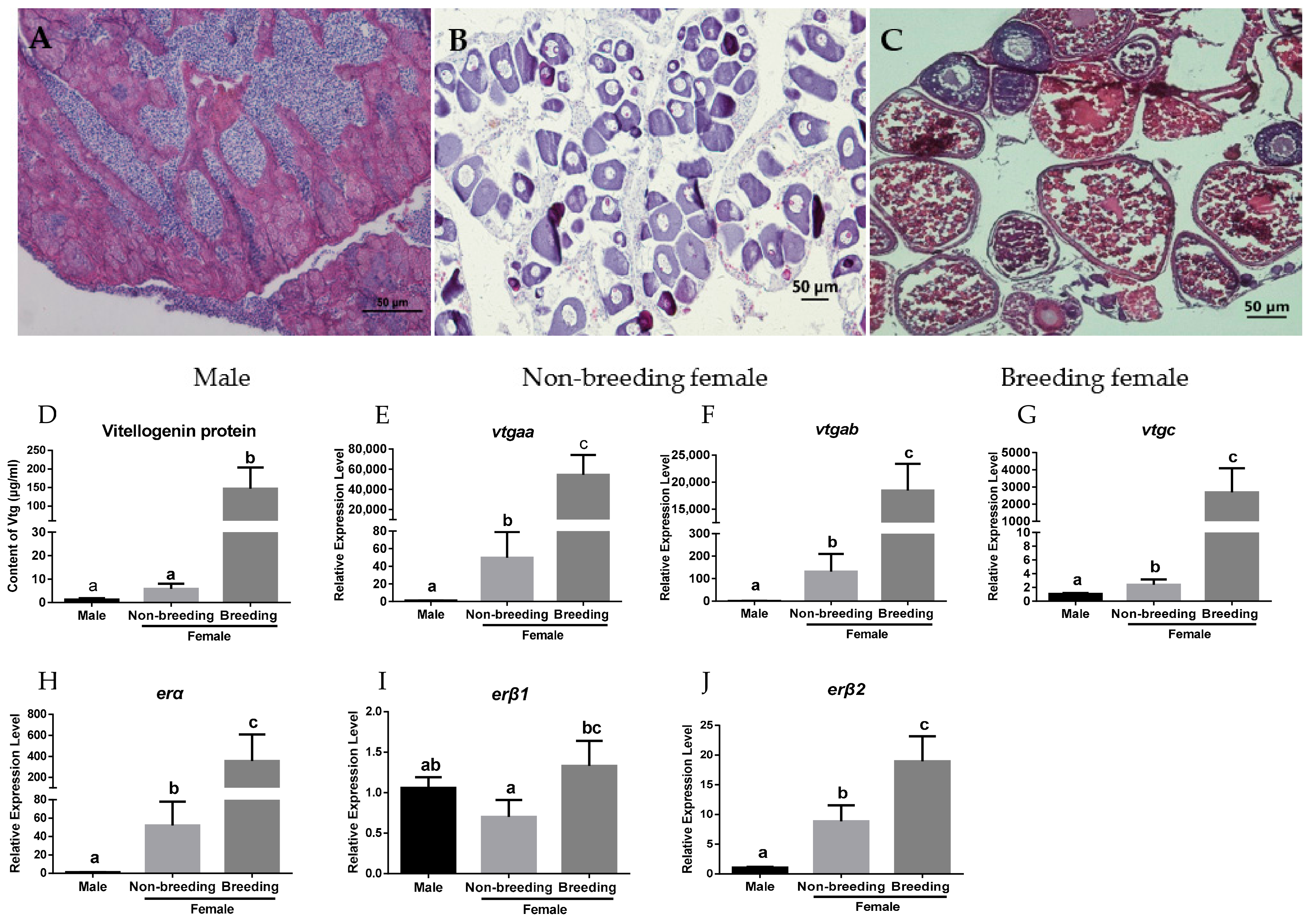
2.1. Expression Profiles of vtgs and ers in the Liver of Orange-Spotted Grouper

To investigate the expression profiles of vtgs and ers in the liver during vitellogenesis, a histological analysis was firstly applied to examine the ovarian developmental status of each fish. The male groupers used as controls showed normal spermatogenesis in the testis (Figure 1A). Primary oocytes were present in the ovaries of non-breeding females (Figure 1B), while the females in the breeding season showed vitellogenesis in the ovaries with vitellogenic oocytes (Figure 1C). The results showed that females in the breeding season had significantly higher VTG protein and mRNA levels compared to females in the non-breeding season and males (Figure 1D–G). All three er genes were expressed in the livers of the groupers. Their mRNA levels in females were also obviously upregulated in the breeding season (Figure 1H–J). Evidently, erα expression showed a dramatic increase in the breeding season compared to the non-breeding season.

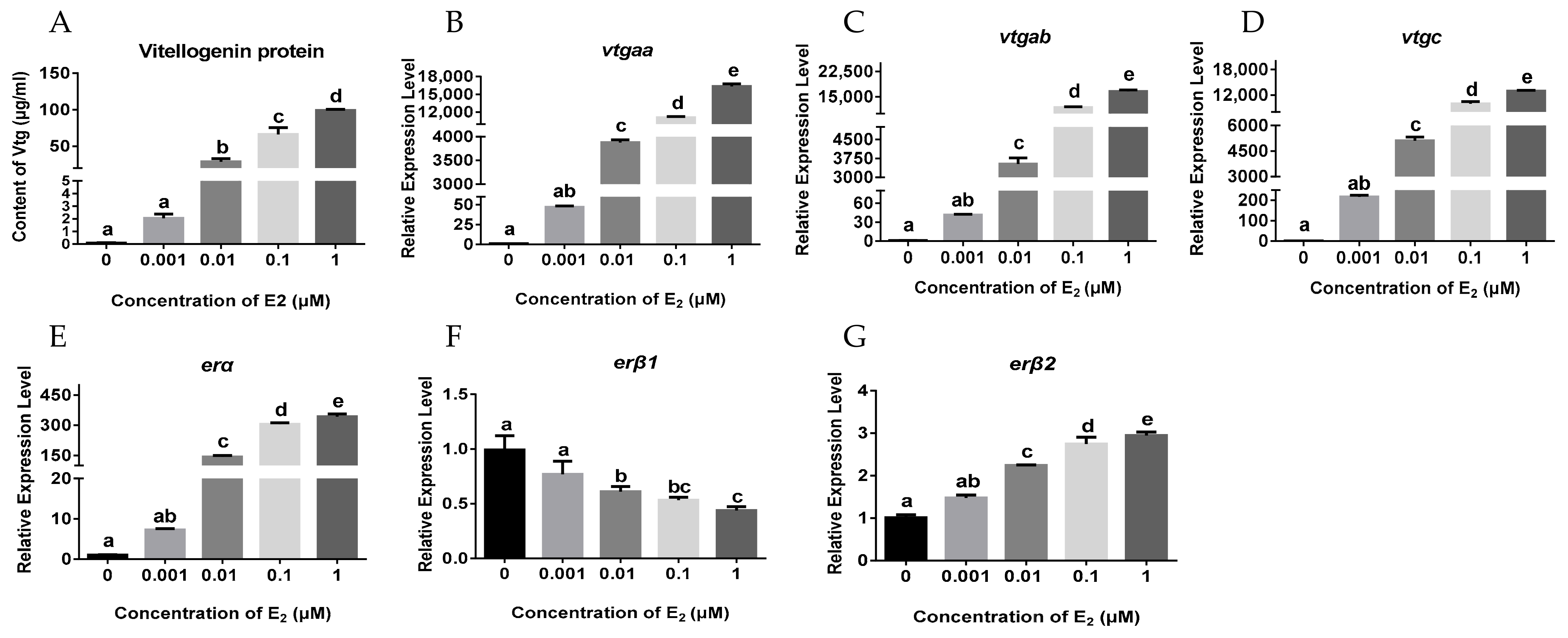
2.2. Effects of E2 on the Hepatic Expression of vtgs and ers in Juvenile Female Orange-Spotted Grouper

To investigate the relationship between ers and vtgs in vivo, the juvenile females received an intraperitoneal injection with a dose of 5 mg/kg E2. The results showed that exogenous estrogen dramatically increased the protein and mRNA levels of vtgs in the liver compared to the control group (Figure 2A–D). However, the er genes in the liver showed distinct expression patterns in response to E2 treatment. A remarkable increase in erα mRNA levels was observed, whereas a significant decrease in erβ1 mRNA levels was found after the E2 administration (Figure 2E,F). The erβ2 mRNA levels were slightly but significantly elevated following the E2 injection (Figure 2G).
Moreover, a primary hepatocyte culture system was established using the livers of the juvenile females, and the cells were incubated with different concentrations of E2 (from 0 to 1 μM) for 24 h. The results showed that E2 treatment significantly stimulated the protein and mRNA expression of vtgs in a dose-dependent manner (Figure 3A–D), and evoked the dose-dependent expression of erα and erβ2 in the hepatocytes (Figure 3E,G). However, a dose-dependent decrease in erβ1 expression was observed with increasing concentrations of E2 treatment (Figure 3F).

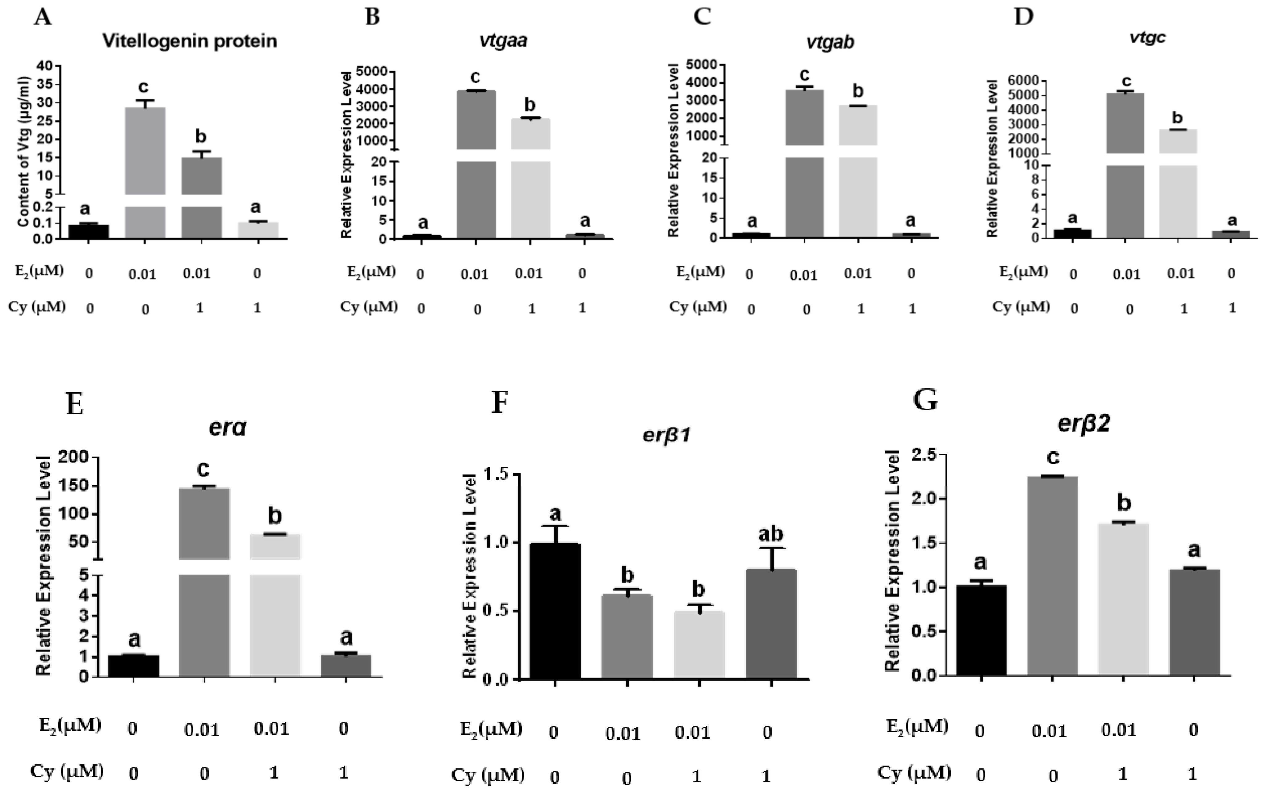
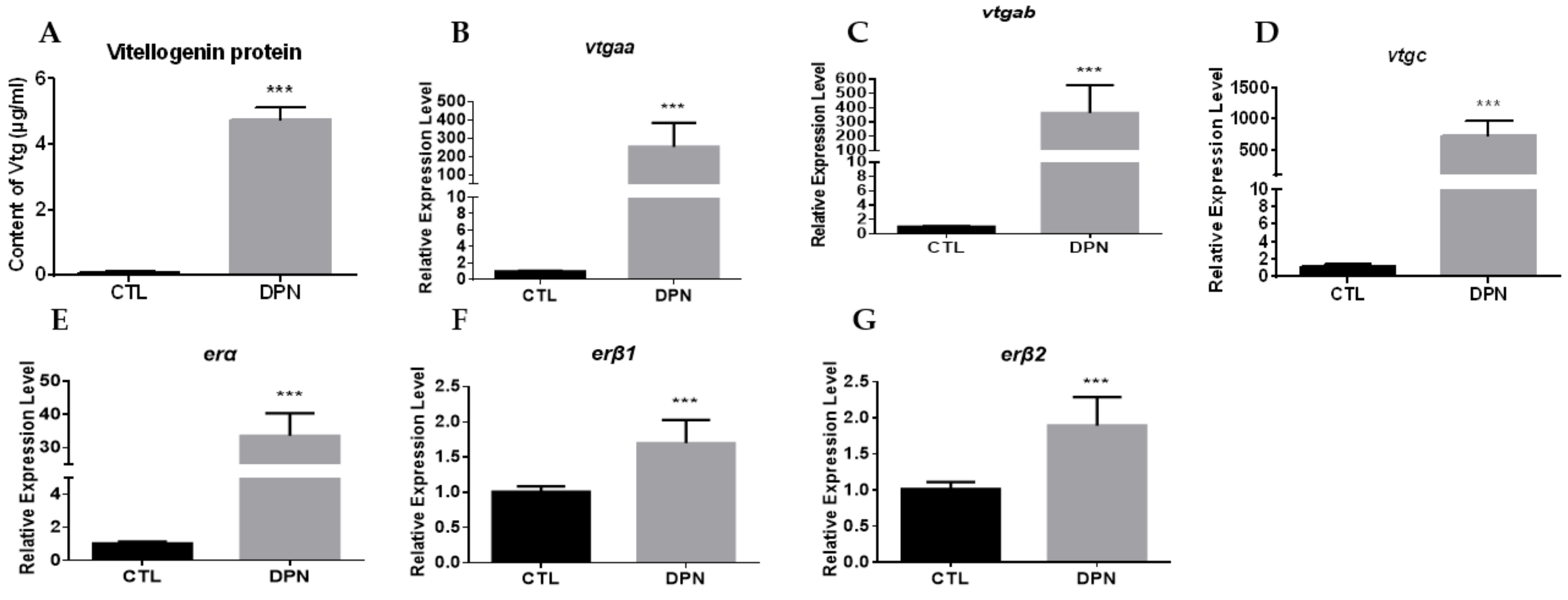
2.3. Effects of ER Antagonists or Agonist on the Expression of vtgs and ers in Primary Hepatocytes

To reveal the roles of different ERs towards the regulation of vtg expression, ER antagonists and one agonist were applied to treat the primary hepatocytes. These drugs altered the er expression and ultimately modulated the vtg expression. As shown in Figure 4, ERα and ERβ antagonist ICI182780 markedly suppressed the increasing protein and mRNA expression of vtgs induced by E2 in the hepatocytes and abolished the stimulatory effects of E2 on the expression of erα and erβ2 but had no effect on the reduced expression of erβ1 caused by E2.
Cyclofenil, an Erβ-specific antagonist, significantly attenuated the stimulatory effects of E2 on the expression of vtgs, erα, and erβ2, but could not change the erβ1 expression patterns following co-incubation with E2 (Figure 5).
DPN, an ERβ specific agonist, stimulated the protein and mRNA expression of vtgs effectively, and significantly upregulated the mRNA expression of three ers in hepatocytes without the presence of E2 (Figure 6).

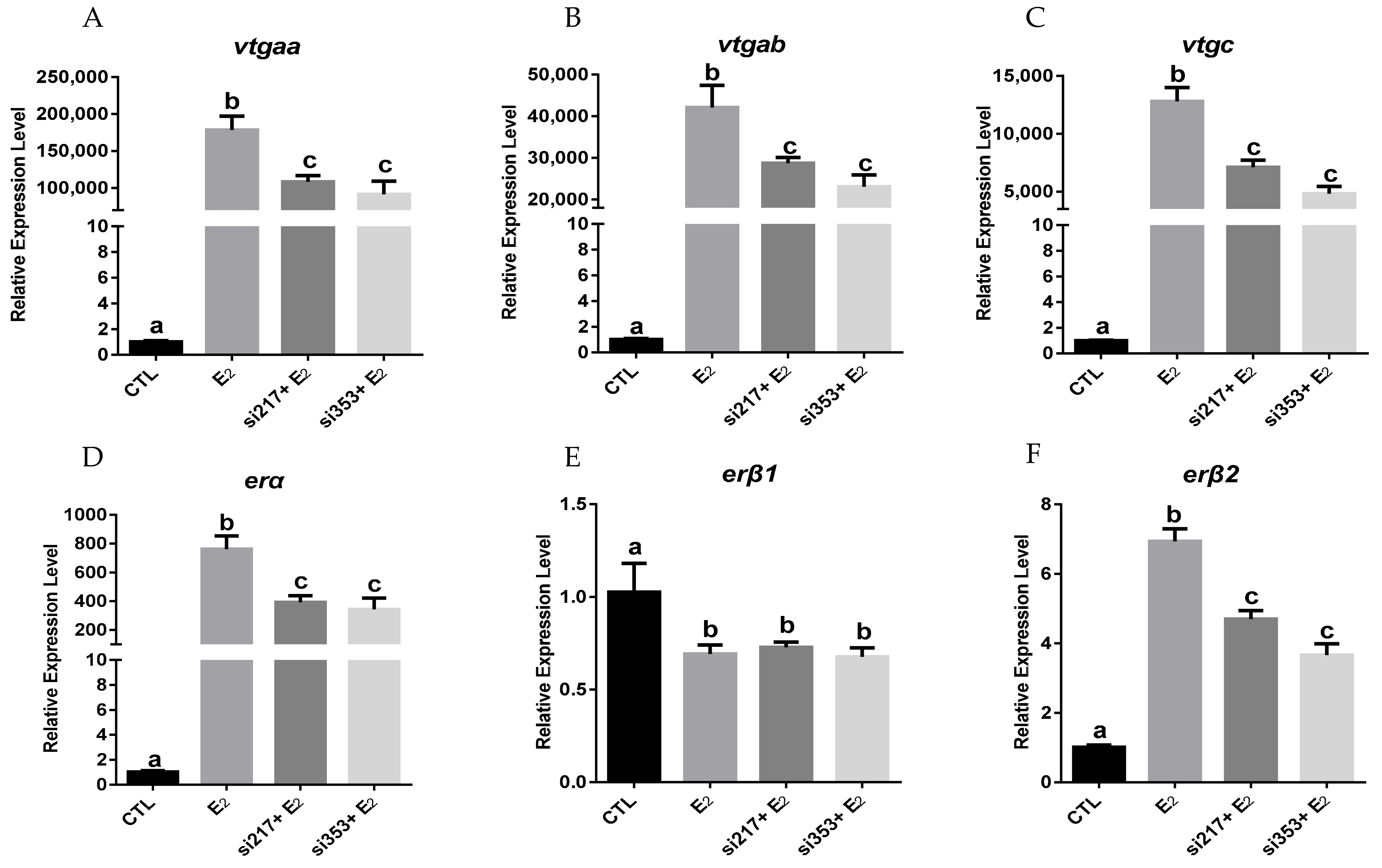
2.4. Effects of erβ2 Knockdown by siRNA on the Expression of vtgs and ers in Primary Hepatocytes

To verify whether ERβ2 could regulate the expression of erα, we established a transfection system that transfers the siRNA into primary grouper hepatocytes using an electroporation method. Flow cytometry was used to evaluate the transfection efficiency in the primary hepatocytes via electroporation. The fluorescence intensity of the hepatocytes in the control group and the siRNA-NC-Cy3 (NC) group was analyzed by flow cytometry (Figure S1A,B), and the P1 range was defined as the strong fluorescence range. As shown in Figure S1, in the P1 range, the cell count of the NC group was significantly higher than that of the control group, indicating that the siRNA was effectively transmitted into the primary hepatocytes. To test the hypothesis wherein E2-induced erβ2 expression would promote the erα expression and thus accelerate the vtg expression, two distinct siRNAs (Si217 and Si353) against erβ2 were separately transfected into the hepatocytes through electroporation. As shown in Figure 7, both siRNAs could significantly reduce the E2-induced erβ2 expression, resulting in an about 32% and 47% reduction compared to the E2 treatment group. As expected, the expression levels of the vtgs and erα simulated by E2 were significantly downregulated in the hepatocytes, and the erβ1 expression was not affected by erβ2 knockdown (Figure 7).

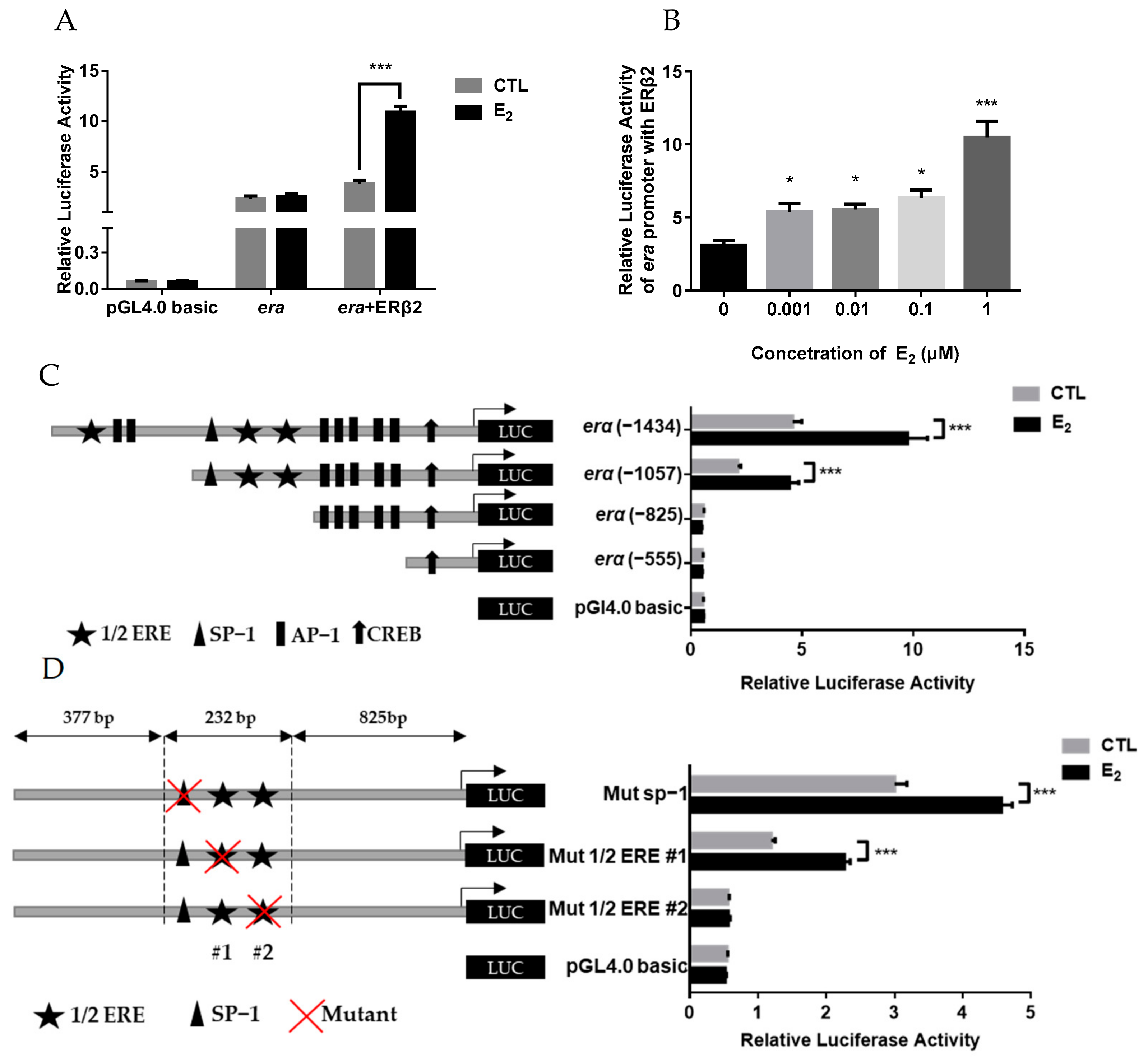
2.5. Estrogen Stimulation on erα Promoter Activity via ERβ2

Our results indicate that ERβ2 could mediate E2 action and then regulate the erα expression, but the transcriptional regulatory mechanism is unknown. Therefore, to reveal the mechanism of E2 in the regulation of erα expression via ERβ2, the 5′-flanking region of the erα gene was isolated. As shown in Figure S2, the cloned sequence upstream of the TSS of erα is 1434 bp in length, containing three half-estrogen response elements (ERE) and several transcription factor binding motifs, such as Activator protein 1 (Ap1), Specificity proteins 1 (Sp1), and cAMP response element binding protein (CREB). Whether the erα promoter activity was regulated by E2 and ERβ2 was further assessed in HEK293T cells. The results showed that E2 treatment significantly increased the erα promoter activity in the cells co-transfected with the ERβ2 (Figure 8A). The promoter activity was significantly elevated by E2 at 0.1 and 1 μM in the presence of ERβ2 (Figure 8B). To identify the E2 response region of the erα promoter, deletion analyses were further performed. The E2 treatment significantly increased the activity of the wild-type erα promoter in the presence of ERβ2, and the deletion of the promoter to position −1057 did not eliminate the E2 stimulatory effect (Figure 8C). However, deleting the promoter to position −825 could abolish the E2-induced promoter activity (Figure 8C), indicating that this region is responsible for mediating the E2 action towards triggering the transcriptional activity of the erα promoter through ERβ2. As the proximal −825 bp region of the erα promoter contains a putative Sp1 binding site and two putative half-ERE sites (marked by #1, #2) (Figure 8D), site-directed mutagenesis analyses were carried out. The mutation of the Sp1 binding site (upstream of TSS from −953 to −944 bp) or ERE#1 site (upstream of TSS from −900 to −895 bp) did not change the E2-induced promoter activity in the presence of ERβ2; however, the stimulatory effect was abolished when ERE#2 (upstream of TSS from −870 to −865 bp) was mutated (Figure 8D), indicating that ERE#2 is responsible for the E2/ ERβ2-induced expression of erα.

3. Discussion

The orange-spotted grouper is a seasonally spawning fish. In the breeding season, the serum estrogen levels in females are significantly elevated [25], but the pathway of estrogen in the regulation of the VTG product in the liver via ERs remains unclear in this species. In the present study, our results showed that (1) erα was abundantly expressed in the liver in the breeding season, (2) hepatic erα expression was more sensitive to E2 treatment than erβs in vivo and in vitro, (3) and that the E2-induced expression of VTG and erα was almost completely inhibited after the ICI182780 treatment, suggesting that hepatic ERα is mainly responsible for mediating the action of estrogen to promote the VTG production. These results are consistent with the findings in other fishes [26].
For erβ1, its expression in females was also significantly increased in the liver in the breeding season compared to non-breeding season. However, the E2 treatment reduced erβ1 expression in vivo and in vitro, and this inhibitory effect was not abolished by ICI182780 and Cyclofenil. These results indicate that the elevated expression of erβ1 in the breeding season may not be induced by estrogen. A study also found that hepatic erβ1 expression was markedly decreased in zebrafish after 48 h of E2 exposure [9]. However, the hepatic erβ1 expression level varied greatly in response to E2 in goldfish. Some studies observed an induction of hepatic erβ1 expression by E2 in juvenile and female goldfish, while others found a downregulation of erβ1 expression in males after E2 implantation, suggesting that Erβ1 possesses hepatic functions in teleosts, but its role in the regulation of VTG production is unclear.
Compared with other ER subtypes, erβ2 showed different expression patterns. Its expression was significantly increased in the breeding season and after E2 treatments, but not as dramatically as erα. The ER antagonists, especially the Cyclofenil (a specific antagonist of ERβ), significantly attenuated the upregulation of erβ2 mRNA expression induced by E2. These data indicate that hepatic ERβ2 expression is regulated by estrogen. Interestingly, Cyclofenil also downregulated the increased expression of the erα and vtgs induced by E2, but DPN upregulated their expression. As the hepatic erβ1 expression was downregulated by E2 and was not sensitive to Cyclofenil, the actions of Cyclofenil and DPN on the expression of erα and vtgs may possibly occur via ERβ2. To verify whether ERβ2 could regulate the expression of the erα and vtgs, we established a transfection system that transfers the siRNA into primary grouper hepatocytes using an electroporation method. Two siRNAs against erβ2 were employed to specifically knock down the hepatic erβ2 expression, and a significant decrease in the E2-induced expression of erα and vtgs in the hepatocytes was also observed. However, unlike ICI182780, neither Cyclofenil nor siRNA knockdown could fully abolish the E2 induction of erα and vtgs expression, and the DPN-mediated upregulation of erα and vtgs expression was not as drastic as the E2 treatment, suggesting that ERβ2 may partially contribute to the estrogenic regulation of erα and vtgs expression in grouper.
The results above showed that ERβ2 could promote the hepatic erα expression by mediating the estrogen action, but the molecular mechanism remains unclear. At present, it is clear that estrogen’s regulation of downstream gene expression occurs through the E2-ER-ERE pathway. The E2-ER dimer complexes act on the ERE site in the promoter to initiate or repress gene expression [27,28]. Studies have shown that there are several ERE sites in the erα promoter region of rainbow trout and zebrafish [28]. However, there is no complete ERE site in grouper’ erα promoter, but three 1/2 ERE sites, and some transcription binding sites that could mediate the estrogen signal, including one Sp1 binding site, nine Ap1 binding sites, and one CRBE binding site, were observed. Subsequent experiments demonstrated that E2 significantly enhanced the grouper erα promoter activities in the presence of ERβ2 in HEK293T cells. Deletion and site-directed mutagenesis analysis indicated that the ERE#2 site is required to maintain the E2 induction of erα promoter activities, suggesting that ERβ2 could modulate erα transcriptional activity through a half-ERE-dependent classical mechanism in grouper. Our previous study on grouper has shown that the half-ERE site in the gene promotor could initiate the estrogen response [29].
The present data suggest that ERβ2 is involved, directly and indirectly, in the regulation of VTG production in grouper. As promoters of vtg genes contain E2 response sites, ERβ2 may bind to these sites and initiate the vtg expression in the hepatocytes directly. In addition, some studies proposed that ERα and ERβ2 may interact as heterodimers to drive the vtg expression [7]. However, this hypothesis requires further evidence in fish species. Indirectly, E2-activated ERβ2 enhances the hepatic erα expression, and then ERα promotes VTG synthesis, as our results demonstrate that E2 could regulate the erα expression in the presence of ERβ2. During the breeding season, VTG is produced rapidly and efficiently in the female bloodstocks. The collaboration between ERα and ERβ subtypes may accelerate this process. In goldfish, a model for E2-mediated vitellogenesis was proposed: both ERβ subtypes can induce some vitellogenesis and are necessary for the normal E2 induction of erα expression. The increased erα expression would sensitize the hepatocyte to further E2 stimulation, and thus promote VTG production in the breeding season. This model may not be entirely suitable for grouper. Our study shows that grouper ERβ1 may not be involved in the regulation of VTG production. Although blocking ERβ2 action and the knockdown of its expression could result in the reduction of erα expression, whether ERβ2 is required for the E2-mediated upregulation of erα expression is uncertain and awaits further study. Therefore, the roles of ER subtypes, especially ERβ receptors, in mediating estrogen action may be different across fish species, suggesting that fish have evolved different mechanisms to regulate VTG synthesis.
In summary, we investigated the roles of ER subtypes towards VTG production in orange-spotted grouper. The results revealed that the physiological actions of estrogen on VTG production are primarily mediated by ERα, and that ERβ2 may play a role in promoting the E2-mediated upregulation of erα and vtg expression. This study will lead to a better understanding of vitellogenesis and paves the way for improving the oocyte quality of groupers.

4. Materials and Methods

4.1. Animal and Sample Collection

Eighteen adult orange-spotted groupers (5-years old; body weight: 3.21~4.46 kg; standard length: 46.93~60.16 cm) were obtained from the Hainan Chenhai Fisheries Co., Ltd., in Sanya (Hainan Province, China). The gonadal status of fish was extracted with a catheter and syringe, immediately fixed in Bouin’s fluid, and checked by histological examination. The methods were carried out as described by Tang et al. [30]. Livers from non-breeding females (n = 6) were collected in February 2021, and livers from males (n = 6) and breeding females (n = 6) were collected in April 2021. Fish were anesthetized with MS-222 (Sigma) and dissected. The samples were frozen in liquid nitrogen and stored at −80 °C for RNA extraction. Juvenile female groupers (1.5-years old; body weight: 0.70~1.01 kg; standard length: 18.35~22.18 cm) used for intraperitoneal injection and primary hepatocyte culture were provided by Yuxiangzi Aquatic Industry Co., Ltd. in Yangjiang (Guangdong Province, China). All animal experiments were conducted in accordance with the guidelines and approval of the Animal Research and Ethics Committees of the Sun Yat-sen University.

4.2. Chemicals

17β-Estradiol (E2) (CAS 50-28-2; purity ≥ 98%) was purchased from Sigma. ICI182780 (CAS 129453-61-8; purity ≥ 99%) was purchased from Abcam. Cyclofenil (CAS 2624-43-3; purity ≥ 99%) and DPN (CAS 1428-67-7; purity ≥ 99%) were purchased from Tocris. E2, ICI182780, Cyclofenil, and DPN were dissolved in DMSO at a final stock concentration of 10 μM.

4.3. Intraperitoneal Injection Experiment

Sixteen healthy juvenile female groupers (1.5-years old; body weight: 0.70~1.01 kg; standard length:18.35~22.18 cm) were randomly divided into two groups, and kept in two 3000 L tanks with continuous seawater and air supply. Groupers were acclimatized for a week before the experiment. The fish were exposed to natural photoperiods and water temperature (25.5–28.2 °C) and fed with commercial feed once a day. E2 was dissolved in DMSO, and then diluted in physiological saline solution. The fish in experimental group were intraperitoneally injected with E2 solution (5 mg/kg body weight). The physiological saline injected group was used as control. The livers were collected at 24 h after injection. The mRNA levels of vtgs and ers were quantified by Quantitative real-time PCR. The content of VTG protein in the liver was measured by using a VTG ELISA kit (Cusabio, Wuhan, China) following manufacturer’s protocol.

4.4. Isolation, Primary Culture, and Static Incubation of Grouper Hepatocytes

Livers from six juvenile female groupers were excised and washed three times in ice-cold Ca2+/Mg2+-free HBSS (Gibco, Carlsbad, CA, USA) containing 1% antibiotic-antimycotic (Gibco). The tissue fragments were further diced to about 1 mm in thickness using a razor blade and washed three times in Ca2+/Mg2+-free HBSS containing 1% antibiotic-antimycotic at room temperature. Subsequently, slices were digested with trypsin-EDTA (0.25%) (Gibco) at 28 °C for 20 min, then were mechanically dispersed into individual cells by gentle pipetting and filtered through a 30 μm sterilized nylon mesh. Cells were harvested by centrifugation (30× g for 5 min, twice) and were resuspended in L-15 medium (Gibco) containing 1% antibiotic-antimycotic and 10% FBS (Gibco). The cell viability test and counting were performed using a trypan blue exclusion test. Only cells with >95% viability were used for subsequent experiments. The number of hepatocytes was adjusted to approximately 1.5 × 10⁶ cells/mL/well and seeded as a monolayer into 24 well-plates precoated with 5 µg/mL PEI (Sigma, Saint Louis, MO, USA). After preincubation at 28 °C for 24 h, the medium was removed and replaced with a fresh medium containing the test drugs (E2, ER antagonists and agonist). In the E2 treatment experiment, the doses of E2 were 0.001, 0.01, 0.1, and 1 µM. In ER antagonists’ experiments, the dose of E2 was 0.01 µM, and 1 µM of ICI182780 or Cyclofenil was used. In ER agonist’s experiment, 1 µM DPN was added into the medium. Each experiment had three groups (4 wells per group). After 24 h incubation, the culture media were collected for VTG protein assay, and cells were harvested for gene expression analysis.

4.5. Knockdown Experiment

Small interfering RNAs (siRNAs) (Table 1) were synthesized by GenePharma according to the sequence of erβ2 (GenBank Accession No GU721078.1). Transfection experiment was performed on an electroporation instrument NEPA21 (NEPAGENE, Chiba, Japan). Primary hepatocytes (1.5 × 10⁷ cells per mL) were suspended in the opti-MEM and mixed with siRNA at room temperature for 10 min. The mixture was transferred into electrode chamber and subjected to electroporation. The program was 195 V, 2.5 ms, 2 times, and 50 ms interval for poring pulse, and 20 V, 50 ms, 5 times, and a 50 ms interval for transfer pulse. After electroporation, the mixture was immediately transferred to L-15 medium containing 10% FBS in a 24-well plate. After preincubation at 28 °C for 24 h, the medium was removed and replaced with a fresh medium containing E2. After 24 h incubation, cells were harvested for gene expression analysis. The electroporation experiment was performed in triplicate.
To analyze the transfection efficiency, siRNA-NC-Cy3 (Cy3-labeled negative control siRNA) was transfected into primary hepatocytes by electroporation according to the protocol above. After 24 h incubation, the fluorescence value was detected by Beckman Cytoflex flow cytometry (Beckman Coulter Inc., Brea, CA, USA), and analyzed by CytExpert software (Beckman Coulter Inc., Brea, CA, USA).

4.6. Quantitative Real-Time PCR

To examine the mRNA levels of vtgs (vtgaa, vtgab, and vtgc) and ers (erα, erβ1, and erβ2), total RNA was extracted with Trizol reagent (Invitrogen, Carlsbad, CA, USA) and was reverse transcribed with a ReverTra Ace qPCR-RT Kit (Toyobo, Osaka, Japan) following the manufacturer’s protocol. Quantitative real-time PCR reactions were performed on a Roche Light-Cycler 480 real-time PCR system using SYBR Green I Master (Roche, Basel, Switzerland) according to the manufacturer’s protocol. Briefly, 500 ng template, 5 µL SYBR Green I Master, and 0.2 µL forward (reverse) primer (10 mM) were mixed in a tube and supplemented with water to 10 µL. The program was as follows: denaturation at 95 °C for 10 min, followed by 40 amplification cycles of 95 °C for 10 s, 58 °C for 20 s, and 72 °C for 20 s. After amplification, melting curve analysis was carried out to confirm the specificity of amplification. Fluorescence signals were converted to threshold cycle (Ct) values. Beta-actin was used as reference gene to normalize the expression values. The relative mRNA levels were calculated by using 2−ΔΔCT method. The sequences of gene-specific primers are listed in Table 2.

4.7. Plasmid Construction and In Silico Analysis of Promoter

Using a SMARTer RACE cDNA Amplification Kit (Takara, Osaka, Japan), the transcriptional start site (TSS) of erα gene were confirmed. The 5′-flanking region (1434 bp) upstream of the transcriptional start site was cloned from the genome of orange-spotted grouper. The transcription factor binding sites in the promoter region were predicted using a virtual database online (http://alggen.lsi.upc.es/cgi-bin/promo_v3/promo/promoinit.cgi?dirDB=TF_8.3 (accessed on 2 July 2021)). Based on the sequence of 5′-flanking region, different erα promoter deletion fragments (1057 bp, 825 bp and 555 bp) upstream of TSS were cloned. These fragments were digested with endonuclease XhoI and KPNI (New England Biolabs, Ipswich, MA, USA) and then inserted into pGL4.10 vector (Promega, Madison, WI, USA). Based on the deletion analysis, three transcription factor binding sites of erα promoter were mutated using Mut Express II Fast Mutagenesis Kits (Vazyme, Nanjing, China) according to the manufacturer’s protocol, and subsequently subcloned into pGL4.10 vector. In addition, the open reading frame (ORF) of erβ2 was cloned and subcloned into pcDNA3.1. All constructs were verified by sequencing to ensure there was no mismatch. Primers used for rapid amplification, site-directed mutagenesis, and plasmid construction are listed in Table 3.

4.8. Dual-Luciferase Assay

Human embryonic kidney cells, (HEK293T) purchased from Shanghai Institute of Biochemistry and Cell Biology (SIBCB), were seeded on a 48-well plate and cultured in phenol red-free DMEM medium (HyClone, Logan, UT, USA) containing 5% FBS and 1% penicillin and streptomycin at 37 °C with 5% CO2 overnight. Cells in each well were co-transfected with 250 ng of promoter plasmid vector, 100 ng of ERβ2 expression vector, and 10 ng of pRL-TK vector (Promega). Six hours after transfection, the medium was replaced with fresh DMEM medium containing different doses of E2. After 24 h incubation, cells were harvested and lysed by cell lysis buffer (Vazyme). The luciferase activity was measured by the Dual-Luciferase Report Assay Kit (Promega).

4.9. Statistical Analysis

All statistical analyses were performed using SPSS 23.0. All data are presented as means ± SEM. Statistical differences were estimated by unpaired Student’s t-test or one-way ANOVA followed by Tukey’s post-hoc test. Values were considered significantly different at p < 0.05.

Supplementary Materials

The supporting information can be downloaded at: https://www.mdpi.com/article/10.3390/ijms23158632/s1.

Author Contributions

Z.Y., S.L. and Y.Z. contributed to design, analysis, interpretation of experiments, and manuscript-writing. Z.Y., T.Z. and Q.W. carried out experiments and analyzed data. H.L. revised the manuscript. All authors have read and agreed to the published version of the manuscript.

Funding

This research is supported by Guangdong Provincial Key R&D Program (2021B0202020002), National Science Foundation of China (No. 32172968, 31972769), The talent team tender grant of Zhanjiang marine equipment and biology (2021E05035), Specific Research Fund of the Innovation Platform for Academicians of Hainan Province (YSPTZX202155), Guangdong Provincial Special Fund for Modern Agriculture Industry Technology Innovation Teams (2019KJ143), and Innovation Group Project of Southern Marine Science and Engineering Guangdong Laboratory (Zhuhai) (311021006).

Institutional Review Board Statement

The animal study protocol was approved by the Animal Research and Ethics Committees of Sun Yat-sen University (SYSU-IACUC-2021-B0495, 24 February 2021).

Informed Consent Statement

Not applicable.

Data Availability Statement

The data underlying this article will be shared on reasonable request to the corresponding author.

Acknowledgments

We wish to thank Hainan Chenhai Fisheries Co., Ltd. for providing valuable parental groupers. In addition, we also thank Yangjiang YuXiangZi Aquatic Science and Technology Industrial Co., Ltd. for providing the adolescent groupers for the primary hepatocyte experiments. Furthermore, we thank Guangdong Haiwei Agricultural Industry Co., Ltd., which provided a captive space and animal management for the injection experiment.

Conflicts of Interest

The authors declare no conflict of interest.

References

  1. Goulas, A.; Triplett, E.L.; Taborsky, G. Isolation and characterization of a vitellogenin cDNA from rainbow trout (Oncorhynchus mykiss) and the complete sequence of a phosvitin coding segment. DNA Cell Biol. 1996, 15, 605–616. [Google Scholar] [CrossRef]
  2. Jalabert, B. Particularities of reproduction and oogenesis in teleost fish compared to mammals. Reprod. Nutr. Dev. 2005, 45, 261–279. [Google Scholar] [CrossRef]
  3. Panprommin, D.; Poompuang, S.; Srisapoome, P. Molecular characterization and seasonal expression of the vitellogenin gene from Günther’s walking catfish Clarias macrocephalus. Aquaculture 2008, 276, 60–68. [Google Scholar] [CrossRef]
  4. Yilmaz, O.; Patinote, A.; Com, E.; Pineau, C.; Bobe, J. Knock out of specific maternal vitellogenins in zebrafish (Danio rerio) evokes vital changes in egg proteomic profiles that resemble the phenotype of poor quality eggs. BMC Genom. 2021, 22, 308. [Google Scholar] [CrossRef]
  5. Finn, R.N.; Kristoffersen, B.A. Vertebrate vitellogenin gene duplication in relation to the “3R hypothesis”: Correlation to the pelagic egg and the oceanic radiation of teleosts. PLoS ONE 2007, 2, 169. [Google Scholar] [CrossRef] [PubMed] [Green Version]
  6. Cakmak, G.; Togan, I.; Severcan, F. 17Beta-estradiol induced compositional, structural and functional changes in rainbow trout liver, revealed by FT-IR spectroscopy: A comparative study with nonylphenol. Aquat. Toxicol. 2006, 77, 53–63. [Google Scholar] [CrossRef] [PubMed]
  7. Roman-Blas, J.A.; Castaneda, S.; Largo, R.; Herrero-Beaumont, G. Osteoarthritis associated with estrogen deficiency. Arthritis Res. Ther. 2009, 11, 241–255. [Google Scholar] [CrossRef] [PubMed] [Green Version]
  8. Choi, C.Y.; Habibi, H.R. Molecular cloning of estrogen receptor α and expression pattern of estrogen receptor subtypes in male and female goldfish. Mol. Cell. Endocrinol. 2003, 204, 169–177. [Google Scholar] [CrossRef]
  9. Menuet, A.; Page, Y.L.; Torres, O.; Kern, L.; Pakdel, F. Analysis of the estrogen regulation of the zebrafish estrogen receptor (ER) reveals distinct effects of ERalpha, ERbeta1 and ERbeta2. J. Mol. Endocrinol. 2004, 32, 975–986. [Google Scholar] [CrossRef] [PubMed] [Green Version]
  10. Sabo-Attwood, T.; Kroll, K.J.; Denslow, N.D. Differential expression of largemouth bass (Micropterus salmoides) estrogen receptor isotypes alpha, beta, and gamma by estradiol. Mol. Cell. Endocrinol. 2004, 218, 107–118. [Google Scholar] [CrossRef] [PubMed]
  11. Davis, L.K.; Katsu, Y.; Iguchi, T.; Lerner, D.T.; Hirano, T.; Grau, E.G. Transcriptional activity and biological effects of mammalian estrogen receptor ligands on three hepatic estrogen receptors in Mozambique tilapia. J. Steroid Biochem. Mol. Biol. 2010, 122, 272–278. [Google Scholar] [CrossRef] [PubMed]
  12. Davis, L.K.; Pierce, A.L.; Hiramatsu, N.; Sullivan, C.V.; Hirano, T.; Grau, E.G. Gender-specific expression of multiple estrogen receptors, growth hormone receptors, insulin-like growth factors and vitellogenins, and effects of 17 beta-estradiol in the male tilapia (Oreochromis mossambicus). Gen. Comp. Endocrinol. 2008, 156, 544–551. [Google Scholar] [CrossRef] [PubMed]
  13. Filby, A.L.; Tyler, C.R. Molecular characterization of estrogen receptors 1, 2a, and 2b and their tissue and ontogenic expression profiles in fathead minnow (Pimephales promelas). Biol. Reprod. 2005, 73, 648–662. [Google Scholar] [CrossRef] [PubMed] [Green Version]
  14. Boyce-Derricott, J.; Nagler, J.J.; Cloud, J.G. Regulation of hepatic estrogen receptor isoform mRNA expression in rainbow trout (Oncorhynchus mykiss). Gen. Comp. Endocrinol. 2009, 161, 73–78. [Google Scholar] [CrossRef]
  15. Andreassen, T.K.; Korsgaard, B. Characterization of a cytosolic estrogen receptor and its up-regulation by 17β-estradiol and the xenoestrogen 4-tert-octylphenol in the liver of eelpout (Zoarces viviparus). Comp. Biochem. Physiol. Part C Pharmacol. Toxicol. Endocrinol. 2000, 125, 300–313. [Google Scholar] [CrossRef]
  16. Mosconi, G.; Carnevali, O.; Habibi, H.R.; Sanyal, R.; Polzonetti-Magni, A.M. Hormonal mechanisms regulating hepatic vitellogenin synthesis in the gilthead sea bream, Sparus aurata. Am. J. Physiol. 2002, 283, 673–678. [Google Scholar] [CrossRef] [Green Version]
  17. Hawkins, M.B.; Thornton, J.W.; Crews, D.; Skipper, J.K. Identification of a third distinct estrogen receptor and reclassification of estrogen receptors in teleosts. Proc. Natl. Acad. Sci. USA 2000, 20, 10751–10756. [Google Scholar] [CrossRef] [Green Version]
  18. Hoegg, S.; Brinkmann, H.; Taylor, J.S.; Meyer, A. Phylogenetic Timing of the Fish-Specific Genome Duplication Correlates with the Diversification of Teleost Fish. J. Mol. Evol. 2004, 59, 190–203. [Google Scholar] [CrossRef] [Green Version]
  19. Nelson, E.R.; Wiehler, W.B.; Cole, W.C.; Habibi, H.R. Homologous regulation of estrogen receptor subtypes in goldfish (Carassius auratus). Mol. Reprod. Dev. 2007, 74, 1105–1112. [Google Scholar] [CrossRef]
  20. Leanos-Castaneda, O.; Van Der Kraak, G. Functional characterization of estrogen receptor subtypes, ERalpha and ERbeta, mediating vitellogenin production in the liver of rainbow trout. Toxicol. Appl. Pharmacol. 2007, 224, 116–125. [Google Scholar] [CrossRef]
  21. Nelson, E.R.; Habibi, H.R. Functional significance of nuclear estrogen receptor subtypes in the liver of goldfish. Endocrinology 2010, 151, 1668–1676. [Google Scholar] [CrossRef] [Green Version]
  22. Griffin, L.B.; January, K.E.; Ho, K.W. Morpholino-mediated knockdown of ERα, ERβa, and ERβb mRNAs in zebrafish (Danio rerio) embryos reveals differential regulation of estrogen-inducible genes. Endocrinology 2013, 154, 4158–4169. [Google Scholar] [CrossRef] [Green Version]
  23. Ding, S.X.; Wang, Y.H.; Wang, J.; Zhuang, X.; You, Y.Z. Molecular phylogenetic relationships of 30 grouper species in China Seas based on 16S rDNA fragment sequences. Acta Zool. Sin. 2006, 52, 504–513. [Google Scholar]
  24. Ding, S.; Liu, Q.; Haohao, W.U.; Meng, Q.U. A review of research advances on the biology and artificial breeding of groupers. J. Fish. Sci. China 2018, 25, 737–752. [Google Scholar] [CrossRef]
  25. Zhao, H.H.; Liu, X.C.; Liufu, Y.Z.; Wang, Y.X.; Lin, H.R. Seasonal cycles of ovarian development and serum sex steroid levels of female grouper Epinephelus coioides. Acta Sci. Nat. Univ. Sunyatseni 2003, 42, 56–76. [Google Scholar]
  26. Nelson, E.R.; Habibi, H.R. Estrogen receptor function and regulation in fish and other vertebrates. Gen. Comp. Endocrinol 2013, 192, 15–24. [Google Scholar] [CrossRef]
  27. Cui, X.F.; Zhao, Y.; Chen, H.P.; Deng, S.P.; Jiang, D.N.; Wu, T.L.; Zhu, C.H.; Li, G.L. Cloning, expression and functional characterization on vitellogenesis of estrogen receptors in Scatophagus argus. Gen. Comp. Endocrinol. 2017, 246, 37–45. [Google Scholar] [CrossRef]
  28. Le Drean, Y.; Lazennec, G.; Kern, L.; Saligaut, D.; Pakdel, F.; Valotaire, Y. Characterization of an estrogen-responsive element implicated in regulation of the rainbow trout estrogen receptor gene. J. Mol. Endocrinol. 1995, 15, 37–47. [Google Scholar] [CrossRef]
  29. Guo, Y.; Wang, Q.; Li, G.; He, M.; Tang, H.; Zhang, H.; Yang, X.; Liu, X.; Lin, H. Molecular mechanism of feedback regulation of 17beta-estradiol on two kiss genes in the protogynous orange-spotted grouper (Epinephelus coioides). Mol. Reprod. Dev. 2017, 84, 495–507. [Google Scholar] [CrossRef]
  30. Tang, L.; Xiao, X.; Shi, H.; Chen, J.; Han, C.; Huang, H.; Lin, H.; Zhang, Y.; Li, S. Induction of oocyte maturation and changes in the biochemical composition, physiology and molecular biology of oocytes during maturation and hydration in the orange-spotted grouper (Epinephelus coioides). Aquaculture 2020, 522, 735–745. [Google Scholar] [CrossRef]
Figure 1. Gonad histology and expression profiles of vtgs and ers in the livers of male and female groupers: (A) male testis (n = 6) with active spermatogenesis; (B) non-breeding female ovaries (n = 6) with primary oocytes; (C) breeding female ovaries (n = 6) with vitellogenic oocytes; (D) VTG protein levels in the liver of males and females; (EJ) the mRNA levels of vtgaa, vtgab, vtgc, erα, erβ1, and erβ2 in the liver of males and females. Data are expressed as the mean ± SEM (n = 6) and analyzed by one-way ANOVA followed by Tukey’s post-hoc test. Different letters above the error bars indicate statistical differences at p < 0.05.
Ijms 23 08632 g001
Figure 2. The content of VTG protein and mRNA expression of vtgs and ers in the livers of juvenile female groupers in injection group (n = 8) (5mg/kg E2) and control group (n = 8): (A) VTG protein levels in the liver; (BG) the mRNA levels of vtgaa, vtgab, vtgc, erα, erβ1, and erβ2 in the liver. Data are expressed as the mean ± SEM (n = 8) and analyzed by unpaired Student’s t-test. Asterisks (*) indicate statistical differences (* p < 0.05, ** p < 0.01, *** p < 0.001).
Ijms 23 08632 g002
Figure 3. Effects of E2 treatment on the expression of vtgs and ers in primary hepatocytes: (A) VTG protein levels in the hepatocytes; (BG) the mRNA levels of vtgaa, vtgab, vtgc, erα, erβ1 and erβ2 in the hepatocytes. The cells were treated with different dose of E2 for 24 h. Data are expressed as the mean ± SEM (n = 4) and analyzed by one-way ANOVA followed by Tukey’s post-hoc test. Different letters above the error bars indicate statistical differences at p < 0.05.
Ijms 23 08632 g003
Figure 4. Effects of ICI182780 (1 μM) on the expression of vtgs and ers in primary hepatocytes: (A) VTG protein levels in the hepatocytes; (BG) the mRNA levels of vtgaa, vtgab, vtgc, erα, erβ1 and erβ2 in the hepatocytes. Data are expressed as the mean ± SEM (n = 4) and analyzed by one-way ANOVA followed by Tukey’s post-hoc test. Different letters above the error bars indicate statistical differences at p < 0.05.
Ijms 23 08632 g004
Figure 5. Effects of Cyclofenil (1 μM) on the expression of vtgs and ers in primary hepatocytes: (A) VTG protein levels in the hepatocytes; (BG) the mRNA levels of vtgaa, vtgab, vtgc, erα, erβ1 and erβ2 in the hepatocytes. Data are expressed as the mean ± SEM (n = 4) and analyzed by one-way ANOVA followed by Tukey’s post-hoc test. Different letters above the error bars indicate statistical differences at p < 0.05.
Ijms 23 08632 g005
Figure 6. Effects of DPN (1 μM) on the expression of vtgs and ers in primary hepatocytes: (A) VTG protein levels in the hepatocytes; (BG) the mRNA levels of vtgaa, vtgab, vtgc, erα, erβ1 and erβ2 in the hepatocytes. Data are expressed as the mean ± SEM (n = 4) and analyzed by unpaired Student’s t-test. Asterisks (*) indicate statistical differences (*** p < 0.001).
Ijms 23 08632 g006
Figure 7. Effects of siRNA knockdown on the expression of vtgs and ers in primary hepatocytes: (AF) the mRNA levels of vtgaa, vtgab, vtgc, erα, erβ1 and erβ2 in the hepatocytes. The cells were transfected with si217 or si353 (5 μg /mL), and then treated with 0.01 μM E2 for 24 h. Data are expressed as the mean ± SEM (n = 4) and analyzed by one-way ANOVA followed by Tukey’s post-hoc test. Different letters above the error bars indicate statistical differences at p < 0.05.
Ijms 23 08632 g007
Figure 8. The luciferase activities of erα promoter in HEK293T cells. Cells in each well were co-transfected with 250 ng of promoter plasmid vector, 100 ng of ERβ2 expression vector, and 10 ng of pRL-TK vector (Promega). (A) Basal activities of erα promoter in transfected HEK293T cells. Transfected cells were treated with ethanol (CTL) or 1 μM E2 for 24 h. (B) The luciferase activities of erα promoter after different dose of E2 treatment in the presence of ERβ2. (C) Deletion analysis of erα promoter. Left panel: schematic representation of erα promoter deletion constructs and the positions of the putative motifs. Right panel: the luciferase activities of corresponding erα promoter deletion constructs. 293T cells. Transfected cells were treated with ethanol (CTL) or 1 μM E2 for 24 h. (D) Mutation analysis of erα promoter. Left panel: schematic representation of erα promoter mutant constructs. Right panel: the luciferase activities of corresponding erα promoter mutant constructs. Transfected cells were treated with ethanol (CTL) or 1 μM of E2 for 24 h. Data were expressed as the mean ± SEM (n = 4) of the relative luminescence activity from each promoter construct and analyzed by unpaired Student’s t-test (A,C,D) or one-way ANOVA followed by Tukey’s post-hoc test (B). * p < 0.05; *** p < 0.001 compared to the corresponding control.
Ijms 23 08632 g008
Table 1. siRNA used in knockdown experiment.
GeneGene AccessionsiRNA (Sense Strand)
erβ2GU721078.1GCCCAUCUGUGCUGAGCUATT
erβ2GU721078.1GCCUCUCGUCUACAAUGAATT
NC UUGUCCGAACGUGUCACGUTT
Table 2. Primers used for Real-Time PCR.
GenePrimer Name5′ to 3′ Sequence
vtgaavtgaa-FGTATCCCAACAAGTTCCAGAGG
vtgaa-RGGACGATGATGGCAAAGGTAG
vtgabvtgab-FGCTGCCCGCCTGAAGATTAC
vtgab-RCCTTTGCCAGGTTTATTTCG
vtgcvtgc-FCTGCGAGCAATGCCTTAT
vtgc-RGGAATGGCCTTGAGATGG
erαerα-FGGACACCATCACAGATGCTCTC
erα-RCTCTGTTTGGGCTCTGGTGGCTG
erβ1erβ1-FGACAAGAACCGCCGTAAGAGC
erβ1-RGAGAAGATAAGTTTCCCTGGATG
erβ2erβ2-FTCACCAACCTGGCAGACAAGGAG
erβ2-RGTACACAGATTGTAGTTAAGGAG
beta-actinactin-FACCATCGGCAATGAGAGGTT
actin-RACATCTGCTGGAAGGTGGAC
Table 3. Primers used for rapid amplification, site-directed mutagenesis, and plasmid construction.
GenePrimer Name5′ to 3′ Sequence
Primers used for rapid amplification of cDNA ends
erαerα-race-R1AGTCATTGTGACCCTGAATGCTCC
erα-race-R2CATACTGTATGCCTCGTCACTG
Primers used for Site-directed mutagenesis
erαMut-sp1-FCCACCtcgataaagCATTTGGGATTGATTGTGTAATTATTG
Mut-sp1-RATGctttatcgaGGTGGCTATTTTAATCAGATGCTG
erαMut-ERE1-FGAGATTgacttcCCTGCTTTGTTTGCTGTGTTATG
Mut-ERE1-RAGCAGGgaagtcAATCTCATTCCAACAGCCAATAATT
erαMut-ERE2-FTTATGGgacttcGGCAGTAAAACACACTGCTGTTTG
Mut-ERE2-RACTGCCgaagtcCCATAACACAGCAAACAAAGCAG
Primers used for plasmid construction
erαerα-1434-FTGGCCTAACTGGCCGGTACCGAGCTCCTGTTGTAACTGGT
erα-1434-RTCTTGATATCCTCGAGGTCACTGAAGGGGGCACGA
erα-1057-FTGGCCTAACTGGCCGGTACCCAATGAAATGTCATGAGG
erα-825-FTGGCCTAACTGGCCGGTACCGCATCTTTAATTTGTTTATC
erα-555-FTGGCCTAACTGGCCGGTACCTTACTGCAGAGTCTCAGG
erβ2erβ2-ORF-FCTAGCTAGCATGGCCTCGTCCCCTGAGCT
erβ2-ORF-RCCGGAATTCCTACTGGTTCCACTGATGGA
Publisher’s Note: MDPI stays neutral with regard to jurisdictional claims in published maps and institutional affiliations.

