Article

Silencing GmBIR1 in Soybean Results in Activated Defense Responses

1
Institute of Plant Genetics and Developmental Biology, College of Chemistry and Life Sciences, Zhejiang Normal University, Jinhua 321004, China
2
Zhejiang Provincial Key Laboratory of Biotechnology on Specialty Economic Plants, Zhejiang Normal University, Jinhua 321004, China
*
Author to whom correspondence should be addressed.
These authors contributed to this work equally.
Int. J. Mol. Sci. 2022, 23(13), 7450; https://doi.org/10.3390/ijms23137450
Submission received: 7 June 2022 / Revised: 29 June 2022 / Accepted: 30 June 2022 / Published: 5 July 2022
(This article belongs to the Special Issue Molecular Genetics and Plant Breeding 2.0)

Abstract

:
Receptor-like kinases (RLKs) are a large group of pattern recognition receptors (PRRs) and play a critical role in recognizing pathogens, transducing defense signals, and mediating the activation of immune defense responses. Although extensively studied in the model plant Arabidopsis, studies of RLKs in crops, including soybean, are limited. When a BAK1-interacting receptor-like kinase (BIR1) homolog (referred to as GmBIR1 hereafter) was silenced by the BPMV (Bean pod mottle virus)-induced gene silencing (BPMV-VIGS), it resulted in phenotypes that were reminiscent of constitutively activated defense responses, including a significantly stunted stature with observable cell death on the leaves of the silenced plants. In addition, both SA and H2O2 were over-accumulated in the leaves of the GmBIR1-silenced plants. Consistent with this autoimmune phenotype, GmBIR1-silenced plants exhibited significantly enhanced resistance to both Pseudomonas syringae pv. glycinea (Psg) and Soybean mosaic virus (SMV), two different types of pathogens, compared to the vector control plants. Together, our results indicated that GmBIR1 is a negative regulator of immunity in soybean and the function of BIR1 homologs is conserved in different plant species.

1. Introduction

Pathogen-associated molecular patterns (PAMPs) are conserved microbial components that can be perceived by plasma membrane-localized pattern recognition receptors (PRRs) and trigger immunity [1]. The PRRs constitute the largest gene family (>600 genes) in Arabidopsis [2]. The PRRs include two types of protein families: the receptor-like kinases (RLKs) and receptor-like proteins (RLPs). While a typical RLK contains an extracellular leucine-rich repeat (LRR) domain, a transmembrane domain (TM), and a cytoplasmic kinase domain, an RLP contains only LRR and TM domains but lacks a cytoplasmic kinase domain. The LRR domain is responsible for perception of PAMPs or ligands, and the cytoplasmic kinase domain is involved in intracellular signal transduction [3]. The lack of a kinase domain in a RLP suggests that an RLP needs to pair with RLKs or another cytoplasmic kinase to transduce signals [3].
Some RLKs have been reported to sense PAMPs either from bacteria or fungi and transduce defense signals [4]. The best studied examples are FLAGELLIN SENSING2 (FLS2) and EF-TU RECEPTOR (EFR). FLS2 recognizes the 22 amino acid peptide flg22 from bacterial flagellin, while EFR recognizes an 18 amino acid peptide, elf18, derived from bacterial translation elongation factor EF-TU [5,6]. BAK1 was originally identified as a BRI1 interacting protein, and it forms a protein complex with BRI1 during perception of Brassinosteroids (BRs) [7,8]. It is now clear that BAK1 serves as a common co-receptor for different RLKs or even RLPs that sense ligands ranging from BRs to PAMPs. Upon ligand or PAMP recognition, BAK1 is recruited to form a complex with RLKs such as FLS2, EFR and CERK1 [9,10,11]. The ligand-induced RLKs–BAK1 complex formation initiates dynamic trans-phosphorylation between the receptor, co-receptor, and receptor-like cytoplasmic kinases (RLCKs) such as BOTRYTIS-INDUCED KINASE1 (BIK1) [12,13], thus leading to the activation of downstream signaling.
Using isotope-coded affinity tag reagents and mass spectrometry, Gao et al. [14] identified BAK1 as BIR1-interacting protein on the plasma membrane (PM), which was subsequently confirmed by BiFC and Co-IP assays [14]. BIR1 belongs to the LRRX group of RLKs, with 620 amino acids and 5 LRRs. Compared to RLKs such as FLS2 or BAK1, BIR1 is a smaller protein with a significantly reduced number of LRR repeats [14]. Loss function of BIR1 in Arabidopsis leads to extensive cell death, induction of PR genes, enhanced accumulation of SA and H2O2 and impairment in the activation of MPK4 by flagellin [14]. Genetic studies show that the bir1 mutant phenotype is dependent on BAK1, EDS1, and PAD4 and loss-of-function mutations in these genes can partially suppress the autoimmune phenotype of bir1 mutant, indicating that over-accumulation of SA is partially responsible for the autoimmune phenotype [14,15]. The kinase activity of BAK1 is required for its function in activation of defense responses in bir1-1.
SOBIR1 (suppressor of BIR1) was identified as a suppressor of bir1-1 autoimmune phenotypes in a suppressor screening. Loss of function in SOBIR1 strongly suppresses cell death in bir1-1. Combining the sobir1 and pad4-1 mutations leads to suppression of all mutant phenotypes of bir1-1, suggesting that SOBIR1 functions in parallel with PAD4. SOBIR1 encodes an RLK with four extracellular LRRs and a cytoplasmic kinase domain [14]. BAK1 interacts with SOBIR1 only when BIR1 is absent [15,16,17]. In the absence of BIR1, inhibition of BAK1 by BIR1 is released and BAK1 forms an active receptor complex with SOBIR1, leading to cell death and defense responses [15]. In addition, BAK1 associates with the BIR1 and the copain-like proteins BONZAI1 (BON1)–BON3, which are required to suppress autoimmunity [18].
BIR2 is an inactive pseudokinase and a substrate of BAK1. It forms a constitutive complex with BAK1 to prevent it from interacting with FLS2. The kinase activity of BAK1 is required for BIR2-BAK1 interaction [19]. BIR2 Is released from BAK1 after PAMP treatment and BIR2 controls BAK1-FLS2 complex formation in a ligand-dependent manner [19]. bir2 mutant plants exhibit enhanced pathogen-associated molecular pattern (PAMP) responses but do not have the dwarf morphology like bir1-1. Unlike BIR2, BIR1 is an active kinase, and kinase activity is at least partially required for its function. flg22 treatment leads to dissociation of BIR2 from BAK1 and over-expression of BIR2 attenuates PAMP-triggered responses [19].
Although extensively studied in the model plant Arabidopsis, the investigations of RLKs in crop plants are limited. Using the BPMV-VIGS system, we have recently identified multiple positive and negative regulators of defense responses in soybean [20,21,22,23,24,25]. Here, using the same strategy, we identified GmBIR1 as a negative regulator of defense responses in soybean. Silencing GmBIR1 led to spontaneous cell death and constitutively activated defense responses. These autoimmune responses are highly correlated with the over-accumulation of both SA and H2O2. Consistent with the autoimmune phenotypes, the GmBIR1-silenced plants displayed a significantly increased resistance to both Soybean mosaic virus (SMV) and Pseudomonas syringae pv. glycinea (Psg). Taken together, our results indicate that GmBIR1 plays a negative regulatory role in soybean immunity, and the function of BIR1 homologs is conserved across plant species.

2. Results

2.1. Silencing Glyma.18g246400 Results in Stunted Stature and Spontaneous Cell Death in Soybean

Through BPMV-VIGS silencing approach, we successfully identified both positive and negative regulators of defense responses in soybean [20,21,22,23,24,25]. Using the same strategy, we found that when Glyma.18g246400 was silenced, a significantly stunted stature (Figure 1A) and spontaneous cell death on the systemic leaves were observed for the silenced plants (Figure 1B). The cell death was further confirmed by Tryplan blue staining, which specifically stains the dead cells. The massive blue patches were observed along the lager veins on the systemic leaves of the silenced plants but not on that of vector control plants (Compare Figure 1C,D). RT-PCR analysis confirmed that the transcript level of Glyma.18g246400 was significantly reduced in the silenced plants (Figure 1E). Stunted stature and spontaneous cell death are the signature characteristics of the constitutively activated defense responses. Consistent with this, a significantly induced expression of the PR1 gene was observed in the silenced plants (Figure 1F). These results indicated that Glyma.18g246400 encodes a negative regulator of cell death and defense responses. Blast search with the full-length cDNA of Glyma.18g246400 revealed that there is a duplicated gene in the soybean genome, Glyma.09g246600, which shares 95% identity with Glyma.18g246400. It has been reported that VIGS can simultaneously silence genes with a homology greater than 85% at the nucleotide level [26,27]. We confirmed that the silenced phenotype observed for the Glyma.18g246400-silenced plants was actually a result of co-silencing both Glyma.18g246400 and Glyma.09g246600 (Figure 1G). Both Glyma.18g246400 and Glyma.09g246600 share the highest homology with Arabidopsis BIR1 (65%), we thus referred to these two genes as GmBIR1a and GmBIR1b, respectively. Because the bir1 mutant in Arabidopsis displays a constitutive activated defense phenotype (Gao et al., 2009), our results indicate that the function of BIR1 homologs is conserved in different plants species.

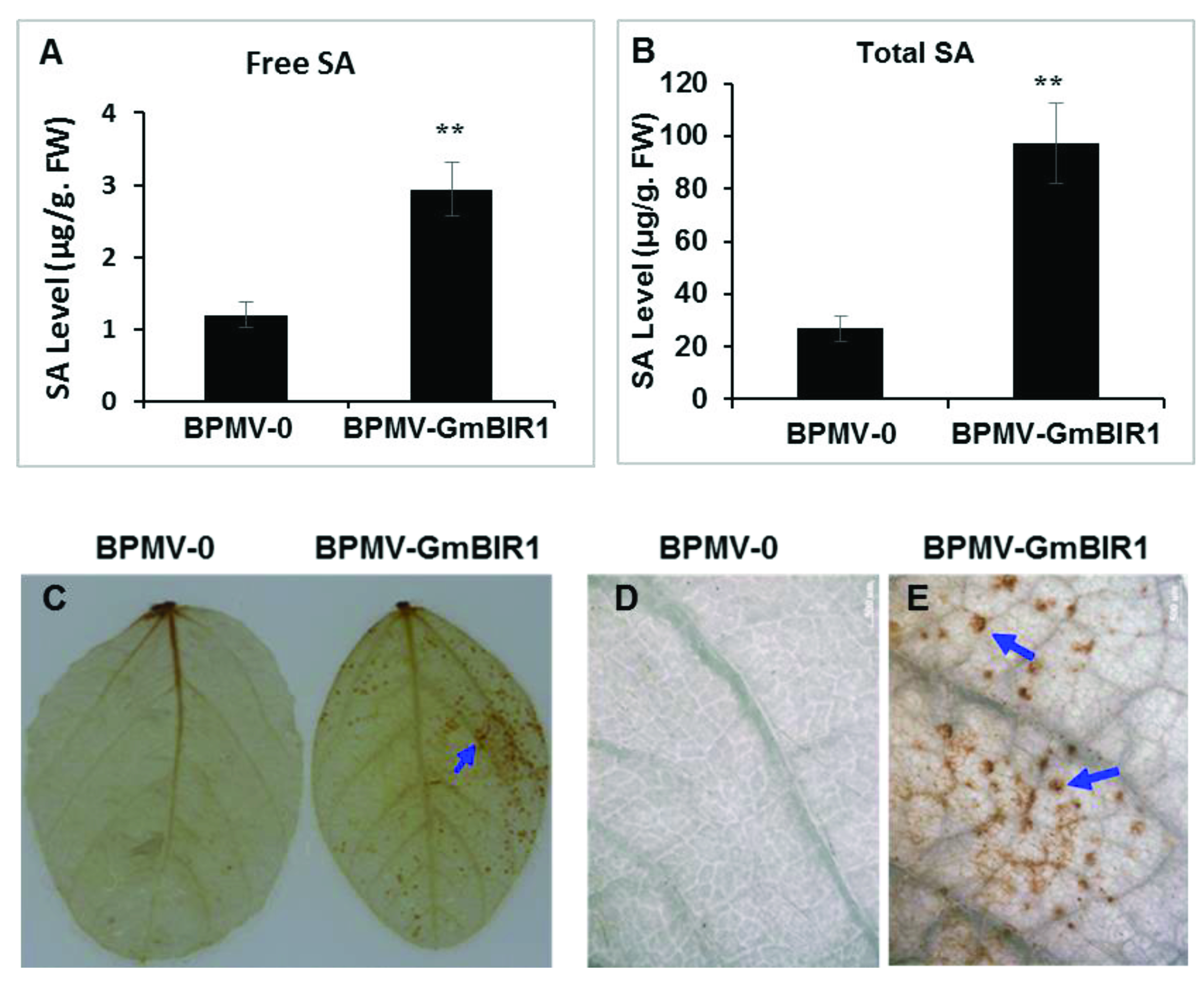
2.2. Both SA and H2O2 Are Over-Accumulated in the GmBIR1-Silenced Plants

SA is a key hormone that positively regulates defense responses against biotrophic pathogens and is over-accumulated in the plants with activated defense responses [20,28]. To examine whether SA over-accumulation occurred in GmBIR1-silenced plants, both free SA and bound SA were quantified, and the levels of free SA and bound SA were 2.46- and 4.1-fold higher in the GmBIR1-silenced plants than in BPMV-0 control plants, respectively (Figure 2A,B). The over-accumulated level of SA suggests that SA is involved in the constitutively activated defense responses observed in GmBIR1-silenced plants.
H2O2 is a potent trigger of cell death [29,30] and H2O2 is over-accumulated in the plants with activated defense responses [20,23]. To examine if H2O2 is over-accumulated in the GmBIR1-silenced plants, the leaves collected from both vector control plants and the GmBIR1-silenced plants were stained with 3,3-diaminobenzidine (DAB) [31,32]. More intense brown spots were observed on the leaves of the GmBIR1-silenced plants compared to the BPMV-0 control plants (Figure 2C), indicating that the cell death observed in the GmBIR1-silenced plants was correlated with increased level of H2O2 accumulation.

2.3. Silencing GmBIR1 Enhances Resistance against SMV and Pseudomonas syringae pv. glycinea (Psg), R4 Strain

Activated defense responses are usually correlated with enhanced disease resistance [20,21,22,23,24,25]. To examine whether disease resistance is enhanced in GmBIR1-silenced plants, SMV strain N tagged with the GUS marker protein (SMV-N-GUS) [33] was inoculated via biolistic bombardment onto three individual leaves collected from both the vector control plants and the GmBIR1-silenced plants. Both the size of the GUS foci and the intensity of GUS staining reflect viral infectivity. At 5 days post inoculation (dpi), the SMV-N-GUS infection was visualized by GUS staining. The GUS infection foci were easily seen on the vector control leaves (Figure 3A, left panels; Figure 3B). However, the GUS spots on the GmBIR1-silenced plants were much smaller and only visible under a dissecting microscopy (Figure 3C). The average diameter of the GUS foci formed on the vector control leaves was four-fold of those formed on the GmBIR1-silenced leaves (Figure 3D). This result indicates that silencing GmBIR1 leads to increased resistance against SMV.
We reported that silencing GmFLS2 compromises soybean resistance against Pseudomonas syringae pv. glycinea (Psg) R4 [24]. To test the effect of GmBIR1 silencing on Psg infection, we inoculated the BPMV-0 control plants and the GmBIR1-silenced plants with Psg. At 4- and 6-dpi, the colony forming unit of Psg on the vector control leaves was significantly higher than on that of the GmBIR1-silenced leaves (Figure 4). Together, these results clearly show that silencing GmBIR1 enhances resistance of soybean plants against two totally different types of pathogens, SMV and Psg.

2.4. The Activated Defense Responses in GmBIR1-Silenced Plants Are Independent of GmMPK3/6 Activation

Activation of MAPK signaling pathway is one of the signature hallmarks of activated defense responses [34]. To test whether the activated MAPK signaling pathway contributes to the activated defense responses and enhanced disease resistance observed in GmBIR1-silenced plants (Figure 1, Figure 2, Figure 3 and Figure 4), the activation of GmMPK3 and GmMPK6 in response to flg22 was examined both in vector control plants and GmBIR1-silenced plants using the Phospho-p44/42 MAP Erk1/2 antibody that recognizes phosphorylation of Arabidopsis and soybean MPK3/4/6 at the sites corresponding to threonine (T) 202/tyrosine (Y) 204 of human (Homo sapiens) p44 MAPK [23,24,35]. Contrary to our expectation, activation of both GmMPK3 and GmMPK6 by flg22 was significantly reduced in GmBIR1-silenced plants compared to vector control plants (Figure 4), indicating that the activated defense responses observed in the GmBIR1-silenced plants are independent of GmMPK3/6 activation.

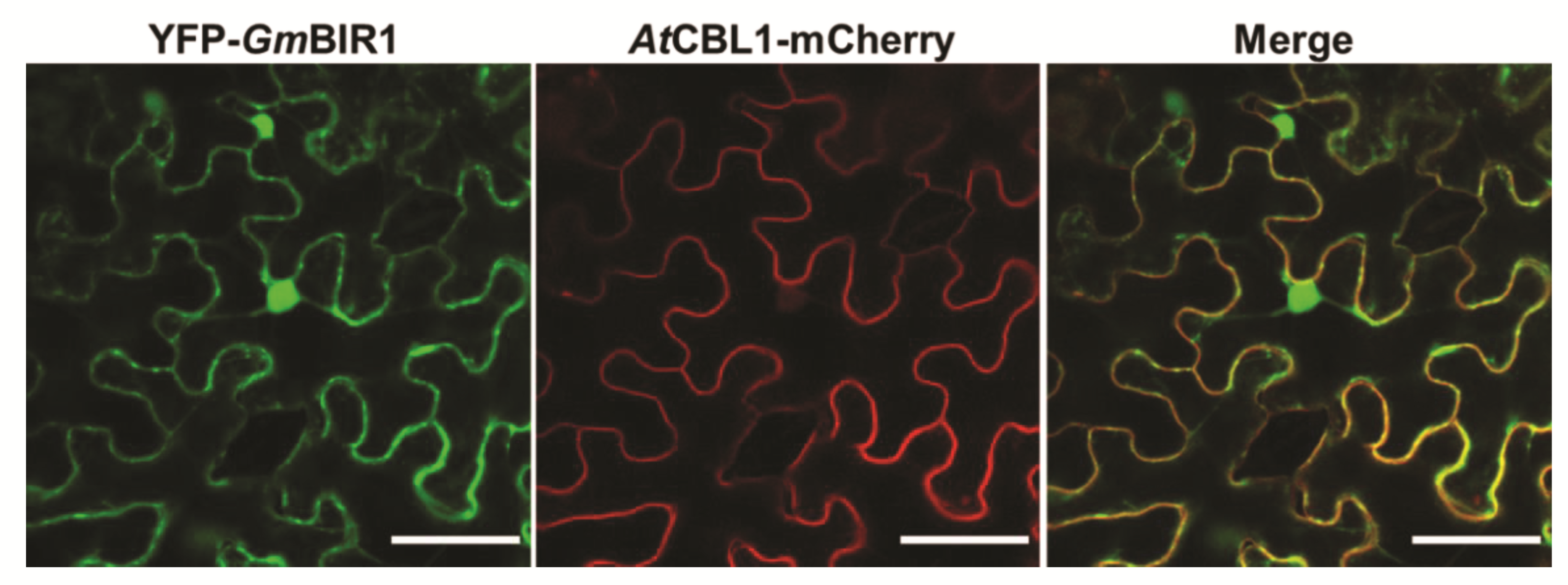
2.5. GmBIR1 Is Localized at the Plasma Membrane (PM) and in the Nucleus

RLKs normally function at the PM. To investigate the subcellular location of GmBIR1, the full-length GmBIR1a was fused to the C-terminus of GFP and transiently co-expressed in N. benthamiana leaves with a PM marker, AtCBL1-mCherry, via agroinfiltration. As shown in Figure 5 and Figure 6, GFP-GmBIR1 was not only co-localized with AtCBL1-mCherry but also presented in the nucleus, indicating that GmBIR1 has dual subcellular localizations at the PM and in the nucleus.

3. Discussion

3.1. The Function of BIR1 Homologs Is Conserved from Different Plant Species

It has been reported that the extensive cell death, induction of PR genes, enhanced accumulation of SA and H2O2, impaired activation of MPK4, and enhanced resistance to different pathogens were observed for Arabidopsis bir1-1 mutant [14,15,36,37,38]. Similarly, spontaneous cell death, constitutively activated autoimmune responses (including induced PR gene expression), accumulated levels of both SA and H2O2, and enhanced disease resistance to both viral and bacterial pathogens were also observed for the GmBIR1-silenced soybean plants (Figure 1, Figure 2 and Figure 3). The phenotypic reminiscence between bir1-1 mutant and the GmBIR1-silenced plants strongly indicate that the functions of BIR1 homologs are highly conserved across plant species in negatively regulating cell death and immunity. Given that cell death occurs in bir1-1 mutants even under sterile conditions, BIR1 may not function in perception of PAPMs (pathogen-associated molecular patterns).

3.2. The Constitutively Activated Cell Death and Defense Responses in GmBIR1-Silenced Plants Is A Result of Co-Silencing of Both GmBIR1a and GmBIR1b

Soybean is a paleopolyploid and 75% of genes in its genome contain at least two copies [39]. Consistent with this, there are two GmBIR1 genes, GmBIR1a and GmBIR1b, in the soybean genome. The open reading frame sequences of GmBIR1a and GmBIR1b share 95% identity at the nucleotide level. The DNA fragment used for GmBIR1 silencing contains four stretches of sequences with 100% identity between GmBIR1a and GmBIR1b that could potentially generate siRNAs of >20 nt in length (Supplementary Figure S1). Therefore, both GmBIR1a and GmBIR1b were silenced using the same construct (Figure 1G). The autoimmune phenotype of the GmBIR1-silenced plants (Figure 1B) actually is a result of co-silencing of both GmBIR1a and GmBIR1b. We have previously shown that multiple copies of GmMPK4, GmMPK6, GmMEKK1, GmFLS2, and GmATG2 genes could be successfully co-silenced using a single BPMV-VIGS construct [20,22,23,24,25]. Given that soybean is polyploidy and is recalcitrant to transformation, VIGS is currently one of the most efficient approaches to interrogate gene functions in soybean [26,27].

3.3. Does the Autoimmunity Observed in GmBIR1-Silenced Plants Result from Activated PTI or Activated ETI?

Activation of either PTI or ETI results in autoimmune responses, although the autoimmune responses resulting from ETI are much stronger and last longer than those resulting from PTI [1]. Structure analysis indicates that Arabidopsis BIR1 and BAK1 directly interact with each other through their ecto-LRR domains [36]. Under no-elicited conditions, BIR1LRR–BAK1LRR interaction sequesters BAK1 and prevents formation of a signal competent complex with FLS2. In the presence of flg22, flg22-bound FLS2LRR outcompetes BIR1LRR for binding to BAK1LRR from the BIR1LRR–BAK1LRR complex and forms a signaling competent flg22–FLS2LRR–BAK1LRR complex [36] Based on this structural study, BIR1 functions to keep BAK1-mediated cell death signaling under tight control, thus preventing undesired autoimmunity, and therefore, it is possible that the autoimmunity in bir1-1 or GmBIR1-silenced plants is a result of activation of BAK1-mediated PTI. Our Psg infection assay supported this possibility (Figure 4). However, inconsistent with this possibility, the flg22-induced activation of GmMPK3 and GmMPK6, which is a hallmark event of activated PTI (Meng et al., 2013), was significantly reduced in GmBIR1-silenced plants relative to vector control plants (Figure 4), and the activation of MPK3 and MPK6 in the Arabidopsis bir1-1 mutant is unchanged compared to WT Col-0 [14]. Therefore, it is still unclear whether the autoimmune responses observed in bir1-1 or GmBIR1-silenced plants are solely due to activated PTI, reflecting the complexity of the involvement of BIR1 in plant immunity.
Several lines of evidence indicate that autoimmune responses in bir1-1 or the GmBIR1-silenced plants could result from misactivation of R proteins. Firstly, loss function of BIR1 in Arabidopsis leads to the impairment in the activation of MPK4 by flagellin [14]. The autoimmune phenotype of bir1-1 could be the consequence of the impaired MPK4 activation given that Arabidopsis MEKK1-MKK1/2-MPK4 signaling module negatively regulates the activation of the R proteins SUMM2 and SMN1/RPS6 [40,41,42]. Misactivation of SUMM2 and SMN1/RPS6 result from mutations in MEKK1-MKK1/2-MPK4 pathway leads to autoimmune responses [40,42]. However, GmMPK4 activity cannot be induced either by flg22 or SA, regardless of concentration and duration of treatments [23]. Therefore, it is unclear whether GmMPK4 plays a similar role as its counterpart in Arabidopsis. As the GmMPK4 activation is also not observed in the vector control plants (Figure 4 and [23]), it is unlikely that the GmBIR1-silenced phenotypes are related to GmMPK4 function. Secondly, in Arabidopsis thaliana, the copain-like protein BONZAI1 (BON1) interacts physically with and phosphorylates BIR1 and bir1-1 mutant is partially suppressed by loss of function mutation in the resistance gene SNC1 [18]. Thirdly, the autoimmune phenotype of bir1-1 can be partially suppressed by elevated temperatures [18], which is a characteristic feature of R protein-mediated responses [43,44]. Lastly, the autoimmune phenotype of bir1-1 is rescued by the loss of function of EDS1 and PAD4 [14], two key players in TIR type of R protein activation [45]. These results strongly suggest that the autoimmune phenotype of the bir1-1 mutant is a result of misactivated R protein(s). Therefore, it is possible that BIR1 might participate in both PTI and ETI through interacting with different proteins, and the autoimmune responses observed in bir1-1 and GmBIR1-silenced plants could be combined effects of activated PTI and ETI. The facts that RLKs/RLPs are required for NLR-mediated immunity [46] and RLKs/RLPs and NLRs mutually potentiate each other [47] support the idea that BIR1 may participate in both PTI and ETI.

3.4. Is the Enhanced Resistance of GmBIR1-Silenced plants to SMV SA-Dependent?

The enhanced resistance of GmBIR1-silenced plants against Psg could be attributed to the increased accumulation of both SA and H2O2 (Figure 2) and is thus SA-dependent. The enhanced SMV resistance in GmBIR1-silenced plants may not be solely due to increased accumulation of both SA and H2O2. In Arabidopsis, Guzman-Benito et al. [38] reported that the enhanced resistance of bir1-1 to Tobacco rattle virus (TRV) is independent of cell death or SA defense priming. The expression of Arabidopsis BIR1 is regulated both at TGS and PTGS level by RNA silencing pathway [38]. A threshold level of BIR1 expression must be properly maintained and an expression beyond the threshold level (either too high or too low) triggers autoimmunity. Whether the enhanced resistance of GmBIR1-silenced plants to SMV is uncoupled from the cell death needs to be further investigated.

4. Materials and Methods

4.1. Plant Materials

Seeds of soybean (Glycine max ‘Williams 82’) used in this study were harvested from greenhouse-grown plants previously indexed for the absence of BPMV and SMV [48,49]. Soybean plants were grown in a growth chamber at 22 degrees with a photoperiod of 16 h.

4.2. Flg22 Peptides

The flg22 peptide was synthesized by HuaBio (Hangzhou, China).

4.3. BPMV-Mediated VIGS

BPMV strains, BPMV VIGS constructs, and inoculation of soybean seedlings with DNA-based BPMV constructs via biolistic particle bombardments using a Biolistic PDS-1000/He system (Bio-Rad Laboratories, Hercules, CA, USA) have been described previously [48]. The orthologs of ArabidopsisBIR1 in the soybean genome were identified by BLASTN in phytozome databases. The primers used for construction of the BPMV-GmBIR1 (Glyma.18g246400.1) are:
  • GmBIR1-F: 5′-aagGGATCCCTCACTGGTCAAATTCCTGCTA-3′
  • GmBIR1-R: 5′-ttgGGTACCCAGGGTCCTCTTCCTTCTTCCTA-3′
The underlined sequences are BamH I and Kpn I restriction sites, respectively, which were used for directional cloning of the PCR fragment into the BPMV VIGS (IA-D35). The letter C in bold indicates an extra nucleotide in reverse primers needed to maintain the correct open reading frame with BPMV genome.

4.4. RNA Isolation and RT-PCR

RNA isolation and RT-PCR were performed as described elsewhere [50]. The RNA samples extracted were treated with DNaseI according to the manufacturer’s instructions (Invitrogen, Carlsbad, CA, USA). Primers used for RT-PCR in this study are:
  • GmELF1b-F: 5′- ACCGAAGAGGGCATCAAATCCC -3′
  • GmELF1b-R: 5′- CTCAACTGTCAAGCGTTCCTC -3′
  • GmBIR1-full-F: 5′- ATAGGAAGAAGGAAGAGGACC -3′
  • GmBIR1-full-R: 5′- TTTAGGAGTAGCCACCAAAGT -3′
  • GmBIR1a-F: 5′- GACTTGCTCAATATCCCAGGATAAGT-3′
  • GmBIR1a-R: 5′- TGTTGAGATTCCTGTAATCTCTTAACCAT -3′
  • GmBIR1b-F: 5′- TACTTCCTCAATATCCCAGGTGG -3′
  • GmBIR1b-R: 5′- TATTGAGATTCCTGCAATCTCTTGAC -3′.
The GmBIR1a-full primers were used for amplifying the full-length cDNA and the GmBIR1a and GmBIR1b primers are used for determining the specific transcript levels of the GmBIR1a and GmBIR1b, respectively.

4.5. Trypan Blue Staining

Cell death was detected specifically by trypan blue staining solution as described by [18]. At 18 dpi with BPMV vector only (BPMV-0) or BPMV-GmBIR1 constructs, second fully expanded soybean trifoliolate leaves counting from the top were detached. The detached leaves were placed in trypan blue staining solution (10 mL glycerol, 10 mL lactic acid, 10 mL water, 10 mg trypan blue powder and 10 g phenol) and boiled for 1–1.5 min. The stained leaves were cleared using tap water and then dipped in the chloral hydrate solution in the shaker overnight to remove the non-specific stains. Images were captured using a stereo microscope (Olympus SZH10, Center Valley, PA, USA).

4.6. SA Quantification

SA was quantified using an Agilent 1260 HPLC system (Agilent Technologies) with a diode array detector and a fluorescence detector and a column as described previously [51]. The column was a 4.6-by-75-mm Zorbax SB-C18 with a mobile phase comprising sodium acetate (0.2 m, pH 5.5) and methanol with a series of concentration gradients. The initial methanol gradient was maintained at 3% (v/v) for 12 min, linearly increased to 7% (v/v) at 12.5 min and maintained until 38 min at a flow rate of 0.8 mL min−1 throughout the process. After 1 min, the initial reaction conditions were restored and the system needed to be equilibrated for 7 min before the next injection. Total SA from the sample treated with β-Glucosidase and free SA were detected with a 296 nm excitation wavelength and 410 nm emission wavelength. The concentration was calculated by determining the HPLC peak area according to a standard curve.

4.7. H2O2 Detection by DAB Staining

H2O2 was detected by an endogenous peroxidase-dependent in situ histochemical staining procedure using DAB (Sigma-Aldrich, St. Louis, MO, USA) [32]. Detached leaves were placed in a solution containing 1 mg mL−1 DAB (pH 5.5) for 2 h. The leaves were cleared by boiling in 96% (v/v) ethanol for 10 min. H2O2 production was visualized as a reddish-brown precipitate on the cleared leaves [32].

4.8. Inoculation of Pseudomonas Syringae pv. Glycinea (Psg) onto the Leaves of Soybean Plants

Psg growth assay was performed as described by [24]. Psg was cultivated at 28 degrees until OD = 1.3. The bacterial culture was centrifuged at 3000 rpm for 10 min and the pellet was re-suspended in double-distilled water with an OD = 1. The vector control or GmBIR1-silenced plants were thoroughly and evenly sprayed with the re-suspended bacterial solution on both upsides and downsides of the leaves. The sprayed plants were immediately covered with transparent plastic bags for at least 24 h.

4.9. SMV-N-GUS Inoculation, GUS Staining, and GUS Foci Measurements

At 18 dpi with BPMV vector only (BPMV-0) or BPMV-GmBIR1 constructs, second fully expanded soybean trifoliolate leaves counting from the top were detached and biolistically inoculated with SMV-N-GUS [33,48]. Following SMV-N-GUS inoculation, the detached leaves were put into petri dishes with moist filter papers and kept on a lighted growth shelf for 5 days before GUS staining. GUS staining was performed as described by [52]. Photographs of the leaves with GUS foci were taken using a stereo microscope (Olympus SZH10, Center Valley, PA, USA). The numbers of GUS foci were counted, and the diameters of GUS foci were measured using Soft Image System analysis (IA Package; Olympus).

4.10. Western Blot Analysis for Detecting Phosphorylated MPKs

Protein was extracted from soybean leaf tissues using extraction buffer (50 mM Tris-MES pH 8.0, 0.5 M sucrose, 1 mM MgCl2, 10 mM EDTA, 5 mM DTT) with protease inhibitor cocktail S8830 (Sigma-Aldrich, St. Louise, MO, USA) added as described by [23]. The extract was centrifuged at 12,000 rpm at 4 °C for 30 min and the supernatant was collected. For immunoblotting, the extracted proteins were separated by SDS-PAGE (10% acrylamide gel) and transferred to PVDF membrane (Millipore, Billerica, MA, USA) by semi-dry electro-transfer (Bio-Rad, Hercules, CA, USA). The membrane was blocked in 1× TBST buffer containing 5% milk powder for 2 h. After washing, the membrane was further incubated with anti–Phospho-p44/p42 MAPK (anti-pTEpY) diluted at 1:3000 (Cell Signaling Technology, Danvers, MA, USA), followed by incubation with secondary antibody diluted at 1:7500. The bands were visualized by incubating with chemiluminescent HRP substrate (Millipore, Billerica, MA, USA).

4.11. Subcellular Localization of GmBIR1

The GmBIR1 (Glyma.18g246400.1) open reading frame was amplified by RT-PCR from total RNA extracted from cv Williams 82 soybean plants using the following primers:
  • GmBIR1-PGDG-F-HinDIII: 5′-AAGCTTATGAAGATGTTTATGGGTGGC-3′
  • GmBIR1-PGDG-R-BamHI: 5′-GGATCCTCAATCATGTCCCTCTCGAG-3′
The letters in bold represent the cleavage sites for different restriction enzymes.
The RT-PCR products were firstly double digested with HindIII + BamHI and subsequently cloned into the PGDG vector [53] to generate the GFP-GmBIR1 fusion construct. These fusion constructs and the AtCBL1-mCherry construct (membrane marker) were coinfiltrated into N. benthamiana leaves as described by [50]. Images were captured with a confocal laser-scanning microscope (Leica TCS SP5 AOBS, Wetzlar, Germany).

Supplementary Materials

The following supporting information can be downloaded at: https://www.mdpi.com/article/10.3390/ijms23137450/s1.

Author Contributions

J.-Z.L. designed the experiments and wrote the original draft; D.-D.L., H.-J.L., H.S.M., M.-Y.Y., X.-Y.D., C.-L.Z. and S.-N.T. performed the experiments; J.-Z.L. and D.-D.L. prepared figures. All authors have read and agreed to the published version of the manuscript.

Funding

This research was funded by the National Natural Science Foundation of China (grant numbers 32170761 and 31571423 to J.Z.L.).

Informed Consent Statement

No applicable.

Data Availability Statement

No applicable.

Acknowledgments

We thank Steven A. Whitham and John Hill for providing the BPMV-VIGS system, Pseudomonas syringae pv. glycinea R4 strain and SMV-N-GUS construct.

Conflicts of Interest

The authors declare no conflict of interest.

References

  1. Jones, J.D.; Dangl, J.L. The plant immune system. Nature 2006, 444, 323–329. [Google Scholar] [CrossRef] [PubMed] [Green Version]
  2. Shiu, S.H.; Bleecker, A.B. Receptor-like kinases from Arabidopsis form a monophyletic gene family related to animal receptor kinases. Proc. Natl. Acad. Sci. USA 2001, 98, 10763–10768. [Google Scholar] [CrossRef] [PubMed] [Green Version]
  3. Jamieson, P.A.; Shan, L.; He, P. Plant cell surface molecular cypher: Receptor-like proteins and their roles in immunity and development. Plant Sci. 2018, 274, 242–251. [Google Scholar] [CrossRef] [PubMed]
  4. Boller, T.; Felix, G. A renaissance of elicitors: Perception of microbe-associated molecular patterns and danger signals by pattern-recognition receptors. Annu. Rev. Plant Biol. 2009, 60, 379–406. [Google Scholar] [CrossRef] [PubMed]
  5. Gómez-Gómez, L.; Boller, T. FLS2: An LRR Receptor–like Kinase Involved in the Perception of the Bacterial Elicitor Flagellin in Arabidopsis. Mol. Cell 2009, 5, 1003–1011. [Google Scholar] [CrossRef]
  6. Zipfel, C.; Kunze, G.; Chinchilla, D.; Caniard, A.; Jones, J.D.; Boller, T.; Felix, G. Perception of the Bacterial PAMP EF-Tu by the Receptor EFR Restricts Agrobacterium-Mediated Transformation. Cell 2006, 125, 749–760. [Google Scholar] [CrossRef]
  7. Li, J.; Wen, J.; Lease, K.A.; Doke, J.T.; Tax, F.E.; Walker, J.C. BAK1, an Arabidopsis LRR Receptor-like Protein Kinase, Interacts with BRI1 and Modulates Brassinosteroid Signaling. Cell 2002, 110, 213–222. [Google Scholar] [CrossRef] [Green Version]
  8. Nam, K.H.; Li, J. BRI1/BAK1, a Receptor Kinase Pair Mediating Brassinosteroid Signaling. Cell 2002, 110, 203–212. [Google Scholar] [CrossRef] [Green Version]
  9. Chinchilla, D.; Zipfel, C.; Robatzek, S.; Kemmerling, B.; Nürnberger, T.; Jones, J.D.G.; Felix, G.; Boller, T. A flagellin-induced complex of the receptor FLS2 and BAK1 initiates plant defence. Nature 2007, 448, 497–500. [Google Scholar] [CrossRef]
  10. Heese, A.; Hann, D.R.; Gimenez-Ibanez, S.; Jones, A.M.; He, K.; Li, J.; Schroeder, J.I.; Peck, S.C.; Rathjen, J.P. The receptor-like kinase SERK3/BAK1 is a central regulator of innate immunity in plants. Proc. Natl. Acad. Sci. USA 2007, 104, 12217–12222. [Google Scholar] [CrossRef] [Green Version]
  11. Roux, M.; Schwessinger, B.; Albrecht, C.; Chinchilla, D.; Jones, A.; Holton, N.; Malinovsky, F.G.; Tör, M.; de Vries, S.; Zipfel, C. The Arabidopsis leucine-rich repeat receptor-like kinase BAK1/SERK3 and BKK1/SERK4 are required for the innate immunity to hemibiotrophic and biotrophic pathogens. Plant Cell 2011, 23, 2440–2455. [Google Scholar] [CrossRef] [PubMed] [Green Version]
  12. Macho, A.P.; Schwessinger, B.; Ntoukakis, V.; Brutus, A.; Segonzac, C.; Roy, S.; Kadota, Y.; Oh, M.H.; Sklenar, J.; Derbyshire, P.; et al. A bacterial tyrosine phosphatase inhibits plant pattern recognition receptor activation. Science 2014, 343, 1509–1512. [Google Scholar] [CrossRef] [PubMed]
  13. Couto, D.; Zipfel, C. Regulation of pattern recognition receptor signalling in plants. Nat. Rev. Immunol. 2016, 16, 537–552. [Google Scholar] [CrossRef] [PubMed]
  14. Gao, M.; Wang, X.; Wang, D.; Xu, F.; Ding, X.; Zhang, Z.; Bi, D.; Cheng, Y.T.; Chen, S.; Li, X.; et al. Regulation of cell death and innate immunity by two receptor-like kinases in Arabidopsis. Cell Host Microbe 2009, 6, 34–44. [Google Scholar] [CrossRef] [PubMed] [Green Version]
  15. Liu, Y.; Huang, X.; Li, M.; He, P.; Zhang, Y. Loss-of-function of Arabidopsis receptor-like kinase BIR1 activates cell death and defense responses mediated by BAK1 and SOBIR1. New Phytol. 2016, 212, 637–645. [Google Scholar] [CrossRef] [PubMed] [Green Version]
  16. Liebrand, T.W.; van den Berg, G.C.; Zhang, Z.; Smit, P.; Cordewener, J.H.; America, A.H.; Sklenar, J.; Jones, A.M.; Tameling, W.I.; Robatzek, S.; et al. Receptor-like kinase SOBIR1/EVR interacts with receptor-like proteins in plant immunity against fungal infection. Proc. Natl. Acad. Sci. USA 2013, 110, 10010–10015. [Google Scholar] [CrossRef] [Green Version]
  17. Liebrand, T.W.; van den Burg, H.A.; Joosten, M.H. Two for all: Receptor-associated kinases SOBIR1 and BAK1. Trends Plant Sci. 2014, 19, 123–132. [Google Scholar] [CrossRef]
  18. Wang, Z.; Meng, P.; Zhang, X.; Ren, D.; Yang, S. BON1 interacts with the protein kinases BIR1 and BAK1 in modulation of temperature-dependent plant growth and cell death in Arabidopsis. Plant J. 2011, 67, 1081–1093. [Google Scholar] [CrossRef]
  19. Halter, T.; Imkampe, J.; Mazzotta, S.; Wierzba, M.; Postel, S.; Bücherl, C.; Kiefer, C.; Stahl, M.; Chinchilla, D.; Wang, X.; et al. The leucine-rich repeat receptor kinase BIR2 is a negative regulator of BAK1 in plant immunity. Curr. Biol. 2014, 24, 134–143. [Google Scholar] [CrossRef] [Green Version]
  20. Liu, J.Z.; Horstman, H.D.; Braun, E.; Graham, M.A.; Zhang, C.; Navarre, D.; Qiu, W.L.; Lee, Y.; Nettleton, D.; Hill, J.H.; et al. Soybean homologs of MPK4 negatively regulate defense responses and positively regulate growth and development. Plant Physiol. 2011, 157, 1363–1378. [Google Scholar] [CrossRef] [Green Version]
  21. Liu, J.Z.; Whitham, S.A. Overexpresson of a soybean nuclear-localized type III DnaJ domain-containing HSP40 reveals it roles in cell death and disease resistance. Plant J. 2013, 74, 110–121. [Google Scholar] [CrossRef] [PubMed]
  22. Liu, J.Z.; Braun, E.; Qiu, W.L.; Shi, Y.F.; Marcelino-Guimarães, F.C.; Navarre, D.; Hill, J.H.; Whitham, S.A. Positive and negative roles for soybean MPK6 in regulating defense responses. Mol. Plant Microbe Interact. 2014, 27, 824–834. [Google Scholar] [CrossRef] [PubMed] [Green Version]
  23. Xu, H.; Zhang, C.; Li, Z.C.; Wan, Z.R.; Jiang, X.X.; Tian, S.N.; Braun, E.; Mei, Y.; Qiu, W.L.; Li, S.; et al. The MAPK kinase kinase GmMEKK1 regulates cell death and defense responses. Plant Physiol. 2018, 178, 907–922. [Google Scholar] [CrossRef] [PubMed] [Green Version]
  24. Tian, S.N.; Liu, D.D.; Zhong, C.L.; Xu, H.Y.; Yang, S.; Fang, Y.; Ran, J.; Liu, J.Z. Silencing GmFLS2 enhances the susceptibility of soybean to bacterial pathogen through attenuating the activation of GmMAPK signaling pathway. Plant Sci. 2019, 292, 110386. [Google Scholar] [CrossRef]
  25. Hashimi, S.M.; Wu, N.N.; Ran, J.; Liu, J.Z. Silencing Autophagy-Related Gene 2 (ATG2) Results in Accelerated Senescence and Enhanced Immunity in Soybean. Int. J. Mol. Sci. 2021, 22, 11749. [Google Scholar] [CrossRef]
  26. Liu, J.Z.; Graham, M.A.; Pedley, K.F.; Whitham, S.A. Gaining insight into soybean defense responses using functional genomics approaches. Brief. Funct. Genom. 2015, 14, 283–290. [Google Scholar] [CrossRef] [Green Version]
  27. Liu, J.Z.; Fang, Y.; Pang, H. The current status of the soybean-soybean mosaic virus (SMV) pathosystem. Front. Microbiol. 2016, 7, 1906. [Google Scholar] [CrossRef]
  28. Petersen, M.; Brodersen, P.; Naested, H.; Andreasson, E.; Lindhart, U.; Johansen, B.; Nielsen, H.B.; Lacy, M.; Austin, M.J.; Parker, J.E.; et al. Arabidopsis map kinase 4 negatively regulates systemic acquired resistance. Cell 2000, 103, 1111–1120. [Google Scholar] [CrossRef] [Green Version]
  29. Lamb, C.; Dixon, R.A. The oxidative burst in plant disease resistance. Annu. Rev. Plant Physiol. Plant Mol. Biol. 1997, 48, 251–275. [Google Scholar] [CrossRef]
  30. Delledonne, M.; Xia, Y.; Dixon, R.A.; Lamb, C. Nitric oxide functions as a signal in plant disease resistance. Nature 1998, 394, 585–588. [Google Scholar] [CrossRef]
  31. Thordal-Christensen, H.; Zhang, Z.G.; Wei, Y.D.; Collinge, D.B. Subcellular localization of H2O2 in plants: H2O2 accumulation in papillae and hypersensitive response during the barley-powdery mildew interaction. Plant J. 1997, 11, 1187–1194. [Google Scholar] [CrossRef]
  32. Ren, D.; Yang, H.; Zhang, S. Cell death mediated by MAPK is associated with hydrogen peroxide production in Arabidopsis. J. Biol. Chem. 2002, 277, 559–565. [Google Scholar] [CrossRef] [PubMed] [Green Version]
  33. Wang, L.; Eggenberger, A.; Hill, J.; Bogdanove, A.J. Pseudomonas syringae effector avrB confers soybean cultivar-specific avirulence on Soybean mosaic virus adapted for transgene expression but effector avrPto does not. Mol. Plant Microbe Interact. 2006, 19, 304–312. [Google Scholar] [CrossRef] [PubMed] [Green Version]
  34. Meng, X.; Zhang, S. MAPK cascades in plant disease resistance signaling. Annu. Rev. Phytopathol. 2013, 51, 245–266. [Google Scholar] [CrossRef]
  35. Zhao, C.; Nie, H.; Shen, Q.; Zhang, S.; Lukowitz, W.; Tang, D. EDR1 physically interacts with MKK4/MKK5 and negatively regulates a MAP kinase cascade to modulate plant innate immunity. PLoS Genet. 2014, 10, e1004389. [Google Scholar] [CrossRef]
  36. Ma, C.; Liu, Y.; Bai, B.; Han, Z.; Tang, J.; Zhang, H.; Yaghmaiean, H.; Zhang, Y.; Chai, J. Structural basis for BIR1-mediated negative regulation of plant immunity. Cell Res. 2017, 27, 1521–1524. [Google Scholar] [CrossRef] [Green Version]
  37. Wierzba, M.P.; Tax, F.E. An allelic series of bak1 mutations differentially alter bir1 cell death, immune response, growth, and root development phenotypes in Arabidopsis thaliana. Genetics 2016, 202, 689–702. [Google Scholar] [CrossRef] [Green Version]
  38. Guzmán-Benito, I.; Donaire, L.; Amorim-Silva, V.; Vallarino, J.G.; Esteban, A.; Wierzbicki, A.T.; Ruiz-Ferrer, V.; Llave, C. The immune repressor BIR1 contributes to antiviral defense and undergoes transcriptional and post-transcriptional regulation during viral infections. New Phytol. 2019, 224, 421–438. [Google Scholar] [CrossRef]
  39. Schmutz, J.; Cannon, S.B.; Schlueter, J.; Ma, J.; Mitros, T.; Nelson, W.; Hyten, D.L.; Song, Q.; Thelen, J.J.; Cheng, J. Genome sequence of the palaeopolyploid soybean. Nature 2010, 463, 178–183. [Google Scholar] [CrossRef] [Green Version]
  40. Zhang, Z.; Wu, Y.; Gao, M.; Zhang, J.; Kong, Q.; Liu, Y.; Ba, H.; Zhou, J.; Zhang, Y. Disruption of PAMP-induced MAP kinase cascade by a Pseudomonas syringae effector activates plant immunity mediated by the NB-LRR protein SUMM2. Cell Host Microbe 2012, 11, 253–263. [Google Scholar] [CrossRef] [Green Version]
  41. Kong, Q.; Qu, N.; Gao, M.; Zhang, Z.; Ding, X.; Yang, F.; Li, Y.; Dong, O.X.; Chen, S.; Li, X.; et al. The MEKK1-MKK1/MKK2-MPK4 kinase cascade negatively regulates immunity mediated by a mitogen-activated protein kinase kinase kinase in Arabidopsis. Plant Cell 2012, 24, 2225–2236. [Google Scholar] [CrossRef] [PubMed] [Green Version]
  42. Takagi, M.; Hamano, K.; Takagi, H.; Morimoto, T.; Akimitsu, K.; Terauchi, R.; Shirasu, K.; Ichimura, K. Disruption of the MAMP-Induced MEKK1-MKK1/MKK2-MPK4 Pathway Activates the TNL Immune Receptor SMN1/RPS6. Plant Cell Physiol. 2019, 60, 778–787. [Google Scholar] [CrossRef] [PubMed]
  43. Whitham, S.; Dinesh-Kumar, S.P.; Choi, D.; Hehl, R.; Corr, C.; Baker, B. The product of the tobacco mosaic virus resistance gene N: Similarity to toll and the interleukin-1 receptor. Cell 1994, 78, 1101–1115. [Google Scholar] [CrossRef]
  44. Yang, S.; Hua, J. A haplotype-specific Resistance gene regulated by BONZAI1 mediates temperature-dependent growth control in Arabidopsis. Plant Cell 2004, 16, 1060–1071. [Google Scholar] [CrossRef] [Green Version]
  45. Wiermer, M.; Feys, B.J.; Parker, J.E. Plant immunity: EDS1 regulatory node. Curr. Opin. Plant Biol. 2005, 8, 383–389. [Google Scholar] [CrossRef] [PubMed] [Green Version]
  46. Yuan, M.; Jiang, Z.; Bi, G.; Nomura, K.; Liu, M.; Wang, Y.; Cai, B.; Zhou, J.M.; He, S.Y.; Xin, X.F. Pattern-recognition receptors are required for NLR-mediated plant immunity. Nature 2021, 592, 105–109. [Google Scholar] [CrossRef]
  47. Ngou, B.P.M.; Ahn, H.K.; Ding, P.; Jones, J.D.G. Mutual potentiation of plant immunity by cell-surface and intracellular receptors. Nature 2021, 592, 110–115. [Google Scholar] [CrossRef]
  48. Zhang, C.; Yang, C.; Whitham, S.A.; Hill, J.H. Development and use of an efficient DNA-based viral gene silencing vector for soybean. Mol. Plant Microbe Interact. 2009, 22, 123–131. [Google Scholar] [CrossRef] [Green Version]
  49. Zhang, C.; Bradshaw, J.D.; Whitham, S.A.; Hill, J.H. The development of an efficient multipurpose bean pod mottle virus viral vector set for foreign gene expression and RNA silencing. Plant Physiol. 2010, 153, 52–65. [Google Scholar] [CrossRef] [Green Version]
  50. Liu, J.Z.; Blancaflor, E.B.; Nelson, R.S. The tobacco mosaic virus 126-kilodalton protein, a constituent of the virus replication complex, alone or within the complex aligns with and traffics along microfilaments. Plant Physiol. 2005, 138, 1853–1865. [Google Scholar] [CrossRef] [Green Version]
  51. Zhang, K.; Halitschke, R.; Yin, C.; Liu, C.J.; Gan, S.S. Salicylic acid 3-hydroxylase regulates Arabidopsis leaf longevity by mediating salicylic acid catabolism. Proc. Natl. Acad. Sci. USA 2013, 110, 14807–14812. [Google Scholar] [CrossRef] [PubMed] [Green Version]
  52. Jefferson, R.A.; Kavanagh, T.A.; Bevan, M.W. GUS fusions: Beta-glucuronidase as a sensitive and versatile gene fusion marker in higher plants. EMBO J. 1987, 6, 3901–3907. [Google Scholar] [CrossRef] [PubMed]
  53. Goodin, M.; Dietzgen, R.G.; Schichnes, D.; Ruzin, S.; Jackson, A.O. pGD vectors: Versatile tools for the expression of green and red fluorescent protein fusions in Agroinfiltrated plant leaves. Plant J. 2002, 31, 375–383. [Google Scholar] [CrossRef] [PubMed]
Figure 1. Silencing GmBIR1 causes both local and systemic hypersensitive response (HR) cell death in soybean. (A) The phenotypes of vector control (BPMV-0) and GmBIR1-silenced (BPMV-GmBIR1) plants at 15 days post infection (dpi); (B) Massive cell death was observed on the systemic leaves of GmBIR1-silenced plants at 25 dpi. Red arrows point to the dead leaves; (C,D) Trypan Blue-stained systemic leaves from vector control (C) and GmBIR1-silenced plants (D). Red arrows point to the dead regions. Bar = 500 μm; (E) RT-PCR result indicates that the GmBIR1 transcript level was reduced in GmBIR1-silenced plants relative to the vector control plants; (F) RT-PCR result indicates that the GmPR1 transcript level was increased in GmBIR1-silenced plants relative to the vector control plants; (G) RT-PCR result indicates that the GmBIR1a and GmBIR1b transcript level were both reduced in GmBIR1-silenced plants relative to the vector control plants. Each experiment was repeated at least twice. Each experiment consisted of 3 replicates and at least 3 plants were used in each replicate.
Ijms 23 07450 g001
Figure 2. Elevated accumulation of SA and H2O2 in GmBIR1-silenced plants. (A,B) Free SA (A) and Total SA (B) levels were quantified in GmBIR1-silenced and BPMV-0 empty vector control plants at 20 days post BPMV inoculation. Error bars represent SD for three independent samples. Asterisks indicate significant differences from the control (**, p < 0.01, Student’s t test). FW, fresh weight. (C) Presence of H2O2 in soybean leaves visualized by staining with DAB. (D,E) The images taken under the stereomicroscope from part of the regions shown in C, Bar = 500 μm. Oxidized DAB formed a reddish-brown deposit. (Examples of these deposits are indicated by the blue arrows).
Ijms 23 07450 g002
Figure 3. Silencing GmBIR1 enhances the resistance of soybean plants to SMV. At 18 dpi with BPMV-0 or BPMV-GmBIR1, SMV-N-GUS was biolistically delivered onto the detached leaves of silenced and non-silenced plants. At 5 dpi with SMV-N-GUS, the replication and movement of SMV-N-GUS in the biolistically inoculated leaves was detected by GUS staining. The GUS foci were counted and measured. (A) The distribution of SMV-N-GUS foci on the leaves of BPMV-0 and BPMV-GmBIR1 plants. (B,C) Infection foci of SMV-N-GUS on the leaves of BPMV-0 and BPMV-GmBIR1 plants. Bar = 2000 μm. (D) Comparison of the diameters of SMV-N-GUS foci on the leaves of BPMV-0 and BPMV-GmBIR1 plants. Error bars represent SD of the diameters of at least 30 GUS foci measured on each of three independent leaves (at least 90 foci). Asterisks indicate a significant difference from the control (***, p < 0.001, Student’s t test). This experiment was repeated 3 times with similar results.
Ijms 23 07450 g003
Figure 4. Silencing GmBIR1 enhanced the resistance of soybean plants to Pseudomonas syringae pv. glycinea (Psg) R4. (A) Comparison of the vector control plants and the GmBIR1-silenced plants 4 days after spraying Psg suspension; (B) Comparison of the leaves from the vector control plants and the GmBIR1-silenced plants 4 days after spraying Psg; (C) Comparison of the colony forming unit (cfu)between the vector control plants and the GmBIR1-silenced plants after different days of Psg infection. * and ** represent p < 0.05 and 0.01, respectively (Student’s t-test). This experiment was repeated 2 times with similar results.
Ijms 23 07450 g004
Figure 5. Silencing GmBIR1 reduces the activation of GmMPK3/GmMPK6 in response to flg22 treatment. Leaf discs from indicated soybean plants were incubated on moist filter paper for 24 h to allow recovery from wounding before being treated with 10 μM flg22 or diluted DMSO. The kinase activities were detected by Western blotting using Phosph-p44/p42 MAP Erk1/2 antibody. Coomassie Blue–stained gel (CBS) used as loading controls. The experiment was repeated 3 times with similar results.
Ijms 23 07450 g005
Figure 6. The subcelluar localization of GFP-GmBIR1. The GFP-GmBIR1 localizes both at plasma membrane (PM) and in the nucleus. The PM-localized protein AtCBL1-mCherry was used as a control. Bar = 50 μm. This experiment was repeated 3 times with similar results.
Ijms 23 07450 g006
Publisher’s Note: MDPI stays neutral with regard to jurisdictional claims in published maps and institutional affiliations.

