Review

Gal-3BP in Viral Infections: An Emerging Role in Severe Acute Respiratory Syndrome Coronavirus 2

by
Valentina Gallo
1,
Alyexandra Arienzo
2,
Stefano Iacobelli
3,
Valentina Iacobelli
3 and
Giovanni Antonini
1,2,*
1
Department of Sciences, Roma Tre University, Viale G. Marconi, 00146 Rome, Italy
2
Biostructures and Biosystems National Institute (INBB), Viale delle Medaglie d’Oro, 00136 Rome, Italy
3
MediaPharma Srl, Via Colle dell’Ara, 66013 Chieti, Italy
*
Author to whom correspondence should be addressed.
Int. J. Mol. Sci. 2022, 23(13), 7314; https://doi.org/10.3390/ijms23137314
Submission received: 9 May 2022 / Revised: 27 June 2022 / Accepted: 27 June 2022 / Published: 30 June 2022
(This article belongs to the Section Molecular Microbiology)

Abstract

:
Galectin-3 binding protein (Gal-3BP) is a multifunctional glycoprotein involved in cell–cell and cell–matrix interactions known to be upregulated in cancer and various viral infections, including HIV-1, HCV, and SARS-CoV-2, with a key role in regulating the antiviral immune response. Studies have identified a direct correlation between circulating levels of Gal-3BP and the severity of disease and/or disease progression for some viral infections, including SARS-CoV-2, suggesting a role of Gal-3BP in these processes. Due to Gal-3BP’s complex biology, the molecular mechanisms underlying its role in viral diseases have been only partially clarified. Gal-3BP induces the expression of interferons (IFNs) and proinflammatory cytokines, including interleukin-6 (IL-6), mainly interacting with galectin-3, targeting the TNF receptor-associated factors (TRAF-6 and TRAF-3) complex, thus having a putative role in the modulation of TGF-β signaling. In addition, an antiviral activity of Gal-3BP has been ascribed to a direct interaction of the protein with virus components. In this review, we explored the role of Gal-3BP in viral infections and the relationship between Gal-3BP upregulation and disease severity and progression, mainly focusing on SARS-CoV-2. Augmented knowledge of Gal-3BP’s role in virus infections can be useful to evaluate its possible use as a prognostic biomarker and as a putative target to block or attenuate severe disease.

1. Introduction

Galectin-3-binding protein (Gal-3BP, Uniprot ID–Q08380), also known as tumor-associated antigen 90K or Mac-2-binding protein, is a multifunctional secreted glycoprotein encoded by LGAL3SBP gene [1,2] involved in cell–cell and cell–matrix interactions [3,4]. Gal-3BP is expressed in various cell types and found in serum and other biological fluids [5,6,7,8,9,10]. The full-length protein is constituted by 585 amino acids, but the molecular weight is 90 to 100 kDa due to extensive glycosylation [11,12]. After translation, the protein undergoes a proteolytic cleavage that causes the release of 18 amino acids from its N-terminal domain, leading the protein through the secretory pathway [1,13]. The structure of Gal-3BP consists of three main functional domains, including an N-terminal scavenger receptor cysteine-rich (SRCR) domain, a BTB/POZ (broad-complex, tramtrack and bric-a-brac/poxvirus and zinc finger) domain and a BACK (BTB and C-terminal Kelch) domain [14] (Figure 1).
The SRCR domain is known to be involved in protein–ligand interactions and to play a role in innate immune response [15,16]. The BTB/POZ domain has been demonstrated to have a role in transcriptional repression, cytoskeleton regulation, in targeting proteins for ubiquitination and degradation [17], and dimerization and oligomerization [18]. Indeed, Gal-3BP has been found not only as a monomeric protein but also in dimeric and oligomeric complexes, the latter being characterized by a ring-shape structure. The presence of these domains is consistent with the diversified functions exerted by the protein, together with the glycosylation state, and both are determinant for Gal-3BP oligomerization. Indeed, structural and functional studies demonstrated that the ring-shape conformation of the oligomer depends on the presence of all the three domains [12,19]. In addition, Gal-3BP contains a C-terminal cleavage site, presumably with regulatory functions, that might justify the existence of smaller forms of the protein (70 and 25 kDa) [1]. These features suggest that Gal-3BP can be finely regulated at diverse levels and that its regulation is based on the formation of multimers; the modification of protein conformation and length that mediate protein interactions; and affinity to different interaction partners, its activity, and turnover [20,21]. This highlights the complex biology of the protein, whose mechanisms of action could vary greatly depending on interactions with different molecular partners, on cell type, and disease considered.
A multitude of epidemiological and clinical studies have documented that patients affected by pathological conditions such as cancer, viral infections, and autoimmune disorders frequently display supranormal levels of Gal-3BP [22,23,24,25,26,27], which are associated with unfavorable clinical outcomes [5,28,29,30,31,32,33,34]. On the contrary, low levels of Gal-3BP have been found to be associated with an augmented response to antiviral therapies [35]. Increased levels of Gal-3BP have been detected in diverse virus infections, including human immunodeficiency virus 1 (HIV-1), hepatitis B virus (HBV), hepatitis C virus (HCV), hantavirus, and dengue virus, and have been associated with an important antiviral significance [26,36,37]. Data have been presented to show that the activities of Gal-3BP in viral infections are mediated by the interaction of the protein with diverse intracellular and extracellular partners, which results in the induction of IFN and proinflammatory cytokines. In particular, interaction with galectin-3 [3], a multifunctional β-galactoside-binding animal lectin that is a modulator of both acute and chronic inflammation, is known to promote cell-to-cell adhesion and induce IL-6 and proinflammatory-signaling cascades [38]. Recently, increased plasma levels of Gal-3BP have been found in SARS-CoV-2-infected patients (COVID-19 patients) and, like in other viral infections, a correlation has been observed between increased levels of Gal-3BP, disease severity, and poor clinical outcome [39,40]. Furthermore, Kuśnierz-Cabala et al. have presented preliminary data suggesting that Gal-3BP could be involved in mediating outcomes of COVID-19 patients, such as lung fibrosis [39]. Some of the detrimental effects of increasing levels of Gal-3BP in COVID-19 patients could be due to its interaction with galectin-3 [41]. Indeed, the Gal-3BP/galectin-3 axis has been demonstrated to activate the antiviral innate immune responses by targeting TNF receptor-associated factors (TRAF6 and TRAF3) complexes, thus inducing expression of IFN and proinflammatory cytokines, including interleukin-6 [42], a known inducer of TGF-β signaling [43,44].
In this review, we investigated the most recent knowledge of the intricate biology of Gal-3BP in viral infections, with the aim of elucidating its mechanisms of action and the relationship with disease severity and progression, particularly in SARS-CoV-2 infection. The evaluation of Gal-3BP early in the course of the infection might represent a novel tool for prognosis and the discovery of new medicines for COVID-19 patients.

2. Gal-3BP and Viral Infections

The involvement of Gal-3BP in viral infections, its role in innate immune response, and the antiviral significance have been extensively investigated and documented, with a renewed surge of interest in the last 5 years (Figure 2).
The protein was originally found to be elevated in patients infected with HIV-1 [22,45]. Subsequent studies reported elevated levels of Gal-3BP in various viral infections, including HBV, HCV [36], hantavirus [37], and dengue virus [26]. These studies showed that Gal-3BP displayed an antiviral activity [46,47,48], which was related to the induction of IFN and proinflammatory cytokines, thus limiting virus expression and replication. Other studies showed that infection by influenza A virus (IAV), vesicular stomatitis virus (VSV), and herpes simplex virus (HSV) was associated with upregulated expression of Gal-3BP mRNA in mouse embryonic fibroblasts (MEFs) and in IAV-infected murine lungs [42]. Recently, increased levels of Gal-3BP have been found during SARS-CoV-2 infection and a correlation between Gal-3BP levels and COVID-19 disease severity has been observed [39,40].
The upregulation of Gal-3BP during viral infection has been demonstrated to be mediated by different cytokines that are commonly induced by virus infection, such as interferons (i.e., IFN-α, IFN-β, and IFN-γ) [47,49,50,51,52], tumor necrosis factor alpha (TNF-α) [53,54], and by double-stranded polynucleotides [55]. However, the mechanisms of action underlying its activities in virus infections are still poorly understood.
Functionally, Gal-3BP intersects various signaling cascade pathways [14], and intra- and extracellular mechanisms have been proposed in which the interaction of the protein with its cellular ligands modulates the expression of proinflammatory and anti-inflammatory cytokines [42,56,57,58] (Figure 3).
Data have been presented to show that secreted Gal-3BP possesses both immunostimulatory and immunosuppressive activities by inducing the release of IL-2 and IL-6 [56,58,59] and suppressing IL-4, IL-5, and IL-13 [60]. Other studies have shown that treatment of CD14+ cells with exogenous Gal-3BP induces the production of IL-1, IL-6, granulocyte-macrophage colony-stimulating factor (GM-CSF), and TNF-α [61] and the induction of proinflammatory genes in human-monocytes-derived macrophages [62]. Even if not all the Gal-3BP interaction partners have been identified, galectin-3 and galectin-1 are the most investigated endogenous ligands [3,63]. The interaction of Gal-3BP with galectin-3 has been demonstrated to be responsible for inducing IL-6 expression and secretion in various cell types, including bone marrow stromal cells (BMSC), neuroblastoma cells, and macrophages. These results are confirmed by the fact that the downregulation of Gal-3BP leads to decreased IL-6 expression. Additionally, data have been presented to show that Gal-3BP/galectin-3-mediated induction of IL-6 comes with a mechanism that involves the Ras-Mek-Erk1/2-signaling pathway [56,57,58]. In addition, secreted Gal-3BP can interact with the dendritic cell (DC)-specific intercellular adhesion molecule (DC-SIGN) with immunosuppressive effects [20,64]. Intracellularly, Gal-3BP has been found to mediate the inactivation and degradation of the transforming growth factor β-activated kinase 1 (TAK1), thus blocking the TAK1-dependent NF-κB-signaling pathway and suppressing inflammation processes [65]. Additionally, Xu et al. [42] demonstrated that intracellular Gal-3BP regulates the production of IFN and inflammatory cytokines, working as a scaffold protein that forms complexes with TRAF6 and TAK1 and with TRAF3 and TANK binding kinase 1 (TBK1), leading to the translocation of the transcription factors NF-κB and IRF3/7 from the cytoplasm to the nucleus. Moreover, Gal-3BP has been demonstrated to exert an antiviral activity by directly interacting with virus components [48], as in the case of adeno-associated viruses, that induces viral particle aggregation and impairment of transduction [66], or HIV-1 Gag that prevents transportation of HIV-1 Gag to the plasma membrane, thus inhibiting HIV-1 virion production [48]. Finally, a direct interaction of Gal-3BP with virus particles has been proposed for SARS-CoV-2 [67,68,69] (Table 1).

3. Gal-3BP and SARS-CoV-2 Infection

Recently, studies have demonstrated that Gal-3BP is one of the proteins abundantly expressed in the circulation of patients infected by SARS-CoV-2 [39,40,71,72]. Gal-3BP plasma levels were measured retrospectively in a cohort of 84 hospitalized COVID-19 patients with nonsevere and severe disease. Compared to healthy controls, Gal-3BP levels were noticeably increased in COVID-19 patients (p < 0.0001), being higher in severe than in nonsevere patients (p < 0.05) [40]. In another study, a longitudinal analysis of hospitalized COVID-19 patients demonstrated that upregulation of Gal-3BP was greater during the early hospitalization and decreased over time [73]. All these studies stated a correlation between Gal-3BP levels and COVID-19 disease severity. Furthermore, patients with severe disease had higher IL-6 plasma levels compared to those with nonsevere disease (p < 0.01) [40]. Increased levels of inflammatory cytokines, especially IL-6, have been associated with the risk of developing severe COVID-19 disease [74,75]. At present, the role of Gal-3BP in SARS-CoV-2 infection remains controversial. The overexpression of Gal-3BP in COVID-19 patients has been demonstrated to impair SARS-CoV-2 spike glycoprotein-induced syncytia formation and spike-pseudo-particle transduction efficiency (while it does not affect SARS-CoV-2 spike-pseudo-particle entry), suggesting an antiviral effect of Gal-3BP on SARS-CoV-2 involving intracellular mechanisms [72]. Other studies suggest that the elevated levels of Gal-3BP in COVID-19 patients may have a role in the progression to a severe disease. In keeping with this last hypothesis, increasing evidence suggests Gal-3BP has a key role in the development of the detrimental outcomes of the diseases, including lung fibrosis [39,41].

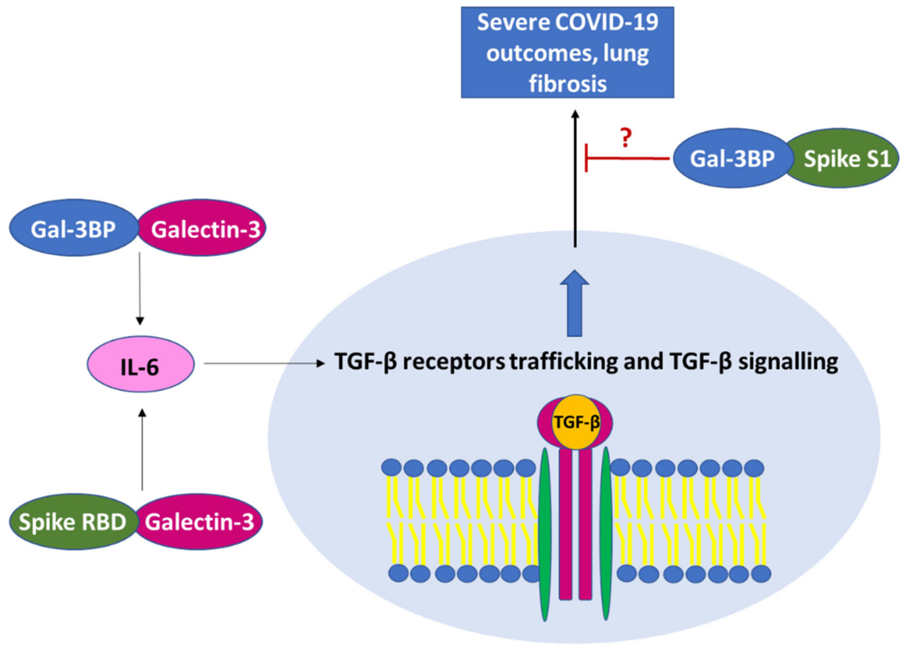
3.1. Role of Gal-3BP in Severe COVID-19 Disease and Pulmonary Fibrosis

The “acute respiratory distress syndrome” (ARDS) is a severe pulmonary complication caused by SARS-CoV-2 virus infection, that in the early pandemic was found in about 5% of COVID patients [76,77]. ARDS has been shown to be mainly the consequence of an exacerbated immune response that leads to a massive release of a specific subset of proinflammatory cytokines, such as TNF-α, IL-2, IL-6, IL-7, IL-10, granulocyte-colony-stimulating factor (G-CSF), interferon-γ-induced protein 10 (IP10), monocyte chemoattractant protein-1 (MCP-1), macrophage inflammatory protein 1 α (MIP-1A) and C-reactive protein (CRP) [78,79,80]. This is accompanied by abnormally increased production and augmented biological activity of anti-inflammatory cytokines, especially TGF-β, as reported in severe COVID-19 disease. TGF-β is a key cytokine in regulating chronic immune reaction in severe COVID-19 and is known to play a crucial role in the development of some peculiar clinical manifestations of the infection, including fatigue, loss of olfactory and taste senses, and in the development of the detrimental pulmonary fibrosis [81,82,83,84,85]. Indirect evidence suggests that Gal-3BP/galectin-3 interaction has a key role in these pathological processes. Indeed, COVID-19 patients who developed pneumonia, particularly those requiring ICU admission, display increased Gal-3BP levels, which are paralleled by increased galectin-3 levels. In addition, patients with high serum levels of galectin-3 (above 35.3 ng/mL) were found to have an increased risk of severe ARDS, admission to ICU, and mortality [39,41]. Furthermore, a positive correlation between galectin-3 and inflammatory markers, including interleukin-6, has been reported by Kuśnierz-Cabala et al. These authors hypothesized that galectin-3 may be involved in severe COVID-19, especially in the development of pulmonary fibrosis [39]. Interestingly, galectin-3, which is an important modulator of acute and chronic inflammation, is the most studied interaction partner of Gal-3BP. The lectin is found in the cytoplasm, in the nucleus, and as a membrane-associated receptor of both normal and malignant cells [86] and plays important roles in signal transduction [87]. Diverse studies reported that during inflammation, galectin-3 loses its proinflammatory functions and promotes tissue healing, inducing the formation of fibrotic tissue [88,89]. Indeed, diverse galectin-3 inhibitors have been reported to counteract fibrotic disorders, including idiopathic pulmonary fibrosis [90]. Further studies reported that galectin-3 induces pulmonary fibrosis by promoting the activity of the transforming growth factor β (TGF-β) [88,89]. Even if the mechanism of action remains still poorly understood, the interaction between Gal-3BP and galectin-3 has been found to be involved in inducing increased expression levels of IL-6 [56]. Interestingly, IL-6 is known to increase trafficking of TGF-β1 receptors to nonlipid raft-associated pools that results in augmented TGF-β signaling [43,44]. These data suggest that the Gal-3BP/galectin-3 axis could be involved in the TGF-β-mediated fibrosis. Interestingly, the putative Gal-3BP/galectin-3 profibrotic mechanism of action resembles those mediated by SARS-CoV-2 in the induction of the detrimental outcome of pulmonary fibrosis, thought to be related to TGF-β increased levels [83,91]. In this regard, a recent study demonstrated that cytokines linked to COVID-19 disease are induced by the S1 subunit of the SARS-CoV-2 spike protein in monocytes. In particular, the S1 subunit of the spike protein, but none of the other spike subunits, induces IL-6, IL-1β, and TNF-α secretion in cultured monocytes [92]. Interestingly, NMR studies showed that the two N-glycans of the receptor-binding domain (RBD) of the spike protein bind to galectin-3 [93], eliciting the possibility that the S1 subunit of spike could interact with galectin-3 with the participation of Gal-3BP. In addition, data showed that the N-terminal domain of SARS-CoV-2 spike protein S1 (i.e., the galectin-like NTD) is highly homologous to galectin-3 [94]. The presence of the galectin-like domain within the NTD of the S1 subunit of the spike protein supports the possibility of Gal-3BP/spike interaction [94] that could possess an antiviral activity. The putative significance of the spike protein’s interaction with Gal-3BP or galectin-3 in miming and/or blocking the effects of the Gal-3BP/galectin-3 signaling is reported schematically in Figure 4.

3.2. Interaction of Gal-3BP with SARS-CoV-2 Components and Spike Protein

Studies carried out on various viruses, including HIV-1 and adeno-associated viruses, have demonstrated that one of the mechanisms by which Gal-3BP exerts its antiviral effects is through a direct interaction with viral particles [48]. To date, there are no studies that confirm this mechanism of action also for SARS-CoV-2. However, data from pull-down experiments, conducted on the plasma of COVID-19 ICU patients, demonstrated that Gal-3BP is consistently retrieved together with the SARS-CoV-2 spike protein, strongly indicating that Gal-3BP is a possible binding partner of the viral protein [72]. This has previously been supported by the presence of a galectin-like domain within the NTD of the S1 subunit of the spike protein. Beside the interaction with the spike protein, a Gal-3BP direct interaction with other virus components could be also plausible, since SARS-CoV-2 is an enveloped virus displaying the glycans produced in the infected cells. Indeed, the N-terminal domain of the spike protein, that is homologous to a region of galectin-3, has been demonstrated to bind other glycans including galectin-7 and 8, Siglec-10, macrophage galactose lectin (MGL), and DC-SIGN [93,95], suggesting that Gal-3BP could interact with SARS-CoV-2 also through these molecules. Even if the significance of the putative interaction between Gal-3BP and SARS-CoV-2 components has not yet been solved, data showed that Gal-3BP overexpression is related to the reduction in spike-mediated syncytia formation, and to a decreased spike-pseudo-particle entry. This can be an important issue, since spike-mediated syncytia formation and entry are the most targeted functions of spike protein in the drug discovery field [70]. In addition, a mechanism of SARS-CoV-2 entry has been proposed that involves the galectin-like spike NTD; data supported a dual attachment model for SARS-CoV2, where the spike CTD domain is involved in ACE-2 receptor recognition, while the NTD region binds gangliosides on the cell surface to stabilize viral adhesion [67]. Indeed, studies reported a great affinity between spike NTD and GM1 ganglioside [68,69]; moreover, a high binding affinity has been demonstrated also between GM1 ganglioside and galectin-3 [96], suggesting that galectin-3 inhibitors could impair virus entry [67]. Altogether, these results allow us to speculate on the possibility that Gal-3BP interaction with the galectin-like NTD region of the spike protein could play inhibitory roles in virus entry. Further studies are necessary to confirm Gal-3BP interaction with SARS-CoV-2 components and its significance in SARS-CoV-2 infection.

3.3. Clinical Significance of Gal-3BP in SARS-CoV-2 Infection

Despite the advancements made from the beginning of the COVID-19 pandemic to control virus spread and reduce mortality, there is still no effective approach for the prevention or treatment of the severe form of the disease, that often leads to life-threatening complications, such as ARDS. The detection of severity-associated biomarkers is one of the most important tools for the early individuation of patients at risk of developing ARDS, that is crucial for timely treatments [97]. Altered values of various laboratory markers, including increased levels of C-reactive protein, lactate dehydrogenase, serum amyloid A, fibrinogen, D-dimer, adenosine deaminase, and lower levels of lymphocyte, eosinophil, platelet counts, calcium, albumin, albumin/globulin, have been associated with COVID-19 severity [98]. In addition, higher levels of diverse inflammatory cytokines are associated with the disease’s severity, especially IL-6, that has been demonstrated to be one of the most accurate predictors of disease course and mortality [99]. Interestingly, also galectin-3 has been proposed as a potential COVID-19-severity-associated marker. Indeed, studies showed that galectin-3, besides being an independent marker of severity, significantly improves the likelihood to assess the risk of developing a severe outcome when used in combination with other markers such as C-reactive protein and albumin [100], hence the importance of extending the panel of already-known severity-associated clinical markers. In this context, the higher levels of Gal-3BP observed in plasma samples of patients with severe compared to those with nonsevere COVID-19 disease, in addition to the established role of the protein in stimulating IL-6 production and secretion through interaction with galectin-3, makes Gal-3BP a new promising prognostic biomarker of COVID-19 severity.

4. Conclusions

Gal-3BP has been demonstrated to be upregulated during several viral infections, including SARS-CoV-2. Various studies demonstrated a direct correlation between Gal-3BP plasma levels and viral infection progression and/or severity of disease, suggesting a key role of Gal-3BP in modulating immune response to viral pathogens other than a role as a prognosticator. However, the complex biology of Gal-3BP, that encompasses different known and unknown interaction partners and signaling cascades, makes it difficult to elucidate its mechanisms of action, although it could explain the various effects ascribed to the protein. Indeed, both an antiviral activity and a role in inducing disease progression and severe clinical manifestations have been reported for various viral diseases, including COVID-19. Besides modulating immune response to viral pathogens by interacting with various extra- and intracellular cell components, including galectin-3, Gal-3BP has been demonstrated to directly interact with virus components, exerting an antiviral activity. The interaction between Gal-3BP and virus components has been reported for different viruses, including SARS-CoV-2.
Data have been presented to show that Gal-3BP interacts with the SARS-CoV-2 spike protein and other SARS-CoV-2 envelope components derived from the cell surface of the infected cells. In this context, it also needs to be considered that the type and amount of glycans can differ depending on cell types and vary greatly between individuals, and this could lead to a diverse individual response to the virus. These facts render Gal-3BP interesting in several respects. While the correlation between Gal-3BP levels and disease severity renders the protein a promising prognostic biomarker, its role in inducing detrimental disease outcomes, such as lung fibrosis in severe COVID-19, makes the protein a good candidate as a target to prevent or block pulmonary fibrosis and the other clinical manifestations observed in severe COVID-19. Furthermore, the possible role of Gal-3BP in intersecting SARS-CoV-2 cell targets could be exploited in the attempt to find possible antiviral inhibitors. In addition, a deepened comprehension of Gal-3BP mechanisms of action can be of uttermost importance for an augmented understanding of the molecular mechanisms underlying the host immune response to viral pathogens. In this context, the role of Gal-3BP in COVID-19 could be a useful model to elucidate the role of Gal-3BP in other viral diseases and, considering its putative role in inducing fibrosis, also in other pathologies, including cancer.
Based on the findings discussed in this work, we believe that future investigations should focus on two main purposes: (i) the evaluation of the clinical significance of Gal-3BP as a new severity-associated prognostic marker; and (ii) the search of inhibitors of Gal-3BP/galectin-3 interaction. Furthermore, studies on the correlation between Gal-3BP level in patients with COVID-19 disease and patients with COVID-19 and HIV or other virus infections should be initiated to enhance this field of research.

Funding

This research was funded by National Funding for Centers of Excellence (Science Department, Roma Tre University—MIUR, Articolo 1, Commi 314–337 Legge 232/2016) and by “ETICO”: Lazio Innova, POR FESR Lazio 2014-2020, Bando Gruppi di Ricerca 2020 (CUP E85F21000870002).

Institutional Review Board Statement

Not applicable.

Informed Consent Statement

Not applicable.

Data Availability Statement

Not applicable.

Conflicts of Interest

The authors declare no conflict of interest.

References

  1. Koths, K.; Taylor, E.; Halenbeck, R.; Casipit, C.; Wang, A. Cloning and characterization of a human Mac-2-binding protein, a new member of the superfamily defined by the macrophage scavenger receptor cysteine-rich domain. J. Biol. Chem. 1993, 268, 14245–14249. [Google Scholar] [CrossRef]
  2. Calabrese, G.; Sures, I.; Pompetti, F.; Natoli, G.; Palka, G.; Iacobelli, S. The gene (LGALS3BP) encoding the serum protein 90K, associated with cancer and infection by the human immunodeficiency virus, maps at 17q25. Cytogenet. Cell. Genet. 1995, 69, 223–225. [Google Scholar] [CrossRef] [PubMed]
  3. Inohara, H.; Akahani, S.; Koths, K.; Raz, A. Interactions between galectin-3 and Mac-2-binding protein mediate cell-cell adhesion. Cancer Res. 1996, 56, 4530–4534. [Google Scholar] [PubMed]
  4. Sasaki, T.; Brakebusch, C.; Engel, J.; Timpl, R. Mac-2 binding protein is a cell-adhesive protein of the extracellular matrix which self-assembles into ring-like structures and binds beta1 integrins, collagens and fibronectin. EMBO J. 1998, 17, 1606–1613. [Google Scholar] [CrossRef] [Green Version]
  5. Iacobelli, S.; Arnò, E.; D’Orazio, A.; Coletti, G. Detection of antigens recognized by a novel monoclonal antibody in tissue and serum from patients with breast cancer. Cancer Res. 1986, 46, 3005–3010. [Google Scholar]
  6. Linsley, P.S.; Horn, D.; Marquardt, H.; Brown, J.P.; Hellstroem, I.; Hellstroem, K.E.; Ochs, V.; Tolentino, E. Identification of a novel serum protein secreted by lung carcinoma cells. Biochemistry 1986, 25, 2978–2986. [Google Scholar] [CrossRef]
  7. Fornarini, B.; Iacobelli, S.; Tinari, N.; Natoli, C.; De Martino, M.; Sabatino, G. Human milk 90K (Mac-2 BP): Possible protective effects against acute respiratory infections. Clin. Exp. Immunol. 1999, 115, 91–94. [Google Scholar] [CrossRef]
  8. Grassadonia, A.; Tinari, N.; Iurisci, I.; Piccolo, E.; Cumashi, A.; Innominato, P.; D’Egidio, M.; Natoli, C.; Piantelli, M.; Iacobelli, S. 90K (Mac-2 BP) and galectins in tumor progression and metastasis. Glycoconj. J. 2002, 19, 551–556. [Google Scholar] [CrossRef]
  9. Bair, E.L.; Nagle, R.B.; Ulmer, T.A.; Laferté, S.; Bowden, G.T. 90K/Mac-2 binding protein is expressed in prostate cancer and induces promatrilysin expression. Prostate 2006, 66, 283–293. [Google Scholar] [CrossRef]
  10. Dumic, J.; Dabelic, S.; Flögel, M. Galectin-3: An open-ended story. Biochim. Biophys. Acta 2006, 1760, 616–635. [Google Scholar] [CrossRef]
  11. Laferté, S.; Loh, L.C.; Keeler, V. Monoclonal antibodies specific for human tumor-associated antigen 90K/Mac-2 binding protein: Tools to examine protein conformation and function. J. Cell. Biochem. 2000, 77, 540–559. [Google Scholar] [CrossRef]
  12. Hellstern, S.; Sasaki, T.; Fauser, C.; Lustig, A.; Timpl, R.; Engel, J. Functional studies on recombinant domains of Mac-2-binding protein. J. Biol. Chem. 2002, 277, 15690–15696. [Google Scholar] [CrossRef] [Green Version]
  13. Yu, B.; Wright, S.D. LPS-dependent interaction of Mac-2-binding protein with immobilized CD14. J. Inflamm. 1995, 45, 115–125. [Google Scholar]
  14. Loimaranta, V.; Hepojoki, J.; Laaksoaho, O.; Pulliainen, A.T. Galectin-3-binding protein: A multitask glycoprotein with innate immunity functions in viral and bacterial infections. J. Leukoc. Biol. 2018, 104, 777–786. [Google Scholar] [CrossRef]
  15. Sarrias, M.R.; Grønlund, J.; Padilla, O.; Madsen, J.; Holmskov, U.; Lozano, F. The Scavenger Receptor Cysteine-Rich (SRCR) domain: An ancient and highly conserved protein module of the innate immune system. Crit. Rev. Immunol. 2004, 24, 1–37. [Google Scholar] [CrossRef]
  16. Martínez, V.G.; Moestrup, S.K.; Holmskov, U.; Mollenhauer, J.; Lozano, F. The conserved scavenger receptor cysteine-rich superfamily in therapy and diagnosis. Pharmacol Rev. 2011, 63, 967–1000. [Google Scholar] [CrossRef] [Green Version]
  17. Stogios, P.J.; Downs, G.S.; Jauhal, J.J.; Nandra, S.K.; Privé, G.G. Sequence and structural analysis of BTB domain proteins. Genome Biol. 2005, 6, R82. [Google Scholar] [CrossRef] [Green Version]
  18. Bardwell, V.J.; Treisman, R. The POZ domain: A conserved protein-protein interaction motif. Genes Dev. 1994, 8, 1664–1677. [Google Scholar] [CrossRef] [Green Version]
  19. Müller, S.A.; Sasaki, T.; Bork, P.; Wolpensinger, B.; Schulthess, T.; Timpl, R.; Engel, A.; Engel, J. Domain organization of Mac-2 binding protein and its oligomerization to linear and ring-like structures. J. Mol. Biol. 1999, 291, 801–813. [Google Scholar] [CrossRef]
  20. Nonaka, M.; Ma, B.Y.; Imaeda, H.; Kawabe, K.; Kawasaki, N.; Hodohara, K.; Kawasaki, N.; Andoh, A.; Fujiyama, Y.; Kawasaki, T. Dendritic cell-specific intercellular adhesion molecule 3-grabbing non-integrin (DC-SIGN) recognizes a novel ligand, Mac-2-binding protein, characteristically expressed on human colorectal carcinomas. J. Biol. Chem. 2011, 286, 22403–22413. [Google Scholar] [CrossRef] [Green Version]
  21. Anthony, R.M.; Wermeling, F.; Karlsson, M.C.; Ravetch, J.V. Identification of a receptor required for the anti-inflammatory activity of IVIG. Proc. Natl. Acad. Sci. USA 2008, 105, 19571–19578. [Google Scholar] [CrossRef] [PubMed] [Green Version]
  22. Natoli, C.; Iacobelli, S.; Ghinelli, F. Unusually high level of a tumor-associated antigen in the serum of human immunodeficiency virus-seropositive individuals. J. Infect. Dis. 1991, 164, 616–617. [Google Scholar] [CrossRef]
  23. Rea, A.; Palmieri, G.; Tinari, N.; Natoli, C.; Tagliaferri, P.; Morabito, A.; Grassadonia, A.; Bianco, A.; Iacobelli, S. 90k is a serum marker of poor-prognosis in non-hodgkins-lymphoma patients. Oncol. Rep. 1994, 1, 723–725. [Google Scholar] [CrossRef]
  24. Marchetti, A.; Tinari, N.; Buttitta, F.; Chella, A.; Angeletti, C.A.; Sacco, R.; Mucilli, F.; Ullrich, A.; Iacobelli, S. Expression of 90K (Mac-2 BP) correlates with distant metastasis and predicts survival in stage I non-small cell lung cancer patients. Cancer Res. 2002, 62, 2535–2539. [Google Scholar]
  25. Ostergaard, O.; Nielsen, C.T.; Iversen, L.V.; Tanassi, J.T.; Knudsen, S.; Jacobsen, S.; Heegaard, N.H.H. Unique protein signature of circulating microparticles in systemic lupus erythematosus. Arthritis Rheum. 2013, 65, 2680–2690. [Google Scholar] [CrossRef] [Green Version]
  26. Liu, K.T.; Liu, Y.H.; Chen, Y.H.; Lin, C.Y.; Huang, C.H.; Yen, M.C.; Kuo, P. Serum Galectin-9 and Galectin-3-Binding Protein in Acute Dengue Virus Infection. Int. J. Mol. Sci. 2016, 17, 832. [Google Scholar] [CrossRef] [Green Version]
  27. Cecamore, C.; Marsili, M.; Salvatore, R.; Troiani, R.; D’Egidio, M.; Tinari, N.; Pelliccia, P.; Chiarelli, F.; Marcovecchio, M.L.; Breda, L. 90K immunostimulatory glycoprotein in children with juvenile idiopathic arthritis. Mod. Rheumatol. 2018, 28, 637–641. [Google Scholar] [CrossRef]
  28. Briggs, N.C.; Natoli, C.; Tinari, N.; D’Egidio, M.; Goedert, J.J.; Iacobelli, S. A 90-kDa protein serum marker for the prediction of progression to AIDS in a cohort of HIV-1+ homosexual men. AIDS Res. Hum. Retrovir. 1993, 9, 811–816. [Google Scholar] [CrossRef]
  29. Natoli, C.; Dianzani, F.; Mazzotta, F.; Balocchini, E.; Pierotti, P.; Antonelli, G.; Iacobelli, S. 90K protein: A new predictor marker of disease progression in human immunodeficiency virus infection. J. Acquir. Immune Defic. Syndr. 1993, 6, 370–375. [Google Scholar]
  30. Cesinaro, A.M.; Natoli, C.; Grassadonia, A.; Tinari, N.; Iacobelli, S.; Trentini, G.P. Expression of the 90K tumor-associated protein in benign and malignant melanocytic lesions. J. Investig. Dermatol. 2002, 119, 187–190. [Google Scholar] [CrossRef] [Green Version]
  31. Mbeunkui, F.; Metge, B.J.; Shevde, L.A.; Pannell, L.K. Identification of differentially secreted biomarkers using LC-MS/MS in isogenic cell lines representing a progression of breast cancer. J. Proteome Res. 2007, 6, 2993–3002. [Google Scholar] [CrossRef] [PubMed] [Green Version]
  32. Woo, J.K.; Jang, J.E.; Kang, J.H.; Seong, J.K.; Yoon, Y.S.; Kim, H.C.; Lee, S.J.; Oh, S.H. Lectin, Galactoside-Binding Soluble 3 Binding Protein Promotes 17-N-Allylamino-17-demethoxygeldanamycin Resistance through PI3K/Akt Pathway in Lung Cancer Cell Line. Mol. Cancer Ther. 2017, 16, 1355–1365. [Google Scholar] [CrossRef] [PubMed] [Green Version]
  33. He, X.; Zhang, S.; Chen, J.; Li, D. Increased LGALS3 expression independently predicts shorter overall survival in patients with the proneural subtype of glioblastoma. Cancer Med. 2019, 8, 2031–2040. [Google Scholar] [CrossRef] [PubMed]
  34. Capone, E.; Iacobelli, S.; Sala, G. Role of galectin 3 binding protein in cancer progression: A potential novel therapeutic target. J. Transl. Med. 2021, 26, 405. [Google Scholar] [CrossRef]
  35. Iacovazzi, P.A.; Cozzolongo, R.; Lanzillotta, E.; Frisullo, S.; Guerra, V.; Correale, M. Serum 90K/Mac-2 binding protein (Mac-2BP) as a response predictor to peginterferon and ribavirin combined treatment in HCV chronic patients. Immunopharmacol. Immunotoxicol. 2008, 30, 687–700. [Google Scholar] [CrossRef]
  36. Artini, M.; Natoli, C.; Tinari, N.; Costanzo, A.; Marinelli, R.; Balsano, C.; Porcari, P.; Angelucci, D.; D’Egidio, M.; Levrero, M.; et al. Elevated serum levels of 90K/MAC-2 BP predict unresponsiveness to alpha-interferon therapy in chronic HCV hepatitis patients. J. Hepatol. 1996, 25, 212–217. [Google Scholar] [CrossRef]
  37. Hepojoki, J.; Strandin, T.; Hetzel, U.; Sironen, T.; Klingström, J.; Sane, J.; Mäkelä, S.; Mustonen, J.; Meri, S.; Lundkvist, A.; et al. Acute hantavirus infection induces galectin-3-binding protein. J. Gen. Virol. 2014, 95, 2356–2364. [Google Scholar] [CrossRef]
  38. Filer, A.; Bik, M.; Parsonage, G.N.; Fitton, J.; Trebilcock, E.; Howlett, K.; Cook, M.; Raza, K.; Simmons, D.L.; Thomas, A.M.C.; et al. Galectin 3 induces a distinctive pattern of cytokine and chemokine production in rheumatoid synovial fibroblasts via selective signaling pathways. Arthritis Rheum. 2009, 60, 1604–1614. [Google Scholar] [CrossRef] [Green Version]
  39. Kuśnierz-Cabala, B.; Maziarz, B.; Dumnicka, P.; Dembiński, M.; Kapusta, M.; Bociąga-Jasik, M.; Winiarski, M.; Garlicki, A.; Grodzicki, T.; Kukla, M. Diagnostic Significance of Serum Galectin-3 in Hospitalized Patients with COVID-19-A Preliminary Study. Biomolecules 2021, 11, 1136. [Google Scholar] [CrossRef]
  40. Gallo, V.; ISERC-Team; Gentile, R.; Antonini, G.; Iacobelli, S. Increased Gal-3BP plasma levels in hospitalized patients infected with SARS-CoV-2. Clin. Exp. Med. 2022, 25, 1–5. [Google Scholar] [CrossRef]
  41. Portacci, A.; Diaferia, F.; Santomasi, C.; Dragonieri, S.; Boniello, E.; Serio, F.D.; Carpagnano, G.E. Galectin-3 as prognostic biomarker in patients with COVID-19 acute respiratory failure. Respir. Med. 2021, 187, 106556. [Google Scholar] [CrossRef]
  42. Xu, G.; Xia, Z.; Deng, F.; Liu, L.; Wang, Q.; Yu, Y.; Wang, F.; Zhu, C.; Liu, W.; Cheng, Z.; et al. Inducible LGALS3BP/90K activates antiviral innate immune responses by targeting TRAF6 and TRAF3 complex. PLoS Pathog. 2019, 15, e1008002. [Google Scholar] [CrossRef] [Green Version]
  43. Zhang, X.L.; Topley, N.; Ito, T.; Phillips, A. Interleukin-6 regulation of transforming growth factor (TGF)-beta receptor compartmentalization and turnover enhances TGF-beta1 signaling. J. Biol. Chem. 2005, 280, 12239–12245. [Google Scholar] [CrossRef] [Green Version]
  44. Luckett-Chastain, L.R.; Gallucci, R.M. Interleukin (IL)-6 modulates transforming growth factor-beta expression in skin and dermal fibroblasts from IL-6-deficient mice. Br. J. Dermatol. 2009, 161, 237–248. [Google Scholar] [CrossRef] [Green Version]
  45. Iacobelli, S.; Natoli, C.; D’Egidio, M.; Tamburrini, E.; Antinori, A.; Ortona, L. Lipoprotein 90K in human immunodeficiency virus-infected patients: A further serologic marker of progression. J. Infect. Dis. 1991, 164, 819. [Google Scholar] [CrossRef]
  46. Pelliccia, P.; Galli, L.; de Martino, M.; Chiarelli, F.; Verrotti, A.; Sabatino, G.; Fornarini, B.; Iacobelli, S.; Natoli, C. Lack of mother-to-child HIV-1 transmission is associated with elevated serum levels of 90 K immune modulatory protein. AIDS 2000, 14, F41–F45. [Google Scholar] [CrossRef]
  47. Lodermeyer, V.; Suhr, K.; Schrott, N.; Kolbe, C.; Stürzel, C.M.; Krnavek, D.; Münch, J.; Dietz, C.; Waldmann, T.; Kirchhoff, F.; et al. 90K, an interferon-stimulated gene product, reduces the infectivity of HIV-1. Retrovirology 2013, 10, 111. [Google Scholar] [CrossRef] [Green Version]
  48. Wang, Q.; Zhang, X.; Han, Y.; Wang, X.; Gao, G. M2BP inhibits HIV-1 virion production in a vimentin filaments-dependent manner. Sci. Rep. 2016, 6, 32736. [Google Scholar] [CrossRef] [Green Version]
  49. Iacobelli, S.; Scambia, G.; Natoli, C.; Panici, P.B.; Baiocchi, G.; Perrone, L.; Mancuso, S. Recombinant human leukocyte interferon-alpha 2b stimulates the synthesis and release of a 90K tumor-associated antigen in human breast cancer cells. Int. J. Cancer 1988, 42, 182–184. [Google Scholar] [CrossRef]
  50. Marth, C.; Dreps, A.; Natoli, C.; Zeimet, A.G.; Lang, T.; Widschwendter, M.; Daxenbichler, G.; Ullrich, A.; Iacobelli, S. Effects of type-I and -II interferons on 90K antigen expression in ovarian carcinoma cells. Int. J. Cancer 1994, 59, 808–813. [Google Scholar] [CrossRef]
  51. D’Ostilio, N.; Natoli, C.; Grassadonia, A.; Rossi, N. Prognostic value of a novel interferon-inducible 90K tumor antigen. Ann. N. Y. Acad. Sci. 1996, 784, 288–293. [Google Scholar] [CrossRef]
  52. Grassadonia, A.; Tinari, N.; Fiorentino, B.; Nakazato, M.; Chung, H.K.; Giuliani, C.; Napolitano, G.; Iacobelli, S.; Howcroft, T.K.; Singer, D.S.; et al. Upstream stimulatory factor regulates constitutive expression and hormonal suppression of the 90K (Mac-2BP) protein. Endocrinology 2007, 148, 3507–3517. [Google Scholar] [CrossRef] [Green Version]
  53. Park, Y.P.; Choi, S.C.; Kim, B.Y.; Kim, J.; Song, E.Y.; Kang, S.O.; Yoon, D.; Paik, S.; Kim, K.D.; Kim, J.W.; et al. Induction of Mac-2BP by nerve growth factor is regulated by the PI3K/Akt/NF-kappaB-dependent pathway in the HEK293 cell line. BMB Rep. 2008, 41, 784–789. [Google Scholar] [CrossRef] [Green Version]
  54. Noma, N.; Simizu, S.; Kambayashi, Y.; Kabe, Y.; Suematsu, M.; Umezawa, K. Involvement of NF-κB-mediated expression of galectin-3-binding protein in TNF-α-induced breast cancer cell adhesion. Oncol. Rep. 2012, 27, 2080–2084. [Google Scholar] [CrossRef] [Green Version]
  55. Grassadonia, A.; Tinari, N.; Fiorentino, B.; Suzuki, K.; Nakazato, M.; De Tursi, M.; Giuliani, C.; Napolitano, G.; Singer, D.S.; Iacobelli, S. The 90K protein increases major histocompatibility complex class I expression and is regulated by hormones, gamma-interferon, and double-strand polynucleotides. Endocrinology 2004, 145, 4728–4736. [Google Scholar] [CrossRef] [Green Version]
  56. Fukaya, Y.; Shimada, H.; Wang, L.C.; Zandi, E.; DeClerck, Y.A. Identification of galectin-3-binding protein as a factor secreted by tumor cells that stimulates interleukin-6 expression in the bone marrow stroma. J. Biol. Chem. 2008, 283, 18573–18581. [Google Scholar] [CrossRef] [Green Version]
  57. Song, L.; Asgharzadeh, S.; Salo, J.; Engell, K.; Wu, H.W.; Sposto, R.; Ara, T.; Silverman, A.M.; DeClerck, Y.A.; Seeger, R.C.; et al. Valpha24-invariant NKT cells mediate antitumor activity via killing of tumor-associated macrophages. J. Clin. Investig. 2009, 119, 1524–1536. [Google Scholar] [CrossRef] [Green Version]
  58. Silverman, A.M.; Nakata, R.; Shimada, H.; Sposto, R.; DeClerck, Y.A. A galectin-3-dependent pathway upregulates interleukin-6 in the microenvironment of human neuroblastoma. Cancer Res. 2012, 72, 2228–2238. [Google Scholar] [CrossRef] [PubMed] [Green Version]
  59. Ullrich, A.; Sures, I.; D’Egidio, M.; Jallal, B.; Powell, T.J.; Herbst, R.; Dreps, A.; Azam, M.; Rubinstein, M.; Natoli, C.; et al. The secreted tumor-associated antigen 90K is a potent immune stimulator. J. Biol. Chem. 1994, 269, 18401–18407. [Google Scholar] [CrossRef]
  60. Kalayci, O.; Birben, E.; Tinari, N.; Oguma, T.; Iacobelli, S.; Lilly, C.M. Role of 90K protein in asthma and TH2-type cytokine expression. Ann. Allergy Asthma Immunol. 2004, 93, 485–492. [Google Scholar] [CrossRef]
  61. Powell, T.J.; Schreck, R.; McCall, M.; Hui, T.; Rice, A.; App, H.; Azam, M.; Ullrich, A.; Shawver, L.K. A tumor-derived protein which provides T-cell costimulation through accessory cell activation. J. Immunother. Emphas. Tumor Immunol. 1995, 17, 209–221. [Google Scholar] [CrossRef]
  62. Gleissner, C.A.C.; Erbel, F.; Linden, G.; Domschke, M.; Akhavanpoor, C.M.; Helmes, C.M.; Doesch, A.O.; Kleber, M.E.; Katus, H.A.; Maerz, W. Galectin-3 binding protein, coronary artery disease and cardiovascular mortality: Insights from the LURIC study Atherosclerosis. J. Atheroscler. 2017, 260, 121–129. [Google Scholar] [CrossRef]
  63. Lin, T.W.; Chang, H.T.; Chen, C.H.; Chen, C.H.; Lin, S.W.; Hsu, T.L.; Wong, C.H. Galectin-3 Binding Protein and Galectin-1 Interaction in Breast Cancer Cell Aggregation and Metastasis. J. Am. Chem. Soc. 2015, 137, 9685–9693. [Google Scholar] [CrossRef]
  64. White, M.J.; Roife, D.; Gomer, R.H. Galectin-3 binding protein secreted by breast cancer cells inhibits monocyte-derived fibrocyte differentiation. J. Immunol. 2015, 195, 1858–1867. [Google Scholar] [CrossRef] [Green Version]
  65. Hong, C.S.; Park, M.; Sun, E.; Choi, W.; Hwang, J.; Bae, W.; Rhee, J.H.; Cho, S.; Chung, I. Gal-3BP Negatively Regulates NF-κB Signaling by Inhibiting the Activation of TAK1. Front. Immunol. 2019, 10, 1760. [Google Scholar] [CrossRef] [Green Version]
  66. Denard, J.; Beley, C.; Kotin, R.; Lai-Kuen, R.; Blot, S.; Leh, H.; Asokan, A.; Samulski, R.J.; Moullier, P.; Voit, T.; et al. Human galectin 3 binding protein interacts with recombinant adeno-associated virus type 6. J. Virol. 2012, 86, 6620–6631. [Google Scholar] [CrossRef] [Green Version]
  67. Caniglia, J.L.; Guda, M.R.; Asuthkar, S.; Tsung, A.J.; Velpula, K.K. A potential role for Galectin-3 inhibitors in the treatment of COVID-19. PeerJ 2020, 15, e9392. [Google Scholar] [CrossRef]
  68. Fantini, J.; Di Scala, C.; Chahinian, H.; Yahi, N. Structural and molecular modelling studies reveal a new mechanism of action of chloroquine and hydroxychloroquine against SARS-CoV-2 infection. Int. J. Antimicrob. Agents 2020, 55, 105960. [Google Scholar] [CrossRef]
  69. Ou, X.; Liu, Y.; Lei, X.; Li, P.; Mi, D.; Ren, L.; Guo, L.; Guo, R.; Chen, T.; Hu, J.; et al. Characterization of spike glycoprotein of SARS-CoV-2 on virus entry and its immune cross-reactivity with SARS-CoV. Nat. Commun. 2020, 11, 1620. [Google Scholar] [CrossRef] [Green Version]
  70. Braga, L.; Ali, H.; Secco, I.; Chiavacci, E.; Neves, G.; Goldhill, D.; Penn, R.; Jimenez-Guardeño, J.M.; Ortega-Prieto, A.M.; Bussani, R.; et al. Drugs that inhibit TMEM16 proteins block SARS-CoV-2 spike-induced syncytia. Nature 2021, 594, 88–93. [Google Scholar] [CrossRef]
  71. Messner, C.B.; Demichev, V.; Wendisch, D.; Michalick, L.; White, M.; Freiwald, A.; Textoris-Taube, K.; Vernardis, S.I.; Egger, A.S.; Kreidl, M. Ultra-High-Throughput Clinical Proteomics Reveals Classifiers of COVID-19 Infection. Cell Syst. 2020, 11, 11–24.e4. [Google Scholar] [CrossRef]
  72. Gutmann, C.; Takov, K.; Burnap, S.A.; Singh, B.; Ali, H.; Theofilatos, K.; Reed, E.; Hasman, M.; Nabeebaccus, A.; Fish, M.; et al. SARS-CoV-2 RNAemia and proteomic trajectories inform prognostication in COVID-19 patients admitted to intensive care. Nat. Commun. 2021, 12, 3406. [Google Scholar] [CrossRef]
  73. Geyer, P.E.; Arend, F.M.; Doll, S.; Louiset, M.L.; Virreira Winter, S.; Müller-Reif, J.B.; Torun, F.M.; Weigand, M.; Eichhorn, P.; Bruegel, M.; et al. High-resolution serum proteome trajectories in COVID-19 reveal patient-specific seroconversion. EMBO Mol. Med. 2021, 9, e14167. [Google Scholar] [CrossRef]
  74. Zeng, F.; Huang, Y.; Guo, Y.; Yin, M.; Chen, X.; Xiao, L.; Deng, G. Association of inflammatory markers with the severity of COVID-19: A meta-analysis. Int. J. Infect. Dis. 2020, 96, 467–474. [Google Scholar] [CrossRef]
  75. Zhang, J.; Hao, Y.; Ou, W.; Ming, F.; Liang, G.; Qian, Y.; Cai, Q.; Dong, S.; Hu, S.; Wang, W.; et al. Serum interleukin-6 is an indicator for severity in 901 patients with SARS-CoV-2 infection: A cohort study. J. Transl. Med. 2020, 18, 406. [Google Scholar] [CrossRef]
  76. Richardson, S.; Hirsch, J.S.; Narasimhan, M.; Crawford, J.M.; McGinn, T.; Davidson, K.W.; the Northwell COVID-19 Research Consortium; Barnaby, D.P.; Becker, L.B.; Chelico, J.D.; et al. The Northwell COVID-19 Research Consortium Presenting Characteristics, Comorbidities, and Outcomes Among 5700 Patients Hospitalized With COVID-19 in the New York City Area. JAMA 2020, 323, 2052–2059. [Google Scholar] [CrossRef]
  77. Osuchowski, M.F.; Winkler, M.S.; Skirecki, T.; Cajander, S.; Shankar-Hari, M.; Lachmann, G.; Monneret, G.; Venet, F.; Bauer, M.; Brunkhorst, F.M.; et al. The COVID-19 puzzle: Deciphering pathophysiology and phenotypes of a new disease entity. Lancet Respir. Med. 2021, 9, 622–642. [Google Scholar] [CrossRef]
  78. Chen, G.; Wu, D.; Guo, W.; Cao, Y.; Huang, D.; Wang, H.; Wang, T.; Zhang, X.; Chen, H.; Yu, H.; et al. Clinical and immunological features of severe and moderate coronavirus disease. J. Clin. Investig. 2019, 130, 2620–2629. [Google Scholar] [CrossRef] [Green Version]
  79. Huang, C.; Wang, Y.; Li, X.; Ren, L.; Zhao, J.; Hu, Y.; Zhang, L.; Fan, G.; Xu, J.; Gu, X.; et al. Clinical features of patients infected with 2019 novel coronavirus in Wuhan, China. Lancet 2020, 395, 497–506. [Google Scholar] [CrossRef] [Green Version]
  80. Li, Y.; Li, J.Z. SARS-CoV-2 Virology. Infect. Dis. Clin. N. Am. 2022, 36, 251–265. [Google Scholar] [CrossRef] [PubMed]
  81. Lee, C.G.; Homer, R.J.; Zhu, Z.; Lanone, S.; Wang, X.; Koteliansky, V.; Shipley, J.M.; Gotwals, P.; Noble, P.; Chen, Q.; et al. Interleukin-13 induces tissue fibrosis by selectively stimulating and activating transforming growth factor beta(1). J. Exp. Med. 2001, 194, 809–821. [Google Scholar] [CrossRef] [PubMed]
  82. Thompson, B.T.; Chambers, R.C.; Liu, K.D. Acute Respiratory Distress Syndrome. N. Engl. J. Med. 2017, 377, 562–572. [Google Scholar] [CrossRef] [PubMed]
  83. Ferreira-Gomes, M.; Kruglov, A.; Durek, P.; Heinrich, F.; Tizian, C.; Heinz, G.A.; Pascual-Reguant, A.; Du, W.; Mothes, R.; Fan, C.; et al. SARS-CoV-2 in severe COVID-19 induces a TGF-β-dominated chronic immune response that does not target itself. Nat. Commun. 2021, 12, 1961. [Google Scholar] [CrossRef] [PubMed]
  84. Shen, W.X.; Luo, R.C.; Wang, J.Q.; Chen, Z.S. Features of Cytokine Storm Identified by Distinguishing Clinical Manifestations in COVID-19. Front. Public. Health 2021, 9, 671788. [Google Scholar] [CrossRef]
  85. Windisch, D.; Dietrich, O.; Mari, T.; von Stillfried, S.; Ibarra, I.L.; Mittermaier, M.; Mache, C.; Chua, R.L.; Knoll, R.; Timm, S.; et al. SARS-CoV-2 infection triggers profibrotic macrophage responses and lung fibrosis. Cell 2021, 184, 6243–6261.e27. [Google Scholar] [CrossRef]
  86. Haudek, K.C.; Spronk, K.J.; Voss,  P.G.; Patterson, R.J.; Wang, J.L.; Arnoys,  E.J. Dynamics of galectin-3 in the nucleus and cytoplasm. Biochim. Biophys. Acta 2010, 1800, 181–189. [Google Scholar] [CrossRef] [Green Version]
  87. Nakahara, S.; Raz, A. On the role of galectins in signal transduction. Methods Enzymol. 2006, 417, 273–289. [Google Scholar] [CrossRef]
  88. Mackinnon, A.C.; Gibbons, M.A.; Farnworth, S.L.; Leffler, H.; Nilsson, U.J.; Delaine, T.; Simpson, A.J.; Forbes, S.J.; Hirani, N.; Gauldie, J.; et al. Regulation of transforming growth factor-β1-driven lung fibrosis by galectin-3. Am. J. Respir. Crit. Care Med. 2012, 185, 537–546. [Google Scholar] [CrossRef] [Green Version]
  89. Gawlik, K.I.; Holmberg, J.; Svensson, M.; Einerborg, M.; Oliveira, B.M.; Deierborg, T.; Durbeej, M. Potent pro-inflammatory and pro-fibrotic molecules, osteopontin and galectin-3, are not major disease modulators of laminin α2 chain-deficient muscular dystrophy. Sci. Rep. 2017, 7, 44059. [Google Scholar] [CrossRef]
  90. Hirani, N.; MacKinnon, A.C.; Nicol, L.; Ford, P.; Schambye, H.; Pedersen, A.; Nilsson, U.J.; Leffler, H.; Sethi, T.; Tantawi, S. Target inhibition of galectin-3 by inhaled TD139 in patients with idiopathic pulmonary fibrosis. Eur. Respir. J. 2021, 57, 2002559. [Google Scholar] [CrossRef]
  91. Xiong, Y.; Liu, Y.; Cao, L.; Wang, D.; Guo, M.; Jiang, A.; Guo, D.; Hu, W.; Yang, J.; Tang, Z.; et al. Transcriptomic characteristics of bronchoalveolar lavage fluid and peripheral blood mononuclear cells in COVID-19 patients. Emerg. Microbes Infect. 2020, 9, 761–770. [Google Scholar] [CrossRef]
  92. Schroeder, J.T.; Bieneman, A.P. The S1 Subunit of the SARS-CoV-2 Spike Protein Activates Human Monocytes to Produce Cytokines Linked to COVID-19: Relevance to Galectin-3. Front. Immunol. 2022, 13, 831766. [Google Scholar] [CrossRef]
  93. Lenza, M.P.; Oyenarte, I.; Diercks, T.; Quintana, J.I.; Gimeno, A.; Coelho, H.; Diniz, A.; Peccati, F.; Delgado, S.; Bosch, A.; et al. Structural characterization of the N-linked glycans in the receptor binding domain of the SARS-CoV-2 spike protein and their interactions with human lectins using NMR spectroscopy. Angew. Chem. Int. Ed. Engl. 2020, 59, 23763–23771. [Google Scholar] [CrossRef]
  94. Caniglia, J.L.; Asuthkar, S.; Tsung, A.J.; Guda, M.R.; Velpula, K.K. Immunopathology of galectin-3: An increasingly promising target in COVID-19. F1000Research 2020, 9, 1078. [Google Scholar] [CrossRef]
  95. Trbojević-Akmačić, I.; Petrović, T.; Lauc, G. SARS-CoV-2 S glycoprotein binding to multiple host receptors enables cell entry and infection. Glycoconj. J. 2021, 38, 611–623. [Google Scholar] [CrossRef]
  96. Wu, G.; Lu, Z.H.; Andre, S.; Gabius, H.J.; Ledeen, R.W. Functional interplay between ganglioside GM1 and cross-linking galectin-1 induces axon-like neuritogenesis via integrin-based signaling and TRPC5-dependent Ca2+ influx. J. Neurochem. 2016, 136, 550–563. [Google Scholar] [CrossRef]
  97. Kim, P.S.; Read, S.W.; Fauci, A.S. Therapy for early COVID-19a critical need. JAMA 2020, 324, 2149–2150. [Google Scholar] [CrossRef]
  98. Qi, J.; He, D.; Yang, D.; Wang, M.; Ma, W.; Cui, H.; Ye, F.; Wang, F.; Xu, J.; Li, Z.; et al. Severity-associated markers and assessment model for predicting the severity of COVID-19: A retrospective study in Hangzhou, China. BMC Infect. Dis. 2021, 21, 774. [Google Scholar] [CrossRef]
  99. Zhou, F.; Yu, T.; Du, R.; Fan, G.; Liu, Y.; Liu, Z.; Xiang, J.; Wang, Y.; Song, B.; Gu, X.; et al. Clinical course and risk factors for mortality of adult inpatients with COVID-19 in Wuhan, China: A retrospective cohort study. Lancet 2020, 395, 1054–1062. [Google Scholar] [CrossRef]
  100. Cervantes-Alvarez, E.; la Rosa, N.L.; la Mora, M.S.; Valdez-Sandoval, P.; Palacios-Jimenez, M.; Rodriguez-Alvarez, F.; Vera-Maldonado, B.I.; Aguirre-Aguilar, E.; Escobar-Valderrama, J.M.; Alanis-Mendizabal, J.; et al. Galectin-3 as a potential prognostic biomarker of severe COVID-19 in SARS-CoV-2 infected patients. Sci. Rep. 2022, 12, 1856. [Google Scholar] [CrossRef]
Figure 1. Human Gal-3BP’s structure consists of three main functional domains: an N-terminal scavenger receptor cysteine-rich (SRCR) domain, a BTB/POZ (broad-complex, tramtrack and bric-a-brac/poxvirus and zinc finger) domain and a BACK domain (BTB and C-terminal Kelch domain). In addition, there is an N-terminal 18 amino acid signal peptide that is cleaved after translation and a C-terminal protease-susceptible area.
Ijms 23 07314 g001
Figure 2. Number of publications per year (from 1977 to 2022) on Gal-3BP and viral infections. The search was performed using PubMed, Scopus, and Google Scholar databases, using the following key words: “(90K OR LGALS3BP) AND virus”.
Ijms 23 07314 g002
Figure 3. The figure shows some of the known interaction partners and signaling pathways of Gal-3BP, which lead to the expression of proinflammatory cytokines during virus infection. The viral DNA/RNA induces IFNs and TNF-α, that in turn induce Gal-3BP. The protein can act both intra- and extracellularly. Extracellularly, Gal-3BP can interact with galectin-3-inducing RAS-signaling pathway and IL-6 expression. Gal-3BP can also directly interact with virus particles reducing their infectivity. Intracellularly, Gal-3BP acts as a scaffold protein interacting with both TRAF-6/transforming growth factor β-activated kinase 1 (TAK1) and TRAF3/TANK binding kinase 1 (TBK1) complexes which leads to the production of IFNs and proinflammatory cytokines.
Ijms 23 07314 g003
Figure 4. Schematic representation of the possible significance of the interaction between the spike RBD glycans and galectin-3 and between the S1 subunit of spike protein and Gal-3BP in miming and/or blocking the Gal-3BP/galectin-3 signaling. Gal-3BP/galectin-3 interaction leads to increased expression and secretion of IL-6, which has been demonstrated to increase TGF-β signaling and play a role in the development of fibrosis. This profibrotic mechanism resembles that mediated by SARS-CoV-2; indeed, the interaction of Spike RBD glycans with galectin-3 has been demonstrated to upregulate IL-6. This led us to hypothesize that SARS-CoV-2 spike could mimic the Gal-3BP/galectin-3 signaling. Furthermore, in vitro studies demonstrated the possible interaction between Gal-3BP and the S1 galectin-like domain of the spike protein. While there is strong evidence of the antiviral significance of this interaction (see the main text for further details), no studies have been conducted to demonstrate the possibility that it could also act differently. However, the findings of Gal-3BP/S1 spike interaction suggest that it could block the Gal-3BP signaling and the detrimental outcomes of severe COVID-19; thus, we believe that it might be interesting to consider this for further investigations.
Ijms 23 07314 g004
Table 1. Gal-3BP’s interaction partners, mechanisms of action, and reported effects during viral infections.
Interaction PartnerMechanism of ActionEffectReferences
Galectin-3Induction of Ras-Mek-Erk1/2-signaling pathway.IL-6 expression and secretion.[56,57,58]
TRAF6/TAK1Translocation of NF-κB to the nucleus.Expression of IFN and inflammatory cytokines.[42]
TRAF3/TBK1Translocation of IRF3/7 to the nucleus.Expression of IFN and inflammatory cytokines.[42]
VirusesDirect binding with adeno-associated
Viruses.
Induces viral particle aggregation and impairment of transduction.[66]
Direct binding with HIV-1.Prevents transportation of HIV-1 Gag to the plasma membrane, inhibiting HIV-1 virion production; reduces the amount of gp120 and gp41 at the plasma membrane.[14,48]
Direct binding with SARS-CoV-2
components (i.e., spike protein).
Reduction in spike-mediated syncytia formation, and to a decreased spike-pseudo-particle entry.[67,68,69,70]
DC-SIGNDC maturation and fibrocyte
differentiation.
Immunosuppression.[14,20,64]
UnknownUnknownInduces Ca2+ mobilization; increases the amount of ICAM-I and MHC-I on the cell surface.[14]
UnknownInduction of IL-1; IL-2; GM-CSF; and TNF-α.Immunostimulatory effects.[56,58,59,61]
UnknownSuppression of IL-4, IL-5, and IL-13.Immunosuppressive effects.[60]
Publisher’s Note: MDPI stays neutral with regard to jurisdictional claims in published maps and institutional affiliations.

