Hypothesis

An Altered Sphingolipid Profile as a Risk Factor for Progressive Neurodegeneration in Long-Chain 3-Hydroxyacyl-CoA Deficiency (LCHADD)

1
Pharmacy, Medical Center, University of Freiburg, 79106 Freiburg, Germany
2
Department of General Pediatrics, Adolescent Medicine and Neonatology, Faculty of Medicine, Medical Centre-University of Freiburg, 79106 Freiburg, Germany
Int. J. Mol. Sci. 2022, 23(13), 7144; https://doi.org/10.3390/ijms23137144
Submission received: 19 May 2022 / Revised: 23 June 2022 / Accepted: 25 June 2022 / Published: 27 June 2022
(This article belongs to the Special Issue Lipid Metabolism in Pathology and Health)

Abstract

:
Long-chain 3-hydroxyacyl-CoA deficiency (LCHADD) and mitochondrial trifunctional protein (MTPD) belong to a group of inherited metabolic diseases affecting the degradation of long-chain chain fatty acids. During metabolic decompensation the incomplete degradation of fatty acids results in life-threatening episodes, coma and death. Despite fast identification at neonatal screening, LCHADD/MTPD present with progressive neurodegenerative symptoms originally attributed to the accumulation of toxic hydroxyl acylcarnitines and energy deficiency. Recently, it has been shown that LCHADD human fibroblasts display a disease-specific alteration of complex lipids. Accumulating fatty acids, due to defective β-oxidation, contribute to a remodeling of several lipid classes including mitochondrial cardiolipins and sphingolipids. In the last years the face of LCHADD/MTPD has changed. The reported dysregulation of complex lipids other than the simple acylcarnitines represents a novel aspect of disease development. Indeed, aberrant lipid profiles have already been associated with other neurodegenerative diseases such as Parkinson’s Disease, Alzheimer’s Disease, amyotrophic lateral sclerosis and retinopathy. Today, the physiopathology that underlies the development of the progressive neuropathic symptoms in LCHADD/MTPD is not fully understood. Here, we hypothesize an alternative disease-causing mechanism that contemplates the interaction of several factors that acting in concert contribute to the heterogeneous clinical phenotype.

1. Introduction

The mitochondrial trifunctional protein (MTP) of the β-oxidation pathway is a α2β2 heterotetramer multienzyme complex encoded by two different genes, HADHA (OMIM 600890) and HADHB (OMIM 143450). This enzyme complex catalyzes three reactions. The long-chain enoyl-CoA hydratase and long-chain 3-hydroxyacyl-CoA dehydrogenase (LCHAD) are carried by the α-subunit, while the long-chain 3-ketoacyl-CoA thiolase (LCKAT) is localized on the β-subunit [1,2]. Mutations in HADHA are responsible for LCHAD deficiency (LCHADD), whereas variants in HADHA and HADHB cause MTP deficiency (MTPD). Both diseases are autosomal recessive disorders of the mitochondrial long-chain fatty acid oxidation (lc-FAODs) affecting the degradation of chain fatty acids with a chain length > C12 [3]. These diseases, with an incidence of 1:250,000 newborns in Germany, are characterized by a wide range of clinical manifestations including cardiomyopathy, liver dysfunction, progressive neuropathy and retinopathy, rhabdomyolysis and sudden death (https://www.screening-dgns.de/Pdf/Screeningreports/DGNS-Screeningreport-d_2019.pdf; accessed on 15 May 2022) [3]. The incomplete oxidation of long chain fatty acids during acute episodes of metabolic decompensation results in the accumulation of toxic acylcarnitines and energy deficiency. The subsequent impaired production of redox equivalents for oxidative phosphorylation may lead to life-threatening episodes, coma and death. Lc-FAODs are included in newborn screening programs of many countries worldwide. The identification of affected patients at neonatal screenings allows therapy to be initiated, thereby avoiding catabolic crises and metabolic derangement. No cure for LCHADD and MTPD is available. However, a number of dietary-based management strategies can be applied. Treatment recommendations include frequent meals, avoiding fasting, restriction of dietary long-chain fatty acids and supplementation with carbohydrates and medium-chain fatty acids (MCT oil) [4,5]. In addition, the use of triheptanoin, a triglyceride with three molecules of heptanoic acid, was recently approved by the FDA for the treatment of lc-FAODs [6].
Despite early diagnosis and therapy initiation, LCHADD and MTPD present with retinopathy and neurodegenerative symptoms originally attributed to the accumulation of toxic acylcarnitines, oxidative stress and energy deficiency. These symptoms specific for LCHADD and MTPD are progressive and cannot be avoided. Studies in the fibroblasts of patients with lc-FAODs have demonstrated that the accumulating fatty acids due to defective β-oxidation are likely involved in remodeling processes leading to an alteration of the profile and content of complex lipids [7,8].
Here, we aim at outlining the importance of these complex molecules in the pathophysiology of lc-FAODs. We summarize the evidence that exists in support of the hypothesis that profile disturbances of mitochondrial cardiolipins and sphingolipids are likely involved in LCHADD/MTPD disease development. Based on these observations, we propose a novel mechanism that considers dysregulated complex lipids as a contributing cause in the progressive neurodegenerative symptoms of LCHADD/MTPD.

2. Mitochondrial Trifunctional Protein (MTP) and Cardiolipins

Very recently the crystal structure of the human MTP has been elucidated [2]. Due to the peculiar arcuate shape of the protein that binds its concave site to the mitochondrial inner membrane, the authors suggested a structure acting as a channel that allows the transition of the substrate among the catalytic units [2]. In particular, the transfer of the shortening acyl-residues from the hydratase to the ketothiolase is possible because of the hydrophobic nature of the substrates. These are able to move among the mitochondrial inner membrane to reach the last catalytic site for the cleavage. Therefore, the integrity of the mitochondrial membrane is essential to ensure a proper enzyme function [2]. In addition to mutations that directly affect the catalytic domain of the enzymes [9,10,11,12], several other variants severely impact the structural stability of the protein with the subsequent loss of the interaction between α and β subunits [1,13,14,15,16]. Maintenance of mitochondrial processes is also ensured by an intact cardiolipin composition as an essential constituent of mitochondrial membranes. The essential role of the MTP α-subunit in mitochondrial cardiolipin profiling can be explained by the identical structure of this enzyme with the newly identified monolysocardiolipin acyltransferase 1 (MLCLAT1) [17]. It is therefore conceivable that sequence variants affecting MTP enzyme activity result in an altered cardiolipin profile [7]. At the same time, an aberrant cardiolipin profile reduces mitochondrial fusion and fission processes [18,19,20] and leads to the destabilization of mitochondrial supercomplexes and the dysregulation of energy metabolism [7,8]. Analysis in human fibroblasts has highlighted that the LCHADD induces cardiolipin remodeling with a subsequent profile alteration and incorporation of the non-metabolized C16 fatty acids into cardiolipin species [7]. In keeping with previous data [18,19,20], LCHADD cell lines have been shown to present with dysregulated mitochondrial dynamics accompanied by mitochondrial dysfunction [21]. However, profile alteration was also observed in other classes of bioactive lipids such as sphingolipids, ceramides and hexosylceramides.

3. Sphingomyelins and Ceramides: New Co-Player for Neurodegeneration in LCHADD?

The aberrant metabolism of sphingolipids characterized, i.e., by the accumulation of specific species with the subsequent alteration of ceramide/sphingomyelin ratio, glucosylceramides, ceramides and glactolsylceramides [22] results in the loss of neurons and their axis [23] or in neuronal dysfunction [24,25]. The accumulation of these lipids in lysosomes is typical for Niemann–Pick disease, Gaucher’s disease, Farber’s disease and Krabbe’s disease, a group of inherited metabolic diseases called lysosomal storage disorders (LSDs). LSDs share the accumulation of sphingolipids and ceramides with neurodegenerative disorders such as Alzheimer’s disease (AD), Parkinson’s Disease (PD) and Multiple Sclerosis (MS) [26]. Ceramides and sphingolipids belong to the group of bioactive lipids acting as messengers and involved in a wide range of cellular functions. Both lipid classes contribute structurally to the bilayer of membrane lipids and lipid rafts, playing a striking role in inflammation, apoptosis, neuronal differentiation and synaptic transmission in neuronal–glial connections [27,28]. These lipid classes are essential for the membrane compartmentalization of transporters and receptors [29,30,31]. Therefore, any perturbation of their metabolism induces the remodeling of the plasma membrane with the subsequent alteration of cellular functions [28,32] and the possible development of several neurological diseases [33]. Studies in fibroblasts from LCHADD patients have shown for the first time the alteration of complex lipids, specifically sphingolipids and ceramides. This finding is indicative of an unexpected common feature between LSDs and FAODs. Long-chain fatty acids that cannot undergo mitochondrial β-oxidation, appear not only to accumulate as acylcarnitines, but also to participate in lipid remodeling processes. Bioactive compounds such as ceramides and sphingomyelins are directly involved in these processes [7,34] which may suggest the participation of aberrant lipid profiles in the onset of neurodegenerative symptoms in LCHADD. Because neural cells are not able to rely efficiently on mitochondrial β-oxidation to energy supply to a great extent [35], we may speculate that in LCHADD the accumulation of the toxic hydroxyacids could play a marginal role in these cells. Indeed, fatty acid degradation is associated with the generation of superoxides, high oxygen consumption and risk of hypoxia that limits the regeneration of ATP by mitochondria during fatty acid degradation [36,37,38].
In addition to MS and amyotrophic lateral sclerosis (ALS) [26], the role of the sphingolipid family in retina degenerative diseases has also been recently reviewed [39]. This phenotype is typical for sphingolipidoses, a group of diseases belonging to LSD presenting with early neurodegeneration, retinal vascular abnormalities, degeneration of ganglion cells and blindness [39,40,41,42,43]. In particular, alterations of the content and composition of ceramides associated with inflammation and apoptotic response [44] by acting directly on mitochondria lead to photoreceptor death [45,46,47] and retinal epithelium dystrophy [48,49]. Our findings demonstrated that fibroblasts from LCHADD patients showed a peculiar alteration of the ceramide/hexosylceramide and sphingomyelin/hexosylceramide ratios, similar to those reported for AD, PD and MS. These data are strongly suggestive of a redirection of the sphingomyelin metabolic flux towards the biosynthesis of hexosylceramides [7]. This results in the alteration of the lipid composition, without however the excessive accumulation of certain lipid species as occurs in LSDs [50]. In particular, the composition of sphingomyelin in LCHADD fibroblasts was shifted towards shorter chains. In addition, the total sphingomyelin content was remarkably reduced but accompanied by a parallel increase in hexosylceramides [7]. These sphingolipids belong specifically to the group of cerebrosides [51]. They are an essential structural component of cell membranes assembled in lipid rafts, serve to maintain the stability of myelin and contribute to the axonal growth of neurons as well as to the proper function of oligodendrocytes [52,53]. The upregulation of ceramide biosynthesis from sphingomyelin has been reported to specifically activate inflammatory processes in neurons and oligodendrocytes leading to cell death [54]. Studies in the plasma of patients with PD demonstrated that the total content of ceramides and hexosylceramides with fatty acid chains between 16 and 26 were significantly higher compared to healthy controls [55]. In a similar manner, the specific elevation of hexosylceramides has been described also for SM, ALS and macular degeneration [55,56,57] in response to oxidative stress, mitochondrial dysfunction [58], immune reactions and neuroinflammation [59]. Because these pathomechanisms are at least in part common to LCHADD/MTPD, it is conceivable to suppose that the disruption of the sphingolipid profiles may contribute to the onset of the specific neurodegenerative symptoms [7].
Although this information does not completely clarify the molecular mechanism, it clearly indicates that a multifactorial system contributes to the clinical feature in these diseases. In contrast to other fatty acid oxidation disorders, LCHADD/MTPD are characterized by the high accumulation of hydroxylated acylcarnitines [60]. Previous studies have shown that these compounds indeed cause oxidative stress in rat brains [61] and lipotoxycity in fatty livers during pregnancy [62]. In this context, oxidative stress has been demonstrated to mediate or amplify neuronal dysfunction and trigger neurodegeneration in AD, PD and ALS due to its effect on calcium homeostasis as well as on mitochondrial energy and respiration [63,64]. Although some points still remain obscure, it appears that oxidative stress may likely influence mitochondrial sphingolipid metabolism. The activity of sphingomyelinase can indeed be upregulated by oxidative stress enhancing the biosynthesis of bioactive metabolites such as ceramides and derivatives [65,66]. At the same time, ceramide and ceramide analogs impair mitochondrial respiration by inhibiting electron transport at complex III [67].
Based on these observations, here we propose a comprehensive pathomechanism for the progressive neurodegeneration in LCHADD/MTPD that includes the experimental evidence reported in the past years (Figure 1). First, oxidative stress induced by the accumulation of long-chain hydroxy acylcarnitines and hampered electron flow results in mitochondrial damage [68]. This is aggravated by the altered content and composition of mitochondrial cardiolipins as a consequence of the accumulation of long-chain fatty acids that cannot enter the β-oxidation cycle [7]. In this regard, it is known that altered cardiolipins affect mitochondrial respiration and dynamics and this effect has been recently described in fibroblasts from LCHADD patients [7,69]. However, cardiolipins are not the only lipid class that is subjected to remodeling. Indeed, sphingolipids change in content and profile and are characterized by an upregulation of the biosynthesis of hexosylceramides [8]. In particular, these lipids have been reported as detrimental for other neurodegenerative diseases [54]. In LCHADD/MTPD they may, therefore, share the co-responsibility for the specific development of retinopathy and peripheral neuropathy. This hypothesis is supported by the fact that the incubation of fibroblasts of LCHADD patients with medium chain fatty acids, especially heptanoate, avoids the biosynthesis and accumulation of long-chain hydroxy acylcarnitines, contributes to the restoration of the sphingolipid content and profile and makes this similar to those observed in the fibroblasts of healthy controls [8]. In accordance to this hypothesis, Francis et al. 2013 reported an improvement in oligodendrocyte survival and myelination in a mouse model of Canavan Disease [70]. The authors suggested that the energy supply via anaplerosis promoted myelination by reducing the metabolic demand placed on the oxidation of glucose by fatty acid synthesis [70]. It is therefore conceivable that this effect may also occur in other cells, thereby modulating the biosynthesis flux of complex lipids. However, studies to evaluate whether mitochondrial function is improved under these conditions are still ongoing and it is still unclear whether this effect may also occur in patients.

4. Conclusions

In the light of the existing literature, it is clear that the dysregulation of complex lipids is a common element in the onset of several neurodegenerative diseases. In this context, the very recent reports on the altered composition of cardiolipins, sphyngomyelin and ceramides described in the fibroblasts of LCHADD patients led us to formulate the hypothesis of a more complex multifactorial pathogenesis behind the progressive neurodegenerative symptoms in this disease also involving the complex lipids. However, it is unclear whether the described alterations are a consequence of oxidative stress, remodeling or to the accumulation of fatty acids rather than an unforeseen independent effect. However, they may explain why despite therapy and metabolic maintenance, the symptoms remain progressive.
We would also highlight the fact that these data have been retrieved from fibroblasts which of course never allow a full extrapolation to patients. Whether the same alteration of complex lipids and modulation by medium-chain fatty acids may also occur in vivo is currently under investigation.

Author Contributions

S.T.: conception and design of the work, interpretation of data, drafting the work and final approval of the version to be published. All authors have read and agreed to the published version of the manuscript.

Funding

The study was financially supported by the DFG (Deutsche Forschungsgemeinschaft TU492/3-1) and Ultragenyx. The main research project was published Alatibi et al. (2021) Cells. 18 May 2021, 10(5):1239 and Alatibi et al. (2021) Int. J. Mol. Sci. 29 September 2021, 22(19):10556. The article processing charge was funded by the Baden-Wuerttemberg Ministry of Science, Research and Art and the University of Freiburg in the funding program Open Access Publishing.

Institutional Review Board Statement

Not applicable.

Informed Consent Statement

Written informed consent has been obtained from the patients for research purposes.

Data Availability Statement

The datasets supporting the conclusions of this article are included in the cited articles Alatibi et al. (2021) Cells. 18 May 2021, 10(5):1239 and Alatibi et al. (2021) Int. J. Mol. Sci. 29 September 2021, 22(19):10556.

Conflicts of Interest

ST has received a grant by Ultragenyx. The funders had no role in the design of the study; in the collection, analyses, or interpretation of data; in the writing of the manuscript, or in the decision to publish the results.

References

  1. Liang, K.; Li, N.; Wang, X.; Dai, J.; Liu, P.; Wang, C.; Chen, X.W.; Gao, N.; Xiao, J. Cryo-EM structure of human mitochondrial trifunctional protein. Proc. Natl. Acad. Sci. USA 2018, 115, 7039–7044. [Google Scholar] [CrossRef] [Green Version]
  2. Xia, C.; Fu, Z.; Battaile, K.P.; Kim, J.P. Crystal structure of human mitochondrial trifunctional protein, a fatty acid beta-oxidation metabolon. Proc. Natl. Acad. Sci. USA 2019, 116, 6069–6074. [Google Scholar] [CrossRef] [Green Version]
  3. Merritt, J.L., 2nd; Norris, M.; Kanungo, S. Fatty acid oxidation disorders. Ann. Transl. Med. 2018, 6, 473. [Google Scholar] [CrossRef]
  4. Arnold, G.L.; Van Hove, J.; Freedenberg, D.; Strauss, A.; Longo, N.; Burton, B.; Garganta, C.; Ficicioglu, C.; Cederbaum, S.; Harding, C.; et al. A Delphi clinical practice protocol for the management of very long chain acyl-CoA dehydrogenase deficiency. Mol. Genet. Metab. 2009, 96, 85–90. [Google Scholar] [CrossRef] [PubMed] [Green Version]
  5. Spiekerkoetter, U.; Lindner, M.; Santer, R.; Grotzke, M.; Baumgartner, M.R.; Boehles, H.; Das, A.; Haase, C.; Hennermann, J.B.; Karall, D.; et al. Treatment recommendations in long-chain fatty acid oxidation defects: Consensus from a workshop. J. Inherit. Metab. Dis. 2009, 32, 498–505. [Google Scholar] [CrossRef] [PubMed]
  6. Zand, D.; Doan, J.; Yi, S.; Wang, J.; Ma, L.; Akinshola, E.; Chakder, S.; Meyer, J.; Pacanowski, M.; Johnson, L.L.; et al. Regulatory news: Dojolvi (triheptanoin) as a source of calories and fatty acids in long-chain fatty acid oxidation disorders: FDA approval summary. J. Inherit. Metab. Dis. 2021, 44, 515–517. [Google Scholar] [CrossRef] [PubMed]
  7. Alatibi, K.I.; Hagenbuchner, J.; Wehbe, Z.; Karall, D.; Ausserlechner, M.J.; Vockley, J.; Spiekerkoetter, U.; Grunert, S.C.; Tucci, S. Different Lipid Signature in Fibroblasts of Long-Chain Fatty Acid Oxidation Disorders. Cells 2021, 10, 1239. [Google Scholar] [CrossRef]
  8. Alatibi, K.I.; Tholen, S.; Wehbe, Z.; Hagenbuchner, J.; Karall, D.; Ausserlechner, M.J.; Schilling, O.; Grunert, S.C.; Vockley, J.; Tucci, S. Lipidomic and Proteomic Alterations Induced by Even and Odd Medium-Chain Fatty Acids on Fibroblasts of Long-Chain Fatty Acid Oxidation Disorders. Int. J. Mol. Sci. 2021, 22, 10556. [Google Scholar] [CrossRef]
  9. Boese, E.A.; Jain, N.; Jia, Y.; Schlechter, C.L.; Harding, C.O.; Gao, S.S.; Patel, R.C.; Huang, D.; Weleber, R.G.; Gillingham, M.B.; et al. Characterization of Chorioretinopathy Associated with Mitochondrial Trifunctional Protein Disorders: Long-Term Follow-up of 21 Cases. Ophthalmology 2016, 123, 2183–2195. [Google Scholar] [CrossRef] [Green Version]
  10. Ibdah, J.A.; Bennett, M.J.; Rinaldo, P.; Zhao, Y.; Gibson, B.; Sims, H.F.; Strauss, A.W. A fetal fatty-acid oxidation disorder as a cause of liver disease in pregnant women. N. Engl. J. Med. 1999, 340, 1723–1731. [Google Scholar] [CrossRef]
  11. Liu, J.; Ghaziani, T.T.; Wolf, J.L. Acute Fatty Liver Disease of Pregnancy: Updates in Pathogenesis, Diagnosis, and Management. Am. J. Gastroenterol. 2017, 112, 838–846. [Google Scholar] [CrossRef] [PubMed]
  12. Tyni, T.; Johnson, M.; Eaton, S.; Pourfarzam, M.; Andrews, R.; Turnbull, D.M. Mitochondrial fatty acid beta-oxidation in the retinal pigment epithelium. Pediatr. Res. 2002, 52, 595–600. [Google Scholar] [CrossRef]
  13. Boutron, A.; Acquaviva, C.; Vianey-Saban, C.; de Lonlay, P.; de Baulny, H.O.; Guffon, N.; Dobbelaere, D.; Feillet, F.; Labarthe, F.; Lamireau, D.; et al. Comprehensive cDNA study and quantitative analysis of mutant HADHA and HADHB transcripts in a French cohort of 52 patients with mitochondrial trifunctional protein deficiency. Mol. Genet. Metab. 2011, 103, 341–348. [Google Scholar] [CrossRef]
  14. Scheuerman, O.; Wanders, R.J.; Waterham, H.R.; Dubnov-Raz, G.; Garty, B.Z. Mitochondrial trifunctional protein deficiency with recurrent rhabdomyolysis. Pediatr. Neurol. 2009, 40, 465–467. [Google Scholar] [CrossRef] [PubMed]
  15. Spiekerkoetter, U.; Sun, B.; Khuchua, Z.; Bennett, M.J.; Strauss, A.W. Molecular and phenotypic heterogeneity in mitochondrial trifunctional protein deficiency due to beta-subunit mutations. Hum. Mutat. 2003, 21, 598–607. [Google Scholar] [CrossRef] [PubMed]
  16. Ushikubo, S.; Aoyama, T.; Kamijo, T.; Wanders, R.J.; Rinaldo, P.; Vockley, J.; Hashimoto, T. Molecular characterization of mitochondrial trifunctional protein deficiency: Formation of the enzyme complex is important for stabilization of both alpha- and beta-subunits. Am. J. Hum. Genet. 1996, 58, 979–988. [Google Scholar]
  17. Taylor, W.A.; Mejia, E.M.; Mitchell, R.W.; Choy, P.C.; Sparagna, G.C.; Hatch, G.M. Human trifunctional protein alpha links cardiolipin remodeling to beta-oxidation. PLoS ONE 2012, 7, e48628. [Google Scholar] [CrossRef] [PubMed] [Green Version]
  18. Ban, T.; Ishihara, T.; Kohno, H.; Saita, S.; Ichimura, A.; Maenaka, K.; Oka, T.; Mihara, K.; Ishihara, N. Molecular basis of selective mitochondrial fusion by heterotypic action between OPA1 and cardiolipin. Nat. Cell Biol. 2017, 19, 856–863. [Google Scholar] [CrossRef]
  19. Kameoka, S.; Adachi, Y.; Okamoto, K.; Iijima, M.; Sesaki, H. Phosphatidic Acid and Cardiolipin Coordinate Mitochondrial Dynamics. Trends Cell Biol. 2018, 28, 67–76. [Google Scholar] [CrossRef]
  20. Musatov, A.; Sedlak, E. Role of cardiolipin in stability of integral membrane proteins. Biochimie 2017, 142, 102–111. [Google Scholar] [CrossRef]
  21. Jheng, H.F.; Tsai, P.J.; Guo, S.M.; Kuo, L.H.; Chang, C.S.; Su, I.J.; Chang, C.R.; Tsai, Y.S. Mitochondrial fission contributes to mitochondrial dysfunction and insulin resistance in skeletal muscle. Mol. Cell Biol. 2012, 32, 309–319. [Google Scholar] [CrossRef] [Green Version]
  22. Alessenko, A.V.; Albi, E. Exploring Sphingolipid Implications in Neurodegeneration. Front. Neurol. 2020, 11, 437. [Google Scholar] [CrossRef] [PubMed]
  23. Orvisky, E.; Park, J.K.; LaMarca, M.E.; Ginns, E.I.; Martin, B.M.; Tayebi, N.; Sidransky, E. Glucosylsphingosine accumulation in tissues from patients with Gaucher disease: Correlation with phenotype and genotype. Mol. Genet. Metab. 2002, 76, 262–270. [Google Scholar] [CrossRef]
  24. Ehlert, K.; Frosch, M.; Fehse, N.; Zander, A.; Roth, J.; Vormoor, J. Farber disease: Clinical presentation, pathogenesis and a new approach to treatment. Pediatr. Rheumatol. Online J. 2007, 5, 15. [Google Scholar] [CrossRef] [PubMed] [Green Version]
  25. Sugita, M.; Dulaney, J.T.; Moser, H.W. Ceramidase deficiency in Farber’s disease (lipogranulomatosis). Science 1972, 178, 1100–1102. [Google Scholar] [CrossRef] [PubMed]
  26. Trompetero, A.; Gordillo, A.; Del Pilar, M.C.; Cristina, V.M.; Bustos Cruz, R.H. Alzheimer’s Disease and Parkinson’s Disease: A Review of Current Treatment Adopting a Nanotechnology Approach. Curr. Pharm. Des. 2018, 24, 22–45. [Google Scholar] [CrossRef]
  27. Olsen, A.S.B.; Faergeman, N.J. Sphingolipids: Membrane microdomains in brain development, function and neurological diseases. Open Biol. 2017, 7, 170069. [Google Scholar] [CrossRef] [Green Version]
  28. Bouscary, A.; Quessada, C.; Rene, F.; Spedding, M.; Turner, B.J.; Henriques, A.; Ngo, S.T.; Loeffler, J.P. Sphingolipids metabolism alteration in the central nervous system: Amyotrophic lateral sclerosis (ALS) and other neurodegenerative diseases. Semin. Cell Dev. Biol. 2020, 112, 82–91. [Google Scholar] [CrossRef]
  29. Bandet, C.L.; Tan-Chen, S.; Bourron, O.; Le Stunff, H.; Hajduch, E. Sphingolipid Metabolism: New Insight into Ceramide-Induced Lipotoxicity in Muscle Cells. Int. J. Mol. Sci. 2019, 20, 479. [Google Scholar] [CrossRef] [Green Version]
  30. Castro, B.M.; Prieto, M.; Silva, L.C. Ceramide: A simple sphingolipid with unique biophysical properties. Prog. Lipid Res. 2014, 54, 53–67. [Google Scholar] [CrossRef]
  31. Hannun, Y.A.; Obeid, L.M. Principles of bioactive lipid signalling: Lessons from sphingolipids. Nat. Rev. Mol. Cell Biol. 2008, 9, 139–150. [Google Scholar] [CrossRef]
  32. Moskot, M.; Bochenska, K.; Jakobkiewicz-Banecka, J.; Banecki, B.; Gabig-Ciminska, M. Abnormal Sphingolipid World in Inflammation Specific for Lysosomal Storage Diseases and Skin Disorders. Int. J. Mol. Sci. 2018, 19, 247. [Google Scholar] [CrossRef] [PubMed] [Green Version]
  33. Hussain, G.; Wang, J.; Rasul, A.; Anwar, H.; Imran, A.; Qasim, M.; Zafar, S.; Kamran, S.K.S.; Razzaq, A.; Aziz, N.; et al. Role of cholesterol and sphingolipids in brain development and neurological diseases. Lipids Health Dis. 2019, 18, 26. [Google Scholar] [CrossRef] [PubMed] [Green Version]
  34. McCoin, C.S.; Piccolo, B.D.; Knotts, T.A.; Matern, D.; Vockley, J.; Gillingham, M.B.; Adams, S.H. Unique plasma metabolomic signatures of individuals with inherited disorders of long-chain fatty acid oxidation. J. Inherit. Metab. Dis. 2016, 39, 399–408. [Google Scholar] [CrossRef] [PubMed] [Green Version]
  35. Romano, A.; Koczwara, J.B.; Gallelli, C.A.; Vergara, D.; Di Bonaventura, M.V.; Gaetani, S.; Giudetti, A.M. Fats for thoughts: An update on brain fatty acid metabolism. Int. J. Biochem. Cell Biol. 2017, 84, 40–45. [Google Scholar] [CrossRef]
  36. Erecinska, M.; Silver, I.A. ATP and brain function. J. Cereb. Blood Flow Metab. 1989, 9, 2–19. [Google Scholar] [CrossRef] [Green Version]
  37. Schonfeld, P.; Reiser, G. Why does brain metabolism not favor burning of fatty acids to provide energy? Reflections on disadvantages of the use of free fatty acids as fuel for brain. J. Cereb. Blood Flow Metab. 2013, 33, 1493–1499. [Google Scholar] [CrossRef] [Green Version]
  38. Siesjo, B.K. Brain energy metabolism and catecholaminergic activity in hypoxia, hypercapnia and ischemia. J. Neural Transm. Suppl. 1978, 17–22. [Google Scholar]
  39. Simón, M.V.; Prado Spalm, F.H.; Vera, M.S.; Rotstein, N.P. Sphingolipids as Emerging Mediators in Retina Degeneration. Front. Cell. Neurosci. 2019, 13, 246. [Google Scholar] [CrossRef]
  40. Brownstein, S.; Carpenter, S.; Polomeno, R.C.; Little, J.M. Sandhoff’s disease (GM2 gangliosidosis type 2). Histopathology and ultrastructure of the eye. Arch. Ophthalmol. 1980, 98, 1089–1097. [Google Scholar] [CrossRef]
  41. Brownstein, S.; Meagher-Villemure, K.; Polomeno, R.C.; Little, J.M. Optic nerve in globoid leukodystrophy (Krabbe’s disease). Ultrastructural changes. Arch. Ophthalmol. 1978, 96, 864–870. [Google Scholar] [CrossRef] [PubMed]
  42. Chen, H.; Chan, A.Y.; Stone, D.U.; Mandal, N.A. Beyond the cherry-red spot: Ocular manifestations of sphingolipid-mediated neurodegenerative and inflammatory disorders. Surv. Ophthalmol. 2014, 59, 64–76. [Google Scholar] [CrossRef] [PubMed] [Green Version]
  43. Harcourt, B.; Ashton, N. Ultrastructure of the optic nerve in Krabbe’s leucodystrophy. Br. J. Ophthalmol. 1973, 57, 885–891. [Google Scholar] [CrossRef] [PubMed] [Green Version]
  44. Galadari, S.; Rahman, A.; Pallichankandy, S.; Thayyullathil, F. Tumor suppressive functions of ceramide: Evidence and mechanisms. Apoptosis 2015, 20, 689–711. [Google Scholar] [CrossRef]
  45. Dasgupta, U.; Bamba, T.; Chiantia, S.; Karim, P.; Tayoun, A.N.; Yonamine, I.; Rawat, S.S.; Rao, R.P.; Nagashima, K.; Fukusaki, E.; et al. Ceramide kinase regulates phospholipase C and phosphatidylinositol 4, 5, bisphosphate in phototransduction. Proc. Natl. Acad. Sci. USA 2009, 106, 20063–20068. [Google Scholar] [CrossRef] [Green Version]
  46. Fan, J.; Wu, B.X.; Crosson, C.E. Suppression of Acid Sphingomyelinase Protects the Retina from Ischemic Injury. Investig. Ophthalmol. Vis. Sci. 2016, 57, 4476–4484. [Google Scholar] [CrossRef] [Green Version]
  47. Ranty, M.L.; Carpentier, S.; Cournot, M.; Rico-Lattes, I.; Malecaze, F.; Levade, T.; Delisle, M.B.; Quintyn, J.C. Ceramide production associated with retinal apoptosis after retinal detachment. Graefes Arch. Clin. Exp. Ophthalmol. 2009, 247, 215–224. [Google Scholar] [CrossRef]
  48. Sugano, E.; Edwards, G.; Saha, S.; Wilmott, L.A.; Grambergs, R.C.; Mondal, K.; Qi, H.; Stiles, M.; Tomita, H.; Mandal, N. Overexpression of acid ceramidase (ASAH1) protects retinal cells (ARPE19) from oxidative stress. J. Lipid Res. 2019, 60, 30–43. [Google Scholar] [CrossRef] [Green Version]
  49. Tomita, H.; Abe, T.; Tamai, M. Ceramide-induced cell death in cultured rat retinal pigment epithelial cells. Tohoku J. Exp. Med. 2000, 190, 223–229. [Google Scholar] [CrossRef] [Green Version]
  50. Abed Rabbo, M.; Khodour, Y.; Kaguni, L.S.; Stiban, J. Sphingolipid lysosomal storage diseases: From bench to bedside. Lipids Health Dis. 2021, 20, 44. [Google Scholar] [CrossRef] [PubMed]
  51. Traversier, M.; Gaslondes, T.; Milesi, S.; Michel, S.; Delannay, E. Polar lipids in cosmetics: Recent trends in extraction, separation, analysis and main applications. Phytochem. Rev. 2018, 17, 1179–1210. [Google Scholar] [CrossRef]
  52. Jurevics, H.; Hostettler, J.; Muse, E.D.; Sammond, D.W.; Matsushima, G.K.; Toews, A.D.; Morell, P. Cerebroside synthesis as a measure of the rate of remyelination following cuprizone-induced demyelination in brain. J. Neurochem. 2001, 77, 1067–1076. [Google Scholar] [CrossRef] [PubMed] [Green Version]
  53. Yanagihara, T.; Cumings, J.N. Fatty acid composition of cerebrosides and cerebroside sulphatides in cerebral oedema. Acta Neuropathol. 1968, 12, 62–67. [Google Scholar] [CrossRef] [PubMed]
  54. Pujol-Lereis, L.M. Alteration of Sphingolipids in Biofluids: Implications for Neurodegenerative Diseases. Int. J. Mol. Sci. 2019, 20, 3564. [Google Scholar] [CrossRef] [PubMed] [Green Version]
  55. Mielke, M.M.; Maetzler, W.; Haughey, N.J.; Bandaru, V.V.; Savica, R.; Deuschle, C.; Gasser, T.; Hauser, A.K.; Graber-Sultan, S.; Schleicher, E.; et al. Plasma ceramide and glucosylceramide metabolism is altered in sporadic Parkinson’s disease and associated with cognitive impairment: A pilot study. PLoS ONE 2013, 8, e73094. [Google Scholar] [CrossRef] [PubMed] [Green Version]
  56. Pujol-Lereis, L.M.; Liebisch, G.; Schick, T.; Lin, Y.; Grassmann, F.; Uchida, K.; Zipfel, P.F.; Fauser, S.; Skerka, C.; Weber, B.H.F. Evaluation of serum sphingolipids and the influence of genetic risk factors in age-related macular degeneration. PLoS ONE 2018, 13, e0200739. [Google Scholar] [CrossRef] [PubMed]
  57. Vidaurre, O.G.; Haines, J.D.; Katz Sand, I.; Adula, K.P.; Huynh, J.L.; McGraw, C.A.; Zhang, F.; Varghese, M.; Sotirchos, E.; Bhargava, P.; et al. Cerebrospinal fluid ceramides from patients with multiple sclerosis impair neuronal bioenergetics. Brain 2014, 137, 2271–2286. [Google Scholar] [CrossRef] [PubMed]
  58. Shi, P.; Gal, J.; Kwinter, D.M.; Liu, X.; Zhu, H. Mitochondrial dysfunction in amyotrophic lateral sclerosis. Biochim. Biophys. Acta 2010, 1802, 45–51. [Google Scholar] [CrossRef] [Green Version]
  59. McCombe, P.A.; Henderson, R.D. The Role of immune and inflammatory mechanisms in ALS. Curr. Mol. Med. 2011, 11, 246–254. [Google Scholar] [CrossRef]
  60. Vissing, C.R.; Duno, M.; Wibrand, F.; Christensen, M.; Vissing, J. Hydroxylated Long-Chain Acylcarnitines are Biomarkers of Mitochondrial Myopathy. J. Clin. Endocrinol. Metab. 2019, 104, 5968–5976. [Google Scholar] [CrossRef]
  61. Tonin, A.M.; Grings, M.; Busanello, E.N.; Moura, A.P.; Ferreira, G.C.; Viegas, C.M.; Fernandes, C.G.; Schuck, P.F.; Wajner, M. Long-chain 3-hydroxy fatty acids accumulating in LCHAD and MTP deficiencies induce oxidative stress in rat brain. Neurochem. Int. 2010, 56, 930–936. [Google Scholar] [CrossRef] [PubMed]
  62. Natarajan, S.K.; Ibdah, J.A. Role of 3-Hydroxy Fatty Acid-Induced Hepatic Lipotoxicity in Acute Fatty Liver of Pregnancy. Int. J. Mol. Sci. 2018, 19, 322. [Google Scholar] [CrossRef] [PubMed] [Green Version]
  63. Guo, C.; Sun, L.; Chen, X.; Zhang, D. Oxidative stress, mitochondrial damage and neurodegenerative diseases. Neural Regen. Res. 2013, 8, 2003–2014. [Google Scholar] [PubMed]
  64. Islam, M.T. Oxidative stress and mitochondrial dysfunction-linked neurodegenerative disorders. Neurol Res. 2017, 39, 73–82. [Google Scholar] [CrossRef] [PubMed]
  65. Ayasolla, K.; Khan, M.; Singh, A.K.; Singh, I. Inflammatory mediator and beta-amyloid (25-35)-induced ceramide generation and iNOS expression are inhibited by vitamin E. Free Radic. Biol. Med. 2004, 37, 325–338. [Google Scholar] [CrossRef]
  66. Bello, R.I.; Gomez-Diaz, C.; Buron, M.I.; Alcain, F.J.; Navas, P.; Villalba, J.M. Enhanced anti-oxidant protection of liver membranes in long-lived rats fed on a coenzyme Q10-supplemented diet. Exp. Gerontol. 2005, 40, 694–706. [Google Scholar] [CrossRef]
  67. Summers, S.A.; Chaurasia, B.; Holland, W.L. Metabolic Messengers: Ceramides. Nat. Metab. 2019, 1, 1051–1058. [Google Scholar] [CrossRef]
  68. Amaral, A.U.; Cecatto, C.; da Silva, J.C.; Wajner, A.; Wagner, M. Mechanistic Bases of Neurotoxicity Provoked by Fatty Acids Accumulating in MCAD and LCHAD Deficiencies. J. Inborn Errors Metab. Screen. 2017, 5, 2326409817701472. [Google Scholar] [CrossRef] [Green Version]
  69. Hagenbuchner, J.; Scholl-Buergi, S.; Karall, D.; Ausserlechner, M.J. Very long-/and long Chain-3-Hydroxy Acyl CoA Dehydrogenase Deficiency correlates with deregulation of the mitochondrial fusion/fission machinery. Sci. Rep. 2018, 8, 3254. [Google Scholar] [CrossRef] [Green Version]
  70. Francis, J.S.; Markov, V.; Leone, P. Dietary triheptanoin rescues oligodendrocyte loss, dysmyelination and motor function in the nur7 mouse model of Canavan disease. J. Inherit. Metab. Dis. 2014, 37, 369–381. [Google Scholar] [CrossRef]
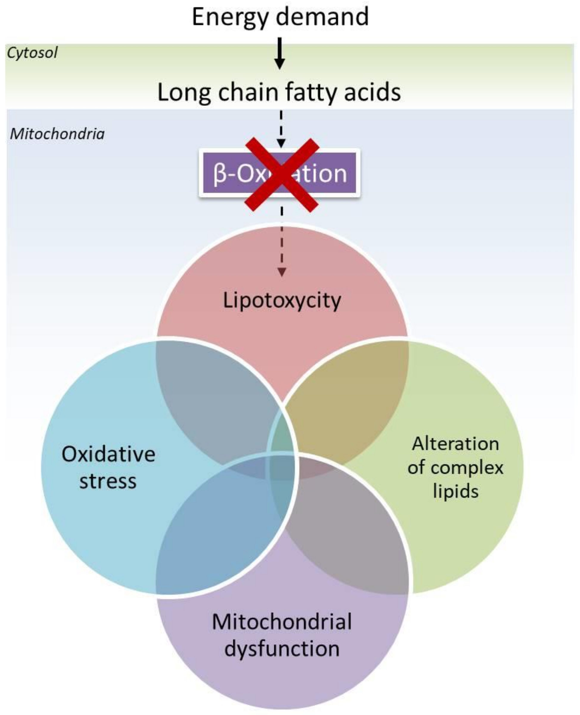
Figure 1. Schematic representation of the proposed working hypothesis on the additional involvement of complex lipids in the onset of neurodegenerative diseases in LCHADD/MTPD. It is known that due to impaired mitochondrial β-oxidation, cells accumulate toxic hydroxyl acylcarnitines. These organelles suffer oxidative stress that leads to mitochondrial dysfunction. At the same time an alteration of content and composition of complex lipids has been observed.
Ijms 23 07144 g001
Publisher’s Note: MDPI stays neutral with regard to jurisdictional claims in published maps and institutional affiliations.