Article

May Staphylococcus lugdunensis Be an Etiological Factor of Chronic Maxillary Sinuses Infection?

by
Maja Kosecka-Strojek
1,*,
Mariola Wolska-Gębarzewska
1,
Adrianna Podbielska-Kubera
2,
Alfred Samet
2,
Beata Krawczyk
3,
Jacek Międzobrodzki
1 and
Michał Michalik
2
1
Department of Microbiology, Faculty of Biochemistry, Biophysics and Biotechnology, Jagiellonian University in Krakow, Gronostajowa 7, 30-387 Krakow, Poland
2
MML Centre, Bagno 2, 00-112 Warsaw, Poland
3
Department of Molecular Biotechnology and Microbiology, Faculty of Chemistry, Gdansk University of Technology, Gabriela Narutowicza 11/12, 80-233 Gdansk, Poland
*
Author to whom correspondence should be addressed.
Int. J. Mol. Sci. 2022, 23(12), 6450; https://doi.org/10.3390/ijms23126450
Submission received: 27 May 2022 / Revised: 3 June 2022 / Accepted: 7 June 2022 / Published: 9 June 2022
(This article belongs to the Special Issue Current and New Knowledge of Biofilm Formation by Staphylococci)

Abstract

:
Staphylococcus lugdunensis is an opportunistic pathogen found in the healthy human skin microbiome bacterial community that is able to cause infections of diverse localization, manifestation, and course, including laryngological infections, such as necrotizing sinusitis. Chronic maxillary sinusitis is a disease present in up to one third of European and American populations, and its etiology is not fully described. Within this study, we aimed to characterize 18 S. lugdunensis strains recovered from maxillary sinuses and evaluate them as etiological agents of chronic disease. We performed MLST analysis, the complex analysis of both phenotypic and genetic virulence factors, antibiotic susceptibility profiles, and biofilm formation assay for the detection of biofilm-associated genes. Altogether, S. lugdunensis strains were clustered into eight different STs, and we demonstrated several virulence factors associated with the chronic disease. All tested strains were able to produce biofilm in vitro with numerous strains with a very strong ability, and overall, they were mostly susceptible to antibiotics, although we found resistance to fosfomycin, erythromycin, and clindamycin in several strains. We believe that further in-depth analysis of S. lugdunensis strains from different niches, including the nasal one, should be performed in the future in order to reduce infection rate and broaden the knowledge about this opportunistic pathogen that is gaining attention.

1. Introduction

Staphylococcus lugdunensis has been known since the late 1980s as a component of the human skin microbiome but also as a potential dangerous pathogen [1]. It is mainly associated with the lower parts of the human body but can also be found in the nasal cavity, causing a wide range of infections, including severe ones with high mortality rates [2,3,4]. Moreover, S. lugdunensis infections may be underestimated since a positive result, as with S. aureus, may be identified with routinely used agglutination tests [5]. The invasive infections are also detected at an insufficient level since coagulase negative staphylococci (CoNS) are often discarded as contaminants. Since the matrix-assisted laser desorption ionization–time of flight mass spectrometry (MALDI TOF MS) method was introduced into routine microbiological practice, S. lugdunensis infections are detected at higher levels [6,7]. Still, chronic infections, such as chronic maxillary sinusitis, are rarely related to S. lugdunensis as an etiological factor [8]. However, it is known that CoNS are responsible for laryngological infections, and there are reports describing S. lugdunensis as responsible for acute necrotizing sinusitis in hospitalized patients with metastatic prostate adenocarcinoma, rhinosinusitis, nasal polyps, and osteomyelitis, which can lead to laryngological implications in terms of significant effects on the head or face bones [8].
Acute rhinosinusitis (ARS) appears the most frequently in the course of a viral cold disease, whereas chronic rhinosinusitis (CRS) is commonly associated with CoNS [9]. Chronic rhinosinusitis is one of the most common chronic disorders, present in up to 28% of European or American populations, and its mechanisms are still under careful molecular analysis [10]. Bacterial infection plays an important role in CRS as either a causative or exacerbating factor. The microbiological analysis of samples from CRS patients revealed that mainly CoNS were identified, followed by S. aureus and gram-negative rods, including Pseudomonas aeruginosa, Stenotrophomonas maltophilia, Escherichia coli, and Serratia marcescens [11,12,13,14]. It was shown that 87% of CRS cases were due to CoNS, with the most frequently isolated being S. epidermidis, S. lugdunensis, S. capitis, Staphylococcus saprophyticus, S. haemolyticus, and Staphylococcus saccharolyticus [8]. Based on previous studies, it was depicted that the pathologic ability of CoNS such as S. epidermidis depends on genes associated with biofilm formation both in CRS but also in all other infections [10,15]. However, knowledge of the importance of bacteria and microbial biofilms in the etiology of CRS is still incomplete. Moreover, it is still not unambiguously shown that the severity of CRS diseases is associated with CoNS solely; further complex analysis based on isolates recovered from patients with CRS are needed. The frequency of CoNS isolation at the infection site and the molecular typing of CoNS to determine the clonality of the isolates is crucial in determining the real etiological factor.
In the case of any chronic infection, not only the detection of biofilm formation but also other virulence factors may play an important role. S. lugdunensis is able to produce several virulence factors such as (i) toxins: haemolysins hlb and hlIII, phenol-soluble modulins—synergistic hemolysins slush; (ii) cell wall modifications: dltA—D-Alanylation of teichoic acids and mprF—multiple peptide resistance factor; (iii) capsular polysaccharide Cap; (iv) adhesins/biofilm synthesis: polysaccharide intercellular adhesin ica, atlL, eno, ebpS, fbl—fibrinogen binding protein of S. lugdunensis, vwbl—von Willebrand factor binding protein of S. lugdunenesis; (v) proteolytic activity: lugdulysin—metalloprotease with homology to the ShpI protease of S. hyicus but not to S. aureus serine or cysteine proteases; and (vi) iron surface determinants: iron-regulated surface determinant Isd proteins (isdC), iron-regulated ECF heme transporter Lha [4].
In the case of S. lugdunensis species, there is one more very interesting feature, namely that it encodes two nonribosomal peptide synthesis systems. One of them is responsible for the synthesis of lugdunin—the new class of cyclic peptide antibiotic with an important antimicrobial and immunomodulatory activity [16]. Lugdunin has antimicrobial activity towards S. aureus via the enhancement of the innate immune response. It was also shown that individuals who are colonized with S. lugdunensis are less likely colonized with S. aureus, and the suppression of S. aureus growth in healthy skin and nares was observed [17,18,19]. In the nasal cavity, it may also depend on the antibiotic action and nutritional competition of these two species. Moreover, it is also considered that predominant niches of S. lugdunensis colonization are skin areas and not the nasal cavity, and S. aureus is rarely found on the skin so it is possible that S. lugdunensis helps to exclude S. aureus from healthy skin [20].
The aim of this study was to characterize S. lugdunensis isolates recovered from chronic maxillary sinusitis. We performed the multilocus sequence typing, complex analysis of both phenotypic and genetic virulence factors, antibiotic susceptibility profiles, and biofilm formation assay with detection of biofilm-associated genes in order to compare those results with laryngological patients’ characteristics.

2. Results

2.1. Isolates and Patients’ Characteristics

All 18 S. lugdunensis isolates were recovered from maxillary sinuses of patients with a chronic disease between 2017 and 2020. The patients were treated in MML Centre due to chronic sinusitis, but some of them also had nasal polyps and snoring problems (Table 1). All patients were male and adult, aged from 26 to 59. Almost all patients had undergone functional endoscopic sinus surgery (FESS). Only two patients did not have any accompanying flora isolated at the time of S. lugdunensis isolation. Altogether, six patients had both S. lugdunensis and S. aureus in maxillary sinuses. Two of them before S. lugdunensis infection, one at the S. lugdunensis isolation, and three were infected with S. aureus after S. lugdunensis isolation.

2.2. Species Identification and Antibiotic Susceptibility

All isolates were identified unambiguously as S. lugdunensis by 16S rRNA and rpoB genes sequencing. Generally, S. lugdunensis strains were mostly susceptible for tested antibiotics. Eight strains were resistant to fosfomycin with a MIC range of 48 to 256 mg/L (including reference strain PL397). A total of four strains were resistant to erythromycin, and all also showed resistance to clindamycin. Single strains showed resistance for amikacin, tetracycline, and tobramycin (Figure 1).

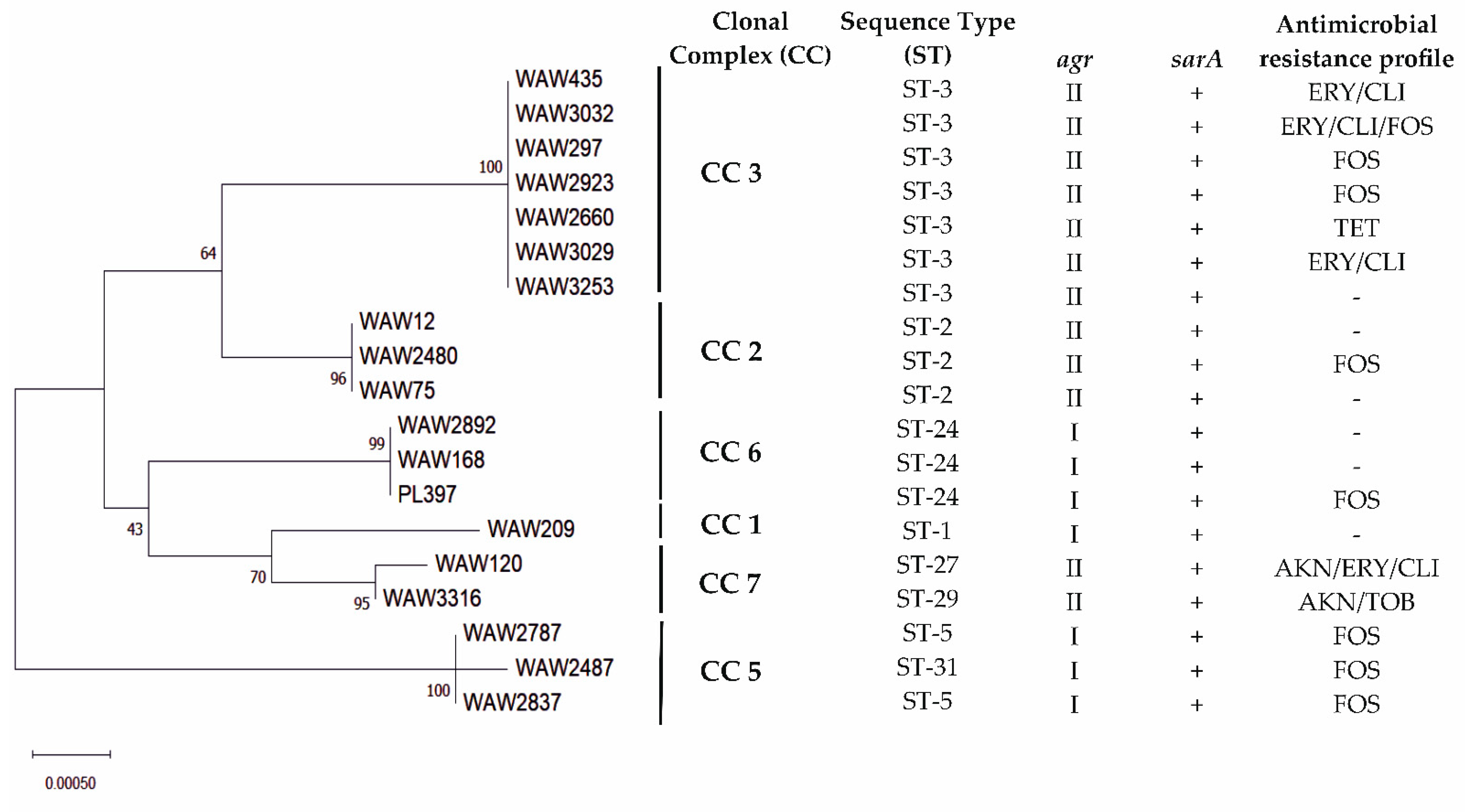
2.3. Multilocus Sequence Types

Based on the S. lugdunensis MLST database, all strains were assigned to six CCs (clonal complexes) including eight STs. The largest cluster was ST3, which grouped seven strains. Next, three strains were assigned to ST2, two to ST24 (together with reference strain PL397), and two to ST5. The remaining four strains were assigned to ST1, ST27, ST29, and ST31 (Figure 1).

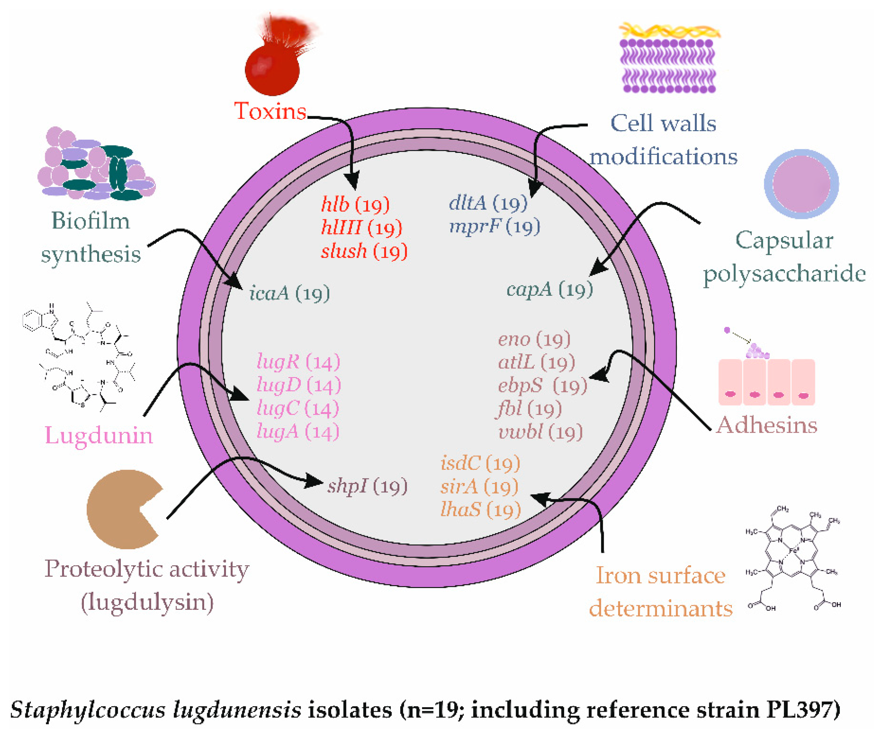
2.4. Virulence Factors Distribution

All S. lugdunensis strains showed beta hemolysis in vitro and had hlb, hlIII, and SLUSH genes (including reference strain PL397). Five strains showed very high (incl. PL397) and nine strains high proteolysis, and five strains did not show proteolysis in vitro. All strains had a sarA gene and most strains had agr type II, with seven strains demonstrating type I (incl. PL397). Altogether, all strains possessed virulence genes associated with: cell wall modifications (dltA, mprF); capsular polysaccharide (capA); adhesion (eno, atlL, ebpS, fbl, vwbl); proteolytic activity (shpI); and iron acquisition (isdC, sirA and lhaS). A total of 14 strains produced lugdunin in vitro and possessed lugRDCA genes (incl. PL397), whereas the remaining five strains did not (Figure 2).

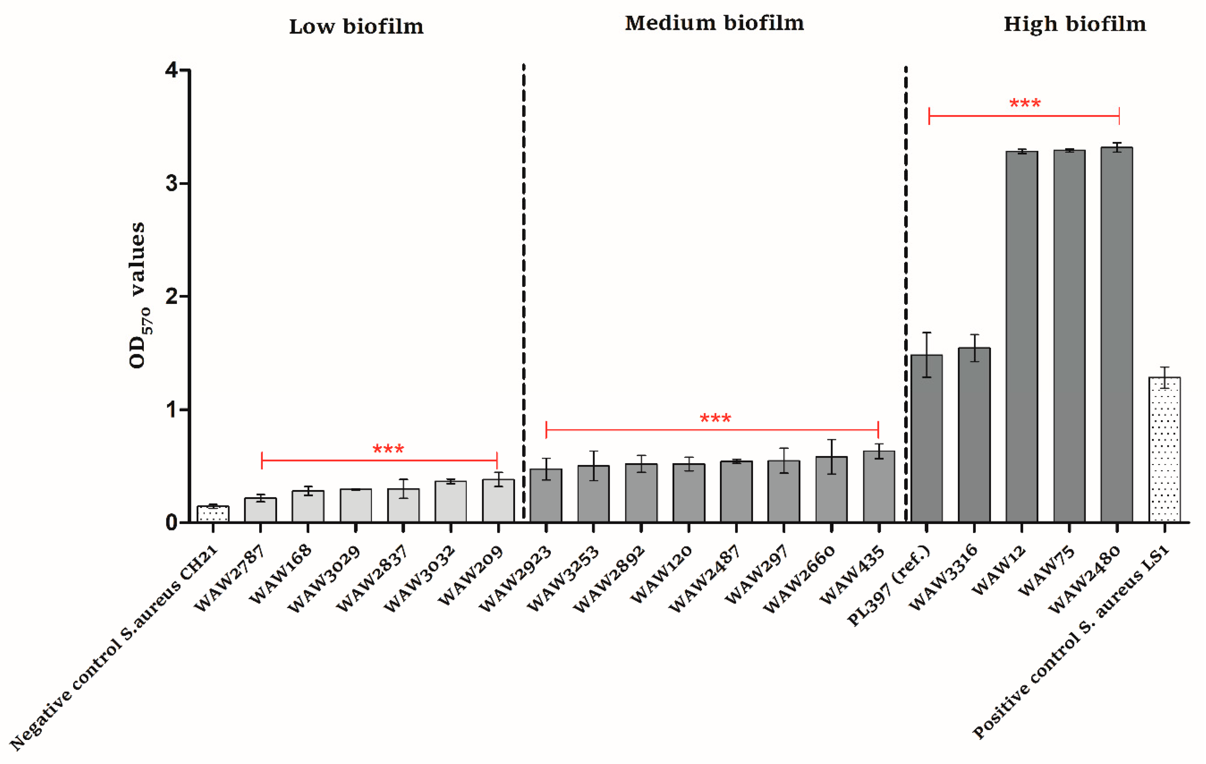
2.5. Biofilm Formation

Compared to the negative and positive controls, all S. lugdunensis strains produced biofilm in vitro (Figure 3). Six strains did so at a low level (OD570 values from 0.15 to 0.38), eight strains at a medium level (OD570 values from 0.39 to 1.27), and four strains at a high level (OD570 values above 1.28). The reference S. lugdunensis strain (PL397) produced biofilm at a high level. Moreover, all strains possessed the icaA gene (Figure 2).

3. Discussion

Four decades after the first description of Staphylococcus lugdunensis, much more is known about its pathogenicity and virulence potential. Based on current research, this species should not be discarded as a harmless contaminant from clinical samples and should be considered as a dangerous opportunistic pathogen [4]. S. lugdunensis represents one of the most aggressive CoNS species, causing severe infections, especially infective endocarditis and soft tissue infections [22,23]. This is due to the fact that S. lugdunensis possesses a number of virulence factors associated mainly with toxic/haemolytic potential, acquisition of heme, cell wall modifications, and adhesins. Although, the number of virulence factors is not as high as that of S. aureus [24]. It is also known that S. aureus and S. lugdunensis can share the niche; this becomes even more interesting in light of the fact that S. lugdunensis produces the antibiotic lugdunin, which kills S. aureus. However, there are also reports where individuals colonized with S. aureus did not have S. lugdunensis in their nose [16].
Chronic maxillary sinusitis is the term generally used to describe nasal congestion or discharge that persists for 8 to 12 weeks. Chronic disease rarely causes symptoms of pain except during acute exacerbations [9]. Chronic sinusitis disease is usually bacterial rather than viral, and the most commonly isolated are Gram-positive cocci. Between them, S. lugdunensis was also observed, but there are not much data regarding the actual recognition of this species as an etiological factor of chronic maxillary sinuses disease [8]. Within this study, we aimed to characterize 18 S. lugdunensis strains recovered from the maxillary sinuses of patients with a chronic disease between 2017 and 2020 treated in MML Centre. First of all, we performed MLST analysis in order to determine the strains clonality. Altogether, the strains were assigned to six CCs including eight different STs, revealing high genetic diversity. The most common were ST3 and ST2, as also demonstrated in other studies in Europe [25]. Based on recent research, S. lugdunensis displays a closed pan genome, and all published genomes are very similar, with similar genes set among the strains [26]. Even though this was not expected, it can explain the very well conserved antimicrobial susceptibility of S. lugdunensis that rarely acquires resistance genes [27]. So, as expected, this study also demonstrated that all S. lugdunensis strains were similar based on genes’ distribution. All strains possessed genes associated with a broad spectrum of virulence such as toxin production, cell wall modifications, capsular polysaccharide, adhesion, proteolytic activity, and iron acquisition. Interestingly, eight strains were resistant to fosfomycin and four strains were resistant to erythromycin; also, all showed resistance to clindamycin. According to a review by Argemi et al., susceptibility to fosfomycin is greatly variable, with resistance reported in >50% of isolates. Moreover, the resistance to erythromycin and clindamycin is also emerging in such strains [26].
Together with antibiotic resistance, the biofilm formation is an important pathogenicity potential of the strain [28]. This feature is mainly important in infections associated with indwelling devices, but it is also known that the eradication of biofilm in chronic infections is of high priority [29]. In the case of our study, all S. lugdunensis strains were able to produce biofilm. Four strains produced biofilm at a high and very high level (OD570 values above 1.28). We depicted the scale of low, medium, and high biofilm formation based on two S. aureus strains, one with OD570 = 0.1446 as a negative control and one with OD570 = 1.2837 as a positive control. It should be noted that three strains produced biofilm at a rate that was two-fold higher than that of the positive control. Although all strains carried the ica locus, it is probably not the only cause of biofilm production. In staphylococci, several molecules other than polysaccharide intracellular adhesin (encoded by the ica operon) molecules have been shown to play an important role in biofilm formation, such as major autolysin Atl (atlL), the enzyme for D-alanine esterification of teichoic acids DltA (dltA), the iron-regulated surface determinant IsdC (isdC) under iron-limited conditions, and also the agr system, which is a component of the quorum-sensing agr system and is known to be a major element in the regulation of pathogenicity [30,31,32]. All tested S. lugdunensis strains possessed atlL, dltA, isdC, and agr genes.
As mentioned previously by Zipperer et al. [16] and later by Heilbronner and Foster in a review article [4], S. lugdunensis might be usable as a probiotic to reduce or eliminate S. aureus carriage when applied to nasal cavities. We decided to check if patients with chronic maxillary sinusitis were colonized with S. aureus at the time of S. lugdunensis isolation or before/after infection caused by S. lugdunensis. We found that eight patients had both S. lugdunensis and S. aureus in maxillary sinuses. Three of them before S. lugdunensis infection, one at the S. lugdunensis isolation, and five were infected with S. aureus after S. lugdunensis isolation.
Even though there are many open questions, it is speculated that S. lugdunensis is adapted to the presence of S. aureus. This may be explained by the fact that both species share the same niche but not the same adhesion sites. The success in nasal colonization by S. aureus is known to be multifactorial but mainly relies on cell wall adhesins such as ClfB or SdrD and wall teichoic acid molecules that interact with epithelial cells receptors [18]. The knowledge about the factors facilitating S. lugdunensis nasal colonization is insufficient. However, S. lugdunensis do not encode ClfB and SdrD, or a homologue of the S. epidermidis Aap protein, so it is not likely that the adhesion strategies are the same as in S. aureus. It is possible that S. lugdunensis uses S. aureus for its own benefit when the bacteria number is low and produces lugdunin to kill S. aureus when its number increases [4]. However, our findings are based on several strains; thus, it should be considered that S. lugdunensis and S. aureus may colonize the same niche, and further complex investigations are warranted.

4. Conclusions

In conclusion, within this study, we demonstrated several virulence factors carried by S. lugdunensis strains recovered from maxillary sinuses and associated with chronic disease. All tested strains were clustered in eight different STs. All tested strains were able to produce biofilm in vitro with numerous strains having a very strong ability. Overall, the strains were susceptible to most antibiotics tested in the study, but we found resistance to fosfomycin, erythromycin, and clindamycin in several strains. Moreover, the co-colonization of S. lugdunensis and S. aureus in the nasal cavity of laryngological patients should be reconsidered in the future. We believe that further in-depth analysis of S. lugdunensis strains from different niches, including the nasal one, should be performed in the future in order to reduce infection rates and broaden the knowledge about this opportunistic pathogen that is gaining attention.

5. Materials and Methods

5.1. Bacterial Isolates

The study included 18 S. lugdunensis clinical isolates recovered between 2017 and 2020 from laryngological patients treated in MML Medical Centre, Warsaw. The collection of bacterial isolates used in this study is described in Table 1. All isolates were recovered from maxillary sinuses. The PCM2430 (PL397 in this study) strain deposited in Polish Collection of Microorganisms (PCM) was used as a reference. All strains were cultivated on blood agar medium with 5% sheep blood (Graso Biotech, Starograd Gdański, Poland) in 37 °C for 20 h.

5.2. Genomic DNA Extraction

Genomic DNA extraction was performed as previously described [33]. Briefly, isolates were grown for 20 h at 37 °C on blood agar plates. A full inoculation loop of 10 μL of bacterial colonies was homogenized with a TissueLyser II (Qiagen, Germantown, MD, USA). The Qiagen DNeasy Blood & Tissue Kit (Qiagen, Germantown, MD, USA) was used for genomic DNA extraction. The subsequent steps were performed according to the manufacturer’s instructions. Purified DNA was stored at −20 °C.

5.3. Species Identification

All isolates were identified at the species level by sequencing the 16S rRNA and rpoB genes as previously described [34,35]. The PCR products were resolved by electrophoresis and purified using the Clean-Up Concentrator purification kit (A&A Biotechnology, Gdynia, Poland). Concentration and purity were measured using a NanoDrop ND-1000 (Thermo Fisher Scientific, Waltham, MA, USA). The PCR products were sequenced with the Sanger method at Genomed S.A. (Warsaw, Poland) with the same primers as those used for PCR.

5.4. Susceptibility Testing

Susceptibility testing was carried out according to the European Committee on Antimicrobial Susceptibility Testing (EUCAST; www.eucast.org/; Accessed on 11 February 2021) recommendations for all S. lugdunensis isolates. Disc diffusion values were performed for amikacin, cefazoline, cefoxitin, chloramphenicol, ciprofloxacin, clindamycin, erythromycin, gentamicin, linezolid, tetracycline, tigecycline, tobramycin, trimethoprim-sulfamethoxazole, rifampicin. Minimum inhibitory concentration (MIC) values for daptomycin, fosfomycin, teicoplanin, and vancomycin were determined using the Etest method.

5.5. Phenotypic Assessment of Secreted Virulence Factors Production

The secretion of virulence factors was evaluated using a culture-dependent assay with specific substratum for hemolysis and proteolysis detections [24]. All isolates were previously grown at 37 °C on TSA (Sigma-Aldrich, St. Louis, MO, USA). For hemolysin production, the S. lugdunensis isolates were cultivated on 5% sheep blood agar (Graso Biotech, Starograd Gdański, Poland), and the aspect of the hemolytic zone surrounding the colony was noted. For proteolysis, the isolates were cultivated onto TSA (Sigma-Aldrich, St. Louis, MO, USA) with 10% milk (0% fat).

5.6. Lugdunin Activity Assay

Lugdunin activity assay was performed as previously described [16], with modifications. Briefly, the antimicrobial activity was investigated against S. aureus USA300 ATCC BAA-1717. All S. lugdunensis strains and S. aureus USA300 were cultivated on TSA (Sigma-Aldrich, St. Louis, MO, USA) in 37 °C for 20 h. Then, a single colony was inoculated in 10 mL Basic Medium (BM: 1% tryptone, 0.5% yeast extract, 0.5% NaCl, 0.1% glucose and 0.1% K2HPO4, pH 7.2). Suspensions were incubated for 24 h at 37 °C with shaking (180 rpm). The S. aureus USA300 ATCC BAA-1717 strain was equated to OD600 = 0.5 (optical density measured at 600 nm wavelength) in fresh BM. In the next step, BM agar (BM: 1% tryptone, 0.5% yeast extract, 0.5% NaCl, 0.1% glucose and 0.1% K2HPO4, 1.5% agar, pH 7.2) supplemented with 200 μM 2,2′-bipyridine was used because, as previously described, isolates’ antimicrobial activity under iron-limiting conditions was higher [15]. So, BM agar plates were inoculated with an equated culture of S. aureus USA300 ATCC BAA-1717 in order to grow a bacterial lawn. All tested S. lugdunensis strains were inoculated in amount of 50 μL drop on the resulting S. aureus bacterial lawn, and the plates were incubated at 37 °C. After 24 h and 48 h incubation, the zone sizes were measured.

5.7. Biofilm Formation Assay

The quantitative assay for biofilm formation was carried out as previously described, with several modifications [36]. Briefly, all S. lugdunensis isolates were grown overnight at 37 °C as pure cultures on Tryptic Soy Agar (TSA) (Sigma-Aldrich, St. Louis, MO, USA). The single colony was inoculated in 10 mL Tryptic Soy Broth (TSB) (Sigma-Aldrich, St. Louis, MO, USA) and incubated for 24 h at 37 °C. After overnight culture, bacterial suspensions were equated to OD600 = 0.5 (optical density measured at 600 nm wavelength) with fresh TSB (Sigma-Aldrich, St. Louis, MO, USA). Then, suspensions were diluted 1:100 with fresh TSB (Sigma-Aldrich, St. Louis, MO, USA) with 1% glucose (POCH, Gliwice, Poland). For each isolate, 200 µL aliquots of prepared suspension were inoculated into four wells of the 96-well tissue culture plates (Biologix, Jinan, China). Each culture plate included an experiment negative control (TSB with 1% glucose) and biofilm negative activity control (S. aureus, CH21 strain). Ap S. aureus (LS1) strain that is a hyperproducer of biofilm formation was used as a positive control. The plates were incubated at 37 °C for 48 h. Afterwards, content of each well was removed by aspiration and the wells were rinsed three times with 300 µL sterile phosphate buffer solution (PBS) (Sigma-Aldrich, St. Louis, MO, USA). The plates were dried in 60 °C for 1 h and afterwards stained with 150 µL 0.5% crystal violet solution (Honeywell Fluka, Charlotte, NC, USA) for 15 min at room temperature. Following staining, the plates were rinsed using distilled water until there was no visible trace of stain. The stain bound to bacteria was dissolved by adding 150 µL of 33% acetic acid solution (POCH, Gliwice, Poland). The OD of each well was measured using a spectrometric reader (FlexiStation 3, Molecular Devices, San Jose, CA, USA) at 570 nm (OD570). Biofilm formation data were analyzed with the Bonferroni’s Multiple Comparison Test using GraphPad Prism 5 software (San Diego, CA, USA).

5.8. MLST Typing

The multilocus sequence typing (MLST) was carried out according to the protocol for S. lugdunensis on the MLST website (https://bigsdb.pasteur.fr/staphlugdunensis/; Accessed on 26 July 2021) and as previously described [25]. For ddl gene amplification, primers ddl-F-5′-CAAAATGTATTAAATGCTAT-3′ and ddl-R-5′-ATTTAATGATATTTCCTTGAT-3′ (product size 421 bp) were used. Briefly, all housekeeping genes were amplified with PCR and sequenced, then, alleles and sequence types (STs) for allelic profiles were assigned according to the S. lugdunensis MLST website.

5.9. Molecular Detection of Virulence Genes

The PCRs targeted to detect the presence of the following genes: the accessory gene regulator (agr) [37], the staphylococcal accessory regulator (sarA) [38], adhesins genes (atlL) [39], haemolysins genes (hlb, hem-III) [40], the genes encoding proteins of the extracellular matrix (fbl, vwbl) [40,41], S. lugdunensis synergistic hemolysins (slush) [40] and proteolytic activity—lugdulysin (shpI) [42], were performed. The agr types were identified based on the expected product sizes (586 bp for agr type I and 771 bp for agr type II). Detection of lugdunin operon (lugRDCA), biofilm synthesis gene (icaA), adhesins genes encoding laminin and elastin binding protein (eno, ebpS), capsular polysaccharide biosynthesis protein gene (capA), cell walls modifications genes (mprF and dltA), and iron surface determinant genes (isdC, sirA, lhaS) was performed with sequencing primers designed in the current study (Table 2). The PCR products were resolved by electrophoresis, and the band patterns were analyzed.

5.10. Nucleotide Sequence Accession Numbers

The 169 sequences for one Staphylococcus lugdunensis were annotated using the NCBI BankIt tool and deposited in the GenBank database (https://www.ncbi.nlm.nih.gov/genbank/ accessed on 26 May 2022) under the following accession numbers: for the 16S rRNA gene, ON584774-ON584791; for the rpoB gene, ON604987-ON605004; for the aroE, dat, ddl, gmk, ldh, recA and yqiL genes, ON605005-ON605137, respectively.

5.11. Statistical Analysis

Biofilm formation experimental data were analyzed using ANOVA test followed by the Bonferroni’s multiple comparison test to evaluate the difference between groups. Before analysis, normality was checked with the Shapiro–Wilk test. All analyses were performed using GraphPad Prism (GraphPad Software, Inc., La Jolla, CA, USA; version 5.03) and data were presented as the mean ± standard deviation. p value < 0.05 was considered to indicate a statistically significant difference.

Author Contributions

Conceptualization, M.K.-S., J.M. and M.M.; methodology, M.K.-S.; validation, M.K.-S., M.W.-G., B.K. and J.M.; formal analysis, M.K.-S. and A.P.-K.; investigation, M.K.-S. and M.W.-G.; resources, A.S., A.P.-K., J.M. and M.M.; data curation, M.K.-S.; writing—original draft preparation, M.K.-S.; writing—review and editing, M.K.-S., M.W.-G., B.K., J.M. and M.M.; visualization, M.K.-S. and M.W.-G.; supervision, M.K.-S.; project administration, M.K.-S.; funding acquisition, M.M. All authors have read and agreed to the published version of the manuscript.

Funding

The APC was funded by MML Centre, Warsaw.

Institutional Review Board Statement

Not applicable.

Informed Consent Statement

Informed consent was obtained from all subjects involved in the study.

Data Availability Statement

The datasets generated for this study can be found in Genbank ON584774-ON584791; ON604987-ON605004; ON605005-ON605137.

Acknowledgments

The authors wish to thank Michał Bukowski from the Department of Analytical Biochemistry, Faculty of Biochemistry, Biophysics and Biotechnology of Jagiellonian University in Krakow for invaluable help with analysis.

Conflicts of Interest

The authors declare no conflict of interest.

References

  1. Taha, L.; Stegger, M.; Söderquist, B. Staphylococcus lugdunensis: Antimicrobial susceptibility and optimal treatment options. Eur. J. Clin. Microbiol. Infect. Dis. 2019, 38, 1449–1455. [Google Scholar] [CrossRef] [PubMed] [Green Version]
  2. Bieber, L.; Kahlmeter, G. Staphylococcus lugdunensis in several niches of the normal skin flora. Clin. Microbiol. Infect. 2010, 16, 385–388. [Google Scholar] [CrossRef] [PubMed] [Green Version]
  3. Liu, C.M.; Price, L.B.; Hungate, B.A.; Abraham, A.G.; Larsen, L.A.; Christensen, K.; Stegger, M.; Skov, R.; Andersen, P.S. Staphylococcus aureus and the ecology of the nasal microbiome. Sci. Adv. 2015, 1, e1400216. [Google Scholar] [CrossRef] [PubMed] [Green Version]
  4. Heilbronner, S.; Foster, T.J. Staphylococcus lugdunensis: A skin commensal with invasive pathogenic potential. Clin. Microbiol. Rev. 2021, 34, e00205-20. [Google Scholar] [CrossRef]
  5. Thean, Y.T.; Siew, Y.N.; He, J. Microbiological characteristics, presumptive identification, and antibiotic susceptibilities of Staphylococcus lugdunensis. J. Clin. Microbiol. 2008, 46, 2393–2395. [Google Scholar] [CrossRef] [Green Version]
  6. Argemi, X.; Riegel, P.; Lavigne, T.; Lefebvre, N.; Grandpré, N.; Hansmann, Y.; Jaulhac, B.; Prévost, G.; Schramm, F. Implementation of matrix-assisted laser desorption ionization-time of flight mass spectrometry in routine clinical laboratories improves identification of coagulase-negative staphylococci and reveals the pathogenic role of Staphylococcus lugdunensis. J. Clin. Microbiol. 2015, 53, 2030–2036. [Google Scholar] [CrossRef] [Green Version]
  7. Clark, A.E.; Kaleta, E.J.; Arora, A.; Wolk, D.M. Matrix-assisted laser desorption ionization-time of flight mass spectrometry: A fundamental shift in the routine practice of clinical microbiology. Clin. Microbiol. Rev. 2013, 26, 547–603. [Google Scholar] [CrossRef] [Green Version]
  8. Michalik, M.; Samet, A.; Podbielska-Kubera, A.; Savini, V.; Międzobrodzki, J.; Kosecka-Strojek, M. Coagulase-negative staphylococci (CoNS) as a significant etiological factor of laryngological infections: A review. Ann. Clin. Microbiol. Antimicrob. 2020, 19, 26. [Google Scholar] [CrossRef]
  9. Bachert, C.; Pawankar, R.; Zhang, L.; Bunnag, C.; Fokkens, W.J.; Hamilos, D.L.; Jirapongsananuruk, O.; Kern, R.; Meltzer, E.O.; Mullol, J.; et al. ICON: Chronic rhinosinusitis. World Allergy Organ. J. 2014, 7, 25. [Google Scholar] [CrossRef] [Green Version]
  10. Dlugaszewska, J.; Leszczynska, M.; Lenkowski, M.; Tatarska, A.; Pastusiak, T.; Szyfter, W. The pathophysiological role of bacterial biofilms in chronic sinusitis. Eur. Arch. Otorhinolaryngol. 2016, 273, 1989–1994. [Google Scholar] [CrossRef] [Green Version]
  11. Coffey, C.S.; Sonnenburg, R.E.; Melroy, C.T.; Dubin, M.G.; Senior, B.A. Endoscopically guided aerobic cultures in postsurgical patients with chronic rhinosinusitis. Am. J. Rhinol. 2006, 20, 72–76. [Google Scholar] [CrossRef] [PubMed]
  12. Jervis-Bardy, J.; Foreman, A.; Field, J.; Wormald, P.J. Impaired mucosal healing and infection associated with Staphylococcus aureus after endoscopic sinus surgery. Am. J. Rhinol. Allergy 2009, 23, 549–552. [Google Scholar] [CrossRef] [PubMed]
  13. Michalik, M.; Samet, A.; Marszałek, A.; Krawczyk, B.; Kotłowski, R.; Nowicki, A.; Anyszek, T.; Nowicki, S.; Kur, J.; Nowicki, B. Intra-operative biopsy in chronic sinusitis detects pathogenic Escherichia coli that carry fimG/H, fyuA and agn43 genes coding biofilm formation. PLoS ONE 2018, 13, e0192899. [Google Scholar] [CrossRef] [PubMed]
  14. Krawczyk, B.; Michalik, M.; Fordon, M.; Wysocka, M.; Samet, A.; Nowicki, B. Escherichia coli strains with virulent factors typical for uropathogens were isolated from sinuses from patients with chronic rhinosinusitis-case report. Pathogens 2020, 9, 318. [Google Scholar] [CrossRef] [PubMed]
  15. Both, A.; Huang, J.; Qi, M.; Lausmann, C.; Weißelberg, S.; Büttner, H.; Lezius, S.; Failla, A.V.; Christner, M.; Stegger, M.; et al. Distinct clonal lineages and within-host diversification shape invasive Staphylococcus epidermidis populations. PLoS Pathog. 2021, 17, e1009304. [Google Scholar] [CrossRef] [PubMed]
  16. Zipperer, A.; Konnerth, M.C.; Laux, C.; Berscheid, A.; Janek, D.; Weidenmaier, C.; Burian, M.; Schilling, N.A.; Slavetinsky, C.; Marschal, M.; et al. Human commensals producing a novel antibiotic impair pathogen colonization. Nature 2016, 535, 511–516. [Google Scholar] [CrossRef]
  17. Nakatsuji, T.; Chen, T.H.; Narala, S.; Chun, K.A.; Two, A.M.; Yun, T.; Shafiq, F.; Kotol, P.F.; Bouslimani, A.; Melnik, A.V.; et al. Antimicrobials from human skin commensal bacteria protect against Staphylococcus aureus and are deficient in atopic dermatitis. Sci. Transl. Med. 2017, 9, eaah4680. [Google Scholar] [CrossRef] [Green Version]
  18. Sakr, A.; Brégeon, F.; Mège, J.L.; Rolain, J.M.; Blin, O. Staphylococcus aureus nasal colonization: An update on mechanisms, epidemiology, risk factors, and subsequent infections. Front. Microbiol. 2018, 9, 2419. [Google Scholar] [CrossRef]
  19. Bitschar, K.; Sauer, B.; Focken, J.; Dehmer, H.; Moos, S.; Konnerth, M.; Schilling, N.A.; Grond, S.; Kalbacher, H.; Kurschus, F.C.; et al. Lugdunin amplifies innate immune responses in the skin in synergy with host- and microbiota-derived factors. Nat. Commun. 2019, 10, 2730. [Google Scholar] [CrossRef]
  20. Heilbronner, S. Staphylococcus lugdunensis. Trends Microbiol. 2021, 29, 1143–1145. [Google Scholar] [CrossRef]
  21. Kumar, S.; Stecher, G.; Li, M.; Knyaz, C.; Tamura, K. MEGA X: Molecular Evolutionary Genetics Analysis across computing platforms. Mol. Biol. Evol. 2018, 35, 1547–1549. [Google Scholar] [CrossRef] [PubMed]
  22. Liu, P.Y.; Huang, Y.F.; Tang, C.W.; Chen, Y.Y.; Hsieh, K.S.; Ger, L.P.; Chen, Y.S.; Liu, Y.C. Staphylococcus lugdunensis infective endocarditis: A literature review and analysis of risk factors. J. Microbiol. Immunol. Infect. 2010, 43, 478–484. [Google Scholar] [CrossRef] [Green Version]
  23. Non, L.R.; Santos, C.A.Q. The occurrence of infective endocarditis with Staphylococcus lugdunensis bacteremia: A retrospective cohort study and systematic review. J. Infect. 2017, 74, 179–186. [Google Scholar] [CrossRef] [PubMed]
  24. Preda, M.; Mihai, M.M.; Popa, L.I.; Dițu, L.-M.; Holban, A.M.; Manolescu, L.S.C.; Popa, G.-L.; Muntean, A.-A.; Gheorghe, I.; Chifiriuc, C.M.; et al. Phenotypic and genotypic virulence features of staphylococcal strains isolated from difficult-to-treat skin and soft tissue infections. PLoS ONE 2021, 16, e0246478. [Google Scholar] [CrossRef] [PubMed]
  25. Chassain, B.; Lemée, L.; Didi, J.; Thiberge, J.M.; Brisse, S.; Pons, J.L.; Pestel-Caron, M. Multilocus sequence typing analysis of Staphylococcus lugdunensis implies a clonal population structure. J. Clin. Microbiol. 2012, 50, 3003–3009. [Google Scholar] [CrossRef] [PubMed] [Green Version]
  26. Argemi, X.; Hansmann, Y.; Prola, K.; Prévost, G. Coagulase-negative staphylococci pathogenomics. Int. J. Mol. Sci. 2019, 20, 1215. [Google Scholar] [CrossRef] [PubMed] [Green Version]
  27. Chang, S.C.; Lee, M.H.; Yeh, C.F.; Liu, T.P.; Lin, J.F.; Ho, C.M.; Lu, J.J. Characterization of two novel variants of staphylococcal cassette chromosome mec elements in oxacillin-resistant Staphylococcus lugdunensis. J. Antimicrob. Chemother. 2017, 72, 3258–3262. [Google Scholar] [CrossRef]
  28. Zapotoczna, M.; Heilbronner, S.; Speziale, P.; Foster, T.J. Iron-regulated surface determinant (Isd) proteins of Staphylococcus lugdunensis. J. Bacteriol. 2012, 194, 6453–6467. [Google Scholar] [CrossRef] [Green Version]
  29. Lebeaux, D.; Ghigo, J.M.; Beloin, C. Biofilm-related infections: Bridging the gap between clinical management and fundamental aspects of recalcitrance toward antibiotics. Microbiol. Mol. Biol. Rev. 2014, 78, 510–543. [Google Scholar] [CrossRef] [Green Version]
  30. Aubourg, M.; Dhalluin, A.; Gravey, F.; Pottier, M.; Thomy, N.; Bernay, B.; Goux, D.; Martineau, M.; Giard, J.-C. Phenotypic and proteomic approaches of the response to iron-limited condition in Staphylococcus lugdunensis. BMC Microbiol. 2020, 20, 328. [Google Scholar] [CrossRef]
  31. Aubourg, M.; Pottier, M.; Léon, A.; Bernay, B.; Dhalluin, A.; Cacaci, M.; Torelli, R.; Ledormand, P.; Martini, C.; Sanguinetti, M.; et al. Inactivation of the response regulator AgrA has a pleiotropic effect on biofilm formation, pathogenesis and stress response in Staphylococcus lugdunensis. Microbiol. Spectr. 2022, 10, e0159821. [Google Scholar] [CrossRef] [PubMed]
  32. Dahyot, S.; Oxaran, V.; Niepceron, M.; Dupart, E.; Legris, S.; Destruel, L.; Didi, J.; Clamens, T.; Lesouhaitier, O.; Zerdoumi, Y.; et al. Role of the LytSR two-component regulatory system in Staphylococcus lugdunensis biofilm formation and pathogenesis. Front. Microbiol. 2020, 11, 39. [Google Scholar] [CrossRef] [PubMed] [Green Version]
  33. Kosecka-Strojek, M.; Sabat, A.J.; Akkerboom, V.; Becker, K.; van Zanten, E.; Wisselink, G.; Miedzobrodzki, J.; Kooistra-Smid, A.; Friedrich, A.W. Development and Validation of a Reference Data Set for Assigning Staphylococcus Species Based on Next-Generation Sequencing of the 16S-23S rRNA Region. Front. Cell. Infect. Microbiol. 2019, 9, 278. [Google Scholar] [CrossRef] [PubMed] [Green Version]
  34. Woo, P.C.; Leung, A.S.; Leung, K.W.; Yuen, K.Y. Identification of slide coagulase positive, tube coagulase negative Staphylococcus aureus by 16S ribosomal RNA gene sequencing. Mol. Pathol. MP 2001, 54, 244–247. [Google Scholar] [CrossRef] [PubMed] [Green Version]
  35. Mellmann, A.; Becker, K.; von Eiff, C.; Keckevoet, U.; Schumann, P.; Harmsen, D. Sequencing and staphylococci identification. Emerg. Infect. Dis. 2006, 12, 333–336. [Google Scholar] [CrossRef] [PubMed]
  36. Stepanović, S.; Vuković, D.; Hola, V.; Di Bonaventura, G.; Djukić, S.; Cirković, I.; Ruzicka, F. Quantification of biofilm in microtiter plates: Overview of testing conditions and practical recommendations for assessment of biofilm production by staphylococci. J. Pathol. Microbiol. Immunol. 2007, 115, 891–899. [Google Scholar] [CrossRef]
  37. Yeh, C.F.; Liu, T.P.; Cheng, C.W.; Chang, S.C.; Lee, M.H.; Lu, J.J. Molecular characteristics of disease-causing and commensal Staphylococcus lugdunensis isolates from 2003 to 2013 at a tertiary hospital in Taiwan. PLoS ONE 2015, 10, e0134859. [Google Scholar] [CrossRef]
  38. Frebourg, N.B.; Lefebvre, S.; Baert, S.; Lemeland, J.F. PCR-Based assay for discrimination between invasive and contaminating Staphylococcus epidermidis strains. J. Clin. Microbiol. 2000, 38, 877–880. [Google Scholar] [CrossRef] [Green Version]
  39. Giormezis, N.; Kolonitsiou, F.; Makri, A.; Vogiatzi, A.; Christofidou, M.; Anastassiou, E.D.; Spiliopoulou, I. Virulence factors among Staphylococcus lugdunensis are associated with infection sites and clonal spread. Eur. J. Clin. Microbiol. Infect. Dis. 2015, 34, 773–778. [Google Scholar] [CrossRef]
  40. Szabados, F.; Nowotny, Y.; Marlinghaus, L.; Korte, M.; Neumann, S.; Kaase, M.; Gatermann, S.G. Occurrence of genes of putative fibrinogen binding proteins and hemolysins, as well as of their phenotypic correlates in isolates of S. lugdunensis of different origins. BMC Res. Notes 2011, 4, 113. [Google Scholar] [CrossRef]
  41. Chatzigeorgiou, K.S.; Siafakas, N.; Petinaki, E.; Zerva, L. fbl gene as a species-specific target for Staphylococcus lugdunensis identification. J. Clin. Lab. Anal. 2010, 24, 119–122. [Google Scholar] [CrossRef] [PubMed]
  42. Argemi, X.; Prévost, G.; Riegel, P.; Keller, D.; Meyer, N.; Baldeyrou, M.; Douiri, N.; Lefebvre, N.; Meghit, K.; Ronde Oustau, C.; et al. VISLISI trial, a prospective clinical study allowing identification of a new metalloprotease and putative virulence factor from Staphylococcus lugdunensis. Clin. Microbiol. Infect. 2017, 23, 334.e1–334.e8. [Google Scholar] [CrossRef] [PubMed] [Green Version]
Figure 1. Sequence types (STs), agr types, sarA gene distribution, and antibiotic resistance profiles of Staphylococcus lugdunensis strains. A phylogenetic tree was constructed based on S. lugdunensis strains MLST results with Neighbor-Joining method in MEGA X (v. 10.2.4) [21]. The percentage of replicate trees in which the associated taxa clustered together in the bootstrap test (1000 replicates) are shown next to the branches. To identify clonal complexes (CC), BURST analysis was conducted in Bacterial Isolate Genome Sequence Database (BIGSdb) curated by Institut Pasteur, Paris, France. Antibiotics, AKN: amikacin; ERY: erythromycin; CLI: clindamycin; FOS: fosfomycin; TET: tetracycline; TOB: tobramycin; The hyphen mark means susceptibility to all antibiotics tested in the study.
Ijms 23 06450 g001
Figure 2. Virulence factors carried by S. lugdunensis strains.
Ijms 23 06450 g002
Figure 3. Biofilm formation in vitro of Staphylococcus lugdunensis strains. Each bar represents mean ± standard deviation. Dotted bars: negative and positive controls; light gray bars: low biofilm formation; medium gray bars: medium biofilm formation; dark gray bars: high biofilm formation. Experiments were performed in four repetitions. Normality was checked with Shapiro–Wilk test. Biofilm formation experimental data were analyzed using ANOVA test followed by the Bonferroni’s multiple comparison test to evaluate the difference between groups (*** p < 0.0001).
Ijms 23 06450 g003
Table 1. Characteristic of patients and isolates from chronic sinusitis.
Clinical Characteristic of PatientsMicrobiological Cultures—Characteristic
Isolate NameDate of Strain IsolationIsolation SiteAgeGenderUnderlying DiseasesAdditional Patient-Associated Risk FactorsSurgeryAccompanying Bacterial Flora at Isolation TimeIsolation of S. aureus
WAW24806 September 2017left maxillary sinus59Mchronic sinusitisnoneFESS, DSN, HCNIS. epidermidis; S. salivarius; S. mitisno
WAW24878 September 2017right maxillary sinus36Mchronic sinusitissleep apneaDSN, HCNI, UPPE. colino
WAW266031 October 2017right maxillary sinus56Mchronic sinusitisnoneFESS, DSNS. epidermidisno
WAW278713 January 2018right maxillary sinus43Mchronic sinusitissleep disturbance; snoringDSN, HCNIno datano data
WAW28378 February 2018left maxillary sinus39Mchronic sinusitisnasal polyps; lower nasal concha hypertrophyFESSS. epidermidis; Moraxella catarrhalis;
C. pseudodiphtheriticum
no
WAW289224 February 2018right maxillary sinus35Mchronic sinusitisnoneFESSS. salivarius S. hominisno
WAW29238 March 2018left maxillary sinus64Mchronic sinusitissnoringFESS, DSN, HCNIno datano data
WAW302913 April 2018right maxillary sinus46Mchronic sinusitissnoringMISTS. epidermidisyes B
WAW303213 April 2018right maxillary sinus31Mchronic sinusitisrecurring respiratory infections; snoringFESS, HCNIS. epidermidis; S. mitis;
S. capitis; Pantoea agglomerans
yes A,C
WAW325313 June 2018left maxillary sinus44Mchronic sinusitisnoneFESS, DSN, HCNI (twice), CELONS. pneumoniae; Propoonibacterium acnes; S. epidermidisyes C
WAW331618 July 2018left maxillary sinus23Mchronic sinusitisnoneEndoscopic sinus catheterization, DSN, HCNIS. epidermidis; C. accolensno
WAW122 April 2019right maxillary sinus36Mchronic sinusitisnoneFESS, DSN, HCNIC. pseudodiphtheriticum; Klebsiella oxytoca;
S. epidermidis
no
WAW7527 April 2019left maxillary sinus34Mchronic sinusitisnoneFESS, DSN, HCNIS. epidermidis; E. cloacaeyes C
WAW12020 May 2019left maxillary sinus43Mchronic sinusitisnasal polyps; lower nasal concha hypertrophyFESS, DSN, HCNICitrobacter freundii;
S. epidermidis
yes C
WAW16812 June 2019right maxillary sinus26Mchronic sinusitisnasal polyps; lower nasal concha hypertrophyFESS, DSN, HCNI-no
WAW20931 July 2019right maxillary sinus56Mchronic sinusitisnasal polypsFESS, DSN, HCNIS. epidermidisno
WAW29715 October 2019left maxillary sinus48Mchronic sinusitisnasal polypsFESS, DSN, HCNI-no
WAW43525 June 2020right maxillary sinus56Mchronic sinusitisnasal polyps; lower nasal concha hypertrophy; sleep apnea.FESSS. epidermidisyes A
A before isolation; B at the isolation time; C after isolation. FESS—Functional Endoscopic Sinus Surgery. DSN—Deviated Septum Surgery. HCNI—Correction of lower nasal turbinates. MIST—Minimally invasive sinus technique. UPP—Uvulopalatopharyngoplasty.
Table 2. The nucleotide sequences of primers designed within this study and used for the detection of the virulence genes.
Virulence Factor(s)Gene(s)PrimersSequence (5′–3′)Tm (°C)Product Size (bp)
LugduninlugRlugR_FTGAAGTCATCATAAGTGCACACAA50296
lugR_RATCCTAAGGCAGAAATCCCTAAAT
lugDlugD_FACACAAGCGAAAGCGTTCAT48717
lugD_RGGCTACTCCCATTCCACCAA
lugClugC_FAAACGCATTCTGGACGGGAT50994
lugC_RTTTGGGTTGCCCGTAGTACC
lugAlugA_FACCACATAATTGCGAAGGCG501396
lugA_RAGCCTCCATGTTTCCATGGTT
Biofilm synthesisicaAicaA _FATGAAATATTTAAATTTGTTAA431224
icaA _RCTAATTTTTTCCTCTGTCTGG
Adhesins/MSCRAMMsenoenoFAGCTACTGCGATGTCAGCAA501059
enoRGCATTAGTGCCATCAGGTGC
ebpSebpSFCGTCAGCGGAACACCAAAAG50969
ebpSRATTTGACTGTGACGCTCCGT
Capsular polysaccharide biosynthesis proteincapAcapAFATGGAAAAAACGCTTGATT40663
capARCTATTTCAATTTATGGATT
Cell walls modificationsdltAdltAFGACGTGCAACACCTACTGGA50925
dltARGATATTGAGCAAGCGCAGCC
mprFmprFFTGCCACAACGACAGGTACAA50728
mprFRTCAATCGCTGGATGCTCGTT
Iron surface determinantsisdCisdCFTCGCAGAGGGTCAGTCACTT50429
isdCRCACTTGCTGCTGAGCCTGTA
sirAsirAFATGAATAAAGTTGTTAACATTAT40993
sirARTTACTTTGATTGTTTATCA
lhaSlhaSFACCTGCCATGATTGGCTTTT50410
lhaSRTGTAACCTAGCCATGCACCAA
Publisher’s Note: MDPI stays neutral with regard to jurisdictional claims in published maps and institutional affiliations.

