
Treatment of Stress Urinary Incontinence with Muscle Stem Cells and Stem Cell Components: Chances, Challenges and Future Prospects

 ,
,  ,
, 
 , and
, and 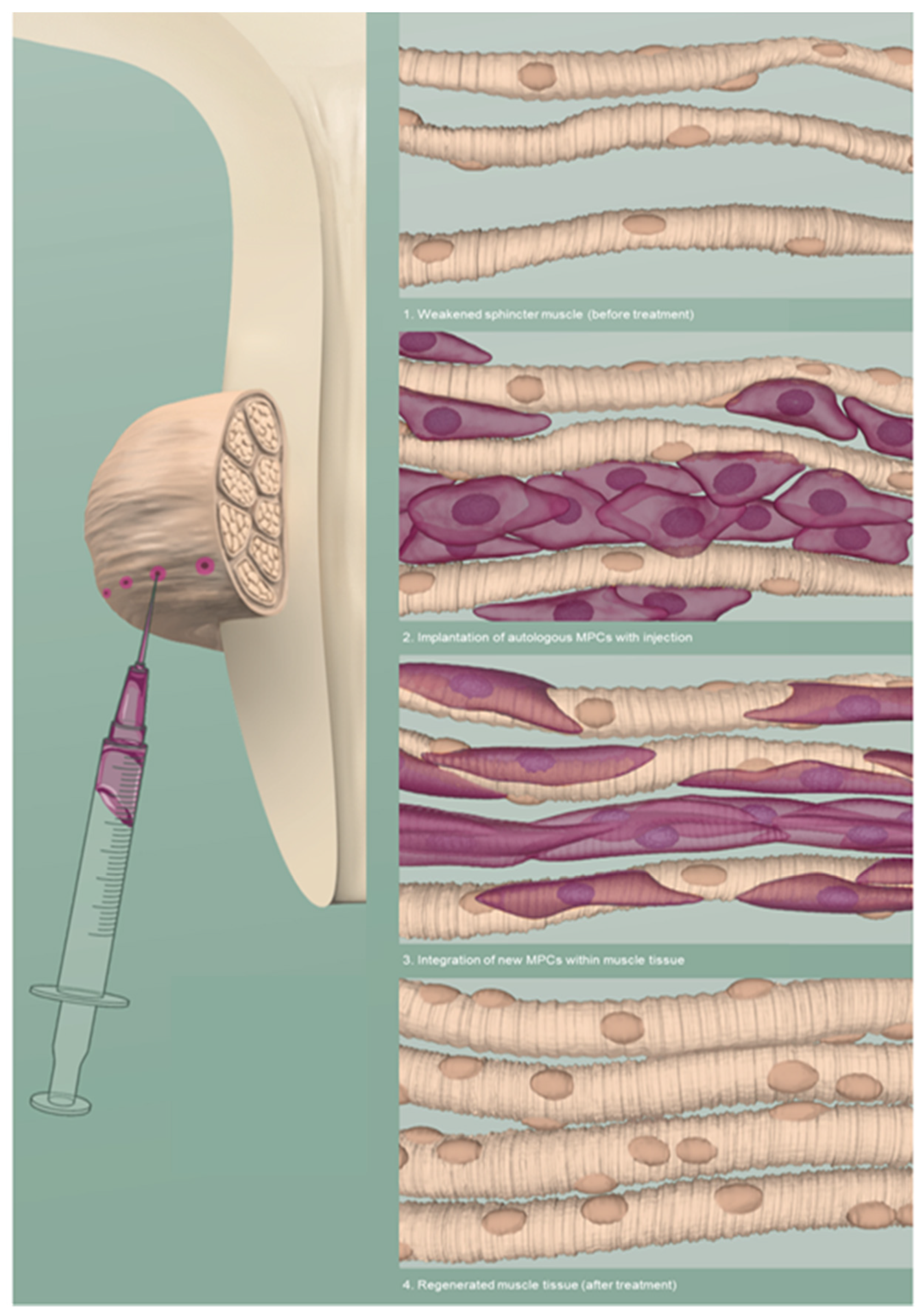
Abstract
1. Introduction
2. Stress Urinary Incontinence
2.1. Pathophysiology
2.2. Etiology and Diagnostics
2.3. Treatment
3. The Regenerative Approach
3.1. Muscle Progenitor Cells (MPCs)
3.1.1. Preclinical Data
3.1.2. Clinical Data
3.2. Routes of Application
3.3. Neuromuscular Electromagnetic Stimulation (NMES)
3.4. Imaging Technologies for MPC Tracking
3.5. Chemokine Therapy
4. Discussion and Future Prospects
5. Conclusions
Supplementary Materials
Author Contributions
Funding
Institutional Review Board Statement
Informed Consent Statement
Data Availability Statement
Acknowledgments
Conflicts of Interest
Abbreviations
| 19F | Fluorine-19 |
| ATMP | advanced therapy medicinal product |
| BMI | body-mass index |
| CXCL12 | C-X-C Motif Chemokine Ligand 12 |
| D2R | dopamine 2 receptors |
| DOA | detrusor overactivity |
| DTI | diffusion tensor imaging |
| EUS | external urethral sphincter |
| FDA | Food and Drug Administration |
| GCP | good clinical practice |
| GMP | good manufacturing practice |
| ICIQ | International Consultation on Incontinence Questionnaire |
| ICIQ-SF | International Consultation on Incontinence Questionnaire–Short Form |
| ICS | International Continence Society |
| ISD | intrinsic sphincter deficiency |
| LPP | leak point pressure |
| MDSC | muscle-derived stem cell |
| MPC | muscle progenitor cell |
| MRI | magnetic resonance imaging |
| MSC | mesenchymal stromal cell |
| MT | magnetization transfer |
| MUCP | maximal urethral closure pressure |
| NMES | neuromuscular electromagnetic stimulation |
| pADSC | porcine adipose tissue-derived stromal cell |
| PET | positron emission tomography |
| PRO | patient reported outcome |
| QoL | quality of life |
| SPIO | superparamagnetic iron oxide |
| SSNRI | selective serotonin noradrenaline inhibitors |
| SUI | stress urinary incontinence |
| TE | tissue engineering |
| TVT | tension free vaginal tape |
| UI | urinary incontinence |
| VLPP | Valsalva leak point pressure |
| µCT | micro-computed tomography |
References
- Norton, P.; Brubaker, L. Urinary incontinence in women. Lancet 2006, 367, 57–67. [Google Scholar] [CrossRef]
- Irwin, D.E.; Kopp, Z.S.; Agatep, B.; Milsom, I.; Abrams, P. Worldwide prevalence estimates of lower urinary tract symptoms, overactive bladder, urinary incontinence and bladder outlet obstruction. BJU Int. 2011, 108, 1132–1138. [Google Scholar] [CrossRef] [PubMed]
- Hunskaar, S.; Lose, G.; Sykes, D.; Voss, S. The prevalence of urinary incontinence in women in four European countries. BJU Int. 2004, 93, 324–330. [Google Scholar] [CrossRef] [PubMed]
- Minassian, V.A.; Stewart, W.F.; Wood, G.C. Urinary incontinence in women: Variation in prevalence estimates and risk factors. Obstet. Gynecol. 2008, 111, 324–331. [Google Scholar] [CrossRef] [PubMed]
- Asoglu, M.R.; Selcuk, S.; Cam, C.; Cogendez, E.; Karateke, A. Effects of urinary incontinence subtypes on women′s quality of life (including sexual life) and psychosocial state. Eur. J. Obstet. Gynecol. Reprod. Biol. 2014, 176, 187–190. [Google Scholar] [CrossRef] [PubMed]
- Mota, R.L. Female urinary incontinence and sexuality. Int. Braz. J. Urol. 2017, 43, 20–28. [Google Scholar] [CrossRef]
- Hannestad, Y.S.; Rortveit, G.; Sandvik, H.; Hunskaar, S. A community-based epidemiological survey of female urinary incontinence: The Norwegian EPINCONT study. Epidemiology of Incontinence in the County of Nord-Trondelag. J. Clin. Epidemiol. 2000, 53, 1150–1157. [Google Scholar] [CrossRef]
- Corcos, J.; Beaulieu, S.; Donovan, J.; Naughton, M.; Gotoh, M. Symptom Quality of Life Assesment Committee of the First International Consultation on, I., Quality of life assessment in men and women with urinary incontinence. J. Urol. 2002, 168, 896–905. [Google Scholar] [CrossRef]
- Wilson, L.; Brown, J.S.; Shin, G.P.; Luc, K.O.; Subak, L.L. Annual direct cost of urinary incontinence. Obstet. Gynecol. 2001, 98, 398–406. [Google Scholar] [PubMed]
- Morrison, A.; Levy, R. Fraction of nursing home admissions attributable to urinary incontinence. Value Health 2006, 9, 272–274. [Google Scholar] [CrossRef]
- Lukacz, E.S.; Santiago-Lastra, Y.; Albo, M.E.; Brubaker, L. Urinary Incontinence in Women: A Review. JAMA 2017, 318, 1592–1604. [Google Scholar] [CrossRef] [PubMed]
- D′Ancona, C.; Haylen, B.; Oelke, M.; Abranches-Monteiro, L.; Arnold, E.; Goldman, H.; Hamid, R.; Homma, Y.; Marcelissen, T.; Rademakers, K.; et al. The International Continence Society (ICS) report on the terminology for adult male lower urinary tract and pelvic floor symptoms and dysfunction. Neurourol. Urodyn. 2019, 38, 433–477. [Google Scholar] [CrossRef] [PubMed]
- Foldspang, A.; Mommsen, S.; Djurhuus, J.C. Prevalent urinary incontinence as a correlate of pregnancy, vaginal childbirth, and obstetric techniques. Am. J. Public Health 1999, 89, 209–212. [Google Scholar] [CrossRef] [PubMed]
- Danforth, K.N.; Townsend, M.K.; Lifford, K.; Curhan, G.C.; Resnick, N.M.; Grodstein, F. Risk factors for urinary incontinence among middle-aged women. Am. J. Obstet. Gynecol. 2006, 194, 339–345. [Google Scholar] [CrossRef] [PubMed]
- Shah, S.M.; Gaunay, G.S. Treatment options for intrinsic sphincter deficiency. Nat. Rev. Urol. 2012, 9, 638–651. [Google Scholar] [CrossRef]
- Morling, J.R.; McAllister, D.A.; Agur, W.; Fischbacher, C.M.; Glazener, C.M.; Guerrero, K.; Hopkins, L.; Wood, R. Adverse events after first, single, mesh and non-mesh surgical procedures for stress urinary incontinence and pelvic organ prolapse in Scotland, 1997–2016: A population-based cohort study. Lancet 2017, 389, 629–640. [Google Scholar] [CrossRef]
- Garriboli, M.; Radford, A.; Southgate, J. Regenerative medicine in urology. Eur. J. Pediatr. Surg. 2014, 24, 227–236. [Google Scholar]
- Linde, J.M.; Nijman, R.J.M.; Trzpis, M.; Broens, P.M.A. Urinary incontinence in the Netherlands: Prevalence and associated risk factors in adults. Neurourol. Urodyn. 2017, 36, 1519–1528. [Google Scholar] [CrossRef]
- Ptak, M.; Brodowska, A.; Ciecwiez, S.; Rotter, I. Quality of Life in Women with Stage 1 Stress Urinary Incontinence after Application of Conservative Treatment-A Randomized Trial. Int. J. Environ. Res. Public Health 2017, 14, 577. [Google Scholar] [CrossRef]
- Levy, R.; Muller, N. Urinary incontinence: Economic burden and new choices in pharmaceutical treatment. Adv. Ther. 2006, 23, 556–573. [Google Scholar] [CrossRef]
- Hu, T.W.; Wagner, T.H.; Bentkover, J.D.; Leblanc, K.; Zhou, S.Z.; Hunt, T. Costs of urinary incontinence and overactive bladder in the United States: A comparative study. Urology 2004, 63, 461–465. [Google Scholar] [CrossRef] [PubMed]
- Jung, J.; Ahn, H.K.; Huh, Y. Clinical and functional anatomy of the urethral sphincter. Int. Neurourol. J. 2012, 16, 102–106. [Google Scholar] [CrossRef] [PubMed]
- Ashton-Miller, J.A.; DeLancey, J.O. Functional anatomy of the female pelvic floor. Ann. N. Y. Acad. Sci. USA 2007, 1101, 266–296. [Google Scholar] [CrossRef]
- Hillary, C.J.; Roman, S.; MacNeil, S.; Aicher, W.K.; Stenzl, A.; Chapple, C.R. Regenerative medicine and injection therapies in stress urinary incontinence. Nat. Rev. Urol. 2020, 17, 151–161. [Google Scholar] [CrossRef]
- Haab, F.; Zimmern, P.E.; Leach, G.E. Female stress urinary incontinence due to intrinsic sphincteric deficiency: Recognition and management. J. Urol. 1996, 156, 3–17. [Google Scholar] [CrossRef]
- Hosker, G. Is it possible to diagnose intrinsic sphincter deficiency in women? Curr. Opin. Urol. 2009, 19, 342–346. [Google Scholar] [CrossRef]
- Parrillo, L.M.; Ramchandani, P.; Smith, A.L. Can intrinsic sphincter deficiency be diagnosed by urodynamics? Urol. Clin. 2014, 41, 375–381. [Google Scholar] [CrossRef]
- Eberli, D.; Aboushwareb, T.; Soker, S.; Yoo, J.J.; Atala, A. Muscle precursor cells for the restoration of irreversibly damaged sphincter function. Cell Transpl. 2012, 21, 2089–2098. [Google Scholar] [CrossRef]
- Padmanabhan, P.; Dmochowski, R. Urinary incontinence in women: A comprehensive review of the pathophysiology, diagnosis and treatment. Minerva Ginecol. 2014, 66, 469–478. [Google Scholar]
- Edwall, L.; Carlstrom, K.; Jonasson, A.F. Different estrogen sensitivity of urogenital tissue from women with and without stress urinary incontinence. Neurourol. Urodyn. 2009, 28, 516–520. [Google Scholar] [CrossRef] [PubMed]
- Hextall, A.; Cardozo, L. The role of estrogen supplementation in lower urinary tract dysfunction. Int. Urogynecol. J. 2001, 12, 258–261. [Google Scholar] [CrossRef]
- Ficarra, V.; Novara, G.; Rosen, R.C.; Artibani, W.; Carroll, P.R.; Costello, A.; Menon, M.; Montorsi, F.; Patel, V.R.; Stolzenburg, J.U.; et al. Systematic review and meta-analysis of studies reporting urinary continence recovery after robot-assisted radical prostatectomy. Eur. Urol. 2012, 62, 405–417. [Google Scholar] [CrossRef]
- Stamey, T.A. Endoscopic suspension of the vesical neck for urinary incontinence. Surg. Gynecol. Obstet. 1973, 136, 547–554. [Google Scholar] [CrossRef] [PubMed]
- Henderson, J.W.; Kane, S.M.; Mangel, J.M.; Kikano, E.G.; Garibay, J.A.; Pollard, R.R.; Mahajan, S.T.; Debanne, S.M.; Hijaz, A.K. A Randomized Comparative Study Evaluating Various Cough Stress Tests and 24-Hour Pad Test with Urodynamics in the Diagnosis of Stress Urinary Incontinence. J. Urol. 2018, 199, 1557–1564. [Google Scholar] [CrossRef] [PubMed]
- Serati, M.; Tarcan, T.; Finazzi-Agro, E.; Soligo, M.; Braga, A.; Athanasiou, S.; Balzarro, M. The bladder is an unreliable witness: The case for urodynamic investigations in female stress urinary incontinence. Eur. J. Obstet. Gynecol. Reprod. Biol. 2019, 244, 35–37. [Google Scholar] [CrossRef] [PubMed]
- Nygaard, I.E.; Heit, M. Stress urinary incontinence. Obstet. Gynecol. 2004, 104, 607–620. [Google Scholar] [CrossRef] [PubMed]
- Nambiar, A.K.; Bosch, R.; Cruz, F.; Lemack, G.E.; Thiruchelvam, N.; Tubaro, A.; Bedretdinova, D.A.; Ambuhl, D.; Farag, F.; Lombardo, R.; et al. EAU Guidelines on Assessment and Nonsurgical Management of Urinary Incontinence. Eur. Urol. 2018, 73, 596–609. [Google Scholar] [CrossRef]
- Kobashi, K.C.; Albo, M.E.; Dmochowski, R.R.; Ginsberg, D.A.; Goldman, H.B.; Gomelsky, A.; Kraus, S.R.; Sandhu, J.S.; Shepler, T.; Treadwell, J.R.; et al. Surgical Treatment of Female Stress Urinary Incontinence: AUA/SUFU Guideline. J. Urol. 2017, 198, 875–883. [Google Scholar] [CrossRef] [PubMed]
- Mischinger, J.; Amend, B.; Reisenauer, C.; Bedke, J.; Naumann, G.; Germann, M.; Kruck, S.; Arenas Desilva, L.F.; Wallwiener, H.; Koelbl, H.; et al. Different surgical approaches for stress urinary incontinence in women. Minerva Ginecol. 2013, 65, 21–28. [Google Scholar]
- Fusco, F.; Abdel-Fattah, M.; Chapple, C.R.; Creta, M.; La Falce, S.; Waltregny, D.; Novara, G. Updated Systematic Review and Meta-analysis of the Comparative Data on Colposuspensions, Pubovaginal Slings, and Midurethral Tapes in the Surgical Treatment of Female Stress Urinary Incontinence. Eur. Urol. 2017, 72, 567–591. [Google Scholar] [CrossRef]
- Hoda, M.R.; Primus, G.; Fischereder, K.; Von Heyden, B.; Mohammed, N.; Schmid, N.; Moll, V.; Hamza, A.; Karsch, J.J.; Brossner, C.; et al. Early results of a European multicentre experience with a new self-anchoring adjustable transobturator system for treatment of stress urinary incontinence in men. BJU Int. 2013, 111, 296–303. [Google Scholar] [CrossRef] [PubMed]
- Ha, Y.S.; Yoo, E.S. Artificial Urinary Sphincter for Postradical Prostatectomy Urinary Incontinence—Is It the Best Option? Int. Neurourol. J. 2019, 23, 265–276. [Google Scholar] [CrossRef] [PubMed]
- Lemack, G.E.; Xu, Y.; Brubaker, L.; Nager, C.; Chai, T.; Moalli, P.; Kraus, S.R.; Kerr, L.; Sirls, L.; Stoddard, A.; et al. Clinical and demographic factors associated with valsalva leak point pressure among women undergoing burch bladder neck suspension or autologous rectus fascial sling procedures. Neurourol. Urodyn. 2007, 26, 392–396. [Google Scholar] [CrossRef] [PubMed]
- Novara, G.; Artibani, W.; Barber, M.D.; Chapple, C.R.; Costantini, E.; Ficarra, V.; Hilton, P.; Nilsson, C.G.; Waltregny, D. Updated systematic review and meta-analysis of the comparative data on colposuspensions, pubovaginal slings, and midurethral tapes in the surgical treatment of female stress urinary incontinence. Eur. Urol. 2010, 58, 218–238. [Google Scholar] [CrossRef]
- Ellington, D.R.; Erekson, E.A.; Richter, H.E. Outcomes of Surgery for Stress Urinary Incontinence in the Older Woman. Clin. Geriatr. Med. 2015, 31, 487–505. [Google Scholar] [CrossRef] [PubMed]
- Marcelissen, T.; Van Kerrebroeck, P. Overactive bladder symptoms after midurethral sling surgery in women: Risk factors and management. Neurourol. Urodyn. 2018, 37, 83–88. [Google Scholar] [CrossRef]
- Hart, M.L.; Izeta, A.; Herrera-Imbroda, B.; Amend, B.; Brinchmann, J.E. Cell Therapy for Stress Urinary Incontinence. Tissue Eng. Part B Rev. 2015, 21, 365–376. [Google Scholar] [CrossRef]
- Zhou, S.; Zhang, K.; Atala, A.; Khoury, O.; Murphy, S.V.; Zhao, W.; Fu, Q. Stem Cell Therapy for Treatment of Stress Urinary Incontinence: The Current Status and Challenges. Stem Cells Int. 2016, 2016. [Google Scholar] [CrossRef]
- Aragon, I.M.; Imbroda, B.H.; Lara, M.F. Cell Therapy Clinical Trials for Stress Urinary Incontinence: Current Status and Perspectives. Int. J. Med. Sci. 2018, 15, 195–204. [Google Scholar] [CrossRef]
- Amend, B.; Vaegler, M.; Fuchs, K.; Mannheim, J.G.; Will, S.; Kramer, U.; Hart, M.L.; Feitz, W.; Chapple, C.; Stenzl, A.; et al. Regeneration of degenerated urinary sphincter muscles: Improved stem cell-based therapies and novel imaging technologies. Cell Transpl. 2015, 24, 2171–2183. [Google Scholar] [CrossRef]
- Amend, B.; Kelp, A.; Vaegler, M.; Klunder, M.; Frajs, V.; Klein, G.; Sievert, K.D.; Sawodny, O.; Stenzl, A.; Aicher, W.K. Precise injection of human mesenchymal stromal cells in the urethral sphincter complex of Gottingen minipigs without unspecific bulking effects. Neurourol. Urodyn. 2017, 36, 1723–1733. [Google Scholar] [CrossRef] [PubMed]
- Arjmand, B.; Safavi, M.; Heidari, R.; Aghayan, H.; S., T.B.; Dehghani, S.; Goodarzi, P.; Mohammadi-Jahani, F.; Heidari, F.; Payab, M.; et al. Concomitant Transurethral and Transvaginal-Periurethral Injection of Autologous Adipose Derived Stem Cells for Treatment of Female Stress Urinary Incontinence: A Phase One Clinical Trial. Acta Med. Iran. 2017, 55, 368–374. [Google Scholar] [PubMed]
- Burdzinska, A.; Dybowski, B.; Zarychta-Wisniewska, W.; Kulesza, A.; Butrym, M.; Zagozdzon, R.; Graczyk-Jarzynka, A.; Radziszewski, P.; Gajewski, Z.; Paczek, L. Intraurethral co-transplantation of bone marrow mesenchymal stem cells and muscle-derived cells improves the urethral closure. Stem Cell Res. Ther. 2018, 9, 1–16. [Google Scholar] [CrossRef] [PubMed]
- Du, X.W.; Wu, H.L.; Zhu, Y.F.; Hu, J.B.; Jin, F.; Lv, R.P.; Sun, S.; Wang, H.Y.; Xu, J.W. Experimental study of therapy of bone marrow mesenchymal stem cells or muscle-like cells/calcium alginate composite gel for the treatment of stress urinary incontinence. Neurourol. Urodyn. 2013, 32, 281–286. [Google Scholar] [CrossRef]
- Furuta, A.; Jankowski, R.J.; Honda, M.; Pruchnic, R.; Yoshimura, N.; Chancellor, M.B. State of the art of where we are at using stem cells for stress urinary incontinence. Neurourol. Urodyn. 2007, 26, 966–971. [Google Scholar] [CrossRef]
- Jager, L.; Linzenbold, W.; Fech, A.; Enderle, M.; Abruzzese, T.; Stenzl, A.; Aicher, W.K. A novel waterjet technology for transurethral cystoscopic injection of viable cells in the urethral sphincter complex. Neurourol. Urodyn. 2020, 39, 594–602. [Google Scholar] [CrossRef]
- Janssen, K.; Lin, D.L.; Hanzlicek, B.; Deng, K.; Balog, B.M.; van der Vaart, C.H.; Damaser, M.S. Multiple doses of stem cells maintain urethral function in a model of neuromuscular injury resulting in stress urinary incontinence. Am. J. Physiol. Ren. Physiol. 2019, 317, F1047–F1057. [Google Scholar] [CrossRef]
- Jin, M.; Chen, Y.; Zhou, Y.; Mei, Y.; Liu, W.; Pan, C.; Hua, X. Transplantation of bone marrow-derived mesenchymal stem cells expressing elastin alleviates pelvic floor dysfunction. Stem Cell Res. Ther. 2016, 7, 1–10. [Google Scholar] [CrossRef]
- Kuismanen, K.; Sartoneva, R.; Haimi, S.; Mannerstrom, B.; Tomas, E.; Miettinen, S.; Nieminen, K. Autologous adipose stem cells in treatment of female stress urinary incontinence: Results of a pilot study. Stem Cells Transl. Med. 2014, 3, 936–941. [Google Scholar] [CrossRef]
- Lee, C.N.; Jang, J.B.; Kim, J.Y.; Koh, C.; Baek, J.Y.; Lee, K.J. Human cord blood stem cell therapy for treatment of stress urinary incontinence. J. Korean Med. Sci. 2010, 25, 813–816. [Google Scholar] [CrossRef]
- Mitterberger, M.; Pinggera, G.M.; Marksteiner, R.; Margreiter, E.; Fussenegger, M.; Frauscher, F.; Ulmer, H.; Hering, S.; Bartsch, G.; Strasser, H. Adult stem cell therapy of female stress urinary incontinence. Eur. Urol. 2008, 53, 169–175. [Google Scholar] [CrossRef]
- Wu, S.; Wang, Z.; Bharadwaj, S.; Hodges, S.J.; Atala, A.; Zhang, Y. Implantation of autologous urine derived stem cells expressing vascular endothelial growth factor for potential use in genitourinary reconstruction. J. Urol. 2011, 186, 640–647. [Google Scholar] [CrossRef] [PubMed]
- Yamamoto, T.; Gotoh, M.; Kato, M.; Majima, T.; Toriyama, K.; Kamei, Y.; Iwaguro, H.; Matsukawa, Y.; Funahashi, Y. Periurethral injection of autologous adipose-derived regenerative cells for the treatment of male stress urinary incontinence: Report of three initial cases. Int. J. Urol. 2012, 19, 652–659. [Google Scholar] [CrossRef] [PubMed]
- Yu, A.; Campeau, L. Bone marrow mesenchymal stem cell therapy for voiding dysfunction. Curr. Urol. Rep. 2015, 16, 49. [Google Scholar] [CrossRef] [PubMed]
- Zhao, W.; Zhang, C.; Jin, C.; Zhang, Z.; Kong, D.; Xu, W.; Xiu, Y. Periurethral injection of autologous adipose-derived stem cells with controlled-release nerve growth factor for the treatment of stress urinary incontinence in a rat model. Eur. Urol. 2011, 59, 155–163. [Google Scholar] [CrossRef]
- Lin, C.S.; Lue, T.F. Stem cell therapy for stress urinary incontinence: A critical review. Stem Cells Dev. 2012, 21, 834–843. [Google Scholar] [CrossRef] [PubMed]
- Williams, J.K.; Dean, A.; Badlani, G.; Andersson, K.E. Regenerative Medicine Therapies for Stress Urinary Incontinence. J. Urol. 2016, 196, 1619–1626. [Google Scholar] [CrossRef]
- Vinarov, A.; Atala, A.; Yoo, J.; Slusarenco, R.; Zhumataev, M.; Zhito, A.; Butnaru, D. Cell therapy for stress urinary incontinence: Present-day frontiers. J. Tissue Eng. Regen. Med. 2018, 12, e1108–e1121. [Google Scholar] [CrossRef]
- Bennington, J.; Williams, J.K.; Andersson, K.E. New concepts in regenerative medicine approaches to the treatment of female stress urinary incontinence. Curr. Opin. Urol. 2019, 29, 380–384. [Google Scholar] [CrossRef]
- Deasy, B.M.; Jankowski, R.J.; Huard, J. Muscle-derived stem cells: Characterization and potential for cell-mediated therapy. Blood Cells Mol. Dis. 2001, 27, 924–933. [Google Scholar] [CrossRef]
- Roca, I.; Requena, J.; Edel, M.J.; Alvarez-Palomo, A.B. Myogenic Precursors from iPS Cells for Skeletal Muscle Cell Replacement Therapy. J. Clin. Med. 2015, 4, 243–259. [Google Scholar] [CrossRef] [PubMed]
- Chancellor, M.B.; Yokoyama, T.; Tirney, S.; Mattes, C.E.; Ozawa, H.; Yoshimura, N.; de Groat, W.C.; Huard, J. Preliminary results of myoblast injection into the urethra and bladder wall: A possible method for the treatment of stress urinary incontinence and impaired detrusor contractility. Neurourol. Urodyn. 2000, 19, 279–287. [Google Scholar] [CrossRef]
- Yokoyama, T.; Yoshimura, N.; Dhir, R.; Qu, Z.; Fraser, M.O.; Kumon, H.; de Groat, W.C.; Huard, J.; Chancellor, M.B. Persistence and survival of autologous muscle derived cells versus bovine collagen as potential treatment of stress urinary incontinence. J. Urol. 2001, 165, 271–276. [Google Scholar] [CrossRef] [PubMed]
- Yiou, R.; Dreyfus, P.; Chopin, D.K.; Abbou, C.C.; Lefaucheur, J.P. Muscle precursor cell autografting in a murine model of urethral sphincter injury. BJU Int. 2002, 89, 298–302. [Google Scholar] [CrossRef]
- Lee, J.Y.; Cannon, T.W.; Pruchnic, R.; Fraser, M.O.; Huard, J.; Chancellor, M.B. The effects of periurethral muscle-derived stem cell injection on leak point pressure in a rat model of stress urinary incontinence. Int. Urogynecol. J. 2003, 14, 31–37. [Google Scholar] [CrossRef]
- Badra, S.; Andersson, K.E.; Dean, A.; Mourad, S.; Williams, J.K. Long-term structural and functional effects of autologous muscle precursor cell therapy in a nonhuman primate model of urinary sphincter deficiency. J. Urol. 2013, 190, 1938–1945. [Google Scholar] [CrossRef]
- Williams, J.K.; Dean, A.; Lankford, S.; Criswell, T.; Badlani, G.; Andersson, K.E. Determinates of muscle precursor cell therapy efficacy in a nonhuman primate model of intrinsic urinary sphincter deficiency. Stem Cell Res. Ther. 2017, 8, 1–8. [Google Scholar] [CrossRef]
- Zhou, F.; Reed-Maldonado, A.B.; Tan, Y.; Yuan, H.; Peng, D.; Banie, L.; Wang, G.; Hou, J.; Lin, G.; Lue, T.F. Development of Male External Urethral Sphincter and Tissue-Resident Stem/Progenitor Cells in Rats. Stem Cells Dev. 2020, 29, 133–143. [Google Scholar] [CrossRef]
- Strasser, H.; Marksteiner, R.; Margreiter, E.; Pinggera, G.M.; Mitterberger, M.; Frauscher, F.; Ulmer, H.; Fussenegger, M.; Kofler, K.; Bartsch, G. Autologous myoblasts and fibroblasts versus collagen for treatment of stress urinary incontinence in women: A randomised controlled trial. Lancet 2007, 369, 2179–2186. [Google Scholar] [CrossRef]
- Strasser, H.; Marksteiner, R.; Margreiter, E.; Mitterberger, M.; Pinggera, G.M.; Frauscher, F.; Fussenegger, M.; Kofler, K.; Bartsch, G. Transurethral ultrasonography-guided injection of adult autologous stem cells versus transurethral endoscopic injection of collagen in treatment of urinary incontinence. World J. Urol. 2007, 25, 385–392. [Google Scholar] [CrossRef]
- Mitterberger, M.; Marksteiner, R.; Margreiter, E.; Pinggera, G.M.; Colleselli, D.; Frauscher, F.; Ulmer, H.; Fussenegger, M.; Bartsch, G.; Strasser, H. Autologous myoblasts and fibroblasts for female stress incontinence: A 1-year follow-up in 123 patients. BJU Int. 2007, 100, 1081–1085. [Google Scholar] [CrossRef] [PubMed]
- Mitterberger, M.; Marksteiner, R.; Margreiter, E.; Pinggera, G.M.; Frauscher, F.; Ulmer, H.; Fussenegger, M.; Bartsch, G.; Strasser, H. Myoblast and fibroblast therapy for post-prostatectomy urinary incontinence: 1-year followup of 63 patients. J. Urol. 2008, 179, 226–231. [Google Scholar] [CrossRef] [PubMed]
- Sebe, P.; Doucet, C.; Cornu, J.N.; Ciofu, C.; Costa, P.; de Medina, S.G.; Pinset, C.; Haab, F. Intrasphincteric injections of autologous muscular cells in women with refractory stress urinary incontinence: A prospective study. Int. Urogynecol. J. 2011, 22, 183–189. [Google Scholar] [CrossRef]
- Blaganje, M.; Lukanovic, A. Intrasphincteric autologous myoblast injections with electrical stimulation for stress urinary incontinence. Int. J. Gynaecol. Obstet. 2012, 117, 164–167. [Google Scholar] [CrossRef]
- Blaganje, M.; Lukanovic, A. Ultrasound-guided autologous myoblast injections into the extrinsic urethral sphincter: Tissue engineering for the treatment of stress urinary incontinence. Int. Urogynecol. J. 2013, 24, 533–535. [Google Scholar] [CrossRef]
- Gerullis, H.; Eimer, C.; Georgas, E.; Homburger, M.; El-Baz, A.G.; Wishahi, M.; Boros, M.; Ecke, T.H.; Otto, T. Muscle-derived cells for treatment of iatrogenic sphincter damage and urinary incontinence in men. Sci. World J. 2012, 2012. [Google Scholar] [CrossRef] [PubMed]
- Carr, L.K.; Robert, M.; Kultgen, P.L.; Herschorn, S.; Birch, C.; Murphy, M.; Chancellor, M.B. Autologous muscle derived cell therapy for stress urinary incontinence: A prospective, dose ranging study. J. Urol. 2013, 189, 595–601. [Google Scholar] [CrossRef]
- Peters, K.M.; Dmochowski, R.R.; Carr, L.K.; Robert, M.; Kaufman, M.R.; Sirls, L.T.; Herschorn, S.; Birch, C.; Kultgen, P.L.; Chancellor, M.B. Autologous muscle derived cells for treatment of stress urinary incontinence in women. J. Urol. 2014, 192, 469–476. [Google Scholar] [CrossRef]
- Yiou, R.; Hogrel, J.Y.; Loche, C.M.; Authier, F.J.; Lecorvoisier, P.; Jouany, P.; Roudot-Thoraval, F.; Lefaucheur, J.P. Periurethral skeletal myofibre implantation in patients with urinary incontinence and intrinsic sphincter deficiency: A phase I clinical trial. BJU Int. 2013, 111, 1105–1116. [Google Scholar] [CrossRef]
- Gras, S.; Klarskov, N.; Lose, G. Intraurethral injection of autologous minced skeletal muscle: A simple surgical treatment for stress urinary incontinence. J. Urol. 2014, 192, 850–855. [Google Scholar] [CrossRef]
- Stangel-Wojcikiewicz, K.; Jarocha, D.; Piwowar, M.; Jach, R.; Uhl, T.; Basta, A.; Majka, M. Autologous muscle-derived cells for the treatment of female stress urinary incontinence: A 2-year follow-up of a Polish investigation. Neurourol. Urodyn. 2014, 33, 324–330. [Google Scholar] [CrossRef] [PubMed]
- Stangel-Wojcikiewicz, K.; Piwowar, M.; Jach, R.; Majka, M.; Basta, A. Quality of life assessment in female patients 2 and 4 years after muscle-derived cell transplants for stress urinary incontinence treatment. Ginekol. Pol. 2016, 87, 183–189. [Google Scholar] [CrossRef] [PubMed]
- Sharifiaghdas, F.; Tajalli, F.; Taheri, M.; Naji, M.; Moghadasali, R.; Aghdami, N.; Baharvand, H.; Azimian, V.; Jaroughi, N. Effect of autologous muscle-derived cells in the treatment of urinary incontinence in female patients with intrinsic sphincter deficiency and epispadias: A prospective study. Int. J. Urol. 2016, 23, 581–586. [Google Scholar] [CrossRef]
- Jankowski, R.J.; Tu, L.M.; Carlson, C.; Robert, M.; Carlson, K.; Quinlan, D.; Eisenhardt, A.; Chen, M.; Snyder, S.; Pruchnic, R.; et al. A double-blind, randomized, placebo-controlled clinical trial evaluating the safety and efficacy of autologous muscle derived cells in female subjects with stress urinary incontinence. Int. Urol. Nephrol. 2018, 50, 2153–2165. [Google Scholar] [CrossRef] [PubMed]
- Schmid, F.A.; Gascho, D.; Zoelch, N.; Prange, J.A.; Colacicco, G.; Eberli, D. Feasibility, technique and accuracy of ultrasound-guided transurethral injections into the urinary sphincter of female cadavers: Proof of concept. BMC Urol. 2020, 20, 167. [Google Scholar] [CrossRef] [PubMed]
- Burdzinska, A.; Dybowski, B.; Zarychta-Wisniewska, W.; Kulesza, A.; Hawryluk, J.; Graczyk-Jarzynka, A.; Kaupa, P.; Gajewski, Z.; Paczek, L. Limited accuracy of transurethral and periurethral intrasphincteric injections of cellular suspension. Neurourol. Urodyn. 2018, 37, 1612–1622. [Google Scholar] [CrossRef] [PubMed]
- Faerber, G.J.; Belville, W.D.; Ohl, D.A.; Plata, A. Comparison of transurethral versus periurethral collagen injection in women with intrinsic sphincter deficiency. Tech. Urol. 1998, 4, 124–127. [Google Scholar] [PubMed]
- Schulz, J.A.; Nager, C.W.; Stanton, S.L.; Baessler, K. Bulking agents for stress urinary incontinence: Short-term results and complications in a randomized comparison of periurethral and transurethral injections. Int. Urogynecol. J. 2004, 15, 261–265. [Google Scholar] [CrossRef]
- Kirchin, V.; Page, T.; Keegan, P.E.; Atiemo, K.O.; Cody, J.D.; McClinton, S.; Aluko, P. Urethral injection therapy for urinary incontinence in women. Cochrane Database Syst. Rev. 2017. [Google Scholar] [CrossRef]
- Linzenbold, W.; Jager, L.; Stoll, H.; Abruzzese, T.; Harland, N.; Beziere, N.; Fech, A.; Enderle, M.; Amend, B.; Stenzl, A.; et al. Rapid and precise delivery of cells in the urethral sphincter complex by a novel needle-free waterjet technology. BJU Int. 2020. [Google Scholar] [CrossRef]
- Hillen, B.K.; Abbas, J.J.; Jung, R. Accelerating locomotor recovery after incomplete spinal injury. Ann. N. Y. Acad Sci. USA 2013, 1279, 164–174. [Google Scholar] [CrossRef] [PubMed]
- Stolting, M.N.; Arnold, A.S.; Haralampieva, D.; Handschin, C.; Sulser, T.; Eberli, D. Magnetic stimulation supports muscle and nerve regeneration after trauma in mice. Muscle Nerve 2016, 53, 598–607. [Google Scholar] [CrossRef]
- Lim, R.; Liong, M.L.; Leong, W.S.; Karim Khan, N.A.; Yuen, K.H. Pulsed Magnetic Stimulation for Stress Urinary Incontinence: 1-Year Followup Results. J. Urol. 2017, 197, 1302–1308. [Google Scholar] [CrossRef] [PubMed]
- Yamanishi, T.; Suzuki, T.; Sato, R.; Kaga, K.; Kaga, M.; Fuse, M. Effects of magnetic stimulation on urodynamic stress incontinence refractory to pelvic floor muscle training in a randomized sham-controlled study. LUTS Low. Urin. Tract Symptoms 2019, 11, 61–65. [Google Scholar] [CrossRef] [PubMed]
- Vadala, M.; Palmieri, B.; Malagoli, A.; Laurino, C. High-power Magnetotherapy: A New Weapon in Urinary Incontinence? LUTS Low. Urin. Tract Symptoms 2018, 10, 266–270. [Google Scholar] [CrossRef] [PubMed]
- Haralampieva, D.; Betzel, T.; Dinulovic, I.; Salemi, S.; Stoelting, M.; Kramer, S.D.; Schibli, R.; Sulser, T.; Handschin, C.; Eberli, D.; et al. Noninvasive PET Imaging and Tracking of Engineered Human Muscle Precursor Cells for Skeletal Muscle Tissue Engineering. J. Nucl. Med. 2016, 57, 1467–1473. [Google Scholar] [CrossRef]
- Pacak, C.A.; Hammer, P.E.; MacKay, A.A.; Dowd, R.P.; Wang, K.R.; Masuzawa, A.; Sill, B.; McCully, J.D.; Cowan, D.B. Superparamagnetic iron oxide nanoparticles function as a long-term, multi-modal imaging label for non-invasive tracking of implanted progenitor cells. PLoS ONE 2014, 9, e108695. [Google Scholar] [CrossRef]
- Gutpell, K.; McGirr, R.; Hoffman, L. Molecular imaging to target transplanted muscle progenitor cells. J. Vis. Exp. JoVE 2013. [Google Scholar] [CrossRef]
- Swider, E.; Daoudi, K.; Staal, A.H.J.; Koshkina, O.; van Riessen, N.K.; van Dinther, E.; de Vries, I.J.M.; de Korte, C.L.; Srinivas, M. Clinically-Applicable Perfluorocarbon-Loaded Nanoparticles For In vivo Photoacoustic, 19F Magnetic Resonance And Fluorescent Imaging. Nanotheranostics 2018, 2, 258–268. [Google Scholar] [CrossRef]
- Constantinides, C.; McNeill, E.; Carnicer, R.; Al Haj Zen, A.; Sainz-Urruela, R.; Shaw, A.; Patel, J.; Swider, E.; Alonaizan, R.; Potamiti, L.; et al. Improved cellular uptake of perfluorocarbon nanoparticles for in vivo murine cardiac (19)F MRS/MRI and temporal tracking of progenitor cells. Nanomedicine 2019, 18, 391–401. [Google Scholar] [CrossRef]
- Koshkina, O.; Lajoinie, G.; Bombelli, F.B.; Swider, E.; Cruz, L.J.; White, P.B.; Schweins, R.; Dolen, Y.; van Dinther, E.A.W.; van Riessen, N.K.; et al. Multicore Liquid Perfluorocarbon-Loaded Multimodal Nanoparticles for Stable Ultrasound and 19F MRI Applied to In Vivo Cell Tracking. Adv. Funct. Mater. 2019, 29, 1806485. [Google Scholar] [CrossRef]
- Koshkina, O.; White, P.B.; Staal, A.H.J.; Schweins, R.; Swider, E.; Tirotta, I.; Tinnemans, P.; Fokkink, R.; Veltien, A.; van Riessen, N.K.; et al. Nanoparticles for “two color” 19F magnetic resonance imaging: Towards combined imaging of biodistribution and degradation. J. Colloid Interface Sci. 2020, 565, 278–287. [Google Scholar] [CrossRef]
- Rousset, P.; Delmas, V.; Buy, J.N.; Rahmouni, A.; Vadrot, D.; Deux, J.F. In vivo visualization of the levator ani muscle subdivisions using MR fiber tractography with diffusion tensor imaging. J. Anat. 2012, 221, 221–228. [Google Scholar] [CrossRef] [PubMed]
- Oudeman, J.; Nederveen, A.J.; Strijkers, G.J.; Maas, M.; Luijten, P.R.; Froeling, M. Techniques and applications of skeletal muscle diffusion tensor imaging: A review. J. Magn. Reson. Imaging 2016, 43, 773–788. [Google Scholar] [CrossRef] [PubMed]
- Sinha, S.; Sinha, U.; Malis, V.; Bhargava, V.; Sakamoto, K.; Rajasekaran, M. Exploration of male urethral sphincter complex using diffusion tensor imaging (DTI)-based fiber-tracking. J. Magn. Reson. Imaging 2018, 48, 1002–1011. [Google Scholar] [CrossRef] [PubMed]
- Zifan, A.; Reisert, M.; Sinha, S.; Ledgerwood-Lee, M.; Cory, E.; Sah, R.; Mittal, R.K. Connectivity of the Superficial Muscles of the Human Perineum: A Diffusion Tensor Imaging-Based Global Tractography Study. Sci. Rep. 2018, 8, 1–10. [Google Scholar]
- Zijta, F.M.; Froeling, M.; Nederveen, A.J.; Stoker, J. Diffusion tensor imaging and fiber tractography for the visualization of the female pelvic floor. Clin. Anat. 2013, 26, 110–114. [Google Scholar] [CrossRef] [PubMed]
- McDaniel, J.D.; Ulmer, J.L.; Prost, R.W.; Franczak, M.B.; Jaradeh, S.; Hamilton, C.A.; Mark, L.P. Magnetization transfer imaging of skeletal muscle in autosomal recessive limb girdle muscular dystrophy. J. Comput. Assist. Tomogr. 1999, 23, 609–614. [Google Scholar] [CrossRef]
- Boss, A.; Martirosian, P.; Kuper, K.; Fierlbeck, G.; Claussen, C.D.; Schick, F. Whole-body magnetization transfer contrast imaging. J. Magn. Reson. Imaging 2006, 24, 1183–1187. [Google Scholar] [CrossRef]
- Basser, P.J.; Pierpaoli, C. Microstructural and physiological features of tissues elucidated by quantitative-diffusion-tensor MRI. J. Magn. Reson. 2011, 213, 560–570. [Google Scholar] [CrossRef]
- Rottmar, M.; Haralampieva, D.; Salemi, S.; Eberhardt, C.; Wurnig, M.C.; Boss, A.; Eberli, D. Magnetization Transfer MR Imaging to Monitor Muscle Tissue Formation during Myogenic in Vivo Differentiation of Muscle Precursor Cells. Radiology 2016, 281, 436–443. [Google Scholar] [CrossRef]
- Christ, G.J.; Saul, J.M.; Furth, M.E.; Andersson, K.E. The pharmacology of regenerative medicine. Pharmacol. Rev. 2013, 65, 1091–1133. [Google Scholar] [CrossRef]
- Lavoie, J.R.; Rosu-Myles, M. Uncovering the secretes of mesenchymal stem cells. Biochimie 2013, 95, 2212–2221. [Google Scholar] [CrossRef]
- Deng, K.; Lin, D.L.; Hanzlicek, B.; Balog, B.; Penn, M.S.; Kiedrowski, M.J.; Hu, Z.; Ye, Z.; Zhu, H.; Damaser, M.S. Mesenchymal stem cells and their secretome partially restore nerve and urethral function in a dual muscle and nerve injury stress urinary incontinence model. Am. J. Physiol. Renal. Physiol. 2015, 308, F92–F100. [Google Scholar] [CrossRef] [PubMed]
- Williams, J.K.; Dean, A.; Badra, S.; Lankford, S.; Poppante, K.; Badlani, G.; Andersson, K.E. Cell versus Chemokine Therapy in a Nonhuman Primate Model of Chronic Intrinsic Urinary Sphincter Deficiency. J. Urol. 2016, 196, 1809–1815. [Google Scholar] [CrossRef] [PubMed]
- Williams, J.K.; Mariya, S.; Suparto, I.; Lankford, S.S.; Andersson, K.E. Cell Versus Chemokine Therapy Effects on Cell Mobilization to Chronically Dysfunctional Urinary Sphincters of Nonhuman Primates. Int. Neurourol. J. 2018, 22, 260–267. [Google Scholar] [CrossRef] [PubMed]
- Liu, Y.; Feng, Q.; Miao, J.; Wu, Q.; Zhou, S.; Shen, W.; Feng, Y.; Hou, F.F.; Liu, Y.; Zhou, L. C-X-C motif chemokine receptor 4 aggravates renal fibrosis through activating JAK/STAT/GSK3beta/beta-catenin pathway. J. Cell. Mol. Med. 2020, 24, 3837–3855. [Google Scholar] [CrossRef] [PubMed]
- Macoska, J.A.; Wang, Z.; Virta, J.; Zacharias, N.; Bjorling, D.E. Inhibition of the CXCL12/CXCR4 axis prevents periurethral collagen accumulation and lower urinary tract dysfunction in vivo. Prostate 2019, 79, 757–767. [Google Scholar] [CrossRef]
- Ray, P.; Lewin, S.A.; Mihalko, L.A.; Lesher-Perez, S.C.; Takayama, S.; Luker, K.E.; Luker, G.D. Secreted CXCL12 (SDF-1) forms dimers under physiological conditions. Biochem. J. 2012, 442, 433–442. [Google Scholar] [CrossRef] [PubMed]
- Veldkamp, C.T.; Seibert, C.; Peterson, F.C.; De la Cruz, N.B.; Haugner, J.C., 3rd; Basnet, H.; Sakmar, T.P.; Volkman, B.F. Structural basis of CXCR4 sulfotyrosine recognition by the chemokine SDF-1/CXCL12. Sci. Signal. 2008. [Google Scholar] [CrossRef]
- Veldkamp, C.T.; Ziarek, J.J.; Su, J.; Basnet, H.; Lennertz, R.; Weiner, J.J.; Peterson, F.C.; Baker, J.E.; Volkman, B.F. Monomeric structure of the cardioprotective chemokine SDF-1/CXCL12. Protein Sci. 2009, 18, 1359–1369. [Google Scholar] [CrossRef] [PubMed]
- Prange, J.A.; Aleandri, S.; Komisarski, M.; Luciani, A.; Käch, A.; Schuh, C.-D.; Hall, A.M.; Mezzenga, R.; Devuyst, O.; Landau, E.M. Overcoming Endocytosis Deficiency by Cubosome Nanocarriers. ACS Appl. Bio Mater. 2019, 2, 2490–2499. [Google Scholar] [CrossRef]

Publisher’s Note: MDPI stays neutral with regard to jurisdictional claims in published maps and institutional affiliations. |
© 2021 by the authors. Licensee MDPI, Basel, Switzerland. This article is an open access article distributed under the terms and conditions of the Creative Commons Attribution (CC BY) license (https://creativecommons.org/licenses/by/4.0/).
Share and Cite
Schmid, F.A.; Williams, J.K.; Kessler, T.M.; Stenzl, A.; Aicher, W.K.; Andersson, K.-E.; Eberli, D. Treatment of Stress Urinary Incontinence with Muscle Stem Cells and Stem Cell Components: Chances, Challenges and Future Prospects. Int. J. Mol. Sci. 2021, 22, 3981. https://doi.org/10.3390/ijms22083981
Schmid FA, Williams JK, Kessler TM, Stenzl A, Aicher WK, Andersson K-E, Eberli D. Treatment of Stress Urinary Incontinence with Muscle Stem Cells and Stem Cell Components: Chances, Challenges and Future Prospects. International Journal of Molecular Sciences. 2021; 22(8):3981. https://doi.org/10.3390/ijms22083981
Chicago/Turabian StyleSchmid, Florian A., J. Koudy Williams, Thomas M. Kessler, Arnulf Stenzl, Wilhelm K. Aicher, Karl-Erik Andersson, and Daniel Eberli. 2021. "Treatment of Stress Urinary Incontinence with Muscle Stem Cells and Stem Cell Components: Chances, Challenges and Future Prospects" International Journal of Molecular Sciences 22, no. 8: 3981. https://doi.org/10.3390/ijms22083981
APA StyleSchmid, F. A., Williams, J. K., Kessler, T. M., Stenzl, A., Aicher, W. K., Andersson, K.-E., & Eberli, D. (2021). Treatment of Stress Urinary Incontinence with Muscle Stem Cells and Stem Cell Components: Chances, Challenges and Future Prospects. International Journal of Molecular Sciences, 22(8), 3981. https://doi.org/10.3390/ijms22083981