Article

Genome-wide Identification and Characterization of FCS-Like Zinc Finger (FLZ) Family Genes in Maize (Zea mays) and Functional Analysis of ZmFLZ25 in Plant Abscisic Acid Response

1
Guangdong Provincial Key Laboratory of Biotechnology for Plant Development, School of Life Sciences, South China Normal University, Guangzhou 510631, China
2
Shenzhen Institute of Molecular Crop Design, Shenzhen 518107, China
*
Authors to whom correspondence should be addressed.
These authors contributed equally to this work.
Int. J. Mol. Sci. 2021, 22(7), 3529; https://doi.org/10.3390/ijms22073529
Submission received: 20 January 2021 / Revised: 13 March 2021 / Accepted: 18 March 2021 / Published: 29 March 2021

Abstract

:
FCS-like zinc finger family proteins (FLZs), a class of plant-specific scaffold of SnRK1 complex, are involved in the regulation of various aspects of plant growth and stress responses. Most information of FLZ family genes was obtained from the studies in Arabidopsis thaliana, whereas little is known about the potential functions of FLZs in crop plants. In this study, 37 maize FLZ (ZmFLZ) genes were identified to be asymmetrically distributed on 10 chromosomes and can be divided into three subfamilies. Protein interaction and subcellular localization assays demonstrated that eight typical ZmFLZs interacted and partially co-localized with ZmKIN10, the catalytic α-subunit of the SnRK1 complex in maize leaf mesophyll cells. Expression profile analysis revealed that several ZmFLZs were differentially expressed across various tissues and actively responded to diverse abiotic stresses. In addition, ectopic overexpression of ZmFLZ25 in Arabidopsis conferred hypersensitivity to exogenous abscisic acid (ABA) and triggered higher expression of ABA-induced genes, pointing to the positive regulatory role of ZmFLZ25 in plant ABA signaling, a scenario further evidenced by the interactions between ZmFLZ25 and ABA receptors. In summary, these data provide the most comprehensive information on FLZ family genes in maize, and shed light on the biological function of ZmFLZ25 in plant ABA signaling.

1. Introduction

The first FLZ-domain containing protein MEDIATOR OF ABA-REGULATED DORMANCY 1 (MARD1) was identified from senescence related enhancer-trapping and found to be implicated in abscisic acid (ABA)-mediated seed dormancy in Arabidopsis [1,2]. MARD1 possesses a novel zinc finger domain that shares high signature motif similarity with MYM-type zinc finger containing FCS sequence motif [2]. Subsequently, this domain was found to be exclusively existed in plants and designated as the FCS-Like Zinc finger (FLZ) domain or Domains of Unknown Function 581 (DUF581), which consists of around 70 amino acid residues harboring an identical CX2CX17-19FCSX2C motif [3,4]. Proteins containing this domain are named as FLZ family proteins, which are found in all taxa except algae [3,4], and are extensively involved in plant growth and stress responses [1,5,6,7,8].
The Arabidopsis genome encodes 18 FLZ proteins [3,9]. Gene expression analysis revealed diverse expression patterns of AtFLZs, and most of them were found to be highly expressed in reproductive organs, such as flower and silique, indicating potential specific functions of AtFLZ genes in regulating the development of these organs [4,9]. Interestingly, the highest expression of 17 AtFLZs occurs in 26 days old rosette, a prerequisite stage of flower bud appearance, indicating the potential important roles of AtFLZ genes in vegetative-to-reproductive phase transition [4]. In addition, AtFLZ genes were found to be highly responsive to plant hormone and nutritional cues, as well as various abiotic stresses, such as ABA, nitrogen starvation and high concentration of salt [4]. These studies highlight the potential roles of FLZ family genes during plant growth and stress response. Overexpression of AtFLZ4/IRM1 (Increased Resistance to Myzus persicae 1) increases the resistance to aphid attack, but leads to a significant reduction in plant growth in Arabidopsis [5]. Ectopic expression of a salt-induced wheat FLZ gene TaSRHP in Arabidopsis results in enhanced tolerance to salt and drought stresses [6]. In addition, AtFLZ genes were also found to be involved in the ABA-mediated seed dormancy and seedling growth [1,2,7]. Both AtFLZ6 and AtFLZ10 knock-down Arabidopsis mutants display higher sensitivity to ABA-repressed root elongation and lower tolerance to osmotic stress [7,8], albeit the underlying molecular mechanisms remain elusive.
Protein-protein interaction screening by yeast-two hybrid (Y2H) assay using AtFLZs as baits has successfully identified several FLZ-interacting proteins in Arabidopsis including the well-characterized energy-sensor SnRK1 kinase [4,9,10]. SnRK1 is a heterotrimeric kinase complex consisting of α catalytic and βγ regulatory subunits [11,12,13]. Further studies revealed that the FLZ domain enables the interactions between FLZ proteins and the catalytic subunit SnRK1α, whereas the intrinsically disordered region (IDR) in the N terminus facilitates their interactions with the β and γ subunits of SnRK1, suggesting a function of FLZ protein as the scaffold of SnRK1 complex [9,14]. However, the molecular mechanisms of how FLZs modulate the activity of SnRK1 complex remain elusive. SnRK1 was found to repress the transcription of sugar-inducible FLZ genes (such as FLZ2, FLZ3, and FLZ8) but induce the expression of sugar-inducible FLZ9 in Arabidopsis [4], suggesting the complicated connection of FLZs with the SnRK1 signaling cascade at the transcriptional level. More interestingly, one recent report illustrated that two carbon starvation-induced FLZ genes, FLZ6 and FLZ10, work as repressors of SnRK1 signaling in Arabidopsis [7]. Knock-down of these two genes resulted in an increase in the protein level of SnRK1α1 and a concomitant enhancement of the SnRK1 activity. In line with these findings, the flz6 and flz10 Arabidopsis mutants were found to display similar phenotype to SnRK1α1 overexpression plants with compromised growth under normal growth condition [7].
Maize is an important food crop and a source of industrial materials worldwide [15]. Although the genome sequencing of maize has been obtained [16,17,18], there is still a lack of relevant research on the FLZ family genes in maize. In this work, we clarified the bioinformatics characteristics of ZmFLZs genes as well as their tissue-specific and stress-responsive expression patterns and protein subcellular localizations in maize. Furthermore, the potential protein-protein interactions between ZmFLZs and ZmSnRK1α were also investigated. Finally, the biological role of ZmFLZ25 in ABA signaling was analyzed in the ectopic expressed Arabidopsis. These studies provide a useful reference for the further functional analysis of the FLZ family genes in maize.

2. Results

2.1. Identification of FLZ Family Genes in Maize Genome

The amino acid sequences of FLZ domain from AtFLZ proteins were used as queries to search the homologous proteins in maize genome against the B73 genome in MaizeGDB (https://www.maizegdb.org/; accessed on 1 September 2019). In total, 37 independent FLZ proteins (ZmFLZs) were identified in maize genome (B73) (Figure 1A and Table S1). These 37 putative ZmFLZs were then submitted to NCBI Batch-CD and Pfam to confirm the existence of FLZ domain (Figure 1B), and their corresponding coding genes were orderly named ZmFLZ1 to ZmFLZ37 in accordance with their locations on the chromosomes of maize (Table S1). Detail information regarding the 37 ZmFLZ genes (gene ID and DNA attributes, such as gene locus, open reading frame, and number of exons) as well as their encoded proteins (protein molecular weight, isoelectric point, and predicted subcellular localization) are listed in Table S1. These 37 ZmFLZ genes are unevenly distributed on the 10 chromosomes, and the highest numbers of them are found on chromosomes 2, 4 and 5 (7 each), while chromosomes 6, 7 and 8 only contain a single ZmFLZ gene (Table S1 and Figure S1). Subsequent sequence analysis showed diverse features of the 37 ZmFLZ genes in open reading frames, ranging from 273 (ZmFLZ7) to 1077 (ZmFLZ13) base pair, and most of them (29 out of 37) contain two exons (Table S1). In addition, the lengths of ZmFLZ proteins range from 90 to 358 amino acids with predicted molecular weights range from 9.6 to 36.45 kDa and isoelectric points from 4.38 to 11.33 (Table S1). All ZmFLZs are predicated to be soluble proteins without transmembrane domains and N-terminal signal peptide (Table S1).

2.2. Phylogenetic and Structural Analysis of FLZ Proteins

To investigate the phylogenetic relationship between the FLZ genes in maize and Arabidopsis, a phylogenetic tree was constructed based on their full-length protein sequences using MEGA-X software according to Neighbor joining method (Figure 1A). As shown in Figure 1A, 55 FLZ proteins (37 ZmFLZs and 18 AtFLZs) are naturally divided into 3 major clades, designated as I, II and III, consisting of 28, 13 and 14 members, respectively. Except the smallest subfamily II that exclusively contains maize FLZs, the other two clades contain FLZs from both maize and Arabidopsis (Figure 1A). However, most of the FLZ members are tended to be clustered in species-specific clades (Figure 1A). The FLZ domain is predicted to be the only conserved functional domain in maize and Arabidopsis FLZ proteins (Figure 1B). A multiple sequence alignment of the FLZ domains revealed that CX2CX3LX4DX3YX5FCSX2CR is a highly conserved motif in the FLZ domain region among those FLZ proteins in maize and Arabidopsis (Figure 1C). Collectively, the above information suggests that the main characteristics of FLZ proteins in maize and Arabidopsis were formed before the divergence of monocots and dicots, and then evolved separately in a species-specific manner.

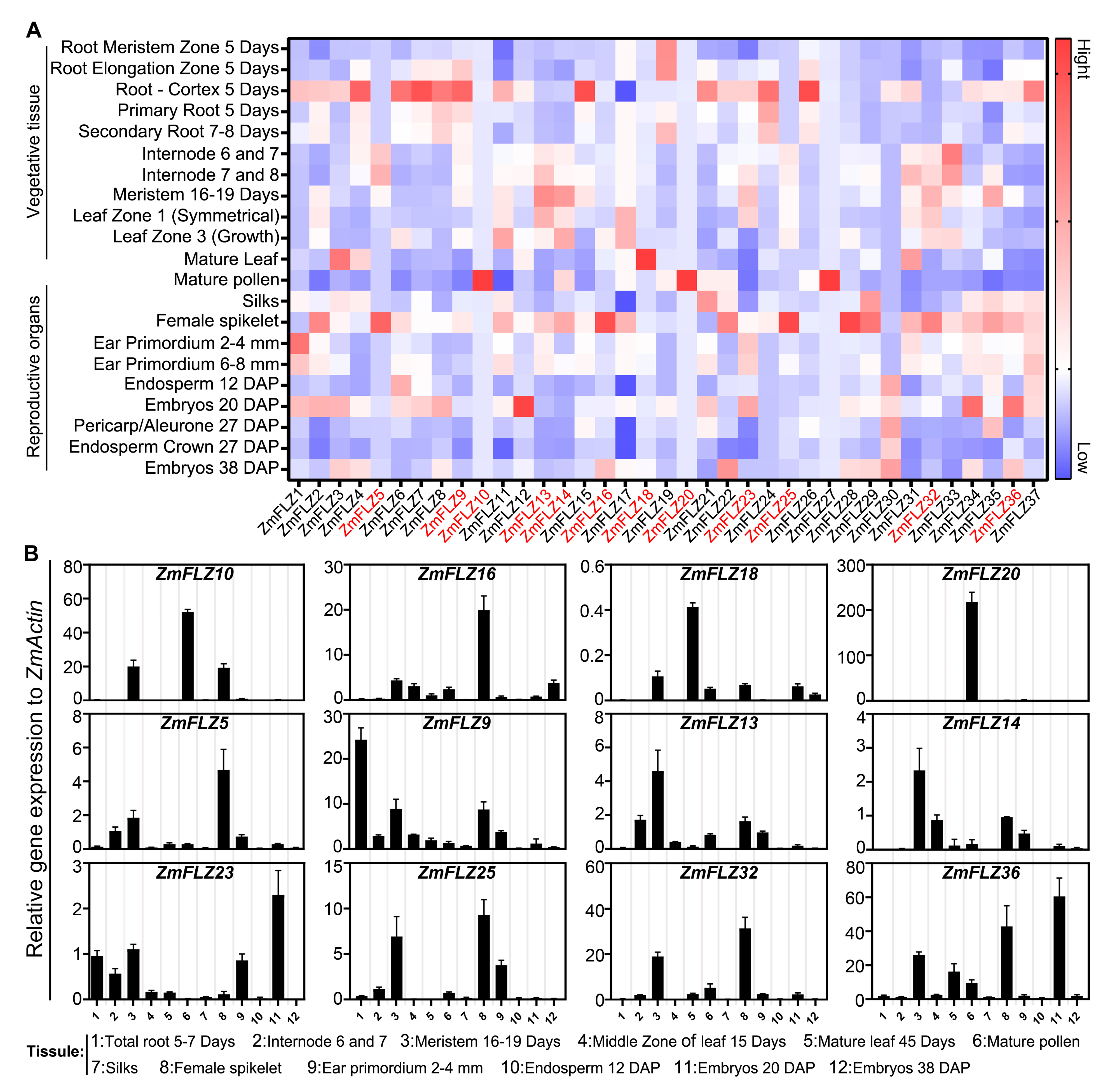
2.3. Tissue Specific Expression Pattern of ZmFLZ Genes

Gene expression pattern provides important clues for understanding potential gene function. To clarify the spatial expression profiles of ZmFLZ genes, we first retrieved the publicly available RNA-seq data from maize the MaizeGDB database (https://www.maizegdb.org/; accessed on 1 September 2019), and constructed an expression map of all the 37 ZmFLZ genes in 21 different tissues, including 11 vegetative organs (like root, internode, and leaf) and 10 reproductive organs (like ear primordium, spikelet, silk, mature pollen, pericarp, endosperm and embryo). As shown in Figure 2A, most of the ZmFLZ genes were ubiquitously expressed in various tissues, but the expression levels in different tissues varied largely. Some ZmFLZ genes showed highly tissue-specific expression patterns, for instance, ZmFLZ12 is highly expressed in embryos 20 days after pollination (DAP), whereas four ZmFLZ genes including ZmFLZ5, ZmFLZ16, ZmFLZ25 and ZmFLZ28 are preferentially expressed in female spikelet. In addition, ZmFLZ10, ZmFLZ20, and ZmFLZ27 are preferentially expressed in mature pollen, while ZmFLZ18 is highly expressed in mature leaf (Figure 2A). To experimentally verify the expression profiles of selected ZmFLZs, we firstly tested the expression data of four ZmFLZs (ZmFLZ10, ZmFLZ16, ZmFLZ18 and ZmFLZ20), all of which have highly specific expression pattern. The results of quantitative real-time PCR (qRT-PCR) showed that these four selected ZmFLZ genes displayed peak expression in specific tissues like ZmFLZ10 and ZmFLZ20 in mature pollen, ZmFLZ16 in female spikelet as well as ZmFLZ18 in mature leaf (Figure 2B), which were largely consistent with results obtained from the publicly available RNA-seq data (Figure 2A). Secondly, according to the phylogenetic analysis, we selected and checked the transcript abundance of eight representative ZmFLZ genes (ZmFLZ9 and ZmFLZ36 from clade I, ZmFLZ5 and ZmFLZ25 from clade II, ZmFLZ13, ZmFLZ14, ZmFLZ23, and ZmFLZ32 from clade III) in 12 different tissues or developmental stages. The results of qRT-PCR showed that the expression patterns of these eight selected ZmFLZ genes were generally in agreement with the data of RNA-seq analysis. For example ZmFLZ36 exhibited the highest expression in embryo 20 DAP (Figure 2B). Relative high expression level of ZmFLZ13 and ZmFLZ14 were observed in vegetative meristem of 16–19 days old maize seedling.

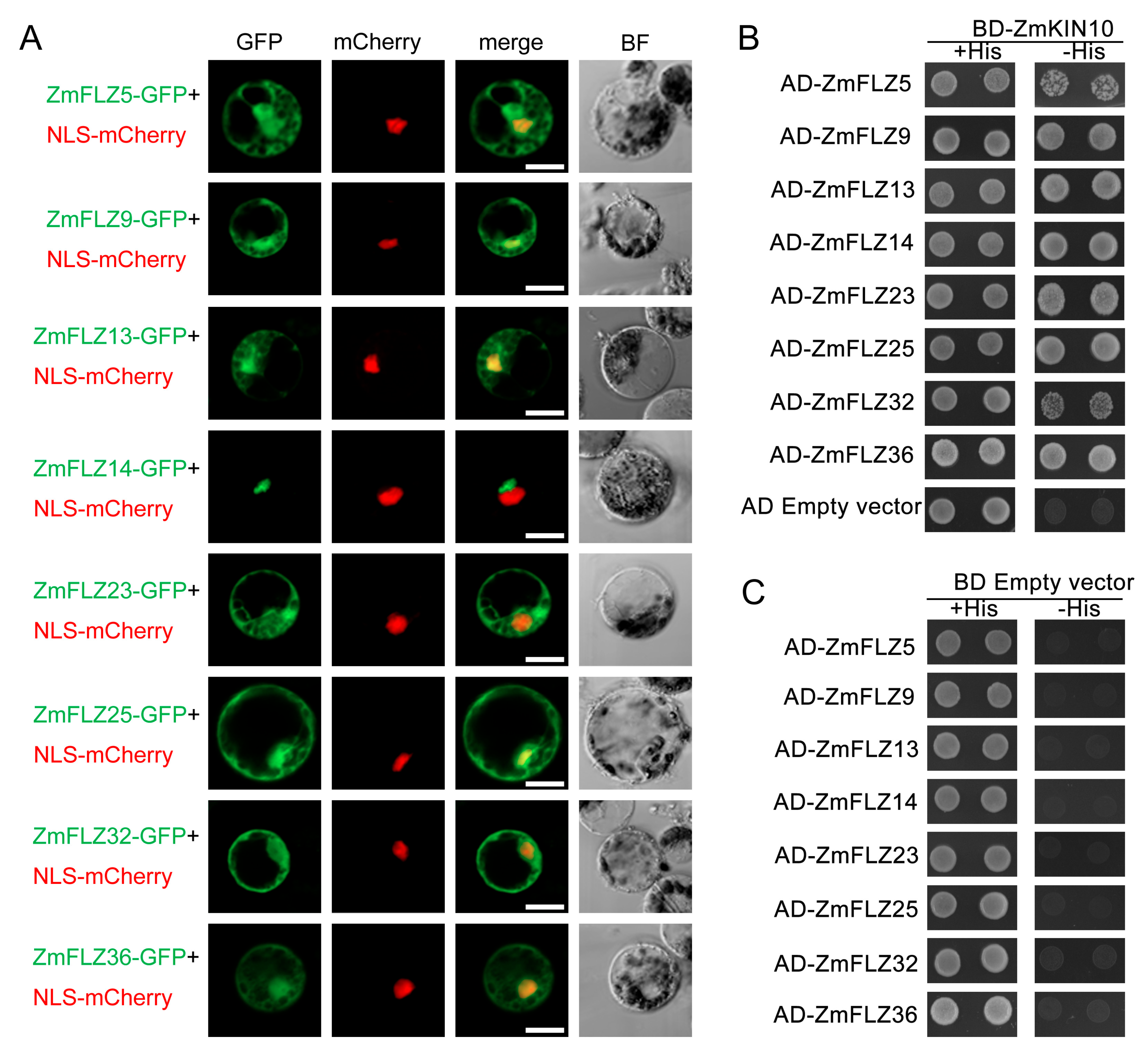
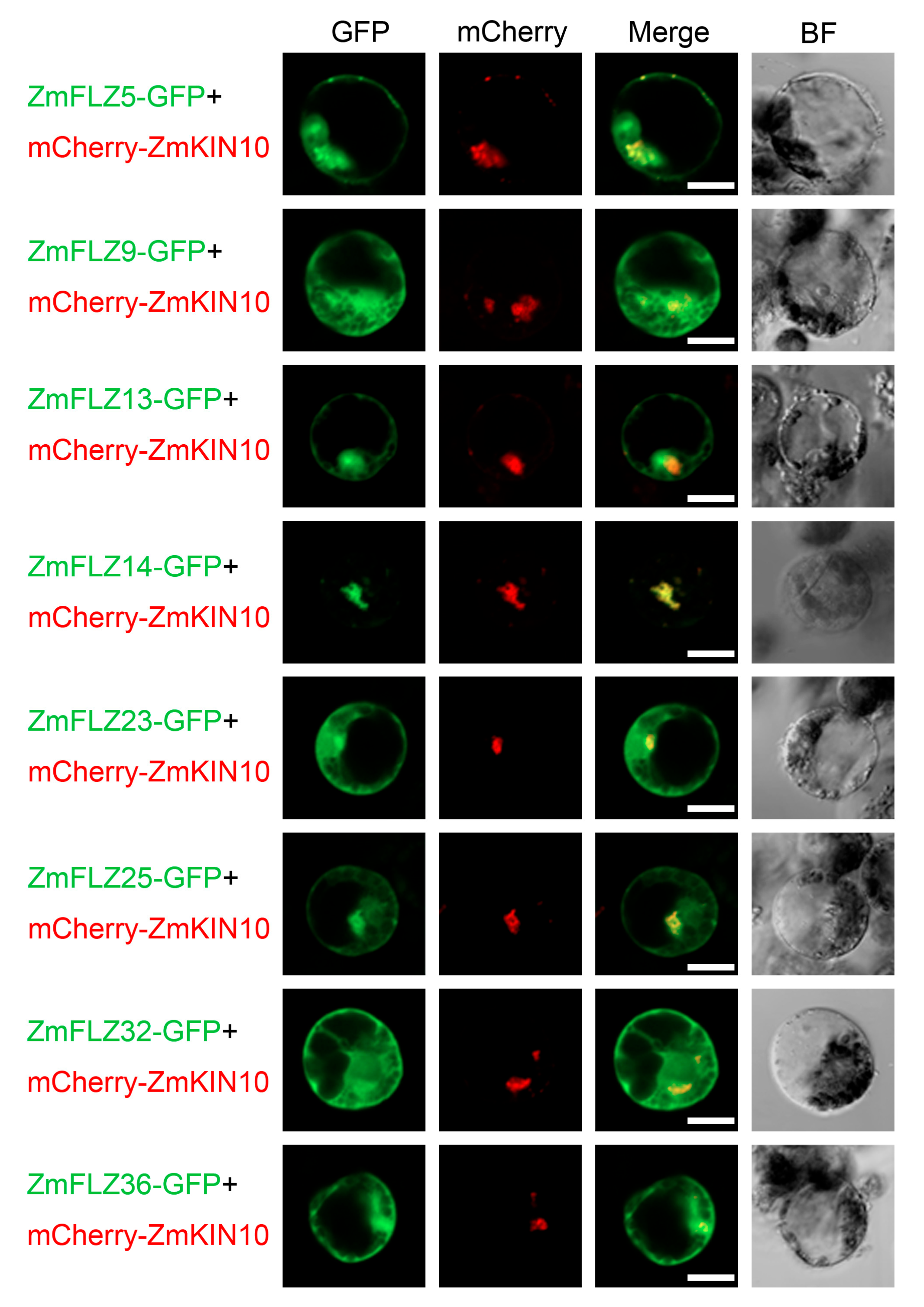
2.4. Subcellular Localizations of Eight Typical ZmFLZ Proteins and Their Interactions with ZmKIN10

Bioinformatics analyses indicate that ZmFLZs are potentially targeted to several subcellular compartments (Table S1). To experimentally determine the subcellular localization of ZmFLZs from different subfamilies, eight typical ZmFLZs were randomly selected from three different clades according to the above phylogenetic analysis. These eight selected members include ZmFLZ9 and ZmFLZ36 from clade I, ZmFLZ5 and ZmFLZ25 from clade II, ZmFLZ13, ZmFLZ14, ZmFLZ23, and ZmFLZ32 from clade III. To this end, the full-length coding sequences of the eight selected typical ZmFLZs were fused in-frame to the N-terminus of Green Fluorescent Protein (GFP) driven by the UBQ10 promoter. Then the resulting fusions were co-expressed with the nuclear marker NLS-mCherry in the maize mesophyll protoplasts. Confocal microscopic analysis showed that GFP-fused ZmFLZ5, 9, 13, 23, 25, 32, and 36 localized in both cytoplasm and nucleus; while the fluorescent signal of ZmFLZ14-GFP was near to but not co-localized with NLS-mCherry (Figure 3A). The results were, in general, consistent with the bioinformatics prediction of the subcellular localization of ZmFLZs (Table S1).
Previous reports demonstrated that FLZ proteins could interact with the α-subunit of SnRK1 kinase complex and function as the scaffolding proteins of SnRK1 complex in Arabidopsis [7,8]. To verify whether ZmFLZs also interact with ZmSnRK1 complex in maize, we used yeast two-hybrid (Y2H) assay to detect the interactions between these eight selected ZmFLZs and the maize SnRK1 subunit ZmKIN10. To this end, the full-length ZmFLZs were fused to GAL4 activation domain (AD-ZmFLZ) and ZmKIN10 was fused to DNA binding domain (BD-ZmKIN10) vectors, respectively. The AD-ZmFLZs were separately co-transformed with the BD-ZmKIN10 into yeast cells, and the interaction of them was evaluated based on the growth of yeast cells on SD/-Trp/-Leu/-His medium plates. As shown in Figure 3B, all the yeast cells harboring BD-ZmKIN10 and AD-ZmFLZ pairs could well grow on the SD/-Trp/-Leu/-His selective medium, suggesting that these ZmFLZs proteins could directly interact with ZmKIN10 in yeast. In order to further explore the functional connection of ZmFLZs and ZmKIN10, ZmFLZs-GFP and ZmKIN10-mCherry were co-transformed into maize leaf protoplasts to observe their subcellular localizations (Figure 4). The obtained results showed that the fluorescent signal of mCherry-ZmKIN10 partially overlapped with the GFP tagged ZmFLZs on the cytoplasmic aggregates (Figure 4), whose shape is obviously different from the smooth and round shape of the nucleus as labeled by the nuclear marker NLS-mCherry (Figure 3). Interestingly, the non-nuclear localized ZmFLZ14-GFP showed a complete colocalization with mCherry-ZmKIN10 when co-expressed in the same cell. These results indicate the close association of these FLZ proteins with ZmKIN10 inside maize leaf mesophyll cells.

2.5. Stress-Responsive Expression Profiles of ZmFLZ Genes

Maize is frequently challenged by abiotic stresses such as shade, drought, and high or low temperature in the field. Recent studies have suggested that AtFLZ genes are widely involved in signaling and response to abiotic stimuli [4]. To study the potential roles of ZmFLZ genes in response to abiotic stresses, we next performed qRT-PCR analysis for the aforementioned eight selected ZmFLZ genes in maize seedlings under multiple abiotic stress conditions, including dark, drought (20% PEG4000), heat (40 °C), cold (4 °C) and ABA (100 μM) (Figure 5). All of these stresses seriously impaired the normal growth of maize seedling and induced the expression of stress-responsive genes (Figure S2). As shown in Figure 5, the transcription of the eight ZmFLZ genes illustrated dynamic and active responses to different abiotic stresses. In the dark treatment, the expressions of ZmFLZ25 and ZmFLZ36 were induced 3~100-fold, while the expression level of ZmFLZ9 was significantly repressed throughout the time course. The expression of ZmFLZ5, ZmFLZ13, ZmFLZ23 and ZmFLZ32 were unregulated during the early time points, and then down-regulated at the later time points, while ZmFLZ14 was down-regulated from 4 h to 12 h after dark exposure. PEG-mimicked drought stress also had strong effect on the expression of these eight examined ZmFLZ genes, in which ZmFLZ13, ZmFLZ32 and ZmFLZ36 were repressed but the expression levels of ZmFLZ5, ZmFLZ9, ZmFLZ23 and ZmFLZ25 were elevated at most of the checked time points. Interestingly, the expression of ZmFLZ14 was increased after PEG treatment for 1 h to 4 h, and then decreased subsequently.
For the heat treatment, ZmFLZ5, ZmFLZ9 and ZmFLZ25 were markedly downregulated at most of the checked time points. In contrast, the expression of ZmFLZ32 was significantly increased, with >20-fold increase after heat treatment for 24 h. Other four ZmFLZ genes (ZmFLZ13, 14, 23 and 36) were differentially expressed at specific time points during heat treatment (Figure 5). Upon the cold treatment, ZmFLZ25 and ZmFLZ36 showed increased expression patterns at almost all checked time points except for the stage of treatment for 12 h. The expression levels of ZmFLZ13, ZmFLZ14 and ZmFLZ32 were slightly increased and then significantly decreased from 6 h after cold treatment. ZmFLZ23 was obviously upregulated at 1 h and 4 h points and then downregulated afterwards (Figure 5). Finally, we evaluated the responses of these eight ZmFLZ genes to ABA, the most important stress-protective phytohormone in plants [19]. ZmFLZ5, ZmFLZ13 and ZmFLZ36 were dramatically suppressed by ABA treatment, while the expressions of other five ZmFLZs were mostly upregulated at the early stages after ABA treatment (Figure 5). These results indicate that different ZmFLZs might have diverse roles in stress response to various stresses.

2.6. Ectopic Expression of ZmFLZ25 in Arabidopsis Confers Plant the Sensitivity to ABA Treatment

ZmFLZ25 belongs to the maize-specific subfamily II (Figure 1A) and is highly responsive to various stress treatments (Figure 5). Therefore, we tried to study the functional role of this gene in plant stress response. To this end, ZmFLZ25-GFP construct driven by the UBQ10 promoter was introduced into Arabidopsis (Columbia-0). We totally obtained 12 individual T3 homozygous transgenic lines.Three independent overexpression lines (OE1, OE2 and OE3) with higher expression level of ZmFLZ25 as confirmed by qRT-PCR assay (Figure S3) were chosen for further study. Compared with the wild-type (WT) plant, these OE lines showed no obvious phenotype under regular growth condition. However, when directly sowed on the 1/2 MS plates containing different concentrations of ABA, the OE lines displayed enhanced sensitivity to ABA-mediated inhibition of seedling establishment when compared to the WT (Figure 6A,B). To further compare the sensitivity of these plants to ABA, we transferred the 2-d-old plants of WT and OE lines to the fresh 1/2 MS plates supplemented with 0, 5 and 10 μM ABA for additional 7 days growth. As shown in Figure 6C,D, ABA treatment seriously inhibited the primary root growth of Arabidopsis seedlings, and such an effect was more obvious seen in the roots of ZmFLZ25-OE seedlings that were significantly shorter than wild-type seedlings (p-value ≤ 0.05). These data demonstrated that the ZmFLZ25-OE lines are hypersensitive to ABA treatment. To explore the possible molecular mechanisms underlying this ABA hypersensitive phenotype, we examined the possible protein interactions between ZmFLZ25 and Arabidopsis ABA-responsive signaling components. To this end, we checked the protein interactions between ZmFLZ25 and the 14 ABA receptors [19,20,21]. Interestingly, we found that the ZmFLZ25 could interact with several ABA receptors including PYL4, PYL8, PYL12 and PYL13 (Figure 6E), indicating a direct connection between ZmFLZ25 and ABA signaling. We also analyzed the expression of ABA-responsive genes ADH1, EM6 and RD29B in ZmFLZ25-OE plants. The obtained results showed that gene expression level of these ABA-responsive marker genes was more obviously enhanced in ZmFLZ25-OE plants than that in the wild-type seedlings in response to ABA treatment (Figure 6F). Taken together, these results suggest that ZmFLZ25 interacts with the ABA receptors and might function as a positive regulator in ABA signaling during seedling establishment and growth.

3. Discussion

In this study, 37 ZmFLZ genes were identified in the maize genome via a homology-search method. These 37 ZmFLZ proteins can be divided into three subfamilies with highly conserved FLZ domain but significantly different protein length. Moreover, we found that ZmFLZ genes have a varied expression patterns with some organ-specific induction trend, while the relative higher expression of most ZmFLZ genes were observed in 5-day-old root cortex and female spikelets (Figure 2A). The qRT-PCR analysis of twelve typical ZmFLZ genes showed that three ZmFLZs, including ZmFLZ10, ZmFLZ16 and ZmFLZ20, display a specifically high expression in reproductive organs, while the remaining nine selected ZmFLZs show high expression levels in vegetative meristem and internodes as well as endosperm (20 DAP) (Figure 2B). These results suggested the differential and specific roles of ZmFLZ family members during maize growth and organ or tissue development. In supporting this noting, one root expressed FLZ gene ZmFLZ14 was recently found to be well associated with maize lateral root branching frequency under well-water conditions [22], highlighting its potential roles in the control of root architecture in maize. In Arabidopsis, it has been shown that several AtFLZs exhibited higher expression levels in the reproductive organs, such as, flower and/or silique [4], and overexpression of the FLZ gene AtFLZ4/IRM1 led to delayed bolting time and smaller size of siliques and less production of seeds in Arabidopsis, indicating an important role of FLZ family genes in plant reproduction [5]. In future, it will be interesting to dissect the roles of those ZmFLZs specifically expressed in reproductive organs in corn development and production.
Previous studies have well established that expression of FLZ genes is differentially regulated by hormones and environmental cues, as well as cellular energy level, suggesting their implication in plant stress signaling or response [4,6,9]. For example, expression of the wheat FLZ gene TaSRHP is salt-induced, and ectopic overexpression of this gene in Arabidopsis conferred enhanced tolerance to salt and drought stresses [6]. Furthermore, one recent report showed that knock-down of two ABA-induced FLZ genes, FLZ6 and FLZ10 resulted in the hypersensitive of seedlings to ABA and osmotic stresses [7,8]. In this study, we found that eight ZmFLZ genes are highly responsive to abiotic stresses, including dark, drought, heat, cold and ABA (Figure 5). For instance, the expression level of ZmFLZ25 dramatically increased in maize seedlings under dark, cold or ABA treatments. These results indicated that some ZmFLZ genes might be involved in stress response to adverse environmental conditions. But up to now, how FLZs are involved in plant stress signaling and response remains largely unknown. For instance, ZmFLZ25 showed the most notable response to all of the five stresses, among them, heat treatment significantly repressed its expression, whereas dark, drought, cold and ABA treatments up-regulated its expression (Figure 5). To clarify the potential roles of ZmFLZ25 in plant stress response, we generated ZmFLZ25-OE transgenic Arabidopsis plants and found that ZmFLZ25-OE plants displayed higher sensitivity to ABA treatment (Figure 6), pointing to the positively regulatory role of ZmFLZ25 in plant ABA signaling. Consistent with the roles of FLZs in the positive regulation of plant ABA signaling, knock-down of the FLZ family gene MARD1 in Arabidopsis conferred resistance to ABA-regulated seed dormancy, and the mutant seeds germinated faster comparing with the WT seeds in the medium supplemented with exogenous ABA [2]. However, the direct link between FLZs and ABA signaling remain totally unclear. Here, we showed that the maize FLZ member ZmFLZ25 could directly interact with several ABA receptors including PYL4, PYL8, PYL12 and PYL13. Considering the positive regulatory role of ZmFLZ25 in Arabidopsis ABA response, it is possible that binding of ZmFLZ25 might result in protein stabilization and less degradation of ABA receptors, or might mimic the FLZ function as a scaffold of SnRK1 kinase complex to enable the enhanced interactions between PYLs and PP2Cs, thereby activating ABA signaling pathway. Future work is needed to dissect the exact roles of FLZ family in plant ABA signaling.
Protein-protein interaction network analysis showed that FLZ proteins might interact with several functional proteins that are involved in well-characterized stress signaling pathways such as SnRK1, TOR and MPK signaling [4,9,10]. In this work, we demonstrated that ZmFLZs interacted with ZmKIN10 in the yeast-two-hybrid assay (Figure 3B,C) and found that several ZmFLZs partially co-localized with ZmKIN10 in maize cells (Figure 4). Although, these results are largely consistent with the results of FLZ-SnRK1 interactions in Arabidopsis [7,8,9], it is worth noting that the subcellular compartment where ZmSnRK1 and ZmFLZs co-localized with each other in maize cells seems to be the cytoplasmic aggregates rather than the nucleus (Figure 4). Interestingly, the Arabidopsis KIN10 has been reported to target to diverse cellular compartments, such as the cytoplasm and nucleus [23,24], some unknown puncta [25,26], the Golgi [27], chloroplasts [28], and the endoplasmic reticulum (ER) [14,29]. Moreover, a recent report has demonstrated that AtKIN10 interacted with AtFLZs in onion epidermal cells, and the fluorescent signals of their interaction site were observed in cytoplasmic foci, which were further confirmed as the ER associated with the nucleus [14]. Based on the evolutionary conservation of FLZ-SnRK1 interaction in different plant species and the localization pattern we observed in this study, we suspected that the cytoplasmic aggregates where ZmFLZs and ZmKIN10 co-localized might be a portion of ER near the nucleus in maize cells. In future, more accurate experiments are needed to further clarify the subcellular distributions of ZmFLZ and ZmSnRK1 in maize.
It is clear that SnRK1 kinase complex is the cellular energy sensor, which can be activated by very diverse abiotic and biotic stress conditions that directly or indirectly cause an energy deficit [11,30,31]. Although FLZs function as the scaffold of SnRK1 complex, how FLZs affect SnRK1 signaling remains elusive. One recent study has revealed that FLZ6 and FLZ10 act as suppressors of SnRK1 to regulate Arabidpsis growth and the responses to ABA and sugar by modulating the protein abundance of SnRK1 [7], but the underling molecular mechanism remains elusive. So in the future, more genetic, molecular and physiological studies are needed to decipher how FLZs affect SnRK1 signaling to modulate plant stress response. As an important food, feeds, and industrial raw material, maize is often exposed to various abiotic stresses such as drought, extreme temperature, and shade, which might cause reduced or even no yield [32,33,34,35]. Further studies of the molecular functional roles of SnRK1-FLZ module in maize will be helpful for future applications of this conserved regulatory module in maize breeding to enhance stress-resistance.

4. Materials and Methods

4.1. Data Search and Analyses

Search of FLZ proteins in maize was done as described previously [36]. Briefly, the protein sequences of FLZ domain from AtFLZs were used as queries to run the BLAST program against the B73 reference genome (RefGen_v4) (https://www.maizegdb.org/; accessed on 1 September 2019) [37]. A self-BLAST of these sequences was also performed to remove redundant sequences, and then the remaining sequences were submitted to the Pfam search (https://pfam.xfam.org/; accessed on 15 October 2019) [38] and BLAST-based NCBI conserved domain search (https://www.ncbi.nlm.nih.gov/Structure/cdd/wrpsb.cgi; accessed on 26 November 2019) to further confirm the presence of the conserved FLZ domain, and the protein structures of them were visualized with the TBtools software [39]. Information about gene locus, chromosome location, nucleotide length, protein length, protein molecular weight, protein isoelectric point was also obtained for each ZmFLZ gene from maize database (https://www.maizegdb.org/; accessed on 1 September 2019). The distribution pattern of ZmFLZ genes on the chromosomes were visualized with a RCircos package in R. All the FLZ protein sequences from maize and Arabidopsis were aligned using ClustalW, and then the alignment result was imported in MEGAX to generate a phylogenetic tree using the Neighbor joining method with partial deletion and 1000 bootstraps.

4.2. Maize Growth Conditions and Treatments

Seeds of maize B73 were obtained from Institute of Germplasm Resources and Biotechnology, Jiangsu Academy of Agricultural Sciences, China. Seedling cultivation was performed according to the methods described previously [40]. Briefly, the surface-sterilized plump seeds were cultured in water in a growth chamber at 28 °C (light) /25 °C (dark) with a 14 h (light)/10 h (dark) photoperiod. After five days, the uniformed seedlings were selected and further cultured with 1/2 Hoagland (Phygene, Shenzhen, China) nutrient solution to the V3 stage. Then the seedlings were planted in the field and the different tissues at the indicated developmental stages were harvested to analyze the gene expression of ZmFLZs.
The stress treatments of maize seedlings were performed as previously described [41]. For each treatment, three uniformed maize plants of B73 at the V3 stage were used, and the untreated maize seedlings at the same time points were used as the control. For dark treatment, seedlings were kept in the full darkness. For drought stress, seedlings were root-cultured in nutrient solution containing 20% PEG4000 (Sangon Biotech, Shanghai, China). Seedlings were transferred to a 40 °C or 4 °C incubator respectively for heat and cold treatments. For ABA treatment, maize plants were root-soaked in the nutrient solution containing 100 μM ABA (Sangon Biotech, Shanghai, China). After 0 h, 1 h, 2 h, 4 h, 6 h, 12 h and 24 h treatments, leaf samples of these treated or control seedlings were harvested and frozen in liquid nitrogen for further RNA isolation.

4.3. Generation of ZmFLZ25 Overexpressed Arabidopsis and ABA Sensitivity Test

The BLAST search against the Arabidopsis database TAIR website (https://www.arabidopsis.org/; accessed on 8 February 2021) showed that ZmFLZ25 shares the highest similarity (59%) to AtFLZ4 (AT5G65040.1). To overexpress ZmFLZ25, the coding sequence (CDS) of ZmFLZ25 was PCR-amplified and inserted into the binary vector pCAMBIA1300 to in-frame fuse with a GFP gene. The resulting pCAMBIA1300-UBQ10-ZmFLZ25-GFP construct was introduced into wild-type Arabidopsis (Col-0) plants by Agrobacteria-mediated floral dipping. The positive transgenic plants were selected on standard 1/2 MS (Murashige and Skoog) plates containing 50 μg/L Hygromycin B (Sangon Biotech, Shanghai, China) and the T3 homozygous transgenic lines were used for further analysis [42].
For ABA sensitivity test during seedling establishment, seeds were surface sterilized and stratified at 4 °C for 48 h in darkness before sowing on standard 1/2 MS plates supplemented with 0, 0.1 and 0.25 μM ABA. Then the plates were placed in a growth chamber at 22 °C under the 16 h (light)/8 h (dark) conditions for five days before photographing and recording the seedling growth rate following the procedure as described previously [43]. For ABA-inhibited root elongation assay, uniformed two-day-old seedlings were transferred to fresh 1/2 MS plates containing 0, 5 and 10 μM ABA for another seven days vertical growth before taking pictures to measure the length of primary roots following the procedure as described previously [44]. These experiments were repeated three times with similar results.

4.4. Subcellular Localization Assays

The possible subcellular localization of ZmFLZs was first predicted using the TargetP-2.0 web server (http://www.cbs.dtu.dk/services/TargetP/; accessed on 15 November 2019) [45]. Nuclear localization of ZmFLZs proteins was predicted by NLStradamus (https://nucpred.bioinfo.se/cgi-bin/single.cgi; accessed on 27 March 2019) [46]. To experimentally analyze the subcellular localization of ZmFLZs and their co-localization with ZmSnRK1α, the cDNAs of the eight ZmFLZs were separately cloned into pCAMBIA1300 vector to in-frame fuse with a GFP gene (ZmFLZ-GFP), while the cDNA of ZmSnRK1α was cloned into pBI221 vector to fuse with a mCherry tag (ZmKIN10-mCherry). Primers used in this study were listed in the Table S2. The plasmid combinations of ZmFLZ-GFP and nuclear marker NLS-mCherry or ZmKIN10-mCherry were introduced into maize leaf protoplasts to determine their subcellular localization. Maize leaf protoplast isolation and PEG-mediated transformation was performed as previously described [47,48]. The transfected protoplast cells were incubated in 1 mL W5 solution (154 mM of NaCl, 125 mM of CaCl2, 5 mM of KCl, and 2 mM of MES at pH 5.7) supplemented with 10 mM sucrose for 12 h in the dark at 26 °C before imaging with a laser scanning confocal microscope (LSM800, Zeiss, Jena, Germany).

4.5. Yeast-Two-Hybrid Assay

The yeast-two-hybrid assay was carried out following the manufacturer’s instructions (TaKaRa, Beijing, China). The cDNA of the ZmSnRK1α was cloned into pGBKT7 as bait and the cDNAs of the ZmFLZs were cloned into pGADT7 as prey, respectively. Primers used for cloning were listed in Table S2. Different pairs of bait and prey vectors indicated were co-transformed into the yeast reporter strain AH109. Then the different yeast transformants were grown on synthetic double drop-out (SD/-Leu/-Trp) and synthetic triple drop-out (SD/-Leu/-Trp/-Ade) solid medium to examine the protein-protein interaction.

4.6. RNA Isolation and Quantitative Real-Time PCR

The samples of maize B73 tissues or organs at specific developmental stages were collected according to the methods as described previously [49]. Total RNA was isolated from the harvested samples using the TransZol Plant kit (TransGen, Beijing, China), then subjected to the cDNA synthesis using the TransScript All-in-One First-Strand cDNA synthesis kit (TaKaRa, Beijing, China) according to the manufacturer’s instructions. Diluted cDNA samples were used for real-time PCR amplification in 10 μL reaction volumes with specific primers and SYBR Green PCR Master Mix (TaKaRa, Beijing, China). Quantitative real-time PCR was run on the Applied Biosystems 7500 system (Prism® 7500, Carlsbad, America) using the following reaction conditions: 95 °C for 1 min followed by 45 cycles of 95 °C for 10 sec and 60 °C for 15 sec. ZmACTIN gene was used as the internal control, and the relative expression level of each gene were calculated using the 2−∆∆CT method [50]. Primers used in this study were listed in Table S2.

4.7. Statistical Analyses

At least three biological replicates were used during the stress treatments. t-test in Graphpad Prism 8 was used to estimate the statistical significance of the difference between the treatment group and the control group and marked with * when p < 0.05.

Supplementary Materials

The following are available online at https://www.mdpi.com/article/10.3390/ijms22073529/s1, Supplementary materials including Figures S1–S3 and Tables S1 and S2 can be found online.

Author Contributions

S.C., C.Y., X.T. and C.G. conceived and designed this research; S.C., X.L., C.Y., and C.L. performed the experiment; S.C., X.L., C.Y. and W.Y. analyzed the data; S.C., C.Y., X.T. and C.G. wrote the manuscript. All authors have read and agreed to the published version of the manuscript.

Funding

This work was supported by Natural Science Foundation of Guangdong Province (grant number 2018B030308008) to X.T., the National Natural Science Foundation of China (31870171) and Fok Ying-Tong Education Foundation for Young Teachers in the Higher Education Institutions of China (171014) to C.G.

Institutional Review Board Statement

Not applicable.

Informed Consent Statement

Not applicable.

Data Availability Statement

Data is contained within the article or Supplementary Materials.

Acknowledgments

We thank Han Zhao, Tifu Zhang, Shuaiqiang Liang from Institute of germplasm resources and biotechnology, Jiangsu Academy of Agricultural Sciences, China for sharing the maize seeds of B73. We would like to thank Chunjue Xu and Man Jin of Shenzhen Institute of Molecular Crop Design for their help in providing experimental materials for this study.

Conflicts of Interest

The authors declare no conflict of interest.

References

  1. He, Y.; Tang, W.; Swain, J.D.; Green, A.L.; Jack, T.; Gan, S. Networking Senescence-Regulating Pathways by Using Arabidopsis Enhancer Trap Lines. Plant Physiol. 2001, 126, 707–716. [Google Scholar] [CrossRef] [Green Version]
  2. He, Y.; Gan, S. A novel zinc-finger protein with a proline-rich domain mediates ABA-regulated seed dormancy in Arabidopsis. Plant Mol. Biol. 2004, 54, 1–9. [Google Scholar] [CrossRef]
  3. Laxmi, A. DUF581 Is Plant Specific FCS-Like Zinc Finger Involved in Protein-Protein Interaction. PLOS ONE 2014, 9, e99074. [Google Scholar]
  4. Jamsheer, K.M.; Laxmi, A. Expression of Arabidopsis FCS-Like Zinc finger genes is differentially regulated by sugars, cellular energy level, and abiotic stress. Front Plant Sci. 2015, 6, 746. [Google Scholar] [CrossRef] [Green Version]
  5. Chen, X.; Zhang, Z.; Visser, R.G.F.; Broekgaarden, C.; Vosman, B. Overexpression of IRM1 Enhances Resistance to Aphids in Arabidopsis thaliana. PLoS ONE 2013, 8, e70914. [Google Scholar] [CrossRef] [Green Version]
  6. Hou, X.; Liang, Y.; He, X.; Shen, Y.; Huang, Z. A Novel ABA-Responsive TaSRHP Gene from Wheat Contributes to Enhanced Resistance to Salt Stress in Arabidopsis thaliana. Plant Mol. Biol. Rep. 2013, 31, 791–801. [Google Scholar] [CrossRef]
  7. Jamsheer, K.M.; Sharma, M.; Singh, D.; Mannully, C.T.; Jindal, S.; Shukla, B.N.; Laxmi, A. FCS-like zinc finger 6 and 10 repress SnRK1 signalling in Arabidopsis. Plant J. 2018, 94, 232–245. [Google Scholar] [CrossRef] [Green Version]
  8. Jamsheer K, M.; Jindal, S.; Laxmi, A. Evolution of TOR-SnRK dynamics in green plants and its integration with phytohormone signaling networks. J. Exp. Bot. 2019, 70, 2239–2259. [Google Scholar] [CrossRef]
  9. Nietzsche, M.; Schiesl, I.; Bornke, F. The complex becomes more complex: Protein-protein interactions of SnRK1 with DUF581 family proteins provide a framework for cell- and stimulus type-specific SnRK1 signaling in plants. Front. Plant Sci. 2014, 5, 54. [Google Scholar] [CrossRef] [PubMed] [Green Version]
  10. Nietzsche, M.; Landgraf, R.; Tohge, T.; Brnke, F. A protein-protein interaction network linking the energy-sensor kinase SnRK1 to multiple signaling pathways in Arabidopsis thaliana. Curr. Plant Biol. 2015, 5, 36–44. [Google Scholar] [CrossRef] [Green Version]
  11. Crepin, N.; Rolland, F. SnRK1 activation, signaling, and networking for energy homeostasis. Curr. Opin. Plant Biol. 2019, 51, 29–36. [Google Scholar] [CrossRef] [PubMed]
  12. Zhu, J. Abiotic Stress Signaling and Responses in Plants. Cell 2016, 167, 313–324. [Google Scholar] [CrossRef] [Green Version]
  13. Margalha, L.; Confraria, A.; Baena-González, E. SnRK1 and TOR: Modulating growth-defense trade-offs in plant stress responses. J. Exp. Bot. 2019, 70, 2261–2274. [Google Scholar] [CrossRef]
  14. Shukla, B.N.; Jindal, S.; Gopan, N.; Mannully, C.T.; Laxmi, A. The FCS-like zinc finger scaffold of the kinase SnRK1 is formed by the coordinated actions of the FLZ domain and intrinsically disordered regions. J. Biol. Chem. 2018, 293, 13134–13150. [Google Scholar]
  15. Jinge, T.; Chenglong, W.; Jinliang, X.; Lishuan, W.; Guanghui, X.; Weihao, W.; Dan, L.; Wenchao, Q.; Xu, H.; Qiuyue, C. Teosinte ligule allele narrows plant architecture and enhances high-density maize yields. Science 2019, 365, 658–664. [Google Scholar]
  16. Schnable, P.S.; Ware, D.; Fulton, R.S.; Stein, J.C.; Wei, F.; Pasternak, S.; Liang, C.; Zhang, J.; Fulton, L.; Graves, T.A. The B73 Maize Genome: Complexity, Diversity, and Dynamics. Science 2009, 326, 1112–1115. [Google Scholar] [CrossRef] [Green Version]
  17. Sun, S.; Zhou, Y.; Chen, J.; Shi, J.; Zhao, H.; Zhao, H.; Song, W.; Zhang, M.; Cui, Y.; Dong, X.; et al. Extensive intraspecific gene order and gene structural variations between Mo17 and other maize genomes. Nat. Genet. 2018, 50, 1289–1295. [Google Scholar] [CrossRef] [PubMed] [Green Version]
  18. Springer, N.M.; Anderson, S.N.; Andorf, C.M.; Ahern, K.R.; Bai, F.; Barad, O.; Barbazuk, W.B.; Bass, H.W.; Baruch, K.; Ben-Zvi, G.; et al. The maize W22 genome provides a foundation for functional genomics and transposon biology. Nat. Genet. 2018, 50, 1282–1288. [Google Scholar] [CrossRef] [Green Version]
  19. Chen, K.; Li, G.J.; Bressan, R.A.; Song, C.P.; Zhu, J.K.; Zhao, Y. Abscisic acid dynamics, signaling, and functions in plants. J. Integr. Plant Biol. 2020, 62, 25–54. [Google Scholar] [CrossRef] [Green Version]
  20. Rodriguez, L.; Gonzalez-Guzman, M.; Diaz, M.; Rodrigues, A.; Izquierdo-Garcia, A.C.; Peirats-Llobet, M.; Fernandez, M.A.; Antoni, R.; Fernandez, D.; Marquez, J.A.; et al. C2-domain abscisic acid-related proteins mediate the interaction of PYR/PYL/RCAR abscisic acid receptors with the plasma membrane and regulate abscisic acid sensitivity in Arabidopsis. Plant Cell 2014, 26, 4802–4820. [Google Scholar] [CrossRef] [Green Version]
  21. Bueso, E.; Rodriguez, L.; Lorenzo-Orts, L.; Gonzalez-Guzman, M.; Sayas, E.; Munoz-Bertomeu, J.; Ibanez, C.; Serrano, R.; Rodriguez, P.L. The single-subunit RING-type E3 ubiquitin ligase RSL1 targets PYL4 and PYR1 ABA receptors in plasma membrane to modulate abscisic acid signaling. Plant J. 2014, 80, 1057–1071. [Google Scholar] [CrossRef]
  22. Schneider, H.M.; Klein, S.P.; Hanlon, M.T.; Nord, E.A.; Shawn, K.; Brown, K.M.; Andrew, W.; Rahul, B.; Lynch, J.P. Genetic control of root architectural plasticity in maize. J. Exp. Bot. 2020, 71, 3185–3197. [Google Scholar] [CrossRef] [Green Version]
  23. Han, C.; Liu, Y.; Shi, W.; Qiao, Y.; Wang, L.; Tian, Y.; Fan, M.; Deng, Z.; Lau, O.S.; De Jaeger, G.; et al. KIN10 promotes stomatal development through stabilization of the SPEECHLESS transcription factor. Nat. Commun. 2020, 11, 4214. [Google Scholar] [CrossRef]
  24. Ramon, M.; Dang, T.; Broeckx, T.; Hulsmans, S.; Crepin, N.; Sheen, J.; Rolland, F. Default Activation and Nuclear Translocation of the Plant Cellular Energy Sensor SnRK1 Regulate Metabolic Stress Responses and Development. Plant Cell 2019, 31, 1614–1632. [Google Scholar] [CrossRef]
  25. Bitrián, M.; Roodbarkelari, F.; Horváth, M.; Koncz, C. BAC-recombineering for studying plant gene regulation: Developmental control and cellular localization of SnRK1 kinase subunits. Plant J. 2011, 65, 829–842. [Google Scholar] [CrossRef]
  26. Williams, S.P.; Rangarajan, P.; Donahue, J.L.; Hess, J.E.; Gillaspy, G.E. Regulation of Sucrose non-Fermenting Related Kinase 1 genes in Arabidopsis thaliana. Front Plant Sci. 2014, 5, 324. [Google Scholar] [CrossRef] [Green Version]
  27. O’Brien, M.; Kaplan-Levy, R.N.; Quon, T.; Sappl, P.G.; Smyth, D.R. PETAL LOSS, a trihelix transcription factor that represses growth in Arabidopsis thaliana, binds the energy-sensing SnRK1 kinase AKIN10. J. Exp. Bot. 2015, 66, 2475–2485. [Google Scholar] [CrossRef] [Green Version]
  28. Fragoso, S.; Espíndola, L.; Páez-Valencia, J.; Gamboa, A.; Camacho, Y.; Martínez-Barajas, E.; Coello, P. SnRK1 isoforms AKIN10 and AKIN11 are differentially regulated in Arabidopsis plants under phosphate starvation. Plant Physiol. 2009, 149, 1906–1916. [Google Scholar] [CrossRef] [Green Version]
  29. Blanco, N.E.; Liebsch, D.; Guinea Díaz, M.; Strand, Å.; Whelan, J. Dual and dynamic intracellular localization of Arabidopsis thaliana SnRK1.1. J. Exp. Bot. 2019, 70, 2325–2338. [Google Scholar] [CrossRef]
  30. Yang, C.; Shen, W.; Yang, L.; Sun, Y.; Li, X.; Lai, M.; Wei, J.; Wang, C.; Xu, Y.; Li, F. HY5-HDA9 Module Transcriptionally Regulates Plant Autophagy in Response to Light-to-dark Conversion and Nitrogen Starvation. Mol. Plant 2020, 13, 515–531. [Google Scholar] [CrossRef]
  31. Wang, X.F.; Xu, M.; Gao, C.J.; Zeng, Y.L.; Cui, Y.; Shen, W.J.; Jiang, L.W. The roles of endomembrane trafficking in plant abiotic stress responses. J. Integr. Plant Biol. 2020, 62, 55–69. [Google Scholar] [CrossRef]
  32. Wang, X.; Wang, H.; Liu, S.; Ferjani, A.; Li, J.; Yan, J.; Yang, X.; Qin, F. Genetic variation in ZmVPP1 contributes to drought tolerance in maize seedlings. Nat. Genet. 2016, 48, 1233–1241. [Google Scholar] [CrossRef]
  33. Qin, F.; Shinozaki, K.; Yamaguchishinozaki, K. Achievements and Challenges in Understanding Plant Abiotic Stress Responses and Tolerance. Plant Cell Physiol. 2011, 52, 1569–1582. [Google Scholar] [CrossRef] [Green Version]
  34. Julia, K.; Claudia, J. Drought, salt, and temperature stress-induced metabolic rearrangements and regulatory networks. J. Exp. Bot. 2012, 63, 1593–1608. [Google Scholar]
  35. Zhao, C.; Zhang, H.; Song, C.; Zhu, J.-K.; Shabala, S. Mechanisms of Plant Responses and Adaptation to Soil Salinity. Innovation 2020, 1, 100017. [Google Scholar] [CrossRef]
  36. Yang, C.; Shen, W.; Chen, H.; Chu, L.; Xu, Y.; Zhou, X.; Liu, C.; Chen, C.; Zeng, J.; Liu, J. Characterization and subcellular localization of histone deacetylases and their roles in response to abiotic stresses in soybean. BMC Plant Biol. 2018, 18, 226. [Google Scholar] [CrossRef]
  37. Lawrence, C.J.; Qunfeng, D.; Polacco, M.L.; Seigfried, T.E.; Volker, B. MaizeGDB, the community database for maize genetics and genomics. Nucleic Acids Res. 2004, 32, D393–D397. [Google Scholar] [CrossRef] [Green Version]
  38. Finn, R.D.; Alex, B.; Jody, C.; Penelope, C.; Eberhardt, R.Y.; Eddy, S.R.; Andreas, H.; Kirstie, H.; Liisa, H.; Jaina, M. Pfam: The protein families database. Nucleic Acids Res. 2014, 42, D222–D230. [Google Scholar] [CrossRef] [Green Version]
  39. Chen, C.; Chen, H.; Zhang, Y.; Thomas, H.R.; Xia, R. TBtools: An Integrative Toolkit Developed for Interactive Analyses of Big Biological Data. Mol. Plant 2020, 13, 1194–1202. [Google Scholar] [CrossRef]
  40. Zhang, X.; Mi, Y.; Mao, H.; Liu, S.; Chen, L.; Qin, F. Genetic variation in ZmTIP1 contributes to root hair elongation and drought tolerance in maize. Plant Biotechnol. J. 2019, 18, 1271–1283. [Google Scholar] [CrossRef] [Green Version]
  41. Ma, H.; Liu, C.; Li, Z.; Ran, Q.; Xie, G.; Wang, B.; Fang, S.; Chu, J.; Zhang, J. ZmbZIP4 Contributes to Stress Resistance in Maize by Regulating ABA Synthesis and Root Development. Plant Physiol. 2018, 178, 753–770. [Google Scholar] [CrossRef] [PubMed] [Green Version]
  42. Gao, C.; Luo, M.; Zhao, Q.; Yang, R.; Cui, Y.; Zeng, Y.; Xia, J.; Jiang, L. A Unique Plant ESCRT Component, FREE1, Regulates Multivesicular Body Protein Sorting and Plant Growth. Curr. Biol. 2014, 24, 2556–2563. [Google Scholar] [CrossRef] [Green Version]
  43. Li, H.; Li, Y.; Zhao, Q.; Li, T.; Wei, J.; Li, B.; Shen, W.; Yang, C.; Zeng, Y.; Rodriguez, P.L. The plant ESCRT component FREE1 shuttles to the nucleus to attenuate abscisic acid signalling. Nat. Plants 2019, 5, 512–524. [Google Scholar] [CrossRef] [PubMed]
  44. Xiao, Z.; Yang, C.; Liu, C.; Yang, L.; Shen, W. SINAT E3 ligases regulate the stability of the ESCRT component FREE1 in response to iron deficiency in plants. J. Integr. Plant Biol. 2020, 62, 1399–1417. [Google Scholar] [CrossRef]
  45. Emanuelsson, O.; Brunak, S.R.; Von Heijne, G.; Nielsen, H. Locating proteins in the cell using TargetP, SignalP and related tools. Nat. Protocols 2007, 2, 953–971. [Google Scholar] [CrossRef]
  46. Brameier, M.; Krings, A.; Maccallum, R.M. NucPred - Predicting Nuclear Localization of Proteins. Bioinformatics 2007, 23, 1159–1160. [Google Scholar] [CrossRef]
  47. Gao, L.; Shen, G.; Zhang, L.; Qi, J.; Zhang, C.; Ma, C.; Li, J.; Wang, L.; Malook, S.U.; Wu, J. An efficient system composed of maize protoplast transfection and HPLC–MS for studying the biosynthesis and regulation of maize benzoxazinoids. Plant Methods 2019, 15, 144. [Google Scholar] [CrossRef] [Green Version]
  48. Shen, W.; Xiao, Z.; Shen, J.; Gao, C. Analysis of Golgi-Mediated Protein Traffic in Plant Cells. Methods Mol. Biol. 2017, 1662, 75–86. [Google Scholar]
  49. Walley, J.W.; Sartor, R.C.; Shen, Z.; Schmitz, R.J.; Wu, K.J.; Urich, M.A.; Nery, J.R.; Smith, L.G.; Schnable, J.C.; Ecker, J.R. Integration of omic networks in a developmental atlas of maize. Science 2016, 353, 814–818. [Google Scholar] [CrossRef] [Green Version]
  50. Livak, K.J.; Schmittgen, T.D. Analysis of relative gene expression data using real-time quantitative PCR and the 2(-Delta Delta C (T)) Method. Methods 2001, 25, 402–408. [Google Scholar] [CrossRef]
Figure 1. Phylogenetic tree and domain feature of FLZs in maize and Arabidopsis. (A) Phylogenetic tree of FLZ proteins in maize and Arabidopsis. The phylogenetic tree was generated using the Neighbor joining method in the MEGA-X software with 1000 replicates bootstrap analysis and complete deletion. The numbers at the nodes indicate the bootstrap values, with cutoff no less than 50. (B) Diagrams of the FLZ domain in the FLZ proteins. The conserved FLZ domains were predicted with NCBI Batch-CD and marked with green boxes. (C) Protein sequences alignment of the FLZ domain. ClustalW was used for protein alignment. The conserved amino acids in FLZ domains were highlighted and marked with asterisks.
Ijms 22 03529 g001
Figure 2. Spatial expression patterns of ZmFLZs. (A) Heat map of the gene expression of ZmFLZs in various tissues or developmental stages. The data were downloaded from the qTeller of MaizeGDB (https://qteller.maizegdb.org/; accessed on 1 September 2019). The expression levels as indicated by Fragments Per Kilobase of exon model per Million mapped fragments (FPKM) were shown from low (blue) to high (red) for each gene. (B) Tissue-specific expression patterns of ZmFLZs by qRT-PCR assay. The x-axis represents different tissues or organs. The y-axis shows the relative gene expression levels normalized to ZmActin. Data represent the mean ± SEM from three independent experiments. DAP, day after pollination.
Ijms 22 03529 g002
Figure 3. Subcellular localization of the eight selected ZmFLZs and ZmFLZs interaction with ZmKIN10 by Y2H assay. (A) ZmFLZs-GFP fusion constructs were used to determine the subcellular localization of ZmFLZs in the protoplast cells isolated from maize leaves. NLS-mCherry was used as a nuclear marker. Fluorescent images of GFP and mCherry were captured with a confocal laser scanning microscopy and shown in green and red, respectively. Scale bars = 25 μm. (B,C) Yeast two hybrid analysis of the binary interactions between the eight selected typical ZmFLZs and ZmKIN10.
Ijms 22 03529 g003
Figure 4. Co-localization analysis of ZmFLZs and ZmKIN10 in maize leaf protoplast cells. Co-expression of mCherry-ZmKIN10 and the GFP fused ZmFLZs in protoplasts followed by confocal imaging. Fluorescent images of GFP and mCherry were captured with a confocal laser scanning microscopy and shown in green and red, respectively. Scale bars = 25 μm.
Ijms 22 03529 g004
Figure 5. Expression pattern of ZmFLZs in response to stress treatments. Seedlings of B73 at V3 stage (three-leaf-stage, around 15 days after germination) were separately subjected to darkness, drought (20% PEG4000), heat (40 °C), cold (4 °C), and 100 μM ABA treatments for 24 h. The leaves were harvested for gene expression analysis by qRT-PCR at 0 h, 1 h, 2 h, 4 h, 6 h, 9 h, 12 h, and 24 h, respectively. Seedlings grown under the normal conditions were harvested at the same time points and used as the controls. Relative gene expression levels of ZmFLZs were calculated as the ratio of “treatment” to “control” after being normalized against ZmActin. The mean ± SEM from three independent experiments were shown for each treatment. Multiple t-test was included to estimate the differences between the expression levels under stresses and untreated (0 h) conditions for each ZmFLZ gene. The red and blue asterisks indicated significantly enhanced and repressed expression under stresses, respectively (p-value < 0.05).
Ijms 22 03529 g005
Figure 6. Overexpression of ZmFLZ25 in Arabidopsis resulted in the hypersensitivity of plant to ABA. (A) Overexpression expression of ZmFLZ25 in Arabidopsis confers plant sensitivity to ABA 5. days after imbibition. The OE1, OE2 and OE3 represent three different ZmFLZ25-overexpression lines. (B) The cotyledon greening rate of WT and OE lines were counted after ABA treatments. Approximately approximately100 seeds were counted for each genotype at 5 days of growth on 1/2 Murashige & Skoog (MS) medium plate, data represents three biologically replicates. (C) Ectopic expression of ZmFLZ25 in Arabidopsis resulted in enhanced root growth inhibition in response to ABA treatment. WT and overexpression seedlings were grown on 1/2 MS plate for 2 days and subsequently transferred to plates containing 5 μM and 10 μM ABA for 7 days. (D) Statistics of the root length of WT and OE lines after ABA treatments corresponding to (C). (E) Protein interactions between ZmFLZ25 and Arabidopsis ABA receptors by yeast two hybrid assay. (F) The expression of ABA-responsive genes (ADH1, EM6, RD29B) (y-axis) in Arabidopsis under treatments with different concentrations of ABA (x-axis) in WT and OE lines. The relative gene expression levels were normalized to the internal gene AtUBQ. All the data are the mean ± SEM from three independent experiments. The asterisks indicated significantly differences between WT and OE lines as determined by t-test (p-value < 0.05).
Ijms 22 03529 g006
Publisher’s Note: MDPI stays neutral with regard to jurisdictional claims in published maps and institutional affiliations.

