Article

Selection and Validation of Reference Genes for qRT-PCR Analysis in the Oil-Rich Tuber Crop Tiger Nut (Cyperus esculentus) Based on Transcriptome Data

1
CAS Key Laboratory of Tropical Plant Resources and Sustainable Use, Xishuangbanna Tropical Botanical Garden, Innovation Academy for Seed Design, Chinese Academy of Sciences, Menglun, Mengla 666303, China
2
College of Life Sciences, University of Chinese Academy of Sciences, Beijing 100049, China
3
Center of Economic Botany, Core Botanical Gardens, Chinese Academy of Sciences, Menglun, Mengla 666303, China
4
State Key Laboratory for Conservation and Utilization of Subtropical Agro-Bioresources, College of Forestry, Guangxi University, Nanning 530004, China
*
Authors to whom correspondence should be addressed.
Int. J. Mol. Sci. 2021, 22(5), 2569; https://doi.org/10.3390/ijms22052569
Submission received: 31 January 2021 / Revised: 24 February 2021 / Accepted: 25 February 2021 / Published: 4 March 2021
(This article belongs to the Section Molecular Plant Sciences)

Abstract

:
Tiger nut (Cyperus esculentus), a perennial C4 plant of the Cyperaceae family, is an unconventional crop that is distinguished by its oil-rich tubers, which also possesses the advantages of strong resistance, wide adaptability, short life periods, and large biomass. To facilitate studies on gene expression in this species, we identified and validated a series of reference genes (RGs) based on transcriptome data, which can be employed as internal controls for qRT-PCR analysis in tiger nut. Fourteen putative candidate RGs were identified and evaluated across nine different tissues of two cultivars, and the RGs were analyzed using three different algorithms (geNorm, NormFinder, and BestKeeper). The stability rankings of the candidate RGs were merged into consensus lists with RankAggreg. For the below-ground storage organ of tiger nut, the optimal RGs were TUB4 and UCE2 in different developmental stages of tubers. UCE2 and UBL5 were the most stably expressed RGs among all tissues, while Rubisco and PGK exhibited the lowest expression stability. UCE2, UBL5 and Rubisco were compared to normalize the expression levels of the caleosin (CLO) and diacylglycerol acyltransferase 2-2 (DGAT2-2) genes across the same tissues. Our results showed that the RGs identified in this study, which exhibit more uniform expression patterns, may be utilized for the normalization of qRT-PCR results, promoting further research on gene expression in various tissues of tiger nut.

1. Introduction

To pursue a better quality of life and a healthier diet for humans, many economically important crops have been intensively studied. Meanwhile, an increasing number of agricultural sources of cooking oil have emerged to meet the increasing market demand. Generally, the majority of plants accumulate abundant oil in their seeds (e.g., soybean, rapeseed, peanut, and sunflower), as well as their mesocarps (e.g., olive, oil palm, and avocado), while a smaller number of oil-rich plants accumulate oil in non-seed vegetable tissues [1,2,3,4,5]. A wild tomato species (Lycopersicon pennellii) accumulates lipids on the epicuticle of leaves [6]. The oil-storage organs of Tetraena mongolica are phloem cells and xylem parenchyma of stems [7]. Tiger nut (Cyperus esculentus) is a perennial herb of Cyperaceae that is widely distributed in almost all middle- and low-latitude areas [8]; this plant stockpiles oil in its tubers as an underground storage organ [9]. Tiger nut is an unconventional crop that is distinguished by its oil-rich tubers, and it is also known as yellow nutsedge and chufa. Oleic acid (C18:1) is abundantly contained in the tuber oil of this plant and is a kind of monounsaturated fatty acids (MUFAs) [10], which has positive effects on human health, especially by reducing the blood lipids [11], blood pressure [12], and the incidence of type 2 diabetes mellitus [13]. At present, the majority of tiger nut researchers have primarily focused on the cultivation technology [14], oil extraction process [15,16], analysis of nutritional components [17] and medicinal values [18], and development of tiger nut milk [19,20]. However, few studies have undertaken gene expression analyses and functional analyses of this species [21,22,23]. To facilitate the studies of new genes and pathways related to lipid accumulation in tiger nut, it is necessary to select optimal reference genes (RGs).
Gene expression analysis is a widely employed and powerful method for elucidating the complex regulatory networks of the genetic, signaling, and metabolic pathway mechanisms that are active during the plant life cycle [24]. Quantitative real-time reverse transcription-polymerase chain reaction (qRT-PCR) has become the most popular method for quantitative gene transcription analysis, and this technique has many advantages, such as high sensitivity, accuracy, and specificity [25]. A prerequisite for the reliable analysis of gene expression is the normalization of qRT-PCR data, which can be utilized to minimize the nonspecific variations caused by variations in the quantity and quality of mRNA and variations in the efficiencies of reverse transcription and PCR [26]. As internal controls, appropriate RGs are necessary to normalize gene expression and, thus, avoid errors caused by various experimental procedures analyzing different plant tissues and plants in different developmental stages.
In general, most of the previously reported RGs are housekeeping genes, such as actin (ACT) [27,28], tubulin (TUB) [29], polyubiquitin (UBQ) [30], elongation factor 1-α (EF1-α) [31], glyceraldehyde-3-phosphate dehydrogenase (GAPDH) [32,33], and ribosomal RNAs (18S rRNA or 28S rRNA) [34,35]. If only a common internal RG is used based on literature reports, it may lead to not only inaccurate test results but also incorrect conclusions [26]. Therefore, researchers first must carefully select RGs from a large number of RGs to suit their respective experimental conditions.
To identify suitable RGs for accurate quantification of target genes in tiger nut, we selected 14 RG candidates according to transcriptome data, namely, 18S ribosomal RNA (18S), actin (ACT), actin-depolymerizing factor 7 (ADF7), elongation factor 1-alpha (EF1α), elongation factor 2 (EF2), glyceraldehyde-3-phosphate dehydrogenase (GAPDH), ribosomal protein L11 (RPL11), ribulose bisphosphate carboxylase/oxygenase (Rubisco), ubiquitin-conjugating enzyme 2 (UCE2), ubiquitin-like protein 5 (UBL5), cyclophilin (CYC), phosphoglycerate kinase (PGK), malate dehydrogenase (MDH), and tubulin beta-4 (TUB4). The expression stability of the selected candidates was evaluated by three different algorithms using a set of cDNAs obtained from different samples of two tiger nut cultivars. Additionally, to verify the expression stability of candidate RGs, the relative expression levels of caleosin (CLO) and diacylglycerol acyltransferase 2-2 (DGAT2-2) in different tissues were normalized by the most stable and least stable RGs and compared with the expression levels revealed by the transcriptome sequencing.

2. Results

2.1. Primer Specificity and PCR Amplification Efficiency

Fourteen candidate RGs (18S, ACT, ADF7, CYC, EF1α, EF2, GAPDH, MDH, PGK, RPL11, Rubisco, TUB4, UCE2, and UBL5) and two target genes (CLO and DGAT2-2) of tiger nut were selected for qRT-PCR normalization. Further details for these genes are presented in Table 1. Melting curve analysis is a means of assessing the dissociation characteristics of double-stranded DNA during heating [36]. The specificity of each primer set was validated by melting curve analysis. For all primer sets, the melting curve exhibited a single amplification peak (Figure 1). The qRT-PCR efficiency for all 14 candidate RGs ranged from 92.54% to 118.22%, and the correlation coefficients varied from 0.9914 to 0.9998 (Table 1).
For each primer pair, a single amplicon of the expected size was obtained via RT-PCR analysis (Figure S3) of the cDNA template prepared from total RNA. Next, the amplified products were column-purified and cloned into the pGEM®-T Easy cloning vector (Promega Corporation, Madison, WI, USA) in preparation for sequencing. Sequencing results showed that the targeted region of each target transcript had been successfully amplified by RT-PCR, with all sequences returning 100% homology to the region targeted for amplification for each assessed candidate RG.

2.2. Threshold Cycle (Ct) Values of Candidate RGs

To assess the expression stability of candidate RGs across all experimental samples, the transcript abundances of the 14 candidate RGs were evaluated based on mean Ct values. The average Ct values for these candidate RGs ranged from 8 to 26, with most values falling between 18 and 22 across all samples. CYC had the lowest expression level with a Ct value as high as 25.84 cycles, while 18S was the most abundantly expressed gene with a Ct value as low as 8.40 cycles (Figure 2). The Ct values of MDH (24.64 ± 0.57) in samples from Xinjiang (XJ) and UCE2 (22.48 ± 0.44) in samples from Yunnan (YN) with the minimum SD indicated that these genes were the most stable genes in the two cultivars. Moreover, ADF7 (21.46 ± 0.48) in samples of YN and UCE2 (24.10 ± 0.65) (Figure 2) in samples of XJ were the next most stable genes. Additionally, the most unstable gene was Rubisco (17.53 ± 2.60) in the XJ cultivar and PGK in the YN cultivar (21.50 ± 2.41) (Figure 2 and Table S2). The results also showed that ADF7 and UCE2 exhibited the smallest variation in expression levels, whereas Rubisco and PGK displayed the largest variation among the tissues of the two cultivars.
To analyze the stability of 14 RGs more accurately, several software programs, including geNorm [26], NormFinder [37], and BestKeeper [38], were utilized for statistical analysis. Each program evaluated the stabilities of these candidate RGs and ranked them from the most stable to the least stable. The data of each RG from different samples were analyzed separately and subsequently integrated by RankAggreg (a package of R software) [39]. To perform a comprehensively analysis, two different cultivars of tiger nut, named “YN” and “XJ”, were collected, and the 14 candidate RGs were evaluated in four experimental sets consisting of samples collected at defined developmental stages. The first experimental set included young leaves, mature leaves, leaf sheathes and stem apexes, collectively known as the aboveground group. The second experimental set consisted of roots, rhizomes and tubers in three developmental stages, collectively known as the underground group. Moreover, tubers in three development stages comprised the third set. The last set consisted of all of the abovementioned plant tissues, known as total. The tissues of the YN and XJ tiger nut cultivars are shown in Figures S1 and S2, respectively. Afterwards, the expression stability of candidate RGs of the two cultivars of tiger nut was analyzed and compared using different software programs.

2.3. Expression Stability of Candidate RGs

2.3.1. GeNorm analysis

GeNorm was mainly used to select the most stable RG by calculating the gene expression stability measure (M) based on the average pairwise expression ratio [26]. The lower the M-values of the candidate gene are, the more stable it is [40]. The M-values of 14 candidate RGs calculated by geNorm were all less than 1.5 except Rubisco in the whole tissues (Table 2). Therefore, all of these candidates meet the basic requirements of RGs, except Rubisco. For the YN tiger nut cultivar, UCE2 (M < 0.403) was the most stable RG in all groups; the second most stable RGs were MDH (M < 0.409 in aboveground and total tissues) and TUB4 (M < 0.062 in underground and tuber tissues) (Table 2). A similarity between two cultivars of tiger nut was that UCE2 (M < 0.267) was the most stable RG in aboveground, underground, and total tissues. There were a number of differences between the two cultivars. The optimal RGs in the tuber development stages of XJ were TUB4 (M = 0.220) and UBL5 (M = 0.241), and another difference was that UBL5 (M < 0.292) and ADF7 (M < 0.268) were the second most suitable RGs in aboveground and total tissues, respectively (Table 2). Generally, UCE2 was the best choice in the geNorm rank list. Conversely, the most unstable gene was Rubisco.
As a single RG may not be sufficient to accurately quantify the level of gene expression, it is necessary to select two or more RGs to ensure reliable normalization [26]. GeNorm provides pairwise comparison (Vn/Vn + 1) analysis to determine the optimal number of RGs. The value of Vn/Vn + 1 was less than 0.15, and the number of optimal internal RGs was n. The pairwise comparison analysis of two tiger nut cultivars indicated that the optimal number of RGs for normalization of qRT-PCR data was two (Figure 3). Therefore, a combination of UCE2 and MDH was optimal for the YN tiger nut cultivar, and the combination of UCE2 and ADF7 was optimal for the XJ tiger nut cultivar among the total samples, as determined from the results obtained by geNorm (Table 2).

2.3.2. NormFinder Analysis

To further confirm the stability of the RGs obtained by geNorm, we applied NormFinder software to identify the optimal normalization genes among the candidates [37]. Using the NormFinder algorithm, the stability value (M) of each candidate RG in each tissue group was calculated and listed (Table 3). This algorithm was similar to geNorm in that it could calculate different stable values and sort them. The difference was that NormFinder selected only one optimal gene, while geNorm selected two or more genes. In particular, UCE2 and UBL5 appeared most frequently in the normFinder rank of the two tiger nut cultivars.
The stability list obtained by normFinder was slightly different from that obtained by geNorm. For the YN cultivar, UCE2 (M < 0.359) and UBL5 (M < 0.409) were the most stable in aboveground and total tissues, while TUB4 (M < 0.221) and RPL11 (M < 0.290) were the top two RGs in both underground tissues and tubers. For the tissues of XJ, the top RGs were the same as with YN, while the second most stable genes were different. UCE2 (M = 0.233) and EF1α (M = 0.292) were optimal in aboveground tissues; TUB4 (M = 0.190) and UBL5 (M = 0.233) were the best in underground tissues; the most stable RGs were TUB4 (M = 0.191) and PGK (M = 0.241) in tubers; and UBL5 (M = 0.231) and UCE2 (M = 0.345) were the top two RGs of all samples. In summary, UCE2, UBL5, and TUB4 appeared most frequently in the optimal RGs. Rubisco, PGK and ADF7 were the most unstable candidates in all sets, as determined by NormFinder.

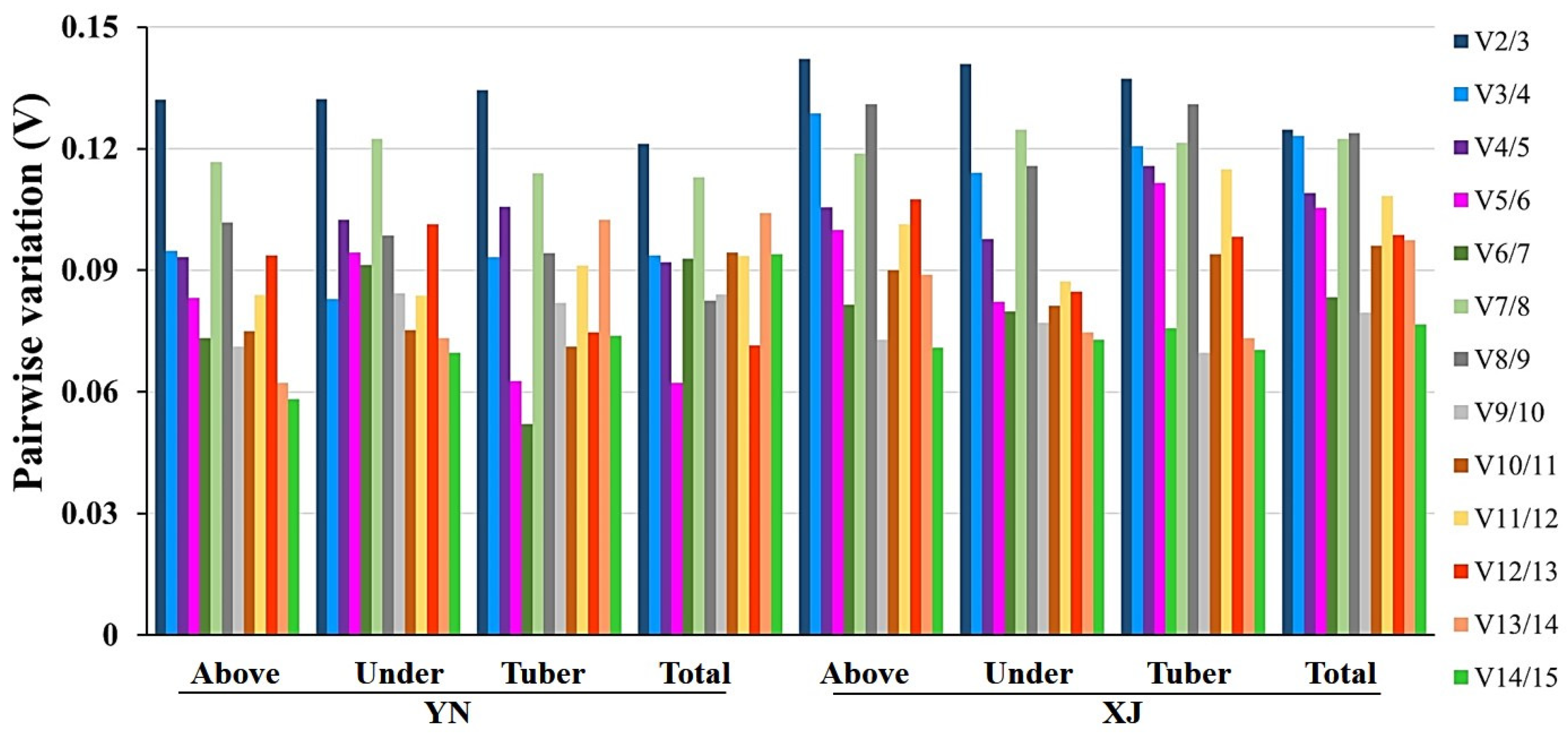
2.3.3. BestKeeper Analysis

BestKeeper is an Excel-based tool that evaluates the stability ranking of RGs based on the coefficient of variance (CV) and the standard deviation (SD) of the average Ct values [38]. The most stable gene exhibited the lowest of these two values, and genes with SD higher than 1.00 were considered to be unacceptable and were excluded. The removed genes included EF1α, TUB4, Rubisco, and PGK in the YN group and PGK, ACT, RPL11, CYC, TUB4, GAPDH, EF2, and Rubisco in the XJ group (Table 4).
For aboveground tissues, MDH and CYC, as well as MDH and EF2, with the lowest SD and CV values, were the top two ranks corresponding to YN and XJ tiger nut cultivars, respectively. For the underground group, the top two candidates of YN and XJ tissues were UCE2 and MDH. By comparison, it was observed that there were some differences in the expression levels of candidate genes between aboveground and underground tissues. Moreover, the optimal RGs were consistent in the tuber tissues of the two cultivars: UCE2 and TUB4. For total tissues, UCE2 and MDH, as well as UCE2 and 18S, were the most stable candidates in YN and XJ tiger nut cultivars, respectively, which exhibited the smallest SD and CV values. Throughout the ranks of BestKeeper, UCE2 and MDH appeared most frequently in the top two, exhibiting the best stability among all groups.

2.4. Unified Rank Lists by RankAggreg

Three different algorithms were used to analyze the expression stability of 14 potential RGs in nine experimental tissues of two cultivators. The results of the aforementioned methods produced too many ranks to analyze. It was necessary that a new algorithm be introduced to unify these results. RankAggreg is an R package for the ranking aggregation algorithm [39], and this software was the best choice to merge these ranking lists in different categories.
RankAggreg integrated rank lists of 14 reference genes in the two cultivars YN and XJ, as well as among four different tissue groups, including aboveground, underground, tuber and total samples (Table 5). The most stable candidate genes were consistently TUB4 in the tubers and UCE2 in the other three groups of two cultivars. In particular, TUB4 was the most stable gene in the tubers of the two cultivars but was low-ranked in the other groups, indicating that TUB4 was a tuber-specific stable gene. In addition, the most unstable genes were Rubisco and PGK in the majority of tissues.
However, the intermediate orderings of each group were distinct. For example, the rankings of ADF7 varied considerably, as this gene was ranked second to last in YN underground tissues but fourth in XJ underground tissues. Similarly, PGK also displayed a higher diversification, as it ranked No. 2 in XJ tubers but No. 7 and Nos. 10–13 in other groups (Table 5). The analysis of two cultivars can remove differences between cultivars to some degree. For a clearer comparison of the stability rankings obtained by different software, all ranking orders of 14 reference genes were integrated by geNorm, NormFinder, BestKeeper and RankAggreg in the YN (Table S3) and XJ (Table S4) tiger nut cultivars, respectively. The most stable and the most unstable reference genes in each part were largely the same. The middle rankings of the stability of RGs varied according to the analysis of different algorithms.

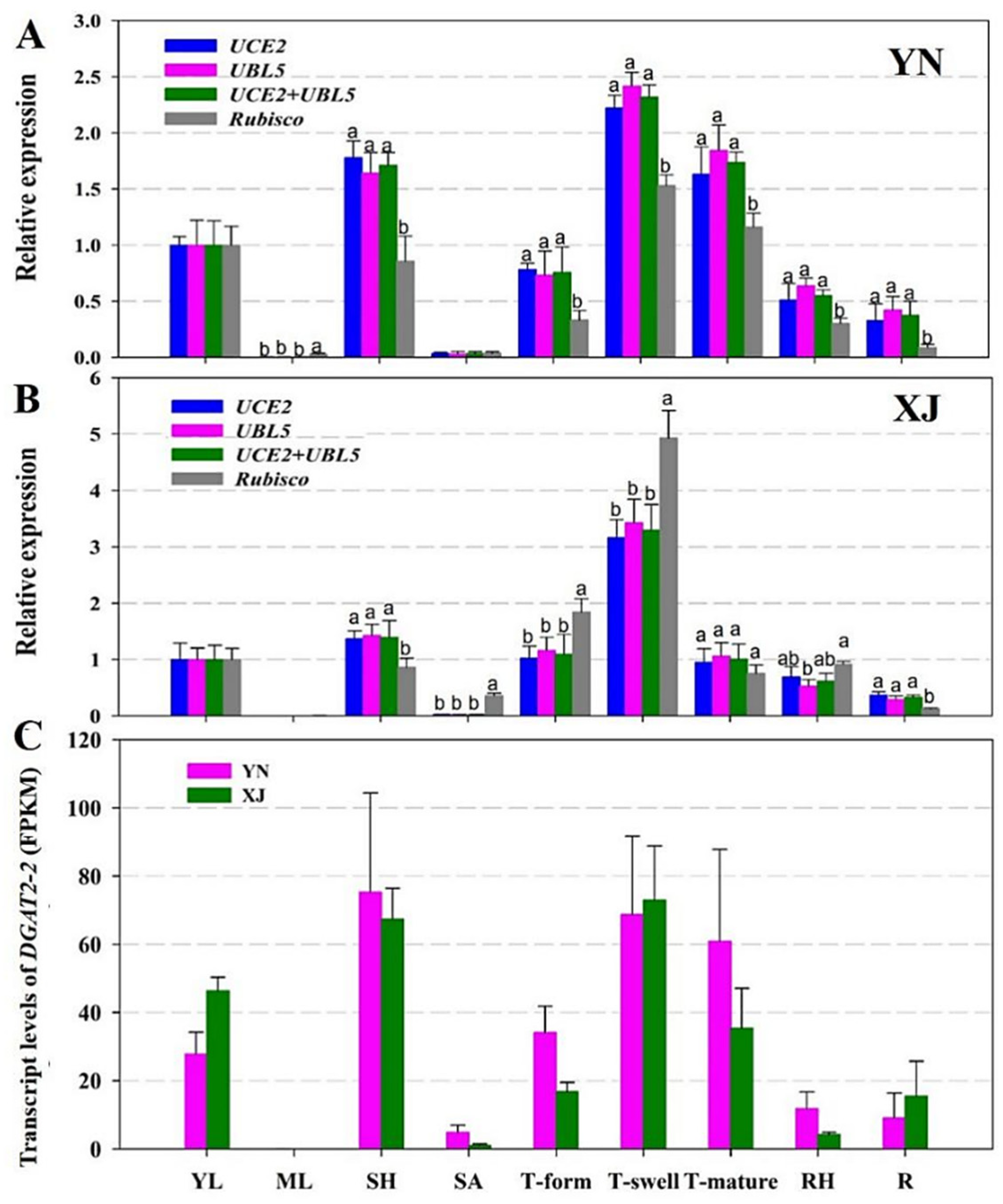
2.5. Validation of Candidate RGs

To determine the expression stability of potential RGs, an oil body membrane protein, caleosin (CLO), and a type II diacylglycerol acyltransferase (DGAT2-2) were selected to verify the stability of selected RGs by qRT-PCR in various tissues and developmental stages of tiger nut tubers. According to the abovementioned analysis, the two most stable RGs (UCE2 and UBL5) and the most unstable RG (Rubisco) were used to normalize the relative expression of the two target genes in the two cultivars of tiger nut (Figure 4 and Figure 5).
CLO is a kind of oil membrane protein that binds to the surface of the oil body in the form of calcium binding protein; the main biological functions of this protein include promoting the degradation of the oil body, maintaining the stability of the oil body, resisting biological stress and participating in the regulation of plant flowering [41,42]. Therefore, the expression of CLO is related to the accumulation of oil. As Figure 4A,B shows, similar relative expression levels of CLO were obtained using single RGs or a combination of RGs (UCE2 and UBL5) to normalize its expression. The expression patterns of CLO differed significantly when using the least stable RG (Rubisco) for normalization (Figure 4A,B). The transcripts of CLO aggregated notably in three developmental stages of tubers, with the highest level being observed in the tuber swelling stage. However, the expression levels of CLO were very low in other non-tuber organs, and the expression levels of underground tissues (including tuber, rhizome, and root) were higher than those of aboveground tissues (young and mature leaf, leaf sheath, and stem apex). The expression levels of CLO in the two tiger nut cultivars revealed by transcriptome analysis, shown as FPKM (fragments per kilobase of exon model per million mapped reads) in Figure 4C, were similar to the profiles characterized by qRT-PCR (Figure 4A,B). Comparing the two cultivars, the expression patterns were uniform, and only a slight difference in expression levels was observed between them. These results showed that using different RGs led to different expression patterns of target genes.
DGAT2-2 is an enzyme catalyzing the limited step in TAG biosynthesis [43]. We also analyzed its expression patterns in two cultivars of tiger nut (Figure 5). Generally, there was an approximately 2.5-fold change in DGAT2-2 expression levels among different tissues in YN tiger nut and a fivefold change in DGAT2-2 expression levels in XJ tiger nut. The similar expression pattern of DGAT2-2 between the two cultivars is reflected in Figure 5: On the one hand, tubers in the swelling stage exhibited the highest expression levels of DGAT2-2 followed by the sheath; on the other hand, mature leaves and shoot apexes showed the lowest transcript levels. The distinctive expression between these two cultivars was mainly observed in mature tubers because mature tubers of YN accumulated more DGAT2-2 transcripts than those of XJ. Comparing the results of different RGs showed that the relative expression of DGAT2-2 displayed significant variation when using the most unstable RG (Rubisco) for normalization, while similar expression patterns of DGAT2-2 were presented using single or a combination of RGs (UCE2 and UBL5), as shown in Figure 5A,B. In the transcriptome data, the transcript level of DGAT2-2 was also higher in leaf sheathes and tubers in the swelling and mature stages (Figure 5C). These results demonstrate that rigorous selection of reliable RGs is crucial to normalize gene expression. If the stability of RGs is poor, the result of normalization will deviate from the real value.

3. Discussion

With the in-depth study of gene functions in various species, the utilization of qRT-PCR to detect gene expression levels has become more extensive [25]. The expression levels of appropriate RGs should be constant under different test conditions and in different tissues and should not be affected by any external or internal factors [44].
Tiger nut is a new type of oil crop; this unique plant accumulates oil efficiently in tubers, which are a kind of nutrient organ [20]. To explore the special biological processes of this plant, such as oil accumulation and carbon distribution, it is urgently important to choose the most suitable RGs for transcript normalization. Tubers, the underground storage organ of tiger nuts, are produced at the expense of the rhizome apical meristem [17]. Considering the special underground organs of tiger nut, we not only selected the aboveground tissues (young leaves, mature leaves, leaf sheaths, and stem apexes) but also selected all underground tissues (roots, rhizomes, and tubers of different developmental stages) (Figures S1 and S2). Unfortunately, tiger nut plants cultivated in the Yunnan area did not produce flowers or fruit during the growth period; thus, reproductive organs were not available. For further improvement of the preciseness of this study, two cultivars of tiger nut with different tuber traits (smaller oval size (Figure S1H) and larger spherical size (Figure S2H)) were selected, planted, sampled, and analyzed and named “YN” and “XJ”, respectively.
To compare the stability of candidate RGs more scientifically, three software tools (geNorm, NormFinder, and BestKeeper) were used to analyze the expression stability of 14 candidate RGs. According to the different calculation rules of the three software programs, the stability rankings of candidate genes were generally close and only slightly different. In the consensus rank lists by RankAggreg (Table 5), the most stable and unstable RGs were roughly consistent in the same group of two cultivars: the most stable RGs were generally UCE2 and UBL5, and the least stable RGs were generally Rubisco and PGK.
The stabilities of UCE2, MDH, and UBL5 in the four groups of YN tiger nuts were the top three, while Rubisco and PGK maintained the lowest stabilities. In addition, TUB4 showed favorable stability in tuber tissues of YN. Among the aboveground tissues of XJ tiger nut, the most stable candidates were UCE2, EF1α, and CYC, while the most unstable were Rubisco and TUB4. For underground tissues of XJ, the most stable genes were UCE2, UBL5, and TUB4, whereas the lowest stability was still observed for Rubisco and GAPDH. Among the tubers at different developmental stages, TUB4, PGK, and UCE2 showed the best stability, while Rubisco and 18S showed the lowest stability. For the total organs of XJ, UCE2, UBL5, and ADF7 were expressed with the best stability, while Rubisco and PGK still exhibited the lowest stability. Combining the rankings of the two cultivars, we found that UCE2 and Rubisco maintained the best and worst stability among non-tuber organs, respectively. Moreover, for the two cultivars, TUB4 exhibited great variability in different types of organs because its stability was the strongest in tuber tissues but the worst after Rubisco and PGK in the aboveground tissues.
These results show that a combination of UCE2 and UBL5 is an appropriate RG for normalizing gene expression in aboveground, underground and total tissues of tiger nuts according to the optimal numbers recommended by geNorm. UCE encodes a ubiquitin-conjugating enzyme domain-containing protein that catalyzes ubiquitin transfer to the substrate or E3 ligase and is also a key enzyme in ubiquitin modification of target proteins [45]. In prior studies, UCE2 showed unstable expression in pepper (Wan et al., 2011), tung trees during seed development [46], and pitaya [47]. However, in keeping with our results, UCE has been reported frequently as the most stable reference gene not only in several herbaceous plants, including Brachypodium distachyon [48], tobacco [49], red clover [50], Lycoris aurea [51], and goosegrass under quizalofop-p-ethyl (a kind of low-toxicity herbicide) stress [52], but also in woody plants, such as poplar [53], Platycladus orientalis [54], sweet osmanthus [55], Plukenetia volubilis [56], olive trees [57], and rubber trees [58]. As another suitable RG, UBL5 is a new type of ubiquitin-like protein discovered in the human iris [59] with multiple biological functions in eukaryotes, such as involvement in mRNA precursor splicing [60,61], regulation of the cell cycle [62], regulation of energy metabolism [63], and stress resistance [64]. UBL had relatively stable expression in Plukenetia volubilis [56], Jatropha curcas [27], and European beech [65]; nevertheless, it had the lowest expression level in Seashore Paspalum [66].
In addition, for tiger nut tuber tissues, TUB4 and UCE2 are a pair of recommended RGs. Tubulin, encoded by TUB [67,68], is the main component of plant microtubules [69], which can maintain cell morphology [70], promote intracellular transport [71], and participate in cell elongation [72], movement, and cell division [73,74,75].
According to the analysis of qRT-PCR results, it has been widely recognized that the combination of multiple RGs can provide more accurate and reliable expression profiles than can a single RG [76]. Based on validation of RGs, when UCE2 or UBL5 was employed alone or in combination as RGs, the expression patterns of the target genes in different tissues were largely consistent. It demonstrated that the expression patterns of the target genes were almost the same as with one or two RGs. However, when Rubisco was used as an RG to normalize target genes, the patterns of CLO and DGAT2-2 expression were clearly inconsistent with that of the pattern obtained with the two optimum RGs, especially in mature leaves and all underground tissues, suggesting the most unstable characteristic of Rubisco.

4. Materials and Methods

4.1. Plant Material

Two cultivars of tiger nut were collected from Shihezi University in Xinjiang (XJ), Northwest China and Heshun County in Yunnan (YN), Southwest China. XJ tiger nut produces larger average spherical tubers (Figure S1H), while YN tiger nut produces smaller oval tubers (Figure S2H). On March 15, 2018, tubers were sown in a field at the Xishuangbanna Tropical Botanical Garden (XTBG, 21° 54′ N, 101° 46′ E, 580 m in altitude) of the Chinese Academy of Sciences in Mengla County, Yunnan Province, Southwest China. Six tissues (young leaf, mature leaf, leaf sheath, root, rhizome, stem apex) were collected on 4 May 2018, and tubers of three developmental stages were collected at 40, 80, and 120 days after sowing (DAS), which are the stages of tuber formation, swelling and maturity in turn. All samples were harvested, washed and surface dried and then frozen in liquid nitrogen and immediately stored at −80 °C until required for further analysis. In total, three biological replicates were collected, and each biological replicate contained tissue sampled from ten individual plants.

4.2. Selection of Candidate RGs and Primer Design

Fourteen candidate RGs were selected based on RNA-Seq data from tiger nut tissues [21], including 18S ribosomal RNA (18S), actin (ACT), actin-depolymerizing factor 7 (ADF7), elongation factor 1-alpha (EF1α), elongation factor 2 (EF2), glyceraldehyde-3-phosphate dehydrogenase (GAPDH), ribosomal protein L11 (RPL11), ribulose bisphosphate carboxylase (Rubisco), ubiquitin-conjugating enzyme 2 (UCE2), ubiquitin-like protein 5 (UBL5), cyclophilin (CYC), phosphoglycerate kinase (PGK), malate dehydrogenase (MDH), and tubulin beta-4 (TUB4). The cDNA sequences of candidate RGs and two genes for validation were obtained from Yang et al. [21] (Table S1). Primers used for qRT-PCR analyses (Table 1) were designed using Primer Premier 5.0 with the following parameters: melting temperature (Tm) values ranging from 50 to 60 °C, GC percent of 40–60%, primer lengths of 18–22 bp and product length of 100–300 bp. Tenfold serial dilutions of cDNA were used to determine the slope of the standard curve to calculate the amplification efficiency of primers (E = 10(−1/slope of the standard curve)).

4.3. RNA Isolation, cDNA Synthesis, and qRT-PCR

Total RNA was extracted by silica particles [77]. Plant tissues (0.2–0.5 g) frozen in liquid nitrogen were ground into powder rapidly and then mixed with 400 μL of RNA extraction buffer (5 M NaCl and 100 mM Tris–HCl [pH = 8.0]), 100 μL of 2-mercaptoethanol, and 300 μL of water-saturated phenol in a 2-mL centrifuge tube. After vortexing and incubating at room temperature (RT) for 5 min, 300 μL of chloroform/isoamyl alcohol (24:1), and 200 μL of 3 M NaAc (pH = 4.0) were added to the mixture. After vortexing and centrifuging (15,000× g, 10 min, 4 °C), the upper aqueous phase was carefully pipetted into a 1.5-mL centrifuge tube, then 200 μL of absolute ethanol and 25 μL of silica suspension (1 g/mL) were added and mixed. After centrifuging (15,000× g, 30 s, RT), the liquid was poured out, and the silica pellet was rinsed twice with 75% ethanol (prepared with DEPC water). The silica pellet dried in vacuum was suspended with 50 μL of DEPC water and centrifuged (15,000× g, 10 min, RT). The supernatant containing the total RNA was transfer to a new centrifuge tube and stored at −80 °C. RNA quality was determined by 2.0% agarose gel electrophoresis. The concentration and purity of total RNA were determined using a NanoDrop 2000c Spectrophotometer (Thermo Scientific, Wilmington, DE, USA). The ratio of OD260/OD280 of total RNA between 1.90 and 2.10 was considered to meet the required quality for further experiments.
Two µg of total RNA from each sample was reverse-transcribed into cDNAs using the PrimeScript® RT Reagent Kit with gDNA Eraser (TaKaRa, Dalian, China). The reverse transcriptional reaction included 4 μL of 5× PrimerScript® Buffer 2, 1 μL of PrimerScript® RT-Enzyme Mix I, 1 μL of RT Primer Mix, 10 μL of the reaction liquid of genomic DNA removal, and RNase-Free ddH2O up to 20 μL, reacting at 37 °C for 15 min and then terminating at 85 °C for 5 s. qRT-PCR was performed using LightCycler® 480 SYBR Green I Master Mix with the Roche 480 real-time PCR detection system according to the manufacturer’s instructions (Roche Diagnostics, Indianapolis, IN, USA). One µL of cDNA was added to each qRT-PCR reaction mix (20 µL), containing 0.25 µM of each primer and 10 µL of SYBR® Premix Ex Taq™ II. Amplifications were performed with the following program: 95 °C for 3 s; 40 cycles of 95 °C for 10 s, 60 °C for 30 s, 72 °C for 3 s. Melting curve analysis was performed as follows: 95 °C for 15 s, 60 °C increased to 95 °C with temperature increment of 0.6 °C/s, cooling at 16 °C.

4.4. Data Analysis

Several software tools were used to evaluate the stability of the 14 selected candidate RGs across all the experimental sets, including geNorm [26], NormFinder [37], and BestKeeper [38]. The expression levels of the candidate RGs were determined by cycle threshold (Ct) values. All the procedures of statistical analyses by the above packages were conducted according to the manufacturer’s instructions.
GeNorm software is specifically used to screen RGs and determine the optimal number of RGs in real-time quantitative PCR. The result of the analysis is that two or more internal RG combinations were selected to correct the data, which makes the relative quantitative results more accurate. The geNorm program selects the optimal RG with stability by calculating the M value of each RG. The criterion is that the smaller the M value is, the better the stability of the RG is; otherwise, the stability is worse. This software can also calculate the paired variation V value of the normalized factor after the introduction of a new internal RG and determine the number of optimal internal RGs based on the Vn/Vn + 1 value. The default V value is 0.15. If the value of Vn/Vn + 1 is less than 0.15, the number of optimal internal RGs is n; if the value is more than 0.15, the number of suitable internal RGs is n + 1.
NormFinder is another program for screening stable internal RGs [37]. The calculation principle of this program is similar to that of the geNorm program. It also obtains the stable value of the internal RG and then selects the value based on the stable value. The most suitable internal RG is determined, and the criterion is that the internal RG with the lowest expression stability value is the most suitable internal RG. The NormFinder program can not only compare the expression differences of candidate internal RGs but also calculate the variation between sample groups, but the program can only screen out one of the most suitable internal RGs.
BestKeeper is a program for analysing the expression of internal RGs [38]. The correlation coefficient (r), standard deviation (SD), and coefficient of variation (CV) that can generate pairings between each gene can be obtained through the calculation of the program. Next, by comparing the values of each value, a stable internal RG is finally determined. The determination principle is that the larger the correlation coefficient, the smaller the standard deviation and the coefficient of variation, the better the stability of the RG, and conversely, the worse the stability; when SD > 1, the expression of the RG is unstable.

4.5. Statistical Method for Rank Aggregation

The RankAggreg package of R software was used to combine the stability measurements obtained from the four methods and establish a consensus rank of RGs. To generate a consensus from the data produced by geNorm, BestKeeper, and NormFinder, we aggregated the obtained ranking lists by applying the RankAggreg (ver. 0.6.5) package of R software as done previously. RankAggreg is a package that provides algorithms able to combine different ranking lists [39].

4.6. Validation of the Candidate RGs

To examine the expression stability of potential RGs, the relative expression levels of two lipid-related genes (CLO and DGAT2-2) were analyzed in various tissues in two cultivars of tiger nut. CLO is a kind of oil-body-related protein [78], and DGAT2-2 is a type II diacylglycerol acyltransferase [79]. The relative expression levels of these two genes were normalized separately to the most stable and least stable RGs analyzed by the mentioned four algorithms. The qRT-PCR amplification conditions of the genes were the same as those described above. The relative expression levels of target genes were calculated according to the 2−ΔΔCt method and presented as fold change.

4.7. Library Construction, Sequencing, and Assembly of Transcriptomes

The total RNAs from 54 samples (nine tissues of each two cultivars, three replicates per tissue) described above were isolated for construction of transcriptome libraries. The quality of the libraries was assessed using an Agilent 2100 Bioanalyzer. Transcriptome sequencing were performed on the Illumina NovaSeq 6000 platform by Novogene Bioinformatics Technology (Tianjin, China) to generate 150-nucleotide-long paired-end sequence reads. Clean reads were obtained from the raw reads by removing adapter sequences and low-quality reads. De novo transcriptome assembly was performed using Trinity (v2.4.0) with default parameters [80]. The clean reads were matched to the Cyperus esculentus unigenes using RSEM software [81]. The frequency of individual reads was normalized to FPKM [82]. RNA-Seq data reported in this study were deposited at NCBI under the BioProject ID PRJNA703731.

5. Conclusions

In this study, our purpose was to select and validate the optimal RGs for normalization of qRT-PCR analysis in tiger nut to establish a foundation for further research on its metabolic pathway and regulatory network. The expression stabilities of 14 potential RGs were evaluated in various tissues from two cultivars of tiger nut, including three leaf-related samples, stem apexes, roots, rhizomes, and tubers of three developmental stages. In the evaluation stage, three kinds of analysis software were utilized for ranking first, and then RankAggreg was used to integrate multiple rankings into a consensus list. UCE2, MDH and UBL5 were the top three stable RGs across all tissues of YN tiger nut. UCE2, UBL5 and ADF7 were the most stable candidates across all tissues of XJ tiger nut. According to the rank list in the “Tissues of two cultivars” column of Table 5, a combination of UCE2 and UBL5 was the best RG for all tissues of the two cultivars. Similarly, a combination of TUB4 and UCE2 was the most suitable for tuber tissues of the two cultivars. Transcriptome data can be used to select candidate RGs and analyze the expression levels of two target genes. These results provide a basis and convenient resource for future research performing gene expression analysis in tiger nut.

Supplementary Materials

Supplementary Materials can be found at https://www.mdpi.com/1422-0067/22/5/2569/s1.

Author Contributions

Z.-F.X., M.T. and X.B. designed the research; X.B., T.C. and Y.W. collected plant materials and performed the experiment. X.B. analyzed the data and wrote the manuscript. Z.-F.X. and M.T. reviewed and edited the manuscript. All authors have read and agreed to the published version of the manuscript.

Funding

This research was supported by the Key Deployment Program of the Chinese Academy of Sciences (CAS) (ZDRW-ZS-2017-2), the Natural Science Foundation of Yunnan Province (2018FB060), the West Light Foundation of Chinese Academy of Science (CAS) Y9XB091, and the Natural Science Foundation of China (31700273, 31771605).

Institutional Review Board Statement

Not applicable.

Informed Consent Statement

Not applicable.

Data Availability Statement

The transcriptome datasets reported in this study are available at the NCBI under the BioProject ID PRJNA703731.

Acknowledgments

We thank Guojiang Wu (South China Botanical Garden), Chengyuan Yang and Qinying Lan (Xishuangbanna Tropical Botanical Garden) for donating tiger nut tubers. We also thank the Central Laboratory of the Xishuangbanna Tropical Botanical Garden for providing the research facilities.

Conflicts of Interest

The authors declare no conflict of interest.

Abbreviations

cDNAComplementary DNA
CLOCaleosin
CtCycle threshold
DASDay after sowing
DGATDiacylglycerol acyltransferase
FPKMFragments per kilobase of exon model per million mapped reads
MUFAMonounsaturated fatty acid
qRT-PCRQuantitative real-time reverse transcription-polymerase chain reaction
RGReference gene
RTRoom temperature
TmMelting temperature

References

  1. Vanhercke, T.; Dyer, J.M.; Mullen, R.T.; Kilaru, A.; Rahman, M.M.; Petrie, J.R.; Green, A.G.; Yurchenko, O.; Singh, S.P. Metabolic engineering for enhanced oil in biomass. Prog. Lipid Res. 2019, 74, 103–129. [Google Scholar] [CrossRef] [PubMed]
  2. Xu, C.; Shanklin, J. Triacylglycerol Metabolism, Function, and Accumulation in Plant Vegetative Tissues. Annu. Rev. Plant Biol. 2016, 67, 179–206. [Google Scholar] [CrossRef]
  3. Zhang, H.; Yasmin, F.; Song, B.H. Neglected treasures in the wild—Legume wild relatives in food security and human health. Curr. Opin. Plant Biol. 2019, 49, 17–26. [Google Scholar] [CrossRef]
  4. Chang, T.; Cheng, Q.; Zhang, Z.; Guan, C.; Zhao, W.; Wang, L. Advance in Breeding of New Rapeseed Varieties with High Oil Content. Mol. Plant Breed. 2019, 17, 4424–4430. [Google Scholar]
  5. Ojiewo, C.O.; Janila, P.; Bhatnagar-Mathur, P.; Pandey, M.K.; Desmae, H.; Okori, P.; Mwololo, J.; Ajeigbe, H.; Njuguna-Mungai, E.; Muricho, G.; et al. Advances in Crop Improvement and Delivery Research for Nutritional Quality and Health Benefits of Groundnut (Arachis hypogaea L.). Front. Plant Sci. 2020, 11, 29. [Google Scholar] [CrossRef] [PubMed] [Green Version]
  6. Fobes, J.F.; Mudd, J.B.; Marsden, M.P. Epicuticular lipid accumulation on the leaves of Lycopersicon pennellii (Corr.) D’Arcy and Lycopersicon esculentum Mill. Plant Physiol. 1985, 77, 567–570. [Google Scholar] [CrossRef] [PubMed] [Green Version]
  7. Wang, G.; Lin, Q.; Xu, Y. Tetraena mongolica Maxim can accumulate large amounts of triacylglycerol in phloem cells and xylem parenchyma of stems. Phytochemistry 2007, 68, 2112–2117. [Google Scholar] [CrossRef]
  8. Defelice, M.S. Yellow nutsedge Cyperus esculentus L.—Snack food of the Gods. Weed Technol. 2002, 16, 901–907. [Google Scholar] [CrossRef]
  9. Stoller, E.W.; Weber, E.J. Differential cold tolerance, starch, sugar, protein, and lipid of yellow and purple nutsedge tubers. Plant Physiol. 1975, 55, 859–863. [Google Scholar] [CrossRef] [Green Version]
  10. Linssen, J.P.H.; Kielman, G.M.; Cozijnsen, J.L.; Pilnik, W. Comparison of chufa and olive oils. Food Chem. 1988, 28, 279–285. [Google Scholar] [CrossRef]
  11. Schwingshackl, L.; Hoffmann, G. Monounsaturated fatty acids and risk of cardiovascular disease: Synopsis of the evidence available from systematic reviews and meta-analyses. Nutrients 2012, 4, 1989–2007. [Google Scholar] [CrossRef] [Green Version]
  12. Terés, S.; Barceló-Coblijn, G.; Benet, M.; Alvarez, R.; Bressani, R.; Halver, J.; Escribá, P. Oleic acid content is responsible for the reduction in blood pressure induced by olive oil. Proc. Natl. Acad. Sci. USA 2008, 105, 13811–13816. [Google Scholar] [CrossRef] [Green Version]
  13. Palomer, X.; Pizarro-Delgado, J.; Barroso, E.; Vazquez-Carrera, M. Palmitic and oleic acid: The yin and yang of fatty acids in type 2 diabetes mellitus. Trends Endocrinol. Metab. 2018, 29, 178–190. [Google Scholar] [CrossRef] [PubMed]
  14. Bo, L.; Shibuya, T.; Yogo, Y.; Hara, T.; Matsuo, K. Effects of light quantity and quality on growth and reproduction of a clonal sedge, Cyperus esculentus. Plant Species Biol. 2001, 16, 69–81. [Google Scholar]
  15. Aljuhaimi, F.; Ghafoor, K.; Ozcan, M.M.; Miseckaite, O.; Babiker, E.E.; Hussain, S. The Effect of solvent type and roasting processes on physico-chemical properties of tigernut (Cyperus esculentus L.) tuber oil. J. Oleo Sci. 2018, 67, 823–828. [Google Scholar] [CrossRef] [Green Version]
  16. Hu, B.; Li, Y.; Song, J.; Li, H.; Zhou, Q.; Li, C.; Zhang, Z.; Liu, Y.; Liu, A.; Zhang, Q.; et al. Oil extraction from tiger nut (Cyperus esculentus L.) using the combination of microwave-ultrasonic assisted aqueous enzymatic method—Design, optimization and quality evaluation. J. Chromatogr. A 2020, 1627, 461380. [Google Scholar] [CrossRef] [PubMed]
  17. Turesson, H.; Marttila, S.; Gustavsson, K.E.; Hofvander, P.; Olsson, M.E.; Bulow, L.; Stymne, S.; Carlsson, A.S. Characterization of oil and starch accumulation in tubers of Cyperus esculentus var. sativus (Cyperaceae): A novel model system to study oil reserves in nonseed tissues. Am. J. Bot. 2010, 97, 1884–1893. [Google Scholar] [CrossRef]
  18. Moral-Anter, D.; Campo-Sabariz, J.; Ferrer, R.; Martin-Venegas, R. Cyperus esculentus L. tubers (tiger nuts) protect epithelial barrier function in Caco-2 cells infected by Salmonella enteritidis and promote Lactobacillus plantarum growth. Nutrients 2021, 13, 71. [Google Scholar] [CrossRef]
  19. Rubert, J.; Monforte, A.; Hurkova, K.; Pérez-Martínez, G.; Blesa, J.; Navarro, J.L.; Stranka, M.; Soriano, J.M.; Hajslova, J. Untargeted metabolomics of fresh and heat treatment Tiger nut (Cyperus esculentus L.) milks reveals further insight into food quality and nutrition. J. Chromatogr. A 2017, 1514, 80–87. [Google Scholar] [CrossRef]
  20. Ezeh, O.; Gordon, M.; Niranjan, K. Enhancing the recovery of tiger nut (Cyperus esculentus) oil by mechanical pressing: Moisture content, particle size, high pressure and enzymatic pre-treatment effects. Food Chem. 2016, 194, 354–361. [Google Scholar] [CrossRef] [PubMed]
  21. Yang, Z.; Ji, H.; Liu, D. Oil biosynthesis in underground oil-rich storage vegetative tissue: Comparison of Cyperus esculentus tuber with oil seeds and fruits. Plant Cell Physiol. 2016, 57, 2519–2540. [Google Scholar] [CrossRef]
  22. Yang, Z.; Liu, D.; Ji, H. Sucrose metabolism in developing oil-rich tubers of Cyperus esculentus: Comparative transcriptome analysis. BMC Plant Biol. 2018, 18, 151. [Google Scholar] [CrossRef] [PubMed] [Green Version]
  23. De Castro, O.; Gargiulo, R.; Del Guacchio, E.; Caputo, P.; De Luca, P. A molecular survey concerning the origin of Cyperus esculentus (Cyperaceae, Poales): Two sides of the same coin (weed vs. crop). Ann. Bot. 2015, 115, 733–745. [Google Scholar] [CrossRef] [Green Version]
  24. Agarwal, P.; Parida, S.K.; Mahto, A.; Das, S.; Mathew, I.E.; Malik, N.; Tyagi, A.K. Expanding frontiers in plant transcriptomics in aid of functional genomics and molecular breeding. Biotechnol. J. 2014, 9, 1480–1492. [Google Scholar] [CrossRef]
  25. Derveaux, S.; Vandesompele, J.; Hellemans, J. How to do successful gene expression analysis using real-time PCR. Methods 2010, 50, 227–230. [Google Scholar] [CrossRef]
  26. Vandesompele, J.; De Preter, K.; Pattyn, F.; Poppe, B.; Van Roy, N.; De Paepe, A.; Speleman, F. Accurate normalization of real-time quantitative RT-PCR data by geometric averaging of multiple internal control genes. Genome Biol. 2002, 3, 1–12. [Google Scholar] [CrossRef] [Green Version]
  27. Zhang, L.; He, L.-L.; Fu, Q.-T.; Xu, Z.-F. Selection of Reliable Reference Genes for Gene Expression Studies in the Biofuel Plant Jatropha curcas Using Real-Time Quantitative PCR. Int. J. Mol. Sci. 2013, 14, 24338–24354. [Google Scholar] [CrossRef] [Green Version]
  28. Tang, M.Y.; Bai, X.; Niu, L.J.; Chai, X.; Chen, M.S.; Xu, Z.F. miR172 Regulates both Vegetative and Reproductive Development in the Perennial Woody Plant Jatropha curcas. Plant Cell Physiol. 2018, 59, 2549–2563. [Google Scholar] [CrossRef] [PubMed]
  29. Llera-Herrera, R.; García-Gasca, A.; Huvet, A.; Ibarra, A.M. Identification of a tubulin-α gene specifically expressed in testis and adductor muscle during stable reference gene selection in the hermaphrodite gonad of the lion’s paw scallop Nodipecten subnodosus. Mar. Genom. 2012, 6, 33–44. [Google Scholar] [CrossRef]
  30. Almas, D.E.; Kamrodi, A.R. Validation of Appropriate Reference Genes for Real-Time Quantitative PCR Gene Expression Analysis in Rice Plants Exposed to Metal Stresses. Russ. J. Plant Physiol. 2018, 65, 890–897. [Google Scholar] [CrossRef]
  31. Morga, B.; Arzul, I.; Faury, N.; Renault, T. Identification of genes from flat oyster Ostrea edulis as suitable housekeeping genes for quantitative real time PCR. Fish. Shellfish Immunol. 2010, 29, 945. [Google Scholar] [CrossRef] [PubMed] [Green Version]
  32. Brattelid, T.; Winer, L.H.; Levy, F.O.; Liestøl, K.; Andersson, K.B. Reference gene alternatives to Gapdh in rodent and human heart failure gene expression studies. Bmc Mol. Biol. 2010, 11, 22. [Google Scholar] [CrossRef] [Green Version]
  33. Pei, D.S.; Sun, Y.H.; Chen, S.P.; Wang, Y.P.; Hu, W.; Zhu, Z.Y. Zebrafish GAPDH can be used as a reference gene for expression analysis in cross-subfamily cloned embryos. Anal. Biochem. 2007, 363, 291–293. [Google Scholar] [CrossRef]
  34. Czechowski, T.; Stitt, M.; Altmann, T.; Udvardi, M.K.; Scheible, W.R. Genome-wide identification and testing of superior reference genes for transcript normalization in Arabidopsis. Plant Physiol. 2005, 139, 5–17. [Google Scholar] [CrossRef] [PubMed] [Green Version]
  35. Kim, B.R.; Nam, H.Y.; Kim, S.U.; Kim, S.I.; Chang, Y.J. Normalization of reverse transcription quantitative-PCR with housekeeping genes in rice. Biotechnol. Lett. 2003, 25, 1869–1872. [Google Scholar] [CrossRef]
  36. Wittwer, C.T. High-Resolution DNA Melting Analysis: Advancements and Limitations. Hum. Mutat. 2009, 30, 857–859. [Google Scholar] [CrossRef]
  37. Andersen, C.L.; Jensen, J.L.; Ørntoft, T.F. Normalization of real-time quantitative reverse transcription-PCR data: A model-based variance estimation approach to identify genes suited for normalization, applied to bladder and colon cancer data sets. Cancer Res. 2004, 64, 5245–5250. [Google Scholar] [CrossRef] [PubMed] [Green Version]
  38. Pfaffl, M.W.; Tichopad, A.; Prgomet, C.; Neuvians, T.P. Determination of stable housekeeping genes, differentially regulated target genes and sample integrity: BestKeeper-Excel-based tool using pair-wise correlations. Biotechnol. Lett. 2004, 26, 509–515. [Google Scholar] [CrossRef] [PubMed]
  39. Pihur, V.; Datta, S.; Datta, S. RankAggreg, an R package for weighted rank aggregation. BMC Bioinform. 2009, 10, 62. [Google Scholar] [CrossRef] [Green Version]
  40. Etschmann, B.; Wilcken, B.; Stoevesand, K.; Von Der Schulenburg, A.; Sterner-Kock, A. Selection of reference genes for quantitative real-time PCR analysis in canine mammary tumors using the GeNorm algorithm. Vet. Pathol. 2006, 43, 934–942. [Google Scholar] [CrossRef] [PubMed] [Green Version]
  41. Shen, Y.; Liu, M.; Wang, L.; Li, Z.; Taylor, D.; Li, Z.; Zhang, M. Identification, duplication, evolution and expression analyses of caleosins in Brassica plants and Arabidopsis subspecies. Mol. Genet. Genom. 2016, 291, 971–988. [Google Scholar] [CrossRef] [Green Version]
  42. Li, C.; Zheng, L.; Wang, X.; Hu, Z.; Zheng, Y.; Chen, Q.; Hao, X.; Xiao, X.; Wang, X.; Wang, G.; et al. Comprehensive expression analysis of Arabidopsis GA2-oxidase genes and their functional insights. Plant Sci. 2019, 285, 1–13. [Google Scholar] [CrossRef] [PubMed]
  43. Shockey, J.M.; Gidda, S.K.; Chapital, D.C.; Kuan, J.-C.; Dhanoa, P.K.; Bland, J.M.; Rothstein, S.J.; Mullen, R.T.; Dyer, J.M. Tung tree DGAT1 and DGAT2 have nonredundant functions in triacylglycerol biosynthesis and are localized to different subdomains of the endoplasmic reticulum. Plant Cell 2006, 18, 2294–2313. [Google Scholar] [CrossRef] [Green Version]
  44. Bustin, S.A.; Benes, V.; Garson, J.; Hellemans, J.; Huggett, J.; Kubista, M.; Mueller, R.; Nolan, T.; Pfaffl, M.W.; Shipley, G.; et al. The need for transparency and good practices in the qPCR literature. Nat. Methods 2013, 10, 1063–1067. [Google Scholar] [CrossRef] [PubMed]
  45. Xu, L.; Ménard, R.; Berr, A.; Fuchs, J.; Shen, W.H. The E2 ubiquitin-conjugating enzymes, AtUBC1 and AtUBC2, play redundant roles and are involved in activation of FLC expression and repression of flowering in Arabidopsis Thaliana. Plant J. 2008, 57, 279–288. [Google Scholar] [CrossRef]
  46. Han, X.; Lu, M.; Chen, Y.; Zhan, Z.; Cui, Q.; Wang, Y. Selection of reliable reference genes for gene expression studies using real-time PCR in tung tree during seed development. PLoS ONE 2012, 7, e43084. [Google Scholar] [CrossRef]
  47. Chen, C.; Wu, J.; Hua, Q.; Tel-Zur, N.; Xie, F.; Zhang, Z.; Chen, J.; Zhang, R.; Hu, G.; Zhao, J.; et al. Identification of reliable reference genes for quantitative real-time PCR normalization in pitaya. Plant Methods 2019, 15, 70. [Google Scholar] [CrossRef] [Green Version]
  48. Hong, S.Y.; Seo, P.J.; Yang, M.S.; Xiang, F.; Park, C.M. Exploring valid reference genes for gene expression studies in Brachypodium distachyon by real-time PCR. BMC Plant Biol. 2008, 8, 11. [Google Scholar] [CrossRef] [Green Version]
  49. Schmidt, G.W.; Delaney, S.K. Stable internal reference genes for normalization of real-time RT-PCR in tobacco (Nicotiana tabacum) during development and abiotic stress. Mol. Genet. Genom. 2010, 283, 233–241. [Google Scholar] [CrossRef] [PubMed]
  50. Khanlou, K.M.; Van Bockstaele, E. A critique of widely used normalization software tools and an alternative method to identify reliable reference genes in red clover (Trifolium pratense L.). Planta 2012, 236, 1381–1393. [Google Scholar] [CrossRef] [PubMed]
  51. Ma, R.; Xu, S.; Zhao, Y.; Xia, B.; Wang, R. Selection and validation of appropriate reference genes for quantitative real-time PCR analysis of gene expression in Lycoris aurea. Front. Plant Sci. 2016, 7, 7. [Google Scholar] [CrossRef] [Green Version]
  52. Chen, J.; Huang, Z.; Huang, H.; Wei, S.; Zhang, C. Selection of relatively exact reference genes for gene expression studies in goosegrass (Eleusine indica) under herbicide stress. Sci. Rep. 2017, 7, 46494. [Google Scholar] [CrossRef] [Green Version]
  53. Basa, B.; Solti, A.; Sarvari, E.; Tamas, L. Housekeeping gene selection in poplar plants under Cd-stress: Comparative study for real-time PCR normalisation. Funct. Plant Biol. 2009, 36, 1079–1087. [Google Scholar] [CrossRef]
  54. Chang, E.; Shi, S.; Liu, J.; Cheng, T.; Xue, L.; Yang, X.; Yang, W.; Lan, Q.; Jiang, Z. Selection of reference genes for quantitative gene expression studies in Platycladus orientalis (Cupressaceae) using real-time PCR. PLoS ONE 2012, 7, e33278. [Google Scholar] [CrossRef] [Green Version]
  55. Zhang, C.; Fu, J.; Wang, Y.; Bao, Z.; Zhao, H. Identification of Suitable Reference Genes for Gene Expression Normalization in the Quantitative Real-Time PCR Analysis of Sweet Osmanthus (Osmanthus fragrans Lour.). PLoS ONE 2015, 10, e0136355. [Google Scholar] [CrossRef] [PubMed] [Green Version]
  56. Niu, L.J.; Tao, Y.B.; Chen, M.S.; Fu, Q.T.; Li, C.Q.; Dong, Y.L.; Wang, X.L.; He, H.Y.; Xu, Z.F. Selection of reliable reference genes for gene expression studies of a promising oilseed crop, Plukenetia volubilis, by real-time quantitative PCR. Int. J. Mol. Sci. 2015, 16, 12513–12530. [Google Scholar] [CrossRef] [Green Version]
  57. Hurkan, K.; Sezer, F.; Ozbilen, A.; Taskin, K.M. Identification of reference genes for real-time quantitative polymerase chain reaction based gene expression studies on various Olive (Olea europaea L.) tissues. J. Hortic. Sci. Biotechnol. 2018, 93, 644–651. [Google Scholar] [CrossRef]
  58. Long, X.; Lu, J.; Kav, N.N.V.; Qin, Y.; Fang, Y. Identification and evaluation of suitable reference genes for gene expression analysis in rubber tree leaf. Mol. Biol. Rep. 2020, 47, 1921–1933. [Google Scholar] [CrossRef] [PubMed]
  59. Friedman, J.S.; Koop, B.F.; Raymond, V.; Walter, M.A. Isolation of a ubiquitin-like (UBL5) gene from a screen identifying highly expressed and conserved iris genes. Genomics 2001, 71, 252–255. [Google Scholar] [CrossRef] [PubMed]
  60. Mishra, S.K.; Ammon, T.; Popowicz, G.M.; Krajewski, M.; Nagel, R.J.; Ares, M., Jr.; Holak, T.A.; Jentsch, S. Role of the ubiquitin-like protein Hub1 in splice-site usage and alternative splicing. Nature 2011, 474, U173–U205. [Google Scholar] [CrossRef] [Green Version]
  61. Wilkinson, C.R.M.; Dittmar, G.A.G.; Ohi, M.D.; Uetz, P.; Jones, N.; Finley, D. Ubiquitin-like protein Hub1 Is required for pre-mRNA splicing and localization of an essential splicing factor in fission yeast. Curr. Biol. 2006, 16, 2283–2288. [Google Scholar] [CrossRef] [Green Version]
  62. Dittmar, G.A.G.; Wilkinson, C.R.M.; Jedrzejewski, P.T.; Finley, D. Role of a ubiquitin-like modification in polarized morphogenesis. Science 2002, 295, 2442–2446. [Google Scholar] [CrossRef]
  63. Benedetti, C.; Haynes, C.M.; Yang, Y.; Harding, H.P.; Ron, D. Ubiquitin-like protein 5 positively regulates chaperone gene expression in the mitochondrial unfolded protein response. Genetics 2006, 174, 229–239. [Google Scholar] [CrossRef] [Green Version]
  64. Kurepa, J.; Walker, J.M.; Smalle, J.; Gosink, M.M.; Davis, S.J.; Durham, T.L.; Sung, D.Y.; Vierstra, R.D. The small ubiquitin-like modifier (SUMO) protein modification system in Arabidopsis. J. Biol. Chem. 2003, 278, 6862–6872. [Google Scholar] [CrossRef] [PubMed] [Green Version]
  65. Olbrich, M.; Gerstner, E.; Welzl, G.; Fleischmann, F.; Oßwald, W.; Bahnweg, G.; Ernst, D. Quantification of mRNAs and housekeeping gene selection for quantitative real-time RT-PCR normalization in European beech (Fagus sylvatica L.) during abiotic and biotic stress. Z. Fur Nat. C J. Biosci. 2008, 63, 574–582. [Google Scholar] [CrossRef] [PubMed] [Green Version]
  66. Liu, Y.; Liu, J.; Xu, L.; Lai, H.; Chen, Y.; Yang, Z.; Huang, B. Identification and validation of reference genes for Seashore Paspalum response to abiotic stresses. Int. J. Mol. Sci. 2017, 18, 1322. [Google Scholar] [CrossRef] [Green Version]
  67. Oakley, C.E.; Oakley, B.R. Identification of gamma-tubulin, a new member of the tubulin superfamily encoded by mipA gene of Aspergillus nidulans. Nature 1989, 338, 662–664. [Google Scholar] [CrossRef]
  68. Cleveland, D.W.; Lopata, M.A.; Macdonald, R.J.; Cowan, N.J.; Rutter, W.J.; Kirschner, M.W. Number and evolutionary conservation of alpha-tubulin and beta-tubulin and cytoplasmic beta-actin and gamma-actin genes using specific cloned cDNA probes. Cell 1980, 20, 95–105. [Google Scholar] [CrossRef]
  69. Dutcher, S.K. The tubulin fraternity: Alpha to eta. Curr. Opin. Cell Biol. 2001, 13, 49–54. [Google Scholar] [CrossRef]
  70. Stoppin-Mellet, V.; Peter, C.; Lambert, A.M. Distribution of gamma-tubulin in higher plant cells: Cytosolic gamma-tubulin is part of high molecular weight complexes. Plant Biol. 2000, 2, 290–296. [Google Scholar] [CrossRef]
  71. Vale, R.D. The molecular motor toolbox for intracellular transport. Cell 2003, 112, 467–480. [Google Scholar] [CrossRef] [Green Version]
  72. Shi, Y.H.; Zhu, S.W.; Mao, X.Z.; Feng, J.X.; Qin, Y.M.; Zhang, L.; Cheng, J.; Wei, L.P.; Wang, Z.Y.; Zhu, Y.X. Transcriptome profiling, molecular biological, and physiological studies reveal a major role for ethylene in cotton fiber cell elongation. Plant Cell 2006, 18, 651–664. [Google Scholar] [CrossRef] [Green Version]
  73. Kilmartin, J.V.; Adams, A.E.M. Structural rearrangements of tubulin and actin during the cell-cycle of the yeast saccharomyces. J. Cell Biol. 1984, 98, 922–933. [Google Scholar] [CrossRef]
  74. Karsenti, E.; Vernos, I. The mitotic spindle: A self-made machine. Science 2001, 294, 543–547. [Google Scholar] [CrossRef]
  75. Eckardt, N.A. Function of gamma-tubulin in plants. Plant Cell 2006, 18, 1327–1329. [Google Scholar] [CrossRef] [Green Version]
  76. Jain, M.; Nijhawan, A.; Tyagi, A.K.; Khurana, J.P. Validation of housekeeping genes as internal control for studying gene expression in rice by quantitative real-time PCR. Biochem. Biophys. Res. Commun. 2006, 345, 646–651. [Google Scholar] [CrossRef] [PubMed]
  77. Ding, L.-W.; Sun, Q.-Y.; Wang, Z.-Y.; Sun, Y.-B.; Xu, Z.-F. Using silica particles to isolate total RNA from plant tissues recalcitrant to extraction in guanidine thiocyanate. Anal. Biochem 2008, 374, 426–428. [Google Scholar] [CrossRef]
  78. Shao, Q.; Liu, X.; Su, T.; Ma, C.; Wang, P. New Insights into the Role of Seed Oil Body Proteins in Metabolism and Plant Development. Front. Plant Sci. 2019, 10, 1568. [Google Scholar] [CrossRef]
  79. Lardizabal, K.D.; Mai, J.T.; Wagner, N.W.; Wyrick, A.; Voelker, T.; Hawkins, D.J. DGAT2 is a new diacylglycerol acyltransferase gene family—Purification, cloning, and expression in insect cells of two polypeptides from Mortierella ramanniana with diacylglycerol acyltransferase activity. J. Biol. Chem. 2001, 276, 38862–38869. [Google Scholar] [CrossRef] [Green Version]
  80. Grabherr, M.G.; Haas, B.J.; Yassour, M.; Levin, J.Z.; Thompson, D.A.; Amit, I.; Adiconis, X.; Fan, L.; Raychowdhury, R.; Zeng, Q. Full-length transcriptome assembly from RNA-Seq data without a reference genome. Nat. Biotechnol. 2011, 29, 644–652. [Google Scholar] [CrossRef] [PubMed] [Green Version]
  81. Dewey, C.N.; Bo, L. RSEM: Accurate transcript quantification from RNA-Seq data with or without a reference genome. BMC Bioinform. 2011, 12, 1–16. [Google Scholar]
  82. Trapnell, C.; Williams, B.; Pertea, G.; Mortazavi, A.; Kwan, G.; van Baren, M.; Salzberg, S.; Wold, B.; Pachter, L. Transcript assembly and quantification by RNA-Seq reveals unannotated transcripts and isoform switching during cell differentiation. Nat. Biotechnol. 2010, 28, 511–515. [Google Scholar] [CrossRef] [PubMed] [Green Version]
Figure 1. Melt curve analysis of 14 candidate reference genes and two target genes assessed across all tissues of two cultivars, “YN” and “XJ”, of tiger nut (including young leaves, mature leaves, leaf sheathes, stem apexes, roots, rhizomes and tubers in three developmental stages) and showed a single peak for each primer pair at a specific annealing temperature. The –(δF/δT) value represents the raw fluorescence (F) versus temperature (T) values.
Ijms 22 02569 g001
Figure 2. Distribution of threshold cycle (Ct) values of 14 candidate reference genes across all 18 samples. Yellow boxes represent XJ tiger nuts, and blue boxes represent YN tiger nuts. The box indicates the 25th and 75th percentiles. The line across the box indicates the median, and the cross depicts the mean.
Ijms 22 02569 g002
Figure 3. Pairwise variation (V) analyses of 14 candidate reference genes to use for data normalization in two cultivars, “YN” and “XJ”, of tiger nut, as computed by geNorm. This chart contains several tissue groups, including above (aboveground), under (underground), tubers, and total samples of two cultivars.
Ijms 22 02569 g003
Figure 4. Expression profiles of CLO in different tissues of YN and XJ tiger nuts. (A) Relative expressions in YN tissues by qRT-PCR. (B) Relative expressions in XJ tissues by qRT-PCR. UCE2, UBL5, and UCE2 + UBL5 were used as the one or two most stable reference genes. Rubisco was used as the least stable reference gene, which is shown in grey bars. (C) Transcript levels of CLO. The horizontal axis shows nine tissues of tiger nut (YL: young leaves, ML: mature leaves, SH: sheath, SA: shoot apexes, T-form: tubers in the formation stage, T-swell: tubers in the swelling stage, T-mature: tubers in the mature stage, RH: rhizome, R: root). Data are represented as the mean ± SD. Bars with different letters (a, b, and c) are significantly different from each other in the expression of the target gene based on three biological replications (p < 0.05, t test; n = 3).
Ijms 22 02569 g004
Figure 5. Expression profiles of DGAT2-2 in different tissues of YN and XJ tiger nut. (A) Relative expressions in YN tissues by qRT-PCR. (B) Relative expressions in XJ tissues by qRT-PCR. UCE2, UBL5, and UCE2 + UBL5 were used as the one or two most stable reference genes. Rubisco was used as the least stable reference gene, which is shown in grey bars. (C) Transcript levels of DGAT2-2. The horizontal axis shows nine tissues of tiger nut (YL: young leaves, ML: mature leaves, SH: sheath, SA: shoot apexes, T-form: tubers in the formation stage, T-swell: tubers in the swelling stage, T-mature: tubers in the mature stage, RH: rhizome, R: root). Data are represented as the mean ± SD. Bars with different letters (a, b, and c) are significantly different from each other in the expression of the target gene based on three biological replications (p < 0.05, t test; n = 3).
Ijms 22 02569 g005
Table 1. Primers used for qRT-PCR analyses in this study. Potential reference genes were the first fourteen, and the last two genes were used to validate reference genes. The details of gene names, description, unigene ID, primer sequence, amplicon length, primers Tm (°C), amplification efficiency (E (%)), and correlation coefficient (R2) are presented.
GeneDescriptionPrimer Sequence (5′-3′;
F, forward; R, Reverse)
Amplicon Length (bp)Primers Tm (°C)E (%)R2
18S18S Ribosomal RNAF: GGAAGTTTGAGGCAATAACAGG14053.71/55.1897.290.9931
R: TATCCCCATCACGATGAAATTTCTC
ACTActinF: CTCAACCCCAAGGCCAACA14653.52/53.81115.70.9923
R: CCATCACCAGAGTCAAGAACAATA
ADF7Actin-depolymerizing factor 7F: GACACCGCAAGGGTAAGG11852.84/52.25103.590.9942
R: CAAGCCCCATCTCAGTAGG
CYCCyclophilinF: GGTGAAAAGGGTATCGGTC14051.73/52.41105.160.9998
R: TTTCCGTAGATGGACTCGC
EF1αElongation factorF: CTGGTATGCTTGTGACATTTGG17554.07/53.9698.640.9931
1-alphaR: TCGTCCTTGGAGTTGGAGG
EF2Elongation factor2F: TGTCCTTCCGTGAGACCGTA20355.85/53.17103.930.9959
R: TCCTTGTCCCATCCGAACT
GAPDHGlyceraldehyde-3-phosphate dehydrogenaseF: ATTCCCAGCAGCACTGGTG9352.75/51.93110.610.9962
R: AGTTGGCACACGGAAAGCC
MDHMalateF: ACCCTCTTGTGTCGGTTCTT18954.44/52.2895.780.9944
DehydrogenaseR: TTGTCATGCCTGGTTTACG
PGKPhosphoglycerateF: AGAAACCAAGGCTTCGTCA16852.24/52.9892.540.992
KinaseR: AAGGGAGTCACAACCATCATT
RPL11Ribosomal proteinF: CTGGATGCTTTGGATTCGG17451.82/53.596.390.9965
L11R: CCTTGGTAACTCTGTGCTGGA
RubiscoRibulose bisphosphate carboxylaseF: ATGTCTACGTGGTGGACTTGAT12452.23/51.92108.650.9973
R: TGTTTCGGCTTGTGCTTTAT
TUB4Tubulin beta-4F: CAGGAAGGAGGCTGAAAAT15652.06/53.6118.220.9931
R: GAGGGGAAGACAGAGAAGGT
UCE2Ubiquitin-conjugating
enzyme 2
F: ATCATCAAGGAGACCCAGCG18355.14/53.3998.210.9998
R: CTTAGGGGCAGCCATAGGA
UBL5Ubiquitin-likeF: ATAATCCCCGTATTTCCACTGC9654.34/55.54109.540.9952
protein 5R: GAATCTATCCTATCCACGCTCTCT
CLOCaleosinF: ACGGCATTGTTTATCCCTGG17853.86/54.32117.020.9914
R: TGTTTGGCTCTGTGTATGTTGTGT
DGAT2-2DiacylglycerolF: CAGGTGGTGTTCAAGAGATGCT12654.85/53.26102.450.9953
O-acyltransferase 2-2R: CAAAGGAGAAAACAGGGACAAGT
Table 2. Expression stability ranking of candidate reference genes in different tissues was calculated using geNorm. The average expression stability (M value) is listed, and the stability decreased from top to bottom.
geNorm
Rank
Tissues of YNTissues of XJ
Aboveground UndergroundTuberTotalAbovegroundUndergroundTuberTotal
GeneMGeneMGeneMGeneMGeneMGeneMGeneMGeneM
1UCE20.316UCE20.048UCE20.041UCE20.403UCE20.292UCE20.215TUB40.220UCE20.267
2MDH0.320TUB40.062TUB40.053MDH0.409UBL50.292TUB40.233UBL50.241ADF70.296
3UBL50.398ACT0.120ACT0.115TUB40.422EF1α0.303ACT0.270UCE20.529UBL50.382
4ADF70.414EF20.380EF20.275UBL50.593CYC0.341UBL50.285PGK0.58918S0.474
5ACT0.463MDH0.514MDH0.373CYC0.665GAPDH0.345ADF70.310ADF70.591CYC0.510
6CYC0.560RPL110.598GAPDH0.461EF20.722ACT0.34618S0.348CYC0.591EF20.534
7TUB40.591CYC0.657CYC0.542RPL110.756MDH0.367EF1α0.375RPL110.679GAPDH0.551
8EF20.593EF1α0.724RPL110.608GAPDH0.818ADF70.382PGK0.386MDH0.711TUB40.574
9EF1α0.623GAPDH0.775Rubisco0.65518S0.869TUB40.384RPL110.397EF1α0.770EF1α0.576
10RPL110.641UBL50.814EF1α0.684ACT0.96918S0.394CYC0.419EF20.831MDH0.612
11GAPDH0.65418S0.878UBL50.727ADF71.014EF20.446EF20.438ACT0.855RPL110.643
1218S0.740PGK0.94018S0.791EF1α1.089RPL110.485MDH0.44818S0.975PGK0.777
13PGK1.163ADF70.995PGK0.865PGK1.365PGK0.513GAPDH0.485GAPDH1.320ACT0.790
14Rubisco1.304Rubisco1.182ADF70.954Rubisco1.552Rubisco0.956Rubisco0.660Rubisco1.409Rubisco0.963
The aboveground part including young leaf, mature leaf, leaf sheath, and stem apex. The underground part including root, rhizome, and all tubers. The tuber part contains three developmental stages of tubers collected at 40, 80, and 120 days after sowing (DAS), which are the stages of tuber formation, growth, and maturity, respectively. The total represents all samples of each cultivar.
Table 3. Stability “M” value of candidate reference genes was determined by NormFinder. The stability decreased from top to bottom.
Norm-
Finder
Rank
Tissues of YNTissues of XJ
AbovegroundUndergroundTuberTotalAbovegroundUndergroundTuberTotal
GeneMGeneMGeneMGeneMGeneMGeneMGeneMGeneM
1UCE20.152TUB40.211TUB40.221UCE20.359UCE20.233TUB40.190TUB40.191UBL50.231
2UBL50.320RPL110.290RPL110.236UBL50.409EF1α0.292UBL50.233PGK0.241UCE20.345
3MDH0.225MDH0.335UCE20.264EF20.494CYC0.475UCE20.409ADF70.772ADF70.358
4ADF70.227UCE20.438MDH0.308MDH0.545PGK0.500CYC0.476CYC0.837CYC0.413
5EF1α0.444ACT0.454EF20.324ACT0.557ACT0.550EF1α0.493UBL50.862EF20.430
6ACT0.450EF20.458EF1α0.350ADF70.592MDH0.606RPL110.549UCE20.86518S0.442
7GAPDH0.591GAPDH0.473GAPDH0.355GAPDH0.606RPL110.63218S0.598RPL110.884ACT0.455
8CYC0.635CYC0.476ACT0.473CYC0.627TUB40.636ACT0.604MDH1.003GAPDH0.479
9EF20.639EF1α0.531UBL50.479EF1α0.643EF20.667EF20.646EF1α1.003EF1α0.485
10TUB40.691UBL50.563CYC0.496TUB40.698GAPDH0.724MDH0.773ACT1.037TUB40.714
11RPL110.694PGK0.679Rubisco0.527RPL110.722UBL50.817PGK0.787EF21.154MDH0.951
1218S0.83318S0.725PGK0.75318S0.75418S0.982GAPDH1.18118S1.163RPL111.054
13PGK1.419ADF70.80018S0.807PGK1.753Rubisco1.327ADF71.194GAPDH1.260Rubisco1.277
14Rubisco1.702Rubisco1.316ADF70.989Rubisco1.936ADF71.619Rubisco1.818Rubisco2.045PGK1.494
Table 4. Expression stability of 14 candidate reference genes in different tissues was determined by BestKeeper. The standard deviation [SD] and coefficient of variation [CV] were given (the lower, the more stable). The stability decreased from top to bottom.
Best-
Keeper Rank
Tissues of YNTissues of XJ
AbovegroundUndergroundTuberTotalAbovegroundUndergroundTuberTotal
GeneSDCVGeneSDCVGeneSDCVGeneSDCVGeneSDCVGeneSDCVGeneSDCVGeneSDCV
1MDH0.271.05UCE20.421.35TUB40.101.16UCE20.441.94MDH0.090.96UCE20.551.31UCE20.231.13UCE20.571.31
2CYC0.301.36MDH0.431.93UCE20.281.54MDH0.482.23EF20.230.96MDH0.631.52TUB40.291.2418S0.651.70
3UCE20.391.85ACT0.455.27EF20.401.80EF20.502.23UCE20.271.37UBL50.783.04PGK0.452.12UBL50.672.06
4ADF70.421.82TUB40.482.20MDH0.411.9218S0.553.07GAPDH0.311.28TUB40.843.02ACT0.512.37ADF70.682.55
5UBL50.603.28ADF70.492.1618S0.522.29UBL50.612.23CYC0.361.7218S0.843.23MDH0.532.86EF20.722.70
6EF20.603.15EF20.563.10GAPDH0.552.38CYC0.642.5218S0.361.38ADF70.883.54CYC0.552.20EF1α0.833.20
718S0.663.73GAPDH0.622.70CYC0.593.29RPL110.743.96ADF70.402.04EF1α0.923.44RPL110.562.19MDH0.853.17
8RPL110.694.46PGK0.633.21RPL110.703.56GAPDH0.774.24EF1α0.522.61ACT0.964.00GAPDH0.623.08CYC0.902.42
9ACT0.785.09UBL50.673.65Rubisco0.742.90ADF70.864.54UBL50.522.70RPL111.003.88UBL50.654.07GAPDH0.973.60
10GAPDH0.965.26RPL110.813.22EF1α0.794.57ACT0.885.67RPL110.753.40PGK1.074.01ADF70.804.24RPL111.224.76
11EF1α1.065.99CYC0.885.54UBL50.814.37TUB41.266.17TUB40.834.31EF21.094.4718S0.894.66PGK1.466.17
12TUB41.105.5918S0.984.96ACT0.924.55EF1α1.407.43PGK1.084.85CYC1.143.37EF1α0.904.63TUB41.697.09
13Rubisco2.317.59EF1α1.054.98PGK0.924.37Rubisco2.347.50ACT2.355.72GAPDH1.254.98EF21.014.83ACT1.817.79
14PGK2.358.03Rubisco1.588.53ADF70.964.75PGK2.418.22Rubisco2.356.30Rubisco2.746.33Rubisco1.216.15Rubisco2.607.83
Table 5. Consensus rank list of 14 reference genes integrated by RankAggreg. It represents comparisons between the two cultivars YN and XJ, as well as among the different tissue groups, including above (aboveground), under (underground), tuber, and total samples. The stability decreased from top to bottom.
RankTissues of YNTissues of XJTissues of Two Cultivars
AboveUnderTuberTotalAboveUnderTuberTotalAboveUnderTuberTotal
1UCE2UCE2TUB4UCE2UCE2UCE2TUB4UCE2UCE2UCE2TUB4UCE2
2MDHTUB4UCE2MDHEF1αUBL5PGKUBL5MDHTUB4UCE2UBL5
3UBL5MDHEF2UBL5CYCTUB4UCE2ADF7EF1αACTMDHEF2
4ADF7ACTMDHEF2MDHADF7CYC18SUBL5UBL5CYCCYC
5ACTEF2RPL11CYCEF218SUBL5EF2CYCMDHRPL11MDH
6EF2RPL11GAPDHGAPDHUBL5ACTADF7CYCEF2RPL11EF2ADF7
7CYCGAPDHACTTUB4GAPDHEF1αMDHEF1αACTADF7PGK18S
8EF1αCYCCYCACTACTRPL11ACTGAPDHADF718SACTGAPDH
9TUB4UBL5EF1αRPL1118SMDHRPL11MDHGAPDHEF1αUBL5TUB4
10GAPDHEF1αRubisco18SPGKCYCEF1αTUB418SEF2GAPDHEF1α
11RPL11PGKUBL5ADF7RPL11PGKEF2ACTRPL11CYCEF1αACT
1218S18S18SEF1αADF7EF2GAPDHRPL11TUB4GAPDHADF7RPL11
13PGKADF7PGKPGKTUB4GAPDH18SPGKPGKPGK18SPGK
14RubiscoRubiscoADF7RubiscoRubiscoRubiscoRubiscoRubiscoRubiscoRubiscoRubiscoRubisco
Publisher’s Note: MDPI stays neutral with regard to jurisdictional claims in published maps and institutional affiliations.

