Review

Paroxetine—Overview of the Molecular Mechanisms of Action

by
Magdalena Kowalska
1,
Jacek Nowaczyk
2,
Łukasz Fijałkowski
1,* and
Alicja Nowaczyk
1,*
1
Department of Organic Chemistry, Faculty of Pharmacy, Ludwik Rydygier Collegium Medicum in Bydgoszcz, Nicolaus Copernicus University in Toruń, 2 dr. A. Jurasza St., 85-094 Bydgoszcz, Poland
2
Faculty of Chemistry, Nicolaus Copernicus University in Toruń, 7 Gagarina Str., 87-100 Torun, Poland
*
Authors to whom correspondence should be addressed.
Int. J. Mol. Sci. 2021, 22(4), 1662; https://doi.org/10.3390/ijms22041662
Submission received: 28 December 2020 / Revised: 31 January 2021 / Accepted: 4 February 2021 / Published: 7 February 2021

Abstract

:
In the 21st century and especially during a pandemic, the diagnosis and treatment of depression is an essential part of the daily practice of many family doctors. It mainly affects patients in the age category 15–44 years, regardless of gender. Anxiety disorders are often diagnosed in children and adolescents. Social phobias can account for up to 13% of these diagnoses. Social anxiety manifests itself in fear of negative social assessment and humiliation, which disrupts the quality of social functioning. Treatment of the above-mentioned disorders is based on psychotherapy and pharmacotherapy. Serious side effects or mortality from antidepressant drug overdose are currently rare. Recent studies indicate that paroxetine (ATC code: N06AB), belonging to the selective serotonin reuptake inhibitors, has promising therapeutic effects and is used off-label in children and adolescents. The purpose of this review is to describe the interaction of paroxetine with several molecular targets in various points of view including the basic chemical and pharmaceutical properties. The central point of the review is focused on the pharmacodynamic analysis based on the molecular mechanism of binding paroxetine to various therapeutic targets.
Keywords:
paroxetine; CYP; MAT; GRK2; EBOV

1. Introduction

Anxiety is an emotional state characterized by feelings of unreasonable fears, feelings of danger, and may have different severity and duration. Once anxiety exceeds personal adaptive abilities, its intensity and duration may be disproportional in relation to the stimulus that has triggered it. This sense of fear is modulated by some regions of the brain such as the amygdala, hippocampus and prefrontal cortex. Incorrect adjustments in the tuning of specific circuit components, including deficiencies in the dampening of amygdala stress responses by prefrontal regions, are involved in alterations in fear response. Clinical anxiety often causes intense need to escape, which may result in immediate relief from symptoms. Such avoidance is so reinforcing that it can quickly become a habit that creates increasingly impaired functioning. Fear is associated with the occurrence of somatization symptoms (rapid heart rate, sweating, tremor, dyspnea, fast breathing) due to vegetative imbalance leading to significant decline in daily functioning [1].
Anxiety disorders (ADs, ICD-10-CM code: F40-F48, [2]) are reported to be the most common mental disorders worldwide. In the USA, they affect 18 percent of the general population, which is more than twice as much as mood disorders (including recurrent depression and bipolar disorder) and twenty-fold more than schizophrenia [3]. The prevalence of ADs in children and adolescents is estimated to be in range from 6 to 20%, placing these diseases among the most common mental disorder/illnesses in developed countries. According to the American Society of Anxiety and Depression (ADAA) estimates, only a third of patients suffering from AD receive adequate help and treatment despite the fact that ADs are highly treatable. In children, before the age of 12, separation anxiety is the most common, and its occurrence decreases with age [3,4]. On the contrary, the most common AD among adolescents is social anxiety disorder (SAD, ICD-10-CM code: F40.1, F40.10, F40.11 [5]) (0.3–13.1%), the spread of which increases with age. The peak incidence occurs at adolescence and early adulthood. Throughout the lifespan, females are more likely to suffer from AD (9.5%) than males (4.9%) [4].
SAD, also called social phobia, is the second most common AD and is characterized by fear of public assessment and humiliation. The disorder leads to significant disturbances in social functioning of a patient [6]. Despite the availability of effective treatments, fewer than 5% of people suffering from SAD seek professional help during the first year after the initial onset [3,7,8].
Treatment of the above-mentioned disorders is based on psychotherapy and pharmacotherapy. In case of child and adolescent patients, only the former is approved as the first line. However, recently, it often happens that pharmacotherapy is introduced using some unapproved substances. Prescription outside the indications occurs when a child receives a drug that has not been approved by the appropriate agency (e.g., FDA or EMA) for a given diagnosis or child patient age. This so-called off-label prescribing frequently occurs despite the lack of information on medication safety, efficacy and proper use in children (e.g., dosing and interactions). Furthermore, off-label prescribing has been associated with adverse drug events [9].
Paroxetine (ATC code: N06AB [10]; DrugBank ID: DB00715 [11]) belongs to the SSRI group (selective serotonin reuptake inhibitors,) and according to the literature, it is one of the most common off-label drugs used in daily clinical practice [12]. The plethora of publications devoted to the drug deliver a variety of information concerning different aspects of the drug’s use and modes of action. Nevertheless, there is a lack of papers devoted to explanation of paroxetine’s mechanism of action on specific targets based on X-ray-supported structural studies. Unapproved prescription of medicines to children has been rated to fall in the range between 11 and 79% worldwide. Serious side effects or mortality from antidepressant drugs overdose are currently rare [13]. Paroxetine was estimated to be among the five most commonly used drugs for depression in children [14]. Recent studies indicate that paroxetine has promising therapeutic effects and is used off-label in children and adolescents [15]. In a natural way, these facts clearly indicate the need to discuss the possibility of using paroxetine as indicated for young patients. For this reason, the main issue of this work is the presentation of data related to the description of paroxetine activities tested at the molecular level based on the collected crystallographic data.
The purpose of this review is focused on description of the interaction of paroxetine with several molecular targets from various perspectives. In the paper, the basic chemical and pharmaceutical properties of paroxetine are discussed to provide a proper foundation for further discussion. Later in the text, detailed pharmacodynamic analyses are drawn based on the molecular mechanism of paroxetine’s binding to all available therapeutic targets that have structure confirmed by X-ray studies. Binding of a ligand to a specific target protein requires a specific arrangement of both the ligand and binding site in the protein. The attractive interactions between ligand and target usually have the nature of noncarbonated contacts (hydrogen bonding, Van der Waals interactions and so on). Detailed characterization of the interaction patterns between paroxetine and analyzed proteins was carried out in the course of this study. For each biological activity discussed here, a detailed 2D representation of protein–ligand interactions is presented using LigPlot+ software [16]. A uniform method of visualization was adopted to better represent the interaction for all the therapeutic targets analyzed.

2. Paroxetine—General Information and History

Paroxetine is a drug indicated for the treatment of variety of anxiety disorders, including generalized anxiety disorder (GAD, ICD-10-CM code: F41.1 [17]), obsessive–compulsive disorder (OCD, ICD-10-CM code: F42.3, F42.2, F42.9, F42.8 [18]), major depressive disorder (MDD, ICD-10-CM code: F32, F33 [19]), premenstrual dysphoric disorder (PDD, ICD-10-CM code: F32.81 [20]), post-traumatic stress disorder (PTSD, ICD-10-CM code:F43.1 [21]), panic disorder (PD, ICD-10-CM code: F41.0 [22]), social anxiety disorder (SAD, ICD-10-CM code: F40.1, F40.10, F40.11 [5]) and vasomotor symptoms [23,24,25,26]. It is worth emphasizing that in the treatment of PTSD there are only two approved pharmacotherapies based on SSRIs, including Paxil (paroxetine hydrochloride). In all of the above disorders, pharmacotherapy is used in children and adolescents with off-label markings. First-line treatments for depression among children are nonpharmacological approaches [13,27].
Preliminary evidence indicates better therapeutic efficacy of paroxetine in children and adolescents with OCD, social phobia or depression when compared to that in adults [28]. However, there have been reports in the literature indicating the influence of paroxetine therapy in children and adolescents suffering from severe depressive disorders on the increase of suicidal tendencies, as compared to placebo [29,30,31,32,33,34,35,36,37]. For this reason, no antidepressants, including paroxetine, are contraindicated in children [34,38,39]. Antidepressants are used to treat depression and prevent disease-induced suicide, the possibility that drug-induced suicide can appear as side effect is a serious issue that needs to be thoroughly investigated [27,38,39,40,41]. Existing studies on the link between suicide and antidepressants vary with different results and continue to cause a lot of controversy [32,33,34,35,36]. In addition, it is estimated that the risk of negative effects of untreated or undertreated depression is usually higher than the risk of drug-induced suicide [27,41].
Paroxetine is an active substance of drugs known by the trade names Aropax, Paxil, Pexeva, Seroxat, Sereupin and Brisdelle. It was first marketed in the U.S. in 1992 under the proprietary name Paxil [42]. It is administered orally as a solid dose tablet, oral suspension, or controlled-release tablet [43]. In its clinical efficacy paroxetine can be compared with tricyclic antidepressants; however, it is safer and has greater acceptance by the patients [44,45]. According to the information provided by Paxil manufacturer GlaxoSmithKline and approved by the FDA, the effectiveness of this drug in MDD has been proven by six placebo-controlled clinical trials. For panic disorder, three 10–12-week studies indicated paroxetine’s superiority to placebo. Similarly, three 12-week trials for adult outpatients with social anxiety disorder demonstrated better response to paroxetine than to placebo [46,47,48]. It has also been used in the treatment of diabetic neuropathy, vasovagal syncope and chronic headache [49]. Paroxetine also has proven effective in the treatment of vasomotor symptoms (e.g., hot flashes, night sweats) in women undergoing menopausal transition and in patients receiving antiestrogenic cancer therapy [50]. Paroxetine is also used as a veterinarian medicine. It has been proven useful in the treatment of canine aggression and stereotyped or another OCD behavior. It has also been used in cats from time to time [51].
In pharmacological studies, various tests were conducted to confirm the expected biological activity, e.g., for serotonin transporter (SERT) inhibition or to test a specific mechanism of action as is the case in Ebolavirus (EBOV) studies. There has also been an accidental discovery of unexpected activity towards disorders in the circulatory system. Table 1 summarizes the results of crystallographic paroxetine studies from different perspectives.
Facts related to therapeutic costs cannot be ignored as well. According to GlobalData Projects Drug estimates increase of sales for PTSD in the seven major markets (7MM: the US, France, Germany, Italy, Spain, the UK and Japan) from $211.4 million in 2018 to $1.2 billion in 2028, at a CAGR of 18.7% [60]. The cited data demonstrate the scale of this issue. Nevertheless, this thread is beyond the scope of the present review.

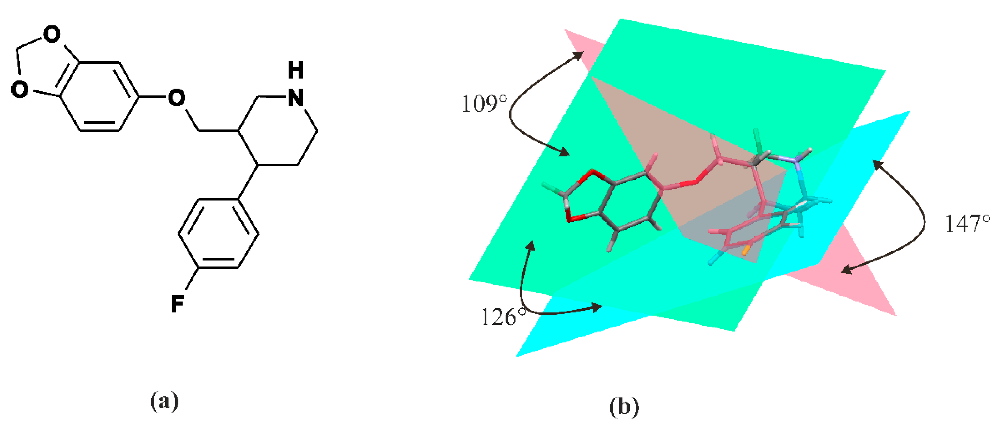
3. Paroxetine—Chemistry

Paroxetine is a phenylpiperidine derivative. It is composed of a secondary amine residing in the piperidine ring, which in turn is connected to benzodioxol and fluorophenyl groups [54] (Figure 1). From a chemical point of view, paroxetine is enantiomerically pure, (−)-(3S,4R)-3-[(2H-1,3-benzodioxol-5-yloxy)methyl]-4-(4-fluorophenyl)piperidine hydrochloride hemihydrate with empirical formula of C19H20FNO3·HCl·½H2O (PubChem CID: 43815 [61]). It is an odorless, off-white powder, having a melting point ranging between 120 and 138 °C. Particularly, paroxetine is a relatively small molecule with molecular weight of 374.8 g/mol (329.4 g/mol as free base). [47]. In addition to hydrochloride, paroxetine mesylate is also available [23]. It can be concluded that nowadays, the structure of paroxetine is well-researched and understood (Figure 1). The compound exists in two crystal forms, i.e., a stable hemihydrate referred to as form I and the anhydrous form called form II (CCDC DN: 125003 [62]) [63,64]. Spectroscopic data are available in the literature and databases: FTIR (SpectraBase Compound ID: 31iWZ0aC88k [65]) NMR ([66]) MS (accession: AU152606 [67]). These data clearly show that each of the three rings present in paroxetine structure is located on a different plane, so the structure of this compound is a highly nonplanar molecule, as is depicted in Figure 1.
It is a lipophilic base amine with both hydrophobic and hydrophilic moieties (pKa is 9.9 and the partition coefficient of paroxetine (log Po/w = 3.95)) [44]. It is slightly soluble in H2O (5.4 mg/mL), sparingly soluble in Me2Cl2 and EtOH (96%) but entirely soluble in MeOH. The chemical properties of this compound make it easy to modify and preserve drug-like properties [43].

4. Paroxetine—Pharmacodynamics Pharmacokinetics and Metabolism

Paroxetine hydrochloride salt ingested orally is almost completely absorbed, with only 2% of dose recovered in feces. Biological availability of commercially available paroxetine as a controlled-release formulation (CR) is distinct. Absorption of paroxetine was found to be insusceptible to influence of food or concomitant antacid treatment. Saturation during pass through liver leads to greater bioavailability. With repeated administration the steady-state concentration of the drug is achieved within 4 to 14 days. There is no further accumulation of the compound. The distribution of paroxetine in the body is extensive and consistent with its lipophilic amine character, with only 1% of the drug remaining in systemic circulation [68,69].
The volume of distribution varies from 3,1 to 28,0 L/kg (after intravenous administration). Mean elimination half-life is estimated to be about 21 h. Almost two-thirds of the drug are eliminated through the kidneys [43,70]. Paroxetine interacts with both cytochrome P450 family 2 subfamily C member 19 (CYP2C19) and cytochrome P450 family 3 subfamily A member 4 (CYP3A4). It is also one of the most potent inhibitors of cytochrome P450 family 2 subfamily B and D member 6 CYP2D6 and CYP2B6 among SSRIs [50]. Up to 95% of the drug is bound to proteins, mainly P-glycoprotein (P-gp), which is involved in transport through the blood–brain barrier (BBB). It is worth emphasizing that paroxetine is both a substrate and inhibitor of P-gp [50,69]. Comprehensive information about human paroxetine metabolites can be found in the Human Metabolome Database [71] under the acronym HMDB0014853 [72] and Kyoto Encyclopedia of Genes and Genomes [73] under the entry D02362 [74].

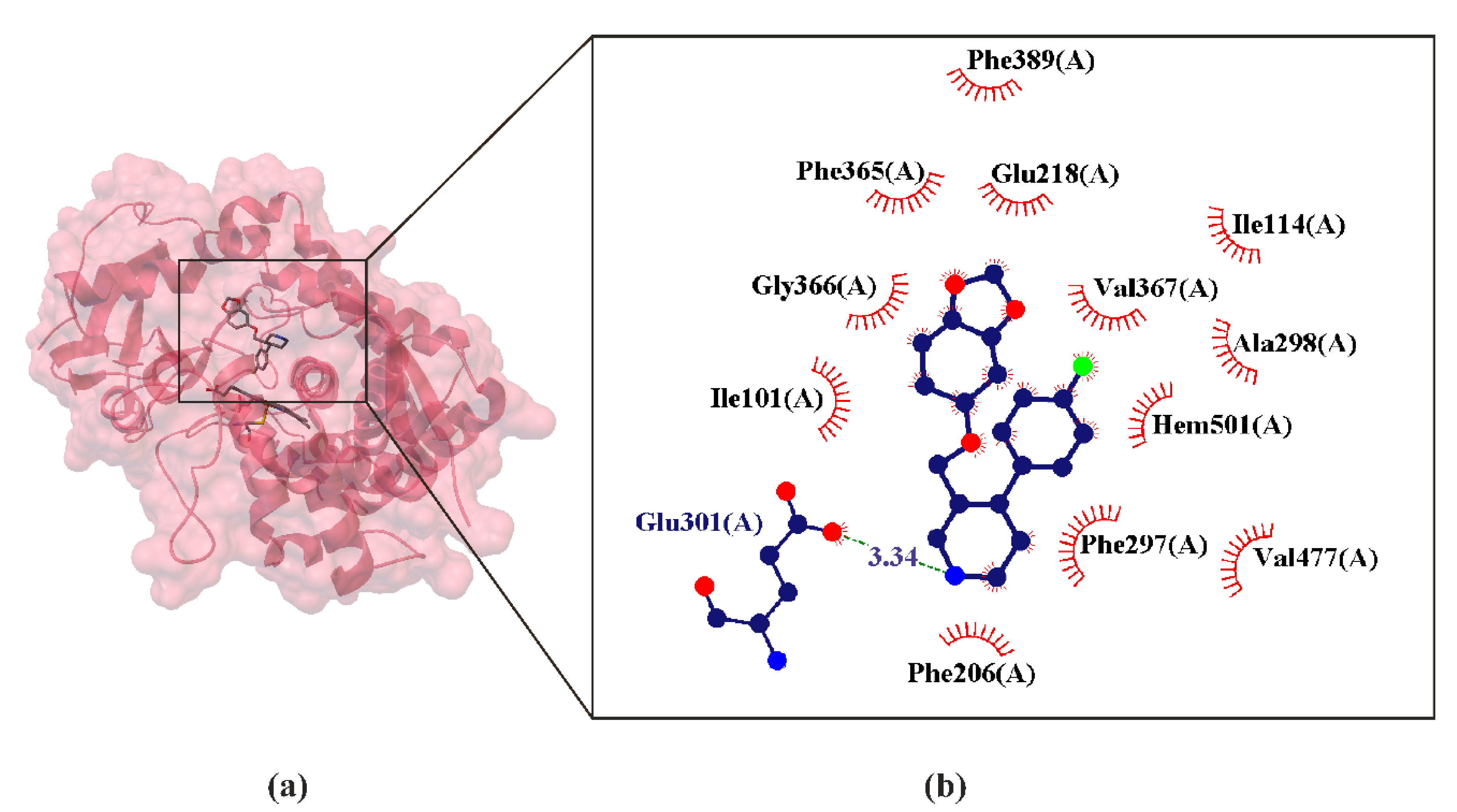
4.1. Paroxetine as P450 Inhibitors

Cytochromes P450 (CYPs) form the main family of enzymes capable of catalyzing oxidative biotransformation of most clinically available drugs (approx. 70–80%), hormones and other lipophilic xenobiotics. For this reason, they are of particular importance to clinical pharmacology. It is widely accepted that there are 57 functional human CYPs, of which about a dozen enzymes are responsible for the biotransformation of most xenobiotics [75,76]. By far the most important are CYP3A4/5, CYP2D6, CYP2C9, CYP1A2 and CYP2B6, which metabolize 30.2%, 20%, 12.8%, 8.9% and 7.2% of currently used drugs, respectively. It has been proven that despite wide and overlapping substrate specificity of these enzymes, many drugs are metabolized at clinically relevant concentrations by only one or more enzymes, which limits the significant redundancy of the oxidation drugs system of phase I [77]. Many issues of great importance in drug treatment determine the metabolism of drugs, such as pharmacokinetics, interindividual variability and drug interactions [78]. Inhibition of metabolism mediated by P450 cytochromes often occurs as a result of interactions between different drugs. A compound that inhibits an enzyme involved in the metabolism of another drug can reduce metabolic excretion of this drug, leading to elevation of the drug’s concentration in the blood, which can cause adverse effects or enhanced therapeutic effect [79]. Paroxetine is almost completely metabolized in animals and the human system [80]. Paroxetine is well-absorbed orally and undergoes extensive first-pass metabolism that is partially saturable [43]. Its metabolites are pharmacologically inactive in vivo [81]. Paroxetine metabolism is mediated in part by CYP2D6, CYP2B6 [55,82].
Numerous crystal structures have shown that all P450s share a common protein fold consisting of a large triangular prism divided into two domains. The α-helical domain (the helices are labeled in the literature A–L) with most of the helices clustered together. The smaller β-sheet domain (the sheets are labeled in the literature 1–4) (Figure 2) [83]. The first structure of a mammalian P450 was determined in 2000 [84]. According to X-ray crystal structure measurements, some P450s adopt multiple conformations, whereas other P450s appear in a single conformation. Consequently, it is difficult to predict how P450s will behave in contact with a new compound [75,76]. Active site of all P450s forms a cavity concealed within the protein molecule [85] (Figure 2). A heme coordination bond is also characteristic of each P450, combining a positively charged iron ion with a negatively charged thioplane sulphur atom [86] (Figure 2). This iron–cysteine bond is the basis of redox states which an iron ion can access during P450 catalysis, resulting in a complex catalytic cycle of P450 enzymes. In each P450, two highly synchronized electron transfer steps are required to enable the P450 catalytic cycle to reach the production stage [87] (Figure 2).
The structural studies available in the literature reveal that in the structure of paroxetine there are two possible pharmacophore points for interaction with P450: methylenedioxo moiety and secondary amine moiety of piperidine. Therefore, two possible routes of P450 inactivation by paroxetine are allowed. One involves the hydroxylation of C-H at the site of methylenedioxo moiety, and the second is N-H hydroxylation. Both C-H and N-H hydroxylation reactions can proceed via oxygen rearrangement or hydrogen atom transfer [88].

4.1.1. Paroxetine as CYP2D6 Inhibitors

The CYP2D6 isoenzyme is highly polymorphic and inhibited by several small molecules and clinically important pharmaceuticals [89,90]. CYP2D6 metabolizes antidepressants, antipsychotics, analgesics, β-blockers and antiarrhythmics. Paroxetine is both a substrate and an inhibitor of cytochrome isoenzyme CYP2D6 [81]. CYP2D6 metabolizes paroxetine via demethylation of the methylenedioxy group of methylenedioxyphenol. This reaction involves oxidation of the methylene bridge to a species that forms a tight but reversible complex with the heme iron atom [91]. Paroxetine also has the highest inhibitory constant of all antidepressants for CYP2D6 (Ki = 0.065) [89]. Clinical drug interaction studies show that paroxetine can inhibit the metabolism of drugs metabolized by CYP2D6, such as desipramine, risperidone and atomoxetine [92]. This extremely high inhibitory binding constant or affinity explains paroxetine’s high interaction profile with substrates for CYP2D6 [93].
Multiple-drug therapy is a common therapeutic practice, particularly in patients with several diseases or conditions. Breast cancer is highly associated with depression and therefore requires antidepressant therapy. In animal studies, antidepressants were observed to increase the incidence and growth of breast cancer in mice. It was suggested then that it is related to the inhibition of enzymes involved in the metabolism of carcinogens and estrogens. The inhibition of the isoenzyme CYP2D6 leads to an increase in their concentrations and serum levels, which increases the risk of breast cancer [42]. Since the early 1990s, many epidemiological studies have been carried out involving women who have been using antidepressants and have been diagnosed with breast cancer. The results obtained were interpreted differently, some studies concluded that there is no association between antidepressant use and breast cancer, while others came to the entirely opposite conclusion [94]. So far, no unequivocal position has been established. Paroxetine has been shown to have estrogenic effects (mimicking estrogen at the estrogen receptors) which may potentially affect the endocrine system and the development of breast tumors in women. Nevertheless, there is no clinically valid evidence for this [95,96].
The crystal structure of CYP2D6 was resolved in 2006 at 3 Å resolution [97]. The structure of CYP2D6 has a well-defined active site cavity above the heme, the shape of which is compared in the literature to the shape of the “right foot”. The “heel” of the foot-shaped cavity lies above the heme, the foot “arch” is formed by the Phe-120 side chain, while the “ball” is bordered by residues from the B′-C loop and the N-terminal end of the I helix. Such detailed information makes it possible to use computational modelling techniques, e.g., docking in the development of drugs involving these CYPs [97].

4.1.2. Paroxetine as CYP2B6 Inhibitors

CYP2B6 participates in the metabolism of a wide range of drug classes, including antiretrovirals, antidepressants, anesthetics, anticancer agents and antismoking agents. There is some evidence that its substrates generally contain a basic N atom and a planar aromatic ring [83]. A good example includes bupropion, cyclophosphamide, ifosfamide, pethidine, ketamine and propofol [89]. This is because this enzyme metabolizes a number of drug substrates which are usually nonplanar, neutral or weakly basic, with one or two hydrogen-bonding acceptors [98]. Paroxetine also has the highest inhibitory constant of all antidepressants for CYP2B6 (Ki = 1.03 μM). The inhibitory constant for CYP2D6, on the other hand, is lower compared to CYP2B6, but it is still high [93]. These high inhibitory binding constants explain paroxetine’s high interaction profile with substrates for CYP2D6 and CYP2B6 [83].
Paroxetine is also a potent inhibitor of CYP3A4 with multiple CYP3A4 substrate interactions. [42,99]. In humans, secondary amine xenobiotics are catalytically metabolized by P450s, leading to the formation of hydroxylamines [88].

4.1.3. Paroxetine as CYP2B4 Inhibitors

No crystalline structure of paroxetine binds with human CYP2D6 and CYP2B6 has been obtained so far [55]. However, in the Research Collaboratory for Structural Bioinformatics (RCSB) base, the crystal of the paroxetine complex with CYB2B4 [83] is deposited. The CYP2B4 catalyst has extremely similar structure to CYP2B6, with the only difference in residue 363, which is Leu in CYP2B6 and Ile in CYP2B4. In particular, the CYP2B4 tertiary structure is the most plastic (i.e., the largest degree of structural flexibility) of any P450 studied by X-ray crystallography [100]. Since the crystal of the paroxetine complex is deposited in the RCSB base with CYB2B4, it seems reasonable to treat it as an appropriate approximation of the interaction of paroxetine with CYP2B4 as well as with CYP2B6 (Figure 2).

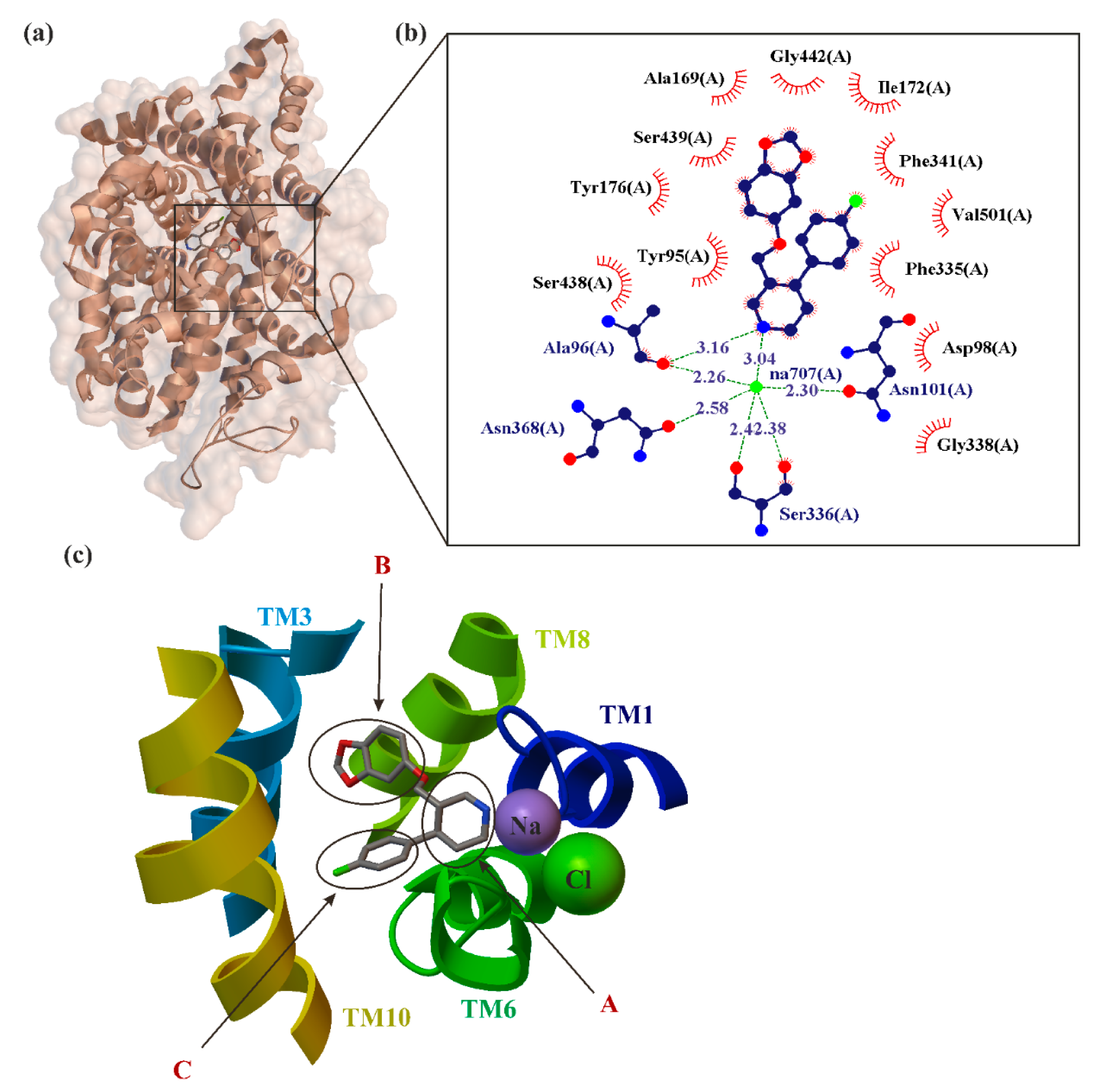
5. Paroxetine as MAT Inhibitions

Selective serotonin reuptake inhibitors (SSRI) are clinically prescribed antidepressants that act by increasing the local concentration of the neurotransmitter at synapses and in extracellular spaces via blockade of the serotonin transporter (SERT) [53]. SERT is a member of the neurotransmitter sodium symporter (NSS) family of transporters. The NSS also includes two more catecholamine transporters such as dopamine (DAT) and norepinephrine (NET) transporters [101] and some amino acid transporters such as glycine transporter (GlyT), γ-aminobutyric acid (GABA) transporter (GAT), leucine transporter (LeuT) and osmolytes such as betaine and creatine [102,103]. Inhibitors of the three monoamine transporters (MATs) increase the extracellular concentration of monoamines, and are widely used in the treatment of psychiatric diseases and as illicit psychostimulant drugs [104].
Paroxetine is an SSRI that exhibits the highest known binding affinity for the central site of SERT (for SERT ≈ 70.2 ± 0.6 pM, for DAT ≈ 490 ± 20.0 nM, for NET ≈ 40 ± 0.2 nM) [105]) compared to any other currently prescribed antidepressants [54]. The selectivity profile of MAT inhibitors across NET, DAT and SERT is critical for their therapeutic profile and/or abuse potential. Binding studies have demonstrated that antidepressants are from 300 to 3500 times more selective for SERT over NET, and generally have low affinity for DAT (from 7800 to 0 times) [106]. Paroxetine acts as a dual serotonin/norepinephrine uptake inhibitor in higher doses (40 mg/day or more) [46]. Furthermore, there is evidence that paroxetine can also operate as an allosteric modulator of SERT, although not as effectively as escitalopram [107]. In vitro studies in animals suggest that it has weak effects on dopamine neuronal reuptake. It may contribute to emotional flattening, apathy and cognitive slowing in some patients. In vitro radioligand binding studies report that paroxetine has minor affinity for dopamine, alpha1-, alpha2-, beta-adrenergic-, muscarinic and histamine (H1) receptors [108,109]. Due to mild anticholinergic actions of paroxetine, the drug may cause a rapid onset of hypnotic and anxiolytic efficacy as well as the occurrence of side effects. Weak antimuscarinic properties may cause sedation, constipation and dry mouth.
The primary target of paroxetine and other SSRI medications is the SERT, which is a type of monoamine transporter that transports serotonin from the synaptic cleft back to the presynaptic neuron [70]. SERT, as the target of many psychoactive agents, has importance to the etiology of affective disorders [52]. SERT belongs to the solute carrier 6 family of transporters (SLC6) [110]. It is an integral membrane protein that exploits pre-existing sodium, chloride and potassium ion gradients to catalyze the thermodynamically unfavorable movement of synaptic serotonin into the presynaptic neuron. The Protein Data Bank provides X-ray structures of the leucine transporter aLeuT (Aquifex aeolicus LeuT) [103] and monoamine transporters (MATs): dDAT (Drosophila DAT) [111] and hSERT (human SERT) [52] crystallized in 2005, 2013 and 2016. The human NET (hNET) crystal structure has not been obtained yet. SERT cotransports molecules of serotonin with one Na+ and Cl ions while a single potassium K+ ion is moved in the opposite direction. Importantly, hSERT was crystallized in complex with the two prototypical SSRIs, escitalopram and paroxetine [52] (Figure 3).
Analysis of the crystalized data amino acid sequences provided evidence for 12 transmembrane segments (TM1-TM12), with the amino and carboxy terminal end located in the extracellular vestibule. Due to high similarities to architecture of LeuT, this TM1-12 arranged often is the so-called LeuT-like structural fold. This pattern is characterized by two inverted 5-TM repeats. The location of drug-binding sites in crystallized MATs is determined according to the scheme originally determined for LeuT by Sørensen et al. [112] in two separate regions. They are marked as S1 and S2 pockets. The former is located approximately halfway across the membrane bilayer and the latter is located in the extracellular vestibule. The S1 site is the central substrate binding pocket which defines the primary binding region in NSSs, while the S2 is an allosteric site approximately 13 Å from S1. S1 is composed of three subsites, formally called A, B, C (Figure 3) [54]. From a chemical point of view, all these subsites represent different natures: A is a polar region surrounding Asp98 (side chains from TMs 1, 6 and 8), whereas subsites B and C are largely hydrophobic regions. B regions are located opposite to subsites C. B regions are formed by residues from TMs 3 and 8. Subsite C is formed by TMs 3, 6 and 10 [113]. In early 2016, the binding mode of paroxetine at the S1 SERT site was investigated simultaneously by two teams: Coleman et al. [52,53] (Figure 3) and Davis et al. [70]. The data obtained from these studies are inconclusive, the binding site and orientation of paroxetine in SERT remain controversial.
Nonplanar structure of the ligand (as in the case of paroxetine, Figure 1) causes the differences between binding poses of the ligand at the binding site obtained by different methods [114]. Piperidine, benzodioxol and fluorophenyl substituents of paroxetine were present [52,53] in subsites A, B and C of the S1 site, respectively—a pose commonly denoted by many authors as ABC (Figure 3). While the homology studies conducted by Davis et al. have proposed an orientation “flipped” from that in the ABC pose, i.e., the piperidine, benzodioxol and fluorophenyl substituents of paroxetine reside in subsites A, C, and B of the S1 site, respectively (often donated ACB) [70]. Additionally, a combination of pharmacological, biochemical and mutagenesis data suggest that amino acids implicated in high-affinity paroxetine binding may not overlap with those thought to be involved in recognizing other inhibitors [70,102].

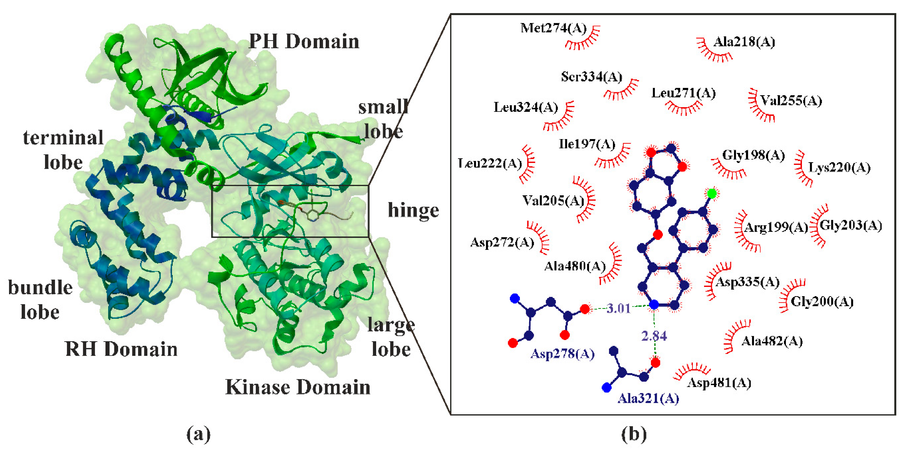
6. Paroxetine as Kinase GRK2 Inhibitors

Currently, there are five main protein families which are key therapeutic targets for most drugs available on the world market. G-protein-coupled receptors (GPCRs) are integral membrane proteins that relay external signals into the cytoplasm of the cell. GPCRs are estimated to be the main therapeutic target for about a third of prescription drugs [115,116]. They are key regulators of cell physiology and control processes ranging from glucose homeostasis to contractility of the heart. A major mechanism for the desensitization of activated GPCRs is their phosphorylation by GPCR kinases (GRKs). Overexpression of G-protein-coupled receptor kinase 2 (GRK2) is strongly linked to both the healthy and failing heart, and it has long been considered a therapeutic target for the treatment of cardiovascular disease [117]. The GRK2 originally known as β-adrenergic receptor kinase 1 (βARK-1) was the first GRK cytoplasmic protein identified in the heart [118]. It belongs to a group of serine/threonine kinases that have a relevant role in the identification and phosphorylation of activated GPCRs [119]. Accordingly, inhibition of GRK2 is considered an important drug target in the treatment of heart failure [120].
From a physiological point of view, the autonomic nervous system affects the frequency of heart contractions: the sympathetic system accelerates heart function while the parasympathetic system decelerates it. In response to a failing heart, the sympathetic nervous system increases the level of circulating catecholamines (norepinephrine and adrenaline). The work of the heart is also affected by β adrenergic receptors (βAR) [121]. There are four types of action β receptors on myocardial cells. These include the following effects: inotropic, chronotropic, bathmotropic, dromotropic. The first two determine the strength and frequency of myocardial contraction, the third affects the threshold of excitability of myocardial cells and the latter is responsible for the rate of conduction in the heart muscle [122]. Binding catecholamines in cardiomyocytes initiate downstream signaling to increase the contraction force of the heart muscle (i.e., inducing a positive inotropic effect) [123]. In the failing heart, activation of βARs also leads to (upregulation) increase in the number of GRK2 and GRK5, which in turn leads to uncoupling of βARs from G proteins, decreased βARs at the cellular membrane and decreased cardiac output in response to hormonal stimulation [124,125,126]. Studies of animal models have shown that reducing GRK2 levels is beneficial in preventing heart failure by renormalizing catecholamine and βAR levels of cell surfaces and improving heart function [127,128].
Paroxetine exhibits 50/60-fold higher selectivity for GRK2 versus other GRKs (such as GRK1 and GRK5) [56]. Paroxetine hydrochloride was identified as a modest GRK2 inhibitor with an IC50 of 1.4 μM [118,127]. It is worth emphasizing that other compounds from the SSRI group have shown no effect in in vitro kinase tests or ex vivo or in vivo myocyte contractility [57]. Due to this, paroxetine (or a paroxetine derivative) could be used in heart failure treatment in the future.
GRK2 is a multidomain protein organized in several domains and regions (Figure 4). It consists of three distinct domains: an RGS homology (RH) domain, a protein kinase domain, and a pleckstrin homology (PH) domain. The RH domain has two subdomains often referred to as the terminal and bundle lobes, whereas the kinase domain is composed of small and large lobes. The terminal lobe of the RH domain also forms an extensive hydrophobic interface with the PH domain. The RH domain is the protein region through which GRK2 binds Gβγ. The RH domain participates in interactions with both the kinase and PH domains, thus playing an important role in the regulation of protein activity. The kinase domains containing the ATP-binding pocket consist of a β-sheet included in the small-terminal lobe (also called N-lobe) and α-helixes dominating the large-terminal lobe (also called C-lobe). Both lobes’ regions are connected via a flexible hinge region [129,130]. Most small-molecule kinase inhibitors target the ATP-binding site (Figure 4). The crystal structure of the GRK2·paroxetine−Gβγ complex revealed that paroxetine binds in the active site of GRK2 (Figure 4) and stabilizes the kinase domain in a novel conformation in which a unique regulatory loop forms part of the ligand-binding site. It was found that paroxetine inhibiting GRK2 increases contractility in isolated cardiomyocytes as well as in myocardial βAR inotropic reserve in living mice [57].

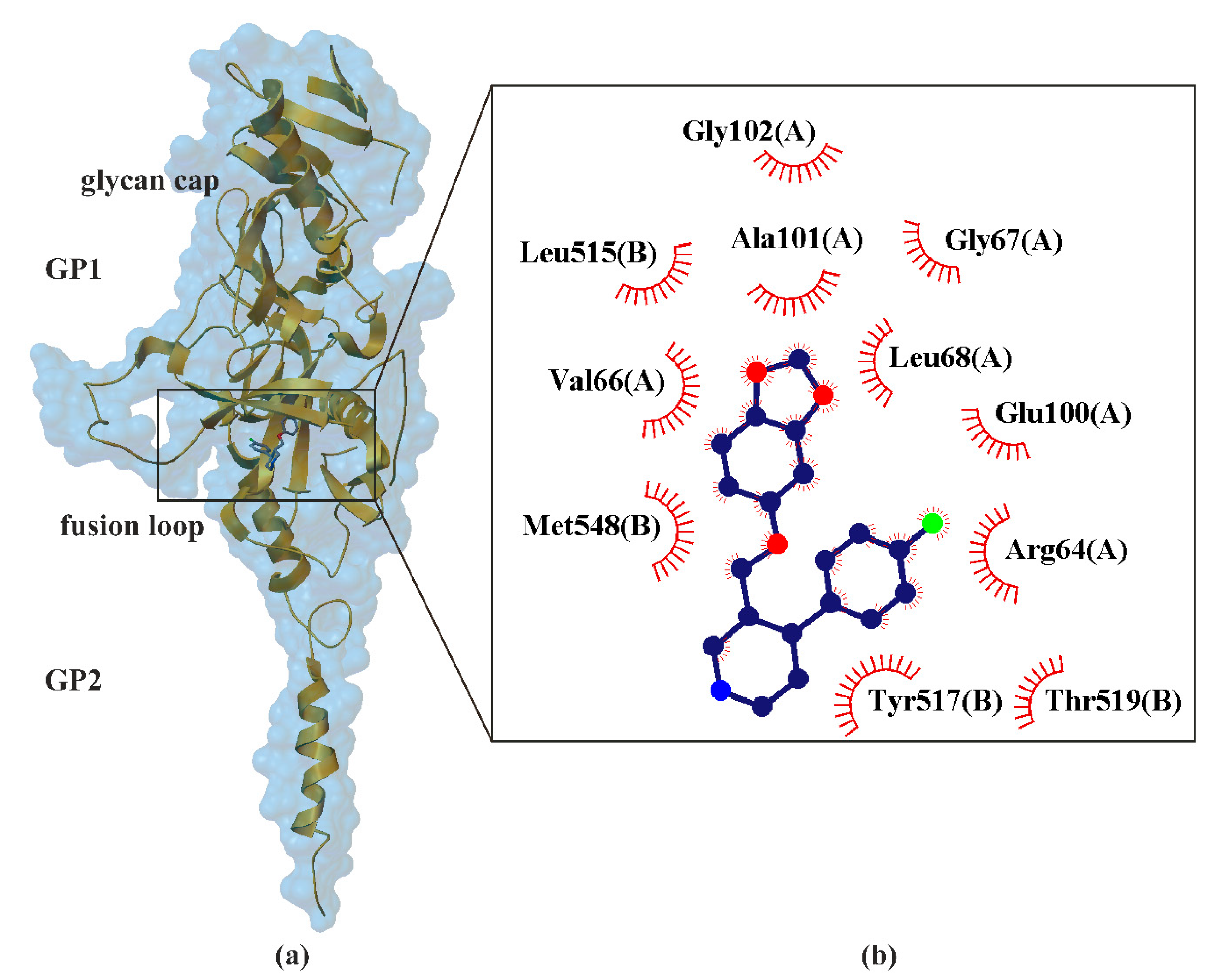
7. Paroxetine as Ebolavirus Inhibitors

EBOV belong to the family Filoviridae and cause severe, often fatal, diseases, e.g., Ebola hemorrhagic fever (EHF) in humans and other mammals, also known as Ebola virus disease (EVD, ICD-10-CM code: A98.4 [131]). EHF is characterized by rapid disease progression and high risk of death, killing between 25% and 90% of those infected, with an average of about 50% [132]. EBOV has caused the majority of human deaths from EVD and was the cause of the 2013–2016 epidemic in western Africa [133] which resulted in at least 28,646 suspected cases and 11,323 confirmed deaths [132]. There are currently no approved therapeutic drugs or vaccines for the disease [134].
EBOV contains single-stranded negative RNA linear genome, about 18–19 kb in size and encode 7 genes such as nucleoprotein (NP), viral protein 35 (VP35), viral protein 40 VP40, viral protein 30 VP30, viral protein 24 (VP24), polymerase (L) and glycoprotein (GP). Fatal human cases of EHF are exemplified by very high viral titers in the blood, liver and spleen, as well as profound immunosuppression [135].
The mechanism of EBOV inhibition is largely unknown. EBOV has a membrane envelope decorated by trimers of a glycoprotein (GP, cleaved by furin to form GP1 and GP2 subunits, Figure 2), which is solely responsible for host cell attachment, endosomal entry and membrane fusion. The trimeric transmembrane GP spike, each approximately 7–10 nm long and spaced at approximately 10 nm intervals, are presented on the surface of the virion and are accountable for cellular attachment and entry. EBOV entry into the cells is initiated by the interaction of the viral GP with receptors on the surface of host cells, and then internalized via the macropinocytosis pathway [136]. GP is thus a primary target for the development of antiviral drugs. Crystallographic studies have shown that five chemically divergent EBOV inhibitors, such as ibuprofen (pKi ≈ 2.22) [137], benztropine (pKi ≈ 2.89), bepridil (pKi ≈ 3.54), paroxetine (pKi ≈ 3.19, Figure 2) and sertraline (pKi ≈ 3.02), interact directly with the Ebolavirus glycoprotein [59]. Binding of these drugs destabilizes the protein, suggesting that it may be an inhibitory mechanism. The study of toremifene (anticancer drug) revealed that the binding affinities (pKi ≈ 4.80, determined by thermal shift assay) correlate with the protein−inhibitor interactions as well as with the antiviral activities determined by virus cell entry assays, supporting the hypothesis that these drugs inhibit viral entry by binding GP and destabilizing the prefusion conformation [137].
The crystal structure of proxetine-EBOV GP complex was determined at 2.4 Å resolution, with good R-factors and stereochemistry. GP1 has three distinct domains: (•) the receptor binding domain (RBD), (•) the glycan cap and (•) the heavily O-linked glycosylated mucin-like domain (MLD). It is predominantly composed of β-strands, forming a large semicircular groove at the center of the subunit. EBOV-GP forms GP-containing microvesicles, so-called virosomes, which are secreted from GP-expressing cells. However, determinants of GP-virosome release and their functionality are poorly understood. RBD promotes GP-virosome secretion, while tetherin suppresses GP-virosomes by interactions involving the GP-transmembrane domain [138]. RBD is responsible for interacting with one or more cellular receptors. It is proof that EBOV-GP-virosomes are immunomodulatory and act as decoys for EBOV-neutralizing antibodies. The glycan cap could protect the receptor binding sites from antibodies and interacts with the internal fusion loop of GP2 which is critical for membrane fusion. In the fusion process, GP2 undergoes conformational changes. Paroxetine binds to GP with its benzodioxol group (Figure 5) and builds interactions with not only the side chains of Val66,
Ala101, Leu515, Tyr517 and Met548, but also with the main-chains of Gly67 and Gly102 and side chain of Leu68 Figure 5 [59]. These results suggest that inhibitor binding destabilizes GP and triggers premature release of GP2, thereby preventing fusion between the viral and endosome membranes. This way of binding paroxetine from EBOV GP is analogous to those previously confirmed for other compounds, including toremifene and ibuprofen, which provides additional confirmation of the EBOV GP inhibition mechanism [137]. Given the weak inhibitory properties (low pKi) of the medicines tested so far, they are not suitable for reducing EBOV infection. Nevertheless, studies on the structures of the acquired complexes reveal the inhibition mechanism and can guide the development of more powerful anti-EBOV drugs.

8. Paroxetine as KIT and JAK Inhibitors

Paroxetine can influence a variety of cancers, including brain tumor [139], colon cancer [140] and breast cancer [141] by blocking some protein kinase signaling pathways involved in tumorigenesis [142].
It has been clinically observed that paroxetine has strong cytotoxicity on human tumor cell lines. This is kinase pathways involved in tumorigenesis. Many studies have established that this is due to the structural similarities (size, shape, physicochemical properties, Figure 6) of paroxetine with many known kinase inhibitors. Consequently, it is not surprising that paroxetine is very compatible with the kinase active site [142]. A good example of paroxetine’s similarity are sunitinib, approved a multitargeted tyrosine kinase inhibitor (KIT inhibitor) [143,144] and tofacitinib (approved Janus kinase inhibitor (JAK inhibitor)) [145] (Figure 6). Sunitinib (ATC code: L01XE04 [146]), a multitargeted tyrosine kinase inhibitor, which is approved by both US and EU regulatory agencies for clinical use, extends survival of patients with metastatic renal cell carcinoma and gastrointestinal stromal tumors, but concerns have arisen about its cardiac safety [143,147,148]. Tofacitinib (ATC code: L04AA29 [146]) is a Janus kinase inhibitor currently approved for the treatment of rheumatoid arthritis, psoriatic arthritis and ulcerative colitis [145,149,150].
Tyrosine kinase proteins are a class of proteins with tyrosine kinase activity that catalyzes the transfer of phosphate groups to ATP to the residues of tyrosine of many important proteins, forming protein phosphorylation and then transmitting a signal regulating cell growth, differentiation, death and a number of physiological and biochemical processes [151]. Paroxetine can interact with these kinases in two so-called binding modes: class I and class II [142]. Five kinases in the C-Src ABL, SRC, KIT, MET and FYN family were identified as targets for paroxetine kinase, which can be inhibited strongly or moderately at IC50 values at the nanomolar or micromolar level. Binding modelling analysis has shown that paroxetine ligands can adopt class I binding modes to interact with KIT, MET, FYN, and class II when interacting with ABL and SRC kinases. This is possible by creating an intensive network of molecular forces: specific (i.e., hydrogen bonds and π–π/cation–π stackings) and nonspecific forces (such as hydrophobic and Van der Waals) [152] in complex kinase–inhibitor interfaces. The division into classes of bonding modes is due to the location of piperidine moieties of paroxetine at the place of active kinase. In class I, piperidine moieties of paroxetine are packed against active sit kinase. While in class II, the same molecular fragment of paroxetine adopts the opposite binding mode. It seems noteworthy that paroxetine class I binding mode shows much higher inhibition potential than that of class II [142].

9. Conclusions and Perspective

Recently, there has been a steadily and rapidly increasing number of prescriptions for approved and unapproved drugs for depression. This is a particularly important issue when it concerns children and young people [9]. This provides the basis for extensive research. In our opinion, we should constantly discuss and analyze current reports on drugs that are used in daily clinical practice as off-label drugs much more often than others. Indisputably, paroxetine belongs to this group of drugs. It is important to be aware that the use of off-label medicines is a multidirectional issue. Sometimes off-label use means a change in the repurpose of the drug, e.g., finding novel therapeutic indications different from the ones for which the drug was already approved [153]. This may also mean unusual use of the drug. This includes, in particular, the use of a different dose, duration of use, frequency of dosing, use of another method of administration (e.g., orally instead of intravenously) or use by another group of patients (e.g., children instead of adults) [15].
Modern scientific research in the medical and health sciences is largely based on the results of research related to the achievements of recent decades and in particular the knowledge of the genomes of both the patients and pathogens. The extraordinary dynamics of the development of this knowledge necessitate the presentation of collective reviews of recent achievements. As we have tried to show in our work, understanding the molecular aspects not only allows for a better understanding of the mechanisms of action of drugs, but also contributes to the creation of new research directions. The presented analysis of molecular studies of paroxetine based on X-ray methods seems to confirm its special place in modern pharmacotherapy. The presented results of paroxetine studies are associated with every possible aspect of studies on the use of off-label procedures. It is to be hoped that the uniqueness of the structure of paroxetine will allow the development of a new drug in heart failure, which we think would be a very valuable achievement. A number of studies of paroxetine on human cell lines of tumors confirmed its strong cytotoxicity, which generates new opportunities for the search for more drugs in oncology. On the other hand, studies on EBOV indicate the possibility of paroxetine activity among these types of pathogens. Considering the confirmed effect of paroxetine on the nervous system makes it clear that the pharmacological profile of this drug coincides with key issues of modern medicine. All this creates promising prospects.

Author Contributions

M.K., J.N., Ł.F., A.N. conceived and directed the project, designed the study, collected the data, analyzed the data, interpreted the results and wrote the manuscript. All authors have read and agreed to the published version of the manuscript.

Funding

This study was supported by the research grant from the EIRU program at the NCU entitled EI DEBUTS “Finding and understanding mechanisms of action of selected compounds active on central nervous system monoamine transporters”.

Institutional Review Board Statement

Not applicable.

Informed Consent Statement

Not applicable.

Data Availability Statement

Not applicable.

Conflicts of Interest

The authors declare no conflict of interests.

Abbreviations

ADAnxiety Disorders
ADAAAmerican Society of Anxiety and Depression
ABLAbl Tyrosine Kinases
aLeuTAquifex Aeolicus Leucine Transporter
ATC codeAnatomical Therapeutic Chemical Classification System
ATPAdenosine Triphosphate
BBBBlood Brain Barrier
C-Src ABLSrc Tyrosine Kinases
CYPCytochromes P450
CYP1A2Cytochrome P450 Family 1 Subfamily A Member 2
CYP2B4Cytochrome P450 Family 2 Subfamily B Member 4
CYP2B6Cytochrome P450 Family 2 Subfamily B Member 6
CYP2C19Cytochrome P450 Family 2 Subfamily C Member 19
CYP2D6Cytochrome P450 Family 2 Subfamily D Member 6
CYP3A4Cytochrome P450 Family 3 Subfamily A Member 4
CYP3A5Cytochrome P450 Family 3 Subfamily A Member 5
DATDopamine Transporter
dDATDrosophila Dopamine Transporter
EBOV GPEbolavirus Glycoprotein
EHFEbola Hemorrhagic Fever
EVDEbola Virus Disease
FDAFood and Drug Administration
FYNFyn Proto-Oncogene, Non-Receptor Tyrosine Kinase
GABAγ-Aminobutyric Acid
GADGeneralized Anxiety Disorder
GATγ-Aminobutyric Acid Transporter
GlyTGlycine Transporter
GPGlycoprotein
GPCRG-Protein-Coupled Receptors
GRKG-Protein-Coupled Receptors Kinase
GRK1G-Protein Coupled Receptor Kinase 1
GRK2G-Protein Coupled Receptor Kinase 2
GRK5G-Protein Coupled Receptor Kinase 5
hDATHuman Dopamine Transporter
HEMHemeproteins
hNETHuman Norepinephrine Transporter
hSERTHuman Serotonin Transporter
ICD-10-CMInternational Classification of Diseases, Tenth Revision, Clinical Modification
JAKJanus Kinase
KiBinding Affinity
KITTyrosine-Kinase Inhibitor
LPolymerase
LeuTLeucine Transporter
MATMonoamine Transporters
MDDMajor Depressive Disorder
MDLO-linked glycosylated mucin-like domain
METMet Proto-Oncogene, Non-Receptor Tyrosine Kinase
NETNorepinephrine Transporter
NPNucleoprotein
NSSNeurotransmitter Sodium Symporter
OCDObsessive-Compulsive Disorder
PDPanic Disorder
PDB IDProtein Data Bank Identifier
PDDPremenstrual Dysphoric Disorder
P-glP-Glycoprotein
PTSDPosttraumatic Stress Disorder
RBDReceptor Binding Domain
RCSBResearch Collaboratory for Structural Bioinformatics
SADSocial Anxiety Disorder
SERTSerotonin Transporter
SLC6Solute Carrier 6 Family of Transporters
SRCSrc Proto-Oncogene, Non-Receptor Tyrosine Kinase
SSRISelective Serotonin Reuptake Inhibitors
TMTransmembrane Segment
VP24Viral Protein 24
VP30Viral Protein 30
VP35Viral Protein 35
VP40Viral Protein 40
βARK-1β-Adrenergic Receptor Kinase 1

References

  1. Gmitrowicz, A.; Janas-Kozik, M. Zaburzenia Psychiczne Dzieci i Młodzieży; Medical Tribune Polska: Warszawa, Poland, 2018; pp. 87–101. [Google Scholar]
  2. Unbound Medicine. F40–F48—Anxiety, Dissociative, Stress-Related, Somatoform and Other Nonpsychotic Mental Disorders. Available online: https://www.unboundmedicine.com/icd/view/ICD-10-CM/934326/all/F40_F48___Anxiety__dissociative__stress_related__somatoform_and_other_nonpsychotic_mental_disorders (accessed on 30 January 2021).
  3. Rockhill, C.; Kodish, I.; DiBattisto, C.; Macias, M.; Varley, C.; Ryan, S. Anxiety Disorders in Children and Adolescents. Curr. Probl. Pediatric Adolesc. Health Care 2010, 40, 66–99. [Google Scholar] [CrossRef]
  4. Adaa.org. Understand the Facts Anxiety and Depression Association of America. Available online: https://adaa.org/understanding-anxiety (accessed on 29 January 2021).
  5. Unbound Medicine.F40.11—Social Phobia, Generalized. Available online: https://www.unboundmedicine.com/icd/view/ICD-10-CM/943473/all/F40_11___Social_phobia_generalized?q=anxiety+disorder+social (accessed on 30 January 2021).
  6. West, K.B.; Wilbanks, J.; Suveg, C. Exposure Therapy for Separation Anxiety Disorder. Exposure Therapy for Children with Anxiety and OCD: Clinician’s Guide to Integrated Treatment; Academic Press: London, UK, 2020; pp. 143–153. [Google Scholar]
  7. Knappe, S.; Beesdo-Baum, K.; Fehm, L.; Lieb, R.; Wittchen, H. Characterizing the association between parenting and adolescent social phobia. J. Anxiety Disord. 2012, 26, 608–616. [Google Scholar] [CrossRef] [Green Version]
  8. Heimberg, R.; Hofmann, S.; Liebowitz, M.; Schneier, F.; Smits, J.; Stein, M.; Hinton, D.; Craske, M. Social anxiety disorder in DSM-5. Depression and Anxiety. Depress. Anxiety 2014, 31, 472–479. [Google Scholar] [CrossRef] [PubMed] [Green Version]
  9. Bazzano, A.T.; Mangione-Smith, R.; Schonlau, M.; Suttorp, M.J.; Brook, R.H. Off-label prescribing to children in the United States outpatient setting. Acad. Pediatrics 2009, 9, 81–88. [Google Scholar] [CrossRef] [PubMed]
  10. ATC Code. Anatomical Therapeutic Chemical Classification System. Available online: http://www.atccode.com/ (accessed on 30 January 2021).
  11. DrugBank. Paroxetine. Available online: https://go.drugbank.com/drugs/DB00715 (accessed on 30 January 2021).
  12. Sharma, S.; Dang, S. Paroxetine loaded PLGA nanoparticles. Mater. Today Proc. 2020, 28, 205–210. [Google Scholar] [CrossRef]
  13. Tang, S.W.; Helmeste, D. Paroxetine. Expert Opin. Pharmacother. 2008, 9, 787–794. [Google Scholar] [CrossRef] [PubMed]
  14. Stone, K.J.; Viera, A.J.; Parman, C.L. Off-label applications for SSRIs. Am. Fam. Physician 2003, 68, 498–504. [Google Scholar] [PubMed]
  15. Skånland, S.S.; Cieślar-Pobuda, A. Off-label uses of drugs for depression. Eur. J. Pharmacol. 2019, 865, 172732. [Google Scholar] [CrossRef]
  16. Laskowski, R.A.; Swindells, M.B. LigPlot+: Multiple ligand–Protein interaction diagrams for drug discovery. J. Chem. Inf. Model. 2011, 51, 2778–2786. [Google Scholar] [CrossRef]
  17. Unbound Medicine. F41.1—Generalized Anxiety Disorder. Available online: https://www.unboundmedicine.com/icd/view/ICD-10-CM/905183/all/F41_1___Generalized_anxiety_disorder?q=300.02 (accessed on 30 January 2021).
  18. Unbound Medicine. 300.3—Obsessive-Compulsive Disorders. Available online: https://www.unboundmedicine.com/icd//search?st=OSS&q=300.3 (accessed on 30 January 2021).
  19. Unbound Medicine. F33—Major Depressive Disorder, Recurrent. Available online: https://www.unboundmedicine.com/icd/view/ICD-10-CM/930155/all/F33___Major_depressive_disorder_recurrent?q=depressive+disorder+major (accessed on 30 January 2021).
  20. Unbound Medicine. F32.81—Premenstrual Dysphoric Disorder. Available online: https://www.unboundmedicine.com/icd/view/ICD-10-CM/968287/all/F32_81___Premenstrual_dysphoric_disorder?q=disorder+dysphoric+premenstrual (accessed on 30 January 2021).
  21. Unbound Medicine. F43.1—Post-Traumatic Stress Disorder (PTSD). Available online: https://www.unboundmedicine.com/icd/view/ICD-10-CM/929135/all/F43_1___Post_traumatic_stress_disorder__PTSD_?q=f43.1 (accessed on 30 January 2021).
  22. Unbound Medicine. F41.0—Panic Disorder [Episodic Paroxysmal Anxiety]. Available online: https://www.unboundmedicine.com/icd/view/ICD-10-CM/915404/all/F41_0___Panic_disorder_[episodic_paroxysmal_anxiety]?q=anxiety+disorder+generalized (accessed on 30 January 2021).
  23. Liu, X.; Li, X.; Zhang, C.; Sun, M.; Sun, Z.; Xu, Y.; Tian, X. Efficacy and tolerability of fluvoxamine in adults with social anxiety disorder: A meta-analysis. Medicine 2018, 97, e11547. [Google Scholar] [CrossRef]
  24. Bandelow, B.; Sher, L.; Bunevicius, R.; Hollander, E.; Kasper, S.; Zohar, J.; Möller, H. Guidelines for the pharmacological treatment of anxiety disorders, obsessive–compulsive disorder and posttraumatic stress disorder in primary care. Int. J. Psychiatry Clin. Pract. 2012, 16, 77–84. [Google Scholar] [CrossRef] [PubMed]
  25. Baldwin, D.S.; Anderson, I.M.; Nutt, D.J.; Bandelow, B.; Bond, A.; Davidson, J.R.; den Boer, J.A.; Fineberg, N.A.; Knapp, M.S.J.; Hu, W. Evidence-based guidelines for the pharmacological treatment of anxiety disorders: Recommendations from the British Association for Psychopharmacology. J. Psychopharmacol. 2005, 19, 567–596. [Google Scholar] [CrossRef] [PubMed] [Green Version]
  26. Yonkers, K.A.; Gullion, C.; Williams, A.; Novak, K.; Rush, A.J. Paroxetine as a treatment for premenstrual dysphoric disorder. J. Clin. Psychopharmacol. 1996, 16, 3–8. [Google Scholar] [CrossRef]
  27. Aghakhani, K.S.S.; Farhidnia, N.; Fallah, F. Successful Suicide in a Child: Depression-Related or ParoxetineInduced? Int. J. Med Toxicol. Forensic Med. 2016, 6, 242–246. [Google Scholar]
  28. Lenox, R.H.; Frazer, A. Neuropsychopharmacology: The Fifth Generation of Progress; Lippincott Williams and Wilkins: Philadelphia, PA, USA, 2010; pp. 1139–1163. [Google Scholar]
  29. Schneeweiss, S.; Patrick, A.R.; Solomon, D.H.; Dormuth, C.R.; Miller, M.; Mehta, J.; Lee, J.C.; Wang, P.S. Comparative safety of antidepressant agents for children and adolescents regarding suicidal acts. Pediatrics 2010, 125, 876–888. [Google Scholar] [CrossRef] [Green Version]
  30. Teicher, M.H.; Glod, C.; Cole, J.O. Emergence of intense suicidal preoccupation during fluoxetine treatment. Am. J. Psychiatry 1990, 147, 207–210. [Google Scholar] [CrossRef] [PubMed]
  31. Hetrick, S.E.; McKenzie, J.E.; Cox, G.R.; Simmons, M.B.; Merry, S.N. Newer generation antidepressants for depressive disorders in children and adolescents. Cochrane Database Syst. Rev. 2012. [Google Scholar] [CrossRef] [PubMed]
  32. Pozzi, M.; Radice, S.; Clementi, E.; Molteni, M.; Nobile, M. Antidepressants and, suicide and self-injury: Causal or casual association? Int. J. Psychiatry Clin. Pract. 2016, 20, 47–51. [Google Scholar] [CrossRef]
  33. Morrison, J.; Schwartz, T.L. Adolescent angst or true intent? Suicidal behavior, risk, and neurobiological mechanisms in depressed children and teenagers taking antidepressants. Int. J. Emerg. Ment. Health 2014, 16, 247–250. [Google Scholar] [PubMed]
  34. Giner, L.; Nichols, C.M.; Zalsman, G.; Oquendo, M.A. Selective serotonin reuptake inhibitors and the risk for suicidality in adolescents: An update. Int. J. Adolesc. Med. Health 2005, 17, 211–220. [Google Scholar] [CrossRef]
  35. McKeown, R.E.; Cuffe, S.P.; Schulz, R.M. US suicide rates by age group, 1970–2002: An examination of recent trends. Am. J. Public Health 2006, 96, 1744–1751. [Google Scholar] [CrossRef]
  36. Hjalmarsson, L.; Corcos, M.; Jeammet, P. Selective serotonin reuptake inhibitors in major depressive disorder in children and adolescents (ratio of benefits/risks). Encephale 2005, 31, 309–316. [Google Scholar] [CrossRef]
  37. Hammad, T.A.; Laughren, T.; Racoosin, J. Suicidality in pediatric patients treated with antidepressant drugs. Arch. Gen. Psychiatry 2006, 63, 332–339. [Google Scholar] [CrossRef] [PubMed]
  38. Termorshuizen, F.; Palmen, S.J.; Heerdink, E.R. Suicide behavior before and after the start with antidepressants: A high persistent risk in the first month of treatment among the young. Int. J. Neuropsychopharmacol. 2016, 19. [Google Scholar] [CrossRef] [PubMed]
  39. Wise, J. Antidepressants may double risk of suicide and aggression in children, study finds. BMJ 2016, 352, i545. [Google Scholar] [CrossRef] [PubMed]
  40. Thomsen, P.H. The treatment of child and adolescent depression: A matter of concern? Acta Psychiatr. Scand. 2007, 115, 169–170. [Google Scholar] [CrossRef]
  41. Li, X.; Hou, Y.; Su, Y.; Liu, H.; Zhang, B.; Fang, S. Efficacy and tolerability of paroxetine in adults with social anxiety disorder: A meta-analysis of randomized controlled trials. Medicine 2020, 99, e19573. [Google Scholar] [CrossRef]
  42. Nevels, R.M.; Gontkovsky, S.T.; Williams, B.E. Paroxetine—the antidepressant from hell? Probably not, but caution required. Psychopharmacol. Bull. 2016, 46, 77. [Google Scholar]
  43. Germann, D.; Ma, G.; Han, F.; Tikhomirova, A. Paroxetine Hydrochloride. Profiles of Drug Substances. Excip. Relat. Methodol. 2013, 38, 367–406. [Google Scholar]
  44. Agrawal, N.; Marco-Peiró, S.; Esteve-Romero, J.; Durgbanshi, A.; Bose, D.; Peris-Vicente, J.; Carda-Broch, S. Determination of paroxetine in blood and urine using micellar liquid chromatography with electrochemical detection. J. Chromatogr. Sci. 2014, 52, 1217–1223. [Google Scholar] [CrossRef] [Green Version]
  45. Pae, C.-U.; Patkar, A.A. Paroxetine: Current status in psychiatry. Expert Rev. Neurother. 2007, 7, 107–120. [Google Scholar] [CrossRef]
  46. Green, B. Focus on paroxetine. Curr. Med Res. Opin. 2003, 19, 13–21. [Google Scholar] [CrossRef]
  47. GlaxoSmithKline. PAXIL (Paroxetine Hydrochloride) Tablets and Oral Suspension: Prescribing Information. Available online: https://www.baumhedlundlaw.com/documents/pdf/dolin-trial-exhibits/JX-3-2007-Paxil-label-prescribing-information.pdf (accessed on 30 January 2021).
  48. GSK Canada. PAXIL—Product monograph. Compend. Pharm. Specialties. Ott. Can. Pharm. Assoc. 2014, 1, 1255–1260. [Google Scholar]
  49. Patetsos, E.; Horjales-Araujo, E. Treating Chronic Pain with SSRIs: What Do We Know? Pain Res. Manag. 2016. [Google Scholar] [CrossRef] [Green Version]
  50. Uttamsingh, V.; Gallegos, R.; Liu, J.F.; Harbeson, S.L.; Bridson, G.W.; Cheng, C.; Wells, D.S.; Graham, P.B.; Zelle, R.; Tung, R. Altering Metabolic Profiles of Drugs by Precision Deuteration: Reducing Mechanism-Based Inhibition of CYP2D6 by Paroxetines. J. Pharmacol. Exp. Ther. 2015, 354, 43–54. [Google Scholar] [CrossRef] [PubMed] [Green Version]
  51. Ogata, N.; de Souza Dantas, L.M.; Crowell-Davis, S.L. Selective serotonin reuptake inhibitors. Vet. Psychopharmacol. 2019, 103–128. [Google Scholar]
  52. Coleman, J.A.; Green, E.M.; Gouaux, E. X-ray structures and mechanism of the human serotonin transporter. Nature 2016, 532, 334–339. [Google Scholar] [CrossRef] [PubMed] [Green Version]
  53. Coleman, J.A.; Gouaux, E. Structural basis for recognition of diverse antidepressants by the human serotonin transporter. Nat. Struct. Mol. Biol. 2018, 25, 170–175. [Google Scholar] [CrossRef]
  54. Coleman, J.A.; Navratna, V.; Antermite, D.; Yang, D.; Bull, J.A.; Gouaux, E. Chemical and structural investigation of the paroxetine-human serotonin transporter complex. eLife 2020, 9, e56427. [Google Scholar] [CrossRef]
  55. Shah, M.B.; Kufareva, I.; Pascual, J.; Zhang, Q.; Stout, C.D.; Halpert, J.R. A structural snapshot of CYP2B4 in complex with paroxetine provides insights into ligand binding and clusters of conformational states. J. Pharmacol. Exp. Ther. 2013, 346, 113–120. [Google Scholar] [CrossRef] [Green Version]
  56. Homan, K.T.; Wu, E.; Wilson, M.W.; Singh, P.; Larsen, S.D.; Tesmer, J.J. Structural and functional analysis of G protein–coupled receptor kinase inhibition by paroxetine and a rationally designed analog. Mol. Pharmacol. 2014, 85, 237–248. [Google Scholar] [CrossRef] [Green Version]
  57. Thal, D.M.; Homan, K.T.; Chen, J.; Wu, E.K.; Hinkle, P.M.; Huang, Z.M.; Chuprun, J.K.; Song, J.; Gao, E.; Cheung, J.Y. Paroxetine is a direct inhibitor of g protein-coupled receptor kinase 2 and increases myocardial contractility. ACS Chem. Biol. 2012, 7, 1830–1839. [Google Scholar] [CrossRef] [PubMed]
  58. Wang, H.; Goehring, A.; Wang, K.H.; Penmatsa, A.; Ressler, R.; Gouaux, E. Structural basis for action by diverse antidepressants on biogenic amine transporters. Nature 2013, 503, 141–145. [Google Scholar] [CrossRef] [PubMed] [Green Version]
  59. Ren, J.; Zhao, Y.; Fry, E.E.; Stuart, D.I. Target identification and mode of action of four chemically divergent drugs against Ebolavirus infection. J. Med. Chem. 2018, 61, 724–733. [Google Scholar] [CrossRef] [PubMed]
  60. GlobalData UK Ltd. Post-Traumatic Stress Disorder (PTSD)—Opportunity Analysis and Forecasts to 2028. Available online: https://store.globaldata.com/report/gdhc095poa--post-traumatic-stress-disorder-ptsd-opportunity-analysis-and-forecasts-to-2028/?utm_source=pharma&utm_medium=Research%20Reports&utm_campaign=RSReportPage?utm_source=pharma&utm_campaign=RShomepage (accessed on 30 January 2021).
  61. PubChem. Paroxetine. Available online: https://pubchem.ncbi.nlm.nih.gov/compound/43815 (accessed on 30 January 2021).
  62. CCDC. Access Structures. Available online: https://www.ccdc.cam.ac.uk/structures/ (accessed on 30 January 2021).
  63. Pina, M.F.; Pinto, J.F.; Sousa, J.J.; Fábián, L.; Zhao, M.; Craig, D.Q. Identification and characterization of stoichiometric and nonstoichiometric hydrate forms of paroxetine HCl: Reversible changes in crystal dimensions as a function of water absorption. Mol. Pharm. 2012, 9, 3515–3525. [Google Scholar] [CrossRef]
  64. Yokota, M.; Uekusa, H.; Ohashi, Y. Structure analyses of two crystal forms of paroxetine hydrochloride. Bull. Chem. Soc. Jpn. 1999, 72, 1731–1736. [Google Scholar] [CrossRef]
  65. SpectraBase. Paroxetine. Available online: https://spectrabase.com/spectrum/3GQhXHaQFxC (accessed on 30 January 2021).
  66. Salsbury, J.S.; Isbester, P.K. Quantitative 1H NMR method for the routine spectroscopic determination of enantiomeric purity of active pharmaceutical ingredients fenfluramine, sertraline, and paroxetine. Magn. Reson. Chem. 2005, 43, 910–917. [Google Scholar] [CrossRef]
  67. MassBank of North America (MoNA). Spectrum AU152606 for Paroxetine. Available online: https://mona.fiehnlab.ucdavis.edu/spectra/display/AU152606 (accessed on 30 January 2021).
  68. Schrapp, A.; Mory, C.; Duflot, T.; Pereira, T.; Imbert, L.; Lamoureux, F. The right blood collection tube for therapeutic drug monitoring and toxicology screening procedures: Standard tubes, gel or mechanical separator? Clin. Chim. Acta 2018, 488, 196–201. [Google Scholar] [CrossRef]
  69. Kaye, C.M.; Haddock, R.E.; Langley, P.F.; Mellows, G.; Tasker, T.C.G.; Zussman, B.D.; Greb, W.H. A review of the metabolism and pharmacokinetics of paroxetine in man. Acta Psychiatr. Scand. 1989, 80, 60–75. [Google Scholar] [CrossRef]
  70. Davis, B.; Nagarajan, A.; Forrest, L.; Singh, S. Mechanism of Paroxetine (Paxil) Inhibition of the Serotonin Transporter. Sci. Rep. 2016, 6, 1–13. [Google Scholar] [CrossRef] [Green Version]
  71. Wishart, D.S.; Feunang, Y.D.; Marcu, A.; Guo, A.C.; Liang, K.; Vázquez-Fresno, R.; Sajed, T.; Johnson, D.; Li, C.; Karu, N. HMDB 4.0: The human metabolome database for 2018. Nucleic Acids Res. 2018, 46, D608–D617. [Google Scholar] [CrossRef]
  72. HMDB. Showing Metabocard for Paroxetine (HMDB0014853). Available online: https://hmdb.ca/metabolites/HMDB0014853#identification (accessed on 30 January 2021).
  73. Kanehisa, M.; Goto, S. KEGG: Kyoto encyclopedia of genes and genomes. Nucleic Acids Res. 2000, 28, 27–30. [Google Scholar] [CrossRef] [PubMed]
  74. KEGG. DRUG: Paroxetine. Available online: https://www.kegg.jp/dbget-bin/www_bget?dr:D02362 (accessed on 30 January 2021).
  75. Human Cytochrome P450s. Available online: https://drnelson.uthsc.edu/human.P450.table.html (accessed on 30 January 2021).
  76. Channels, D.B. Available online: https://webchemdev.ncbr.muni.cz/ChannelsDB/ (accessed on 30 January 2021).
  77. Zanger, U.M.; Schwab, M. Cytochrome P450 enzymes in drug metabolism: Regulation of gene expression, enzyme activities, and impact of genetic variation. Pharmacol. Ther. 2013, 138, 103–141. [Google Scholar] [CrossRef] [PubMed]
  78. Pelkonen, O.; Mäeenpäeä, J.; Taavitsainen, P.; Rautio, A.; Raunio, H. Inhibition and induction of human cytochrome P450 (CYP) enzymes. Xenobiotica 1998, 28, 1203–1253. [Google Scholar] [CrossRef]
  79. Lin, J.H.; Lu, A.Y. Inhibition and induction of cytochrome P450 and the clinical implications. Clin. Pharmacokinet. 1998, 35, 361–390. [Google Scholar] [CrossRef] [PubMed]
  80. Haddock, R.; Johnson, A.; Langley, P.; Nelson, D.; Pope, J.; Thomas, D.; Woods, F. Metabolic pathway of paroxetine in animals and man and the comparative pharmacological properties of its metabolites. Acta Psychiatr. Scand. 1989, 80, 24–26. [Google Scholar] [CrossRef]
  81. Bourin, M.; Chue, P.; Guillon, Y. Paroxetine: A review. Cns Drug Rev. 2001, 7, 25–47. [Google Scholar] [CrossRef]
  82. Taylor, C.; Crosby, I.; Yip, V.; Maguire, P.; Pirmohamed, M.; Turner, R.M. A Review of the Important Role of CYP2D6 in Pharmacogenomics. Genes 2020, 11, 1295. [Google Scholar] [CrossRef]
  83. Gay, S.C.; Roberts, A.G.; Halpert, J.R. Structural features of cytochromes P450 and ligands that affect drug metabolism as revealed by X-ray crystallography and NMR. Future Med. Chem. 2010, 2, 1451–1468. [Google Scholar] [CrossRef] [Green Version]
  84. Urban, P.; Lautier, T.; Pompon, D.; Truan, G. Ligand access channels in cytochrome P450 enzymes: A review. Int. J. Mol. Sci. 2018, 19, 1617. [Google Scholar] [CrossRef] [Green Version]
  85. Poulos, T.L. Cytochrome P450: Molecular architecture, mechanism, and prospects for rational inhibitor design. Pharm. Res. 1988, 5, 67–75. [Google Scholar] [CrossRef]
  86. Yosca, T.H.; Ledray, A.P.; Ngo, J.; Green, M.T. A new look at the role of thiolate ligation in cytochrome P450. Jbic J. Biol. Inorg. Chem. 2017, 22, 209–220. [Google Scholar] [CrossRef] [Green Version]
  87. Ortiz de Montellano, P.R.; De Voss, J.J. Oxidizing species in the mechanism of cytochrome P450. Nat. Prod. Rep. 2002, 19, 477–493. [Google Scholar] [CrossRef]
  88. Kamel, E.M.; Lamsabhi, A.M. The quasi-irreversible inactivation of cytochrome P450 enzymes by paroxetine: A computational approach. Org. Biomol. Chem. 2020, 18, 3334–3345. [Google Scholar] [CrossRef]
  89. Turpeinen, M.; Zanger, U.M. Cytochrome P450 2B6: Function, genetics, and clinical relevance. Drug Metab. Pers. Ther. 2012, 27, 185–197. [Google Scholar] [CrossRef]
  90. Wang, H.; Tompkins, L.M. CYP2B6: New insights into a historically overlooked cytochrome P450 isozyme. Curr. Drug Metab. 2008, 9, 598–610. [Google Scholar] [CrossRef] [PubMed] [Green Version]
  91. Correia, M.A.; de Montellano, P.R.O. Inhibition of cytochrome P450 enzymes. In Cytochrome P450; Springer: Berlin/Heidelberg, Germany, 2005; pp. 247–322. [Google Scholar]
  92. Stahl, S.M. Essential Psychopharmacology: Neuroscientific Basis and Practical Applications; Cambridge University Press: Cambridge, UK, 2000. [Google Scholar]
  93. Walsky, R.L.; Astuccio, A.V.; Obach, R.S. Evaluation of 227 Drugs for In Vitro Inhibition of Cytochrome P450 2B6. Drug Metab. 2006, 7, 705–714. [Google Scholar] [CrossRef] [PubMed]
  94. Cho, Y.-W.; Kim, E.-J.; Nyiramana, M.M.; Shin, E.-J.; Jin, H.; Ryu, J.H.; Kang, K.R.; Lee, G.-W.; Kim, H.J.; Han, J. Paroxetine induces apoptosis of human breast cancer mcf-7 cells through Ca2+-and p38 map kinase-dependent ros generation. Cancers 2019, 11, 64. [Google Scholar] [CrossRef] [PubMed] [Green Version]
  95. Chen, S.; Hsieh, J.-H.; Huang, R.; Sakamuru, S.; Hsin, L.-Y.; Xia, M.; Shockley, K.R.; Auerbach, S.; Kanaya, N.; Lu, H. Cell-based high-throughput screening for aromatase inhibitors in the Tox21 10K library. Toxicol. Sci. 2015, 147, 446–457. [Google Scholar] [CrossRef] [PubMed] [Green Version]
  96. Chen, S.; Zhou, D.; Hsin, L.-Y.; Kanaya, N.; Wong, C.; Yip, R.; Sakamuru, S.; Xia, M.; Yuan, Y.-C.; Witt, K. AroER tri-screen is a biologically relevant assay for endocrine disrupting chemicals modulating the activity of aromatase and/or the estrogen receptor. Toxicol. Sci. 2014, 139, 198–209. [Google Scholar] [CrossRef]
  97. Rowland, P.; Blaney, F.E.; Smyth, M.G.; Jones, J.J.; Leydon, V.R.; Oxbrow, A.K.; Lewis, C.J.; Tennant, M.G.; Modi, S.; Eggleston, D.S. Crystal structure of human cytochrome P450 2D6. J. Biol. Chem. 2006, 281, 7614–7622. [Google Scholar] [CrossRef] [PubMed] [Green Version]
  98. Turpeinen, M.; Raunio, H.; Pelkonen, O. The functional role of CYP2B6 in human drug metabolism: Substrates and inhibitors in vitro, in vivo and in silico. Curr. Drug Metab. 2006, 7, 705–714. [Google Scholar] [CrossRef]
  99. Zhou, S.-F. Drugs behave as substrates, inhibitors and inducers of human cytochrome P450 3A4. Curr. Drug Metab. 2008, 9, 310–322. [Google Scholar] [CrossRef]
  100. Wilderman, R.P.; Halpert, R.J. Plasticity of CYP2B enzymes: Structural and solution biophysical methods. Curr. Drug Metab. 2012, 13, 167–176. [Google Scholar] [CrossRef]
  101. Ravna, A.W.; Sylte, I.; Dahl, S.G. Structure and localisation of drug binding sites on neurotransmitter transporters. J. Mol. Modeling 2009, 15, 1155–1164. [Google Scholar] [CrossRef] [PubMed]
  102. Navratna, V.; Gouaux, E. Insights into the mechanism and pharmacology of neurotransmitter sodium symporters. Curr. Opin. Struct. Biol. 2019, 54, 161–170. [Google Scholar] [CrossRef] [PubMed]
  103. Yamashita, A.; Singh, S.K.; Kawate, T.; Jin, Y.; Gouaux, E. Crystal structure of a bacterial homologue of Na+/Cl-dependent neurotransmitter transporters. Nature 2005, 437, 215–223. [Google Scholar] [CrossRef]
  104. Andersen, J.; Ringsted, K.B.; Bang-Andersen, B.; Strømgaard, K.; Kristensen, A.S. Binding site residues control inhibitor selectivity in the human norepinephrine transporter but not in the human dopamine transporter. Sci. Rep. 2015, 5, 15650. [Google Scholar] [CrossRef] [Green Version]
  105. Cool, D.R.; Leibach, F.H.; Ganapathy, V. High-affinity paroxetine binding to the human placental serotonin transporter. Am. J. Physiol. Cell Physiol. 1990, 259, C196–C204. [Google Scholar] [CrossRef]
  106. Tatsumi, M.; Groshan, K.; Blakely, R.D.; Richelson, E. Pharmacological profile of antidepressants and related compounds at human monoamine transporters. Eur. J. Pharmacol. 1997, 340, 249–258. [Google Scholar] [CrossRef]
  107. Hoehn-Saric, R.; Merchant, A.F.; Keyser, M.L. Effects of Clonidine on Anxiety Disorders. Arch. Gen. Psychiatry 1981, 38, 1278–1282. [Google Scholar] [CrossRef]
  108. Leerssen, E.C.M.; Tak, R.O.; Breur, J.M. Severe transient neonatal long QT syndrome due to maternal paroxetine usage: A case report. Cardiol. Young 2019, 29, 1300–1301. [Google Scholar] [CrossRef]
  109. Patel, N.; Wiśniowska, B.; Jamei, M.; Polak, S. Real Patient and its Virtual Twin: Application of Quantitative Systems Toxicology Modelling in the Cardiac Safety Assessment of Citalopram. Aaps J. 2018, 20, 6. [Google Scholar] [CrossRef] [Green Version]
  110. Rannversson, H.; Andersen, J.; Bang-Andersen, B.; Strømgaard, K. Mapping the binding site for escitalopram and paroxetine in the human serotonin transporter using genetically encoded photo-cross-linkers. ACS Chem. Biol. 2017, 12, 2558–2562. [Google Scholar] [CrossRef] [PubMed]
  111. Penmatsa, A.; Wang, K.H.; Gouaux, E. X-ray structure of dopamine transporter elucidates antidepressant mechanism. Nature 2013, 503, 85–90. [Google Scholar] [CrossRef] [PubMed] [Green Version]
  112. Sørensen, L.; Andersen, J.; Thomsen, M.; Hansen, S.M.; Zhao, X.; Sandelin, A.; Strømgaard, K.; Kristensen, A.S. Interaction of Antidepressants with the Serotonin and Norepinephrine Transporters mutational studies of the S1 substrate binding pocket. J. Biol. Chem. 2012, 287, 43694–43707. [Google Scholar] [CrossRef] [PubMed] [Green Version]
  113. Abramyan, A.M.; Slack, R.D.; Meena, S.; Davis, B.A.; Newman, A.H.; Singh, S.K.; Shi, L. Computation-guided analysis of paroxetine binding to hSERT reveals functionally important structural elements and dynamics. Neuropharmacology 2019, 161, 107411. [Google Scholar] [CrossRef] [PubMed]
  114. Kolb, P.; Irwin, J.J. Docking screens: Right for the right reasons? Curr. Top. Med. Chem. 2009, 9, 755–770. [Google Scholar] [CrossRef] [Green Version]
  115. Hauser, A.S.; Attwood, M.M.; Rask-Andersen, M.; Schiöth, H.B.; Gloriam, D.E. Trends in GPCR drug discovery: New agents, targets and indications. Nat. Rev. Drug Discov. 2017, 16, 829–842. [Google Scholar] [CrossRef]
  116. Sriram, K.; Insel, P.A. G protein-coupled receptors as targets for approved drugs: How many targets and how many drugs? Mol. Pharmacol. 2018, 93, 251–258. [Google Scholar] [CrossRef] [Green Version]
  117. Dorn, G.W. GRK mythology: G-protein receptor kinases in cardiovascular disease. J. Mol. Med. 2009, 87, 455–463. [Google Scholar] [CrossRef]
  118. Thal, D.M.; Yeow, R.Y.; Schoenau, C.; Huber, J.; Tesmer, J.J. Molecular mechanism of selectivity among G protein-coupled receptor kinase 2 inhibitors. Mol. Pharmacol. 2011, 80, 294–303. [Google Scholar] [CrossRef] [Green Version]
  119. Cannavo, A.; Komici, K.; Bencivenga, L.; D’amico, M.L.; Gambino, G.; Liccardo, D.; Ferrara, N.; Rengo, G. GRK2 as a therapeutic target for heart failure. Expert Opin. Ther. Targets 2018, 22, 75–83. [Google Scholar] [CrossRef]
  120. Keretsu, S.; Bhujbal, S.P.; Cho, S.J. Computational study of paroxetine-like inhibitors reveals new molecular insight to inhibit GRK2 with selectivity over ROCK1. Sci. Rep. 2019, 9, 1–14. [Google Scholar] [CrossRef]
  121. Port, J.D.; Bristow, M.R. Altered beta-adrenergic receptor gene regulation and signaling in chronic heart failure. J. Mol. Cell Cardiol. 2001, 33, 887–905. [Google Scholar] [CrossRef] [PubMed]
  122. Gordan, R.; Gwathmey, J.K.; Xie, L.-H. Autonomic and endocrine control of cardiovascular function. World J. Cardiol. 2015, 7, 204. [Google Scholar] [CrossRef]
  123. Sutherland, E.W.; Robison, G.A.; Butcher, R.W. Some aspects of the biological role of adenosine 3’,5’-monophosphate (cyclic AMP). Circulation 1968, 37, 279–306. [Google Scholar] [CrossRef] [Green Version]
  124. Rockman, H.A.; Koch, W.J.; Lefkowitz, R.J. Seven-transmembrane-spanning receptors and heart function. Nature 2002, 415, 206–212. [Google Scholar] [CrossRef] [PubMed]
  125. Hullmann, J.; Traynham, C.J.; Coleman, R.C.; Koch, W.J. The expanding GRK interactome: Implications in cardiovascular disease and potential for therapeutic development. Pharmacol. Res. 2016, 110, 52–64. [Google Scholar] [CrossRef] [PubMed] [Green Version]
  126. Lymperopoulos, A.; Rengo, G.; Funakoshi, H.; Eckhart, A.D.; Koch, W.J. Adrenal GRK2 upregulation mediates sympathetic overdrive in heart failure. Nat. Med. 2007, 13, 315–323. [Google Scholar] [CrossRef]
  127. Waldschmidt, H.V.; Homan, K.T.; Cato, M.C.; Cruz-Rodríguez, O.; Cannavo, A.; Wilson, M.W.; Song, J.; Cheung, J.Y.; Koch, W.J.; Tesmer, J.J. Structure-based design of highly selective and potent G protein-coupled receptor kinase 2 inhibitors based on paroxetine. J. Med. Chem. 2017, 60, 3052–3069. [Google Scholar] [CrossRef] [Green Version]
  128. Raake, P.W.; Vinge, L.E.; Gao, E.; Boucher, M.; Rengo, G.; Chen, X.; DeGeorge, B.R.J.; Matkovich, S.; Houser, S.R.; Most, P. G protein–coupled receptor kinase 2 ablation in cardiac myocytes before or after myocardial infarction prevents heart failure. Circ. Res. 2008, 103, 413–422. [Google Scholar] [CrossRef] [Green Version]
  129. Lodowski, D.T.; Barnhill, J.F.; Pyskadlo, R.M.; Ghirlando, R.; Sterne-Marr, R.; Tesmer, J.J. The role of Gβγ and domain interfaces in the activation of G protein-coupled receptor kinase 2. Biochemistry 2005, 44, 6958–6970. [Google Scholar] [CrossRef] [PubMed]
  130. Murga, C.; Arcones, A.C.; Cruces-Sande, M.; Briones, A.M.; Salaices, M.; Mayor, F.J. G protein-coupled receptor kinase 2 (GRK2) as a potential therapeutic target in cardiovascular and metabolic diseases. Front. Pharmacol. 2019, 10, 112. [Google Scholar] [CrossRef] [PubMed] [Green Version]
  131. Unbound Medicine. A98.4—Ebola Virus Disease. Available online: https://www.unboundmedicine.com/icd/view/ICD-10-CM/888247/all/Ebola%20(virus) (accessed on 30 January 2021).
  132. World Health Organization. Ebola Virus Disease. Available online: https://www.who.int/en/news-room/fact-sheets/detail/ebola-virus-disease (accessed on 30 January 2021).
  133. Na, W.; Park, N.; Yeom, M.; Song, D. Ebola outbreak in Western Africa 2014: What is going on with Ebola virus? Clin. Exp. Vaccine Res. 2015, 4, 17. [Google Scholar] [CrossRef] [PubMed] [Green Version]
  134. Jagga, S.; Sharma, A.R.; Chakraborty, C.; Lee, S.-S. Ebola virus disease: Recent advances in diagnostics and therapeutics. Asian Pac. J. Trop. Med. 2019, 12, 385. [Google Scholar]
  135. Hartman, A.L.; Bird, B.H.; Towner, J.S.; Antoniadou, Z.-A.; Zaki, S.R.; Nichol, S.T. Inhibition of IRF-3 activation by VP35 is critical for the high level of virulence of ebola virus. J. Virol. 2008, 82, 2699–2704. [Google Scholar] [CrossRef] [Green Version]
  136. Yu, D.-S.; Weng, T.-H.; Wu, X.-X.; Wang, F.X.; Lu, X.-Y.; Wu, H.-B.; Wu, N.-P.; Li, L.-J.; Yao, H.-P. The lifecycle of the Ebola virus in host cells. Oncotarget 2017, 8, 55750. [Google Scholar] [CrossRef] [Green Version]
  137. Zhao, Y.; Ren, J.; Harlos, K.; Jones, D.M.; Zeltina, A.; Bowden, T.A.; Padilla-Parra, S.; Fry, E.E.; Stuart, D.I. Toremifene interacts with and destabilizes the Ebola virus glycoprotein. Nature 2016, 535, 169–172. [Google Scholar] [CrossRef] [Green Version]
  138. Nehls, J.; Businger, R.; Hoffmann, M.; Brinkmann, C.; Fehrenbacher, B.; Schaller, M.; Maurer, B.; Schönfeld, C.; Kramer, D.; Hailfinger, S. Release of immunomodulatory Ebola virus glycoprotein-containing microvesicles is suppressed by tetherin in a species-specific manner. Cell Rep. 2019, 26, 1841–1853.e6. [Google Scholar] [CrossRef] [Green Version]
  139. Rooney, A.; Grant, R. Pharmacological treatment of depression in patients with a primary brain tumour. Cochrane Database Syst. Rev. 2010. [Google Scholar] [CrossRef] [Green Version]
  140. Jang, W.J.; Jung, S.K.; Vo, T.T.L.; Jeong, C.H. Anticancer activity of paroxetine in human colon cancer cells: Involvement of MET and ERBB3. J. Cell Mol. Med. 2019, 23, 1106–1115. [Google Scholar] [CrossRef] [Green Version]
  141. Ciccarelli, M.; Sorriento, D.; Fiordelisi, A.; Gambardella, J.; Franco, A.; del Giudice, C.; Oliveti, M. Pharmacological inhibition of GRK2 improves cardiac metabolism and function in experimental heart failure. ESC Heart Fail. 2020, 7, 1571–1584. [Google Scholar] [CrossRef]
  142. Zhou, W.; Yang, H.; Wang, H. Inverse in silico–in vitro fishing of unexpected paroxetine kinase targets from tumor druggable kinome. J. Mol. Modeling 2020, 26, 1–9. [Google Scholar] [CrossRef]
  143. Hao, Z.; Sadek, I. Sunitinib: The antiangiogenic effects and beyond. Oncotargets Ther. 2016, 9, 5495. [Google Scholar] [CrossRef] [Green Version]
  144. Faivre, S.; Demetri, G.; Sargent, W.; Raymond, E. Molecular basis for sunitinib efficacy and future clinical development. Nat. Rev. Drug Discov. 2007, 6, 734–745. [Google Scholar] [CrossRef]
  145. Plante, J.; Eason, C.; Snyder, A.; Elston, D. Tofacitinib in the treatment of lichen planopilaris: A retrospective review. J. Am. Acad. Dermatol. 2020, 83, 1487–1489. [Google Scholar] [CrossRef] [PubMed]
  146. FDA. Drug Approval Package. Available online: https://www.accessdata.fda.gov/drugsatfda_docs/nda/99/20-936_Paxil.cfm (accessed on 30 January 2021).
  147. Chow, L.Q.; Eckhardt, S.G. Sunitinib: From rational design to clinical efficacy. J. Clin. Oncol. 2007, 25, 884–896. [Google Scholar] [CrossRef] [PubMed]
  148. Wang, D.; Xiao, F.; Feng, Z.; Li, M.; Kong, L.; Huang, L.; Li, H.; Liu, F.; Zhang, H.; Zhang, W. Sunitinib facilitates metastatic breast cancer spreading by inducing endothelial cell senescence. Breast Cancer Res. 2020, 22, 1–13. [Google Scholar] [CrossRef] [PubMed]
  149. Kaur, K.; Kalra, S.; Kaushal, S. Systematic review of tofacitinib: A new drug for the management of rheumatoid arthritis. Clin. Ther. 2014, 36, 1074–1086. [Google Scholar] [CrossRef] [PubMed]
  150. Dhillon, S. Tofacitinib: A review in rheumatoid arthritis. Drugs 2017, 77, 1987–2001. [Google Scholar] [CrossRef]
  151. Jiao, Q.; Bi, L.; Ren, Y.; Song, S.; Wang, Q.; Wang, Y.-S. Advances in studies of tyrosine kinase inhibitors and their acquired resistance. Mol. Cancer 2018, 17, 1–12. [Google Scholar] [CrossRef]
  152. Ren, Y.; Chen, X.; Feng, M.; Wang, Q.; Zhou, P. Gaussian process: A promising approach for the modeling and prediction of peptide binding affinity to MHC proteins. Protein Pept. Lett. 2011, 18, 670–678. [Google Scholar] [CrossRef] [PubMed]
  153. Oprea, T.; Mestres, J. Drug repurposing: Far beyond new targets for old drugs. Aaps J. 2012, 14, 759–763. [Google Scholar] [CrossRef] [PubMed] [Green Version]
Figure 1. Molecular structure of paroxetine: 2D semi-structural scheme (a) and 3D stick representation with indicated planes of rings. The pink plane corresponds to the piperidine ring, green one to the benzodioxol ring and blue one to the fluorophenyl ring (b).
Ijms 22 01662 g001
Figure 2. X-ray structure of paroxetine (8PR505) bind in the active site cavity of the CYP2B4 crystal (a), with the enlarged area showing the structural elements around the ligand-biding site (PDB ID: 4JLT, 2.14 Å) [55]. Residues that form hydrogen bonds (dashed lines) with paroxetine are shown in ball-and-stick representation with the interatomic distances shown in Å. Residues forming Van der Waals interactions with paroxetine are shown as labeled arcs with radial spokes that point toward the ligand atoms (b).
Ijms 22 01662 g002
Figure 3. X-ray structure of paroxetine bind in the binding site of the serotonin transporter (SERT) crystal (a) with the enlarged area showing the structural elements around the ligand-biding site (PDB ID: 5i6x, 3.14 Å) [52]. Residues that form hydrogen bonds (dashed lines) with paroxetine are shown in ball-and-stick representation with the interatomic distances shown in Å. Residues forming Van der Waals interactions with paroxetine are shown as labeled arcs with radial spokes that point toward the ligand atoms (b). Schematic representation of drug interactions in the primary binding pocket of SERT (c) [54].
Ijms 22 01662 g003
Figure 4. X-ray structure of paroxetine bind in the hinge region of G-protein-coupled receptor kinase 2 (GRK2) (a), with the enlarged area showing the structural elements around the ligand-biding site (PDB ID: 3V5W, 2.07 Å) [56,57]. Residues that form hydrogen bonds (dashed lines) with paroxetine are shown in ball-and-stick representation with the interatomic distances shown in Å. Residues forming Van der Waals interactions with paroxetine are shown as labeled arcs with radial spokes that point toward the ligand atoms (b).
Ijms 22 01662 g004
Figure 5. X-ray structure of paroxetine bind in the hinge region of the Ebolavirus (EBOV) glycoprotein (GP) (a), with the enlarged area showing the structural elements around the ligand-biding site (PDB ID: 6F6I, 2.40 Å) [59]. Residues that form hydrogen bonds (dashed lines) with paroxetine are shown in ball-and-stick representation with the interatomic distances shown in Å. Residues forming Van der Waals interactions with paroxetine are shown as labeled arcs with radial spokes that point toward the ligand atoms (b).
Ijms 22 01662 g005
Figure 6. Superimposing of the paroxetine with selected tyrosine kinase (KIT) and Janus kinase (JAK) structure inhibitors: paroxetine (ATC code: N06AB05 [146], SSRI inhibitor, red), sunitinib (ATC code: L01XE04; [146] KIT inhibitor, blue) and tofacitinib (ATC code: L04AA29 [146], JAK inhibitor, green).
Ijms 22 01662 g006
Table 1. The list of the crystal structure of target bounded Paroxetine.
PDB IDTargetResolutionOrganismReference
5I6X, 5I6ZSERT3.14 Å, 4.53 ÅHomo sapiens, Mus musculus[52]
6AWNSERT3.62 ÅHomo sapiens, Mus musculus[53]
6VRHSERT3.30 ÅHomo sapiens, Mus musculus[54]
4JLTP450 2B42.14 ÅOryctolagus cuniculus[55]
4L9IGRK12.32 ÅBos taurus[56]
3V5WGRK22.07 ÅBos taurus, Homo sapiens[57]
4MM4LeuBAT2.89 ÅAquifex aeolicus VF5[58]
6F6IEBOV GP2.40 ÅEbola virus[59]
Publisher’s Note: MDPI stays neutral with regard to jurisdictional claims in published maps and institutional affiliations.

