Article

Diazotroph Paenibacillus triticisoli BJ-18 Drives the Variation in Bacterial, Diazotrophic and Fungal Communities in the Rhizosphere and Root/Shoot Endosphere of Maize

State Key Laboratory for Agrobiotechnology and College of Biological Sciences, China Agricultural University, Beijing 100094, China
*
Author to whom correspondence should be addressed.
These authors contributed equally to this work.
Int. J. Mol. Sci. 2021, 22(3), 1460; https://doi.org/10.3390/ijms22031460
Submission received: 24 December 2020 / Revised: 26 January 2021 / Accepted: 27 January 2021 / Published: 2 February 2021
(This article belongs to the Special Issue Plant Microbe Interaction 4.0)

Abstract

:
Application of diazotrophs (N2-fixing microorganisms) can decrease the overuse of nitrogen (N) fertilizer. Until now, there are few studies on the effects of diazotroph application on microbial communities of major crops. In this study, the diazotrophic and endospore-forming Paenibacillus triticisoli BJ-18 was inoculated into maize soils containing different N levels. The effects of inoculation on the composition and abundance of the bacterial, diazotrophic and fungal communities in the rhizosphere and root/shoot endosphere of maize were evaluated by sequencing the 16S rRNA, nifH gene and ITS (Inter Transcribed Spacer) region. P. triticisoli BJ-18 survived and propagated in all the compartments of the maize rhizosphere, root and shoot. The abundances and diversities of the bacterial and diazotrophic communities in the rhizosphere were significantly higher than in both root and shoot endospheres. Each compartment of the rhizosphere, root and shoot had its specific bacterial and diazotrophic communities. Our results showed that inoculation reshaped the structures of the bacterial, diazotrophic and fungal communities in the maize rhizosphere and endosphere. Inoculation reduced the interactions of the bacteria and diazotrophs in the rhizosphere and endosphere, while it increased the fungal interactions. After inoculation, the abundances of Pseudomonas, Bacillus and Paenibacillus in all three compartments, Klebsiella in the rhizosphere and Paenibacillus in the root and shoot were significantly increased, while the abundances of Fusarium and Giberella were greatly reduced. Paenibacillus was significantly correlated with plant dry weight, nitrogenase, N2-fixing rate, P solubilization and other properties of the soil and plant.

1. Introduction

Plants host distinct microbial communities on and inside their tissues, designated as the plant microbiome [1,2,3]. The microorganisms in the plant microbiome could be beneficial, harmful or neutral, according to their effects on plant development [4]. Rhizobacteria in the plant rhizosphere are more active in this zone and possess diverse metabolic capabilities and thus play a crucial role in plant health and soil fertility [5]. Many rhizobacteria are PGRB (plant growth-promoting rhizobacteria), inhabiting around or on the root surface, and may directly or indirectly promote plant growth and development in the vicinity of the rhizosphere [6,7]. Endophytes that live inside plant tissues for at least part of their life cycles do not cause harm and could establish a mutualistic association [8,9]. The mechanisms used by endophytic bacteria to promote plant growth are similar to PGPR. Furthermore, the endophytic bacteria can also provide plants with some advantages in resistance against drought and salinity [10,11]. PGPR and endophytic bacteria have great usage as agricultural inoculants, such as biofertilization and biocontrol of pathogens [12,13]. Commercialized PGPR strains mainly include members of the Agrobacterium, Azospirillum, Bacillus, Paenibacillus, Pseudomonas and Streptomyces [14,15].
It has been demonstrated that plant bacterial endophytes are mainly recruited from soil, which then migrate to stems and leaves via the apoplast in xylem vessels [16,17,18]. Thus, microbiomes in the plant leaf/shoot endosphere have significant overlaps with those in roots [19,20,21]. The abundance and diversity of the bacteria in the root and leaf endospheres are significantly less diverse than those in the rhizosphere and bulk soil [22]. The number of bacterial cells in bulk soil is about 106–109 bacterial cells/g soil and in the rhizosphere about 106–109 bacterial cells/g soil), while the number of bacterial cells within the root endosphere is about 104–108 per gram of root tissue [10,23]. This suggest that plant roots act as “gatekeepers” to screen soil bacteria that enter root and leaf tissues from the rhizosphere [10].
Soil is one of the richest microbial ecosystems on earth [24]. The specificity of the microbiomes in the plant rhizosphere is modulated by the complex interactions between plant hosts and soil type [25]. Each plant species has its specific microbial community [26,27,28]. Plant root exudates are a driving force in this process [29,30,31,32], since plant roots secrete a wide range of compounds, including organic acid ions, inorganic ions, sugars, vitamins, amino acids and siderophores [33]. For example, dicarboxylates in tomato root exudates favored the growth of Pseudomonas biocontrol strains [34,35] and legumes secreted flavonoids to establish symbioses with N2-fixing rhizobia [36]. Benzoxazinoids, a group of secondary metabolites present in maize, affected microbial communities in the rhizosphere and in roots/shoots, shown by analyzing different genotypes (benzoxazinoid knock-out mutants and their parental lines) [37]. Different plant organs are also associated with different endophytic microbial communities [23]. Edwards et al. [27] analyzed the bacterial and archaeal microbiomes from three root-associated compartments of crop rice—the endosphere (root interior), rhizoplane (root surface) and rhizosphere (soil close to the root surface)—and found that each harbors a distinct microbiome; i.e., varying considerably in the composition of the bacterial community. Studies on the rhizosphere bacterial diversity of 27 modern maize inbreds grown under field conditions showed substantial variation in bacterial richness, diversity and relative abundances of taxa between bulk soil and the maize rhizosphere, as well as between fields [26]. Comparative analysis of the bacterial community structure in the maize rhizosphere and soil showed that PGPR, such as Nonomuraea, Thiobacillus and Bradyrhizobium, appeared to be more abundant in the rhizosphere than bulk soil, indicating that the rhizosphere has more impact on soil microorganisms [38]. Furthermore, application of the beneficial microbes reshaped the rhizosphere microbiome. For example, Bacillus amyloliquefaciens-enriched bio-fertilizer and Trichoderma guizhouense-enriched bio-fertilizer treatments significantly reduced the cumulative incidence of vanilla and banana Fusarium wilt diseases by reshaping the soil microbiome [39,40]. Inoculation with N2-fixing Pseudomonas stutzeri A1501 significantly altered the composition of the diazotrophic communities of the rhizosphere and root surface where P. stutzeri A1501 became dominant in the rhizosphere, and also increased the population of indigenous diazotrophs and ammonia oxidizers [41].
Biological nitrogen (N) fixation plays an important role in the N cycle. In addition to symbiotic N2-fixing rhizobia associated with legumes, the non-symbiotic diazotrophs were also important contributors to the N nutrition of non-legumes [18,42,43]. N2-fixing bacteria were mainly identified in soils or plant issues by analyzing the nitrogenase (nifH) genes through PCR [44]. The non-symbiotic diazotrophs are highly diverse and associated with plants in different ways. For example, Azospirillum brasillense and Azotobacter spp. are associative with the plant root, and Azoarcus, Gluconacetobacter and Herbaspirillum are endophytic diazotrophs living within root and shoot tissues [45,46]. Endophytic N2-fixing bacteria constitute only a small proportion of total endophytic bacteria [47,48]. Endophytic N2-fixing bacteria may have an advantage over associative diazotrophic bacteria and rhizobacteria, since they live within plant tissues where better niches are established for N2 fixation and assimilation of the fixed N2 by the plant [49,50]. Biological N fixation quantification experiments showed that associative and endophytic bacteria can fix N2 in plant tissues with higher efficiency [45]. For example, inoculation with endophytic Gluconacetobacter diazotrophicus enhanced sugarcane yield by providing 50–80% N from biological N fixation [51]. N2-fixing bacteria present in the mucilage of aerial roots contributed 29–82% of the N nutrition for Sierra Mixe maize [52]. Diazotrophs are generally considered as a subset of PGPR by N fixation. Research on diazotrophs has mainly focused on quantifying the amount of N2 fixed, identification of the diazotrophs and regulation mechanisms [45,46]. However, studies on whether diazotrophs can induce variation in the plant microbiome are very limited.
Paenibacillus triticisoli BJ-18 is a diazotrophic and endospore-forming species isolated from the wheat rhizosphere by our laboratory [53]. Recent studies have shown that P. triticisoli BJ-18 produced siderophores and indolic acids that play roles in plant growth promotion [53,54,55]. GFP-labelling showed that P. triticisoli BJ-18 could colonize on the plant root surface and inside the roots, stems and leaves of maize, wheat and cucumber to promote their growth, and 15N isotope enrichment analysis showed that this bacterium provided plants with 12.9–36.4% of their N by biological N fixation [18].
In this study, the P. triticisoli BJ-18 cells were inoculated into maize soils containing low N, medium N and high N levels of fertilizer, respectively, with non-inoculated treatments as controls. The population dynamics of the P. triticisoli BJ-18 cells in the rhizosphere and root/shoot endosphere of maize were investigated. The effects of the inoculation on the structures of the bacterial, diazotrophic and fungal communities in three compartments were evaluated by sequencing the 16S rRNA gene, ITS region and nifH gene coding for a subunit of nitrogenase. The relationships of the microbial communities with environmental factors were also investigated. Our study revealed that reshaping the microbiome structure is another major mechanism used by diazotrophs to promote plant growth.

2. Results

2.1. Population Dynamics of P. triticisoli BJ-18 and Copy Numbers of 16S rRNA, nifH Gene and ITS

The population densities of P. triticisoli BJ-18 in the maize rhizosphere and root/shoot endosphere were determined by qPCR with the specific primers for the nifH gene (encoding a subunit of nitrogenase) of P. triticisoli BJ-18 during the plant growth period from Days 7 to 35. We found that P. triticisoli BJ-18 was specifically detected in the inoculated samples rather than non-inoculated samples, suggesting that the primers for the nifH gene were highly specific. The densities of P. triticisoli BJ-18 in all the rhizosphere, root and shoot samples increased firstly, then declined and finally persisted (Figure 1), suggesting that this diazotroph was able to multiply in the soil and plant tissues. The highest copy number of P. triticisoli BJ-18 reached 8.64 × 10³ per gram rhizosphere (Figure 1A), 5.80 × 10² per gram root (Figure 1B) and 1.73 × 10² per gram shoot (Figure 1C) under low N, suggesting that soil N status affected the population densities in the rhizosphere and root/shoot endosphere. The data also suggest that the population densities of P. triticisoli BJ-18 in the rhizosphere were much higher than in both the root and shoot.
Copy numbers of the 16S rRNA, nifH and ITS in the maize rhizosphere and root/shoot endosphere were determined by qPCR. As shown in Figure 2, copy numbers of the 16S rRNA gene per gram of rhizosphere soil, root and shoot were 109, 105 and 104, respectively (Figure 2A–C). Whereas copy numbers of nifH gene in the rhizosphere, root and shoot were 104, 102 and 101, respectively (Figure 2D–F). Inoculation significantly increased the copy numbers of both 16S rRNA and nifH under both low and medium N levels, but this effect was weakened under a high N level. Our findings that the rhizosphere had the highest bacterial and diazotrophic copy numbers among the three habitats support the observation that the roots are effective habitat filters, restricting the movement of bacteria from the soil to the roots [56]. We found that the copy numbers of ITS (fungi) in the rhizosphere were in the range of 4.96 × 10⁶ to 6.56 × 10⁶, and inoculation decreased the copy numbers of ITS under a low N level (Figure 2G).

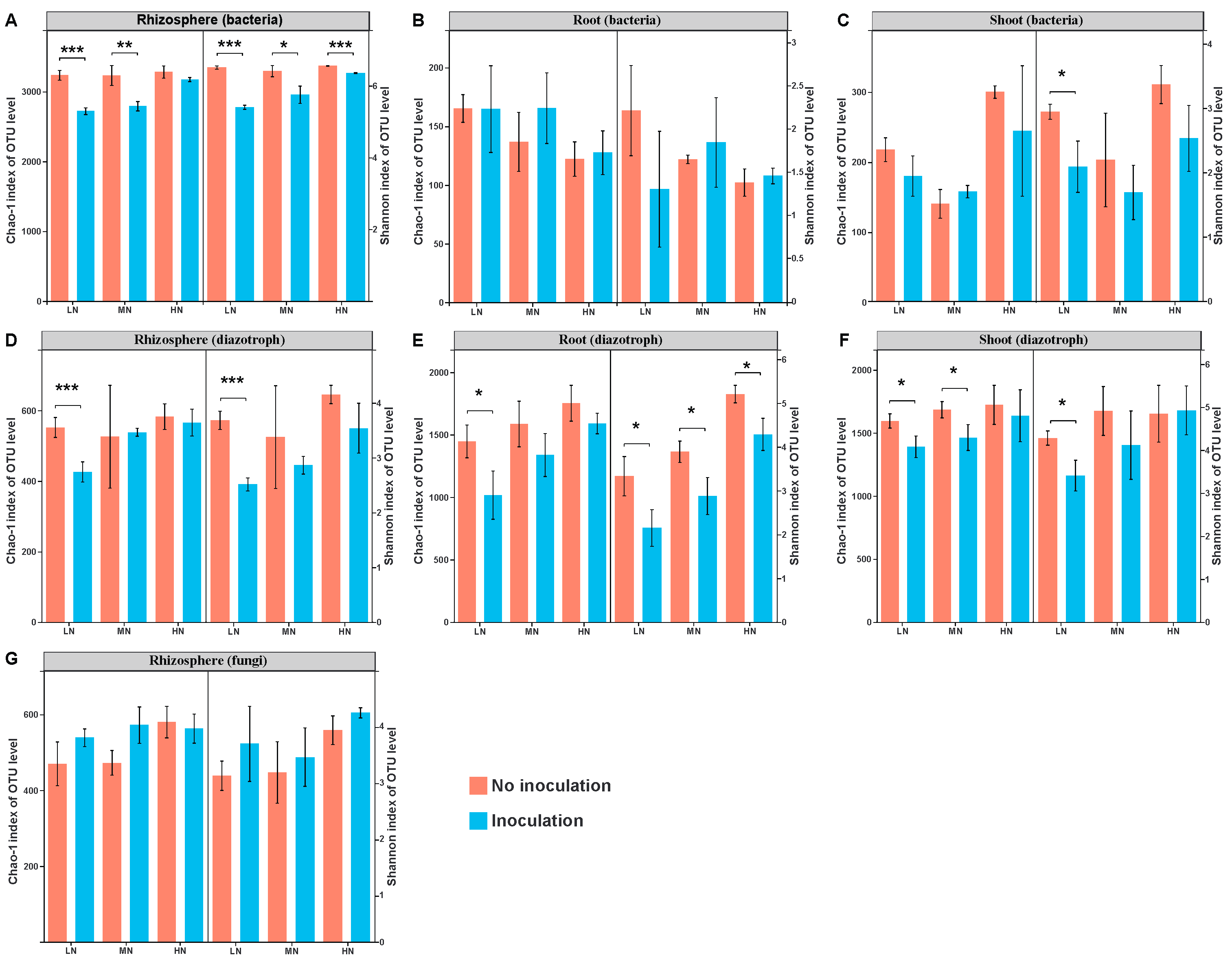
2.2. Microbial Diversity

Samples of the maize rhizosphere, root and shoot were collected. MiSeq amplicon sequencing of each of the 16S rRNA, nifH gene and ITS region from 18 rhizosphere samples was carried out in order to characterize the rhizosphere bacterial, diazotrophic and fungal communities, respectively. Similarly, the 16S rRNA and nifH genes from 18 root and 18 shoot samples were MiSeq amplicon sequenced, respectively. The Good’s coverage (Table S2) values were in the range of 0.974–0.978 (the rhizosphere bacteria), 0.998–0.999 (the root bacteria), 0.997–1.000 (the shoot bacteria), 0.994–0.996 (the rhizosphere diazotrophs), 0.986–0.993 (the root diazotrophs), 0.986–0.993 (the shoot diazotrophs) and 0.998–0.999 (the rhizosphere fungi), respectively. These data indicate that the current numbers of sequences were enough to reflect maize microbiome diversity. The rarefaction curves (Figure S1) also suggested that the microbial profiles were sufficient to represent the microbial communities.
Microbial community α-diversity, as evaluated by the Chao-1 and Shannon indices, is shown in Figure 3. The richness (Chao-1 indices) and diversity (Shannon indices) of bacteria, diazotrophs and fungi were calculated based on the rarefied sequences. Inoculation with P. triticisoli BJ-18 significantly reduced the bacterial Chao-1 richness and Shannon diversity in the rhizosphere under low N, medium N and high N levels (t-test, p < 0.05, hereafter), except Chao-1 under high N (Figure 3A). There were no significant effects on Chao-1 richness and Shannon diversity of the root endophytic bacteria between the inoculation and non-inoculation treatments under three N levels (Figure 3B). Inoculation significantly reduced the Shannon diversity of the shoot endophytic bacterial community (Figure 3C). Similarly, inoculation reduced the Chao-1 richness and Shannon diversity of diazotrophic communities in the rhizosphere, root and shoot, and significant levels were observed in low N (Figure 3D–F). There were no significant differences between the inoculated and uninoculated treatments in the fungal community of the rhizosphere (Figure 3G).
Microbial community β-diversity, as evaluated by principal coordinate analysis (PCoA), is shown in Figure 4. The PCoA and ANOSIM analyses clearly show that inoculation with P. triticisoli BJ-18 led to variation in the structure of the bacterial communities in the rhizosphere, root and shoot under both low N and medium N levels (Figure 4A–C). Similar results were obtained in the diazotrophic and fungal communities (Figure 4D–F).

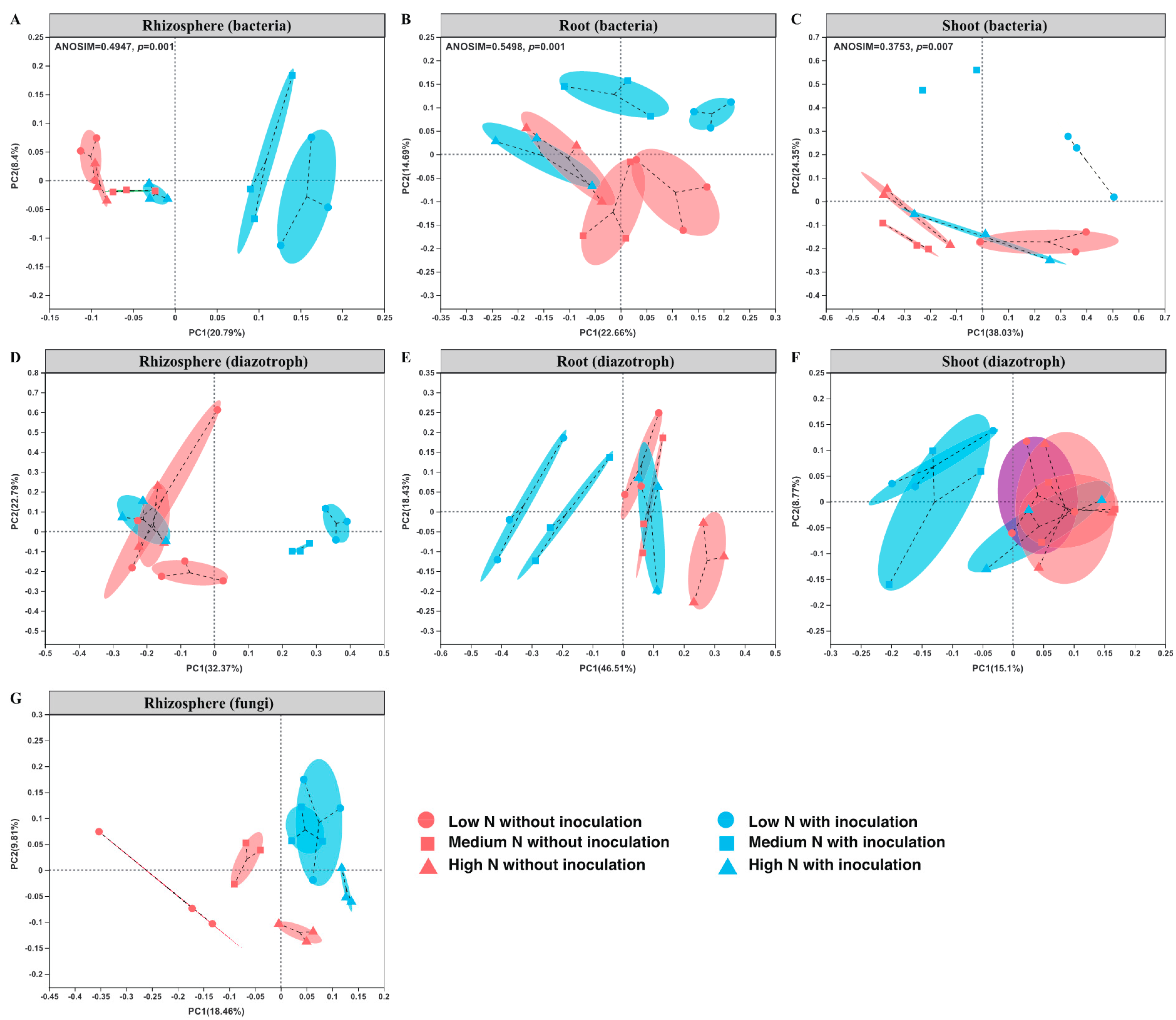
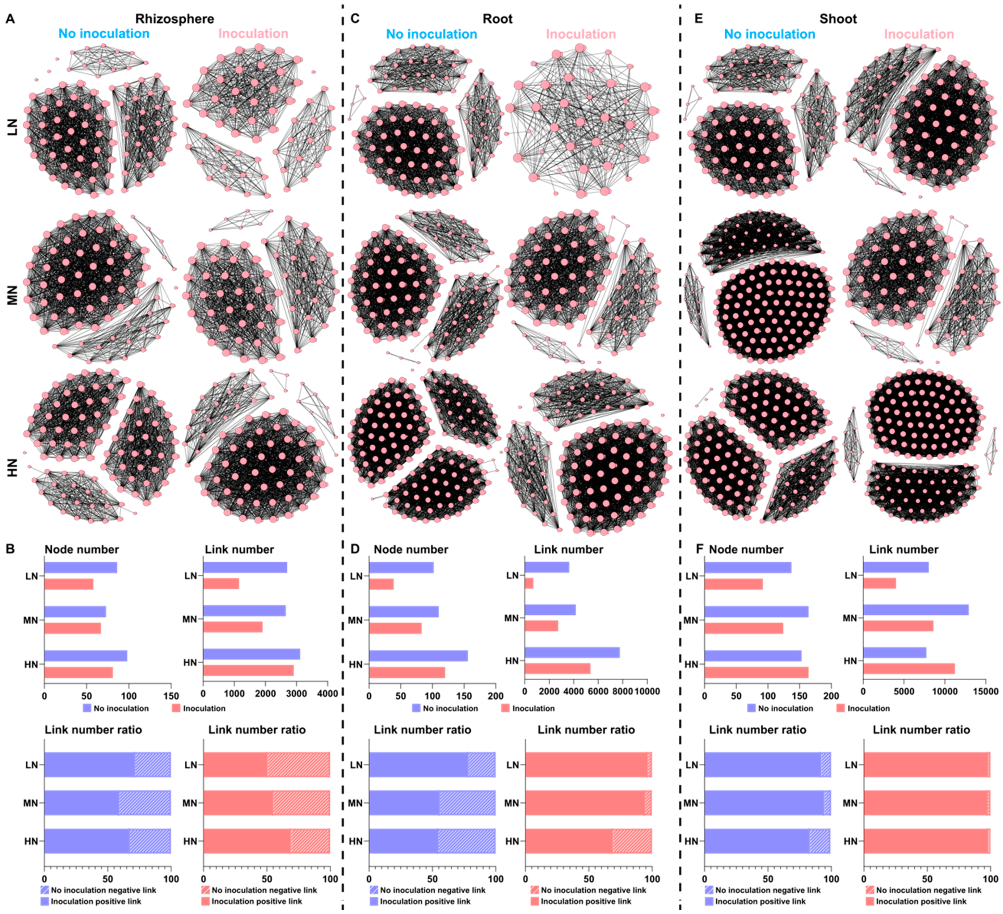
2.3. Bacterial Communities in the Rhizosphere and Root/Shoot Endosphere

We found that there are obvious differences in the composition and diversity of the bacterial communities across the rhizosphere and root/shoot endosphere. No matter whether maize was inoculated with the P. triticisoli BJ-18 cells or not, the abundance and diversity of the bacterial phyla in the rhizosphere were much higher than in maize root/shoot endospheres (Figure 5A–C). The results are consistent with the observation that microbial population decreases from the rhizosphere to the endosphere [10]. In the non-inoculated treatments, Proteobacteria, Actinobacteria, Acidobacteria, Chloroflexi, Bacteroidetes, Firmicutes, Gemmatimonadetes, Planctomycetes, Nitrospirae and Cyanobacteria, in rank order, were the abundant phyla in the rhizosphere (Figure 5A), whereas Proteobacteria was mostly dominated in both root and shoot endospheres with the minor phyla Bacteroidetes, Actinobacteria and Firmicutes (Figure 5B,C). We found that inoculation with the P. triticisoli BJ-18 cells altered the structures of the bacterial microbiomes across all three compartments under both low N and medium N fertilizer conditions. Notably, inoculation enriched the Firmicutes, whose members include the Bacillus and Paenibacillus genera across all three compartments, especially in the shoot endosphere.
Pseudomonas (Proteobacteria phylum) was significantly dominated across three compartments. Notably, Pseudarthrobacter (Actinobacteria phylum) was the most abundant genus in the rhizosphere (Figure 5D), whereas Pantoea (Proteobacteria phylum) was mostly dominated in both root and shoot endospheres (Figure 5E,F). The data suggested that each habitat of the rhizosphere, root and shoot had its bacterial community, supporting that different plant organs were associated with different microbial communities [23]. Inoculation significantly increased the relative abundances of Pseudomonas (low N and medium N), Salinimicrobium (low N and medium N), Bacillus (low N, medium N, and high N), Lysobacter (low N and high N) and Devosia (low N) in the rhizosphere. Whereas Pseudomonas (low N) within root endosphere and Bacillus (low N and medium N) within shoot were mostly enriched, especially under low N level. It is well known that many members of Pseudomonas and Bacillus are PGPR used as biofertilizer or biocontrol agents.

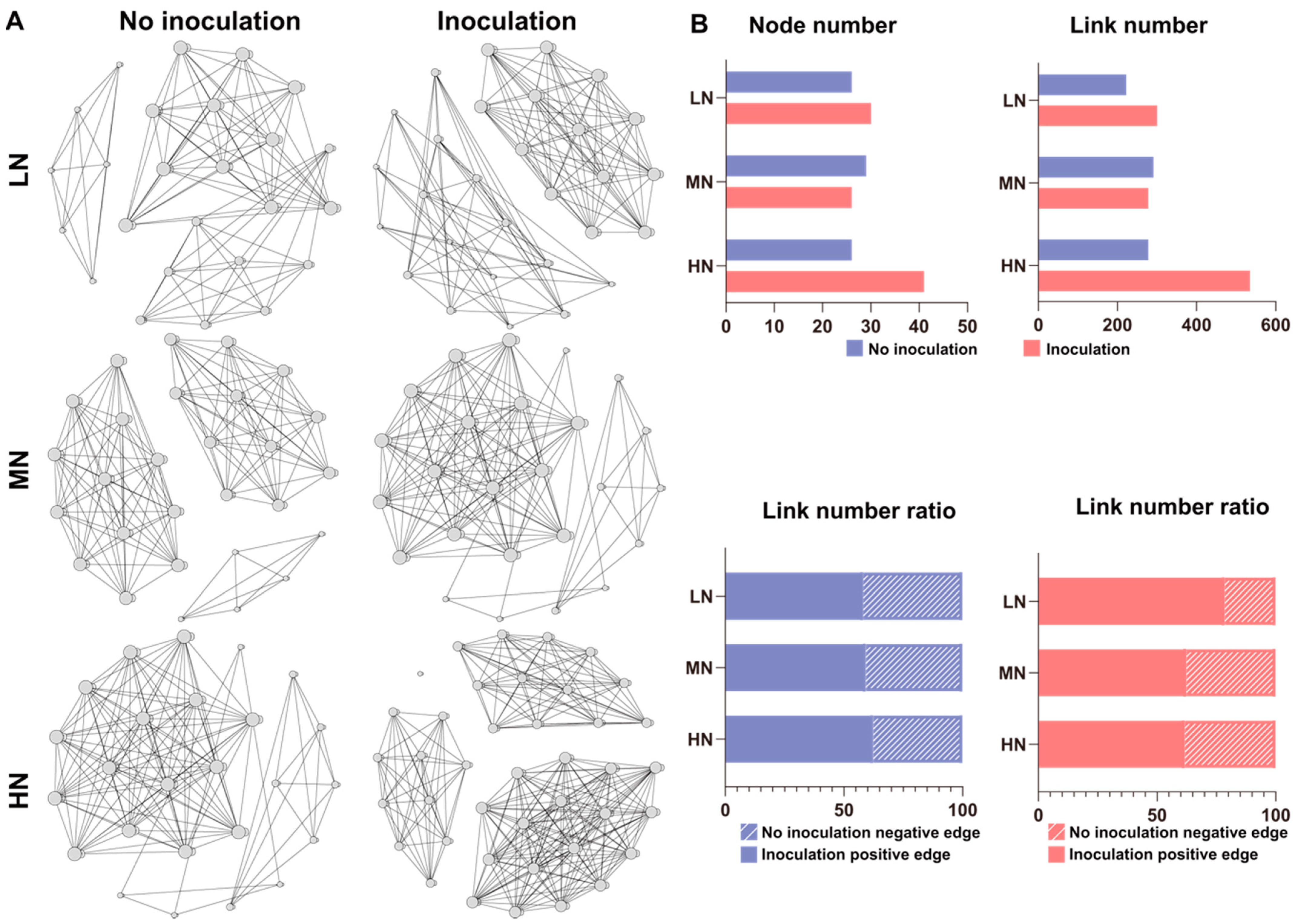
2.4. Diazotrophic Communities in the Rhizosphere and Root/Shoot Endosphere

We analyzed the diazotrophic communities in the rhizosphere and root/shoot endosphere and found that there were obvious variations in the composition and diversity of the diazotrophic taxa across these compartments; also, inoculation significantly altered the abundance and diversity of diazotrophs across these compartments. The data were consistent with the results obtained in the bacterial communities among these compartments. The rhizosphere was significantly dominated by the phylum Proteobacteria, which was followed by Cyanobacteria, Verrucomicrobia and Firmicutes, whereas both root and shoot endospheres were mostly dominated by Proteobacteria (Figure 6A–C). Inoculation obviously increased the relative abundances of Proteobacteria (low N) in the rhizosphere, while inoculation significantly enriched Firmicutes within both the root (low N and medium N) and shoot (low N, medium N, and high N).
The dominant diazotrophs at the genus level in the rhizosphere mainly included Klebsiella, Bradyrhizobium, Azotobacter, Azohydromonas, Skermanella, Trichormus and Azoarcus, followed by Paenibacillus, Leptothrix and Nostoc, in rank order (Figure 6D). Whereas, both the root and shoot endospheres were dominated by both genera Klebsiella and Paenibacillus (~90% in relative abundance), followed by Azotobacter, Skermanella, Azospirillum, Azoarus, Azohydromonas, Decloromonas, Bradyrhizobium and Methylobacter (Figure 6E,F). Klebsiella, a member of Proteobacteria phylum, was the top genus dominated across all three compartments. It is well known that many members of Klebsiella, Azotobacter, Azospirillum, Azoarus and Bradyrhizobium are N2-fixing bacteria. Notably, inoculation significantly enriched Klebsiella (low N and medium N) in the rhizosphere. Whereas the relative abundance of Paenibacillus was significantly increased by inoculation within the root (low N and medium N) and shoot (low N, medium N, and high N). Other diazotrophs, such as Azotobacter, were also significantly enriched across all the compartments. The influences of inoculation on the composition of the diazotrophic communities were gradually weakened as the N level increased generally, consistent with the findings that the colonization rate of P. triticisoli BJ-18 was controlled by soil N status.

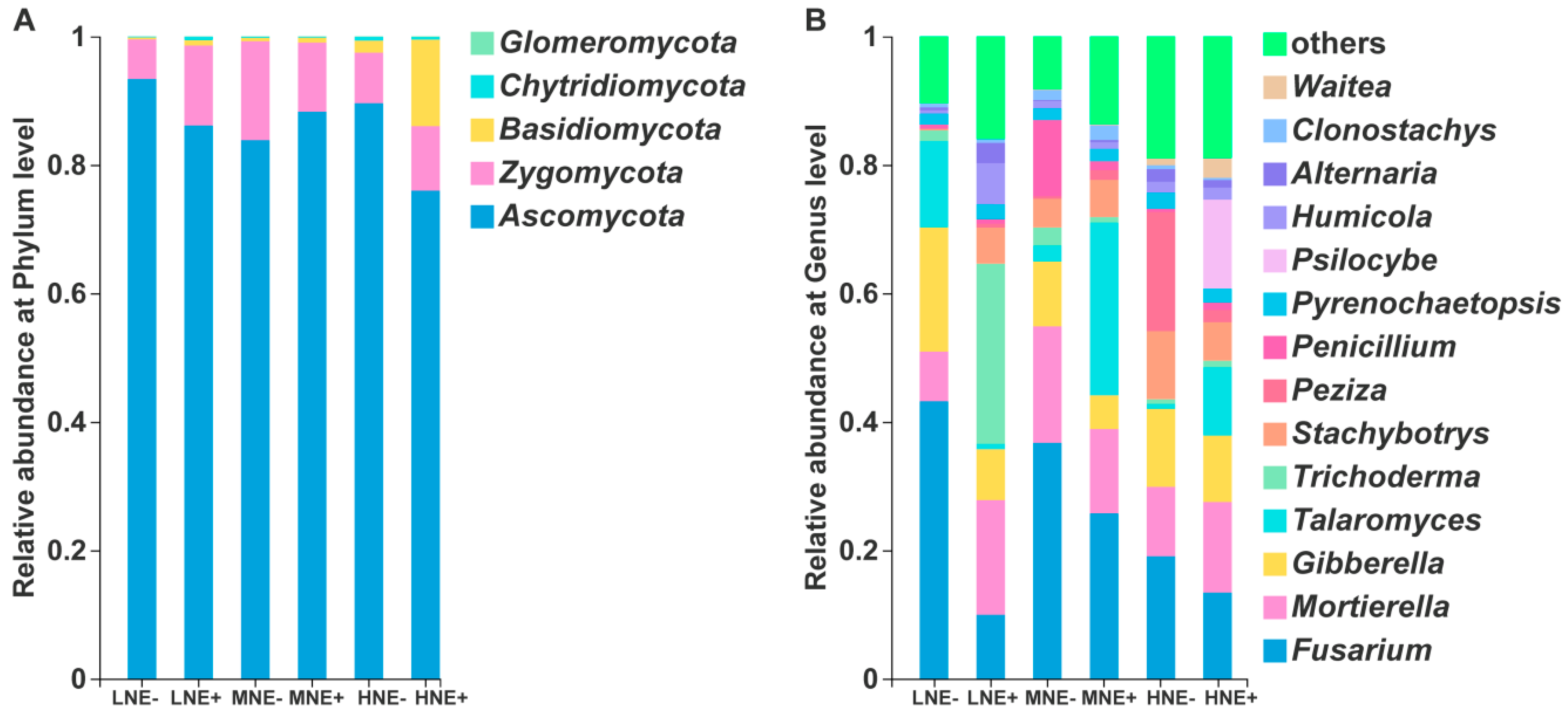
2.5. Fungal Community in the Maize Rhizosphere

In the rhizosphere fungal community, the phyla Ascomycota, Zygomycota and Basidiomycota, in rank order, were abundant (Figure 7A). At the genus level, the ten most abundant fungal genera were Fusarium, Mortierlla, Gibberella, Talaromyces, Stachybotrys, Trichoderma, Peziza, Penicillium, Psilocybe and Humicola, in rank order (Figure 7B). Inoculation significantly decreased the relative abundances of plant pathogen Fusarium (low N: p < 0.05) and Gibberella (low N: p < 0.01) under low N level.

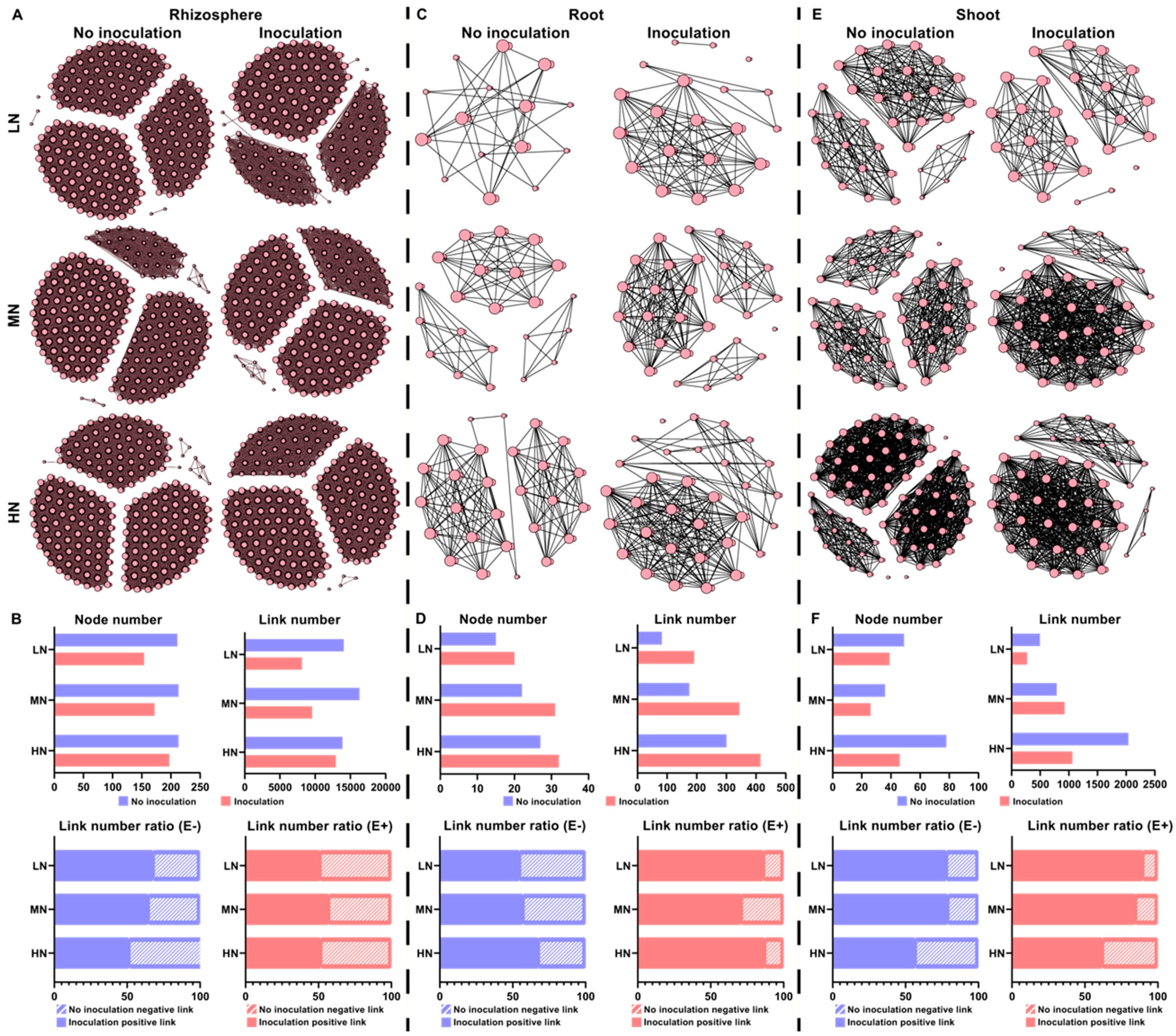
2.6. Microbial Interaction Networks

The above results provide important insight into how a single taxon of a microbial community responds to P. triticisoli BJ-18 inoculation. Therefore, a multitude of direct and indirect interactions that occur in the bacterial, diazotrophic and fungal communities of the rhizosphere, root and shoot were further investigated (Figure 8, Figure 9 and Figure 10). In the rhizosphere, the bacterial, diazotrophic and fungal interactions changed little from low N to high N, as indicated by the connections between the nodes and links (Figure 8A,B, Figure 9A,B and Figure 10A,B). However, obvious changing patterns were observed in the root and shoot (Figure 8C–F, Figure 9C–F and Figure 10C–F). The bacterial interactions were stronger in the rhizosphere than those in the root and shoot (Figure 8), while the opposite results were observed in the diazotrophic communities (Figure 9). In the rhizosphere and endosphere, the potential interactions of the bacteria and diazotrophs in the P. triticisoli BJ-18-inoculated treatments were weaker than those in the uninoculated treatments, particularly in the low N level (Figure 8 and Figure 9). However, P. triticisoli BJ-18 inoculation promoted fungal interactions in the high N level (Figure 10). P. triticisoli BJ-18 inoculation reduced the rhizosphere bacterial and diazotrophic positive edge number ratios, while it increased the endosphere bacterial and diazotrophic and rhizosphere fungal positive edge number ratios.

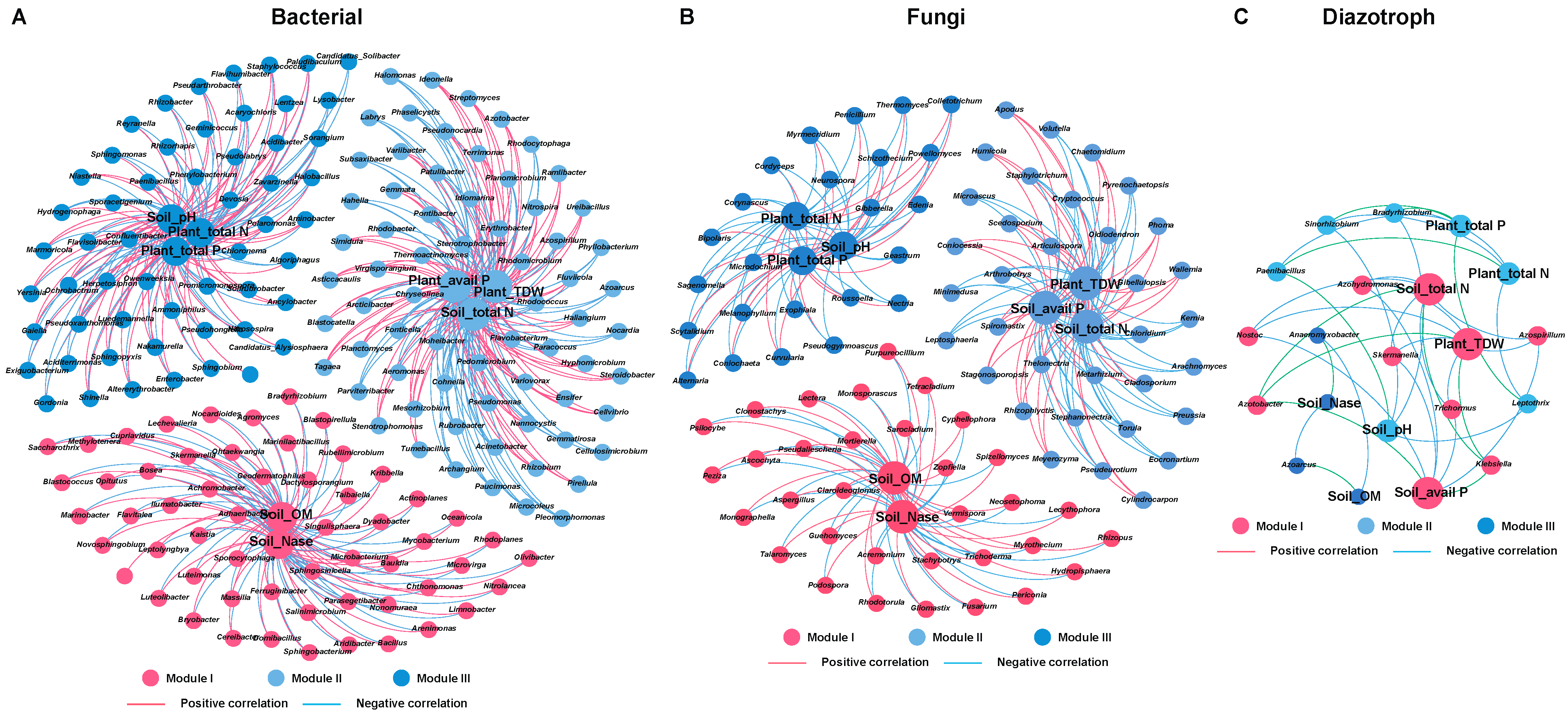
2.7. Relationships between Microbial Community and Environmental Variables in the Rhizosphere and Root/Shoot Endosphere of Maize

The effects of inoculation with P. triticisoli on the soil properties and maize biomass and nutrition were investigated for the rhizosphere soil and maize seedling samples collected on Day 35 after planting. Nitrogenase activity of the rhizosphere was measured by using the acetylene reduction method, and the N2 fixation rate was performed by using the 15N2 incorporation assay. Compared to non-inoculation treatments, inoculation with P. triticisoli BJ-18 significantly increased the nitrogenase activities (Figure 11A) and N2 fixation rates (Figure 11B) under low N and medium N levels. Inoculation increased the soil pH, total N, organic matter and available P (Table S3). Similarly, the N and P contents of maize seedlings were increased (Table S4). After application of P. triticisoli BJ-18, the dry weights (biomass) of the root, shoot and total plant under all N levels were increased by 25.2%, 23.3% and 23.5%, respectively, under a low N level. Generally, the effects of inoculation on the soil and plant properties were most obvious under low N and more obvious under medium N than under high N.
To explore the relationships between microbial community structures and environmental factors (soil properties: pH, organic matter, total N, available P and plant properties: dry weight, N content and P content), a redundancy analysis (RDA) and Spearman’s correlation analysis were conducted. The RDA ordination showed that soil available P was the important factor in influencing the rhizosphere bacterial community, which was followed by soil total N (Figure 12A, Table S5). Root dry weight was the key factor in influencing root bacterial community (Figure 12B, Table S6). Whereas total N and total P were the major factors in influencing shoot bacterial community (Figure 12C, Table S6). In the rhizosphere diazotrophic community, soil available P was the major influencing factor, followed by soil nitrogenase activity (Figure 12D, Table S5). In the root endophytic diazotrophic community, root dry weight was the only significantly influencing factor (Figure 12E, Table S6). In the shoot endophytic diazotrophic community, the influence factors were shoot total P, dry weight and total N (Figure 12F, Table S6). In the rhizosphere fungal community, plant total dry weight was the major influencing factor, followed by soil total N and plant total P content (Figure 12G, Table S5).
In the microbial communities of rhizosphere, the co-occurrence networks of environment–biology were further analyzed (Figure 13). Many bacterial genera in the rhizosphere, such as Pseudomonas, Lysobacter and Bacillus, were significantly and positively correlated with all or part of the soil properties (pH, available P, organic matter and total N) and plant properties (total dry weight and total P) (Figure 13A). In the fungal community, the abundances of Gibberella and Fusarium were significantly negatively correlated with soil nitrogenase activity, plant total N and plant total P (Figure 13B). In the diazotrophic community, the abundances of Paenibacillus, Klebsiella, and Azotobacter were significantly positively correlated with soil total N, soil available P and plant total dry weight (Figure 13C).

3. Discussion

In this study, the diazotrophic P. triticisoli BJ-18 was inoculated into maize grown in soil containing three different levels of N fertilizer. Then we examined the survival of the inoculant P. triticisoli BJ-18 and the effects of this inoculant on the composition and abundance of the bacterial and diazotrophic communities in all three compartments (rhizosphere, root and shoot), but fungi were only measured in the rhizosphere. All of the bacteria, diazotrophs and fungi in the rhizosphere were measured, since the rhizosphere is a large living site for microorganisms. The fungi were only measured, since the population of endophytic fungi in plant tissues is usually low. We found that the P. triticisoli BJ-18 cells survived and propagated in all habitats of the rhizosphere, root and shoot. Inoculation with P. triticisoli BJ-18 not only had influences on the soil and plant properties, but also significantly altered the structures of the bacterial, diazotrophic and fungal communities in all of these habitats. As far as we know, this is the first systematic study on the effects of the application of N2-fixing bacteria on the bacterial, diazotrophic and fungal communities in the rhizosphere and root/shoot endosphere of plants.
Firstly, we found that the P. triticisoli BJ-18 cells effectively colonized and propagated not only in the rhizosphere, but also in the maize root and shoot endospheres. The population densities of P. triticisoli BJ-18 across these compartments were significantly higher under low N than under high N, suggesting that the colonization of maize plants by this bacterium was controlled by the soil N status and consistent with the report that colonization of sugarcane by Acetobacter diazotrophicus was inhibited by high N fertilization [57]. Inoculation with P. triticisoli significantly increased the soil pH, total N, available P, organic matter, N2-fixation rate and nitrogenase activity. The N and P contents and biomass of the maize seedlings were also significantly enhanced by the inoculation. These good effects produced by inoculation of P. triticisoli BJ-18 are mainly due to the roles of P. triticisoli BJ-18 and the reshaped microbial community. P. triticisoli BJ-18 can provide N and Fe for plants by fixing nitrogen and by producing siderophores. We deduce that nitrogen fixation is more important in promoting plant growth than indolic acids, since P. triticisoli BJ-18 produces very low amounts of IAA [54]; also, we deduce that the altered microbial community play a very important role in improving soil and plant features. Especially, inoculation significantly decreased the relative abundances of the plant pathogen Fusarium. The antimicrobial compounds produced by P. triticisoli BJ-18 may be a contributor to the reduction in Fusarium density [54]. The enhanced populations of the diazotrophs and effective bacteria should be the contributors to improvement of the soil and plant properties. Our previous study showed that the ability of phosphate solubilization by P. triticisoli BJ-18 is very low [54]. The increased phosphorus in soil and plants may be due to the roles of phosphate solubilization by other effective microbes in the altered microbial community.
We comparatively analyzed the bacterial communities in the rhizosphere, root/shoot endosphere of maize seedlings grown in different N levels of fertilizer, with inoculation or without inoculation. No matter if the maize seedlings were inoculated or not, the abundance and diversity of the bacterial community in the rhizosphere were significantly higher than in root/shoot endospheres, supporting that the root provides an active and robust selection of bacteria for entering into plant tissues from the soil. Interestingly, the shoot endosphere was much higher than the root endosphere in diversity of the bacterial taxa, consistent with the reports that each plant tissue has its microbiome [10]. Notably, Proteobacteria was the dominant phylum across all the compartments, especially in root/shoot endospheres. In the non-inoculation treatments, the major bacterial phyla in the rhizosphere comprised Proteobacteria, Actinobacteria, Acidobacteria, Chloroflexi, Bacteroidetes, Firmicutes and Gemmatimonadetes. These major bacterial phyla observed in our study were also found in the bacterial community of the maize rhizosphere [38,58,59]. Our study revealed that the ten most dominant bacterial genera in the rhizosphere bacterial community were Pseudarthrobacter, Pseudomonas, Nitrospira, Salinimicrobium, Bacillus, Lysobacter, Devosia, Gaiella, Nocardioides and Marmoricola. Our results were a little different from the reports that Chitinophaga, Flavisolibacter, Nitrospira, Pseudomonas and Streptomyces were predominantly found in the maize rhizosphere and bulk soil [38]. We deduced that the difference in compositions of the bacterial communities may be due to soil type, which was one of the major factors in shaping the microbial community. Our findings that Pseudarthrobacter, a member of Actinobacteria phylum, was the dominant genus in the maize rhizosphere was also observed in beet soils [60]. Some members of the Pseudarthrobacter genus could degrade methane and benzoate [61]. Unlike in the rhizosphere bacterial community, Pantoea was the most abundant genus in both root and shoot bacterial communities. It was reported that Pantoea spp. as endophytes were carried by seeds of rice, wheat and crabgrass [62,63,64]. Some members of Pantoea, belonging to the PGPR, were used as an effective biocontrol agent, whereas Pantoea stewartii was a pathogen of maize [65,66,67]. Inoculation with P. triticisoli significantly enriched Pseudomonas in both the rhizosphere and root endosphere under all N levels, especially under low N and medium N levels. Whereas inoculation obviously increased the relative abundance of Pseudomonas in the shoot endosphere under high level and significantly enriched Bacillus in the shoot endosphere under low N and medium N levels. Many members of both the Pseudomonas and Bacillus genera as PGPR are widely used as biofertilizers or biocontrol agents [14,15]. The relative abundances of Salinimicrobium, Lysobacter and Enterobacter were also significantly increased by inoculation with P. triticisoli. Some members of Lysobacter were used as biocontrol agents against fungal diseases, since they could secrete antimicrobial compounds and extracellular enzymes against bacteria, fungi, oomycetes and nematodes [68,69]. We found that the abundances of Pseudomonas and Bacillus were significantly positively correlated with soil total N, soil available P, plant total P and plant biomass.
Although diazotrophs as PGPB are in common use as inoculants to improve crop yield and to reduce the consumption of chemical nitrogen fertilization, the studies on the diazotrophic community in the rhizosphere, especially in the root/shoot endosphere, are very few. Here, our study revealed that the abundance and diversity of the diazotrophic communities in the rhizosphere is much higher than in root/shoot endospheres, consistent with the observation in the bacterial community. However, the composition of the diazotrophic community structure across all the compartments was much simpler than that of the bacterial community structure, supporting that the diazotrophic community is only a small part of the bacterial community. Inoculation significantly enriched Klebsiella in the rhizosphere, some members (e.g., Klebsiella oxytoca) of which are well-known N2-fixing bacteria. Inoculation also significantly increased the relative abundance of the genus Paenibacillus across all the compartments, especially in the root/shoot endospheres, consistent with the observation that the inoculant P. triticisoli BJ-18 cells survived and propagated in all the habitats. In addition to P. triticisoli BJ-18, the diazotrophic Paenibacillus includes many members, such as P. polymyxa, P. macerans, P. azotofixans, P. sabinae, P. zanthoxyli, P. forsythiae, P. sonchi, P. sophorae, P. jilunlii, P. taohuashanense and P. brasilensis [53,70,71,72,73,74,75,76,77,78]. Certainly, the P. triticisoli BJ-18 cells were the main part of the Paenibacillus population in the inoculated rhizosphere and maize tissues. Other members, such as diazotrophic P. polymyxa and P. brasilensismay, also contribute to the enrichment of Paenibacillus across the compartments. Moreover, the other dominant genera, such as Azospirillum, Azotobacter, Bradyrhizobium, Azoarcus, Azohydromonas and Skermanella, were detected in the diazotrophic communities in the rhizosphere or in root/shoot endosphere. Our results are consistent with the reports that Azohydromonas, Skermanella, Azotobacter and Bradyrhizobium were the dominant genera in the diazotrophic community in a legume–oat intercropping soil [79]. Many members of these genera (e.g., Azospirillum brasilense, Azotobacter chroococcum) are used as biofertilizer to improve crop yield and to reduce the consumption of chemical nitrogen fertilization [80,81]. Bradyrhizobium is generally symbiotic with legumes, but some members of Bradyrhizobium are also free-living N2-fixers. Bradyrhizobium was also found in the rhizosphere of other non-legumes, such as maize, sugarcane and citrus [82,83,84]. We found that the diazotrophic communities across all the compartments included Azoarcus, which is also an important endophyte in rice [85,86]. Our data showed that the abundance of Paenibacillus and other diazotrophic genera, such as Klebsiella, Azotobacter, Bradyrhizobium and Azoarcus, were positively correlated with soil available P, nitrogenase activity, plant biomass and plant P and plant total N, suggesting that these diazotrophs are beneficial for plant growth. It is well known that some members of Pseudomonas, such as P. stutzeri A1501, are N2-fixng bacteria [41]. However, our study found that Pseudomonas was the main genus in the bacterial communities but was not dominant in the diazotrophic communities across all the compartments. The reason why Pseudomonas was not dominant in the diazotrophic communities may be due to the diazotrophic Pseudomonas species and strains forming only a very small part of the large Pseudomonas group.
Furthermore, the effects of inoculation with P. triticisoli BJ-18 on the rhizosphere fungal community were here investigated. Inoculation decreased the population density of fungi in the rhizosphere, consistent with the reports that application of Bacillus amyloliquefaciens NJN-6 decreased the fungal abundance in the rhizosphere soil of banana [87]. Especially, inoculation significantly decreased the relative abundances of Fusarium and Gibberella under a low N level, suggesting that P. triticisoli BJ-18 played an important role in inhibiting these plant pathogens. Fusarium and Gibberella are the anamorphic and teleomorphic state of the fungus, respectively [88]. Fusarium graminearum and Fusarium moniliforme caused Gibberella stalk rot, one of the most destructive soil-borne diseases of maize [89]. Many Fusarium species are pathogens of lots of plants, such as maize, wheat, cucumber and banana [88]. Some Fusarium species are not plant pathogens and even they are beneficial to plants. However, we could not distinguish which Fusarium species is pathogenic or beneficial in this study. The current study showed that the abundances of Fusarium and Gibberella were negatively correlated with soil available P, soil total N, plant total N, plant total P and plant biomass. Thus, we deduce that a reduction in the relative abundance of Fusarium and Gibberella in the maize rhizosphere by inoculation might be due to the antimicrobial substances produced by P. triticisoli BJ-18 [54]. In addition, other microbes, such as Pseudomonas and Bacillus, may also play roles in inhibiting plant pathogens, since these microorganisms were antagonistic microbes against Fusarium wilt [39,90].

4. Materials and Methods

4.1. Plant Plantation and Inoculation with P. triticisoli BJ-18 Cells

P. triticisoli BJ-18 cells were cultured in Luria Bertani broth medium [91] overnight at 30 °C, and then were harvested by centrifugation at 4000× g for 5 min, and adjusted to 108 cells mL−1 with sterile normal saline solution. The concentration 108 cell mL−1 was calculated by spectrophotometry and spread plate method.
The soil used in the pot experiments was low N-content sandy loam that was topsoil (0–20 cm depth) taken from the Shangzhuang Experimental Station of China Agricultural University, Beijing, China (40°08′12.15″ N, 116°10′44.83″ E, 50.21 m above sea level). The collected soil was air-dried at room temperature, and then was screened by a 10-mesh sieve to remove plant residues and reduce the heterogeneity of the soil. Before planting maize, P (50 mg Na2HPO4 per kg soil) and K (17 mg KCl per kg soil) were applied to the soil as base fertilizers. N fertilizers ((NH4)2SO4) were applied to the soil as base fertilizer at three N levels: high N (250 mg N kg−1 soil), medium N (166 mg N kg−1 soil) and low N (83 mg N kg−1 soil).
Maize seeds (Zea mays L., genotype hybrid Zhengdan 958 Henan Shangke Seed Co., Ltd., Shangqiu, China) were surface-sterilized with 10% sodium hypochlorite (NaClO) for 10 min, washed with sterile deionized water three times, and sprouted on sterile Petri dishes containing moist filter papers at room temperature (25 °C). After germination, uniform and vigorous seedlings were selected and divided into two groups at random. For the inoculated group (E+), the maize seedlings were soaked in the suspension (108 cells mL−1) of P. triticisoli BJ-18 for 30 min to facilitate colonization. The non-inoculated group (E−) indicated that the maize seedlings were soaked in sterile saline solution. Then, the inoculated seedlings or non-inoculated seedlings were transplanted into square plastic pots (length of 55 cm; width of 30 cm; high of 17 cm) and 6 maize seedlings were planted in each per pot. On Day 7, 120 mL of the bacterial suspension (108 cells mL−1) was applied to the pot containing the inoculated seedlings, and 120 mL of deionized water was applied to the pot containing the non-inoculated seedlings as the non-inoculated control. Each pot contained about 7.5 kg soil that was collected and supplemented with fertilizers of K, P and N as the base fertilizer, as described above. The seedlings were regularly watered (tap water) to 40% relative soil moisture by the weighing method [92] every 5 days. Three replications of each treatment were conducted in the greenhouse under optimum conditions (15 h light/9 h dark cycle and 25 °C–30 °C/15 °C–20 °C day/night temperature).

4.2. Sample Collection

For determination of survival and propagation of P. triticisoli BJ-18 in the maize rhizosphere and root/shoot endosphere, the maize seedlings were harvested from each treatment on Days 7, 12, 17, 27, 32 and 35 after transplanting, respectively. Whole maize seedlings were uprooted and separated into shoots and roots, both of which were then washed with deionized water to remove the adhering soil particles. In order to sterilize the outer surface of maize tissues, the roots and shoots were immersed in 70% ethanol for 30 s and subsequently in 2% NaClO solution added with Tween 80 (one droplet per 100 mL solution) for 10 min [93]. Subsequently, they were rinsed three times with sterile distilled water for 1 min. Finally, 0.1 mL of the final wash was spread on Luria Bertani agar plates to check whether the microorganisms were completely removed. These maize roots and shoots were immediately frozen in liquid N and then maintained at −80 °C for DNA extraction. At the same time, the tightly adhering soil on the maize roots was gathered as rhizosphere soil on Days 7, 12, 17, 27, 32 and 35 and then maintained at −80 °C.
The samples of the rhizosphere soil and maize root and shoot collected at Day 35 were used to extract DNA for analysis of the bacterial, diazotrophic and fungal communities. For assaying plant biomass (dry weight), the plant tissues were oven-dried at 105 °C for 30 min and then dried at 65 °C until constant weight.

4.3. Soil and Plant Physicochemical Property

Appropriate amount of soil and plant samples were digested by an H2SO4-H2O2 mixture at 370 °C, and then the N concentration was determined using the modified Kjeldahl method [94] and P concentration was determined using the standard method [95]. Available P was extracted with resin and measured according to the description [96]. The organic matter was measured according to the description [97]. Soil pH was measured using SevenMulti (Mettler-Toledo GmbH, Schwerzenbach, Switzerland) with soil:water of 1:1 [98].

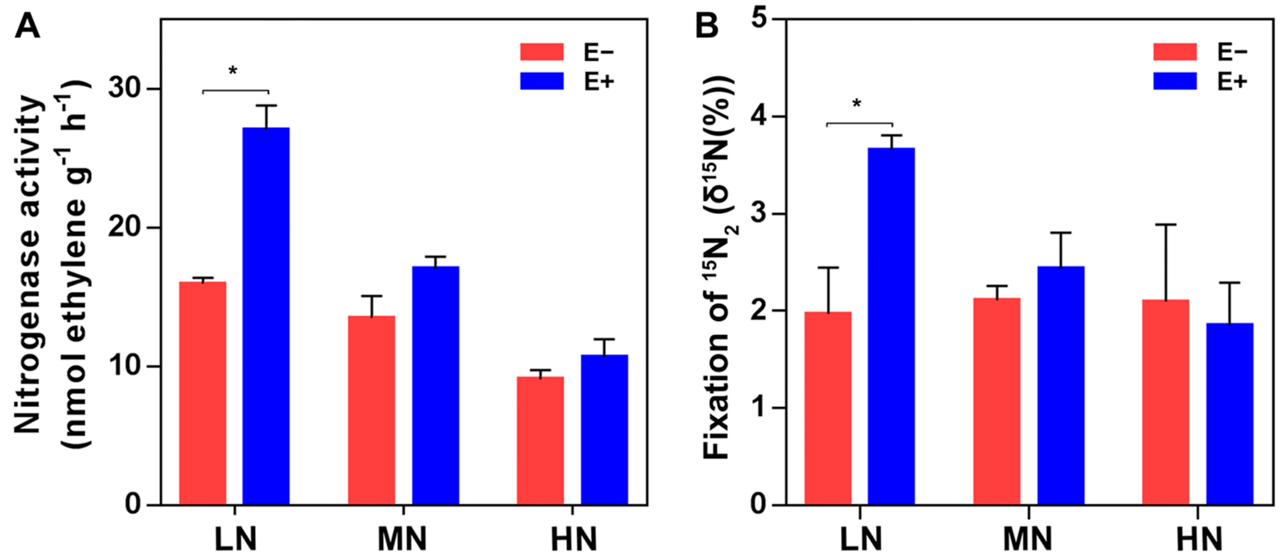
4.4. Nitrogenase Activity Assay

Nitrogenase activity of the rhizosphere soil collected on Day 35 was measured by using the acetylene reduction method [99,100], with slight modification. Specifically, 2 g rhizosphere soil was added to a 26 mL glass jar fitted with septa for gas sampling and then sterile deionized water was added to 240% moisture content. After incubating the cultures for 24 h at 28 °C, with shaking at 180 rpm, the air in the glass jar was replaced with argon gas, and 10% (v/v) of the headspace argon gas in the tube was replaced with acetylene gas. After incubation for 2 h, 100 μL of gas from the tube was collected to measure the ethylene production using a GC-2010 Plus gas chromatograph (Shimadzu Corp., Kyoto, Japan).

4.5. 15N2 Incorporation Assay

A 15N2 incorporation assay was used to measure the incorporation of N2 gas into organic N by the N2-fixing microbes in the soil. Firstly, 2 g rhizosphere soil was added to a 26 mL glass jar fitted with septa for gas sampling and then sterile deionized water was added to 240% moisture content. After incubating the cultures for 24 h at 28 °C, with shaking at 180 rpm, the air in the glass jar was replaced with argon gas, and 10% (v/v) of the headspace argon gas in the tube was replaced with 15N2 (99%+, Shanghai Engineering Research Center for Stable Isotope, Shanghai, China). After 72 h of incubation at 28 °C, the soil was collected, oven-dried, ground, weighed and sealed into tin capsules. 15N enrichment (δ15N value) of soil was determined using a DELTA V Advantage isotope ratio mass spectrometer (Thermo Fisher Scientific, Inc., Waltham, MA, USA).

4.6. Extraction of DNA

Total DNA was extracted from the rhizosphere soil or plant tissue samples using the FastDNA® SPIN Kit for soil (MP Biomedicals, Santa Ana, CA, USA), according to the manufacturer’s recommendation. DNA concentration and quality were measured using a spectrophotometer (Nanodrop 2000, Thermo Fisher Scientific, Waltham, MA, USA).

4.7. Quantitative-PCR (qPCR) Analysis

The qPCR method was used to quantify the abundance of total bacterial 16S rRNA [101], fungal ITS [101], total diazotrophic nifH [102] and P. triticisoli BJ-18 nifH gene. The primers used for the qPCR are listed in Table S1. The fragments of 200 bp (bacteria), 300 bp (fungi), 450 bp (total diazotrophic bacteria) and 217 bp (P. triticisoli BJ-18) were amplified by conventional PCR, respectively. The products were ligated to PMD 19-T vector (TaKaRa, Otsu, Japan), and recombinant plasmids were obtained using a TIANprep Mini Plasmid Kit (Tiangen Biotech (Beijing) Co., Ltd., Beijing, China). Then the standard curve (R2 > 0.99) was generated with a dilution range of the recombinant plasmids from 1 × 101 to 10 × 107 copies. qPCR was performed using the 7500 Real-Time PCR detection system (Applied Biosystems, Foster City, CA, USA) and the program was as follows: 95 °C for 1 min, followed by 40 cycles of 94 °C for 15 s, 55 °C for 34 s and 72 °C for 15 s [103]. The 20 μL reaction mixture contained the SYBR® Premix Ex Taq™ (Takara, Kyoto, Japan), primer pair, DNA and ddH2O. Each treatment had three biological replicates, with three technical replicates for each biological replicate. Gene copy numbers were calculated based on the standard curves. Each treatment had three biological replicates, with three technical replicates for each biological replicate.

4.8. Amplification and Sequencing of 16S rRNA, nifH Gene and ITS Region

The V4-V5 region of the bacterial 16S rRNA gene was amplified from the DNA isolated from the maize rhizosphere by using the primer pair 515F and 907R [104]. Primers 799F/1392R and 799F/1193R were used for analyzing the endophytic bacteria of the plant tissues [56,105,106]. The universal primers nifHF and nifHR for nifH genes were used to amplify the nifH gene from the rhizosphere DNA and plant genomic-DNA for analyzing the diazotrophic communities of the maize rhizosphere, root and shoot [107]. The primers ITS1F and ITS1R were used for amplification of the fungal ITS region from soil DNA [108]. PCR was carried out using ABI GeneAmp 9700 (Applied Biosystems, Foster City, CA, USA). Then amplicons were measured using QuantiFluor™-ST (Promega, Wisconsin, WI, USA), pooled in equimolar amounts and paired-end sequenced on an Illumina MiSeq platform (Illumina, San Diego, CA, USA) by Majorbio Co., Ltd. in Shanghai, China. Three independent biological replicates were provided for each treatment except for shoot endophytic bacteria (two independent biological replicates). These sequence data have been submitted to the GenBank databases under accession numbers SRP218893, SRP218883, SRP223202.

4.9. Bioinformatic Analysis

Raw reads of bacteria, diazotrophs and fungi were analyzed on the free online platform of Majorbio I-Sanger Cloud Platform (www.i-sanger.com). The sequences of the 16S rRNA gene were aligned against the Silva database, the sequences of the ITS against the Unite database and the sequences of the nifH gene against the functional gene database. Then they were analyzed at the phylum, class, order, family and genus levels by RDP Classifier [109]. The alpha (α) diversity indices of microbial communities were estimated by Mothur [110,111]. Shannon was used for estimating community diversity, Chao-1 for community richness and Coverage for sequencing depth. The differences between groups were performed in R with the ‘’stats” package. Principal coordinate analysis (PCoA) was calculated based on Bray–Curtis matrices and generated in R (RStudio, Inc., Boston, MA, USA) with the “ade4” package (v. 1.7.13) and ANOSIM analysis was performed in R with the “vegan” package (v. 2.5.4).

4.10. Statistical Analysis

The data of the soil physicochemical properties, plant biomass and nutrition, gene copies and α-diversity index were statistically analyzed for differences between groups by two-way analysis of variance in SPSS software version 20 (SPSS Inc., Chicago, IL, USA), and the results with p < 0.05 were considered to be statistically significant.
The interactions of “biology–biology” and “environment–biology” were visualized by a co-occurrence network [112]. In the network, a connected link denotes a significant (p < 0.05) and strong (0.6 < |r| <1) Spearman’s correlation between two variables. The size of each node is proportional to the number of connections (i.e., degree). The thickness of each link is proportional to the absolute value of Spearman’s correlation coefficient. The number of nodes and links, centralization of closeness/betweenness/degree and modularity were output with the igraph package in R [113]. The resulting networks were visualized using the interactive platform Gephi [114].

5. Conclusions

P. triticisoli BJ-18 inoculation altered the structures of the bacterial, diazotrophic and fungal communities in the maize rhizosphere, root and shoot. The bacterial and diazotrophic communities were specific in each compartment, and their abundance and diversity were significantly higher in the rhizosphere than in the root and shoot. Inoculation significantly enriched the Proteobacteria, Bacteroidetes and Firmicutes in the rhizosphere bacterial community, while inoculation obviously increased the relative abundance of the Firmicutes in both root and shoot bacterial communities. Compared to the non-inoculation treatments, the relative abundances of the genera Paenibacillus, Pseudomonas and Bacillus were significantly increased and the relative abundance of Fusarium/Gibberella was decreased by the inoculation. The abundance of Paenibacillus was significantly correlated with plant dry weight, nitrogenase, N2-fixing rate, P solubilization and other properties of the soil and plant. Our study provides insight into the plant growth-promotion mechanisms used by diazotrophs—not only through fixing N, but also by reshaping the microbiome structure.

Supplementary Materials

The following are available online at https://www.mdpi.com/1422-0067/22/3/1460/s1, Figure S1: The rarefaction curves of the bacterial, diazotrophic and fungal communities in the maize rhizosphere, root endosphere and shoot endosphere, Figure S2: The statistical comparison of the relative abundance of bacteria in the rhizosphere (A), roots (C) and shoots (E) at the genus level, Figure S3: The statistical comparison of the relative abundance of diazotroph in the rhizosphere (A), roots (B) and shoots (C) at the genus level, Figure S4: The composition of the diazotrophic communities in the rhizosphere (A), root endosphere (B) and shoot endosphere (C) at genus level, Figure S5: The statistical comparison of the relative abundance of fungi in the rhizosphere at the genus level, Table S1: Sequences of oligonucleotide primers for quantitative PCR, Table S2: Coverage of microbial communities in the maize rhizosphere, root and shoot, Table S3: Soil physicochemical properties, Table S4: Maize biomass and nutrition, Table S5: Environmental factors of Envfit in the rhizosphere microbial community, Table S6: Environmental factors of Envfit in the root and shoot endophytic microbial community.

Author Contributions

Conceptualization, S.C.; methodology, Y.L. and Q.L.; formal analysis: Y.L., Q.L. and S.C.; investigation: Y.L.; Writing—Original draft preparation, Y.L. and S.C.; Writing—Review and editing, S.C.; project administration: S.C.; funding acquisition: S.C. All authors have read and agreed to the published version of the manuscript.

Funding

This work was supported by the National Key Research and Development Program of China (Grant No. 2017YFD0200800) and State Key Laboratory of Agrobiotechnology and College of Biological Sciences (Grant No. 2019SKLAB6-16).

Data Availability Statement

The DNA reads have been deposited at NCBI under the SRA accession no. SRP218893, SRP218883, SRP223202.

Conflicts of Interest

The authors declare no conflict of interest.

References

  1. Berendsen, R.L.; Pieterse, C.M.J.; Bakker, P. The rhizosphere microbiome and plant health. Trends Plant Sci. 2012, 17, 478–486. [Google Scholar] [CrossRef] [PubMed]
  2. Mendes, R.; Garbeva, P.; Raaijmakers, J.M. The rhizosphere microbiome: Significance of plant beneficial, plant pathogenic, and human pathogenic microorganisms. FEMS Microbiol. Rev. 2013, 37, 634–663. [Google Scholar] [CrossRef] [PubMed]
  3. Bakker, P.; Pieterse, C.M.J.; de Jonge, R.; Berendsen, R.L. The soil-borne legacy. Cell 2018, 172, 1178–1180. [Google Scholar] [CrossRef] [PubMed] [Green Version]
  4. Dobbelaere, S.; Vanderleyden, J.; Okon, Y. Plant growth-promoting effects of diazotrophs in the rhizosphere. Crit. Rev. Plant Sci. 2003, 22, 107–149. [Google Scholar] [CrossRef]
  5. Dennis, P.G.; Miller, A.J.; Hirsch, P.R. Are root exudates more important than other sources of rhizodeposits in structuring rhizosphere bacterial communities? FEMS Microbiol. Ecol. 2010, 72, 313–327. [Google Scholar] [CrossRef] [PubMed] [Green Version]
  6. Glick, B.R. Plant growth-promoting bacteria: Mechanisms and applications. Scientifica 2012, 2012, 963401. [Google Scholar] [CrossRef] [PubMed] [Green Version]
  7. Etesami, H.; Maheshwari, D.K. Use of plant growth promoting rhizobacteria (PGPRs) with multiple plant growth promoting traits in stress agriculture: Action mechanisms and future prospects. Ecotoxicol. Environ. Saf. 2018, 156, 225–246. [Google Scholar] [CrossRef] [PubMed]
  8. Rosenblueth, M.; Martinez-Romero, E. Bacterial endophytes and their interactions with hosts. Mol. Plant Microbe Interact. 2006, 19, 827–837. [Google Scholar] [CrossRef] [Green Version]
  9. Hallmann, J.; Quadt, A.; Mahaffee, W.; Kloepper, J. Endophytic bacteria in agricultural crops. Can. J. Microbiol. 1997, 43, 895–914. [Google Scholar] [CrossRef]
  10. Liu, H.W.; Carvalhais, L.C.; Crawford, M.; Singh, E.; Dennis, P.G.; Pieterse, C.M.J.; Schenk, P.M. Inner plant values: Diversity, colonization and benefits from endophytic bacteria. Front. Microbiol. 2017, 8, 2552. [Google Scholar] [CrossRef]
  11. Hardoim, P.R.; van Overbeek, L.S.; van Elsas, J.D. Properties of bacterial endophytes and their proposed role in plant growth. Trends Microbiol. 2008, 16, 463–471. [Google Scholar] [CrossRef] [PubMed]
  12. Backer, R.; Rokem, J.S.; Ilangumaran, G.; Lamont, J.; Praslickova, D.; Ricci, E.; Subramanian, S.; Smith, D.L. Plant growth-promoting rhizobacteria: Context, mechanisms of action, and roadmap to commercialization of biostimulants for sustainable agriculture. Front. Plant Sci. 2018, 9, 1473. [Google Scholar] [CrossRef] [PubMed] [Green Version]
  13. Glick, B.R. Bacteria with ACC deaminase can promote plant growth and help to feed the world. Microbiol. Res. 2014, 169, 30–39. [Google Scholar] [CrossRef]
  14. Lucy, M.; Reed, E.; Glick, B.R. Applications of free living plant growth-promoting rhizobacteria. Antonie Van Leeuwenhoek 2004, 86, 1–25. [Google Scholar] [CrossRef] [PubMed]
  15. Banerjee, M.R.; Yesmin, L.; Vessey, J.K.; Rai, M. Plant-growth-promoting rhizobacteria as biofertilizers and biopesticides. In Handbook of Microbial Biofertilizers; Rai, M.K., Ed.; Food Products Press: Binghamton, NY, USA, 2005; pp. 137–181. [Google Scholar]
  16. Chi, F.; Shen, S.H.; Cheng, H.P.; Jing, Y.X.; Yanni, Y.G.; Dazzo, F.B. Ascending migration of endophytic rhizobia, from roots to leaves, inside rice plants and assessment of benefits to rice growth physiology. Appl. Environ. Microbiol. 2005, 71, 7271–7278. [Google Scholar] [CrossRef] [Green Version]
  17. Hao, T.; Chen, S. Colonization of wheat, maize and cucumber by Paenibacillus polymyxa WLY78. PLoS ONE 2017, 12, e0169980. [Google Scholar] [CrossRef]
  18. Li, Y.; Li, Y.; Zhang, H.; Wang, M.; Chen, S. Diazotrophic Paenibacillus beijingensis BJ-18 provides nitrogen for plant and promotes plant growth, nitrogen uptake and metabolism. Front. Microbiol. 2019, 10, 1119. [Google Scholar] [CrossRef]
  19. Bodenhausen, N.; Horton, M.W.; Bergelson, J. Bacterial communities associated with the leaves and the roots of Arabidopsis thaliana. PLoS ONE 2013, 8, e56329. [Google Scholar] [CrossRef]
  20. Bai, Y.; Muller, D.B.; Srinivas, G.; Garrido-Oter, R.; Potthoff, E.; Rott, M.; Dombrowski, N.; Munch, P.C.; Spaepen, S.; Remus-Emsermann, M.; et al. Functional overlap of the Arabidopsis leaf and root microbiota. Nature 2015, 528, 364–370. [Google Scholar] [CrossRef]
  21. Bulgari, D.; Casati, P.; Quaglino, F.; Bianco, P.A. Endophytic bacterial community of grapevine leaves influenced by sampling date and phytoplasma infection process. BMC Microbiol. 2014, 14, 198. [Google Scholar]
  22. Liu, H.; Carvalhais, L.C.; Schenk, P.M.; Dennis, P.G. Effects of jasmonic acid signalling on the wheat microbiome differ between body sites. Sci. Rep. 2017, 7, 41766. [Google Scholar] [CrossRef] [PubMed]
  23. Bulgarelli, D.; Schlaeppi, K.; Spaepen, S.; van Themaat, E.V.L.; Schulze-Lefert, P. Structure and functions of the bacterial microbiota of plants. Annu. Rev. Plant Biol. 2013, 64, 807–838. [Google Scholar] [CrossRef] [PubMed] [Green Version]
  24. Gans, J.; Wolinsky, M.; Dunbar, J. Computational improvements reveal great bacterial diversity and high metal toxicity in soil. Science 2005, 309, 1387–1390. [Google Scholar] [CrossRef]
  25. Berg, G.; Smalla, K. Plant species and soil type cooperatively shape the structure and function of microbial communities in the rhizosphere. FEMS Microbiol. Ecol. 2009, 68, 1–13. [Google Scholar] [CrossRef] [PubMed] [Green Version]
  26. Peiffer, J.A.; Spor, A.; Koren, O.; Jin, Z.; Tringe, S.G.; Dangl, J.L.; Buckler, E.S.; Ley, R.E. Diversity and heritability of the maize rhizosphere microbiome under field conditions. Proc. Natl. Acad. Sci. USA 2013, 110, 6548–6553. [Google Scholar] [CrossRef] [Green Version]
  27. Edwards, J.; Johnson, C.; Santos-Medellin, C.; Lurie, E.; Podishetty, N.K.; Bhatnagar, S.; Eisen, J.A.; Sundaresan, V. Structure, variation, and assembly of the root-associated microbiomes of rice. Proc. Natl. Acad. Sci. USA 2015, 112, 911–920. [Google Scholar] [CrossRef] [Green Version]
  28. Veach, A.M.; Morris, R.; Yip, D.Z.; Yang, Z.K.; Engle, N.L.; Cregger, M.A.; Tschaplinski, T.J.; Schadt, C.W. Rhizosphere microbiomes diverge among Populus trichocarpa plant-host genotypes and chemotypes, but it depends on soil origin. Microbiome 2019, 7, 76. [Google Scholar] [CrossRef]
  29. Lynch, J.M.; Whipps, J.M. Substrate flow in the rhizosphere. Plant Soil 1990, 129, 1–10. [Google Scholar] [CrossRef]
  30. Bever, J.D.; Platt, T.G.; Morton, E.R. Microbial population and community dynamics on plant roots and their feedbacks on plant communities. Annu. Rev. Microbiol. 2012, 66, 265–283. [Google Scholar]
  31. Miransari, M. Soil microbes and the availability of soil nutrients. Acta Physiol. Plant 2013, 35, 3075–3084. [Google Scholar] [CrossRef]
  32. Badri, D.V.; Chaparro, J.M.; Zhang, R.F.; Shen, Q.R.; Vivanco, J.M. Application of natural blends of phytochemicals derived from the root exudates of arabidopsis to the soil reveal that phenolic-related compounds predominantly modulate the soil microbiome. J. Biol. Chem. 2013, 288, 4502–4512. [Google Scholar] [CrossRef] [Green Version]
  33. Dakora, F.D.; Phillips, D.A. Root exudates as mediators of mineral acquisition in low-nutrient environments. Plant Soil 2002, 245, 35–47. [Google Scholar] [CrossRef]
  34. Kamilova, F.; Validov, S.; Azarova, T.; Mulders, I.; Lugtenberg, B. Enrichment for enhanced competitive plant root tip colonizers selects for a new class of biocontrol bacteria. Environ. Microbiol. 2005, 7, 1809–1817. [Google Scholar] [CrossRef] [PubMed]
  35. Kamilova, F.; Kravchenko, L.V.; Shaposhnikov, A.I.; Azarova, T.; Makarova, N.; Lugtenberg, B. Organic acids, sugars, and L-tryptophane in exudates of vegetables growing on stonewool and their effects on activities of rhizosphere bacteria. Mol. Plant Microbe Interact. 2006, 19, 250–256. [Google Scholar] [CrossRef] [PubMed] [Green Version]
  36. Hassan, S.; Mathesius, U. The role of flavonoids in root-rhizosphere signalling: Opportunities and challenges for improving plant-microbe interactions. J. Exp. Bot. 2012, 63, 3429–3444. [Google Scholar] [CrossRef] [PubMed] [Green Version]
  37. Kudjordjie, E.N.; Sapkota, R.; Steffensen, S.K.; Fomsgaard, I.S.; Nicolaisen, M. Maize synthesized benzoxazinoids affect the host associated microbiome. Microbiome 2019, 7, 59. [Google Scholar] [CrossRef]
  38. Yang, Y.; Wang, N.; Guo, X.Y.; Zhang, Y.; Ye, B.P. Comparative analysis of bacterial community structure in the rhizosphere of maize by high-throughput pyrosequencing. PLoS ONE 2017, 12, e0178425. [Google Scholar] [CrossRef]
  39. Xiong, W.; Guo, S.; Jousset, A.; Zhao, Q.Y.; Wu, H.S.; Li, R.; Kowalchuk, G.A.; Shen, Q.R. Bio-fertilizer application induces soil suppressiveness against Fusarium wilt disease by reshaping the soil microbiome. Soil Biol. Biochem. 2017, 114, 238–247. [Google Scholar] [CrossRef]
  40. Fu, L.; Penton, C.R.; Ruan, Y.; Shen, Z.; Xue, C.; Li, R.; Shen, Q. Inducing the rhizosphere microbiome by biofertilizer application to suppress banana Fusarium wilt disease. Soil Biol. Biochem. 2017, 104, 39–48. [Google Scholar] [CrossRef]
  41. Ke, X.; Feng, S.; Wang, J.; Lu, W.; Zhang, W.; Chen, M.; Lin, M. Effect of inoculation with nitrogen–fixing bacterium Pseudomonas stutzeri A1501 on maize plant growth and the microbiome indigenous to the rhizosphere. Syst. Appl. Microbiol. 2019, 42, 248–260. [Google Scholar] [CrossRef]
  42. Gupta, V.; Roper, M.M.; Roget, D.K. Potential for non-symbiotic N2-fixation in different agroecological zones of southern Australia. Aust. J. Soil Res. 2006, 44, 343–354. [Google Scholar] [CrossRef]
  43. Hurek, T.; Handley, L.L.; Reinhold-Hurek, B.; Piche, Y. Azoarcus grass endophytes contribute fixed nitrogen to the plant in an unculturable state. Mol. Plant Microbe Interact. 2002, 15, 233–242. [Google Scholar] [CrossRef] [PubMed] [Green Version]
  44. Reiter, B.; Bürgmann, H.; Burg, K.; Sessitsch, A. Endophytic nifH gene diversity in African sweetpotato. Can. J. Microbiol. 2003, 49, 549–555. [Google Scholar] [CrossRef] [PubMed] [Green Version]
  45. Carvalho, T.L.G.; Balsemao-Pires, E.; Saraiva, R.M.; Ferreira, P.C.G.; Hemerly, A.S. Nitrogen signalling in plant interactions with associative and endophytic diazotrophic bacteria. J. Exp. Bot. 2014, 65, 5631–5642. [Google Scholar] [CrossRef] [PubMed] [Green Version]
  46. Mus, F.; Crook, M.B.; Garcia, K.; Costas, A.G.; Geddes, B.A.; Kouri, E.D.; Paramasivan, P.; Ryu, M.H.; Oldroyd, G.E.D.; Poole, P.S.; et al. Symbiotic nitrogen fixation and the challenges to its extension to nonlegumes. Appl. Environ. Microbiol. 2016, 82, 3698–3710. [Google Scholar] [CrossRef] [PubMed] [Green Version]
  47. Barraquio, W.; Revilla, L.; Ladha, J. Isolation of endophytic diazotrophic bacteria from wetland rice. Plant. Soil 1997, 194, 15–24. [Google Scholar] [CrossRef]
  48. Ladha, J.K.; Barraquio, W.L.; Watanabe, I. Isolation and identification of nitrogen-fixing Enterobacter cloacae and Klebsiella planticola associated with rice plants. Can. J. Microbiol. 1983, 29, 1301–1308. [Google Scholar] [CrossRef]
  49. Reinhold-Hurek, B.; Hurek, T. Life in grasses: Diazotrophic endophytes. Trends Microbiol. 1998, 6, 202. [Google Scholar] [CrossRef]
  50. Reinhold-Hurek, B.; Hurek, T. Living inside plants: Bacterial endophytes. Curr. Opin. Plant Biol. 2011, 14, 435–443. [Google Scholar] [CrossRef]
  51. Boddey, R.M.; de Oliveira, O.C.; Urquiaga, S.; Reis, V.M.; de Olivares, F.L.; Baldani, V.L.D.; Döbereiner, J. Biological nitrogen fixation associated with sugar cane and rice: Contributions and prospects for improvement. Plant Soil 1995, 174, 195–209. [Google Scholar] [CrossRef]
  52. Van Deynze, A.; Zamora, P.; Delaux, P.M.; Heitmann, C.; Jayaraman, D.; Rajasekar, S.; Graham, D.; Maeda, J.; Gibson, D.; Schwartz, K.D.; et al. Nitrogen fixation in a landrace of maize is supported by a mucilage-associated diazotrophic microbiota. PLoS Biol. 2018, 16, e2006352. [Google Scholar] [CrossRef] [PubMed] [Green Version]
  53. Wang, L.Y.; Li, J.; Li, Q.X.; Chen, S.F. Paenibacillus beijingensis sp. nov., a nitrogen–fixing species isolated from wheat rhizosphere soil. Antonie Van Leeuwenhoek 2013, 105, 675–683. [Google Scholar] [CrossRef] [PubMed]
  54. Xie, J.; Shi, H.; Du, Z.; Wang, T.; Liu, X.; Chen, S. Comparative genomic and functional analysis reveal conservation of plant growth promoting traits in Paenibacillus polymyxa and its closely related species. Sci. Rep. 2016, 6, 21329. [Google Scholar] [CrossRef] [PubMed]
  55. Yunzhi, Z.; Jinwei, R.; Wenzhao, W.; Baosong, C.; Erwei, L.; Chen, S. Siderophore and indolic acid production by Paenibacillus triticisoli BJ-18 and their plant growth-promoting and antimicrobe abilities. PeerJ 2020, 8, e9403. [Google Scholar]
  56. Bulgarelli, D.; Rott, M.; Schlaeppi, K.; van Themaat, E.V.L.; Ahmadinejad, N.; Assenza, F.; Rauf, P.; Huettel, B.; Reinhardt, R.; Schmelzer, E.; et al. Revealing structure and assembly cues for Arabidopsis root–inhabiting bacterial microbiota. Nature 2012, 488, 91–95. [Google Scholar] [CrossRef] [PubMed]
  57. Fuentes-Ramirez, L.E.; Caballero-Mellado, J.; Sepulveda, J.; Martinez-Romero, E. Colonization of sugarcane by Acetobacter diazotrophicus is inhibited by high N-fertilization. FEMS Microbiol. Ecol. 1999, 29, 117–128. [Google Scholar] [CrossRef]
  58. Johnston-Monje, D.; Lundberg, D.S.; Lazarovits, G.; Reis, V.M.; Raizada, M.N. Bacterial populations in juvenile maize rhizospheres originate from both seed and soil. Plant Soil 2016, 405, 337–355. [Google Scholar] [CrossRef] [Green Version]
  59. Walters, W.A.; Jin, Z.; Youngblut, N.; Wallace, J.G.; Sutter, J.; Zhang, W.; Gonzalez-Pena, A.; Peiffer, J.; Koren, O.; Shi, Q.J.; et al. Large-scale replicated field study of maize rhizosphere identifies heritable microbes. Proc. Natl. Acad. Sci. USA 2018, 115, 7368–7373. [Google Scholar] [CrossRef] [Green Version]
  60. Miao, L.; Fuzhen, Y.; Wang, Q.; Hai, Y.; Yang, L. Bacterial diversity of three beet soils based on microorganism isolation and culture. J. Anhui Agri. Sci. 2019, 47, 157–161. [Google Scholar]
  61. Jing, M.; Sun, X.; Li, S.; Hao, L. Draft genome sequence of Paenarthrobacter nicotinovorans Hce-1. Genome Announc. 2017, 5, e00727-17. [Google Scholar]
  62. Verma, S.K.; Kingsley, K.; Irizarry, I.; Bergen, M.; Kharwar, R.N.; White, J.F. Seed-vectored endophytic bacteria modulate development of rice seedlings. J. App. Microbiol. 2017, 122, 1680–1691. [Google Scholar] [CrossRef] [PubMed]
  63. Gerna, D.; Roach, T.; Mitter, B.; Stoggl, W.M.; Kranner, I. Hydrogen peroxide metabolism in inter–kingdom interaction between bacteria and wheat seeds and seedlings. Mol. Plant Microbe Interact. 2019, 33, 336–348. [Google Scholar] [CrossRef] [PubMed]
  64. Elmore, M.T.; White, J.F.; Kingsley, K.L.; Diehl, K.H.; Verma, S.K. Pantoea spp. Associated with smooth crabgrass (Digitaria ischaemum) seed inhibit competitor plant species. Microorganisms 2019, 7, 143. [Google Scholar] [CrossRef] [PubMed] [Green Version]
  65. Jiang, L.; Jeong, J.C.; Lee, J.S.; Park, J.M.; Yang, J.W.; Lee, M.H.; Choi, S.H.; Kim, C.Y.; Kim, D.H.; Kim, S.W.; et al. Potential of Pantoea dispersa as an effective biocontrol agent for black rot in sweet potato. Sci. Rep. 2019, 9, 16354. [Google Scholar] [CrossRef] [Green Version]
  66. Luziatelli, F.; Ficca, A.G.; Melini, F.; Ruzzi, M. Genome sequence of the plant growth-promoting rhizobacterium Pantoea agglomerans C1. Microbiol. Resour. Announc. 2019, 8, e00828-19. [Google Scholar] [CrossRef] [Green Version]
  67. Doblas-Ibanez, P.; Deng, K.; Vasquez, M.F.; Giese, L.; Cobine, P.A.; Kolkman, J.M.; King, H.; Jamann, T.M.; Balint-Kurti, P.; De, L.; et al. Dominant, heritable resistance to stewart’s wilt in maize is associated with an enhanced vascular defense response to infection with Pantoea stewartii. Mol. Plant Microbe Interact. 2019, 32, 1581–1597. [Google Scholar] [CrossRef] [Green Version]
  68. Yassin, A.F.; Chen, W.M.; Hupfer, H.; Siering, C.; Kroppenstedt, R.M.; Arun, A.B.; Lai, W.A.; Shen, F.T.; Rekha, P.D.; Young, C.C. Lysobacter defluvii sp. nov., isolated from municipal solid waste. Int. J. Syst. Evol. Microbiol. 2007, 57, 1131–1136. [Google Scholar] [CrossRef]
  69. Yuen, G.Y.; Zhang, Z. Control of brown patch disease using the bacterium Stenotrophomonas maltophilia strain C3 and culture fluid. Int. Turfgrass Soc. Res. J. 2001, 9, 742–747. [Google Scholar]
  70. Ash, C.; Priest, F.G.; Collins, M.D. Molecular identification of rRNA group 3 bacilli (Ash, Farrow, Wallbanks and Collins) using a PCR probe test. Antonie Van Leeuwenhoek 1993, 64, 253–260. [Google Scholar] [CrossRef]
  71. Ma, Y.; Xia, Z.; Liu, X.; Chen, S. Paenibacillus sabinae sp. nov., a nitrogen-fixing species isolated from the rhizosphere soils of shrubs. Int. J. Syst. Evol. Microbiol. 2007, 57, 6–11. [Google Scholar] [CrossRef]
  72. Ma, Y.; Zhang, J.; Chen, S. Paenibacillus zanthoxyli sp. nov., a novel nitrogen-fixing species isolated from the rhizosphere of Zanthoxylum simulans. Int. J. Syst. Evol. Microbiol. 2007, 57, 873–877. [Google Scholar] [CrossRef] [PubMed] [Green Version]
  73. Ma, Y.C.; Chen, S.F. Paenibacillus forsythiae sp. nov., a nitrogen-fixing species isolated from rhizosphere soil of Forsythia mira. Int. J. Syst. Evol. Microbiol. 2008, 58, 319–323. [Google Scholar] [CrossRef] [PubMed]
  74. Hong, Y.Y.; Ma, Y.C.; Zhou, Y.G.; Gao, F.; Liu, H.C.; Chen, S.F. Paenibacillus sonchi sp. nov., a nitrogen-fixing species isolated from the rhizosphere of Sonchus oleraceus. Int. J. Syst. Evol. Microbiol. 2009, 59, 2656–2661. [Google Scholar] [CrossRef] [PubMed] [Green Version]
  75. Jin, H.J.; Lv, J.; Chen, S.F. Paenibacillus sophorae sp. nov., a nitrogen-fixing species isolated from the rhizosphere of Sophora japonica. Int. J. Syst. Evol. Microbiol. 2011, 61, 767–771. [Google Scholar] [CrossRef] [PubMed] [Green Version]
  76. Jin, H.J.; Zhou, Y.G.; Liu, H.C.; Chen, S.F. Paenibacillus jilunlii sp. nov., a nitrogen-fixing species isolated from the rhizosphere of Begonia semperflorens. Int. J. Syst. Evol. Microbiol. 2011, 61, 1350–1355. [Google Scholar] [CrossRef] [Green Version]
  77. Xie, J.B.; Zhang, L.H.; Zhou, Y.G.; Liu, H.C.; Chen, S.F. Paenibacillus taohuashanense sp. nov., a nitrogen–fixing species isolated from rhizosphere soil of the root of Caragana kansuensis Pojark. Antonie Van Leeuwenhoek 2012, 102, 735–741. [Google Scholar] [CrossRef]
  78. von der Weid, I.; Duarte, G.F.; van Elsas, J.D.; Seldin, L. Paenibacillus brasilensis sp. nov., a novel nitrogen-fixing species isolated from the maize rhizosphere in Brazil. Int. J. Syst. Evol. Microbiol. 2002, 52, 2147–2153. [Google Scholar]
  79. Yang, Y.D.; Feng, X.M.; Hu, Y.; Ren, C.Z.; Zeng, Z. Effects of legume-oat intercropping on abundance and community structure of soil N2-fixing bacteria. Chin. J. Appl. Ecol. 2017, 28, 957–965. [Google Scholar]
  80. Zeffa, D.M.; Perini, L.J.; Silva, M.B.; de Sousa, N.V.; Scapim, C.A.; Oliveira, A.L.M.; Júnior, A.T.D.; Azeredo, G.L.S. Azospirillum brasilense promotes increases in growth and nitrogen use efficiency of maize genotypes. PLoS ONE 2019, 14, e0215332. [Google Scholar] [CrossRef] [Green Version]
  81. Romero-Perdomo, F.; Abril, J.; Camelo, M.; Moreno-Galván, A.; Pastrana, I.; Rojas-Tapias, D.; Bonilla, R. Azotobacter chroococcum as a potentially useful bacterial biofertilizer for cotton (Gossypium hirsutum): Effect in reducing N fertilization Azotobacter chroococcum como biofertilizante bacteriano potencialmente útil para el algodón (Gossypium hirsutum L.): Efecto en la reducción de la fertilización nitrogenada. Rev. Argent. Microbiol. 2017, 49, 377–383. [Google Scholar]
  82. de Alencar, M.J.I.; de Matos, F.G.; de Freitas, M.K.; da Conceicao, J.E.; Rouws, L.F.M. Occurrence of diverse Bradyrhizobium spp. in roots and rhizospheres of two commercial Brazilian sugarcane cultivars. Braz. J. Microbiol. 2019, 50, 759–767. [Google Scholar] [CrossRef] [PubMed]
  83. Gaworzewska, E.T.; Carlile, M.J. Positive chemotaxis of Rhizobium leguminosarum and other bacteria towards root exudates from legumes and other plants. J. Gen. Appl. Microbiol. 1982, 128, 1179–1188. [Google Scholar] [CrossRef] [Green Version]
  84. Xu, J.; Zhang, Y.; Zhang, P.; Trivedi, P.; Riera, N.; Wang, Y.; Liu, X.; Fan, G.; Tang, J.; Coletta-Filho, H.D.; et al. The structure and function of the global citrus rhizosphere microbiome. Nat. Commun. 2018, 9, 4894. [Google Scholar] [CrossRef] [Green Version]
  85. Sarkar, A.; Marszalkowska, M.; Schaefer, M.; Pees, T.; Klingenberg, H.; Macht, F.; Reinhold-Hurek, B. Global expression analysis of the response to microaerobiosis reveals an important cue for endophytic establishment of Azoarcus sp. BH72. Environ. Microbiol. 2017, 19, 198–217. [Google Scholar] [CrossRef] [PubMed]
  86. Jha, P.N.; Gomaa, A.B.; Yanni, Y.G.; El-Saadany, A.E.Y.; Stedtfeld, T.M.; Stedtfeld, R.D.; Gantner, S.; Chai, B.; Cole, J.; Hashsham, S.A.; et al. Alterations in the endophyte-enriched root-associated microbiome of rice receiving growth–promoting treatments of urea fertilizer and rhizobium biofertilizer. Microb. Ecol. 2020, 79, 367–382. [Google Scholar] [CrossRef]
  87. Shen, Z.Z.; Ruan, Y.Z.; Chao, X.; Zhang, J.; Li, R.; Shen, Q.R. Rhizosphere microbial community manipulated by 2 years of consecutive biofertilizer application associated with banana Fusarium wilt disease suppression. Biol. Fert. Soils 2015, 51, 553–562. [Google Scholar] [CrossRef]
  88. Mohammadi, A.; Shams-Ghahfarokhi, M.; Nazarian-Firouzabadi, F.; Kachuei, R.; Gholami-Shabani, M.; Razzaghi-Abyaneh, M. Giberella fujikuroi species complex isolated from maize and wheat in Iran: Distribution, molecular identification and fumonisin B1 in vitro biosynthesis. J. Sci. Food Agric. 2016, 96, 1333–1340. [Google Scholar] [CrossRef]
  89. Liu, Y.; Guo, Y.; Ma, C.; Zhang, D.; Wang, C.; Yang, Q. Transcriptome analysis of maize resistance to Fusarium graminearum. BMC Genom. 2016, 17, 477. [Google Scholar]
  90. Thangavelu, R.; Palaniswami, A.; Velazhahan, R. Mass production of Trichoderma harzianum for managing fusarium wilt of banana. Agr. Ecosyst. Environ. 2004, 103, 259–263. [Google Scholar] [CrossRef]
  91. Sezonov, G.; Joseleau-Petit, D.; d’Ari, R. Escherichia coli physiology in Luria-Bertani broth. J. Bacteriol. 2007, 189, 8746–8749. [Google Scholar] [CrossRef] [Green Version]
  92. Earl, H.J. A precise gravimetric method for simulating drought stress in pot experiments. Crop. Sci. 2003, 43, 1868–1873. [Google Scholar] [CrossRef]
  93. Evdokia, S.; Stavros, C.; Georgia, G.; Sofie, T.; Nele, W.; Jaco, V.; Nicolas, K. Exploitation of endophytic bacteria to enhance the phytoremediation potential of the wetland helophyte Juncus acutus. Front. Microbiol. 2016, 7, 1016. [Google Scholar]
  94. Baker, W.; Thompson, T. Determination of total nitrogen in plant samples by Kjeldahl. Plant Anal. Ref. Proced. South. Reg. United States South. Coop. Ser. Bull. 1992, 368, 13–16. [Google Scholar]
  95. Hanson, W.C. The photometric determination of phosphorus in fertilizers using the phosphovanado-molybdate complex. J. Sci. Food Agric. 1950, 1, 172–173. [Google Scholar] [CrossRef]
  96. Hedley, M.J.; Stewart, J.W.B. Method to measure microbial phosphate in soils. Soil Biol. Biochem. 1982, 14, 377–385. [Google Scholar] [CrossRef]
  97. Strickland, T.C.; Sollins, P. Improved method for separating light– and heavy–fraction organic material from soil. Soil Sci. Soc. Am. J. 1987, 51, 1390–1393. [Google Scholar] [CrossRef]
  98. Li, X.Y.; Deng, Y.; Li, Q.; Lu, C.Y.; Wang, J.J.; Zhang, H.W.; Zhu, J.G.; Zhou, J.H.; He, Z.L. Shifts of functional gene representation in wheat rhizosphere microbial communities under elevated ozone. ISME J. 2013, 7, 660–671. [Google Scholar] [CrossRef] [Green Version]
  99. Perakis, S.S.; Pett-Ridge, J.C.; Catricala, C.E. Nutrient feedbacks to soil heterotrophic nitrogen fixation in forests. Biogeochemistry 2017, 134, 41–55. [Google Scholar] [CrossRef]
  100. Jean, M.E.; Phalyvong, K.; Forest-Drolet, J.; Bellenger, J.P. Molybdenum and phosphorus limitation of asymbiotic nitrogen fixation in forests of eastern Canada: Influence of vegetative cover and seasonal variability. Soil Biol. Biochem. 2013, 67, 140–146. [Google Scholar] [CrossRef]
  101. Fierer, N.; Jackson, J.A.; Vilgalys, R.; Jackson, R.B. Assessment of soil microbial community structure by use of taxon-specific quantitative PCR assays. Appl. Environ. Microbiol. 2005, 71, 4117–4120. [Google Scholar] [CrossRef] [Green Version]
  102. Teng, Y.; Zhang, M.; Yang, G.; Wang, J.; Christie, P.; Luo, Y. Successive chlorothalonil applications inhibit soil nitrification and discrepantly affect abundances of functional genes in soil nitrogen cycling. Environ. Sci. Pollut. Res. 2017, 24, 3562–3571. [Google Scholar] [CrossRef] [PubMed]
  103. Lauber, C.L.; Ramirez, K.S.; Aanderud, Z.; Lennon, J.; Fierer, N. Temporal variability in soil microbial communities across land-use types. ISME J. 2013, 7, 1641–1650. [Google Scholar] [CrossRef] [PubMed]
  104. Yusoff, M.Z.M.; Hu, A.Y.; Feng, C.J.; Maeda, T.; Shirai, Y.; Hassan, M.A.; Yu, C.P. Influence of pretreated activated sludge for electricity generation in microbial fuel cell application. Bioresour. Technol. 2013, 145, 90–96. [Google Scholar] [CrossRef] [PubMed]
  105. Lundberg, D.S.; Lebeis, S.L.; Paredes, S.H.; Yourstone, S.; Gehring, J.; Malfatti, S.; Tremblay, J.; Engelbrektson, A.; Kunin, V.; del Rio, T.G.; et al. Defining the core Arabidopsis thaliana root microbiome. Nature 2012, 488, 86. [Google Scholar] [CrossRef] [Green Version]
  106. Bulgarelli, D.; Garrido-Oter, R.; Muench, P.C.; Weiman, A.; Droege, J.; Pan, Y.; McHardy, A.C.; Schulze-Lefert, P. Structure and function of the bacterial root microbiota in wild and domesticated barley. Cell Host Microbe 2015, 17, 392–403. [Google Scholar] [CrossRef] [Green Version]
  107. Roesch, C.; Mergel, A.; Bothe, H. Biodiversity of denitrifying and dinitrogen–fixing bacteria in an acid forest soil. Appl. Environ. Microbiol. 2002, 68, 3818–3829. [Google Scholar]
  108. Adams, R.I.; Miletto, M.; Taylor, J.W.; Bruns, T.D. Dispersal in microbes: Fungi in indoor air are dominated by outdoor air and show dispersal limitation at short distances. ISME J. 2013, 7, 1262–1273. [Google Scholar] [CrossRef] [Green Version]
  109. Wang, Q.; Garrity, G.M.; Tiedje, J.M.; Cole, J.R. Naive Bayesian classifier for rapid assignment of rRNA sequences into the new bacterial taxonomy. Appl. Environ. Microbiol. 2007, 73, 5261–5267. [Google Scholar] [CrossRef] [Green Version]
  110. Schloss, P.D.; Westcott, S.L.; Ryabin, T.; Hall, J.R.; Hartmann, M.; Hollister, E.B.; Lesniewski, R.A.; Oakley, B.B.; Parks, D.H.; Robinson, C.J.; et al. Introducing mothur: Open-source, platform-independent, community-supported software for describing and comparing microbial communities. Appl. Environ. Microbiol. 2009, 75, 7537–7541. [Google Scholar] [CrossRef] [Green Version]
  111. Yousuf, B.; Sanadhya, P.; Keshri, J.; Jha, B. Comparative molecular analysis of chemolithoautotrophic bacterial diversity and community structure from coastal saline soils, Gujarat, India. BMC Microbiol. 2012, 12, 150. [Google Scholar] [CrossRef] [Green Version]
  112. Banerjee, S.; Schlaeppi, K.; Van der Heijden, M. Keystone taxa as drivers of microbiome structure and functioning. Nat. Rev. Microbiol. 2018, 16, 1. [Google Scholar] [CrossRef] [PubMed]
  113. Csardi, G.; Nepusz, T. The igraph software package for complex network research. InterJournal Complex. Syst. 2005, 1695, 1–9. [Google Scholar]
  114. Newman, M.E.J. Modularity and community structure in networks. Proc. Natl. Acad. Sci. USA 2006, 103, 8577. [Google Scholar] [CrossRef] [PubMed] [Green Version]
Figure 1. The population dynamics of P. triticisoli BJ-18 during the plant growth period from Days 7 to 35 in the maize rhizosphere (A), root (B) and shoot (C) during the plant growth period from Days 7 to 35. The population density is indicated by the copy number of the specific nifH gene determined by qPCR. The values are given as means of three independent biological replicates. Different letters (a, b and c) indicate significant differences between different N treatments at a certain day according to the LSD test (p < 0.05).
Ijms 22 01460 g001
Figure 2. Copy numbers of the 16S rRNA gene (bacteria), nifH gene (diazotrophs) and ITS (fungi) in the maize rhizosphere, root and shoot: (A) the rhizosphere bacteria; (B) the root endophytic bacteria; (C) the shoot endophytic bacteria; (D) the rhizosphere diazotrophs; (E) the root endophytic diazotrophs; (F) the shoot endophytic diazotrophs; (G) the rhizosphere fungi. The values are given as the means of three independent biological replicates. The asterisk(s) (* or **) indicate significant differences between the inoculated and uninoculated groups determined by Student’s t at p < 0.05 or p < 0.01. LN: low nitrogen; MN: medium nitrogen; HN: high nitrogen.
Ijms 22 01460 g002
Figure 3. Alpha diversity of the bacterial, diazotrophic and fungal communities in the maize rhizosphere, root and shoot: (A) the rhizosphere bacteria; (B) the root endophytic bacteria; (C) the shoot endophytic bacteria; (D) the rhizosphere diazotrophs; (E) the root endophytic diazotrophs; (F) the shoot endophytic diazotrophs; (G) the rhizosphere fungi. The values are given as the means of three independent biological replicates except for shoot endophytic bacteria (two independent biological replicates). The asterisk(s) (* or ** or ***) indicate significant differences between the inoculated and uninoculated groups determined by Student’s t at p < 0.05 or p < 0.01 or p < 0.001. LN: low nitrogen; MN: medium nitrogen; HN: high nitrogen.
Ijms 22 01460 g003
Figure 4. Principal coordinate analysis (PCoA) of the bacterial, diazotrophic and fungal communities in the maize rhizosphere, root and shoot: (A) the rhizosphere bacterial community; (B) the root endophytic bacterial community; (C) the shoot endophytic bacterial community; (D) the rhizosphere diazotrophic community; (E) the root endophytic diazotrophic community; (F) the shoot endophytic diazotrophic community; (G) the rhizosphere fungal community. Three independent biological replicates were provided for each treatment except for shoot endophytic bacteria (two independent biological replicates).
Ijms 22 01460 g004
Figure 5. The composition of the bacterial communities in the rhizosphere and root/shoot endosphere at the phylum level (AC) and genus level (DF). LNE−: low nitrogen without inoculation; LNE+: low nitrogen with inoculation; MNE−: moderate nitrogen without inoculation; MNE+: moderate nitrogen with inoculation; HNE−: high nitrogen without inoculation; HNE+: high nitrogen with inoculation. The results were obtained based on 16s rRNA sequencing. The statistical comparison of the relative abundance of bacteria in the rhizosphere, roots and shoots at the genus level is shown in Figure S2.
Ijms 22 01460 g005
Figure 6. The composition of the diazotrophic communities in the rhizosphere and root/shoot endosphere at the phylum level (AC) and genus level (D–F). LNE−: low nitrogen without inoculation; LNE+: low nitrogen with inoculation; MNE−: moderate nitrogen without inoculation; MNE+: moderate nitrogen with inoculation; HNE−: high nitrogen without inoculation; HNE+: high nitrogen with inoculation. The results were obtained based on the nifH sequencing. The statistical comparison of the relative abundance of diazotrophs in the rhizosphere, roots and shoots at the genus level is shown in Figure S3. The composition of the diazotrophic communities filtered out from all of the Paenibacillus reads are shown in Figure S4.
Ijms 22 01460 g006
Figure 7. The composition of the fungal microbiome in the rhizosphere at the phylum level (A) and genus level (B). LNE−: low nitrogen without inoculation; LNE+: low nitrogen with inoculation; MNE−: moderate nitrogen without inoculation; MNE+: moderate nitrogen with inoculation; HNE−: high nitrogen without inoculation; HNE+: high nitrogen with inoculation. The results were obtained based on ITS sequencing. The statistical comparison of the relative abundance of fungi in the rhizosphere at the genus level is shown in Figure S5.
Ijms 22 01460 g007
Figure 8. Co-occurrence network of biology–biology, representing the bacterial interactions in the rhizosphere (A), root (C) and shoot (E) at different nitrogen levels. The topological characterizations include the node number, link number and link number ratio (positive link versus negative link; (B,D,F)). The connections are Spearman correlations with significance (p < 0.05) and high significance (0.6 < |r| < 1).
Ijms 22 01460 g008
Figure 9. Co-occurrence network of biology–biology, representing the diazotrophic interactions in the rhizosphere (A), root (C) and shoot (E) at different nitrogen levels. The topological characterizations include the node number, link number and link number ratio (positive link versus negative link; (B,D,F)). The connections are Spearman correlations with significance (p < 0.05) and high significance (0.6 < |r| < 1).
Ijms 22 01460 g009
Figure 10. Co-occurrence network of biology–biology, representing the fungal interactions in the rhizosphere at different nitrogen levels (A). The topological characterizations include the node number, link number and link number ratio (positive link versus negative link) (B). The connections are Spearman correlations with significance (p < 0.05) and high significance (0.6 < |r| < 1).
Ijms 22 01460 g010
Figure 11. The nitrogen-fixing ability of maize rhizosphere soil. Nitrogenase activity was determined with an acetylene reduction assay (A) and 15N2 incorporation assay (B). The values are given as the means of three independent biological replicates. The asterisk (*) indicate significant differences between the inoculated (E+) and non-inoculated (E−) groups, determined by Student’s t at p < 0.05.
Ijms 22 01460 g011
Figure 12. Redundancy analysis (RDA) of the bacterial, diazotrophic and fungal communities in the maize rhizosphere and root/shoot endosphere: (A) the rhizosphere bacteria; (B) the root endophytic bacteria; (C) the shoot endophytic bacteria; (D) the rhizosphere diazotrophs; (E) the root endophytic diazotrophs; (F) the shoot endophytic diazotrophs; (G) the rhizosphere fungi. Soil_total N: soil total nitrogen; Soil_avail P: soil available phosphorous; Soil_OM: soil organic matter; Soil_Nase: soil nitrogenase activity; Plant_TDW: plant total dry weight; Plant_total N: plant total nitrogen content; Plant_total P: plant total phosphorous content; Root_DW: root dry weigh; Root_N: root nitrogen content; Root_P: root phosphorous content; Shoot_DW: shoot dry weight; Shoot _N: shoot nitrogen content; Shoot _P: shoot phosphorous content.
Ijms 22 01460 g012
Figure 13. Co-occurrence network of environment–biology, representing the correlations between the environment factors and the relative abundances of the bacteria (A), fungi (B) and diazotrophs (C). The connections are Spearman correlations with significance (p < 0.05) and high significance (0.6 < |r| < 1, red line: positive correlation, blue line: negative correlation). The network is color coded by module, which means that the nodes clustered in the same module share the same color.
Ijms 22 01460 g013
Publisher’s Note: MDPI stays neutral with regard to jurisdictional claims in published maps and institutional affiliations.

