
Graphene-Doped Poly (Methyl-Methacrylate) (Pmma) Implants: A Micro-CT and Histomorphometrical Study in Rabbits

 ,
,  ,
,  and
and 
Abstract
1. Introduction
2. Results
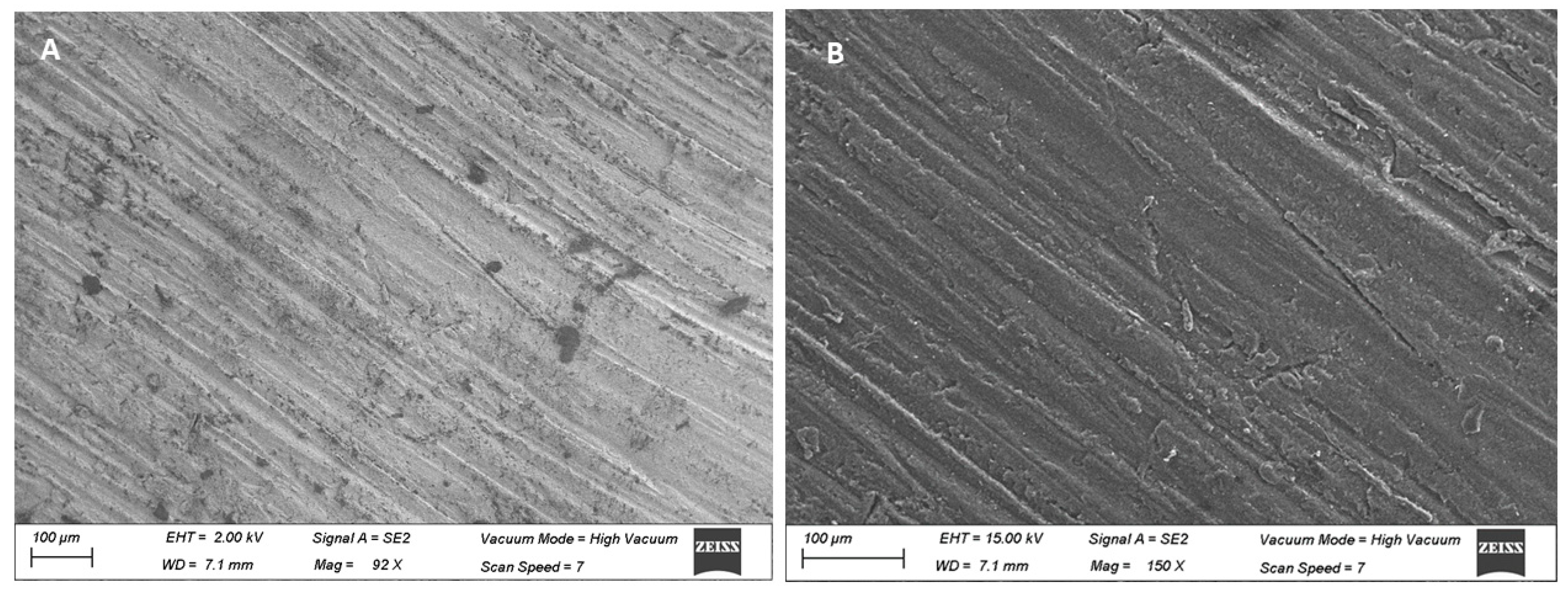
2.1. Scanning Electron Microscopy
2.2. Atomic Force Microscopy (AFM)
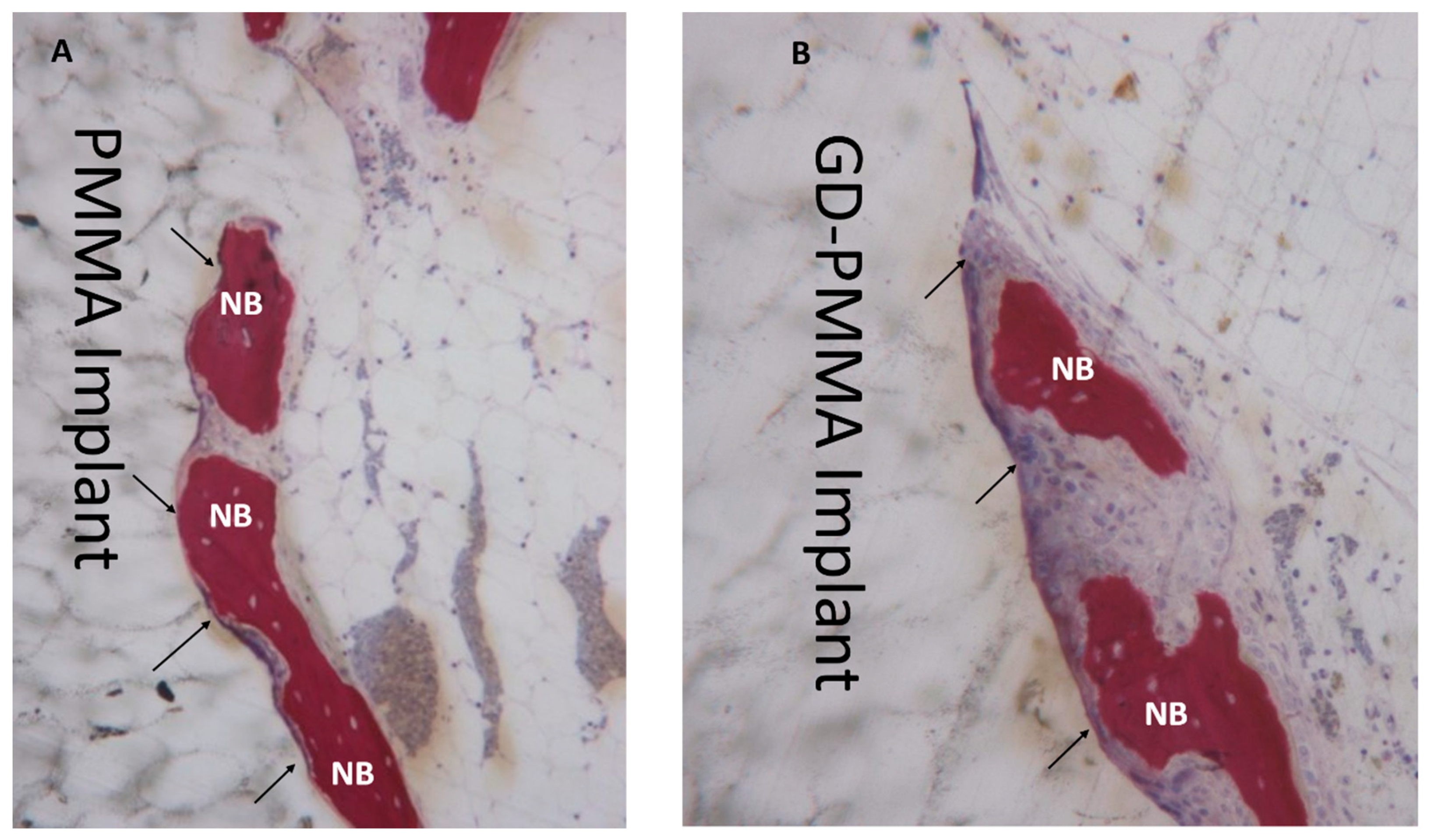
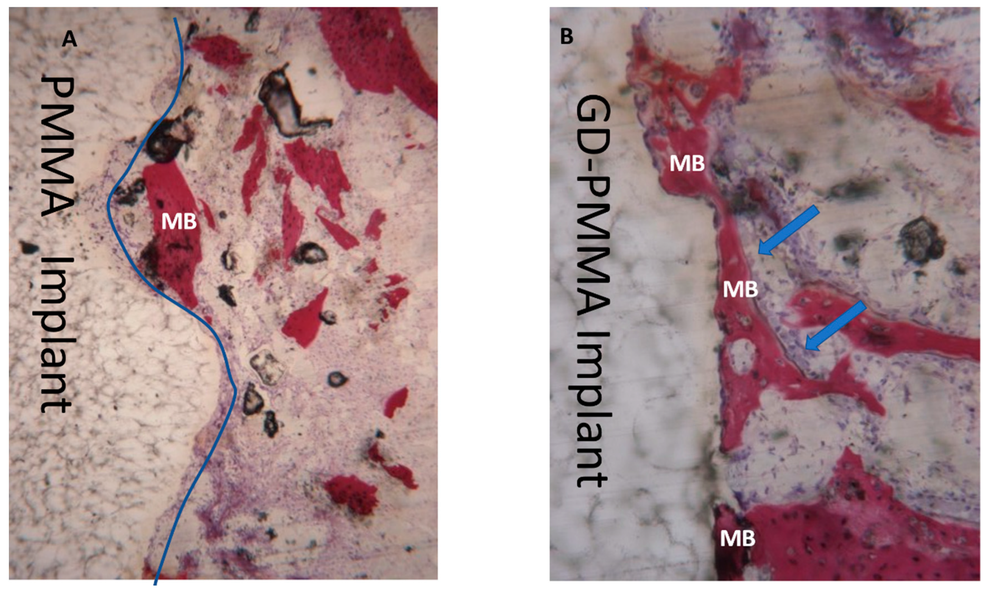
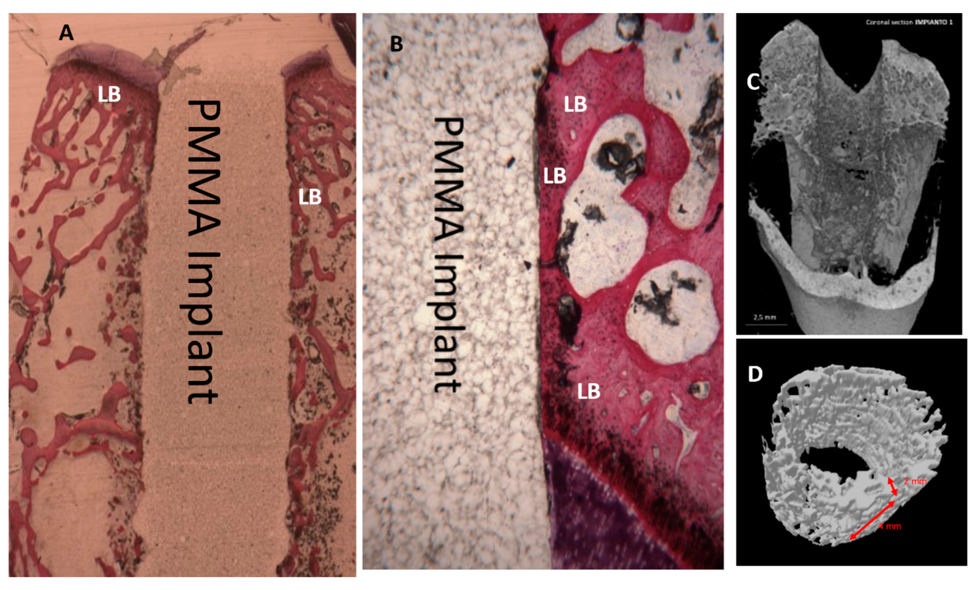
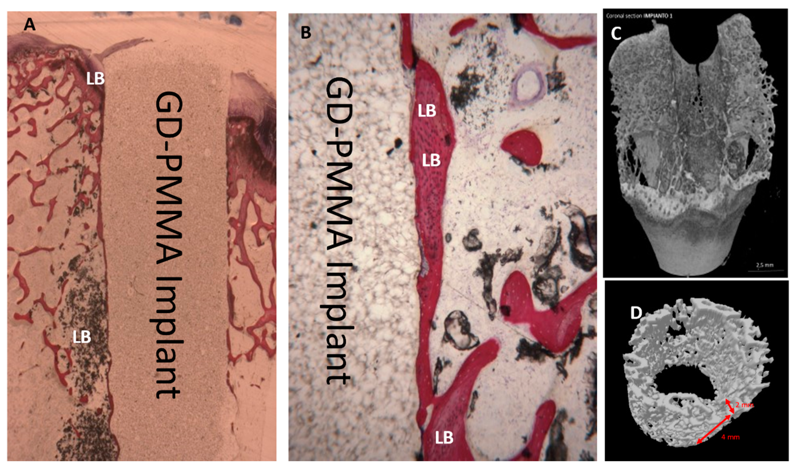
2.3. Histological Evaluation
2.3.1. 15 Days
2.3.2. 30 Days
2.3.3. 60 Days
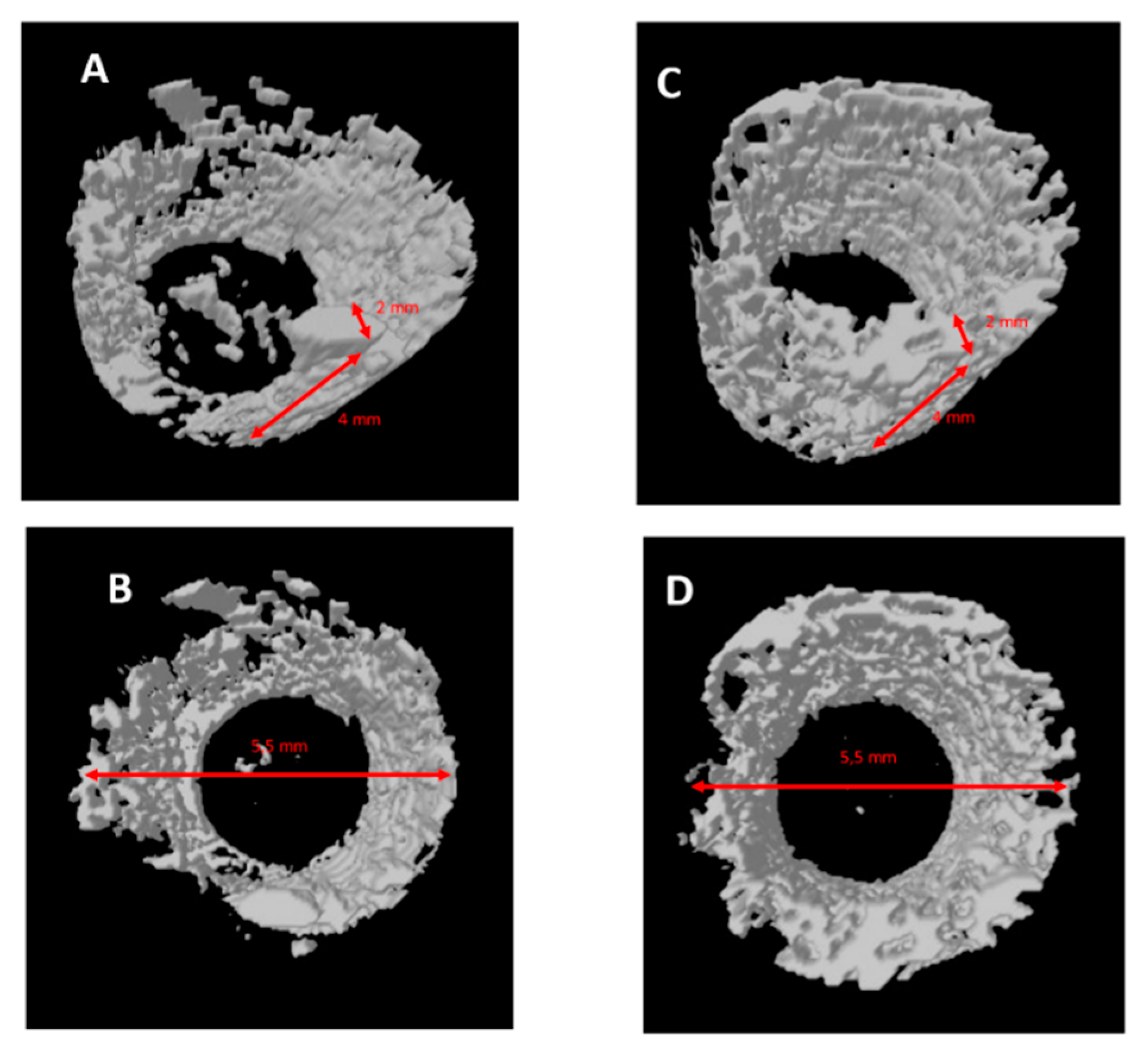
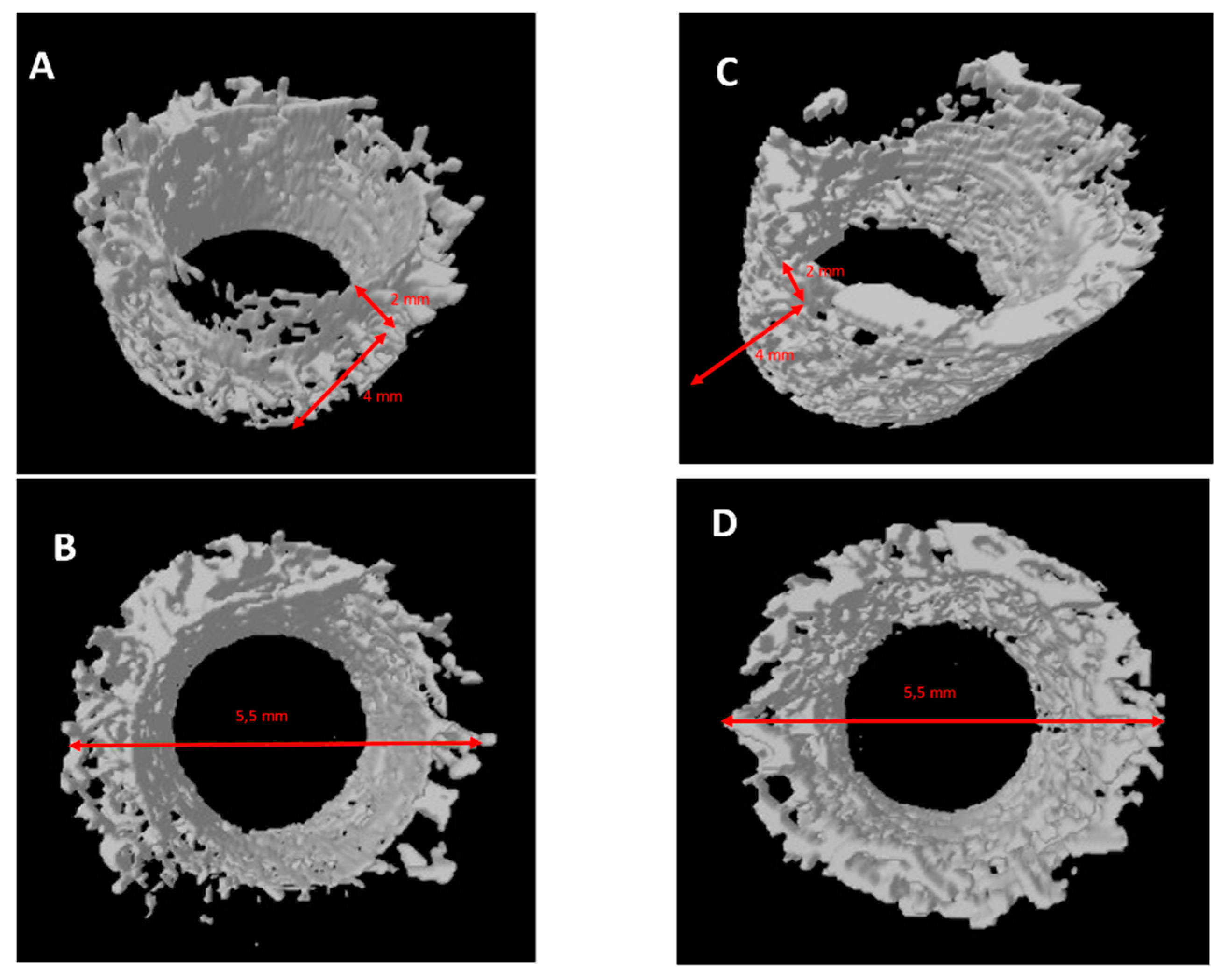
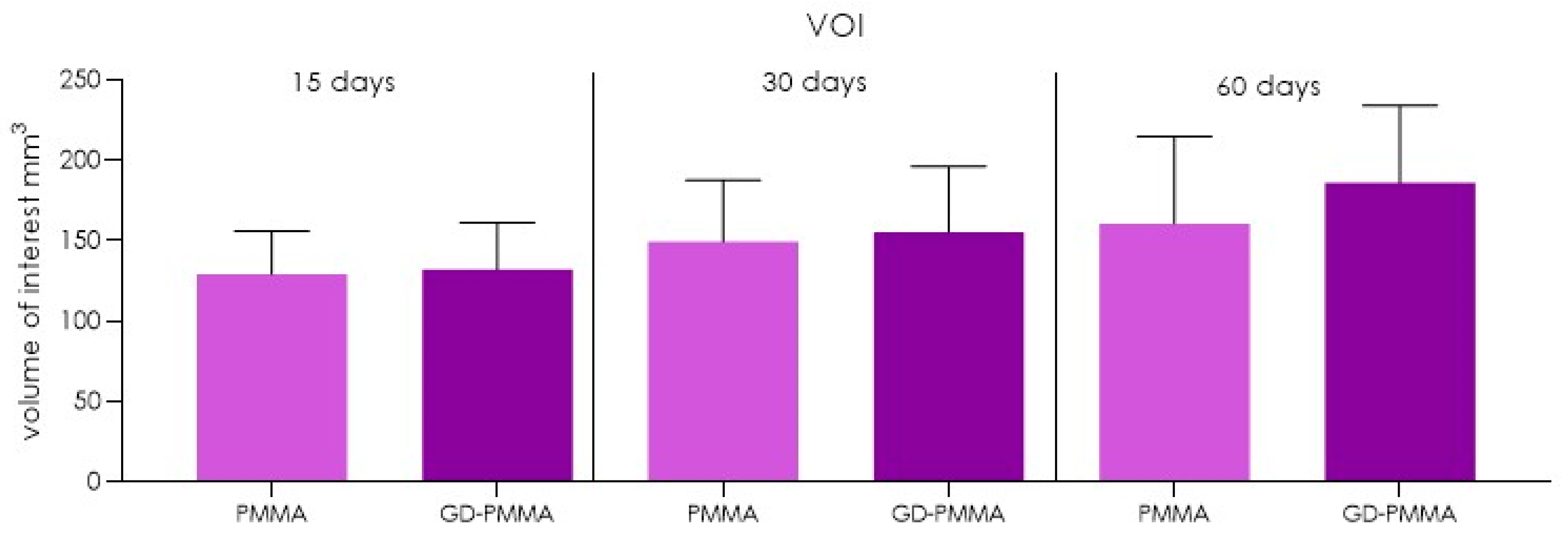
2.4. Micro-CT Evaluation
3. Discussion
4. Materials and Methods
4.1. Scanning Electron Microscopy
4.2. Atomic Force Microscopy (AFM)
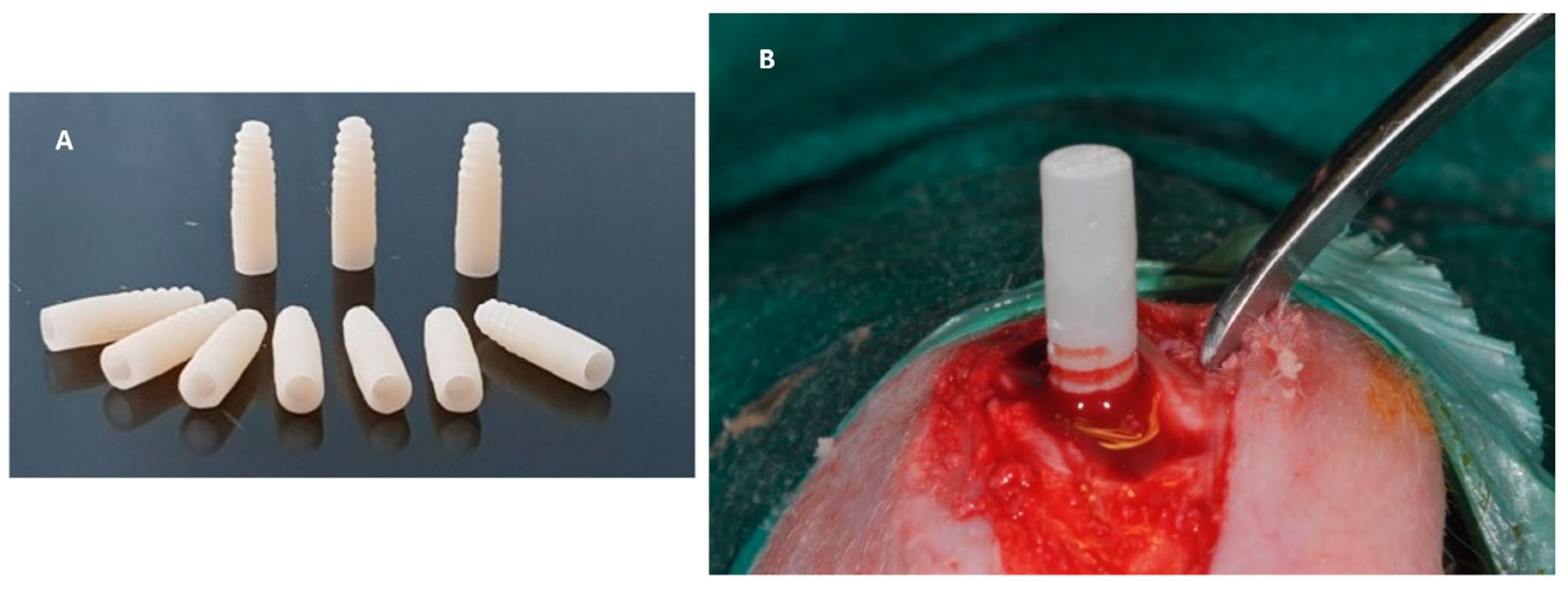
4.3. In Vivo Experiment
4.4. Specimen Processing
4.5. Statistical Evaluation
4.6. Micro-CT Evaluation
5. Conclusions
Author Contributions
Funding
Institutional Review Board Statement
Data Availability Statement
Conflicts of Interest
References
- Albrektsson, T.; Sennerby, L. State of the Art in Oral Implants. J. Clin. Periodontol. 1991, 18, 474–481. [Google Scholar] [CrossRef] [PubMed]
- Buser, D.; Sennerby, L.; De Bruyn, H. Modern Implant Dentistry Based on Osseointegration: 50 Years of Progress, Current Trends and Open Questions. Periodontol. 2000 2017, 73, 7–21. [Google Scholar] [CrossRef] [PubMed]
- Albrektsson, T.; Zarb, G.; Worthington, P.; Eriksson, A.R. The Long-Term Efficacy of Currently Used Dental Implants: A Review and Proposed Criteria of Success. Int. J. Oral Maxillofac. Implants 1986, 1, 11–25. [Google Scholar] [PubMed]
- Beschnidt, S.M.; Cacaci, C.; Dedeoglu, K.; Hildebrand, D.; Hulla, H.; Iglhaut, G.; Krennmair, G.; Schlee, M.; Sipos, P.; Stricker, A.; et al. Implant Success and Survival Rates in Daily Dental Practice: 5-Year Results of a Non-Interventional Study Using CAMLOG SCREW-LINE Implants with or without Platform-Switching Abutments. Int. J. Implant Dent. 2018, 4, 33. [Google Scholar] [CrossRef]
- Lollobrigida, M.; Lamazza, L.; Capuano, C.; Formisano, G.; Serra, E.; Laurito, D.; Romanelli, M.; Molinari, A.; De Biase, A. Physical Profile and Impact of a Calcium-Incorporated Implant Surface on Preosteoblastic Cell Morphologic and Differentiation Parameters: A Comparative Analysis. Int. J. Oral Maxillofac. Implants 2016, 31, 223–231. [Google Scholar] [CrossRef]
- Zhang, W.; Wang, G.; Liu, Y.; Zhao, X.; Zou, D.; Zhu, C.; Jin, Y.; Huang, Q.; Sun, J.; Liu, X.; et al. The Synergistic Effect of Hierarchical Micro/Nano-Topography and Bioactive Ions for Enhanced Osseointegration. Biomaterials 2013, 34, 3184–3195. [Google Scholar] [CrossRef]
- Yoshizawa, S.; Brown, A.; Barchowsky, A.; Sfeir, C. Magnesium Ion Stimulation of Bone Marrow Stromal Cells Enhances Osteogenic Activity, Simulating the Effect of Magnesium Alloy Degradation. Acta Biomater. 2014, 10, 2834–2842. [Google Scholar] [CrossRef]
- Scarano, A.; Lorusso, F.; Orsini, T.; Morra, M.; Iviglia, G.; Valbonetti, L. Biomimetic Surfaces Coated with Covalently Immobilized Collagen Type I: An X-ray Photoelectron Spectroscopy, Atomic Force Microscopy, Micro-CT and Histomorphometrical Study in Rabbits. Int. J. Mol. Sci. 2019, 20, 724. [Google Scholar] [CrossRef]
- Noumbissi, S.; Scarano, A.; Gupta, S. A Literature Review Study on Atomic Ions Dissolution of Titanium and Its Alloys in Implant Dentistry. Materials 2019, 12, 368. [Google Scholar] [CrossRef]
- Lorusso, F.; Noumbissi, S.; Francesco, I.; Rapone, B.; Khater, A.G.A.; Scarano, A. Scientific Trends in Clinical Research on Zirconia Dental Implants: A Bibliometric Review. Materials 2020, 13, 5534. [Google Scholar] [CrossRef]
- Scarano, A.; Di Domizio, P.; Petrone, G.; Iezzi, G.; Piattelli, A. Implant Periapical Lesion: A Clinical and Histologic Case Report. J. Oral Implantol. 2000, 26, 109–113. [Google Scholar] [CrossRef]
- Albrektsson, T.; Berglundh, T.; Lindhe, J. Osseointegration: Historic Background and Current Concepts. Clin. Periodontol. Implant Dent. 2003, 4, 809–820. [Google Scholar]
- Brånemark, P.I. Osseointegration and Its Experimental Background. J. Prosthet. Dent. 1983, 50, 399–410. [Google Scholar] [CrossRef]
- Scarano, A.; Piattelli, A.; Quaranta, A.; Lorusso, F. Bone Response to Two Dental Implants with Different Sandblasted/Acid-Etched Implant Surfaces: A Histological and Histomorphometrical Study in Rabbits. BioMed Res. Int. 2017, 2017, 8724951. [Google Scholar] [CrossRef]
- Frazer, R.Q.; Byron, R.T.; Osborne, P.B.; West, K.P. PMMA: An Essential Material in Medicine and Dentistry. J. Long Term Eff. Med. Implants 2005, 15, 629–639. [Google Scholar] [CrossRef]
- Ghosh, S.; Pramanick, D.; Ray, A.; Burman, R.; Saha, A. Fronto-Orbital Reconstruction Using Polymethyl Methacrylate Implant. Natl. J. Maxillofac. Surg. 2017, 8, 153–156. [Google Scholar] [CrossRef]
- Hallur, N.; Goudar, G.; Sikkerimath, B.; Gudi, S.S.; Patil, R.S. Reconstruction of Large Cranial Defect with Alloplastic Material (Bone Cement-Cold Cure Polymethyl-Methacrylate Resin). J. Maxillofac. Oral Surg. 2010, 9, 191–194. [Google Scholar] [CrossRef][Green Version]
- Eppley, B.L. Craniofacial Reconstruction with Computer-Generated HTR Patient-Matched Implants: Use in Primary Bony Tumor Excision. J. Craniofac. Surg. 2002, 13, 650–657. [Google Scholar] [CrossRef]
- Leigh, J.A. Use of PMMA in Expansion Dental Implants. J. Biomed. Mater. Res. 1975, 9, 233–242. [Google Scholar] [CrossRef]
- Vaidyanathan, T.; Vaidyanathan, J.; Manasse, M. Analysis of Stress Relaxation in Temporization Materials in Dentistry. Dent. Mater. 2015, 31, e55–e62. [Google Scholar] [CrossRef]
- Oh, J.S.; Kim, K.N.; Yeom, G.Y. Graphene Doping Methods and Device Applications. J. Nanosci. Nanotechnol. 2014, 14, 1120–1133. [Google Scholar] [CrossRef]
- Lee, C.; Wei, X.; Kysar, J.W.; Hone, J. Measurement of the Elastic Properties and Intrinsic Strength of Monolayer Graphene. Science 2008, 321, 385–388. [Google Scholar] [CrossRef]
- Balandin, A.A.; Ghosh, S.; Bao, W.; Calizo, I.; Teweldebrhan, D.; Miao, F.; Lau, C.N. Superior Thermal Conductivity of Single-Layer Graphene. Nano Lett. 2008, 8, 902–907. [Google Scholar] [CrossRef]
- Bolotin, K.I.; Sikes, K.J.; Jiang, Z.; Klima, M.; Fudenberg, G.; Hone, J.E.A.; Kim, P.; Stormer, H.L. Ultrahigh Electron Mobility in Suspended Graphene. Solid State Commun. 2008, 146, 351–355. [Google Scholar] [CrossRef]
- Tahriri, M.; Del Monico, M.; Moghanian, A.; Tavakkoli Yaraki, M.; Torres, R.; Yadegari, A.; Tayebi, L. Graphene and Its Derivatives: Opportunities and Challenges in Dentistry. Mater. Sci. Eng. C Mater. Biol. Appl. 2019, 102, 171–185. [Google Scholar] [CrossRef]
- Ettorre, V.; De Marco, P.; Zara, S.; Perrotti, V.; Scarano, A.; Di Crescenzo, A.; Petrini, M.; Hadad, C.; Bosco, D.; Zavan, B. In Vitro and in Vivo Characterization of Graphene Oxide Coated Porcine Bone Granules. Carbon 2016, 103, 291–298. [Google Scholar] [CrossRef]
- Steinmassl, P.-A.; Wiedemair, V.; Huck, C.; Klaunzer, F.; Steinmassl, O.; Grunert, I.; Dumfahrt, H. Do CAD/CAM Dentures Really Release Less Monomer than Conventional Dentures? Clin. Oral Investig. 2017, 21, 1697–1705. [Google Scholar] [CrossRef]
- Zhang, X.; Kang, T.; Liang, P.; Tang, Y.; Quan, C. Biological Activity of an Injectable Biphasic Calcium Phosphate/PMMA Bone Cement for Induced Osteogensis in Rabbit Model. Macromol. Biosci. 2018, 18, 1700331. [Google Scholar] [CrossRef]
- Fottner, A.; Nies, B.; Kitanovic, D.; Steinbrück, A.; Mayer-Wagner, S.; Schröder, C.; Heinemann, S.; Pohl, U.; Jansson, V. Performance of Bioactive PMMA-Based Bone Cement under Load-Bearing Conditions: An in Vivo Evaluation and FE Simulation. J. Mater. Sci. Mater. Med. 2016, 27, 138. [Google Scholar] [CrossRef]
- Kim, S.; Hwang, Y.; Kashif, M.; Jeong, D.; Kim, G. Evaluation of Bone Regeneration on Polyhydroxyethyl-Polymethyl Methacrylate Membrane in a Rabbit Calvarial Defect Model. In Vivo 2016, 30, 587–591. [Google Scholar]
- Li, Q.; Wang, Z. Involvement of FAK/P38 Signaling Pathways in Mediating the Enhanced Osteogenesis Induced by Nano-Graphene Oxide Modification on Titanium Implant Surface. Int. J. Nanomed. 2020, 15, 4659–4676. [Google Scholar] [CrossRef]
- La, W.-G.; Jin, M.; Park, S.; Yoon, H.-H.; Jeong, G.-J.; Bhang, S.H.; Park, H.; Char, K.; Kim, B.-S. Delivery of Bone Morphogenetic Protein-2 and Substance P Using Graphene Oxide for Bone Regeneration. Int. J. Nanomed. 2014, 9 (Suppl. 1), 107–116. [Google Scholar] [CrossRef]
- Li, K.; Wang, C.; Yan, J.; Zhang, Q.; Dang, B.; Wang, Z.; Yao, Y.; Lin, K.; Guo, Z.; Bi, L.; et al. Evaluation of the Osteogenesis and Osseointegration of Titanium Alloys Coated with Graphene: An in Vivo Study. Sci. Rep. 2018, 8, 1843. [Google Scholar] [CrossRef]
- Malhotra, R.; Han, Y.M.; Morin, J.L.P.; Luong-Van, E.K.; Chew, R.J.J.; Castro Neto, A.H.; Nijhuis, C.A.; Rosa, V. Inhibiting Corrosion of Biomedical-Grade Ti-6Al-4V Alloys with Graphene Nanocoating. J. Dent. Res. 2020, 99, 285–292. [Google Scholar] [CrossRef]
- Asgar, H.; Deen, K.M.; Rahman, Z.U.; Shah, U.H.; Raza, M.A.; Haider, W. Functionalized Graphene Oxide Coating on Ti6Al4V Alloy for Improved Biocompatibility and Corrosion Resistance. Mater. Sci. Eng. C Mater. Biol. Appl. 2019, 94, 920–928. [Google Scholar] [CrossRef]
- Park, C.; Park, S.; Lee, D.; Choi, K.S.; Lim, H.-P.; Kim, J. Graphene as an Enabling Strategy for Dental Implant and Tissue Regeneration. Tissue Eng. Regen. Med. 2017, 14, 481–493. [Google Scholar] [CrossRef]
- Scarano, A.; Carinci, F.; Orsini, T.; Valbonetti, L.; Qorri, E.; Bignozzi, C.A.; Lorusso, F. Titanium Implants Coated with a Bifunctional Molecule with Antimicrobic Activity: A Rabbit Study. Materials 2020, 13, 3613. [Google Scholar] [CrossRef]
- Kilkenny, C.; Browne, W.J.; Cuthill, I.C.; Emerson, M.; Altman, D.G. Improving Bioscience Research Reporting: The ARRIVE Guidelines for Reporting Animal Research. PLoS Biol. 2010, 8, e1000412. [Google Scholar] [CrossRef]
- Percie du Sert, N.P.; Hurst, V.; Ahluwalia, A.; Alam, S.; Avey, M.T.; Baker, M.; Browne, W.J.; Clark, A.; Cuthill, I.C.; Dirnagl, U.; et al. The ARRIVE Guidelines 2.0: Updated Guidelines for Reporting Animal Research. PLOS Biol. 2020, 18, e3000410. [Google Scholar] [CrossRef]
- Piattelli, A.; Scarano, A.; Piattelli, M. Detection of Alkaline and Acid Phosphatases around Titanium Implants: A Light Microscopical and Histochemical Study in Rabbits. Biomaterials 1995, 16, 1333–1338. [Google Scholar] [CrossRef]


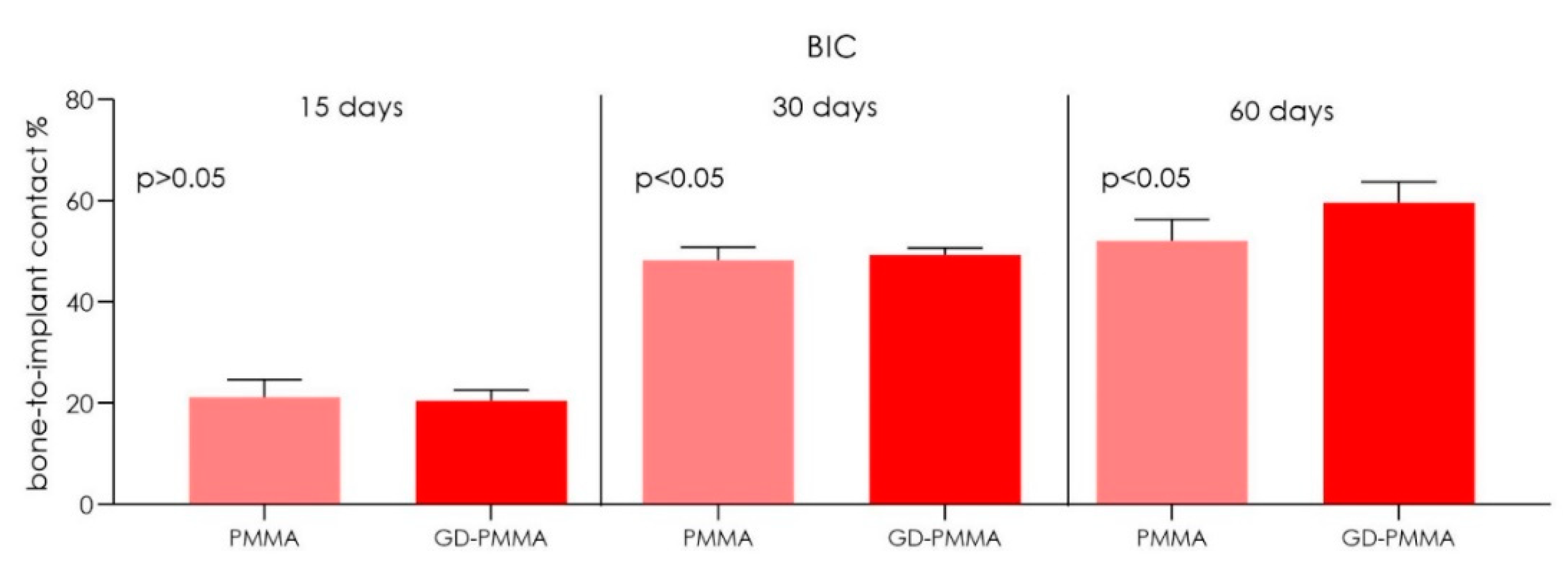
| BIC | 15 Days | 30 Days | 60 Days | |||||||||
|---|---|---|---|---|---|---|---|---|---|---|---|---|
| PMMA | GD-PMMA | PMMA | GD-PMMA | PMMA | GD-PMMA | |||||||
| Mean (SD) | 21.22 | (3.5) | 20.5 | (2.1) | 48.2 | (2.6) | 49.3 | (1.4) | 52.1 | (4.2) | 59.6 | (4.1) |
| 95% CI | (17.52–24.92) | (18.26–22.74) | (45.51–50.89) | (47.8–50.8) | (50.65–59.55) | (55.25–63.95) | ||||||
| p value | p > 0.05 | p < 0.05 | p < 0.05 | |||||||||
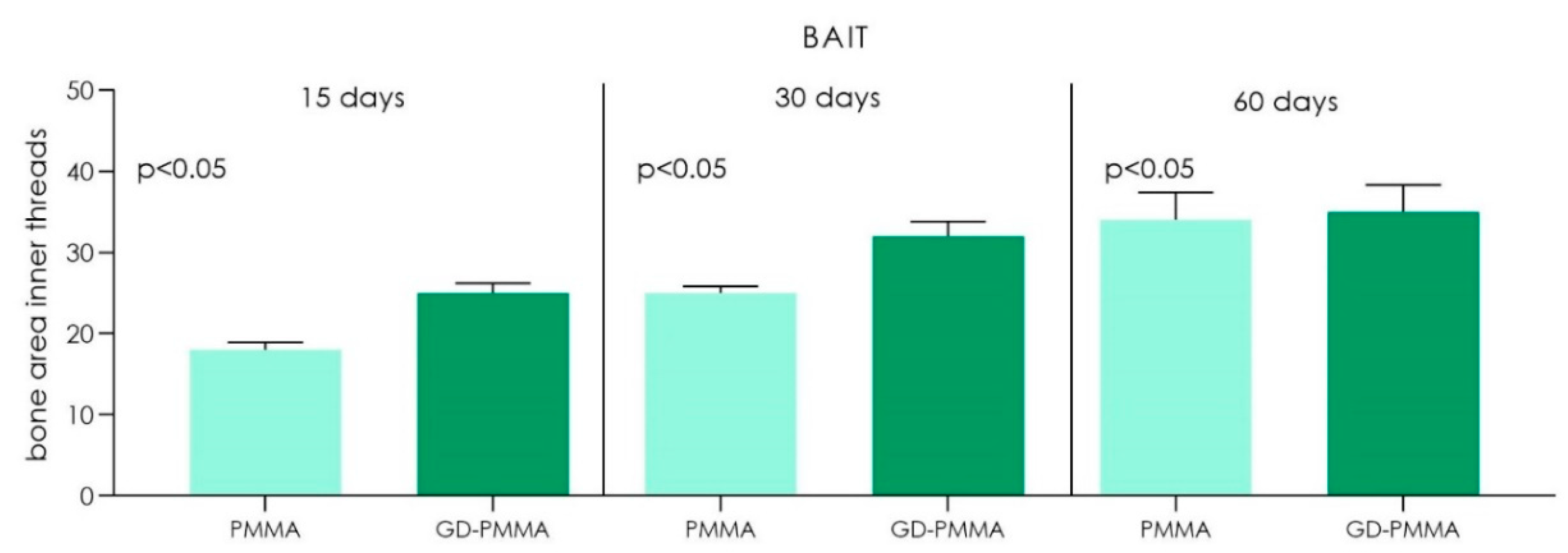
| BAIT | 15 Days | 30 Days | 60 Days | |||||||||
|---|---|---|---|---|---|---|---|---|---|---|---|---|
| PMMA | GD-PMMA | PMMA | GD-PMMA | PMMA | GD-PMMA | |||||||
| Mean (SD) | 18 | (0.9) | 25 | (1.2) | 25 | (0.8) | 32 | (1.8) | 34 | (3.4) | 35 | (3.3) |
| 95% CI | (17.07–18.93) | (23.78–26.22) | (24.18–25.82) | (30.16–33.84) | (30.47–37.53) | (21.57–28.43) | ||||||
| p value | p < 0.05 | p < 0.05 | p < 0.05 | |||||||||
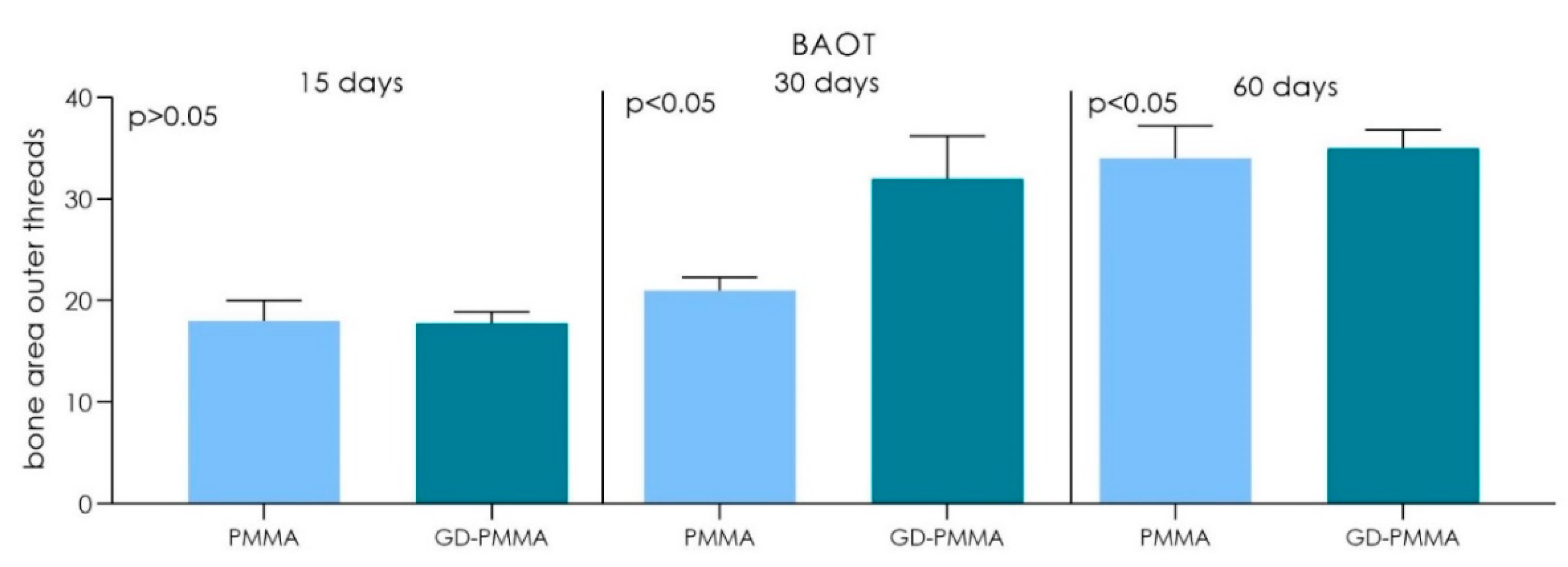
| BAOT | 15 Days | 30 Days | 60 Days | |||||||||
|---|---|---|---|---|---|---|---|---|---|---|---|---|
| PMMA | GD-PMMA | PMMA | GD-PMMA | PMMA | GD-PMMA | |||||||
| Mean (SD) | 18 | (2) | 17.8 | (1.1) | 21 | (1.3) | 32 | (4.2) | 34 | (3.2) | 35 | (1.8) |
| 95% CI | (17.78–18.22) | (16.69–18.91) | (19.64–22.36) | (27.56–36.44) | (30.47–37.53) | (21.57–28.43) | ||||||
| p value | p > 0.05 | p < 0.05 | p < 0.05 | |||||||||
| VOI | 15 Days | 30 Days | 60 Days | |||||||||
|---|---|---|---|---|---|---|---|---|---|---|---|---|
| PMMA | GD-PMMA | PMMA | GD-PMMA | PMMA | GD-PMMA | |||||||
| Mean (SD) | 129 | (27.0) | 132 | (29.0) | 149.2 | (38.1) | 155 | (41.2) | 160.5 | (54.1) | 185.7 | (48.1) |
| 95% CI | (100.3–157.7) | (101.4–162.6) | (109.2–189.2) | (111.7–198.3) | (103.7–217.3) | (135.3–236.1) | ||||||
| p value | p > 0.05 | p > 0.05 | p < 0.05 | |||||||||
Publisher’s Note: MDPI stays neutral with regard to jurisdictional claims in published maps and institutional affiliations. |
© 2021 by the authors. Licensee MDPI, Basel, Switzerland. This article is an open access article distributed under the terms and conditions of the Creative Commons Attribution (CC BY) license (http://creativecommons.org/licenses/by/4.0/).
Share and Cite
Scarano, A.; Orsini, T.; Di Carlo, F.; Valbonetti, L.; Lorusso, F. Graphene-Doped Poly (Methyl-Methacrylate) (Pmma) Implants: A Micro-CT and Histomorphometrical Study in Rabbits. Int. J. Mol. Sci. 2021, 22, 1441. https://doi.org/10.3390/ijms22031441
Scarano A, Orsini T, Di Carlo F, Valbonetti L, Lorusso F. Graphene-Doped Poly (Methyl-Methacrylate) (Pmma) Implants: A Micro-CT and Histomorphometrical Study in Rabbits. International Journal of Molecular Sciences. 2021; 22(3):1441. https://doi.org/10.3390/ijms22031441
Chicago/Turabian StyleScarano, Antonio, Tiziana Orsini, Fabio Di Carlo, Luca Valbonetti, and Felice Lorusso. 2021. "Graphene-Doped Poly (Methyl-Methacrylate) (Pmma) Implants: A Micro-CT and Histomorphometrical Study in Rabbits" International Journal of Molecular Sciences 22, no. 3: 1441. https://doi.org/10.3390/ijms22031441
APA StyleScarano, A., Orsini, T., Di Carlo, F., Valbonetti, L., & Lorusso, F. (2021). Graphene-Doped Poly (Methyl-Methacrylate) (Pmma) Implants: A Micro-CT and Histomorphometrical Study in Rabbits. International Journal of Molecular Sciences, 22(3), 1441. https://doi.org/10.3390/ijms22031441