Article

Expression of Thomsen–Friedenreich Antigen in Colorectal Cancer and Association with Microsatellite Instability

1
Faculty of Medicine, University of Porto, 4200-319 Porto, Portugal
2
i3S—Instituto de Investigação e Inovação em Saúde, Universidade do Porto, 4200-135 Porto, Portugal
3
Department of Pathology, Centro Hospitalar Vila Nova de Gaia/Espinho, 4434-502 Vila Nova de Gaia, Portugal
4
IPATIMUP—Institute of Molecular Pathology and Immunology, University of Porto, 4200-135 Porto, Portugal
5
Department of Pathology, Centro Hospitalar Universitário São João, 4200-319 Porto, Portugal
6
Department of Pathology, Faculty of Medicine, University of Porto, 4200-319 Porto, Portugal
7
Department of Diagnostics and Public Health, Section of Pathology, University and Hospital Trust of Verona, 37134 Verona, Italy
8
Instituto de Ciências Biomédicas Abel Salazar, Universidade do Porto, 4050-313 Porto, Portugal
*
Author to whom correspondence should be addressed.
Current address: IMBA, Institute of Molecular Biotechnology of the Austrian Academy of Sciences, 1030 Vienna, Austria.
Int. J. Mol. Sci. 2021, 22(3), 1340; https://doi.org/10.3390/ijms22031340
Submission received: 29 December 2020 / Revised: 21 January 2021 / Accepted: 26 January 2021 / Published: 29 January 2021
(This article belongs to the Collection Feature Papers in Molecular Oncology)

Abstract

:
Microsatellite instability (MSI) is a molecular phenotype due to a deficient DNA mismatch repair (dMMR). In colorectal cancer (CRC), dMMR/MSI is associated with several clinical and histopathological features, influences prognosis, and is a predictive factor of response to therapy. In daily practice, dMMR/MSI profiles are identified by immunohistochemistry and/or multiplex PCR. The Thomsen–Friedenreich (TF) antigen was previously found to be a potential single marker to identify MSI-high gastric cancers. Therefore, in this study, we aimed to disclose a possible association between TF expression and MSI status in CRC. Furthermore, we evaluated the relationship between TF expression and other clinicopathological features, including patient survival. We evaluated the expression of the TF antigen in a cohort of 25 MSI-high and 71 microsatellite stable (MSS) CRCs. No association was observed between the expression of the TF antigen and MSI-high status in CRC. The survival analysis revealed that patients with MSI-high CRC showed improved survival when the TF antigen was expressed. This finding holds promise as it indicates the potential use of the TF antigen as a biomarker of better prognosis in MSI-high CRCs that should be validated in an independent and larger CRC cohort.

1. Introduction

Colorectal cancer (CRC) is a worldwide health burden disease, being the third most incident cancer and the second cause of cancer-related death [1]. Environmental factors such as diet, obesity, and sedentary behavior are risk factors for the development of CRC [2] that occurs via stepwise accumulation of genetic and epigenetic alterations [3]. The consensus molecular classification of CRC encompasses four distinct subtypes: Consensus Molecular Subtype (CMS) 1 (microsatellite instability, 14%), CMS2 (canonical, 37%), CMS3 (metabolic, 13%), and CMS4 (mesenchymal, 23%) [4]. Almost all hypermutated CRCs with microsatellite-instability (MSI-high) fall into the first category (CMS1). The microsatellite stable (MSS) cancers are subcategorized into the three other groups, CMS2 to CMS4, with a residual unclassified group (mixed features, 13%) that may represent either a transition phenotype or intratumoral heterogeneity. The hypermutated pathway is caused by a defect in the DNA mismatch repair (dMMR) mechanism and can be either sporadic (≈12%) or hereditary (≈3%) [5,6].
MSI is a molecular phenotype of tumors resulting from indel mutations in tandemly repeated nucleotide sequences present throughout the genome called microsatellites, caused by the impairment of the DNA mismatch repair (MMR) machinery. Major MMR genes encompass MLH1, MSH2, MSH6, and PMS2 [7,8]. MSI status is a determining factor in CRC, influencing the clinical outcome [9], namely response to therapy and prognosis [9,10]. These factors are taken into consideration for planning the treatment of CRC patients, which requires a multidisciplinary approach [11]. Several studies have reported MSI-high as a predictive marker for the lack of response to fluorouracil-based adjuvant therapy in CRC, indicating that the efficacy of this type of chemotherapy differs according to MSI status [12,13]. A number of retrospective studies, including a systematic review and a meta-analysis, support the favorable stage-adjusted prognosis of MSI-high compared to MSS CRC patients [11,14,15,16,17].
The gastrointestinal mucosa is covered by a mucous layer rich in high extensive O-glycosylated proteins, called mucins, that constitute a protective layer over the epithelium [18]. In the process of carcinogenesis, several changes in the protein glycosylation machinery occur, resulting in aberrant cell surface glycosylation profiles [19]. These alterations are characterized by increased sialylation, fucosylation or truncation of O-glycans and are often observed in gastrointestinal tumors [19,20]. Moreover, previous studies have shown a correlation between altered glycans and tumor progression in gastrointestinal cancer [18,21,22,23,24,25,26,27]
One illustrative example of aberrant O-glycans signatures is the simple disaccharide antigen Thomsen–Friedenreich antigen (TF antigen), also named antigen T or Core 1 [28]. This antigen is an intermediate product that appears in the Golgi apparatus during the maturation of mucin-type-O-glycans [19]. The TF antigen is rarely detected in normal cells but is frequently expressed in tumor cells, with pathological and clinical consequences [29]. Moreover, TF antigen expression in liver metastasis, the most common hematogenic dissemination in CRC, was described in a pilot study that demonstrated that CRC liver metastasis expressed the TF antigen at a significantly higher rate (91%) than in primary CRC (60%) [30]. For this reason, this truncated O-glycan arises as biomarker of malignancy with possible implications in the diagnosis, prognosis, and follow-up.
A previous study by Mereiter et al. [31] showed, in gastric cancer, a strong association between the expression of the TF antigen and the MSI-high status (specificity of 94% and sensitivity of 69%), suggesting that TF antigen is a single specific and sensitive marker for the MSI-high status in gastric cancer [31]. In the literature, there is no data on the association between the expression of TF antigen and the MSI status in CRC. Therefore, we aimed to perform an exploratory study to evaluate whether such an association is also present in CRC. Furthermore, we evaluated the association between TF expression and additional clinicopathological variables, including survival analysis. We performed histochemistry analysis using the Peanut agglutinin (PNA) lectin that preferentially binds to the galactosyl (β-1,3) N-acetylgalactosamine structure, the TF antigen [32], to evaluate if the detection of this biomarker could be a tool in the identification of MSI-high CRC.

2. Results

2.1. TF Expression in Colorectal Cancer

The expression of TF epitope, detected by histochemistry with PNA lectin, was evaluated in 96 colorectal carcinomas and detected in 55 cases (57%). Several parameters were assessed regarding the TF expression in CRC tumors: the percentage of labeled cancer cells, intensity of the staining, and the subcellular localization. The TF expression in the extracellular mucus secretion was also evaluated and recorded as “intraglandular” (in the lumen of glands), localized in “mucin pools” or both. (Supplementary Materials, Table S1). TF expression in the neoplastic cells was considered positive when more than 5% of tumor cells were stained.
The TF antigen was observed in the nonneoplastic mucosa adjacent to the tumors (Figure 1a). In this localization, the antigen expressed a perinuclear staining in what appears to be the Golgi apparatus (Figure 1b). Low-grade carcinomas typically showed a strong ectopic expression in the apical membrane (Figure 1c). In contrast, high-grade carcinomas evidenced cytoplasmatic staining (Figure 1d) or both membranous and cytoplasmatic staining (Figure 1e). Regarding extracellular mucus staining, the TF expression was mainly positive in intraglandular mucus in low-grade carcinomas (Figure 1f) and mucin pools were marked prominently for the TF antigen, as shown in Figure 1g, in the mucinous component of a low-grade carcinoma. The TF expression was detected also in carcinomas with signet ring cells (Figure 1h).
The whole series (96 patients) consisted of 85 (89%) low-grade carcinomas and 11 (11%) high-grade carcinomas. The TF expression was analyzed according to the tumor grading of the tumor (Table S2), and we did not find any significant association with % and intensity of positive cells or with the type and location of intracellular and extracellular staining.
The whole series (96 patients) was also assessed to find possible associations between TF expression and clinicopathological variables (Table 1).
Concerning macroscopic type, we found a significant statistical association between TF expression and macroscopic type (p = 0.02), as ulcerated tumors showed more frequently TF expression. Regarding tumor desmoplasia, we found also a statistical association between the expression of TF and this feature, as TF positive tumors showed a moderate/strong desmoplasia than TF negative tumors (p = 0.04).
Regarding TNM staging, a significant statistical association between TF expression and T staging was found (p = 0.02), with higher TF expression in tumor progression (7% in pT1, 26% in pT2, and 51% in pT3).
Regarding other clinicopathological features evaluated in this cohort (Table 1), we did not find any statistically significant association with the TF status.
The MSI status was evaluated and compared with the expression of the TF antigen (Table 2). This cohort encompasses 25 cases (26%) classified as MSI-high and 71 cases (74%) as MSS. The expression of TF antigen was found in 15 (60%) MSI-high cases and 40 (56%) MSS cases (Table 2). No statistically significant association was found between the expression of TF antigen and the MSI status (p = 0.47). The sensitivity for MSI-high detection using TF antigen histochemistry was 60% (among the 25 MSI-high cases, only 15 had TF expression) and the specificity was 43.7% (among the 71 MSS cases, 31 did not have TF expression). The positive and negative predictive values were, respectively, 27.3% (15/55) and 75.6% (31/41).

2.2. Survival Analysis

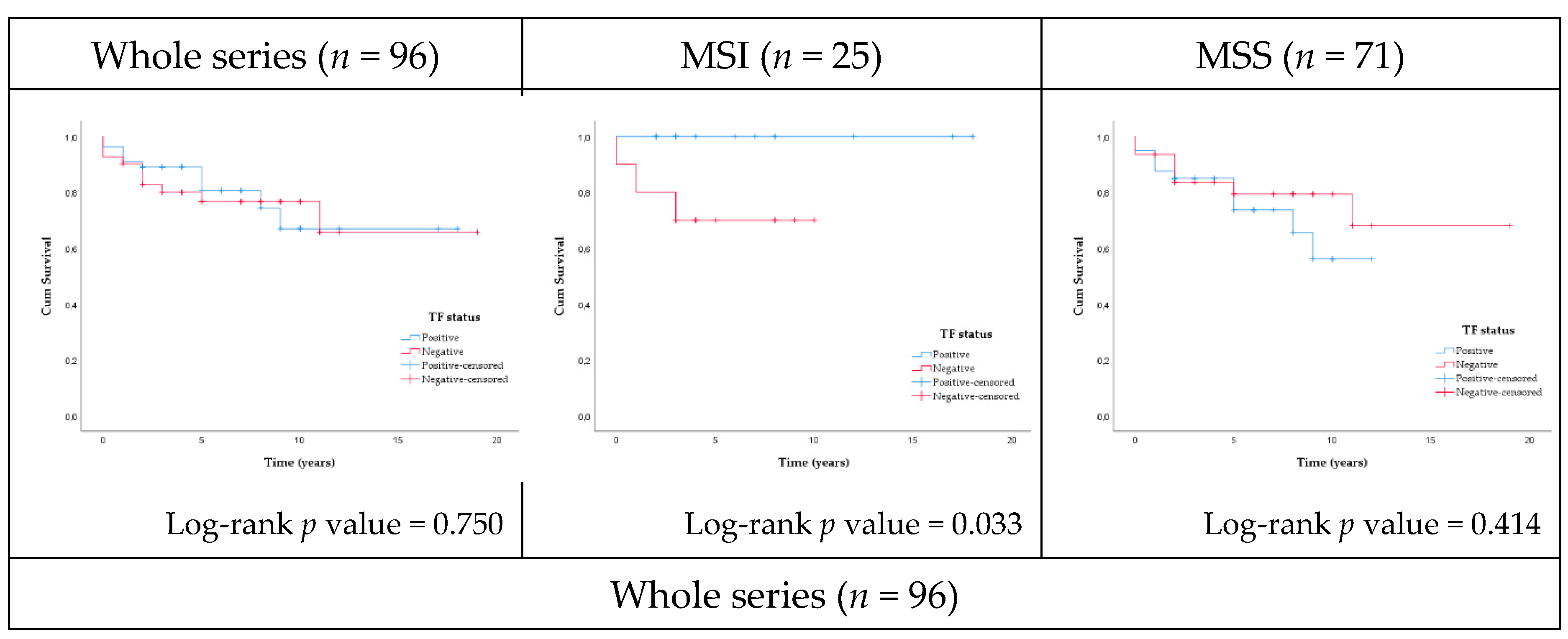
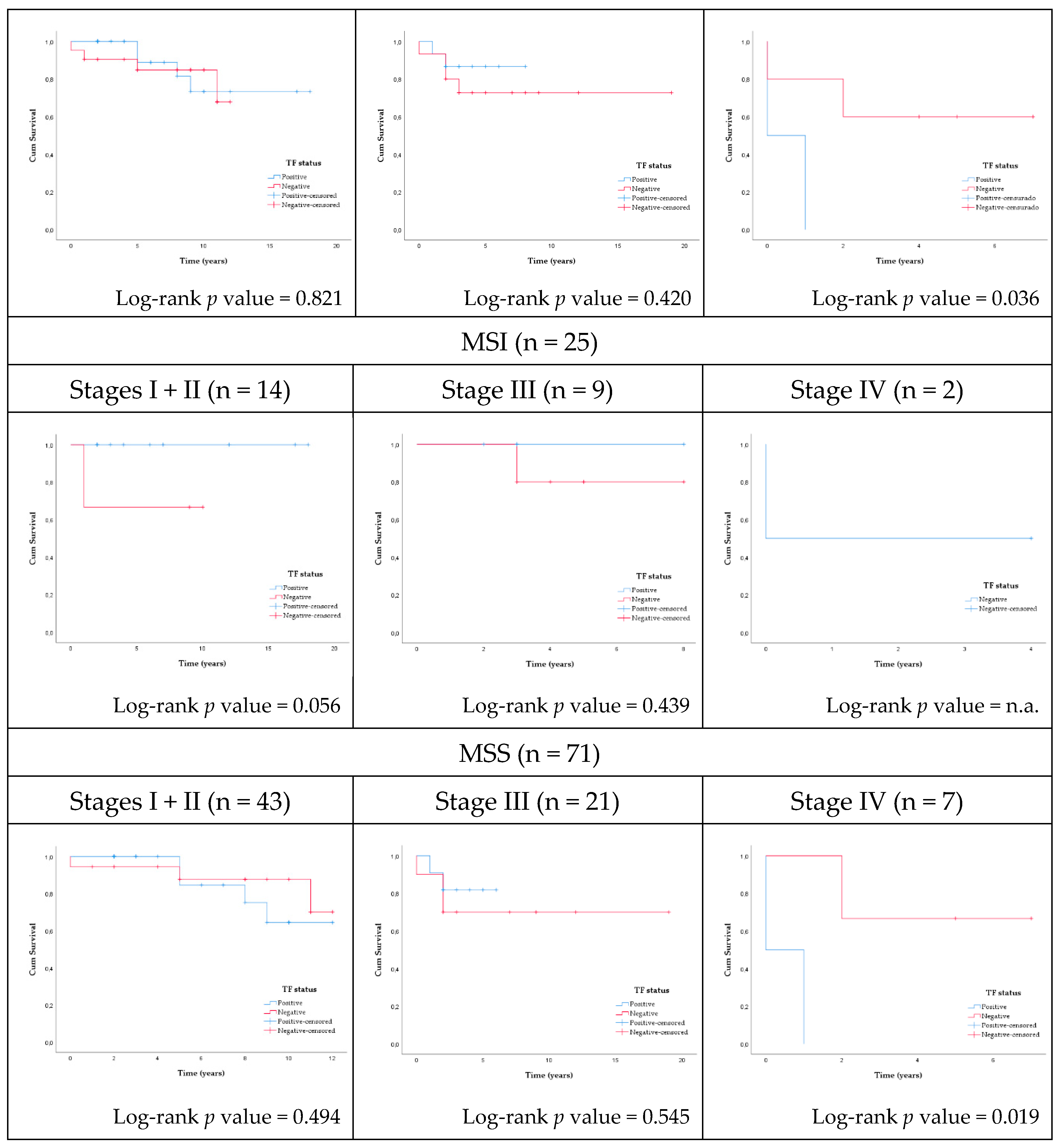
Survival was evaluated in the whole series and according to MSI status (MSI-high and MSS) and TNM stages (stages I + II, stage III, and stage IV) (Figure 2). In this exploratory cohort, the 5-year survival rate decreased with TNM stages: early stages (I/II)—88%, stage III—79%, and stage IV—33%, in keeping with data reported in the literature [33].
In the whole series, no association was found between TF expression and the overall survival of the patients. In the MSI-high subset, the survival of patients harboring TF-positive tumors was significantly better than in the negative cases (log rank p-value = 0.033). When MSI-high cases were stratified by TNM stage, within the patients harboring early stage (I + II) tumors, a better survival was observed in those with TF-positive tumors (log-rank p value = 0.056).
When the whole series was stratified by stage, a significant lower survival was observed in stage IV patients with TF-positive tumors (mean survival time 0.50 years) compared with stage IV patients harboring TF-negative tumors (mean survival time 3.60 years) (p = 0.036). Additionally, in stage IV MSS cases, the survival of patients harboring TF-positive tumors (mean survival time 0.50 years) was significantly lower than in TF-negative cases (mean survival time 4.67 years) (p = 0.019).

3. Discussion

Glycoconjugates are major components of the cell, playing important roles in various biological processes [34]. Altered glycosylation has been shown to influence cellular behavior, affecting and controlling numerous pathophysiological aspects of cancer, including progression [19], immune escape [35,36], tumoral invasion, and metastases [37]. The understanding of these mechanisms is fundamental and may contribute to the implementation of glycosylation modifications in clinical practice [38]. Aberrant cancer-associated glycans and glycoproteins have been used in the clinical context, mostly as serological markers. An example is the use as a biomarker of the carcinoembryonic antigen (CEA), a glycoprotein involved in cell adhesion [39]. It is overexpressed in carcinomas of the colon, rectum, breast, and lung [40]. In CRC, it is present in most patients, being used in the evaluation of prognosis and follow up, particularly after surgical resection [41]. In this study, we evaluated in situ the expression of TF antigen, a truncated O-glycan that has been reported to be expressed in different tumors, such as ovarian cancer, breast cancer, CRC, and acute lymphoblastic leukemia (T-cell) [42]. Regarding CRC, the TF antigen was found to be expressed in 60% of cases [43]. Concerning its role, the TF antigen has been implicated in cell adhesion, as it favors the attachment of tumor cells to the endothelium through the expression of galectin-3 by endothelial cells, supporting its role in tumor invasion [44,45] and therefore contributing to metastasis [30].
The predictive biomarkers used in clinical practice for CRC patients include mutations of the NRAS, KRAS, and BRAF genes as well as MSI status. Particularly, MSI status is relevant as a prognostic factor and predictive biomarker for therapy response.
MSI can be identified by two methods [9]: immunohistochemistry (IHC)-based detection of MMR proteins (MLH1, MSH2, MSH6, and PMS2) in tumor cells or molecular assays using polymerase chain reaction (PCR) or next-generation sequencing (NGS) for the evaluation of alterations in the microsatellites. Although both approaches provide reliable results for diagnostic purposes, these methods have some limitations [46]. MSI testing by multiplex PCR or NGS does not give information on which MMR gene may be involved, is not readily available in all laboratories, and has lower sensitivity than IHC in low tumor purity cases [46,47]. By contrast, IHC indicates which MMR gene may be abnormal, is generally available in all laboratories, and requires lower turnaround times. However, IHC may lead to less consistent results due to pre-analytic and analytic variables as well as interobserver variability [48]. Therefore, these limitations raise the interest in additional biomarkers to detect MSI in the clinical setting [49].
In this exploratory study, we identified 25 MSI-high cases (26%) and 71 MSS cases (74%). All cases were evaluated by both methods and only two discrepant cases (2%) were found. Both cases showed loss of MLH1 and PMS2 nuclear expression but were considered MSS by PCR testing. Possible explanations for the discordant results include tumoral heterogeneity [50] and/or underrepresentation of tumor cells in the sample [51,52]. We have a higher frequency of MSI-high cases in this series than reported in the literature (15–20%) [5]. The limited number of cases within this exploratory analysis highlights the importance of an independent cohort for further validation.
Currently, the treatment of CRC rests on two pillars: surgery and chemotherapy [53]. For therapeutic decision, MSI status has been pointed out as a factor that impacts clinical response to conventional treatments [11]. The decision of adjuvant therapy differs according to MSI status in stage II CRC in intermediate risk patients, as it is not a recommended adjuvant therapy in this group of patients [11]. It has become evident that the “one-size-fits-all” approach is no longer acceptable in the treatment of CRC that is evolving to a more personalized approach, taking into consideration the neoplastic genomic landscape that gained momentum in the treatment strategy. Moreover, immunotherapy is evolving at an enthusiastic speed in the field of oncology. In CRC with MSI, especially in metastatic chemorefractory MSI-high CRC [54], immunotherapy using PD-1 and PD-L1 checkpoint inhibitors is providing promising results regarding sustained clinical response [55] due to the fact that, in MSI-high tumors, there is upregulation of immune checkpoints [56].
Here, we present for the first time a study that correlates the expression of the TF antigen in CRC with MSI. Our data indicate that the TF antigen is not a predictor of MSI in CRC, contrary to what has been described in gastric cancer with MSI [31]. However, our results showed that patients harboring MSI-high tumors that express TF antigen have a significantly better survival than TF-negative cases. Taking into consideration the size of the sample in this exploratory study, this finding should be evaluated in the future in larger cohorts. This will be important to define the potential use of the TF antigen as a biomarker of better prognosis in MSI cases of CRC.
Implications of patient survival in CRC tumors harboring TF expression may be related to cancer immunity. Previous reports have shown higher sensitivity to natural killer (NK) cells towards human carcinoma cell lines expressing the TF antigen [57]. Moreover, it was proposed that the TF antigen also participates in the recognition of endogenous lectins expressed by the immune cells [58] and therefore modulate the immune response. Additionally, MSI-high tumors are characteristically more immunogenic due to a high production of mutated peptides that act as tumor-specific neoantigens that stimulate a more vigorous immune response, both adaptive and innate, leading to a better prognosis [59]. This antigen-driven immune response is mediated by the lymphocytic infiltrate that is observed in MSI-high CRC [60]. Therefore, the simultaneous TF expression and MSI status may contribute to better prognosis due to modulation of the immune response to malignant cells.
Overall, our finding holds promise as it indicates the potential use of the TF antigen as a biomarker of better prognosis in MSI CRC cases. However, further studies validating the obtained predictive results in independent and larger CRC cohorts are warranted in order to be considered for potential clinical application.

4. Materials and Methods

4.1. Patients Samples

The series included 96 colorectal carcinomas retrieved retrospectively from the archives of the Department of Pathology of Centro Hospitalar Universitário de São João (CHUSJ). The patients included in the study were diagnosed with CRC (from January 2001 to December 2018) in which immunohistochemistry for MMR proteins had been previously performed in order to evaluate the MMR deficient status of the tumors. Patients submitted to neoadjuvant therapy and with Lynch syndrome were excluded.
The detailed clinicopathologic features, including gender; age of diagnosis; tumor site; macroscopic type; World Health Organization (WHO) histological classification [61]; tumor grading; growth pattern (ulcerated versus infiltrative); amount of desmoplastic reaction; amount of inflammatory infiltrate; pTNM classification based on the AJCC/UICC TNM classification, 8th edition [62]; residual (R) tumour status, lymphatic and/or venous invasion; perineural invasion; Dukes classification [63]; Jass/Morson classification [64]; adjuvant therapy; and CRC family history were collected using the Database of the Department of Pathology and the Clinical Files system of CHUSJ.
The study, which included access to clinicopathological data, was approved by the ethics committee of CHUSJ (no. 366/19).

4.2. Immunohistochemistry for the Detection of MMR Proteins

The immunohistochemistry for MMR proteins (MLH1, MSH2, MSH6, and PMS2) was assessed by evaluating the presence of a nuclear staining pattern in the tumor cells and classified as (1) intact expression, when ≥10% of the tumor cells showed preserved nuclear expression, or (2) abnormal expression, when the tumor showed complete loss of expression, expression in <10% of tumor cells [65], weaker staining compared with the internal control, or abnormal staining in the nucleoli or nuclear membrane. The presence of an appropriate positive internal control, namely nuclear staining in stromal cells, was consistently verified and compared to the staining of tumor cells.

4.3. MSI Testing

Molecular testing was performed using the IdyllaTM MSI Test [66] in which 7 biomarkers (ACVR2A, BTBD7, DIDO1, MRE11, RYR3, SEC31A, and SULF2) were amplified via PCR for a downstream melting curve analysis. Then, the analysis software detected the mutation status of each biomarker by calculating a probability score (MSI score) derived from the melting curve analysis, expressing the probability of a melting pattern being the wild type or mutant. Consistent with previously established criteria [67] within the software, the detection of at least two mutated markers classified the sample as MSI-high. Otherwise, if less than two markers were mutated, the sample was classified as MSS.

4.4. Histochemistry Profiling of the TF Antigen

In this study, the expression of the TF antigen was assessed through staining with the PNA lectin [32].
A 3 µm section was prepared from one representative formalin-fixed paraffin-embedded (FFPE) block for each sample. Sections were deparaffinated, rehydrated, and endogenous peroxidases were inactivated with 3% hydrogen peroxide in methanol. Tissue sections were blocked for 30 min in room temperature with normal rabbit serum in phosphate-buffered saline (PBS) with 10% bovine serum albumin (BSA). Tissue sections were incubated with 2 µg/mL biotinylated PNA (Vector Labs’, Burlingame, CA, USA) in PBS supplemented with 0.1 mM CaCl2 and 0.01 mM of MnCl2 for 1 h at room temperature. Then, the sections were incubated with ABC (avidine-biotin peroxidase) for an additional 30 min at room temperature. Finally, the sections were stained by 3,3′-iaminobenzidine tetrahydrochloride (DAB) (Sigma Aldrich, St. Louis, MO, USA) and counterstained with Gill’s hematoxylin solution for nuclear contrast. The slides were mounted using Entellan solution and examined using a Zeiss Optical Microscope.
The criteria used to assess the positivity for TF antigen in the tumor was the presence of more than 5% of positive cancer cells. A semiquantitative evaluation of the percentage of positive cancer cells was applied for the following groups: 0% to ≤5% positive cancer cells, >5% to <50% positive cancer cells, and ≥50% positive cancer cells. TF expression in the neoplastic cells was considered positive when more than 5% of the tumor cells were stained. Staining intensity was classified as absent, weak, or strong. The intracellular staining was assessed as membranous, cytoplasmic, or both. The TF expression in the extracellular mucus secretion was also evaluated and recorded as “intraglandular” (in the lumen of glands), localized in “mucin pools” or both.

4.5. Statistical Analysis

Statistical analysis was performed with IBM SPSS STATISTICS (version 26.0 for Windows; SPSS, Chicago, IL, USA).
To assess the presence of an association with statistical significance between clinicopathological variables and TF expression (positive versus negative), Fisher’s Exact Test and Pearson chi-squared test were applied, as appropriate. For numerical variables with normal distribution, Independent Samples t Test was used, while for numerical variables without a normal distribution, Mann–Whitney–Wilcoxon test was used. All tests were two-sided, and differences were considered significant when p < 0.05.

Supplementary Materials

Supplementary materials can be found at https://www.mdpi.com/1422-0067/22/3/1340/s1.

Author Contributions

Conceptualization, S.M., F.C. and C.A.R.; methodology, B.L., I.G., S.M., F.D., J.G., F.C. and C.A.R.; PCR analysis, P.P.; slide preparation, G.G.; materials selection, B.L. and C.C.; histochemistry experiment, H.O.D.; anatomopathological analysis, B.L., X.W., I.G., C.C. and F.C.; statistical analysis B.L., J.G., F.C. and C.A.R.; writing—original draft preparation B.L.; writing—review and editing, H.O.D., I.G., S.M., J.G., F.C. and C.A.R.; supervision J.G., F.C. and C.A.R.; funding acquisition, F.C. and C.A.R. All authors have read and agreed to the published version of the manuscript.

Funding

This research was funded by FEDER funds through the Operational Programme for Competitiveness Factors COMPETE 2020 (POCI-01-0145-FEDER-007274; POCI-01-0145-FEDER-016585) and national funds through the Foundation for Science and Technology (FCT), under the projects UID/BIM/4293 and PTDC/BBB-EBI/0567/2014 and the project NORTE-01-0145-FEDER-000029, supported by Norte Portugal Regional Programme (NORTE 2020), under the PORTUGAL 2020 Partnership Agreement, through the European Regional Development Fund (ERDF).

Institutional Review Board Statement

The study was conducted according to the guidelines of the Declaration of Helsinki, and approved by the Ethics Committee of Centro Hospitalar Universitário de São João (no. 366/19, date of approval 25/11/2019).

Informed Consent Statement

Patient consent was waived due to the retrospective nature of this study.

Data Availability Statement

Data sharing is not applicable to this article.

Acknowledgments

The authors would like to acknowledge the Tumour Bank of the Department of Pathology of Centro Hospitalar Universitário São João for providing the clinical samples and Biocartis for providing the MSI tests.

Conflicts of Interest

The authors declare no conflict of interest. The funders had no role in the design of the study; in the collection, analyses, or interpretation of data; in the writing of the manuscript; or in the decision to publish the results.

Abbreviations

ABCAvidine-Biotin Peroxidase
BSABovine serum albumin
CEACarcinoembryonic antigen
CHUSJCentro Hospitalar Universitário de São João
CMSConsensus Molecular Subtype
CRCColorectal Cancer
DAB3,3′-diaminobenzidine tetrahydrochloride
FFPEPhosphate-buffered saline
IHCImmunohistochemistry
MMRMismatch Repair
MSIMicrosatellite instability
MSSMicrosatellite stable
NGSNew generation sequencing
NKNatural killer cells
PBSPhosphate-buffered saline
PNAPeanut agglutinin
PCRPolymerase chain reaction
TFThomsen–Friedenreich antigen
WHOWorld Health Organization

References

  1. Bray, F.; Ferlay, J.; Soerjomataram, I.; Siegel, R.L.; Torre, L.A.; Jemal, A. Global cancer statistics 2018: GLOBOCAN estimates of incidence and mortality worldwide for 36 cancers in 185 countries. CA Cancer J. Clin. 2018, 68, 394–424. [Google Scholar] [CrossRef] [PubMed] [Green Version]
  2. Murphy, N.; Moreno, V.; Hughes, D.J.; Vodicka, L.; Vodicka, P.; Aglago, E.K.; Gunter, M.J.; Jenab, M. Lifestyle and dietary environmental factors in colorectal cancer susceptibility. Mol. Asp. Med. 2019, 69, 2–9. [Google Scholar] [CrossRef] [PubMed]
  3. Nguyen, L.H.; Goel, A.; Chung, D.C. Pathways of Colorectal Carcinogenesis. Gastroenterology 2020, 158, 291–302. [Google Scholar] [CrossRef] [PubMed]
  4. Guinney, J.; Dienstmann, R.; Wang, X.; de Reyniès, A.; Schlicker, A.; Soneson, C.; Marisa, L.; Roepman, P.; Nyamundanda, G.; Angelino, P.; et al. The consensus molecular subtypes of colorectal cancer. Nat. Med. 2015, 21, 1350–1356. [Google Scholar] [CrossRef] [PubMed]
  5. Chang, L.; Chang, M.; Chang, H.M.; Chang, F. Expending Role of Microsatellite Instability in Diagnosis and Treatment of Colorectal Cancers. J. Gastrointest. Cancer 2017, 48, 305–313. [Google Scholar] [CrossRef] [Green Version]
  6. Boland, C.R.; Goel, A. Microsatellite instability in colorectal cancer. Gastroenterology 2010, 138, 2073–2087.e2073. [Google Scholar] [CrossRef]
  7. Thibodeau, S.N.; Bren, G.; Schaid, D. Microsatellite instability in cancer of the proximal colon. Science 1993, 260, 816–819. [Google Scholar] [CrossRef]
  8. Ionov, Y.; Peinado, M.A.; Malkhosyan, S.; Shibata, D.; Perucho, M. Ubiquitous somatic mutations in simple repeated sequences reveal a new mechanism for colonic carcinogenesis. Nature 1993, 363, 558–561. [Google Scholar] [CrossRef]
  9. Kawakami, H.; Zaanan, A.; Sinicrope, F.A. Microsatellite instability testing and its role in the management of colorectal cancer. Curr. Treat. Options Oncol. 2015, 16, 30. [Google Scholar] [CrossRef]
  10. Okita, A.; Takahashi, S.; Ouchi, K.; Inoue, M.; Watanabe, M.; Endo, M.; Honda, H.; Yamada, Y.; Ishioka, C. Consensus molecular subtypes classification of colorectal cancer as a predictive factor for chemotherapeutic efficacy against metastatic colorectal cancer. Oncotarget 2018, 9. [Google Scholar] [CrossRef]
  11. Argilés, G.; Tabernero, J.; Labianca, R.; Hochhauser, D.; Salazar, R.; Iveson, T.; Laurent-Puig, P.; Quirke, P.; Yoshino, T.; Taieb, J.; et al. Localised colon cancer: ESMO Clinical Practice Guidelines for diagnosis, treatment and follow-up. Ann. Oncol. 2020, 31, 1291–1305. [Google Scholar] [CrossRef] [PubMed]
  12. Ribic, C.M.; Sargent, D.J.; Moore, M.J.; Thibodeau, S.N.; French, A.J.; Goldberg, R.M.; Hamilton, S.R.; Laurent-Puig, P.; Gryfe, R.; Shepherd, L.E.; et al. Tumor microsatellite-instability status as a predictor of benefit from fluorouracil-based adjuvant chemotherapy for colon cancer. N. Engl. J. Med. 2003, 349, 247–257. [Google Scholar] [CrossRef] [PubMed] [Green Version]
  13. Sargent, D.J.; Marsoni, S.; Monges, G.; Thibodeau, S.N.; Labianca, R.; Hamilton, S.R.; French, A.J.; Kabat, B.; Foster, N.R.; Torri, V.; et al. Defective mismatch repair as a predictive marker for lack of efficacy of fluorouracil-based adjuvant therapy in colon cancer. J. Clin. Oncol. 2010, 28, 3219–3226. [Google Scholar] [CrossRef] [PubMed] [Green Version]
  14. Tejpar, S.; Saridaki, Z.; Delorenzi, M.; Bosman, F.; Roth, A.D. Microsatellite instability, prognosis and drug sensitivity of stage II and III colorectal cancer: More complexity to the puzzle. J. Natl. Cancer Inst. 2011, 103, 841–844. [Google Scholar] [CrossRef] [PubMed]
  15. Mouradov, D.; Domingo, E.; Gibbs, P.; Jorissen, R.N.; Li, S.; Soo, P.Y.; Lipton, L.; Desai, J.; Danielsen, H.E.; Oukrif, D.; et al. Survival in stage II/III colorectal cancer is independently predicted by chromosomal and microsatellite instability, but not by specific driver mutations. Am. J. Gastroenterol. 2013, 108, 1785–1793. [Google Scholar] [CrossRef]
  16. Popat, S.; Hubner, R.; Houlston, R.S. Systematic review of microsatellite instability and colorectal cancer prognosis. J. Clin. Oncol. 2005, 23, 609–618. [Google Scholar] [CrossRef]
  17. Guastadisegni, C.; Colafranceschi, M.; Ottini, L.; Dogliotti, E. Microsatellite instability as a marker of prognosis and response to therapy: A meta-analysis of colorectal cancer survival data. Eur. J. Cancer 2010, 46, 2788–2798. [Google Scholar] [CrossRef]
  18. Brockhausen, I. Mucin-type O-glycans in human colon and breast cancer: Glycodynamics and functions. EMBO Rep. 2006, 7, 599–604. [Google Scholar] [CrossRef] [Green Version]
  19. Pinho, S.S.; Reis, C.A. Glycosylation in cancer: Mechanisms and clinical implications. Nat. Rev. Cancer 2015, 15, 540–555. [Google Scholar] [CrossRef]
  20. Mereiter, S.; Balmaña, M.; Campos, D.; Gomes, J.; Reis, C.A. Glycosylation in the Era of Cancer-Targeted Therapy: Where Are We Heading? Cancer Cell 2019, 36, 6–16. [Google Scholar] [CrossRef]
  21. Mereiter, S.; Magalhães, A.; Adamczyk, B.; Jin, C.; Almeida, A.; Drici, L.; Ibáñez-Vea, M.; Gomes, C.; Ferreira, J.A.; Afonso, L.P.; et al. Glycomic analysis of gastric carcinoma cells discloses glycans as modulators of RON receptor tyrosine kinase activation in cancer. Biochim. Biophys. Acta 2016, 1860, 1795–1808. [Google Scholar] [CrossRef] [PubMed]
  22. Campos, D.; Freitas, D.; Gomes, J.; Magalhães, A.; Steentoft, C.; Gomes, C.; Vester-Christensen, M.B.; Ferreira, J.A.; Afonso, L.P.; Santos, L.L.; et al. Probing the O-Glycoproteome of Gastric Cancer Cell Lines for Biomarker Discovery. Mol. Cell. Proteom. 2015, 14, 1616. [Google Scholar] [CrossRef] [PubMed] [Green Version]
  23. Marcos, N.T.; Pinho, S.; Grandela, C.; Cruz, A.; Samyn-Petit, B.; Harduin-Lepers, A.; Almeida, R.; Silva, F.; Morais, V.; Costa, J.; et al. Role of the Human ST6GalNAc-I and ST6GalNAc-II in the Synthesis of the Cancer-Associated Sialyl-Tn Antigen. Cancer Res. 2004, 64, 7050. [Google Scholar] [CrossRef] [PubMed] [Green Version]
  24. Marcos, N.T.; Bennett, E.P.; Gomes, J.; Magalhaes, A.; Gomes, C.; David, L.; Dar, I.; Jeanneau, C.; DeFrees, S.; Krustrup, D.; et al. ST6GalNAc-I controls expression of sialyl-Tn antigen in gastrointestinal tissues. Front. Biosci. 2011, 3, 1443–1455. [Google Scholar] [CrossRef]
  25. Hung, J.S.; Huang, J.; Lin, Y.C.; Huang, M.J.; Lee, P.H.; Lai, H.S.; Liang, J.T.; Huang, M.C. C1GALT1 overexpression promotes the invasive behavior of colon cancer cells through modifying O-glycosylation of FGFR2. Oncotarget 2014, 5, 2096–2106. [Google Scholar] [CrossRef] [Green Version]
  26. Barrow, H.; Tam, B.; Duckworth, C.A.; Rhodes, J.M.; Yu, L.-G. Suppression of Core 1 Gal-Transferase Is Associated with Reduction of TF and Reciprocal Increase of Tn, sialyl-Tn and Core 3 Glycans in Human Colon Cancer Cells. PLoS ONE 2013, 8, e59792. [Google Scholar] [CrossRef] [Green Version]
  27. Holst, S.; Wuhrer, M.; Rombouts, Y. Glycosylation characteristics of colorectal cancer. Adv. Cancer Res. 2015, 126, 203–256. [Google Scholar] [CrossRef]
  28. Kudelka, M.R.; Ju, T.; Heimburg-Molinaro, J.; Cummings, R.D. Simple sugars to complex disease--mucin-type O-glycans in cancer. Adv. Cancer Res. 2015, 126, 53–135. [Google Scholar] [CrossRef] [Green Version]
  29. Cao, Y.; Stosiek, P.; Springer, G.F.; Karsten, U. Thomsen-Friedenreich-related carbohydrate antigens in normal adult human tissues: A systematic and comparative study. Histochem. Cell Biol. 1996, 106, 197–207. [Google Scholar] [CrossRef]
  30. Cao, Y.; Karsten, U.R.; Liebrich, W.; Haensch, W.; Springer, G.F.; Schlag, P.M. Expression of Thomsen-Friedenreich-related antigens in primary and metastatic colorectal carcinomas. A reevaluation. Cancer 1995, 76, 1700–1708. [Google Scholar] [CrossRef]
  31. Mereiter, S.; Polom, K.; Williams, C.; Polonia, A.; Guergova-Kuras, M.; Karlsson, N.G.; Roviello, F.; Magalhaes, A.; Reis, C.A. The Thomsen-Friedenreich Antigen: A Highly Sensitive and Specific Predictor of Microsatellite Instability in Gastric Cancer. J. Clin. Med. 2018, 7, 256. [Google Scholar] [CrossRef] [PubMed] [Green Version]
  32. Cano, M.E.; Varela, O.; García-Moreno, M.I.; García Fernández, J.M.; Kovensky, J.; Uhrig, M.L. Synthesis of β-galactosylamides as ligands of the peanut lectin. Insights into the recognition process. Carbohydr. Res. 2017, 443–444, 58–67. [Google Scholar] [CrossRef] [PubMed]
  33. Howlader, N.; Noone, A.M.; Krapcho, M.; Miller, D.; Brest, A.; Yu, M.; Ruhl, J.; Tatalovich, Z.; Mariotto, A.; Lewis, D.R.; et al. (Eds.) SEER Cancer Statistics Review, 1975–2016. Available online: https://seer.cancer.gov/csr/1975_2016 (accessed on 19 January 2020).
  34. Varki, A. Biological roles of oligosaccharides: All of the theories are correct. Glycobiology 1993, 3, 97–130. [Google Scholar] [CrossRef] [PubMed]
  35. Li, C.W.; Lim, S.O.; Xia, W.; Lee, H.H.; Chan, L.C.; Kuo, C.W.; Khoo, K.H.; Chang, S.S.; Cha, J.H.; Kim, T.; et al. Glycosylation and stabilization of programmed death ligand-1 suppresses T-cell activity. Nat. Commun. 2016, 7, 12632. [Google Scholar] [CrossRef] [PubMed] [Green Version]
  36. Rodriguez, E.; Schetters, S.T.T.; van Kooyk, Y. The tumour glyco-code as a novel immune checkpoint for immunotherapy. Nat. Rev. Immunol. 2018, 18, 204–211. [Google Scholar] [CrossRef]
  37. Paszek, M.J.; DuFort, C.C.; Rossier, O.; Bainer, R.; Mouw, J.K.; Godula, K.; Hudak, J.E.; Lakins, J.N.; Wijekoon, A.C.; Cassereau, L.; et al. The cancer glycocalyx mechanically primes integrin-mediated growth and survival. Nature 2014, 511, 319–325. [Google Scholar] [CrossRef] [PubMed] [Green Version]
  38. Taniguchi, N.; Hancock, W.; Lubman, D.M.; Rudd, P.M. The second golden age of glycomics: From functional glycomics to clinical applications. J. Proteome Res. 2009, 8, 425–426. [Google Scholar] [CrossRef] [Green Version]
  39. Reis, C.A.; Osorio, H.; Silva, L.; Gomes, C.; David, L. Alterations in glycosylation as biomarkers for cancer detection. J. Clin. Pathol. 2010, 63, 322–329. [Google Scholar] [CrossRef] [PubMed]
  40. Youssef, E.M.I.; Ewieda, G.H.; Ali, H.A.A.; Tawfik, A.M.; El-fatah, W.M.E.-d.A.; Ezzat, A.A.; Tash, R.M.E.; El-Khouly, N. Comparison between CEA, CA 19-9 and CA 72-4 in Patients with Colon Cancer. Int. J. Tumor Ther. 2013, 2, 26–34. [Google Scholar] [CrossRef]
  41. Newton, K.F.; Newman, W.; Hill, J. Review of biomarkers in colorectal cancer. Colorectal Dis. 2012, 14, 3–17. [Google Scholar] [CrossRef]
  42. Karsten, U.; Goletz, S. What controls the expression of the core-1 (Thomsen-Friedenreich) glycotope on tumor cells? Biochemistry 2015, 80, 801–807. [Google Scholar] [CrossRef] [PubMed]
  43. Fu, C.; Zhao, H.; Wang, Y.; Cai, H.; Xiao, Y.; Zeng, Y.; Chen, H. Tumor-associated antigens: Tn antigen, sTn antigen, and T antigen. HLA 2016, 88, 275–286. [Google Scholar] [CrossRef] [PubMed]
  44. Yu, L.G.; Andrews, N.; Zhao, Q.; McKean, D.; Williams, J.F.; Connor, L.J.; Gerasimenko, O.V.; Hilkens, J.; Hirabayashi, J.; Kasai, K.; et al. Galectin-3 interaction with Thomsen-Friedenreich disaccharide on cancer-associated MUC1 causes increased cancer cell endothelial adhesion. J. Biol. Chem. 2007, 282, 773–781. [Google Scholar] [CrossRef] [Green Version]
  45. Khaldoyanidi, S.K.; Glinsky, V.V.; Sikora, L.; Glinskii, A.B.; Mossine, V.V.; Quinn, T.P.; Glinsky, G.V.; Sriramarao, P. MDA-MB-435 human breast carcinoma cell homo- and heterotypic adhesion under flow conditions is mediated in part by Thomsen-Friedenreich antigen-galectin-3 interactions. J. Biol. Chem. 2003, 278, 4127–4134. [Google Scholar] [CrossRef] [PubMed] [Green Version]
  46. Buza, N.; Ziai, J.; Hui, P. Mismatch repair deficiency testing in clinical practice. Expert Rev. Mol. Diagn. 2016, 16, 591–604. [Google Scholar] [CrossRef]
  47. Shia, J. Immunohistochemistry versus microsatellite instability testing for screening colorectal cancer patients at risk for hereditary nonpolyposis colorectal cancer syndrome. Part, I. The utility of immunohistochemistry. J. Mol. Diagn. 2008, 10, 293–300. [Google Scholar] [CrossRef] [Green Version]
  48. Klarskov, L.; Ladelund, S.; Holck, S.; Roenlund, K.; Lindebjerg, J.; Elebro, J.; Halvarsson, B.; von Salome, J.; Bernstein, I.; Nilbert, M. Interobserver variability in the evaluation of mismatch repair protein immunostaining. Hum. Pathol. 2010, 41, 1387–1396. [Google Scholar] [CrossRef]
  49. Ferreira, J.A.; Magalhães, A.; Gomes, J.; Peixoto, A.; Gaiteiro, C.; Fernandes, E.; Santos, L.L.; Reis, C.A. Protein glycosylation in gastric and colorectal cancers: Toward cancer detection and targeted therapeutics. Cancer Lett. 2017, 387, 32–45. [Google Scholar] [CrossRef]
  50. Tachon, G.; Frouin, E.; Karayan-Tapon, L.; Auriault, M.L.; Godet, J.; Moulin, V.; Wang, Q.; Tougeron, D. Heterogeneity of mismatch repair defect in colorectal cancer and its implications in clinical practice. Eur. J. Cancer 2018, 95, 112–116. [Google Scholar] [CrossRef]
  51. Loughrey, M.B.; McGrath, J.; Coleman, H.G.; Bankhead, P.; Maxwell, P.; McGready, C.; Bingham, V.; Humphries, M.P.; Craig, S.G.; McQuaid, S.; et al. Identifying mismatch repair-deficient colon cancer: Near-perfect concordance between immunohistochemistry and microsatellite instability testing in a large, population-based series. Histopathology 2020. [Google Scholar] [CrossRef]
  52. Evrard, C.; Tachon, G.; Randrian, V.; Karayan-Tapon, L.; Tougeron, D. Microsatellite Instability: Diagnosis, Heterogeneity, Discordance, and Clinical Impact in Colorectal Cancer. Cancers 2019, 11, 1567. [Google Scholar] [CrossRef] [PubMed] [Green Version]
  53. Schmoll, H.J.; Van Cutsem, E.; Stein, A.; Valentini, V.; Glimelius, B.; Haustermans, K.; Nordlinger, B.; van de Velde, C.J.; Balmana, J.; Regula, J.; et al. ESMO Consensus Guidelines for management of patients with colon and rectal cancer. a personalized approach to clinical decision making. Ann. Oncol. 2012, 23, 2479–2516. [Google Scholar] [CrossRef] [PubMed]
  54. Oliveira, A.F.; Bretes, L.; Furtado, I. Review of PD-1/PD-L1 Inhibitors in Metastatic dMMR/MSI-H Colorectal Cancer. Front. Oncol. 2019, 9, 396. [Google Scholar] [CrossRef] [PubMed] [Green Version]
  55. Le, D.T.; Uram, J.N.; Wang, H.; Bartlett, B.R.; Kemberling, H.; Eyring, A.D.; Skora, A.D.; Luber, B.S.; Azad, N.S.; Laheru, D.; et al. PD-1 Blockade in Tumors with Mismatch-Repair Deficiency. N. Engl. J. Med. 2015, 372, 2509–2520. [Google Scholar] [CrossRef] [PubMed]
  56. Sclafani, F. PD-1 inhibition in metastatic dMMR/MSI-H colorectal cancer. Lancet Oncol. 2017, 18, 1141–1142. [Google Scholar] [CrossRef]
  57. Sotiriadis, J.; Shin, S.C.; Yim, D.; Sieber, D.; Kim, Y.B. Thomsen-Friedenreich (T) antigen expression increases sensitivity of natural killer cell lysis of cancer cells. Int. J. Cancer 2004, 111, 388–397. [Google Scholar] [CrossRef]
  58. Saeland, E.; van Vliet, S.J.; Bäckström, M.; van den Berg, V.C.; Geijtenbeek, T.B.; Meijer, G.A.; van Kooyk, Y. The C-type lectin MGL expressed by dendritic cells detects glycan changes on MUC1 in colon carcinoma. Cancer Immunol. Immunother. 2007, 56, 1225–1236. [Google Scholar] [CrossRef]
  59. Banerjea, A.; Ahmed, S.; Hands, R.E.; Huang, F.; Han, X.; Shaw, P.M.; Feakins, R.; Bustin, S.A.; Dorudi, S. Colorectal cancers with microsatellite instability display mRNA expression signatures characteristic of increased immunogenicity. Mol. Cancer 2004, 3, 21. [Google Scholar] [CrossRef] [Green Version]
  60. Phillips, S.M.; Banerjea, A.; Feakins, R.; Li, S.R.; Bustin, S.A.; Dorudi, S. Tumour-infiltrating lymphocytes in colorectal cancer with microsatellite instability are activated and cytotoxic. Br. J. Surg. 2004, 91, 469–475. [Google Scholar] [CrossRef]
  61. Nagtegaal, I.D.; Odze, R.D.; Klimstra, D.; Paradis, V.; Rugge, M.; Schirmacher, P.; Washington, K.M.; Carneiro, F.; Cree, I.A.; WHO Classification of Tumours Editorial Board. The 2019 WHO classification of tumours of the digestive system. Histopathology 2020, 76, 182–188. [Google Scholar] [CrossRef] [Green Version]
  62. Ajani, J.A.; Sano, T.; Amin, M.B.; Edge, S.; Greene, F.; Byrd, D.R.; Brookland, R.K.; Washington, M.K.; Gershenwald, J.E.; Compton, C.C.; et al. (Eds.) Stomach. In AJCC Cancer Staging Manual, 8th ed.; Springer International Publishing: Cham, Switzerland, 2017. [Google Scholar]
  63. Dukes, C.E. The classification of cancer of the rectum. J. Pathol. Bacteriol. 1932, 35, 323–332. [Google Scholar] [CrossRef]
  64. Jass, J.R.; Morson, B.C. Reporting colorectal cancer. J. Clin. Pathol. 1987, 40, 1016–1023. [Google Scholar] [CrossRef] [PubMed] [Green Version]
  65. Sarode, V.R.; Robinson, L. Screening for Lynch Syndrome by Immunohistochemistry of Mismatch Repair Proteins: Significance of Indeterminate Result and Correlation with Mutational Studies. Arch. Pathol. Lab. Med. 2019, 143, 1225–1233. [Google Scholar] [CrossRef] [PubMed] [Green Version]
  66. Mindiola-Romero, M.A.; Green, B.D.; Al-Turkmani, M.; Godwin, B.K.; Mackay, B.A.; Tafe, M.L.; Ren, M.B.; Tsongalis, G. Novel Biocartis Idylla™ cartridge-based assay for detection of microsatellite instability in colorectal cancer tissues. Exp. Mol. Pathol. 2020, 116, 104519. [Google Scholar] [CrossRef] [PubMed]
  67. Umar, A.; Boland, C.R.; Terdiman, J.P.; Syngal, S.; de la Chapelle, A.; Ruschoff, J.; Fishel, R.; Lindor, N.M.; Burgart, L.J.; Hamelin, R.; et al. Revised Bethesda Guidelines for hereditary nonpolyposis colorectal cancer (Lynch syndrome) and microsatellite instability. J. Natl. Cancer Inst. 2004, 96, 261–268. [Google Scholar] [CrossRef] [PubMed]
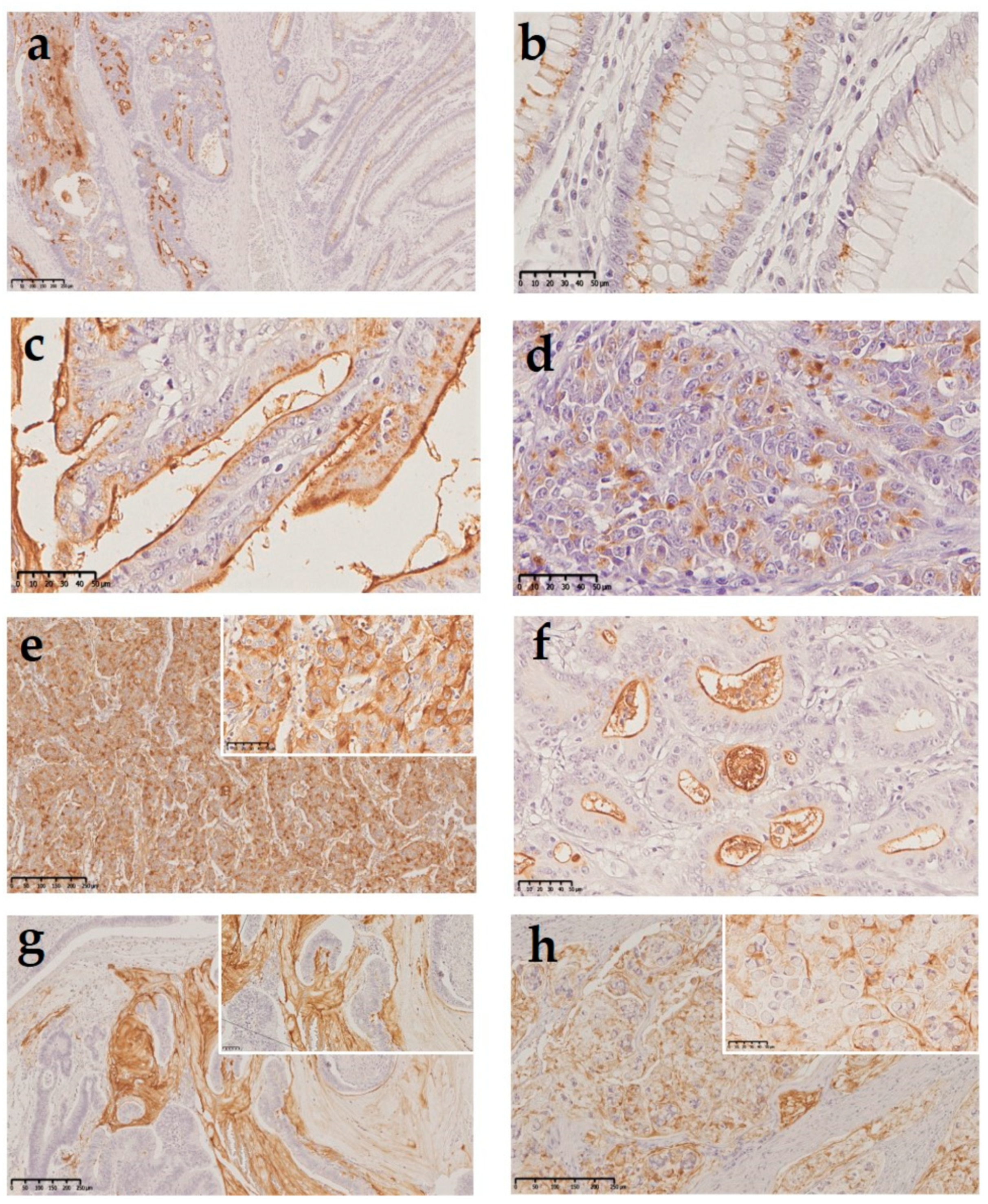
Figure 1. Thomsen–Friedenreich antigen (TF) expression in human colorectal tissue samples: (a) colorectal adenocarcinoma (left) and nonneoplastic mucosa adjacent to the tumor (right), both expressing the TF antigen (100× magnification); (b) high power of the nonneoplastic mucosa displaying perinuclear staining of the TF antigen in the Golgi apparatus (400× magnification); (c) strong expression in the apical membrane in a low-grade carcinoma (400× magnification); (d) cytoplasmatic staining in a high-grade carcinoma with a solid tumoral component (400× magnification); (e) cytoplasmatic and membranous staining in a high-grade adenocarcinoma with trabecular structure (200× magnification—insert 400×); (f) TF expression in intraglandular mucus in a low-grade adenocarcinoma (400× magnification); (g) TF expression in mucin pools in the mucinous component of a low-grade adenocarcinoma (50× magnification—insert 400×); and (h) TF expression in signet ring cells (100× magnification—insert 400×).
Ijms 22 01340 g001
Figure 2. Kaplan–Meier curves of the overall survival of the patients according to TF expression in the tumors.
Ijms 22 01340 g002aIjms 22 01340 g002b
Table 1. Thomsen–Friedenreich (TF) expression and clinicopathological features.
CategoriesTotalTF PositiveTF Negativep-Value 1
n (%)n (%)n (%)
96 (100%)55 (57%)41 (43%)
GenderF44 (46%)24 (44%)20 (49%)0.38
M52 (54%)31 (56%)21 (51%)
Age of diagnosisMean value (Years)54.853.556.70.23
Tumor SiteRight Hemicolon45 (47%)24 (44%)21 (51%)0.24
Left Hemicolon39 (41%)21 (38%)18 (44%)
Rectum9 (9%)7 (13%)2 (5%)
Colon NOS3 (3%)3 (5%)0 (0%)
WHO ClassificationAdenocarcinoma NOS80 (83%)45 (82%)35 (85%)0.66
Mucinous15 (16%)9 (16%)6 (15%)
Undifferentiated1 (1%)1 (2%)0 (0%)
Macroscopic TypeUlcerated60 (62%)40 (73%)20 (49%)0.02 *
Vegetant20 (21%)9 (16%)11 (27%)
Infiltrative2 (2%)2 (4%)0 (0%)
Polypoid14 (15%)4 (7%)10 (24%)
Tumor gradingLow-grade85 (88%)49 (89%)36 (88%)0.55
High-grade11 (12%)6 (11%)5 (12%)
R statusR091 (95%)52 (95%)39 (95%)0.66
R14 (4%)2 (4%)2 (5%)
R21 (1%)1 (1%)0 (0%)
Growth patternInfiltrative69 (72%)37 (67%)32 (78%)0.18
Expansive27 (28%)18 (33%)9 (22%)
DesmoplasiaAbsent/Mild38 (40%)17 (31%)21 (51%)0.04 *
Moderate/Strong58 (60%)38 (69%)20 (49%)
Inflammatory infiltrateAbsent/Mild49 (51%)27 (49%)22 (54%)0.41
Moderate/Strong47 (49%)28 (51%)19 (46%)
pT (TNM Classification)pT117 (18%)4 (7%)13 (32%)0.02 *
pT220 (21%)14 (26%)6 (15%)
pT343 (45%)28 (51%)15 (36%)
pT416 (16%)9 (16%)7 (17%)
pN (TNM Classification)pN057 (59%)36 (65%)21 (51%)0.36
pN128 (29%)14 (26%)14 (34%)
pN211 (12%)5 (9%)6 (15%)
pM (TNM Classification)M086 (90%)51 (93%)35 (85%)0.20
M110 (10%)4 (7%)6 (15%)
StagingEarly (I & II)57 (59%)36 (66%)21 (51%)0.36
III30 (31%)15 (27%)15 (37%)
IV9 (10%)4 (7%)5 (12%)
Peritoneal ImplantsPresent3 (3%)2 (4%)1 (2%)0.61
Absent93 (97%)53 (96%)40 (98%)
Lymphatic and/or venous invasionPresent60 (62%)36 (66%)24 (58%)0.32
Absent36 (38%)19 (34%)17 (42%)
Perineural invasionPresent29 (30%)14 (26%)15 (37%)0.17
Absent67 (70%)41 (74%)26 (63%)
Adjuvant TherapyPerformed54 (56%)30 (54%)24 (58%)0.43
Not performed42 (44%)25 (46%)17 (42%)
Dukes classificationA28 (29%)13 (24%)15 (37%)0.07
B29 (30%)22 (40%)7 (17%)
C38 (40%)19 (34%)19 (46%)
D1 (1%)1 (2%)0 (0%)
Jass/Morson classificationI27 (28%)16 (29%)11 (27%)0.99
II25 (26%)14 (25%)11 (27%)
III25 (26%)14 (26%)11 (27%)
IV19 (20%)11 (20%)8 (19%)
CRC Family HistoryPresent26 (27%)16 (29%)10 (24%)0.39
Absent70 (73%)39 (71%)31 (76%)
Survival time Mean value (Years)5.44.86.10.09
Statistical significant results (p < 0.05) are marked with an asterisk (*). 1 Pearson chi-squared test, Fisher’s Exact Test, Independent Samples T Test (age of diagnosis), and Mann–Whitney–Wilcoxon test (survival time).
Table 2. Expression of TF antigen according to microsatellite instability (MSI) status.
CategoriesTotalTF PositiveTF Negativep-Value 1
n (%)n (%)n (%)
96 (100%)55 (57%)41 (43%)
MSI statusHigh25 (26%)15 (27%)10 (24%)0.47
Stable71 (74%)40 (73%)31 (76%)
1 Fisher’s Exact Test.
Publisher’s Note: MDPI stays neutral with regard to jurisdictional claims in published maps and institutional affiliations.