Review

Regenerative Potential of Mesenchymal Stem Cells’ (MSCs) Secretome for Liver Fibrosis Therapies

by
Simona-Rebeca Nazarie (Ignat)
1,†,
Sami Gharbia
1,2,†,
Anca Hermenean
1,2,
Sorina Dinescu
1,3,* and
Marieta Costache
1,3
1
Department of Biochemistry and Molecular Biology, University of Bucharest, 050663 Bucharest, Romania
2
“Aurel Ardelean” Institute of Life Sciences, “Vasile Goldiș” Western University of Arad, 310025 Arad, Romania
3
The Research Institute of the University of Bucharest (ICUB), University of Bucharest, 050663 Bucharest, Romania
*
Author to whom correspondence should be addressed.
These authors have contributed equally to this paper and share the first author position.
Int. J. Mol. Sci. 2021, 22(24), 13292; https://doi.org/10.3390/ijms222413292
Submission received: 19 November 2021 / Revised: 7 December 2021 / Accepted: 8 December 2021 / Published: 10 December 2021

Abstract

:
Chronic liver injuries lead to liver fibrosis and then to end-stage liver cirrhosis. Liver transplantation is often needed as a course of treatment for patients in critical conditions, but limitations associated with transplantation prompted the continuous search for alternative therapeutic strategies. Cell therapy with stem cells has emerged as an attractive option in order to stimulate tissue regeneration and liver repair. Transplanted mesenchymal stem cells (MSCs) could trans-differentiate into hepatocyte-like cells and, moreover, show anti-fibrotic and immunomodulatory effects. However, cell transplantation may lead to some uncontrolled side effects, risks associated with tumorigenesis, and cell rejection. MSCs’ secretome includes a large number of soluble factors and extracellular vesicles (EVs), through which they exert their therapeutic role. This could represent a cell-free strategy, which is safer and more effective than MSC transplantation. In this review, we focus on cell therapies based on MSCs and how the MSCs’ secretome impacts the mechanisms associated with liver diseases. Moreover, we discuss the important therapeutic role of EVs and how their properties could be further used in liver regeneration.

1. Introduction

Liver fibrosis is a wound healing response that degenerates, and is characterized by excessive accumulation of extracellular matrix (ECM) components that form scar tissue [1]. There are many causes for liver fibrosis, such as alcohol abuse, non-alcoholic steatohepatitis (NASH), viral or autoimmune hepatitis, non-alcoholic fatty liver disease (NAFLD), ischemic injury, and congenital syndromes [2,3]. Liver fibrosis, left untreated, can progress towards irreversible end-stages of liver failure, liver cirrhosis and hepatocellular carcinoma [4]. Most frequently, either removing the cause or the use of antifibrotic drugs would not be completely effective in treating the disease, and liver transplantation is often recommended [5].
Cell therapies have emerged as an alternative to liver transplantation in order to induce tissue regeneration (Figure 1). Numerous cell sources have been investigated for their regenerative potential, either hepatocytes [6], or different types of stem cells [7,8]. There are two major categories of stem cells, embryonic stem cells (ESCs) and adult stem cells (ASCs). Two sources of pluripotent stem cells that can differentiate into hepatocyte-like cells are ESCs, derived from embryos [9,10,11], and induced pluripotent stem cells (iPSCs) [12]. ASCs are multipotent stem cells and have a more limited cell differentiation potential than ESCs. ASCs considered for liver regeneration are liver stem cells (LSCs) [13,14] and mesenchymal stem cells (MSCs) [15,16] from different tissue sources, such as bone marrow (BM-MSCs) [17], adipose tissue (ADSCs) [18,19,20], umbilical cords (UC-SCs), peripheral blood, cartilage, etc. MSCs show low immunogenicity, have a self-renewal ability, are easily obtainable, and can be used without ethical issues, which make them the perfect candidate for liver regeneration [21]. They can migrate to injury sites as a response to cellular damage signals, and they have been shown to promote other cells’ migration to liver sites, differentiate in hepatocyte-like cells and participate in liver regeneration via paracrine mechanisms [22,23,24].
Cell therapies based on MSCs could help liver regeneration directly, by hepatogenic differentiation or cell migration to liver sites, or indirectly, in a paracrine manner via its secretome [23]. MSC-sourced secretome contains many soluble molecules and extracellular vesicles (EVs) that direct tissue repair and regeneration [9,25]. Cell based therapies come with a number of limitations and risks, and cell-free strategies, such as MSC-sourced secretome, could represent an improved alternative. The use of secretome rather than cells avoids the risks associated with tumorigenesis or potential differentiation into pro-fibrotic cells [26,27]. MSC-sourced secretome can be stored over long periods of time without toxic cryopreservative agents and also, it could be produced in large quantities over a short period of time with low costs. Moreover, the content of MSC-sourced secretome could be modified for specific desired therapeutic effects [27]. Cell-based therapies face an additional challenge in regard to its method of transplantation to patients, as such, an effective method has not yet been identified, and cells may end up in other organs [7].
The aim of this review is to present an updated view of cell-based strategies for liver regeneration, with special emphasis on MSC-sourced secretome and derived EVs, and the way they interact with specific pathways involved in liver diseases.

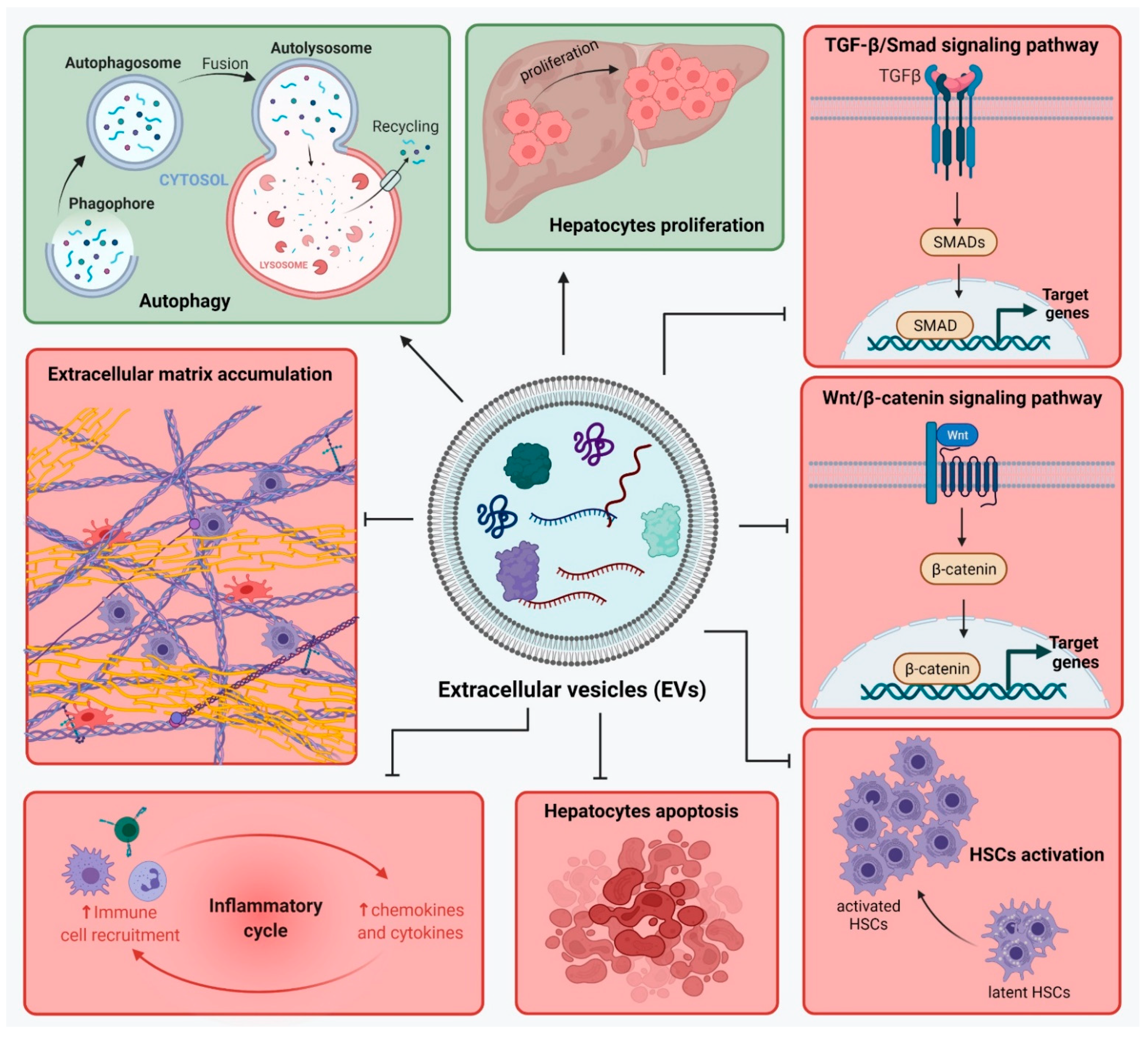
2. Liver Fibrosis and Signaling Pathways

Liver fibrosis occurs after complex interplay between different hepatic cells, and many signaling pathways are involved in its progress [5]. Hepatotoxic agents affect the hepatocytes in the liver and induce their apoptosis [28]. Apoptotic bodies and reactive oxygen species (ROS) further activate hepatic stellate cells (HSCs) and stimulate the infiltration of inflammatory cells [28]. Inflammatory cells release a number of inflammatory factors which will maintain high levels of inflammation in the liver and stimulate HSCs to produce collagen [29]. Hepatic macrophages, such as Kupffer cells (KCs) or monocyte-derived, can be classified as pro-inflammatory macrophages (M1) and immunoregulatory macrophages (M2) [30]. They can contribute to the progress of liver fibrosis and maintain the activated state of HSCs [31].
Liver fibrosis is characterized by the high levels of ECM, and most components are produced by activated HSCs [32]. HSCs produce collagen and other ECM proteins in the normal liver as well, but during liver fibrosis, they transdifferentiate towards a myofibroblast-like phenotype [33]. Activated HSCs lose their ability to store vitamin A-droplets, acquire contractile, proinflammatory and fibrogenic properties, and express α-smooth muscle actin (α-SMA) [34,35]. ECM changes its composition in liver injuries, and adds up to six times more than in normal conditions [36]. It is rich in collagens (I, III, IV), fibronectin, vimentin, elastin, and laminin, and metalloproteinases (MMPs) activity is reduced while the expression of tissue inhibitors of MMPs (TIMPs) is upregulated [37,38].
There are several signaling pathways that play an important role in liver fibrosis development, and especially in HSCs activation. Activated KCs and activated HSCs produce a platelet-derived growth factor (PDGF) [39]. This factor can further activate other signaling pathways in HSCs, such as PI3K/Akt, JAK/STAT, and Ras/Raf systems, that will in turn regulate the expression of fibrotic markers (collagen I, MMPs, TIMPs) [40].
One of the most important signaling pathways in the development of liver fibrosis is the transforming growth factor β (TGF-β)/SMAD pathway. TGF-β is produced by macrophages, hepatocytes and even activated HSCs, as a latent precursor which is stored in the ECM [41]. Its activated form is cleaved by proteases and bound to the specific receptors, which will activate the signaling pathway through phosphorylation of SMAD molecules. A protein complex with phosphorylated SMAD2 and SMAD3, translocates in the nucleus and regulates the expression of fibrotic markers [42,43].
Another pathway involved in liver fibrosis is the Wnt/β-catenin signaling pathway. β-catenin is an adhesion molecule and a transcription factor, and its functions are mainly regulated by Wnt proteins [44]. When the pathway is activated, Wnt inhibits β-catenin phosphorylation, which in turn will increase the levels of unphosphorylated β-catenin in the cytoplasm. This will induce its translocation in the nucleus and the transcription of genes such as Cyclin D1, c-Myc, Axin-2, and c-Jun [45]. Wnt/β-catenin signaling is upregulated in activated HSC, and it can lead to the overexpression of α-SMA and collagen [46,47].

3. Composition of MSCs’ Secretome (Derived Soluble Factors CM/EV)

MSCs secrete a number of molecules, such as soluble proteins, free nucleic acids, lipids and EVs, all accounting for MSCs’ secretome [9,27,48]. MSCs can be isolated from many types of tissues and organs, and even though they share many characteristics and show regenerative potential, their secretome content may vary based on their origin. Differences in MSC-sourced secretome have been pointed out between ADSCs, BM-MSCs and UC-SCs [10,12,49]. All types of MSCs secrete factors such as cytokines, chemokines, growth factors, and anti-inflammatory factors. Overall, it was proved that MSC-sourced secretome shows immunomodulatory, anti-inflammatory and anti-apoptotic activity, regulates angiogenesis, stimulates wound healing and tissue repair, and has antitumor and anti-microbial effects [27]. However, the exact mechanisms through which MSC-sourced secretome exerts its role are not fully understood, and the exact molecules responsible for each effect are not specifically correlated.
MSCs secrete not only a number of anti-inflammatory, but also pro-inflammatory cytokines which mediate its immunomodulatory effect. The secretome contains anti-inflammatory cytokines, such as tumor necrosis factor β1 (TNFβ1), interleukin (IL) 13, IL18 binding protein (IL18BP), ciliary neutrophic factor (CNTF), neurotrophin 3 (NT-3) factor, IL10, IL12p70, IL17E, IL27, IL1. MSCs also secrete pro-inflammatory cytokines, such as IL1b, IL6, IL8 and IL9 [27] and the pleiotropic cytokine transforming growth factor β (TGF-β) [49]. Moreover, MSCs secrete growth factors with regenerative potential, such as vascular endothelial growth factor (VEGF), hepatocyte growth factor-1 (HGF-1), leukemia inhibitory factor (LIF) [49], and keratinocyte growth factor (KGF) [50]. HGF shows anti-fibrotic properties, and can induce apoptosis of activated HSCs and promote hepatocyte proliferation [51,52].
MSC-sourced secretome contains different types of EVs: exosomes (exo), microvesicles (MVs) and apoptotic bodies, classified based on their size and biogenesis, but not on other specific markers [26,53]. Recent studies suggest that MSC-derived EVs, and especially exosomes, play an important role in the therapeutic effect of MSCs through paracrine mechanism. They are easily obtainable through filtering and ultracentrifugation protocols [27,54]. EVs are small particles originated from the plasma membrane, which are secreted from cells, either as part of a disposal mechanisms or as communication mediators between cells [55]. Exosomes are small spherical bilipid membrane vesicles (40–150 nm) which emerge from fusion with early endosomes and form multivesicular endosomes (MVEs). These will fuse with the cell membrane and exosomes will be expelled from the cell [56]. MVs are bigger vesicles (100–1000 nm) formed from direct outward budding of the plasma membrane [26].
Apoptotic bodies (1–5 µm) are formed during the self-destructive actions of a cell during apoptosis, by the outward budding of the plasma membrane. Opposed to exosomes and MVs, apoptotic bodies contain components of cell degradation from dying cells and undergo phagocytosis by macrophages [57].
EVs hold an important role in intercellular communication as their cargo includes proteins, lipids, mRNA, microRNA and they can be safely transferred between cells. Their cargo is determined by their tissue source and influences their effects. Moreover, MSCs can be modified in order to manipulate the EVs cargo to contain specific molecules [26,58,59].

4. Role of MSCs-Sourced Secretome in Liver Regeneration

4.1. MSCs in Liver Disease Treatment

The practice of MSC transplantation emerged as a way to improve liver function, in terms of bringing an extra source of cells with differentiation potential that would help speed up the regeneration process. In addition, it was known that stem cells-sourced secretome is rich in molecules also involved in regenerative mechanisms.
MSCs could be induced to differentiate into hepatocyte-like cells that can contribute to liver regeneration [21,60]. The source of MSCs greatly influences their differentiation potential and the type of cytokines and growth factors needed to induce it [61]. There is no standard for the growth factor cocktail, which is different based on the MSCs’ source and between different studies. Most of the studies use fibroblast growth factor (FGF) and epidermal growth factor (EGF) in a primary induction step that stimulates the proliferation of MSCs [62]. HGF is a pleiotropic cytokine which regulates proliferation, differentiation and migration of MSCs, and it is frequently used in differentiation studies [63]. Other factors used for differentiation are nicotinamide (NTA) and insulin-transferrin selenium (ITS) which promote the proliferation and survival of primary hepatocytes [64]. Oncostatin M (OSM) and dexamethasone (Dex) also help with the maturation process of developing hepatocytes [65].
However, in many studies, MSCs are not differentiated before being transplanted at the injury site, rather their differentiation is directed in vivo by ECM and interactions with other liver cells [66]. MSCs transplantation has been tried via many routes and in many dosages, which influenced the outcome of each study [67]. MSCs transplantation into the liver has been realized by intravenous, intrahepatic, intraperitoneal, intrasplenic and portal vein injection. Most frequently, the peripheral vein and hepatic artery are used as transplantation routes [21].
The potential of MSC transplantation as an anti-fibrotic therapy was evaluated both in vitro, by establishing co-culture systems between HSCs and MSCs, and in vivo, by an MSC injection to liver fibrosis animal models. In a study by Wang et al. [68], human BM-MSCs were co-cultured with LPS-activated human hepatic stellate cells from the LX2 cell line. Expression of fibrotic markers, such as α-SMA, Col-1, and TLR4, were significantly decreased compared to activated HSCs, suggesting the hBM-MSCs inhibit HSCs’ activation. Moreover, hBM-MSCs inhibited the activation of the NF-kB pathway (reduced the expression of downstream molecules—cyclinD1, c-Myc, Mmp9, CXCR4, Cox2 and VEGF) in LX2 cells. Another study by Lin et al. [69] co-cultured human BM-MSCs and activated human HSCs. They found that BM-MSCs inhibit the proliferation of activated HSCs and promote, instead, their apoptosis by increasing the activity of caspase 3/7.
In a study by Wu et al. [70], AD-MSCs were injected intravenously to mice with CCl4-induced liver fibrosis. Up to 3 weeks after transplantation, liver fibrosis was significantly ameliorated and AD-MSCs were proven to migrate, survive and differentiate into hepatic cells. MSCs isolated from BM were also proven to positively influence liver regeneration in rats with thioacetamide-induced liver fibrosis. BM-MSCs were injected intraperitoneally and, for up to 6 weeks, the number of inflammatory cells was reduced and liver function improved [71]. In another study by Hao et al. [72], the ability to attenuate liver fibrosis was investigated in BM-MSCs and AD-MSCs, both in vitro and in vivo. In the in vitro co-culture system, AD-MSCs proved to be more efficient than BM-MSCs in inhibiting proliferation and activation of HSCs, and also in promoting HSCs apoptosis. For the in vivo evaluation, the two types of MSCs were injected through the portal vein twice in 4 weeks in rats with CCl4-induced liver fibrosis. Inflammation and fibrosis were reduced in a similar manner after treatment with either AD-MSCs or BM-MSCs.
Furthermore, clinical trials have established positive roles of MSCs transplantation to patients with chronic liver conditions [73,74,75,76]. Autologous BM-MSCs transplantation was performed for alcohol-related liver cirrhosis via arterial injections. The patient groups that received BM-MSCs showed improved reductions in the proportion of collagen, as an indicator of fibrosis, and Child-Pugh scores were significantly improved as well [75]. In another study by Jang et al. [73], autologous BM-MSCs were amplified for one month and then injected via the hepatic artery to patients with alcoholic cirrhosis. The Child-Pugh score was improved after transplantation, and also gene expression levels of TGF-β1, collagen type I and α-SMA were significantly decreased.
Nonetheless, there are other clinical studies that show no improvement in liver function after MSC transplantation [77,78]. More extensive studies are needed to confirm the safety and efficiency of MSC transplantation to patients with chronic liver injuries.
Many studies indicated that hepatocytes differentiated from MSC represent only ~ 1% of the total liver mass after transplantation [79]. Therefore, the focus switched from the efficiency in terms of MSCs differentiation and MSCs transplantation to the overall effect MSC-sourced secretome.

4.2. Effect of MSC-Derived Conditioned Medium (MSC-CM)

The conditioned medium (CM) from MSCs contains all the components of the secretome, and is easily obtainable from cultured MSCs by centrifugation and filtration [27]. CM was frequently used in studies to evaluate the effects of MSC-sourced secretome in liver regeneration, without the risks associated with cell transplantation.
UC-MSCs were isolated and differentiated in vitro into hepatocyte-like cells, and their secretome was obtained in a study by An et al. [80]. CM-MSCs, from differentiated and undifferentiated UC-MSCs, were distributed to TGF-β1-activated HSCs and mice with thioacetamide (TAA) and CCl4-induced liver fibrosis. CM-MSCs inhibited HSCs activation and reduced the expression of fibrotic factors such as α-SMA, collagens, metalloproteinases, TGFβ, and Smad proteins in the TGFβ signaling pathway. One highly expressed protein was identified in the UC-MSCs secretome, milk fat globule-EGF factor 8 (MFGE8), and it was proven, in the study, to show anti-fibrotic action similar to the effect of CM-MSC.
In a study by Pinheiro et al. [81], CM from AD-MSCs was injected to mice with cholestatic liver fibrosis induced by bile duct ligation. CM-MSC treatment decreased levels of hepatic enzymes and collagen deposition in the liver, and pro-fibrotic IL-17A was decreased and IL-6 and IL-4 were increased. Similarly, CM from BM-MSCs injected into mice with CCl4-induced liver fibrosis, led to an immunosuppressive response, correlated with the upregulation of M2-type macrophages which will secrete anti-inflammatory molecules such as CCL-1 and IL-10. Moreover, CM-MSC stimulated the liver repair system, inhibited inflammatory infiltration and promoted the apoptosis of activated HSCs [82].
A specific interest in some studies was focused on determining the role of some factors from the MSCs secretome. One such factor is HGF, a potent growth factor involved in liver regeneration [83]. One study co-cultured HSCs with BM-MSCs and identified MSC-secreted HGF to be responsible for suppressing HSCs activation [68]. Furthermore, another study by Yin et al. [84] showed that HGF-transfected hUC-MSCs co-cultured with HSCs promoted HSCs apoptosis and inhibited their activation. Moreover, they downregulated the expression of molecules involved in the TGF-β1/SMAD pathway, such as collagen I, collagen III, TGF-β1, Smad2 and Smad3.

4.3. Pre-Treatment of MSCs for Improved Secretome Content

Therapeutic effects of MSCs can be improved by changing the cell culture conditions of MSCs that will further modulate the content of MSCs secretome. Preconditioning can prepare the cells for transplantation in vivo and improve their survival rate and its paracrine effects [85]. Some pro-survival strategies include methods in which cells are exposed to physical or environmental shocks and pharmacological modulators of targeted molecules. One strategy is a thermal preconditioning method at 42 °C for 1–2 h before transplantation, proven to promote cell survival in vivo [86]. Next, hypoxic preconditioning mimics the natural in vivo conditions (1–12% in vivo compared to 21% in vitro) and improves MSCs multipotency and self-renewal abilities [87,88].
Other cell culture conditions can modulate the immunomodulatory properties of MSCs. LPS is a representative Toll-like receptor 4 (TLR4) agonist and can induce MSCs to exert an immunosuppressive phenotype, necessary in order to reduce liver fibrosis-related inflammation [89,90]. In a study by Lee et al. [91], AD-MSCs were pre-cultured for 24 h with a low dose of lipopolysaccharide (LPS). LPS preconditioning of AD-MSCs upregulated the expression of inflammation-related mediators such as IL-6, TNF-α, HGF, and VEGF. CM from LPS-preconditioned AD-MSCs was injected to partially hepatectomized mice, and liver regeneration was found to be improved compared to the control. It was proposed that pro-regenerative effects of the LPS-preconditioned CM were mediated through IL-6/STAT3 activation.

4.4. Effect of EVs

A lot of the recent interest has been focused on the EVs in the MSCs-sourced secretome and their specific role in modulating liver repair (Table 1). It was shown that MSC-EVs modulate liver regeneration by regulating many pathways involved in liver fibrosis development. These effects were confirmed on EVs from different types of MSCs derived from adipose tissue, bone marrow, umbilical cord blood, placenta or amnion.
MSC-EVs act upon the TGF-β1/Smad signaling pathway [92,93] and Wnt/β-catenin pathway [94,95], stimulate autophagy, inhibit hepatocyte apoptosis [96], reduce collagen deposition [92,94,97], inhibit activation of HSCs [98], modulate the inflammation by reducing levels of inflammatory factors [99,100,101], and inhibit activation of NLRP3 inflammasome [102] (Figure 2).
One of the first effects of MSC-EVs studied is on HSCs activation [98], which is inhibited. Additionally, MSC-EVs downregulate the expression of fibrotic markers, such as α-SMA, collagen I, collagen III, TGF-β [92,94,97]. In a study by Fiore et al. [93], rat CFSC-2G hepatic stellate cells and mice with TAA-induced liver fibrosis were treated with EVs isolated from hUC-MSCs. EVs from hUC-MSCs inhibited HSCs’ activation in vitro by reducing the expression of α-SMA, collagen I and TGF-β1. The same results were observed in the in vivo model of TAA liver fibrosis-induced BALB/c mice, with significant downregulation of the fibrotic markers’ expression. Other studies used exosomes isolated from hUC-MSCs and treated animal models with CCl4-induced liver fibrosis [92,96,97]. They acted upon collagen, reducing its expression. Moreover, expression of α-SMA was reduced in mice and rats with CCl4-induced liver fibrosis [92,96], and the TGF-β1/Smad signaling pathway was inhibited in mice with CCl4-induced liver fibrosis [97]. EVs from amnion derived MSCs (AMSCs) were used on rat HSCs and rats with CCl4-induced liver fibrosis, and successfully reduced expression of α-SMA, collagen I, and timp 1, and stimulated the expression of mmp2 [100]. Other studies with EVs from MSCs, used in vitro or in vivo, also report reduced collagen levels and attenuated HSC activation in liver fibrosis models of CCl4-induced Sprague-Dawley albino rats and S. japonicum-infected mice [98,103].
Another effect of MSC-EVs is to inhibit hepatocyte apoptosis [96], and instead promote their proliferation [101]. Exosomes isolated from hUC-MSCs were used to treat rats with CCl4-induced liver fibrosis. The expression of caspase-3 and Bax was significantly reduced, and the expression of Bcl-2 was increased, which suggest that the apoptosis of hepatocytes, usually associated with liver fibrosis, was inhibited [96]. In another in vivo study on BALB/c mice with liver fibrosis induced by CCl4, exosomes from hUC-MSC inhibited hepatocyte apoptosis by reducing the expression of activated caspase 3, and Bax and 8-Oxo-2’-deoxyguanosine (8-OHdG) production [97].
Moreover, EVs derived from MSCs can modulate the expression of Wnt/β-catenin pathway components. Exosomes derived from BM-MSCs inhibited the expression of PPARγ, Wnt3a, Wnt10b, β-catenin, WISP1, and Cyclin D1 in a rat model with CCl4-induced liver fibrosis [94]. MSC-EVs can also reduce inflammation by suppressing the infiltration of inflammatory cells [97] and downregulating pro-inflammatory cytokines [99,100,101]. In Kupffer cells, the expression of inflammatory cytokines, such as Tnf-α, Il-1β, and Mcp-1, was downregulated after treatment with EVs isolated from AMSCs [100]. In addition, exosomes from hUC-MSCs reduced the infiltration of inflammatory cells in mice with CCl4-induced liver fibrosis [97]. In an extensive study by Jin et al. [101], EVs were isolated from hAD-MSCs and used to treat rats with D-aminogalactose (GalN)-induced acute liver failure (ALF). AD-MSC-EVs significantly influenced the expression level of inflammatory mediators by reducing the expression of IL-1ra, IL-1α, IL-1β, IL-6 and IL-17. They also downregulated the expression of chemotactic factors such as CCL20, CINC-1, CINC-2α/β, CINC-3, CNTF, CX3CL1, CXCL7, CXCL9, CXCL10 and LECAM-1.
Table 1. Effects of extracellular vesicles from mesenchymal stem cells (MSC-EVs) in chronic liver injury models in vitro and in vivo.
Source of EVType of EVLiver injury ModelMechanism of ActionReferences
hAD-MSCsExosomes with miR-122LX2 cell lineDown-regulated the expression of miR-122 target genes (P4HA1, IGF1R and CCNG1) which are involved in the proliferation and collagen maturation of HSCs[104]
hUC-MSCsEVsLX2 cell lineSuppressed HSCs proliferation and activation[103]
Amnion-MSCsEVsRat HSCs and KCs activated with LPSInhibits HSCs activation (reduced expression of α-SMA, collagen I, increased expression of mmp-2)Downregulated the expression levels of inflammatory cytokines (Tnf-α, Il-1β, and Mcp-1) in KC[100]
hUC-MSCsEVs with Insulin Growth Factorlike-I (IGF-I) Rat CFSC-2G hepatic stellate cell lineReduced the expression of fibrotic markers (collagen I, α-SMA and TGF-β1, and of pro-inflammatory cytokines IL-6 and TNF-α[93]
hAD-MSCsExosomes with miR-181-5pMouse HSCs (HST-T6)Inhibited HSCs activation by downregulating the expression of miR-181-5p target genes (Stat3 and Bcl-2) and activated autophagy (upregulation of Beclin1)[99]
Amnion-MSCsEVsRats with CCl4-induced liver fibrosisReduced expression of α-SMA and attenuated formation of fibrotic septa and pseudolobules[100]
Rat BM-MSCsEVsRats with CCl4-induced liver fibrosisReduced collagen deposition and attenuated HSC activation[98]
hBM-MSCsExosomesRats with CCl4-induced liver fibrosisInhibited the expression of Wnt/β-catenin pathway components (PPARγ, Wnt3a, Wnt10b, β-catenin, WISP1, Cyclin D1), α-SMA, and collagen I[94]
hUC-MSCsExosomesMice with CCl4-induced liver fibrosisReduced the expression of collagen I and III, inhibited TGF-β1/Smad signaling pathway and epithelial-to-mesenchymal transition (EMT)[92]
hUC-MSCsExosomesMice with CCl4-induced liver fibrosisReduced oxidative stress, decreased TGF-β levels, and inhibited hepatocyte apoptosis and infiltration of inflammatory cells[97]
hUC-MSCsExosomes Rats with CCl4-induced liver fibrosisReduced collagen accumulation and reduced α-SMA and collagen I expression, inhibited inflammation, apoptosis, caspase-3 and Bax expression, and increased Bcl-2 expression[96]
hUC-MSCsEVs with Insulin Growth Factorlike-I (IGF-I) Mice with TAA-induced liver fibrosis Reduced the expression of collagen I, α-SMA and the profibrogenic cytokine TGF-β1[93]
hAD-MSCsEVs with lncRNA-H19D-aminogalactose (GalN)-induced ALFDownregulated the expression of inflammatory mediators (IL-1ra, IL-1α, IL-1β, IL-6 and IL-17) and chemotactic factors (CCL20, CINC-1, CINC-2α/β, CINC-3, CNTF, CX3CL1, CXCL7, CXCL9, CXCL10 and LECAM-1), inhibited tissue necrosis, promoted hepatocyte proliferation[101]
AD-MSCsExosomes with miR-17Mice with LPS/GalN-induced ALFmiR-17 from exosomes inhibited NLRP3 inflammasome activation by targeting TXNIIP in hepatic macrophages[102]
hUC-MSCsExosomes with upregulated miR-145-5pRats with CCl4-induced liver fibrosisInhibited the process of liver fibrosis via miR-145-5p-mediated fascin actin-bundling protein 1 (FSCN1) downregulation[96]
AD-MSCsExosomes with overexpressed mmu_circ_0000623Mice with CCl4-induced liver fibrosisRegulated autophagy mediated by miR-125/ATG4D, inhibited α-SMA expression[105]
hAD-MSCsExosomes with miR-122Mice with CCl4-induced liver fibrosisReduced the expression of TGF-β1 and α-SMA and suppressed the serum levels of HA, P-III-P, ALT, AST and liver hydroxyproline content [104]
hAD-MSCsExosomes with miR-181-5pMice with CCl4-induced liver fibrosisDownregulated expression of fibrotic markers (collagen I, vimentin, α-SMA and fibronectin) and of pro-inflammatory factors (TNFa, IL-6 and IL-17)[99]

4.5. Exosomes from MSCs with Specific Overexpressed Cargo Such as miRNAs

An increasing number of studies report enhancing the expression of certain therapeutic genes or miRNAs in MSCs, in order to modulate the cargo of EVs. In addition, the overexpression of specific proteins or miRNAs offers the basis to study their way of action in liver regeneration and how they interact with molecules in signaling pathways.
The expression of miR-122 is reduced in advanced liver diseases, such as cirrhosis, and it is a molecule associated with anti-fibrotic potential [106,107]. MiR-122 was overexpressed in AD-MSCs by using a lentivirus (LV)-mediated transfer of pre-miR-122 precursor molecules (LV-miR-122), and exosomes were isolated from the supernatant of the cells using ExoQuick-TC Kit [104]. Exosomes from miR-122-modified AD-MSCs specifically downregulated, in the LX2 HSC cell line, the expression of miR-122 target genes P4HA1, IGF1R and CCNG1, which are involved in HSCs activation and proliferation. Moreover, these exosomes with miR-122 acted in vivo in mice with CCl4-induced liver fibrosis, and reduced the expression of TGF-β1 and α-SMA, and suppressed the serum levels of HA, P-III-P, ALT, AST and liver hydroxyproline content.
The effect of miR-181-5p is associated with maintaining an undifferentiated state of hepatic progenitor cells, and was investigated in another study [99]. AD-MSCs were transfected with a plasmid encoding miR-181-5p and their exosomes were isolated with ExoQuick-TC Kit. Exosomes from miR-181-5p-modified AD-MSCs suppressed HSCs’ activation in vitro in a mouse cell line, through direct targeting of Bcl-2 and STAT3, and activated autophagy by upregulating the expression of Beclin1. AD-MSCsExo also inhibited the expression of collagen I, vimentin, α-SMA and fibronectin, and of the pro-inflammatory factors, TNFa, IL-6 and IL-17.
Other studies investigated the role of miR-17 and miR-145-5p from exosomes, which are part of MSC-sourced secretome. miR-17 from AD-MSC-Exo was found to inhibit NLRP3 inflammasome activation by targeting TXNIIP in mice with LPS/GalN-induced ALF [102]. miR-145-5p from UC-MSC-Exo was found to attenuate liver fibrosis by downregulation of fascin actin-bundling protein 1 (FSCN1) [96].
EVs also contain long non-coding RNAs that play significant roles in modulating the liver fibrosis mechanisms. In a study by Jin et al. [101], AD-MSCs were induced to overexpress lncRNA H19. They discovered that these EVs can enhance the hepatocyte proliferation and inhibit hepatocyte apoptosis in rats with GalN-induced ALF. They also observed the upregulation of the HGF/c-Met pathway and other downstream pathways, such as PI3K/AKT and STAT3, after treatment with the lncRNA H19-enriched EVs.

5. Conclusions

Liver fibrosis is a result of liver injury and an altered wound healing response, which leads to ECM proteins’ accumulation. There are complex interactions between liver cells in liver fibrosis development and many signaling pathways are involved in maintaining the inflammatory state. Therapies for liver fibrosis are constantly under investigation, and the use of strategies based on MSCs show great promise. MSC transplantation showed to improve liver function as it brings a source of cells with differentiation potential towards hepatocyte-like cells. In addition to contributing in restoring the number of hepatocytes in the injured liver, MSC-sourced secretome is rich in molecules involved in regenerative mechanisms by action on liver cells such as hepatocytes, HSCs and macrophages. MSC-secretome acts upon hepatocytes and promotes their survival and proliferation. Especially when MSC-EVs were used, the expression of apoptotic markers was inhibited. In addition, MSCs show immunomodulatory and anti-inflammatory activity though the action of their secretome. They stimulate the production of M2-type macrophages, which will secrete anti-inflammatory molecules. Moreover, MSC-secretome inhibits the infiltration of inflammatory cells and suppresses the expression of inflammatory cytokines in KCs. Activated HSCs represent another important cellular target of the MSC-sourced secretome. Not only does it help reduce their number in the fibrotic liver by stimulating their apoptosis, but it also further inhibits their activation and proliferation, thus helping control the overall pool of HSCs. HSCs are also responsible for the ECM production in liver fibrosis, and MSC-secretome inhibits the production of most ECM proteins and fibrotic markers. In such a manner, MSCs contribute in different ways (direct MSC transplantation, MSC-sourced secretome, CM from pre-treated MSCs, MSC-EVs) to alleviate liver damage and promote tissue regeneration. Cell-free therapies based on MSC-sourced secretome help overcome a number of limitations and risks associated with MSCs transplantation. The use of secretome could mimic the anti-fibrotic effects observed after MSCs transplantation. MSCs secretome is rich not only in cytokines, chemokines, growth factors, and anti-inflammatory factors, but in EVs as well. The use of MSCs-EVs as a treatment for liver fibrosis may be more effective than MSCs therapy, as they can pass through biological barriers and deliver their anti-fibrotic cargo to specific target cells. MSC-EVs were shown to regulate signaling pathways such as TGF-β1/Smad and Wnt/β-catenin, stimulate autophagy and hepatocyte apoptosis, reduce collagen deposition, inhibit activation of HSCs, and modulate the inflammation. Moreover, MSC-EVs cargo can be modified in order to deliver specific proteins of miRNAs with anti-fibrotic properties. So far, these strategies show great promise, but more research is needed to confirm their efficiency and safety, especially for the use of EVs with modified cargo.

Author Contributions

Conceptualization, S.-R.N., S.G., S.D.; investigation, S.-R.N., S.G.; resources, A.H., M.C.; data curation, S.-R.N., S.G. and S.D.; writing—original draft preparation, S.-R.N. and S.G.; writing—review and editing, S.D., A.H., M.C.; visualization, S.-R.N. and S.D.; supervision, A.H. and M.C.; project administration, S.G.; funding acquisition, S.G., A.H., M.C. All authors have read and agreed to the published version of the manuscript.

Funding

This work was supported by a grant of the Romanian Ministry of Education and Research, CNCS—UEFISCDI, project number PN-III-P1-1.1-PD-2019-0337, within PNCDI III.

Data Availability Statement

Not applicable.

Conflicts of Interest

The authors declare no conflict of interest.

References

  1. Sun, M.; Kisseleva, T. Reversibility of liver fibrosis. Clin. Res. Hepatol. Gastroenterol. 2015, 39, S60–S63. [Google Scholar] [CrossRef] [PubMed] [Green Version]
  2. Asrani, S.K.; Devarbhavi, H.; Eaton, J.; Kamath, P.S. Burden of liver diseases in the world. J. Hepatol. 2019, 70, 151–171. [Google Scholar] [CrossRef] [PubMed]
  3. Ignat, S.-R.; Dinescu, S.; Hermenean, A.; Costache, M. Cellular Interplay as a Consequence of Inflammatory Signals Leading to Liver Fibrosis Development. Cells 2020, 9, 461. [Google Scholar] [CrossRef] [PubMed] [Green Version]
  4. Bataller, R.; Brenner, D.A. Liver fibrosis. J. Clin. Invest. 2005, 115, 209–218. [Google Scholar] [CrossRef]
  5. Al-Dhamin, Z.; Liu, L.-D.; Li, D.-D.; Zhang, S.-Y.; Dong, S.-M.; Nan, Y.-M. Therapeutic efficiency of bone marrow-derived mesenchymal stem cells for liver fibrosis: A systematic review of in vivo studies. World J. Gastroenterol. 2020, 26, 7444–7469. [Google Scholar] [CrossRef] [PubMed]
  6. Forbes, S.J.; Alison, M.R. Knocking on the door to successful hepatocyte transplantation. Nat. Rev. Gastroenterol. Hepatol. 2014, 11, 277–278. [Google Scholar] [CrossRef] [PubMed]
  7. Wang, J.; Sun, M.; Liu, W.; Li, Y.; Li, M. Stem Cell-Based Therapies for Liver Diseases: An Overview and Update. Tissue Eng. Regen. Med. 2019, 16, 107–118. [Google Scholar] [CrossRef]
  8. Tsolaki, E. Stem cell-based regenerative opportunities for the liver: State of the art and beyond. World J. Gastroenterol. 2015, 21, 12334. [Google Scholar] [CrossRef]
  9. Beer, L.; Mildner, M.; Ankersmit, H.J. Cell secretome based drug substances in regenerative medicine: When regulatory affairs meet basic science. Ann. Transl. Med. 2017, 5, 170–170. [Google Scholar] [CrossRef] [PubMed] [Green Version]
  10. Fierabracci, A.; Fattore, A.; Muraca, M. The Immunoregulatory Activity of Mesenchymal Stem Cells: ‘State of Art’ and ‘Future Avenues. ’ Curr. Med. Chem. 2016, 23, 3014–3024. [Google Scholar] [CrossRef] [PubMed]
  11. Wang, M.; Yang, X.; Zhang, P.; Cai, L.; Yang, X.; Chen, Y.; Jing, Y.; Kong, J.; Yang, X.; Sun, F. Sustained Delivery Growth Factors with Polyethyleneimine-Modified Nanoparticles Promote Embryonic Stem Cells Differentiation and Liver Regeneration. Adv. Sci. 2016, 3, 1500393. [Google Scholar] [CrossRef] [PubMed] [Green Version]
  12. Assoni, A.; Coatti, G.; Valadares, M.C.; Beccari, M.; Gomes, J.; Pelatti, M.; Mitne-Neto, M.; Carvalho, V.M.; Zatz, M. Different Donors Mesenchymal Stromal Cells Secretomes Reveal Heterogeneous Profile of Relevance for Therapeutic Use. Stem Cells Dev. 2017, 26, 206–214. [Google Scholar] [CrossRef]
  13. Lu, W.-Y.; Bird, T.G.; Boulter, L.; Tsuchiya, A.; Cole, A.M.; Hay, T.; Guest, R.V.; Wojtacha, D.; Man, T.Y.; Mackinnon, A.; et al. Hepatic progenitor cells of biliary origin with liver repopulation capacity. Nat. Cell Biol. 2015, 17, 971–983. [Google Scholar] [CrossRef] [PubMed]
  14. Carraro, A.; Flaibani, M.; Cillo, U.; Michelotto, L.; Magrofuoco, E.; Buggio, M.; Abatangelo, G.; Cortivo, R.; Herrera, M.B.; Tetta, C.; et al. A Combining Method to Enhance the In Vitro Differentiation of Hepatic Precursor Cells. Tissue Eng. Part C Methods 2010, 16, 1543–1551. [Google Scholar] [CrossRef]
  15. Liu, Z.-J.; Zhuge, Y.; Velazquez, O.C. Trafficking and differentiation of mesenchymal stem cells. J. Cell. Biochem. 2009, 106, 984–991. [Google Scholar] [CrossRef] [PubMed]
  16. Kobolak, J.; Dinnyes, A.; Memic, A.; Khademhosseini, A.; Mobasheri, A. Mesenchymal stem cells: Identification, phenotypic characterization, biological properties and potential for regenerative medicine through biomaterial micro-engineering of their niche. Methods 2016, 99, 62–68. [Google Scholar] [CrossRef] [PubMed]
  17. Dowidar, M.; El-Belbasi, H.; Ayoub, A.; Rashed, L.; Elged, D. Biochemical and Molecular Studies on Bone Marrow Derived Stromal Stem Cells on Liver Injuries in Rats. Zagazig Vet. J. 2017, 45, 355–365. [Google Scholar] [CrossRef] [Green Version]
  18. Lee, S.K.; Lee, S.C.; Kim, S.-J. A novel cell-free strategy for promoting mouse liver regeneration: Utilization of a conditioned medium from adipose-derived stem cells. Hepatol. Int. 2015, 9, 310–320. [Google Scholar] [CrossRef] [PubMed]
  19. Yin, L.; Zhu, Y.; Yang, J.; Ni, Y.; Zhou, Z.; Chen, Y.; Wen, L. Adipose tissue-derived mesenchymal stem cells differentiated into hepatocyte-like cells in vivo and in vitro. Mol. Med. Rep. 2015, 11, 1722–1732. [Google Scholar] [CrossRef] [PubMed]
  20. Galateanu, B.; Dinescu, S.; Cimpean, A.; Dinischiotu, A.; Costache, M. Modulation of Adipogenic Conditions for Prospective Use of hADSCs in Adipose Tissue Engineering. Int. J. Mol. Sci. 2012, 13, 15881–15900. [Google Scholar] [CrossRef] [PubMed] [Green Version]
  21. Kang, S.H.; Kim, M.Y.; Eom, Y.W.; Baik, S.K. Mesenchymal Stem Cells for the Treatment of Liver Disease: Present and Perspectives. Gut Liver 2020, 14, 306–315. [Google Scholar] [CrossRef] [PubMed]
  22. Eggenhofer, E.; Benseler, V.; Kroemer, A.; Popp, F.C.; Geissler, E.K.; Schlitt, H.J.; Baan, C.C.; Dahlke, M.H.; Hoogduijn, M.J. Mesenchymal stem cells are short-lived and do not migrate beyond the lungs after intravenous infusion. Front. Immunol. 2012, 3, 297. [Google Scholar] [CrossRef] [Green Version]
  23. Hu, C.; Zhao, L.; Zhang, L.; Bao, Q.; Li, L. Mesenchymal stem cell-based cell-free strategies: Safe and effective treatments for liver injury. Stem Cell Res. Ther. 2020, 11, 377. [Google Scholar] [CrossRef] [PubMed]
  24. Zhu, M.; Hua, T.; Ouyang, T.; Qian, H.; Yu, B. Applications of Mesenchymal Stem Cells in Liver Fibrosis: Novel Strategies, Mechanisms, and Clinical Practice. Stem Cells Int. 2021, 2021, 6546780. [Google Scholar] [CrossRef]
  25. Dinescu, S.; Dobranici, A.; Tecucianu, R.; Selaru, A.; Balahura, R.; Ignat, S.; Costache, M. Exosomes as Part of the Human Adipose-Derived Stem Cells Secretome- Opening New Perspectives for Cell-Free Regenerative Applications. 2020, pp. 139–163. Available online: https://link.springer.com/chapter/10.1007/5584_2020_588 (accessed on 5 December 2021).
  26. Chiabotto, G.; Pasquino, C.; Camussi, G.; Bruno, S. Molecular Pathways Modulated by Mesenchymal Stromal Cells and Their Extracellular Vesicles in Experimental Models of Liver Fibrosis. Front. Cell Dev. Biol. 2020, 8. [Google Scholar] [CrossRef]
  27. Vizoso, F.; Eiro, N.; Cid, S.; Schneider, J.; Perez-Fernandez, R. Mesenchymal Stem Cell Secretome: Toward Cell-Free Therapeutic Strategies in Regenerative Medicine. Int. J. Mol. Sci. 2017, 18, 1852. [Google Scholar] [CrossRef] [Green Version]
  28. Higuchi, H.; Gores, G.J. Mechanisms of Liver Injury: An Overview. Curr. Mol. Med. 2003, 3, 483–490. [Google Scholar] [CrossRef]
  29. Viñas, O. Human hepatic stellate cells show features of antigen-presenting cells and stimulate lymphocyte proliferation. Hepatology 2003, 38, 919–929. [Google Scholar] [CrossRef]
  30. Krenkel, O.; Tacke, F. Liver macrophages in tissue homeostasis and disease. Nat. Rev. Immunol. 2017, 17, 306–321. [Google Scholar] [CrossRef] [PubMed]
  31. Sun, Y.-Y.; Li, X.-F.; Meng, X.-M.; Huang, C.; Zhang, L.; Li, J. Macrophage Phenotype in Liver Injury and Repair. Scand. J. Immunol. 2017, 85, 166–174. [Google Scholar] [CrossRef]
  32. Rippe, R.A. Liver nbsp fibrosis signals leading to the amplification of the fibrogenic hepatic stellate cell. Front. Biosci. 2003, 8, 887. [Google Scholar] [CrossRef] [PubMed] [Green Version]
  33. Milani, S.; Herbst, H.; Schuppan, D.; Kim, K.Y.; Riecken, E.O.; Stein, H. Procollagen expression by nonparenchymal rat liver cells in experimental biliary fibrosis. Gastroenterology 1990, 98, 175–184. [Google Scholar] [CrossRef]
  34. Mederacke, I.; Hsu, C.C.; Troeger, J.S.; Huebener, P.; Mu, X.; Dapito, D.H.; Pradere, J.-P.; Schwabe, R.F. Fate tracing reveals hepatic stellate cells as dominant contributors to liver fibrosis independent of its aetiology. Nat. Commun. 2013, 4, 2823. [Google Scholar] [CrossRef] [PubMed] [Green Version]
  35. Weiskirchen, R.; Tacke, F. Cellular and molecular functions of hepatic stellate cells in inflammatory responses and liver immunology. Hepatobiliary Surg. Nutr. 2014, 3, 344–363. [Google Scholar] [CrossRef] [PubMed]
  36. Benyon, R.C. Is liver fibrosis reversible? Gut 2000, 46, 443–446. [Google Scholar] [CrossRef] [Green Version]
  37. Lindquist, J.N.; Marzluff, W.F.; Stefanovic, B. III. Posttranscriptional regulation of type I collagen. Am. J. Physiol. Liver Physiol. 2000, 279, G471–G476. [Google Scholar] [CrossRef] [PubMed]
  38. Arthur, M.J.P. Fibrogenesis II. Metalloproteinases and their inhibitors in liver fibrosis. Am. J. Physiol. Liver Physiol. 2000, 279, G245–G249. [Google Scholar] [CrossRef]
  39. Campbell, J.S.; Hughes, S.D.; Gilbertson, D.G.; Palmer, T.E.; Holdren, M.S.; Haran, A.C.; Odell, M.M.; Bauer, R.L.; Ren, H.-P.; Haugen, H.S.; et al. Platelet-derived growth factor C induces liver fibrosis, steatosis, and hepatocellular carcinoma. Proc. Natl. Acad. Sci. USA 2005, 102, 3389–3394. [Google Scholar] [CrossRef] [PubMed] [Green Version]
  40. Ying, H.-Z.; Chen, Q.; Zhang, W.-Y.; Zhang, H.-H.; Ma, Y.; Zhang, S.-Z.; Fang, J.; Yu, C.-H. PDGF signaling pathway in hepatic fibrosis pathogenesis and therapeutics (Review). Mol. Med. Rep. 2017, 16, 7879–7889. [Google Scholar] [CrossRef] [Green Version]
  41. Fabregat, I.; Moreno-Càceres, J.; Sánchez, A.; Dooley, S.; Dewidar, B.; Giannelli, G.; ten Dijke, P. TGF-β signalling and liver disease. FEBS J. 2016, 283, 2219–2232. [Google Scholar] [CrossRef] [Green Version]
  42. Hu, H.-H.; Chen, D.-Q.; Wang, Y.-N.; Feng, Y.-L.; Cao, G.; Vaziri, N.D.; Zhao, Y.-Y. New insights into TGF-β/Smad signaling in tissue fibrosis. Chem. Biol. Interact. 2018, 292, 76–83. [Google Scholar] [CrossRef] [PubMed] [Green Version]
  43. Tzavlaki, K.; Moustakas, A. TGF-β Signaling. Biomolecules 2020, 10, 487. [Google Scholar] [CrossRef] [PubMed] [Green Version]
  44. Nishikawa, K.; Osawa, Y.; Kimura, K. Wnt/β-Catenin Signaling as a Potential Target for the Treatment of Liver Cirrhosis Using Antifibrotic Drugs. Int. J. Mol. Sci. 2018, 19, 3103. [Google Scholar] [CrossRef] [PubMed] [Green Version]
  45. Bedford, D.C.; Kasper, L.H.; Fukuyama, T.; Brindle, P.K. Target gene context influences the transcriptional requirement for the KAT3 family of CBP and p300 histone acetyltransferases. Epigenetics 2010, 5, 9–15. [Google Scholar] [CrossRef] [PubMed]
  46. Berg, T.; DeLanghe, S.; Al Alam, D.; Utley, S.; Estrada, J.; Wang, K.S. β-Catenin Regulates Mesenchymal Progenitor Cell Differentiation During Hepatogenesis. J. Surg. Res. 2010, 164, 276–285. [Google Scholar] [CrossRef] [Green Version]
  47. Cheng, J.H.; She, H.; Han, Y.-P.; Wang, J.; Xiong, S.; Asahina, K.; Tsukamoto, H. Wnt antagonism inhibits hepatic stellate cell activation and liver fibrosis. Am. J. Physiol. Liver Physiol. 2008, 294, G39–G49. [Google Scholar] [CrossRef]
  48. Dinescu, S.; Hermenean, A.; Costache, M. Human Adipose-Derived Stem Cells for Tissue Engineering Approaches: Current Challenges and Perspectives. In Stem Cells in Clinical Practice and Tissue Engineering; InTech: London, UK, 2018. [Google Scholar]
  49. Eleuteri, S.; Fierabracci, A. Insights into the Secretome of Mesenchymal Stem Cells and Its Potential Applications. Int. J. Mol. Sci. 2019, 20, 4597. [Google Scholar] [CrossRef] [PubMed] [Green Version]
  50. Dubey, N.; Mishra, V.; Dubey, R.; Deng, Y.-H.; Tsai, F.-C.; Deng, W.-P. Revisiting the Advances in Isolation, Characterization and Secretome of Adipose-Derived Stromal/Stem Cells. Int. J. Mol. Sci. 2018, 19, 2200. [Google Scholar] [CrossRef] [Green Version]
  51. Rabani, V.; Shahsavani, M.; Gharavi, M.; Piryaei, A.; Azhdari, Z.; Baharvand, H. Mesenchymal stem cell infusion therapy in a carbon tetrachloride-induced liver fibrosis model affects matrix metalloproteinase expression. Cell Biol. Int. 2010, 34, 601–605. [Google Scholar] [CrossRef]
  52. Lee, E.J.; Hwang, I.; Lee, J.Y.; Park, J.N.; Kim, K.C.; Kim, G.-H.; Kang, C.-M.; Kim, I.; Lee, S.-Y.; Kim, H.-S. Hepatocyte Growth Factor Improves the Therapeutic Efficacy of Human Bone Marrow Mesenchymal Stem Cells via RAD51. Mol. Ther. 2018, 26, 845–859. [Google Scholar] [CrossRef]
  53. Meldolesi, J. Exosomes and Ectosomes in Intercellular Communication. Curr. Biol. 2018, 28, R435–R444. [Google Scholar] [CrossRef] [Green Version]
  54. Lombardi, F.; Palumbo, P.; Augello, F.R.; Cifone, M.G.; Cinque, B.; Giuliani, M. Secretome of Adipose Tissue-Derived Stem Cells (ASCs) as a Novel Trend in Chronic Non-Healing Wounds: An Overview of Experimental In Vitro and In Vivo Studies and Methodological Variables. Int. J. Mol. Sci. 2019, 20, 3721. [Google Scholar] [CrossRef] [PubMed] [Green Version]
  55. Abels, E.R.; Breakefield, X.O. Introduction to Extracellular Vesicles: Biogenesis, RNA Cargo Selection, Content, Release, and Uptake. Cell. Mol. Neurobiol. 2016, 36, 301–312. [Google Scholar] [CrossRef] [PubMed]
  56. Psaraki, A.; Ntari, L.; Karakostas, C.; Korrou-Karava, D.; Roubelakis, M.G. Extracellular vesicles derived from Mesenchymal Stem/Stromal Cells: The regenerative impact in liver diseases. Hepatology 2021. [Google Scholar] [CrossRef]
  57. Brigstock, D.R. Extracellular Vesicles in Organ Fibrosis: Mechanisms, Therapies, and Diagnostics. Cells 2021, 10, 1596. [Google Scholar] [CrossRef] [PubMed]
  58. Rautiainen, S.; Laaksonen, T.; Koivuniemi, R. Angiogenic Effects and Crosstalk of Adipose-Derived Mesenchymal Stem/Stromal Cells and Their Extracellular Vesicles with Endothelial Cells. Int. J. Mol. Sci. 2021, 22, 10890. [Google Scholar] [CrossRef]
  59. Raposo, G.; Stoorvogel, W. Extracellular vesicles: Exosomes, microvesicles, and friends. J. Cell Biol. 2013, 200, 373–383. [Google Scholar] [CrossRef] [PubMed] [Green Version]
  60. Eom, Y.W.; Shim, K.Y.; Baik, S.K. Mesenchymal stem cell therapy for liver fibrosis. Korean J. Intern. Med. 2015, 30, 580–589. [Google Scholar] [CrossRef] [PubMed]
  61. Afshari, A.; Shamdani, S.; Uzan, G.; Naserian, S.; Azarpira, N. Different approaches for transformation of mesenchymal stem cells into hepatocyte-like cells. Stem Cell Res. Ther. 2020, 11, 54. [Google Scholar] [CrossRef] [PubMed] [Green Version]
  62. Salehinejad, P.; Alitheen, N.B.; Mandegary, A.; Nematollahi-mahani, S.N.; Janzamin, E. Effect of EGF and FGF on the expansion properties of human umbilical cord mesenchymal cells. Vitr. Cell. Dev. Biol. Anim. 2013, 49, 515–523. [Google Scholar] [CrossRef] [Green Version]
  63. Neuss, S.; Becher, E.; Wöltje, M.; Tietze, L.; Jahnen-Dechent, W. Functional Expression of HGF and HGF Receptor/c-met in Adult Human Mesenchymal Stem Cells Suggests a Role in Cell Mobilization, Tissue Repair, and Wound Healing. Stem Cells 2004, 22, 405–414. [Google Scholar] [CrossRef]
  64. Hong, S.H.; Gang, E.J.; Jeong, J.A.; Ahn, C.; Hwang, S.H.; Yang, I.H.; Park, H.K.; Han, H.; Kim, H. In vitro differentiation of human umbilical cord blood-derived mesenchymal stem cells into hepatocyte-like cells. Biochem. Biophys. Res. Commun. 2005, 330, 1153–1161. [Google Scholar] [CrossRef]
  65. Miyajima, A.; Kinoshita, T.; Tanaka, M.; Kamiya, A.; Mukouyama, Y.; Hara, T. Role of Oncostatin M in hematopoiesis and liver development. Cytokine Growth Factor Rev. 2000, 11, 177–183. [Google Scholar] [CrossRef]
  66. Lange, C. Liver-specific gene expression in mesenchymal stem cells is induced by liver cells. World J. Gastroenterol. 2005, 11, 4497. [Google Scholar] [CrossRef] [PubMed]
  67. Meier, R.P.H.; Müller, Y.D.; Morel, P.; Gonelle-Gispert, C.; Bühler, L.H. Transplantation of mesenchymal stem cells for the treatment of liver diseases, is there enough evidence? Stem Cell Res. 2013, 11, 1348–1364. [Google Scholar] [CrossRef]
  68. Wang, P.; Xie, D.; Liang, X.-J.; Peng, L.; Zhang, G.; Ye, Y.; Xie, C.; Gao, Z. HGF and Direct Mesenchymal Stem Cells Contact Synergize to Inhibit Hepatic Stellate Cells Activation through TLR4/NF-kB Pathway. PLoS ONE 2012, 7, e43408. [Google Scholar] [CrossRef] [PubMed]
  69. Lin, N.; Hu, K.; Chen, S.; Xie, S.; Tang, Z.; Lin, J.; Xu, R. Nerve growth factor-mediated paracrine regulation of hepatic stellate cells by multipotent mesenchymal stromal cells. Life Sci. 2009, 85, 291–295. [Google Scholar] [CrossRef]
  70. Wu, X.; Zhang, S.; Lai, J.; Lu, H.; Sun, Y.; Guan, W. Therapeutic Potential of Bama Pig Adipose-Derived Mesenchymal Stem Cells for the Treatment of Carbon Tetrachloride-Induced Liver Fibrosis. Exp. Clin. Transplant. 2020, 18, 823–831. [Google Scholar] [CrossRef]
  71. Mehrabani, D.; Khajehahmadi, Z.; Tajik, P.; Tamadon, A.; Rahmanifar, F.; Ashraf, M.; Tanideh, N.; Zare, S. Regenerative Effect of Bone Marrow-derived Mesenchymal Stem Cells in Thioacetamide-induced Liver Fibrosis of Rats. Arch. Razi Inst. 2019, 74, 279–286. [Google Scholar] [CrossRef]
  72. Hao, T.; Chen, J.; Zhi, S.; Zhang, Q.; Chen, G.; Yu, F. Comparison of bone marrow-vs. adipose tissue-derived mesenchymal stem cells for attenuating liver fibrosis. Exp. Ther. Med. 2017, 14, 5956–5964. [Google Scholar] [CrossRef] [PubMed] [Green Version]
  73. Jang, Y.O.; Kim, Y.J.; Baik, S.K.; Kim, M.Y.; Eom, Y.W.; Cho, M.Y.; Park, H.J.; Park, S.Y.; Kim, B.R.; Kim, J.W.; et al. Histological improvement following administration of autologous bone marrow-derived mesenchymal stem cells for alcoholic cirrhosis: A pilot study. Liver Int. 2014, 34, 33–41. [Google Scholar] [CrossRef] [PubMed]
  74. Amin, M.A.; Sabry, D.; Rashed, L.A.; Aref, W.M.; El-Ghobary, M.A.; Farhan, M.S.; Fouad, H.A.; Youssef, Y.A.-A. Short-term evaluation of autologous transplantation of bone marrow-derived mesenchymal stem cells in patients with cirrhosis: Egyptian study. Clin. Transplant. 2013, 27, 607–612. [Google Scholar] [CrossRef]
  75. Suk, K.T.; Yoon, J.-H.; Kim, M.Y.; Kim, C.W.; Kim, J.K.; Park, H.; Hwang, S.G.; Kim, D.J.; Lee, B.S.; Lee, S.H.; et al. Transplantation with autologous bone marrow-derived mesenchymal stem cells for alcoholic cirrhosis: Phase 2 trial. Hepatology 2016, 64, 2185–2197. [Google Scholar] [CrossRef]
  76. Amer, M.-E.M.; El-Sayed, S.Z.; El-Kheir, W.A.; Gabr, H.; Gomaa, A.A.; El-Noomani, N.; Hegazy, M. Clinical and laboratory evaluation of patients with end-stage liver cell failure injected with bone marrow-derived hepatocyte-like cells. Eur. J. Gastroenterol. Hepatol. 2011, 23, 936–941. [Google Scholar] [CrossRef]
  77. Lanthier, N.; Lin-Marq, N.; Rubbia-Brandt, L.; Clément, S.; Goossens, N.; Spahr, L. Autologous bone marrow-derived cell transplantation in decompensated alcoholic liver disease: What is the impact on liver histology and gene expression patterns? Stem Cell Res. Ther. 2017, 8, 88. [Google Scholar] [CrossRef] [Green Version]
  78. Mohamadnejad, M.; Alimoghaddam, K.; Bagheri, M.; Ashrafi, M.; Abdollahzadeh, L.; Akhlaghpoor, S.; Bashtar, M.; Ghavamzadeh, A.; Malekzadeh, R. Randomized placebo-controlled trial of mesenchymal stem cell transplantation in decompensated cirrhosis. Liver Int. 2013, 33, 1490–1496. [Google Scholar] [CrossRef] [PubMed]
  79. Dai, L.-J.; Li, H.Y.; Guan, L.-X.; Ritchie, G.; Zhou, J.X. The therapeutic potential of bone marrow-derived mesenchymal stem cells on hepatic cirrhosis. Stem Cell Res. 2009, 2, 16–25. [Google Scholar] [CrossRef] [PubMed] [Green Version]
  80. An, S.Y.; Jang, Y.J.; Lim, H.-J.; Han, J.; Lee, J.; Lee, G.; Park, J.Y.; Park, S.-Y.; Kim, J.H.; Do, B.-R.; et al. Milk Fat Globule-EGF Factor 8, Secreted by Mesenchymal Stem Cells, Protects Against Liver Fibrosis in Mice. Gastroenterology 2017, 152, 1174–1186. [Google Scholar] [CrossRef] [PubMed] [Green Version]
  81. Pinheiro, D.; Dias, I.; Freire, T.; Thole, A.A.; Stumbo, A.C.; Cortez, E.A.C.; de Carvalho, L.; de Carvalho, S.N. Effects of mesenchymal stem cells conditioned medium treatment in mice with cholestatic liver fibrosis. Life Sci. 2021, 281, 119768. [Google Scholar] [CrossRef] [PubMed]
  82. Huang, B.; Cheng, X.; Wang, H.; Huang, W.; la Ga hu, Z.; Wang, D.; Zhang, K.; Zhang, H.; Xue, Z.; Da, Y.; et al. Mesenchymal stem cells and their secreted molecules predominantly ameliorate fulminant hepatic failure and chronic liver fibrosis in mice respectively. J. Transl. Med. 2016, 14, 45. [Google Scholar] [CrossRef] [Green Version]
  83. Liu, Y.; Dulchavsky, D.S.; Gao, X.; Kwon, D.; Chopp, M.; Dulchavsky, S.; Gautam, S.C. Wound Repair by Bone Marrow Stromal Cells through Growth Factor Production. J. Surg. Res. 2006, 136, 336–341. [Google Scholar] [CrossRef] [PubMed]
  84. Yin, F.; Mao, L.-C.; Cai, Q.-Q.; Jiang, W.-H. Effect of Hepatocyte Growth Factor-Transfected Human Umbilical Cord Mesenchymal Stem Cells on Hepatic Stellate Cells by Regulating Transforming Growth Factor-β1/Smads Signaling Pathway. Stem Cells Dev. 2021, 30, 1070–1081. [Google Scholar] [CrossRef]
  85. Yu, S.P.; Wei, Z.; Wei, L. Preconditioning Strategy in Stem Cell Transplantation Therapy. Transl. Stroke Res. 2013, 4, 76–88. [Google Scholar] [CrossRef] [Green Version]
  86. Feng, Y.; Huang, W.; Meng, W.; Jegga, A.G.; Wang, Y.; Cai, W.; Kim, H.W.; Pasha, Z.; Wen, Z.; Rao, F.; et al. Heat Shock Improves Sca-1 + Stem Cell Survival and Directs Ischemic Cardiomyocytes Toward a Prosurvival Phenotype Via Exosomal Transfer: A Critical Role for HSF1/miR-34a/HSP70 Pathway. Stem Cells 2014, 32, 462–472. [Google Scholar] [CrossRef] [PubMed] [Green Version]
  87. Hawkins, K.E.; Sharp, T.V.; McKay, T.R. The role of hypoxia in stem cell potency and differentiation. Regen. Med. 2013, 8, 771–782. [Google Scholar] [CrossRef] [PubMed]
  88. Das, R.; Jahr, H.; van Osch, G.J.V.M.; Farrell, E. The Role of Hypoxia in Bone Marrow–Derived Mesenchymal Stem Cells: Considerations for Regenerative Medicine Approaches. Tissue Eng. Part B Rev. 2010, 16, 159–168. [Google Scholar] [CrossRef] [PubMed]
  89. Bunnell, B.A.; Betancourt, A.M.; Sullivan, D.E. New concepts on the immune modulation mediated by mesenchymal stem cells. Stem Cell Res. Ther. 2010, 1, 34. [Google Scholar] [CrossRef] [Green Version]
  90. Ha, T.; Hua, F.; Liu, X.; Ma, J.; McMullen, J.R.; Shioi, T.; Izumo, S.; Kelley, J.; Gao, X.; Browder, W.; et al. Lipopolysaccharide-induced myocardial protection against ischaemia/reperfusion injury is mediated through a PI3K/Akt-dependent mechanism. Cardiovasc. Res. 2008, 78, 546–553. [Google Scholar] [CrossRef] [Green Version]
  91. Lee, S.C.; Jeong, H.J.; Lee, S.K.; Kim, S.-J. Lipopolysaccharide preconditioning of adipose-derived stem cells improves liver-regenerating activity of the secretome. Stem Cell Res. Ther. 2015, 6, 75. [Google Scholar] [CrossRef] [PubMed] [Green Version]
  92. Li, T.; Yan, Y.; Wang, B.; Qian, H.; Zhang, X.; Shen, L.; Wang, M.; Zhou, Y.; Zhu, W.; Li, W.; et al. Exosomes Derived from Human Umbilical Cord Mesenchymal Stem Cells Alleviate Liver Fibrosis. Stem Cells Dev. 2013, 22, 845–854. [Google Scholar] [CrossRef] [Green Version]
  93. Fiore, E.; Domínguez, L.M.; Bayo, J.; Malvicini, M.; Atorrasagasti, C.; Rodriguez, M.; Cantero, M.J.; García, M.; Yannarelli, G.; Mazzolini, G. Human umbilical cord perivascular cells-derived extracellular vesicles mediate the transfer of IGF-I to the liver and ameliorate hepatic fibrogenesis in mice. Gene Ther. 2020, 27, 62–73. [Google Scholar] [CrossRef] [PubMed]
  94. Rong, X.; Liu, J.; Yao, X.; Jiang, T.; Wang, Y.; Xie, F. Human bone marrow mesenchymal stem cells-derived exosomes alleviate liver fibrosis through the Wnt/β-catenin pathway. Stem Cell Res. Ther. 2019, 10, 98. [Google Scholar] [CrossRef] [Green Version]
  95. Jun, J.H.; Kim, J.Y.; Choi, J.H.; Lim, J.-Y.; Kim, K.; Kim, G.J. Exosomes from Placenta-Derived Mesenchymal Stem Cells Are Involved in Liver Regeneration in Hepatic Failure Induced by Bile Duct Ligation. Stem Cells Int. 2020, 2020, 5485738. [Google Scholar] [CrossRef] [PubMed]
  96. Zhang, P.; Gan, Z.; Tang, L.; Zhou, L.; Huang, X.; Wang, J. Exosomes from microRNA-145-5p-modified HUCB-MSCs attenuate CCl4-induced hepatic fibrosis via down-regulating FSCN1 expression. Life Sci. 2021, 119404. [Google Scholar] [CrossRef]
  97. Jiang, W.; Tan, Y.; Cai, M.; Zhao, T.; Mao, F.; Zhang, X.; Xu, W.; Yan, Z.; Qian, H.; Yan, Y. Human Umbilical Cord MSC-Derived Exosomes Suppress the Development of CCl 4 -Induced Liver Injury through Antioxidant Effect. Stem Cells Int. 2018, 2018, 6079642. [Google Scholar] [CrossRef] [PubMed] [Green Version]
  98. Rostom, D.M.; Attia, N.; Khalifa, H.M.; Abou Nazel, M.W.; El Sabaawy, E.A. The Therapeutic Potential of Extracellular Vesicles Versus Mesenchymal Stem Cells in Liver Damage. Tissue Eng. Regen. Med. 2020, 17, 537–552. [Google Scholar] [CrossRef]
  99. Qu, Y.; Zhang, Q.; Cai, X.; Li, F.; Ma, Z.; Xu, M.; Lu, L. Exosomes derived from miR-181-5p-modified adipose-derived mesenchymal stem cells prevent liver fibrosis via autophagy activation. J. Cell. Mol. Med. 2017, 21, 2491–2502. [Google Scholar] [CrossRef] [PubMed]
  100. Ohara, M.; Ohnishi, S.; Hosono, H.; Yamamoto, K.; Yuyama, K.; Nakamura, H.; Fu, Q.; Maehara, O.; Suda, G.; Sakamoto, N. Extracellular Vesicles from Amnion-Derived Mesenchymal Stem Cells Ameliorate Hepatic Inflammation and Fibrosis in Rats. Stem Cells Int. 2018, 2018, 3212643. [Google Scholar] [CrossRef] [PubMed] [Green Version]
  101. Jin, Y.; Wang, J.; Li, H.; Gao, S.; Shi, R.; Yang, D.; Wang, X.; Wang, X.; Zhu, L.; Wang, X.; et al. Extracellular Vesicles Secreted by Human Adipose-derived Stem Cells (hASCs) Improve Survival Rate of Rats with Acute Liver Failure by Releasing lncRNA H19. EBioMedicine 2018, 34, 231–242. [Google Scholar] [CrossRef] [PubMed] [Green Version]
  102. Liu, Y.; Lou, G.; Li, A.; Zhang, T.; Qi, J.; Ye, D.; Zheng, M.; Chen, Z. AMSC-derived exosomes alleviate lipopolysaccharide/d-galactosamine-induced acute liver failure by miR-17-mediated reduction of TXNIP/NLRP3 inflammasome activation in macrophages. EBioMedicine 2018, 36, 140–150. [Google Scholar] [CrossRef] [Green Version]
  103. Dong, L.; Pu, Y.; Chen, X.; Qi, X.; Zhang, L.; Xu, L.; Li, W.; Ma, Y.; Zhou, S.; Zhu, J.; et al. hUCMSC-extracellular vesicles downregulated hepatic stellate cell activation and reduced liver injury in S. japonicum-infected mice. Stem Cell Res. Ther. 2020, 11, 21. [Google Scholar] [CrossRef] [PubMed]
  104. Lou, G.; Yang, Y.; Liu, F.; Ye, B.; Chen, Z.; Zheng, M.; Liu, Y. MiR-122 modification enhances the therapeutic efficacy of adipose tissue-derived mesenchymal stem cells against liver fibrosis. J. Cell. Mol. Med. 2017, 21, 2963–2973. [Google Scholar] [CrossRef] [PubMed]
  105. Zhu, M.; Liu, X.; Li, W.; Wang, L. Exosomes derived from mmu_circ_0000623-modified ADSCs prevent liver fibrosis via activating autophagy. Hum. Exp. Toxicol. 2020, 39, 1619–1627. [Google Scholar] [CrossRef] [PubMed]
  106. Hsu, S.; Wang, B.; Kota, J.; Yu, J.; Costinean, S.; Kutay, H.; Yu, L.; Bai, S.; La Perle, K.; Chivukula, R.R.; et al. Essential metabolic, anti-inflammatory, and anti-tumorigenic functions of miR-122 in liver. J. Clin. Invest. 2012, 122, 2871–2883. [Google Scholar] [CrossRef] [Green Version]
  107. Bandiera, S.; Pfeffer, S.; Baumert, T.F.; Zeisel, M.B. miR-122—A key factor and therapeutic target in liver disease. J. Hepatol. 2015, 62, 448–457. [Google Scholar] [CrossRef] [PubMed] [Green Version]
Figure 1. Liver fibrosis therapies based on mesenchymal stem cells (MSCs). MSCs can be isolated from many organs and tissues such as bone marrow, adipose tissue, peripheral blood, placenta, umbilical cord etc. MSC-based therapy strategies: (1) MSCs transplantation directly to the patient; (2) Use of MSC-sourced secretome (soluble molecules and extracellular vesicles (EVs)); (3) Use of medium from pre-conditioned MSCs (physical or environmental shock and pharmacological modulators); (4) Use of MSC-sourced EVs; (5) Use of MSC-sourced EVs with upregulated expression of genes or miRNAs. Figure created with BioRender.com on 6 December 2021.
Ijms 22 13292 g001
Figure 2. Extracellular vesicles from mesenchymal stem cells (MSC-EVs) modulate liver regeneration by regulating pathways involved in liver fibrosis development. EVs’ content is rich in proteins, lipids, mRNA and microRNA. MSC-EVs may act in two distinct ways: Green—Inhibition of: TGF-β1/Smad signaling pathway, Wnt/β-catenin pathway, hepatic stellate cells (HSCs) activation, hepatocyte apoptosis, inflammatory factors secretion, collagen deposition; Red—Activation of: autophagy and hepatocyte proliferation. Figure created with BioRender.com on 17 November 2021.
Ijms 22 13292 g002
Publisher’s Note: MDPI stays neutral with regard to jurisdictional claims in published maps and institutional affiliations.

