Article

Increased Frequency of Copy Number Variations Revealed by Array Comparative Genomic Hybridization in the Offspring of Male Mice Exposed to Low Dose-Rate Ionizing Radiation

1
Department of Radiobiology, Institute for Environmental Sciences, Aomori 039-3213, Japan
2
JAC Co., Ltd., Tokyo 101-0051, Japan
*
Author to whom correspondence should be addressed.
Current Address: Tohoku Research Center, Forest Research and Management Organization, Iwate 020-0123, Japan.
Int. J. Mol. Sci. 2021, 22(22), 12437; https://doi.org/10.3390/ijms222212437
Submission received: 21 October 2021 / Revised: 16 November 2021 / Accepted: 16 November 2021 / Published: 18 November 2021

Abstract

:
There is very little information on the transgenerational or genetic effects of low dose-rate ionizing radiation. We report the detection of the transgenerational effects of chronic low dose-rate irradiation in mice, at the molecular level in the whole genome, using array comparative genomic hybridization technology. We observed that the number of the mice with de novo copy number variations (specifically, deletions) was significantly increased in the offspring of C57BL/6J male mice exposed to 20 mGy/day gamma-rays for 400 days (total dose: 8000 mGy), as compared to non-irradiated controls. We did not detect any difference in the size of the de novo deletions between the irradiated and the non-irradiated groups. An analysis of the life span of the offspring suggested a possibility that de novo copy-number variations may be associated with shorter life spans.

1. Introduction

The transgenerational or genetic effects of ionizing radiation exposure have been a serious concern since the first scientific study, using Drosophila as the test system, was reported in 1927 [1]. It became more concerning after a large number of people were exposed to radiation from the atomic bombs in 1945. To date, induction of germline mutations in human populations exposed to radiation has not been clearly demonstrated [2,3,4]. The current radiation protection system [5,6] uses radiation-induced mutation rates in mice in conjunction with direct data on spontaneous human mutation rates on the assumption that, for humans, the genetic doubling dose (defined as the amount of radiation required to produce as many mutations as those occurring spontaneously in a generation) is the same as that for mice. Data on radiation-induced mutation rates in mice are derived from only a small number of studies, including those performed at Oak Ridge [7] and at Harwell [8]. These studies used classic genetic methods to analyze the induction of mutations (mostly recessive) in the loci for visible phenotypes and for enzymes in mice.
Extensive studies, including analyses of chromosome aberrations [9], blood protein polymorphisms [10], and minisatellite DNA repeats [11], have been performed on the children of the atomic bomb survivors in Hiroshima and Nagasaki, the largest cohort of the radiation-exposed population, but no significant transgenerational effects have been found [2,3,4]. A recent whole-genome sequencing (WGS) analysis of three families of atomic-bomb survivors [12] also did not find any significant change.
Offspring cohorts of cancer survivors who received radiotherapy [13], soldiers exposed to radiation from the amplifier tubes of military radar systems [14], and those exposed to radiation from the Chernobyl [15,16] and the Goiania [17,18] accidents have been the subject of transgenerational studies, wherein some cases were analyzed by using new sensitive technologies, such as array comparative genomic hybridization (CGH), single-nucleotide polymorphism (SNP) arrays, and WGS. So far, the results have been inconclusive, probably because the radiation dose or the number of individuals examined was insufficient to detect mutation events, which are induced at very low rates.
Rodent studies, usually at higher doses and, in some cases, with larger numbers of animals, have clearly demonstrated transgenerational effects of radiation exposure [2,3,4]. Recently, transgenerational effects in the offspring of male mice exposed to radiation doses of 3 or 4 Gy at high dose-rates were successfully detected by WGS and by array CGH. WGS demonstrated that radiation exposure induces clusters of single-nucleotide variants/insertions/deletions [19,20]. Array CGH showed that large (>200 kb) deletions are induced [19,21,22], as suggested in previous studies [5,23,24]. These new technologies, however, have not been applied to the analysis of transgenerational effects in the offspring of the mice chronically exposed to low dose-rate radiation. Thus, our knowledge on the transgenerational effects of low dose-rate radiation is still mainly based on the large classic mouse study performed at Oak Ridge [7].
In the present study, we analyzed the genomes of the offspring of male mice exposed chronically to low dose-rate gamma-rays through the use of array CGH, focusing on large deletions, which have been shown to be most characteristic to ionizing radiation [5,19,22,25] and to be influenced by dose-rates [24]. We detected an increase in the number of mice with de novo copy-number variations (CNVs) in the offspring of the irradiated mice.

2. Results

Figure 1 shows the schematic diagram of the experimental design. First, we analyzed the DNA samples of the parents and offspring in each family by array CGH, using ~1 million autosomal probes (probe spacing: ~2 kb). For each probe that was considered positive (with a difference in the log2 ratio between the DNA sample of the mouse and the reference DNA) in this first array CGH, we selected 10 or more neighboring probes (probe spacing: ~0.1 kb), and then we performed the second array CGH to filter out the false-positives in the first array CGH. The positive probe that passed the first array CGH screening was considered positive only if the same probe and one or more newly selected probes in the neighboring region were positive in the second array CGH screening.
To identify the positive probes that differed between parents and offspring (de novo changes), we performed family analysis, after which the locations of de novo CNV candidates in the genome were identified by using the Agilent Genomic Workbench software. Large de novo CNV candidates, with four or more positive probes in the first array CGH, were called by the software. Smaller de novo CNV candidates were identified by visual inspection of the array plots. Final confirmations of the de novo CNVs were performed by using quantitative PCR (qPCR).
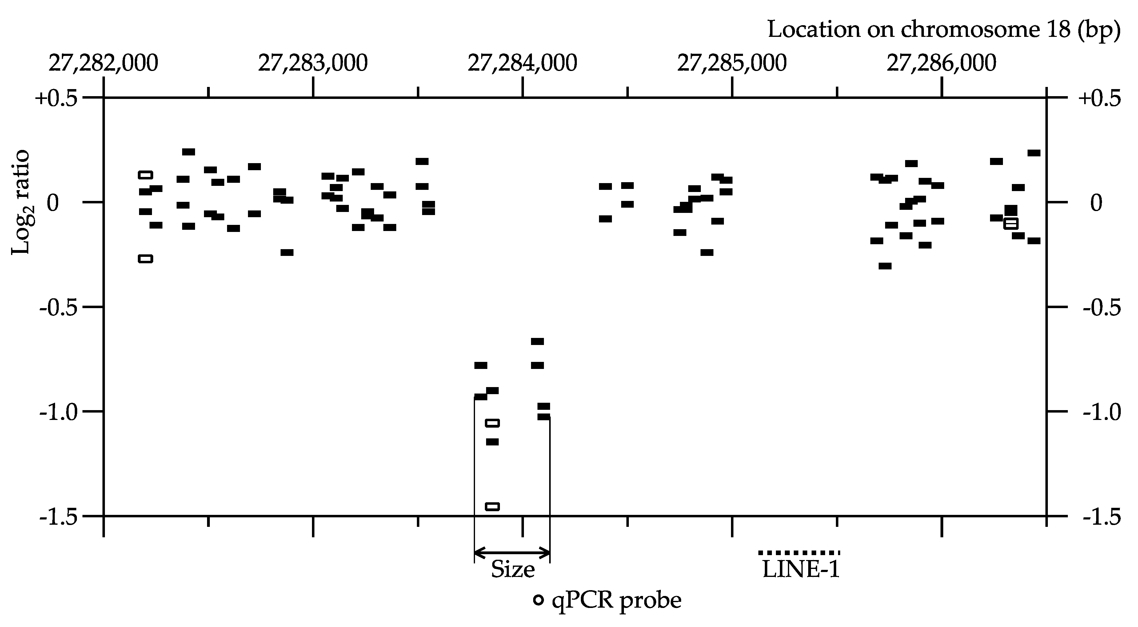
Figure 2 is an example of a small de novo CNV, with only one positive probe in the first array CGH. In this sample deletion, the probe at position 27,283,827, but not the probes at positions 27,282,170 and 27,286,300, showed lower log2 ratios in the first array CGH. Three additional neighboring probes also showed lower log2 ratios in the second array CGH.
De novo CNVs detected by array CGH and confirmed by qPCR are listed in Table 1, including F1 mouse identity, mutation type (deletion or duplication), number of the positive probes in the first and second array CGH, estimated size, chromosomal location, and involved genes. Most of the confirmed de novo CNVs were deletions. In the non-irradiated group, 11 deletions and three duplications were found, while 31 deletions and one duplication were found in the irradiated group.
Table 2 shows the result of the classification of these confirmed CNVs and also the CNV candidates that were detected by using array CGH but were not confirmed by qPCR, according to the number of the positive probes in the first and the second array CGH. All of large CNV candidates, with two or more positive probes in the first array CGH, were confirmed by qPCR. Some of smaller CNV candidates, with only one positive probe in the first array CGH and four or less positive probes in the second array CGH, were not confirmed by qPCR. In this study, we named the CNVs found in the first array CGH with only one positive probe as “Type S” and those with two or more positive probes as “Type L”.
Table 1 and Table 2 include four mice (0mGyH5, 20mGyAA4, 20mGyN4, and 20mGyY1) with two CNVs (deletions) each. Table 3 lists five mice with four or more CNVs. We found 9–108 de novo CNV candidates (all were Type-S deletions) in each of these mice, using array CGH. Further analysis of some of these de novo CNV candidates, using qPCR, confirmed that each mouse had at least four de novo CNVs (Supplementary Materials Table S1). Assuming that the number of de novo CNVs in each mouse follows a Poison distribution, we suspect that there is a possibility that the origins of the CNVs in these five mice with multiple CNVs may be somewhat different from those in mice with only two or less CNVs.
When comparing “mutation rates” between the non-irradiated and irradiated groups, we elected to use the number of F1 mice with de novo CNV(s) (Table 4), instead of the number of de novo CNVs, in order to preclude the influence of mice with multiple CNVs. In the non-irradiated control group, 7.1% of the F1 mice (11 out of 156) had de novo deletion(s), whereas, 20.4% (29 out of 142) F1 mice from the irradiated group had de novo deletion(s). This represents a significant 2.9-fold increase in the number of F1 mice with de novo deletion(s) in the irradiated group.
CNVs detected in other studies [19,21] appear to correspond to our Type-L CNVs, so we performed similar calculations based on the number of F1 mice with Type-L CNVs. De novo Type-L deletions were found in 5.1% (8 out of 156) of the F1 mice in the non-irradiated control group and 15.5% (22 out of 142) of the F1 mice in the irradiated group, showing a significant 3.0-fold increase (Table 4).
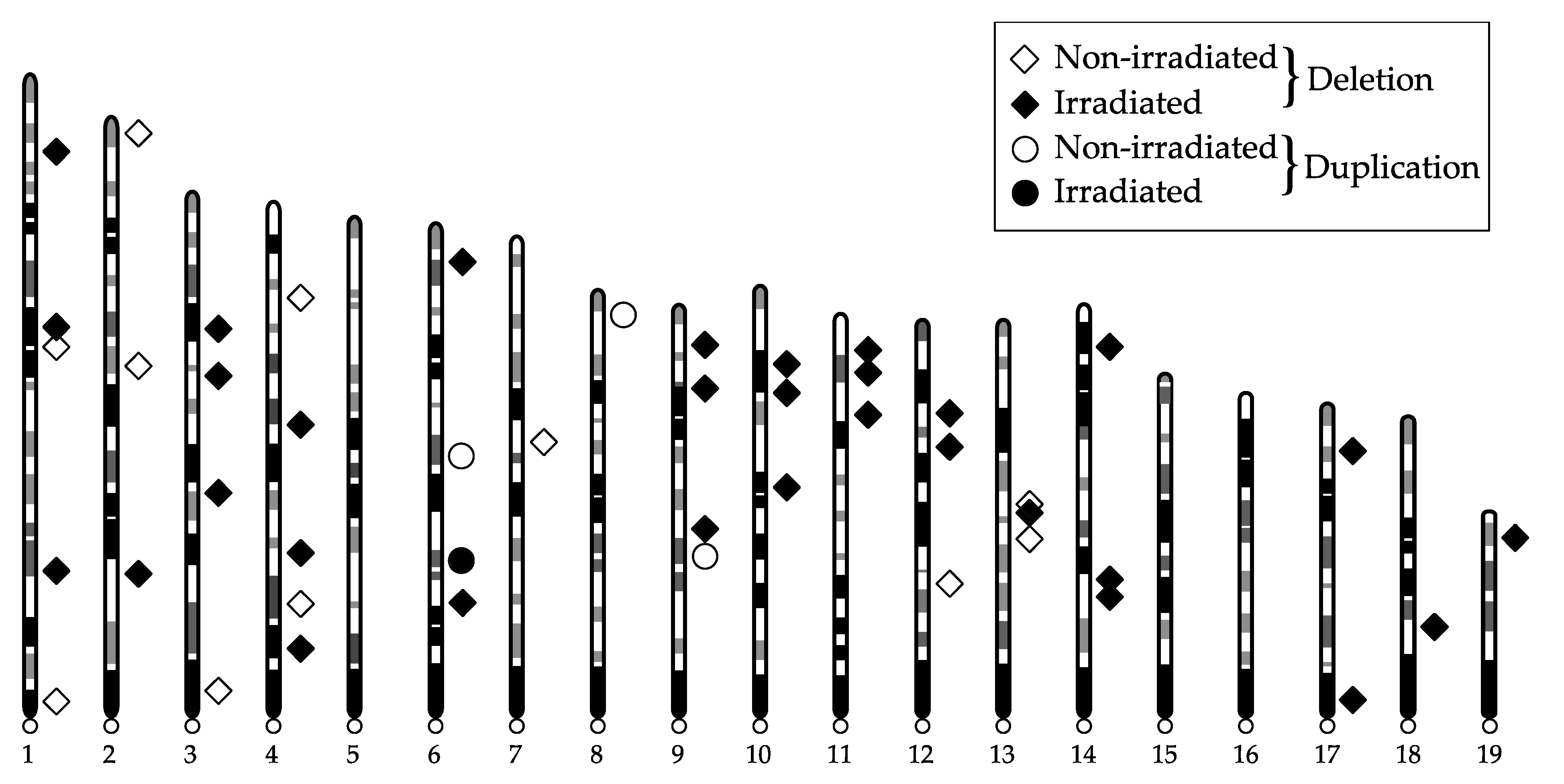
The distribution of de novo CNVs on individual chromosomes is illustrated in Figure 3. We did not observe any clustering of CNVs in any specific chromosome.
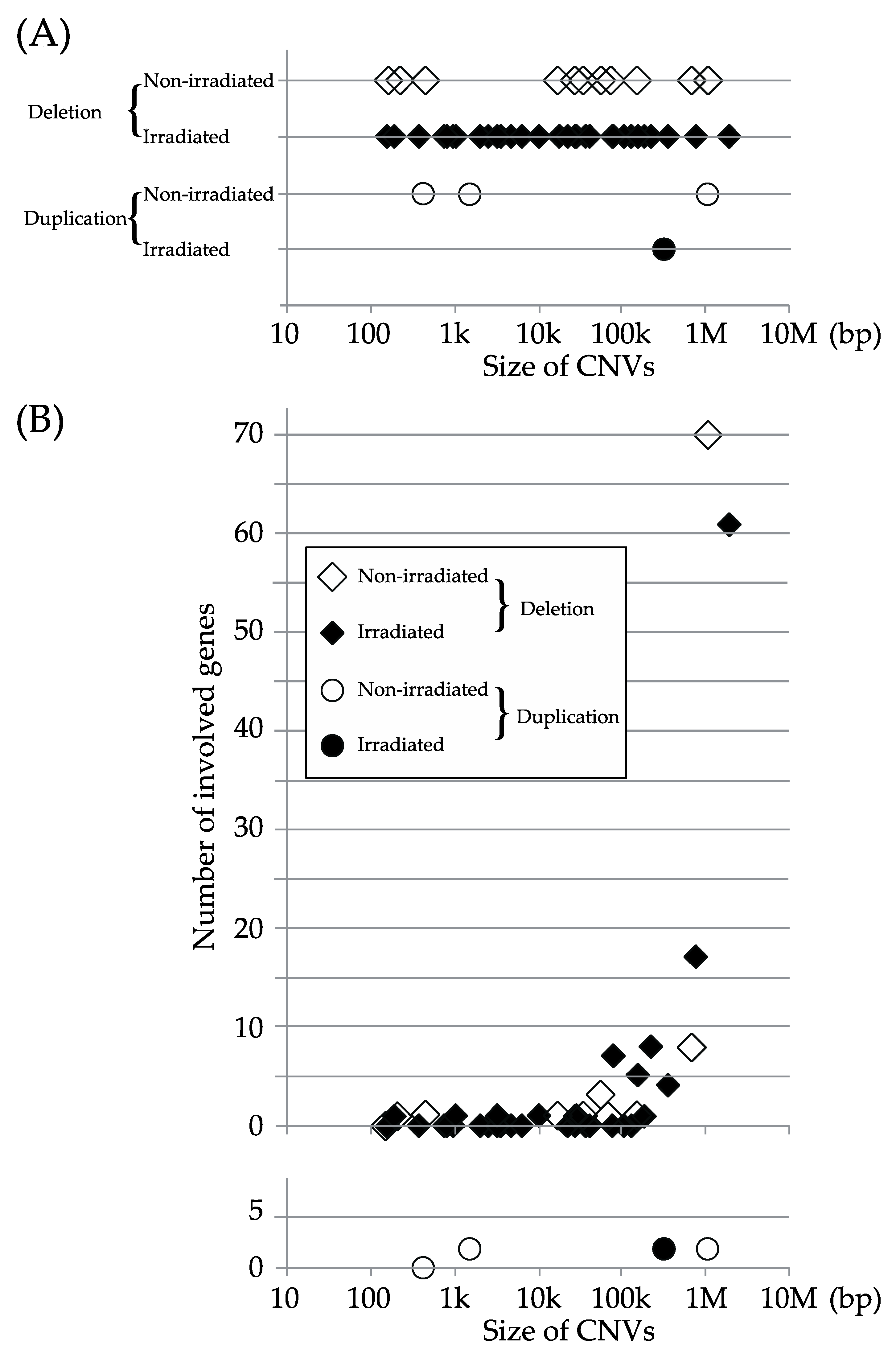
Figure 4A depicts the distribution of CNVs based on size, and Figure 4B shows the relationship between the number of genes in the region of each CNV and the size of the CNV. Most of the CNVs contained 0–2 genes, but some of large CNVs (>50,000 bp) contained 3–70 genes. The sizes of the deletions were compared between the non-irradiated and irradiated groups, using log-transformed values, since there were considerable variations in size. No significant difference was detected (p = 0.6758) in the size of the deletions between the groups, using the t-test (Table 5).
Since the samples used in the present study were part of a large experiment wherein both the sires (F0) and the F1 mice were allowed to live out their natural life span, we compared the life spans of the F1 mice analyzed in this study. Table 6 shows the mean life span of the F1 mice classified based on radiation exposure, sex, and the presence of CNVs. The effects of these three factors were evaluated by using the Cox proportional hazard regression analysis and are summarized in Table 7. The effect of sex on life span, expressed in terms of hazard ratio, was highly significant (p < 0.0001), wherein males had longer life spans than females. The effect of radiation exposure on life span was not significant (p = 0.1899), probably because of the small number of mice studied. The effect of CNVs on life span, however, was significant (p = 0.0019), suggesting a possibility that the presence of CNV(s) may be associated with shorter life spans.

3. Discussion

In this study, by using array CGH, we detected an increase in the number of mice bearing de novo CNVs (deletions) in the progeny of the male mice exposed chronically to low dose-rate radiation.
Recently, two groups [19,21] have reported the use of array CGH to detect increases in the number of de novo CNVs (deletions) in the progeny of the male mice exposed to acute high dose-rate radiation. Adewoye et al. [19] exposed male C57BL/6 mice to 3 Gy of X-rays at a high dose-rate and then bred them to CBA/Ca females and detected one de novo deletion (1.1%) in the progeny (n = 93) of the non-irradiated mice and 16 de novo deletions (9.5%) in the progeny (n = 169) of the irradiated male mice, using Roche NimbleGen CGH arrays (probe spacing: ~1.1 kb, Roche NimbleGen, Inc., Pleasanton, CA, USA). Asakawa et al. [21] exposed male C57BL/6J mice to 4 Gy of gamma-rays at a high dose-rate and then bred them to C3H females and detected six de novo deletions (6%) in the progeny (n = 100) of the non-irradiated mice and eight de novo deletions (8%) in the progeny (n = 100) of the irradiated mice, using Roche NimbleGen CGH arrays (probe spacing: ~1 kb or ~1.5 kb). The present study extends their findings from the effects at high dose-rates to a low dose-rate.
Our results show that the numbers of mice with Type-L deletions, which we considered similar to those detected in the previous reports, were 5.1% (8 out of 156 mice) and 15.5% (22 out of 142 mice) in the F1 mice of the non-irradiated control and the irradiated (20 mGy/day × 400 days; total dose: 8 Gy) groups, respectively. The spontaneous and induced mutation rates in these studies [19,21], including the current study, appear to be of the same magnitude, but it is difficult to conclude whether radiation exposures at acute high dose-rates are more effective in inducing deletions than chronic low dose-rates [5,6,7], because the numbers of the observed mutation events were insufficient and the dose–response relationships were not obtained in these studies. A difference has been suggested between high dose(-rate) radiation and low dose(-rate) radiation: low dose(-rate) radiation cannot fully induce the DNA damage response in cells [26], resulting in insufficient DNA repair [27]. The influence of such a phenomenon on mutation frequency, however, would be difficult to estimate, since non-homologous end joining, which is the major repair pathway of DNA double-strand breaks in higher eukaryotes and is probably responsible for the formation of many of large deletions, is error-prone or rather mutagenic [28,29].
We were unable to detect any significant difference in the size of deletions between the non-irradiated control and the low dose-rate irradiated groups. This is in sharp contrast with the results of the studies on the transgenerational effects of radiation exposures at high dose-rates, including the two studies by Adewoye et al. [19] and Kodaira et al. [22], and is in accordance with the notion that acute irradiation at high dose-rates predominantly induces larger deletions, whereas chronic irradiation at low dose-rates does not [24]. A more detailed analysis on the molecular nature of a large number of deletions is deemed necessary.
Currently, WGS techniques cannot detect deletions larger than ~50 bp in size, whereas array CGH analysis systems cannot detect deletions smaller than ~5 kb in size, resulting in a “gap” in the size of the deletions that can be identified and analyzed in mutation induction research. In an attempt to overcome this difficulty, we used arrays with very short probe spacing (~0.1 kb) in the second array CGH, so that we could detect CNVs smaller than 1 kb. Due to the prohibitive costs of the fine analysis, we had to use the arrays with larger or common probe spacing (~2 kb) for the first array CGH, such that most of small deletions would remain undetected and lost during this step. Nevertheless, the successful detection of some small deletions suggests that the use of high density probes or “tiling arrays” is one potential approach that may fill the gap in the size of detectable deletions. In the present study, we found five F1 mice bearing multiple (four or more) small (Type S) deletions. We suspect that the use of the arrays with common probe spacing in the single CGH step used in other studies might preclude the detection of such a phenomenon, which could be related to radiation-induced genome instability [30,31]. We hope that, in the future, a whole spectrum of radiation-induced mutations, from single-nucleotide changes to gross chromosomal alterations, can be revealed through the use of high density tiling arrays, together with improved WGS and chromosome analysis.
Cox proportional hazard regression analysis of the life span of the F1 mice suggested a possible association of the presence of CNV(s) with shorter life spans. Since all the analyzed F1 mice were viable, it is clear that they did not have classic dominant lethal mutations. In the list of the genes in the CNV regions (Table 1), we did not find any genes that appeared dominant deleterious. We cannot, however, discount the possibility that the alterations in copy number of some of the genes could be weakly deleterious and might slightly affect the life span. Although very preliminary, this approach might be helpful in elucidating the relationships between genotype and phenotype that have been considered to be one of the most important factors in assessing the transgenerational effects of radiation [5].
At the present time, there is no clear evidence of the transgenerational effects of radiation in humans, because of various technical difficulties. Data from laboratory animal experiments are far from sufficient. Thus, uncertainty and controversy still exist. For example, the dose–response relationships and the dose-rate effect are not certain especially in the range of low doses or low dose-rates, and there are a variety of models and opinions on this matter [30,31,32,33]. We hope that the experimental approaches described in this report will contribute towards finding a solution to these controversies.

4. Materials and Methods

4.1. Animals, Irradiation and DNA Isolation

The samples analyzed in the present study were selected from a large experiment on the transgenerational effects on life span and cancer incidence in the progeny of male mice chronically exposed to low dose-rate radiation (unpublished) referred to in a review [34]. Briefly, we irradiated specific pathogen-free male C57BL/6J/Nrs mice with 137Cs gamma-rays at a low dose-rate of 20 mGy/day for 400 days from 8 weeks of age. Immediately after completion of the 400-day irradiation period, the male mice were bred to non-irradiated 8-week-old virgin female C57BL/6J/Nrs mice to produce F1 mice. Tail samples were collected from dams (F0) at the time of euthanasia after weaning, and from sires (F0) and F1 mice at the time of natural death, and they were stored frozen at −80 °C until analyzed. Genomic DNA was extracted from the tail samples by a modified phenol-chloroform method with the NR-201 animal tissue DNA isolation reagent kit (Kurabo Industries Ltd., Osaka, Japan). Animal experiments were conducted according to legal regulations in Japan and the Guidelines for Animal Experiments of the Institute for Environmental Sciences (IES).

4.2. Array CGH

Array CGH data were deposited in the Gene Expression Omnibus (GEO) repository (accession number: GSE89425).
For the first screening by array CGH, we designed a custom 1 × 1 M array, using the eArray (currently updated to SureDesign, Agilent Technologies, Santa Clara, CA, USA) tool and database. We selected ~1 million autosomal probes (probe spacing: ~2 kb) from the ~19.1 million mouse CGH probes (probe spacing: ~0.1 kb) in the database. A small number of sex-linked probes for the determination of the sex of the mice were also selected. Array CGH experiments were performed according to the Agilent Oligonucleotide Array-Based CGH for Genomic DNA Analysis, Protocol (version 4.0.8, Agilent Technologies, Santa Clara, CA, USA). Using reference DNA from a male mouse from the in-house breeding colony at IES, we performed dye-swap hybridization experiments. An Agilent G2565BA scanner and its Feature Extraction software (version 10.7.1.1, Agilent Technologies, Santa Clara, CA, USA) and Genomic Workbench software (version 7.0, Agilent Technologies, Santa Clara, CA, USA) were used for image acquisition, data extraction, and data analysis, respectively. When both of values of |log2 ratio| for a probe, obtained in two dye-swap hybridization experiments, exceeded 0.8, the probe was considered positive (in an aberrant region). When all of the values of |log2 ratio| for two adjacent probes, in two dye-swap hybridization experiments, exceeded 0.5, these probes were also considered positive. For each of these positive probes, we selected 10 or more neighboring probes (probe spacing: ~0.1 kb) from the Agilent SureDesign database and designed custom 4 × 144 k or 8 × 60 k arrays. These arrays were used in the dye-swap hybridization experiments in the second screening. A positive probe found in the first screening was considered positive in the second screening when these two conditions were satisfied: (1) a probe positive in the first screening showed the |log2 ratio| values exceeding 0.5 in the second screening, and (2) at least one newly selected neighboring probe (within 3 probes) showed the |log2 ratio| values exceeding 0.5 in the second screening.

4.3. Quantitative PCR (qPCR)

We performed real-time qPCR to confirm the CNV candidates detected by array CGH, using the predesigned TaqMan Copy Number Assays probes (Thermo Fisher Scientific, Waltham, MA, USA) and the Applied Biosystems 7500 instrument (Thermo Fisher Scientific, Waltham, MA, USA). The locations of the TaqMan probes are described in Table 1. Transferrin receptor protein 1 (Tfrc1) was used as a reference gene. It is an autosomal single-copy housekeeping gene that is widely used as a reference for quantitative analysis of DNA and RNA. Amplification was performed according to the manufacturer’s protocol, in a dilution series (1.25, 2.5, and 5 ng (or 2.5, 5, and 10 ng in some experiments) of genomic DNA in a reaction volume of 20 μL), in duplex (the reference probe and the target probe were labeled with VIC [2′-chloro-7′-phenyl-1,4-dichloro-6-carboxyfluorescein] and FAM [5-(and-6)-carboxyfluorescein], respectively), and in triplicate. The relative standard curve method was used for data analyses.

4.4. Statistics

We compared the numbers of the mice with CNV(s) in the non-irradiated group and the irradiated group by using Fisher’s exact test. To compare the sizes of the deletions, we used geometric means instead of arithmetic means, since the sizes varied widely from 153 to 1,908,155 bp (4 orders of magnitude). The geometric means were compared by t-test on the log-transformed values after confirmation of the equality of variances by F-test. We analyzed the effects of sex, irradiation, and CNV(s) on the life span of the mice, using the Cox proportional hazard regression model. We conducted the tests on the deletion size and on the life span in R software v.4.0.0 (The R Foundation, Vienna, Austria) [35]. We used 0.05 as the threshold of statistical significance.

Supplementary Materials

The following is available online at https://www.mdpi.com/article/10.3390/ijms222212437/s1.

Author Contributions

Conceptualization, K.O. and S.T.; methodology, K.O.; validation, K.O.; formal analysis, Y.A. and S.T.; investigation, K.O. and C.H.; resources, S.T.; data curation, K.O.; writing—original draft preparation, K.O. and J.-i.K.; writing—review and editing, I.B.T.III; visualization, K.O. and J.-i.K.; supervision, J.-i.K. All authors have read and agreed to the published version of the manuscript.

Funding

This study was performed under a contract with the government of Aomori Prefecture, Japan.

Institutional Review Board Statement

Animal experiments were conducted according to legal regulations in Japan and the Guidelines for Animal Experiments of the Institute for Environmental Sciences.

Data Availability Statement

Array CGH data were deposited in the Gene Expression Omnibus (GEO) repository (accession number: GSE89425).

Conflicts of Interest

The authors declare no conflict of interest.

References

  1. Muller, H.J. Artificial transmutation of the gene. Science 1927, 66, 84–87. [Google Scholar] [CrossRef] [PubMed]
  2. Nakamura, N.; Suyama, A.; Noda, A.; Kodama, Y. Radiation effects on human heredity. Annu. Rev. Genet. 2013, 47, 33–50. [Google Scholar] [CrossRef] [Green Version]
  3. Nakamura, N. Why genetic effects of radiation are observed in mice but not in humans. Radiat. Res. 2018, 189, 117–127. [Google Scholar] [CrossRef] [PubMed] [Green Version]
  4. Nakamura, N. History of radiation genetics: Light and darkness. Int. J. Radiat. Biol. 2019, 95, 999–1014. [Google Scholar] [CrossRef] [PubMed]
  5. United Nations Scientific Committee on the Effects of Atomic Radiation. UNSCEAR 2001 Report to the General Assembly, with Scientific Annex, Hereditary Effects of Radiation; United Nations: New York, NY, USA, 2001. [Google Scholar]
  6. International Commission on Radiological Protection. ICRP Publication 103, The 2007 Recommendations of the International Commission on Radiological Protection. Ann. ICRP 2007, 37, 2–4. [Google Scholar]
  7. Russell, W.L.; Kelly, E.M. Mutation frequencies in male mice and the estimation of genetic hazards of radiation in men. Proc. Natl. Acad. Sci. USA 1982, 79, 542–544. [Google Scholar] [CrossRef] [Green Version]
  8. Lyon, M.F.; Morris, T. Gene and chromosome mutation after large fractionated or unfractionated radiation doses to mouse spermatogonia. Mutat. Res. 1969, 8, 191–198. [Google Scholar] [CrossRef]
  9. Awa, A.A.; Honda, T.; Neriishi, S.; Sufuni, T.; Shimba, H.; Ohtaki, K.; Nakano, M.; Kodama, Y.; Itoh, M.; Hamilton, H.B. Cytogenetic Study of the offspring of atomic bomb survivors, Hiroshima and Nagasaki. In The Children of Atomic Bomb Survivors: A Genetic Study; Neel, J.V., Schull, W.J., Eds.; National Academies Press: Washington, DC, USA, 1991; pp. 344–362. [Google Scholar]
  10. Neel, J.V.; Satoh, C.; Goriki, K.; Asakawa, J.; Fujita, M.; Takahashi, N.; Kageoka, T.; Hazama, R. Search for mutations altering protein charge and/or function in children of atomic bomb survivors: Final report. Am. J. Hum. Genet. 1988, 42, 663–676. [Google Scholar]
  11. Kodaira, M.; Shizue Izumi, S.; Takahashi, N.; Nakamura, N. No evidence of radiation effect on mutation rates at hypervariable minisatellite loci in the germ cells of atomic bomb survivors. Radiat. Res. 2004, 162, 350–356. [Google Scholar] [CrossRef]
  12. Horai, M.; Mishima, H.; Hayashida, C.; Kinoshita, A.; Nakane, Y.; Matsuo, T.; Tsuruda, K.; Yanagihara, K.; Sato, S.; Imanishi, D.; et al. Detection of de novo single nucleotide variants in offspring of atomic-bomb survivors close to the hypocenter by whole-genome sequencing. J. Hum. Genet. 2018, 63, 357–363. [Google Scholar] [CrossRef] [Green Version]
  13. Winther, J.F.; Olsen, J.H.; Wu, H.; Shyr, Y.; Mulvihill, J.J.; Stovall, M.; Nielsen, A.; Schmiegelow, M.; Boice, J.B.J. Genetic disease in the children of Danish survivors of childhood and adolescent cancer. J. Clin. Oncol. 2012, 30, 27–33. [Google Scholar] [CrossRef] [Green Version]
  14. Holtgrewe, M.; Knaus, A.; Hildebrand, G.; Pantel, J.T.; de los Santos, M.R.; Neveling, K.; Goldmann, J.; Schubach, M.; Jäger, M.; Coutelier, M.; et al. Multisite de novo mutations in human offspring after paternal exposure to ionizing radiation. Sci. Rep. 2018, 8, 14611. [Google Scholar] [CrossRef] [Green Version]
  15. Dubrova, Y.E.; Nesterov, V.N.; Krouchinsky, N.G.; Valdislav, A.; Ostapenko, V.A.; Neumann, R.; Neil, D.L.; Jeffreys, A.J. Human minisatellite mutation rate after the Chernobyl accident. Nature 1996, 380, 683–686. [Google Scholar] [CrossRef] [PubMed]
  16. Yeager, M.; Machiela, M.J.; Kothiyal, P.; Dean, M.; Bodelon, C.; Suman, S.; Wang, M.; Mirabello, L.; Nelson, C.W.; Zhou, W.; et al. Lack of transgenerational effects of ionizing radiation exposure from the Chernobyl accident. Science 2021, 372, 725–729. [Google Scholar] [CrossRef] [PubMed]
  17. Costa, E.O.A.; Pinto, I.P.; Gonçalves, M.W.; da Silva, J.F.; Oliveira, L.G.; da Cruz, A.S.; Silva, D.M.E.; da Silva, C.C.; Pereira, R.W.; da Cruz, A.D. Small de novo CNVs as biomarkers of parental exposure to low doses of ionizing radiation of caesium-137. Sci. Rep. 2018, 8, 5914. [Google Scholar] [CrossRef] [PubMed] [Green Version]
  18. Leite Filho, H.P.; Pinto, I.P.; Oliveira, L.G.; Costa, E.O.A.; da Cruz, A.S.; Silva, D.M.; da Silva, C.C.; Caetano, A.R.; da Cruz, A.D. Deviation from Mendelian transmission of autosomal SNPs can be used to estimate germline mutations in humans exposed to ionizing radiation. PLoS ONE 2020, 15, e0233941. [Google Scholar] [CrossRef]
  19. Adewoye, A.B.; Lindsay, S.J.; Dubrova, Y.E.; Matthew, E.; Hurles, M.E. The genome-wide effects of ionizing radiation on mutation induction in the mammalian germline. Nat. Commun. 2015, 6, 6684. [Google Scholar] [CrossRef] [Green Version]
  20. Satoh, Y.; Asakawa, J.; Nishimura, M.; Kuo, T.; Shinkai, N.; Cullings, H.M.; Minakuchi, Y.; Sese, J.; Toyoda, A.; Shimada, Y.; et al. Characteristics of induced mutations in offspring derived from irradiated mouse spermatogonia and mature oocytes. Sci. Rep. 2020, 10, 37. [Google Scholar] [CrossRef]
  21. Asakawa, J.; Kodaira, M.; Miura, A.; Tsuji, T.; Nakamoto, Y.; Imanaka, M.; Kitamura, J.; Cullings, H.; Nishimura, M.; Shimada, Y.; et al. Genome-wide deletion screening with the array CGH method in mouse offspring derived from irradiated spermatogonia indicates that mutagenic responses are highly variable among genes. Radiat. Res. 2016, 186, 568–576. [Google Scholar] [CrossRef] [Green Version]
  22. Kodaira, M.; Asakawa, J.; Nakamura, N. Radiation-induced deletions in mouse spermatogonia are usually large (over 200 kb) and contain little sequence similarity at the junctions. Radiat. Res. 2017, 187, 722–731. [Google Scholar] [CrossRef] [Green Version]
  23. Strobel, M.C.; Seperack, P.K.; Copeland, N.G.; Jenkins, N.A. Molecular analysis of two mouse dilute locus deletion mutations: Spontaneous dilute lethal20J and radiation-induced dilute prenatal lethal Aa2 alleles. Mol. Cell. Biol. 1990, 10, 501–509. [Google Scholar]
  24. Russell, L.B.; Hunsicker, P.R. The effect of dose rate on the frequency of specific-locus mutations induced in mouse spermatogonia is restricted to larger lesions; a retrospective analysis of historical data. Radiat. Res. 2012, 177, 555–564. [Google Scholar] [CrossRef] [PubMed]
  25. Sankaranarayanan, K.; Wassom, J.S. Ionizing radiation and genetic risks XIV. Potential research directions in the post-genome era based on knowledge of repair of radiation-induced DNA double-strand breaks in mammalian somatic cells and the origin of deletions associated with human genomic disorders. Mutat. Res. 2005, 578, 333–370. [Google Scholar]
  26. Collis, S.J.; Schwaninger, J.M.; Ntambi, A.J.; Keller, T.W.; Nelson, W.G.; Dillehay, L.E.; DeWeese, T.L. Evasion of early cellular response mechanisms following low level radiation-induced DNA damage. J. Biol. Chem. 2004, 279, 49624–49632. [Google Scholar] [CrossRef] [Green Version]
  27. Grudzenski, S.; Raths, A.; Conrad, S.; Rübe, C.E.; Löbrich, M. Inducible response required for repair of low-dose radiation damage in human fibroblasts. Proc. Natl. Acad. Sci. USA 2010, 107, 14205–14210. [Google Scholar] [CrossRef] [Green Version]
  28. Lieber, M.R. The mechanism of double-strand DNA break repair by the nonhomologous DNA end-joining pathway. Annu. Rev. Biochem. 2010, 79, 181–211. [Google Scholar] [CrossRef] [Green Version]
  29. Duncan, J.R.; Lieber, M.R.; Adachi, N.; Wahl, R.L. Radiation dose does matter: Mechanistic insights into DNA damage and repair support the linear no-threshold model of low-dose radiation health risks. J. Nucl. Med. 2018, 59, 1014–1016. [Google Scholar] [CrossRef] [Green Version]
  30. Dubrova, Y.E. Radiation-induced transgenerational instability. Oncogene 2003, 22, 7087–7093. [Google Scholar] [CrossRef] [Green Version]
  31. Niwa, O. Radiation Induced Dynamic Mutations and Transgenerational Effects. J. Radiat. Res. 2006, 47 (Suppl. B), 25–30. [Google Scholar] [CrossRef] [Green Version]
  32. Vilenchik, M.M.; Knudson, A.G.J. Inverse radiation dose-rate effects on somatic and germ-line mutations and DNA damage rates. Proc. Natl. Acad. Sci. USA 2000, 97, 5381–5386. [Google Scholar] [CrossRef] [Green Version]
  33. Ogura, K.; Magae, J.; Kawakami, Y.; Koana, T. Reduction in mutation frequency by very low-dose gamma irradiation of Drosophila melanogaster germ cells. Radiat. Res. 2009, 171, 1–8. [Google Scholar] [CrossRef] [PubMed]
  34. Tanaka, I.B., III; Tanaka, S.; Kohda, A.; Takai, D.; Nakamura, S.; Ono, T.; Tanaka, K.; Komura, J. Experimental studies on the biological effects of chronic low dose-rate radiation exposure in mice: Overview of the studies at the Institute for Environmental Sciences. Int. J. Radiat. Biol. 2018, 94, 423–433. [Google Scholar] [CrossRef] [PubMed]
  35. The R Project for Statistical Computing. Available online: https://www.R-project.org/ (accessed on 18 June 2021).
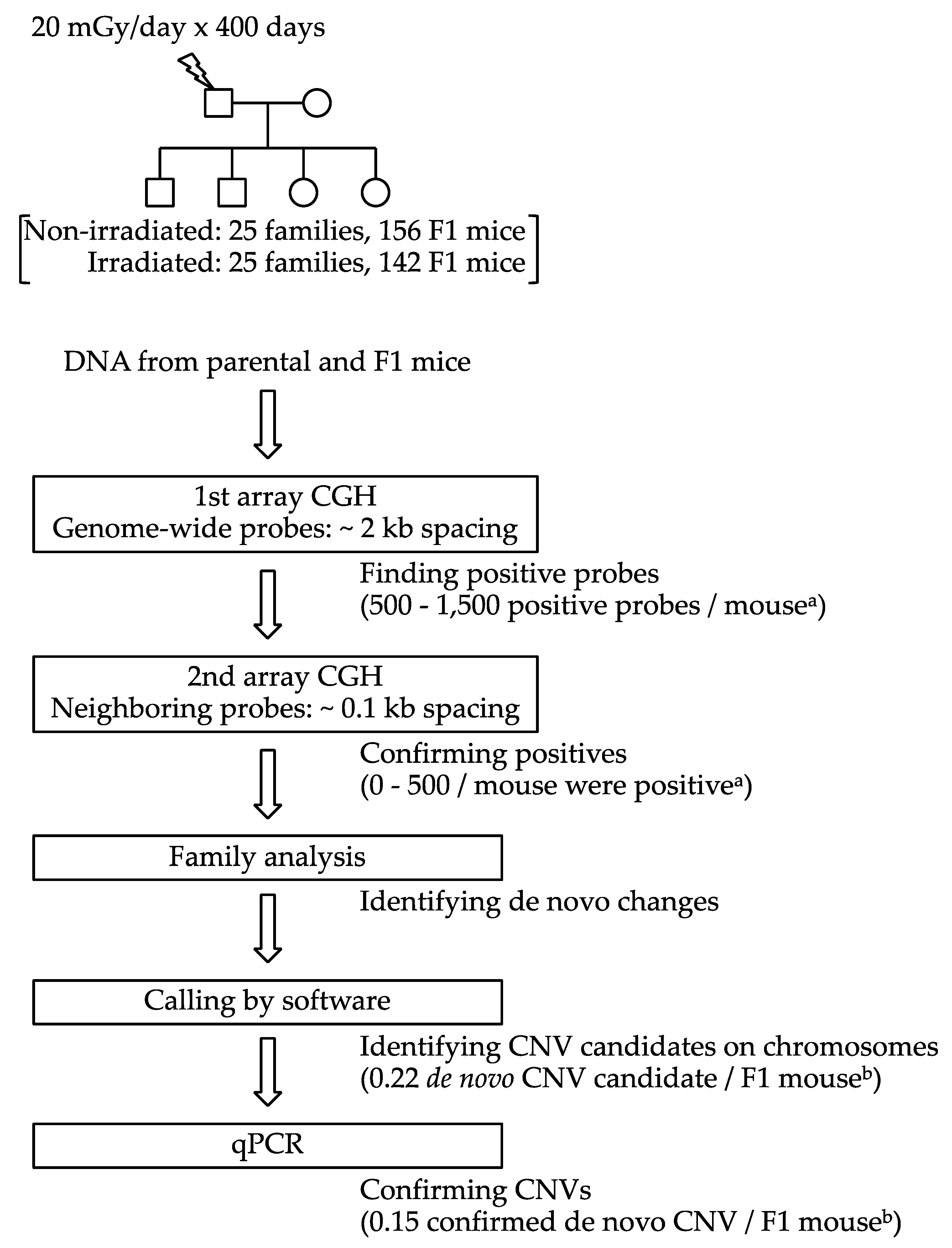
Figure 1. Experimental design for the identification of de novo copy number variations (CNVs). a Approximate numbers of probes for the first array CGH. b F1 mice bearing 4 or more confirmed de novo CNVs are excluded.
Ijms 22 12437 g001
Figure 2. Example of a de novo CNV. The copy number profile of one of the deletions found in an F1 mouse (ID: 20mGyY1). The horizontal axis represents the chromosomal location, and the vertical axis represents the Log2 ratio of fluorescence between the sample and the reference for each probe. The results of two hybridization experiments with dye swapping are plotted separately. Open rectangles indicate the results of the first array CGH; black rectangles indicate the results of the second array CGH; the double-headed arrow indicates the estimated size of the CNV; the small open circle indicates the location of the qPCR probe (TaqMan probe); and the dotted line indicates the location of the LINE-1 repetitive sequence, which might hamper probe design.
Ijms 22 12437 g002
Figure 3. Chromosomal distribution of de novo CNVs.
Ijms 22 12437 g003
Figure 4. (A) Size of de novo CNVs. (B) Number of genes in the region of each de novo CNV plotted against the CNV size.
Ijms 22 12437 g004
Table 1. List of confirmed de novo CNVs.
F1 MouseNo. of Positive ProbesSize (bp)Chromosomal Location (GRCm38)qPCR Probe Location (GRCm38)Involved Genes
IDSex1st Array CGH2nd Array CGH
Non-IrradiatedDeletion0mGyX3M11>501,103,254Chr13:66,699,838–67,803,09266,700,397Gm40988, Gm48404, Gm40989, Gm7896, Gm48413, Gm40989, Vmn2r-ps108, Gm48412, Gm48414, Gm10323, Vmn2r-ps109, Cbx3-ps4, Gm5451, Gm7911, Uqcrb, Gm10767, Mterf3, Ptdss1, 4933433G19Rik, Zfp712, Gm48705, Gm46440, Zfp708, Gm28557, Gm17938, Rslcan18, Zfp759, Gm48732, Gm48733, Rsl1, Gm49646, Gm9626, Zfp455, Gm49064, Zfp458, F630042J09Rik, Zfp457, Gm48824, Zfp595, Gm28044, Zfp593, Zfp456, Gm17039, Zfp953, Gm28041, Zfp429, Gm48900, Zfp459, Zfp874a, Gm38307, Gm7928, Zfp874b, Zfp58, Gm26965, Zfp87, Zfp748, 9430065F17Rik, Zfp729b, Gm49345, Zfp729a, Zfp738, Gm48095, Gm26875, Zfp65, Gm48894, Zfp85, Gm9894, Zfp493, 4930525G20Rik
0mGyH5 aM7>50666,200Chr2:177,329,278–177,995,478178,041,392Gm14414, Zfp970, Gm14403, Gm14324, Gm14322, Gm14326, Gm14327, Rps8-ps5
0mGyH5 aM22>50146,136Chr12:40,469,827–40,615,96340,596,701Dock4
0mGyG2M19>5074,519Chr1:112,845,241–112,919,760112,877,741Gm8204
0mGyS6F14>5056,272Chr7:102,192,468–102,248,740102,193,027Nup98, Pgap2, Rhog
0mGyK4F9>5034,160Chr3:5,533,449–5,567,6095,560,033Pex2
0mGyK5F10>5025,412Chr13:54,919,455–54,944,86754,942,2914930526F13Rik
0mGyJ3F6>5017,346Chr4:128,415,985–128,433,331128,416,544Csmd2
0mGyV2M12461Chr2:126,675,275–126,675,736126,675,329Gabpb1
0mGyA3F12212Chr4:34,844,952–34,845,16434,845,005Zfp292
0mGyO7F12153Chr1:7,323,388–7,323,5417,323,321
Duplication0mGyZ1M158>501,100,262Chr6:79,988,064–81,088,32679,988,623Lrrtm4, Gm43900
0mGyE4F1131535Chr8:123,427,701–123,429,236123,429,010Galnt2, Def8
0mGyG5F13412Chr9:49,721,288–49,721,70049,721,288Ncam1
IrradiatedDeletion20mGyL5M575>501,908,155Chr13:63,399,812–65,307,96765,307,023Fancc, patch1, A930032L01 Rik, Gm30655, Gm30709, 1700024I08 Rik, Gm47387, Gm47390, Gm47417, Gm47418, Ercc6l2, Gm7695, Hsd17b3, Slc35d2, Zfp367, Gm47513, Habp4, Cdc14b, Gm46424, 1810034E14 Rik, Gm47004,Gm47003, Gm25654, Prxl2c, Zfp182, Gm47123, Gm49230, Ctsl, 1700015C15 Rik, Cdk20, Gm31218, Fam240b, Gm7712, Gm47190, Gm4810, Gm47191, Gm47193, Gm5791, Gm4935, Gm47194, Gm7065, Cntnap3, Spata31, Eif1-ps2, Prss47, Mfsd14b, Spata31d1c, Gm6888, Gm3785, Olfr465-ps1, Olfr466, Gm24130, Nlrp4f, Gm47249, Gm36445, Gm47250, Gm47251, Gm47254, Gm10775, Zfp369, Gm47258
20mGyI5F200>50742,233Chr10:99,214,798–99,957,03199,959,613Gm34777, Gm34574, Gm48884, Dusp6, Gm48089, B530045E10 Rik, Gm34921, Gad1, Gm34983, Gm35035, Gm48085, Gm20110, Gm18409, Gm35101, Gm47578, Csl, Gm47579
20mGyL8F145>50353,159Chr3:105,510,798–105,863,957105,511,357AK018929, Kcnd3
20mGyE1M83>50235,104Chr9:57,625,823–57,860,92757,854,366Csk, Cyp1a2, Cyp1a1, Edc3, Clk3, Gm17231, Arid3b
20mGyL2M36>50145,401Chr14:37,914,599–38,060,00037,936,448Gm47974
20mGyA1M12>50133,572Chr9:114,085,760–114,219,332114,213,599
20mGyAA4 aF34>50102,921Chr1:117,376,753–117,479,674117,377,055
20mGyU6F35>5076,287Chr1:172,286,528–172,362,815172,287,087Igsf8, Atp1a2, Kcnj9, Gm36937, Kcnj10
20mGyV6F29>5075,893Chr2:45,606,923–45,682,81645,607,482
20mGyW1M14>5040,175Chr19:54,228,002–54,268,17754,228,561
20mGyL4M8>5032,881Chr6:138,709,973–138,742,854138,741,398
20mGyU8F7>5029,202Chr9:100,793,927–100,823,129100,794,486Stag1
20mGyX1M12>5028,507Chr12:92,023,170–92,051,67792,047,867
20mGyV1M8>5022,299Chr11:111,762,690–111,784,989111,763,249
20mGyN4 aM63210,224Chr6:34,932,670–34,942,89434,941,090Stra8
20mGyV4F4>509424Chr10:107,833,661–107843085107,834,220Otogl
20mGyX2F3405965Chr14:112,914,840–112,920,805112,919,755
20mGyAA4aF2334495Chr17:81,065,337–81,069,83281,069,694
20mGyU5M2233469Chr11:92,816,645–92,820,11492,817,204
20mGyY1 aM2163370Chr11:105,950,869–105,954,239105,951,424
20mGyG2M2163286Chr14:40,993,918–40,997,20440,996,166Prxl2a
20mGyN4 aM1192367Chr4:50,541,636–50,544,00350,543,175
20mGyG7F1171878Chr4:90,232,441–90,234,31990,234,260
20mGyB5F131040Chr10:69,420,081–69,421,12169,420,140Ank3-246
20mGyG5F19948Chr1:44,804,456–44,805,40444,804,512
20mGyX7F15732Chr4:21,163,257–21,163,98921,163,816
20mGyH3M18716Chr3:119,702,208–119,702,924119,702,263
20mGyY1 aM14356Chr18:27,125,326–27,125,68227,125,885
20mGyX5F12191Chr12:81,490,882–81,491,07381,491,441Synj2bp
20mGyB2F12156Chr17:4,507,025–4,507,1814,507,139
Duplication20mGyE5F86>50305,969Chr6:48,055,971–48,361,94048,360,175Zfp746, Gm16630
a Mice with 2 CNVs (mice with 4 or more CNVs are not included in this table).
Table 2. Number of the (candidate) CNVs detected by array CGH and confirmed by qPCR.
No. of Positive Probes in 1st Array CGH≥2 (Type L)1 (Type S)(Total)
No. of Positive Probes in 2nd Array CGH ≥5432(Total)
Deletion (del) or Duplication (dup)DelDupDelDupDelDupDelDupDelDupDelDupDelDup
Non-irradiated
(n = 156)
Detected by array CGH81011002134147228
Confirmed by qPCR810100013032113
Irradiated
(n = 142)
Detected by array CGH22150202030120341
Confirmed by qPCR2215010102090311
Table 3. List of the F1 mice bearing 4 or more confirmed de novo CNVs, with the number of the (candidate) CNVs detected by array CGH, analyzed by qPCR, and confirmed by qPCR.
No. of Positive Probes in 1st Array CGH≥2 (Type L)1 (Type S)(Total)
No. of Positive Probes in 2nd Array CGH ≥5432(Total)
Deletion (Del) or Duplication (Dup)DelDupDelDupDelDupDelDupDelDupDelDupDelDup
Non-irradiated0mGyJ4 (F)Detected by array CGH00001050130190190
Analyzed by qPCR00001050107070
Confirmed by qPCR00000030104040
0mGyX5 (F)Detected by array CGH00003090430550550
Analyzed by qPCR0000302050100100
Confirmed by qPCR00002020105050
Irradiated20mGyA2 (M)Detected by array CGH0014018027049010801080
Analyzed by qPCR00303050110220220
Confirmed by qPCR0030101060110110
20mGyF5 (F)Detected by array CGH00102030100160160
Analyzed by qPCR00102000100130130
Confirmed by qPCR00102000205050
20mGyL1 (M)Detected by array CGH00100020609090
Analyzed by qPCR00000020608080
Confirmed by qPCR00000010304040
Table 4. Number of the F1 mice with confirmed de novo CNVs.
No. of Positive Probes in 1st Array CGH≥2 (Type L)1 (Type S)(Total)
Deletion (Del) or Duplication (Dup)DelDupDelDupDelDup
Sex nn%pan%pan%pan%pan%pan%pa
MNon-irradiated7545.30.01811.3>0.111.3>0.100.0ND b56.60.02411.3>0.1
Irradiated751317.300.034.000.014 c18.700.0
FNon-irradiated8144.9>0.100.0>0.122.50.08522.4>0.167.40.00922.4>0.1
Irradiated67913.411.469.000.01522.411.4
TotalNon-irradiated15685.10.00410.6>0.131.90.07521.2>0.1117.10.00131.9>0.1
Irradiated1422215.510.796.300.02920.410.7
a Fisher’s exact test; irradiated group vs. corresponding non-irradiated group. b Not performed. c Two mice had both Type-L and Type-S deletions.
Table 5. Comparison of the sizes of the deletions found in the F1 mice of non-irradiated and irradiated groups.
No. of DeletionsDeletion Size (bp)
GM aGSD b
Non-irradiated1118,76212.181p = 0.6758 c
Irradiated3112,67421.484
a Geometric mean; b geometric standard deviation; c t-test on log-transformed data.
Table 6. Life span of F1 mice a.
Group of F1 MiceNo. of F1 MiceLife Span (Days)
(Mean ± SD)
MNon-irradiatedCNV− b70914±170
CNV+ c4859±161
IrradiatedCNV-56824±163
CNV+16881±155
FNon-irradiatedCNV-72813±132
CNV+8772±124
IrradiatedCNV-54833±140
CNV+10738±125
a Very short-lived (300 days or less) mice were excluded from analysis, after they were rejected as outliers (Smirnoff–Grubbs rejection test). b Without de novo CNVs. c With de novo CNV(s).
Table 7. Effects of sex, irradiation, and CNVs on life span of F1 mice.
FactorHazard RatioZ Valuep Value
Sex a0.507−5.40<0.0001
Irradiation a0.851−1.320.187
CNVs a0.697−2.000.045
a Factors of sex, irradiation, and CNVs are coded as nominal variables 0 and 1. Male, non-irradiated, and absence of de novo CNVs are assigned 0.
Publisher’s Note: MDPI stays neutral with regard to jurisdictional claims in published maps and institutional affiliations.