Article

Global Transcriptomic Analysis Reveals Differentially Expressed Genes Involved in Embryogenic Callus Induction in Drumstick (Moringa oleifera Lam.)

1
Department of Forestry, College of Forestry and Landscape Architecture, South China Agricultural University, Guangzhou 510642, China
2
Guangdong Key Laboratory for Innovative Development and Utilization of Forest Plant Germplasm, Guangzhou 510642, China
3
Guangdong Province Research Center of Woody Forage Engineering Technology, Guangzhou 510642, China
4
State Key Laboratory for Conservation and Utilization of Subtropical Agro-Bioresources, South China Agricultural University, Guangzhou 510642, China
*
Authors to whom correspondence should be addressed.
Int. J. Mol. Sci. 2021, 22(22), 12130; https://doi.org/10.3390/ijms222212130
Submission received: 9 October 2021 / Revised: 1 November 2021 / Accepted: 6 November 2021 / Published: 9 November 2021
(This article belongs to the Section Biochemistry)

Abstract

:
The plant embryogenic callus (EC) is an irregular embryogenic cell mass with strong regenerative ability that can be used for propagation and genetic transformation. However, difficulties with EC induction have hindered the breeding of drumstick, a tree with diverse potential commercial uses. In this study, three drumstick EC cDNA libraries were sequenced using an Illumina NovaSeq 6000 system. A total of 7191 differentially expressed genes (DEGs) for embryogenic callus development were identified, of which 2325 were mapped to the KEGG database, with the categories of plant hormone signal transduction and Plant-pathogen interaction being well-represented. The results obtained suggest that auxin and cytokinin metabolism and several embryogenesis-labeled genes are involved in embryogenic callus induction. Additionally, 589 transcription factors from 20 different families were differentially expressed during EC formation. The differential expression of 16 unigenes related to auxin signaling pathways was validated experimentally by quantitative real time PCR (qRT-PCR) using samples representing three sequential developmental stages of drumstick EC, supporting their apparent involvement in drumstick EC formation. Our study provides valuable information about the molecular mechanism of EC formation and has revealed new genes involved in this process.

1. Introduction

The plant embryogenic callus is an irregular embryogenic cell mass with strong regenerative ability. Under certain conditions, embryogenic callus cultured in vitro can regenerate into adventitious buds or form complete plants after somatic embryogenesis [1]. Although a callus can be generated at explant wound sites under the stimulation of exogenous hormones, only a small proportion of callus cells can differentiate into embryogenic callus. In addition to its frequent use for propagating various economically important species, embryogenic callus-based genetic transformation has emerged as the most successful plant transformation system in the last decade [2,3,4]. In plant tissue cultures, embryogenic callus formation is generally induced by optimizing the ratio of hormones in the medium [5,6,7]. Although hormones play central roles in embryogenic callus induction, this non-genetic method of optimizing culture conditions cannot provide culture schemes suitable for all genotypes [8,9]. Recent evidence indicates that embryogenic callus formation is a complex dynamic process that involves many intracellular metabolic changes and is influenced by various external environment factors [10,11].
Moringa oleifera Lam., commonly known as drumstick, belongs to the monogeneric family Moringaceae and is naturally distributed in the sub-Himalayan areas of India and various tropical African countries [12]. Drumstick is a tree with many potential commercial uses [13]. The vegetative organs have high levels of protein, vitamins, minerals, and phytochemicals and the seeds have high oil content [14]. In addition, this plant has a number of medicinal properties linked to antimicrobial [15], anti-inflammatory [16], detoxification [17], and anticancer activities [18]. Although it is a fast-growing tree species, it is difficult to quickly obtain good varieties through conventional breeding. Drumstick is dominated by outcrossing [19], the cultivated populations are separated in traits, and the seed activity declines quickly. It is difficult to obtain seedlings with consistent and uniform traits in production. Genetic engineering is an effective alternative method for introducing value-adding genes to modify important agronomic traits. Agrobacterium-mediated transformation via embryogenic callus has been used as a standard and reproducible method for transformation of many plants [20,21]. Embryogenic callus induction is therefore seen as a key step in transformation. We have developed an Agrobacterium-mediated transformation system for drumstick [22], but it has not been widely used due to its low transformation efficiency, which is largely due to the difficulty of inducing embryogenic callus formation. Embryogenic callus induction is thus a key bottleneck in drumstick regeneration and hinders its transgenic breeding. Ways of improving the rate of embryogenic callus induction are therefore needed to facilitate genetic engineering of this species.
Preliminary studies on drumstick embryogenic callus induction revealed that exogenous hormones had strong effects on induction, but the rates of induction and differentiation of embryogenic callus were low and genotype-dependent. Moreover, the changes in cellular structures and physiological status that occur during embryogenic callus induction are largely unknown, as are the details of the regulation of this process at the molecular level.
Callus induction is a complex biological process of cell dedifferentiation and redifferentiation that is regulated by genetic and epigenetic mechanisms [11,23]. Despite the importance of EC for drumstick genetic engineering, few studies have investigated the mechanisms underlying EC induction in this species. Transcriptome sequencing, assembly, and annotation is valuable because it allows large amounts of information to be gathered on transcribed regions relatively quickly and inexpensively. Therefore, to clarify the molecular mechanisms underlying acquisition of embryogenic potential during this process, a comparative analysis of transcriptome dynamics at different stages of EC induction was conducted. The major pathways and key events in embryogenic callus development were identified through deep analysis of differentially expressed genes (DEGs) together with histological and chemical analyses of samples representing different stages of embryogenic callus formation. This led to new insights into the molecular regulatory networks regulating embryogenic callus formation. Our results provide valuable information on the molecular mechanism by which somatic cells are converted into embryogenic callus in drumstick and reveal novel genes involved in this process.

2. Results

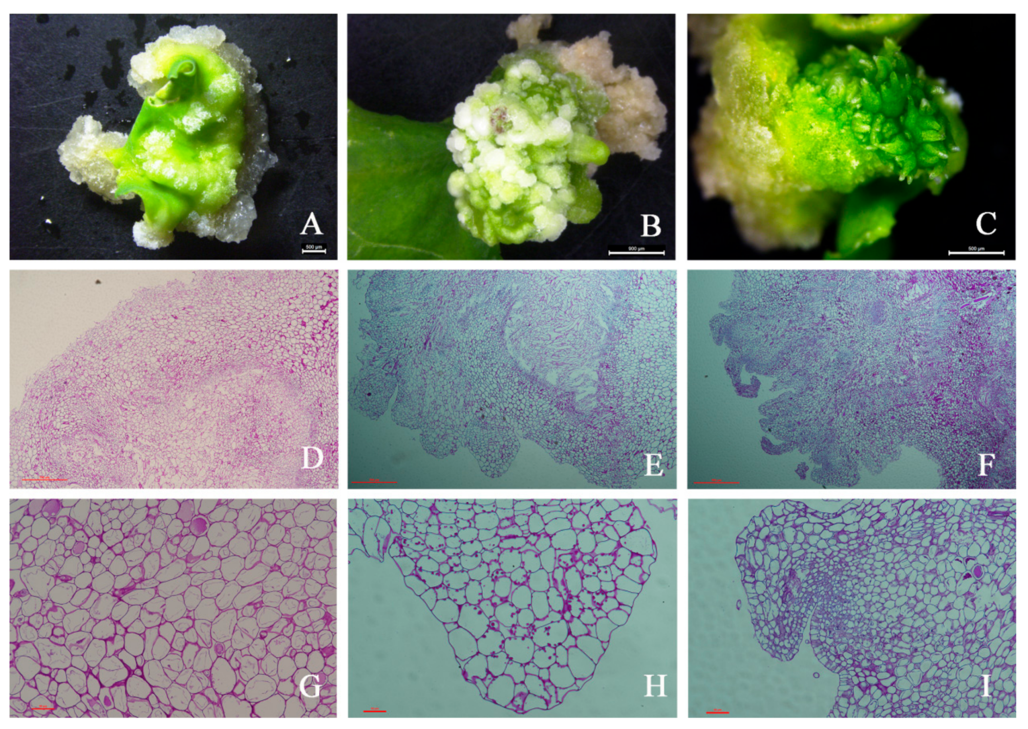
2.1. Morphological, Histological, and Biochemical Features

The first morphological changes during the regeneration process occurred during the first ten days of culture, when the leaf explants became slightly swollen and intense cell proliferation was observed at the cut explants surface, giving rise to the callus (Figure 1A). Callus growth progressed and after ten days of culture, the first meristemoids were observed as small structures on the surface of the explant (Figure 1B). These structures continued developing, originating shoot buds (Figure 1C).
Callus cells appeared in the outer layer of the leaf explants. These cells were more variable in size and shape than their progenitors and had a large central vacuole (Figure 1D,G). Unlike the initially loosely arranged cells, the outermost cells were relatively small and exhibited a high capacity for division, enabling expansion of the outer layer. During this period, the cells were uniformly colored and arranged neatly and regularly without intercellular space. The callus continued to differentiate and formed the bud primordium (Figure 1E,H). The bud primordia cells continued to divide, resulting in a substantial increase in the number of cells and the formation of one or more buds (Figure 1F,I). Histological examinations were performed using light microscopy on the initial leaf explant and at two stages of callus of drumstick development by staining with the Periodic acid Schiff (PAS) reagent to view starch reserves. Starch granules were detected in the parenchymatous cells in stage 2 callus (Figure 1H), indicating that embryogenic cells display metabolic competence.

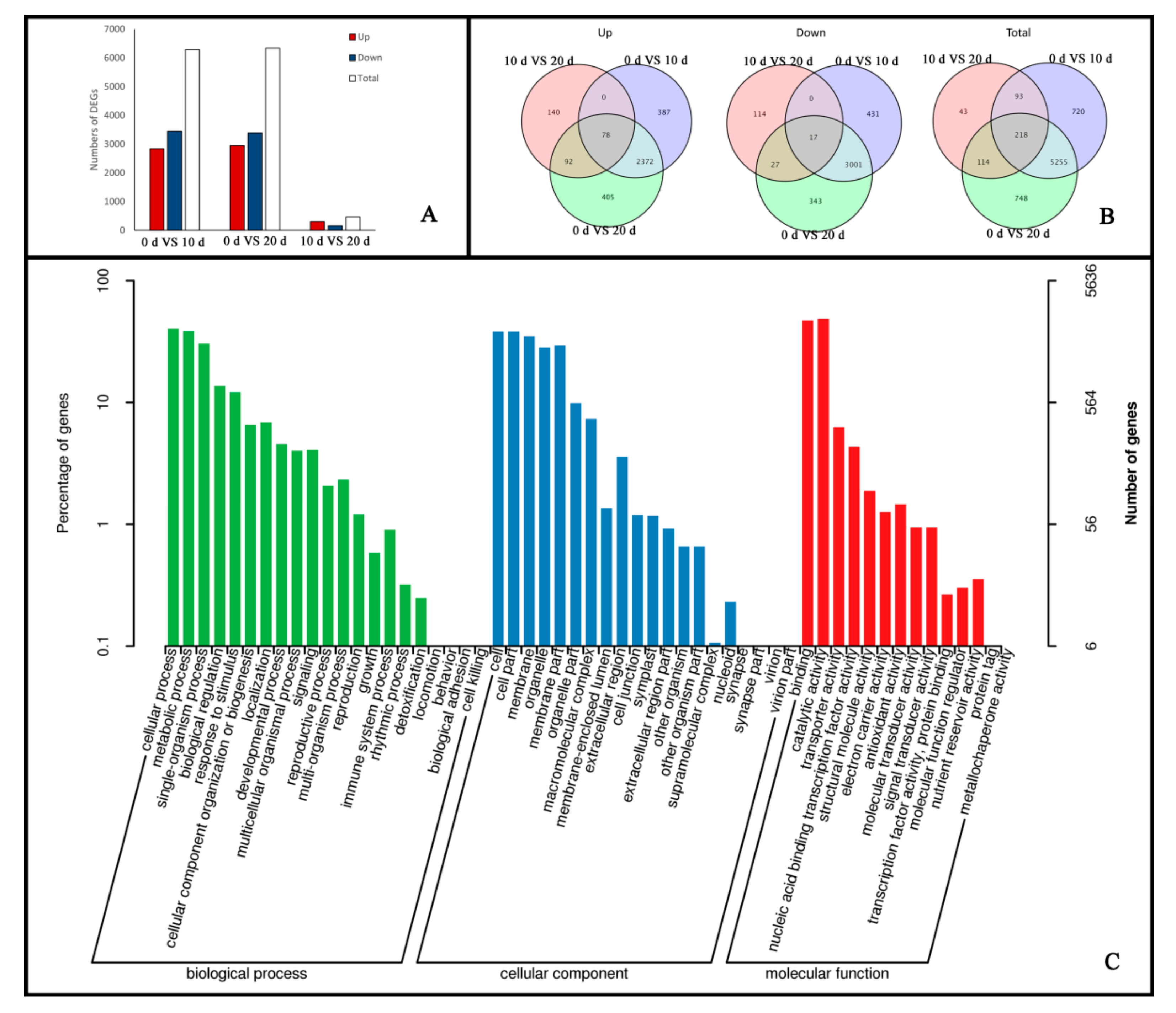
2.2. Global Transcriptomic Changes at Different Stages of Embryogenic Callus Induction Revealed by Transcriptome Sequencing Analysis

To obtain a comprehensive overview of the drumstick transcriptome, RNA was extracted from the three different explants. The Illumina NovaSeq 6000 platform generated 44,952,482 clean reads containing a total of 57.76 Gb clean data with 94.86% Q30 bases (base quality > 30) after performing stringent quality assessment and data filtering. The quality of the sequencing data is summarized in Table 1. The clean reads were mapped to the drumstick genome using the HISAT2 tool. The average mapping ratio ranged from 93.33 to 95.58%. StringTie was used to perform transcript assembly based on the read alignments. After optimal gene structure prediction and alternative splicing analysis, 23,591 genes were identified including 4126 new genes.
To validate and annotate the assembled transcriptome library, we searched against the non-redundant (Nr) peptide database (ftp://ftp.ncbi.nih.gov/blast/db) (14 January 2021), the Swiss-Prot protein database (http://www.uniprot.org) (14 January 2021), KOG (http://www.ncbi.nlm.nih.gov/KOG) (14 January 2021), and KEGG (https://www.genome.jp/kegg) (14 January 2021) using BLASTx (http://blast.ncbi.nlm.nih.gov/Blast.cgi) (14 January 2021) with a cutoff E-value of 10−5. It was found that 90.88% of the transcripts had significant similarity to at least one target from these databases (Table 2).

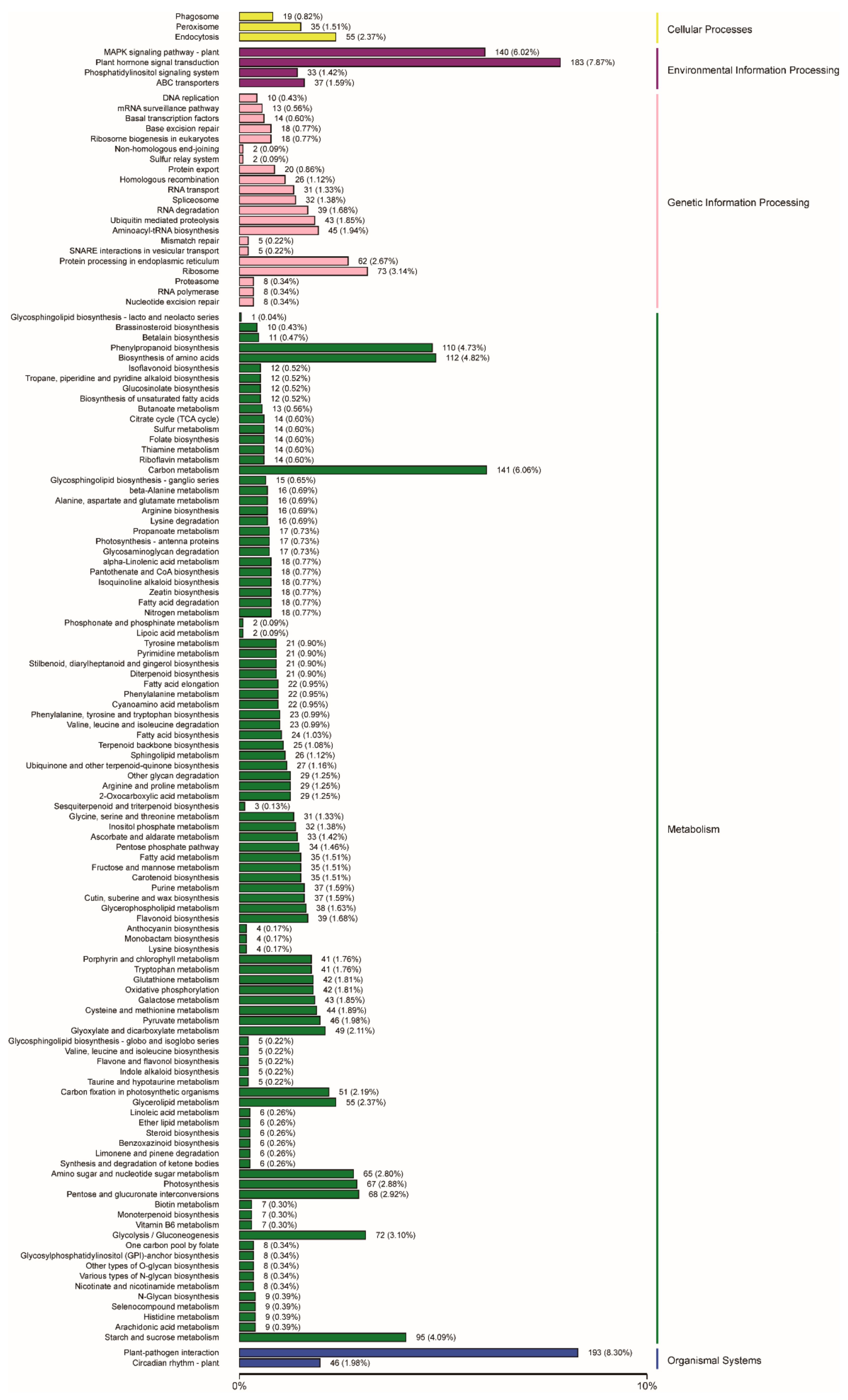
2.3. Functional Annotation and Classification of Genes Related to Embryogenic Callus Induction

DEGs were classified as embryogenic callus induction responsive genes if they exhibited a Fold Change ≥ 2 and FDR < 0.01 at any development point when compared to the control according to DESeq2. In total, 7191 DEGs were identified during embryogenic callus development. Of these DEGs, 6286 were differentially expressed between zero-day and ten-day explants, with 2837 being upregulated and 3449 downregulated. Only 468 DEGs were identified from ten-day explants to twenty days, of which 310 DEGs were upregulated and 158 DEGs were downregulated (Figure 2A). Additionally, 5975 DEGs were only differentially expressed in ten-day explants and 157 were only differentially expressed in twenty-day explants; 78 genes showed increased expression in all developmental stages and 17 showed reduced expression in all stages (Figure 2B). These results suggest that there are significant differences in gene expression at different stages of embryogenic callus development and that many genes are needed to ensure complete differentiation.
To functionally classify the identified genes related to embryogenic callus induction, the DEGs were matched to the Gene Ontology (GO) database (http://www.geneontology.org) (14 January 2021). Three categories were represented: biological processes, cellular components, and molecular functions. Within these main GO categories, cellular process, cell, and catalytic activity had the greatest number of matches among the DEGs (Figure 2C).
To identify metabolic pathways potentially related to embryogenic callus formation, the DEGs were analyzed using the KEGG (https://www.genome.jp/kegg) (14 January 2021) pathway analysis tool—a compilation of manually verified pathway maps used to categorize gene functions, with an emphasis on biochemical pathways. A total of 2325 (32.33%) DEGs were mapped to the KEGG database, with significant enrichment in the “metabolism” and “genetic information processing” clusters. In the “environmental information processing” category, “plant hormone signal transduction” genes were most highly represented among the DEGs, while “plant-pathogen interaction” genes were most highly represented in the “organismal systems” category (Figure 3 and Figure 4).

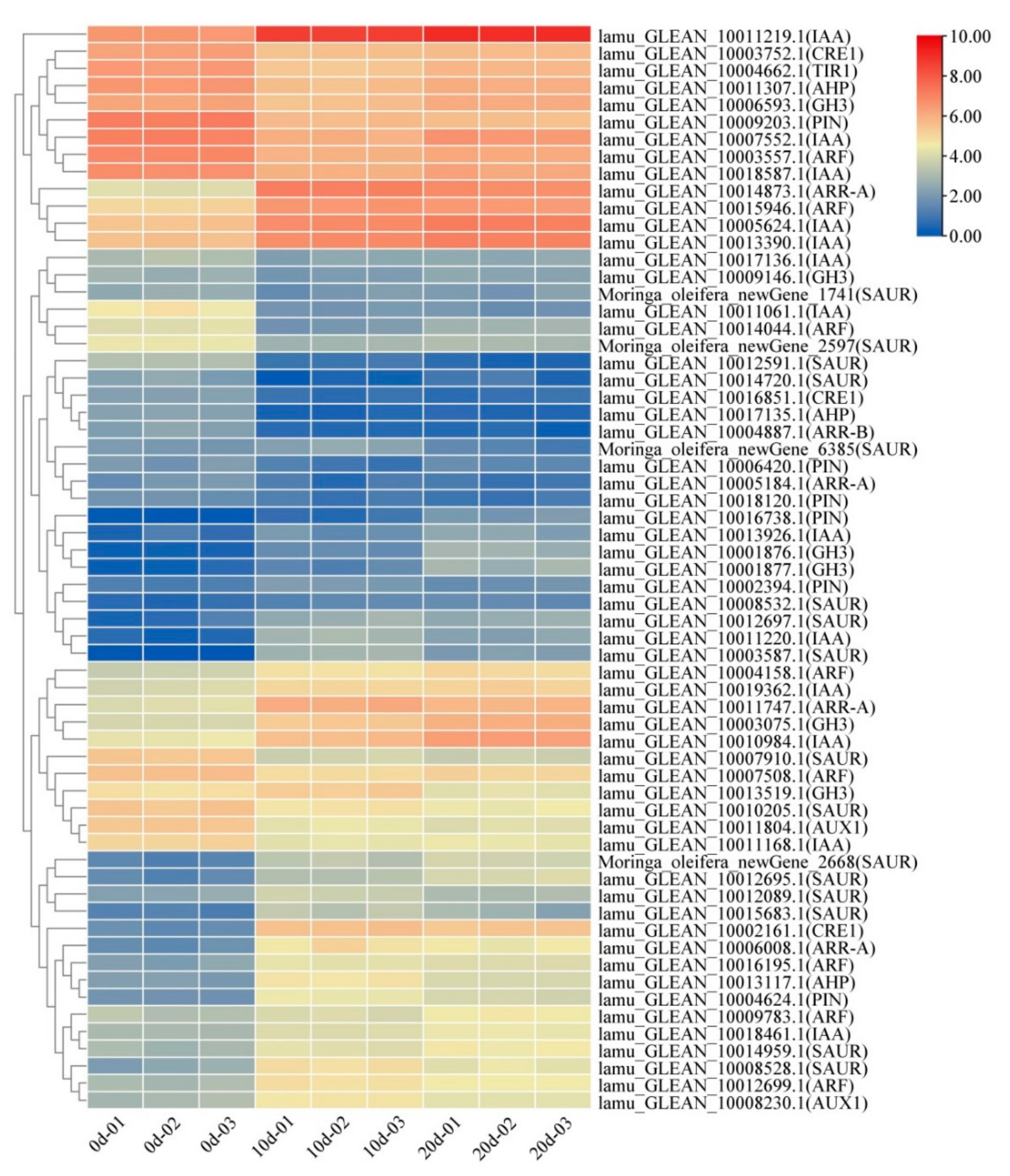
2.4. Auxin and Cytokinin Metabolism Involved in Embryogenic Callus Induction

Auxin is a key hormone that is included in EC induction media and must be maintained in balance with cytokinins during EC development. Fifty-one genes (2 Aux/Lax genes, 13 Aux/IAA genes, 8 ARF genes, 6 PIN genes, 6 GH3 genes, and 16 SAUR genes) related to auxin signaling pathways were differentially expressed during embryogenic callus formation (Figure 4). Among them, one Aux/Lax gene, four Aux/IAA genes, three ARF genes, two PIN genes, three GH3 genes, and eight SAUR genes were significantly up-regulated when comparing leaf explants to ten-day callus samples, and most of them remained highly expression in twenty-day callus samples. Changes in the expression of auxin signaling pathway related genes were visualized using a heatmap and those with similar expression patterns were categorized. Members of the same gene families showed different expression patterns, suggesting that the regulation of auxin signaling networks during callus differentiation is very complex. It is worth noting that the IAA gene (LAMU_GLEAN_10011219.1) was significantly up-regulated during callus formation and remained strongly expressed during callus differentiation, suggesting that it may be a key regulatory factor in the initiation and maintenance of callus regeneration. Further experimental studies are needed to confirm the function of this gene.
Because cytokinin plays a major role in EC induction, the expression patterns of genes encoding proteins involved in cytokinin signaling were also investigated. Eleven genes (four Type A-ARR genes, three CRE1 genes, three AHP genes, and one Type B-ARR genes) related to cytokinin signaling pathways were differentially expressed. Cytokinins rapidly activate the expression of type-A ARRs, a family of genes encoding proteins thought to act as feedback repressors of cytokinin responses. Four type A-ARR genes were differentially expressed, and three were upregulated significantly during EC formation. One of these genes, LAMU_GLEAN_10014873.1, was strongly expressed at both stages of callus development, being upregulated by more than a factor of seven.

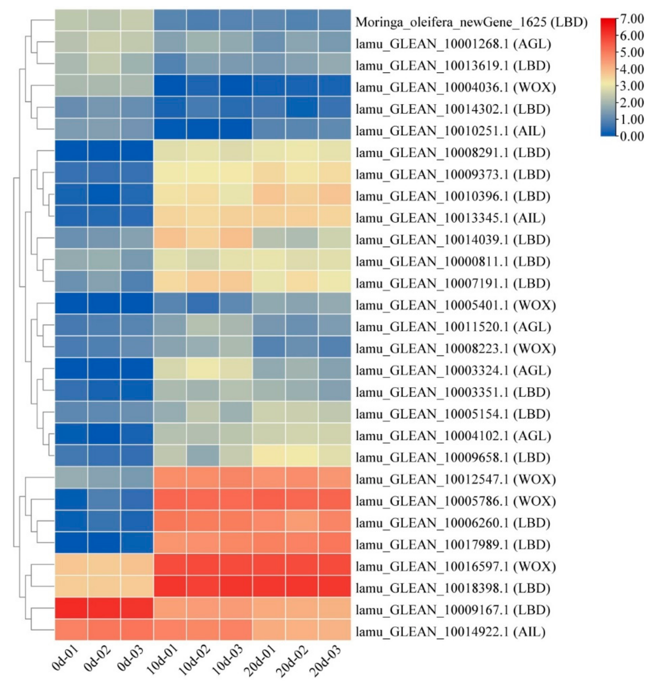
2.5. Embryogenesis-Labeled Genes Involved in Embryogenic Callus Induction

In addition to plant hormone signaling pathway genes, several embryogenesis-labeled genes have been reported to play roles in embryogenic callus induction. To characterize the embryogenesis-labeled genes during drumstick EC induction, a comparative analysis of expression levels at different stages of induction was performed. Twenty-nine differentially expressed embryogenesis-labeled genes were found, belonging to the LBD, AGL, WOX, and AIL gene families (Figure 5). Most of these DEGs such as WOX (LAMU_GLEAN_10016597.1) and LBD (LAMU_GLEAN_10019398.1) were up-regulated at the callus initiation stage and remained highly expressed at the later stage, indicating that these genes were not only involved in EC induction, but also played important roles in maintaining the cells’ embryonic state.

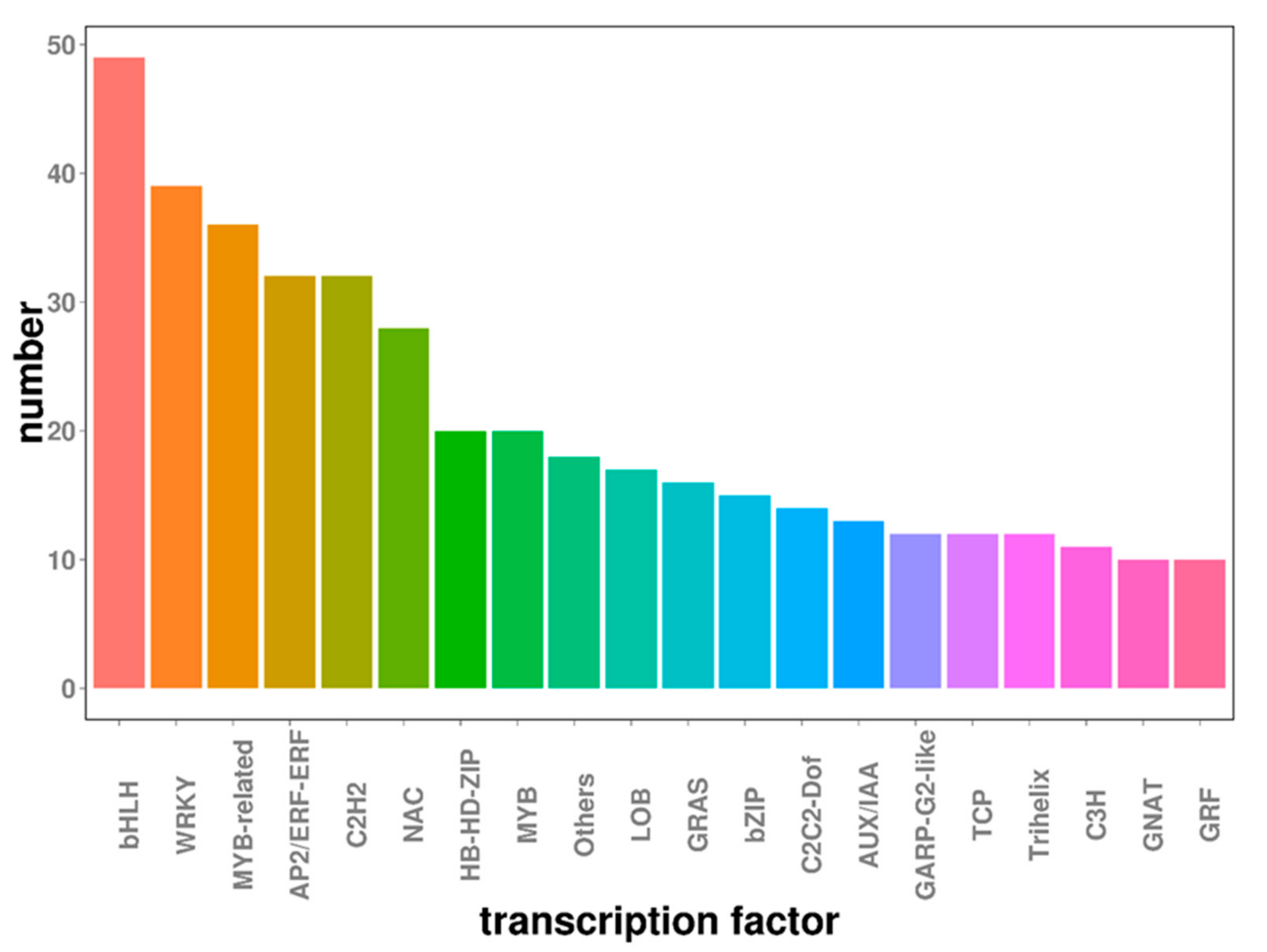
2.6. Transcription Factors Involved in Embryogenic Callus Induction

Transcription factors are proteins that bind specifically to a particular gene sequence and enable spatiotemporal control over the target gene’s expression. Previous studies confirmed that transcription factors play important roles in embryogenic callus formation. There were 589 transcription factors from 20 different families among the DEGs identified in this study (Figure 6). The three transcription factor families most heavily represented among this group were the bHLH, WRKY, and MYB-related families, which collectively accounted for 21% of all differentially expressed transcription factors. Previous studies showed that these gene families were differentially expressed under stress conditions and served to link stress to developmental pathways. All major TF families were represented in the mRNA transcriptome at each developmental stage and several TF families were specifically expressed during particular developmental periods.

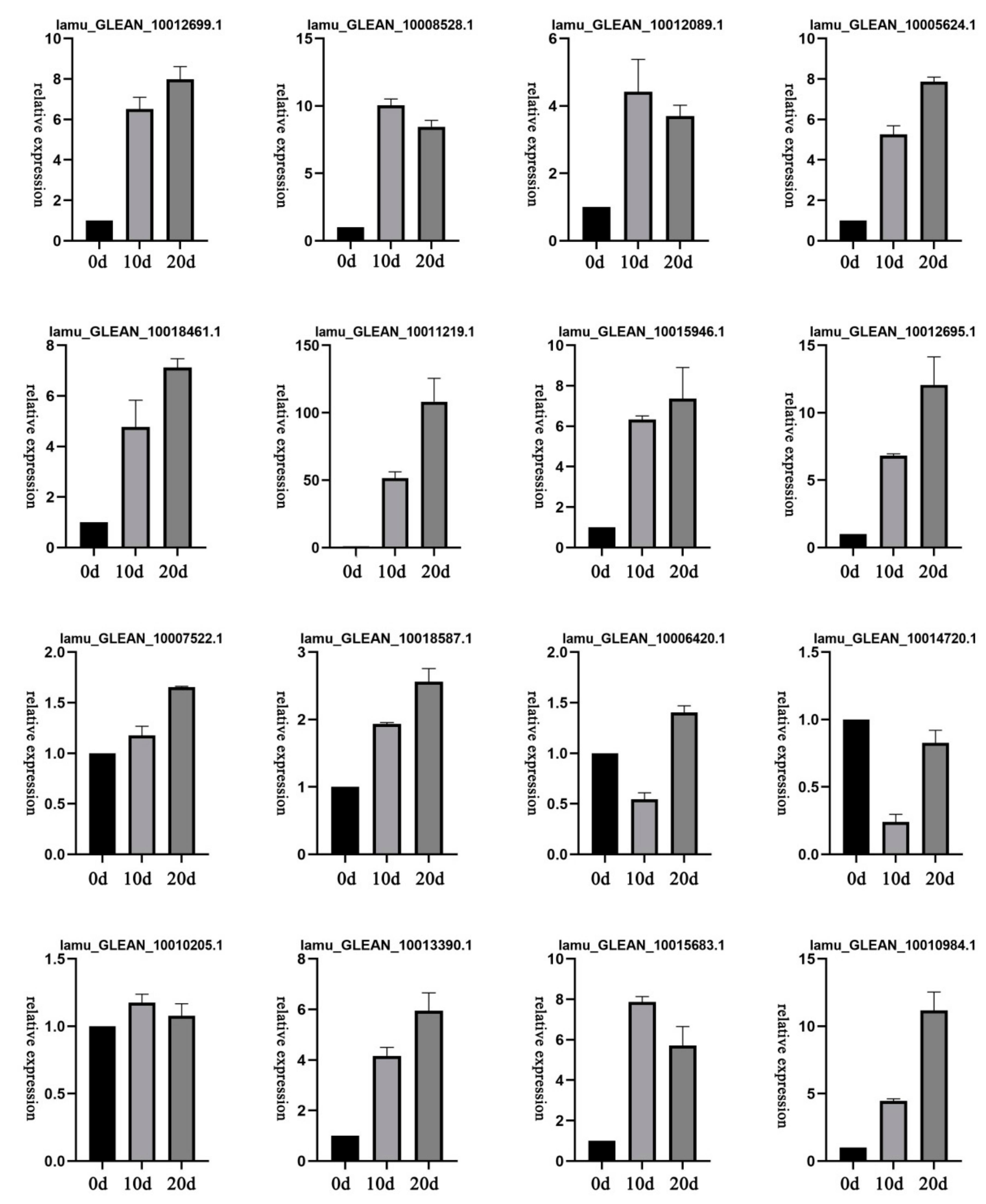
2.7. Expression Patterns of Selected DEGs

To experimentally confirm the expression of the unigenes identified by sequencing and computational analysis, 16 representative DEGs related to auxin signaling pathways including Aux/IAA, SAUR, ARF, and PIN were selected for qRT-PCR analysis across the three sequential developmental stages of drumstick EC.
Based on the analyzed qRT-PCR data (S1, Figure 7), all of these DEGs were expressed at varying levels in different stages. Moreover, the qRT-PCR results were generally consistent with the RNA-seq data, indicating the reliability of the transcriptome analysis. These results also suggest that all of the tested genes may play important roles in auxin transport and contribute to asymmetric auxin distributions that lead to EC induction.

3. Discussion

3.1. Auxin and Cytokinin Play an Important Role in Drumstick EC Formation

Auxin and cytokinins play key roles in plant cell division and differentiation [9]. It is known that auxin activates the expression of three major families of primary response genes: the Aux/IAA genes, the GH3 genes, and the SAUR genes [24]. Aux/IAA family members are short-lived nuclear proteins that for homo- and heterodimers with other Aux/IAA and ARF genes to suppress transcription of downstream auxin-responsive genes [25]. In our study, 13 Aux/IAA genes were differently expressed during EC formation, while 5 Aux/IAA genes were significantly up-regulated in ten-day callus and remained strongly expressed in twenty-day callus. The expression level of one Aux/IAA family member was significantly higher than that of other genes in the pathway, indicating that this gene may play an important role in callus induction. The SAUR family is the largest known family of early auxin-inducible genes [26], and OsSAUR39 was reported to negatively regulate auxin biosynthesis and transport [27]. Interestingly, several SAUR genes were differentially expressed in the early stage of drumstick callus induction and showed different expression patterns. Consequently, their roles in this process need further study. ARF transcription factors are key components of the auxin signal transduction pathway that inhibit or activate the expression of downstream genes by binding to upstream response elements of auxin response genes [28]. Four ARF genes were up-regulated during EC induction, suggesting these genes may play important roles in callus development.
Cytokinins regulate plant development via classic two-component regulatory systems [24]. Previous studies have shown that A-ARR genes are up-regulated under cytokinin stimulus, which would repress WUS expression to maintain normal shoot apical development. Moreover, CRE1 is an upstream regulator of A-ARR together with AHP and B-ARR [29]. One CRE1 gene and 3 A-ARR genes were significantly up-regulated during EC induction, suggesting that they play important roles in cytokinin signaling during this process. These results will provide a foundation for further studies on the effects of cytokinin signaling in drumstick.

3.2. Embryogenesis-Labeled Genes Play Important Roles in Drumstick EC Formation

Several genes have been identified as molecular markers of SE, including somatic embryogenesis receptor-like kinase (SERK), leafy cotyledon 1 (LEC1), BABYBOOM (BBM), wuschel (WUS), and WUS-related homeobox (WOX) [30]. SERK is known to play a key role in the acquisition of embryogenic competence in plant cells [31]. However, no genes of this family were differentially expressed during the developmental stages examined in this work. Conversely, LBD, AGL, WOX, and AIL genes were differentially expressed in different induction stages. Some LBD genes have been identified as key regulators of callus formation in tissue culture. For example, LBDs reportedly interact physically with the AtbZIP59 transcription factor to mediate auxin-induced callus formation in Arabidopsis [32]. AGL15 is a MDAS-BOX protein that is preferentially expressed in developing embryos and promotes somatic embryo (SE) initiation [33]. WOX genes were markers of cell fate during early embryogenesis in Arabidopsis [34], and WOX2 and WOX9 were highly expressed in the early stage of SE development in Picea abies, suggesting that these genes may function together during conifer embryo patterning [35]. WOX4 is associated with callus proliferation, and WOX1 is involved in root primordium initiation in the shrub species Jasminum sambac [36].

3.3. Stress-Related Transcription Factors Play an Important Role in Drumstick EC Formation

Stress is an important regulatory signal for cell dedifferentiation that can induce the expression of many transcription factors, cell fate reorganization, and embryogenic acquisition [37]. It is clear that excising tissue from a parent plant, cutting it into pieces, and putting into an artificial culture environment imposes considerable stress on the plant tissue. The connection between stress and EC initiation has received increasing attention in recent years.
In this study, several transcription factors exhibited dynamic changes in expression during different developmental stages of EC formation; examples include bHLH, WRKY, the MYB-related family, and AP2/ERF. Previous studies have shown that these transcription factors play important roles in responding to various stresses [38,39]—they are differentially expressed under stress conditions and act to link stress to developmental pathways. bHLH participates in the process of adaptation to various stresses and also has pleiotropic effects during cell proliferation and differentiation [38]. In this study, 19 bHLH members were upregulated and 17 were downregulated when comparing leaf explants to ten-day callus tissue. Conversely, when comparing ten and twenty-day callus samples, two bHLH members were upregulated and only one was downregulated. These results indicated that more bHLH transcription factors are needed to initiate differentiation in the early stage of callus differentiation. WRKY is involved in responses to biotic and abiotic stresses and hormone signaling [39]. WRKY transcription factors exhibited complex expression patterns during drumstick callus induction, suggesting that these genes may perform different regulatory functions. MYB-related is involved in hormone and environmental responses [40]. Previous studies have confirmed that the AP2/ERF transcription factor plays an important role in callus induction and adventitious bud regeneration. WIND1 is an AP2/ERF transcription factor that was shown to promote Arabidopsis shoot regeneration [41]. In this study, we identified 40 differentially expressed AP2/ERF transcription factors, most of which were upregulated during EC initiation. It is notable that the ARF, Aux/IAA and LBD families also have roles in auxin-mediated signaling [42]. Further study of these genes is expected to reveal ways of improving the reliability and efficiency of embryogenic callus induction in drumstick.

4. Material and Methods

4.1. Induction of Embryogenic Callus and Plant Regeneration

The donor plants used in this study were in vitro-grown shoots of clonal drumstick tree M-2 (South China Agricultural University). Stage 2 leaves from M-2 were wounded and precultured on Murashige and Skoog (MS) basal medium supplemented with 0.8 mg/L 6-benzyladenine (BA), 0.2 mg/L kinetin (KT), and 0.05 mg/L 1-naphthaleneacetic acid (NAA) to heal the cutting wound and initiate cell division. All media used were adjusted to pH 5.8–6.0 with 1 N NaOH or 1 N HCl solution, set using 0.6% (w/v) agar, 30 g/L sucrose and autoclaved at 121 °C for 20 min. All cultures were kept under cool white light (about 50 µmol m−2 s−1) with a 12 h photoperiod and a temperature of 25–27 °C.

4.2. Histomorphological and Histochemical Analyses

Fresh explants were observed under an Olympus SZX10 (Olympus, Guangzhou, China) stereomicroscope after preparation for microscopic evaluation. Explants for light microscopic examination were dissected and fixed in glutaraldehyde, dehydrated in a series of ethanol solutions, then infiltrated and embedded in paraffin. Sections (thickness: 5 μm) were cut using a Leica RM2235 (Leica, Shanghai, China) rotary microtome. Periodic acid-Schiff (PAS) was used to detect insoluble polysaccharides.

4.3. Library Preparation and Transcriptome Sequencing

Drumstick leaf explants cultured for ten and twenty days exhibited clearly distinct morphology, so three sets of explants were collected at each of the three sequential developmental stages (0, 10, and 20 d). For RNA seq analysis, fresh samples were collected, immediately immersed in liquid nitrogen, and then stored at −80 °C. RNA was extracted using the Simple Total RNA Kit (OMEGA, Guangzhou, China) according to the manufacturer’s instructions. RNA concentration and purity were measured using a NanoDrop 2000 system (Thermo Fisher Scientific, Wilmington, DE, USA). RNA integrity was assessed using the RNA Nano 6000 Assay Kit of the Agilent Bioanalyzer 2100 system (Agilent Technologies, CA, USA). Afterwards, 1 µg RNA of per sample was employed for the RNA-seq analysis using Illumina NovaSeq 6000 system by Biomarker Technologies (Beijing, China). The maximum read length of this system is 2 × 250 bp. Sequencing libraries were generated using the NEBNext UltraTM RNA Library Prep Kit for Illumina (NEB, Ipswich, MA, USA). Clustering of the index-coded samples was performed on a cBot Cluster Generation System using the TruSeq PE Cluster Kit v.4-cBotHS (Illumina). Libraries were sequenced on an Illumina platform and paired-end reads were generated. HISAT2 v2.0.5 (http://ccb.jhu.edu/software/hisat2/index.shtml) (4 November 2016) tools software was used to map the sequences to the reference genome, which has been deposited in National Center for Biotechnology Information database (NCBI) under the accession number PRJNA268707. Gene function was annotated from commonly used databases. DEGs were observed using DESeq2 software and parameters were a false discovery rate (FDR) ≤ 0.01 and |log2FC| (FC, fold change) ≥ 1. Finally, RNA-seq data in this study had been submitted to https://www.ncbi.nlm.nih.gov/ (4 November 2016) and the accession number was PRJNA771463.

4.4. Real-Time Quantitative PCR

Sixteen genes with potential roles in drumstick embryogenic callus were chosen for validation using real-time quantitative PCR (RT-qPCR) in three cultured stages that were consistent with transcriptome sequencing. The cDNA first-strand was synthesized from total RNA using First-Strand cDNA Synthesis Kits and oligo-dT primers (Takara, Guangzhou, China). RT-qPCR was performed, and ACP2 was amplified as a reference gene [43]. The forward and reverse primers listed in Table 3 were designed using Primer 5.0 and blasted in reference genome to verify that primers cover adjacent exons. Three technical replicates were performed for the test and reference genes of each sample to obtain precise and reproducible results. Relative expression levels for each gene were estimated using the 2−ΔΔCT method. Statistical analysis was carried out using SPSS 22.0 software (SPSS Inc., Chicago, IL, USA), Duncan’s multiple range test was used to detect differences between means; p-values < 0.05 were considered significant.

Supplementary Materials

The following are available online at https://www.mdpi.com/article/10.3390/ijms222212130/s1.

Author Contributions

X.C. and J.Z. conceived and designed research; E.Y., M.Z. and X.Z. conducted experiments; E.Y., X.H. and H.Y. collected and analyzed the data; J.Z. and X.C. wrote the manuscript; J.Z. and X.C. obtained funding. All authors have read and agreed to the published version of the manuscript.

Funding

This work was funded by the Forestry Technology Innovation Program, Department of Forestry of Guangdong Province (2018KJCX001); and the Youth Innovation Program (Natural Science), Department of Education of Guangdong Province (2019KQNCX010).

Data Availability Statement

All the raw data of the transcriptome are deposited in the NCBI under the accession number PRJNA771463.

Conflicts of Interest

No potential conflict of interest are reported by the authors.

References

  1. Zimmerman, J.L. Somatic embryogenesis: A model for early development in higher plants. Plant Cell 1993, 5, 1411–1423. [Google Scholar] [CrossRef] [PubMed] [Green Version]
  2. Al Abdallat, A.M.; Sawwan, J.S.; Al Zoubi, B. Agrobacterium tumefaciens-mediated transformation of callus cells of Crataegus aronia. Plant Cell Tissue Organ Cult. 2011, 104. [Google Scholar] [CrossRef]
  3. Elegba, W.; McCallum, E.; Gruissem, W. Efficient genetic transformation and regeneration of a farmer-preferred Cassava cultivar from Ghana. Front. Plant Sci. 2021, 12, 668042. [Google Scholar] [CrossRef] [PubMed]
  4. Saika, H.; Toki, S. Mature seed-derived callus of the model indica rice variety Kasalath is highly competent in Agrobacterium-mediated transformation. Plant Cell Rep. 2010, 29, 1351–1364. [Google Scholar] [CrossRef] [Green Version]
  5. Li, J.; Zhang, D.; Que, Q.; Chen, X.; Ouyang, K. Plant regeneration and Agrobacterium-mediated transformation of the miracle tree Neolamarckia cadamba. Ind. Crop. Prod. 2019, 130, 443–449. [Google Scholar] [CrossRef]
  6. Passamani, L.Z.; Reis, R.S.; Vale, E.M.; Sousa, K.R.; Aragao, V.P.M.; Santa-Catarina, C.; Silveira, V. Long-term culture with 2,4-dichlorophenoxyacetic acid affects embryogenic competence in sugarcane callus via changes in starch, polyamine and protein profiles. Plant Cell Tissue Organ Cult. 2019, 140, 415–429. [Google Scholar] [CrossRef]
  7. Wang, Y.; Ruemmele, B.A.; Chandlee, J.M.; Sullivan, W.M.; Knapp, J.E.; Kausch, A.P. Embryogenic callus induction and plant regeneration media for bentgrasses and annual bluegrass. In Vitro Cell. Dev. Biol.-Plant 2002, 38, 460–467. [Google Scholar] [CrossRef]
  8. Jabeen, N.; Chaudhry, Z.; Mirza, B.; Rashid, H. Effect of genotype and explant type on in vitro shoot regeneration of tomato (Lycopersicon esculentum Mill.). Pak. J. Bot. 2005, 37, 899–903. [Google Scholar]
  9. Kadhimi, A.A.; Alhasnawi, A.N.; Isahak, A.; Ashraf, M.F.; Mohamad, A.; Doni, F.; Yusoffi, W.M.W. Effect of Genotype and Growth Regulators in Induction of Embryogenic Callus in Rice. Pure Appl. Microbiol. 2014, 8, 4573–4578. [Google Scholar]
  10. Kang, H.; Lee, C.; Kwon, S.; Park, J.; Kang, K.; Shim, D. Comparative transcriptome analysis during developmental stages of direct somatic embryogenesis in Tilia amurensis Rupr. Sci. Rep. 2021, 11, 6359. [Google Scholar] [CrossRef]
  11. Lai, Z.; Lin, Y. Analysis of the global transcriptome of longan (Dimocarpus longan Lour.) embryogenic callus using Illumina paired-end sequencing. BMC Genom. 2013, 14, 561. [Google Scholar] [CrossRef] [Green Version]
  12. Zhang, J.; Yang, Y.; Lin, M.; Li, S.; Tang, Y.; Chen, H.; Chen, X. An efficient micropropagation protocol for direct organogenesis from leaf explants of an economically valuable plant, drumstick (Moringa oleifera Lam.). Ind. Crop. Prod. 2017, 103, 59–63. [Google Scholar] [CrossRef]
  13. Lin, M.; Zhang, J.; Chen, X. Bioactive flavonoids in Moringa oleifera and their health-promoting properties. J. Funct. Foods 2018, 47, 469–479. [Google Scholar] [CrossRef]
  14. Leone, A.; Spada, A.; Battezzati, A.; Schiraldi, A.; Aristil, J.; Bertoli, S. Cultivation, genetic, ethnopharmacology, phytochemistry and pharmacology of Moringa oleifera leaves: An overview. Int. J. Mol. Sci. 2015, 16, 12791–12835. [Google Scholar] [CrossRef]
  15. Sengupta, M.E.; Keraita, B.; Olsen, A.; Boateng, O.K.; Thamsborg, S.M.; Pálsdóttir, G.R.; Dalsgaard, A. Use of Moringa oleifera seed extracts to reduce helminth egg numbers and turbidity in irrigation water. Water Res. 2012, 46, 3646–3656. [Google Scholar] [CrossRef]
  16. Bakre, A.G.; Aderibigbe, A.O.; Ademowo, O.G. Studies on neuropharmacological profile of ethanol extract of Moringa oleifera leaves in mice. J. Ethnopharmacol. 2013, 149, 783–789. [Google Scholar] [CrossRef]
  17. Kituyi, J.L.; Foulkes, M.; Worsfold, P.; Ongulu, R.A.; Kiplagat, A.; Gachanja, A. Efficiency of pre-treated Moringa oleifera for the removal of Cd2+ and Zn2+ ions from waste waters. Ecohydrol. Hydrobiol. 2013, 13, 267–271. [Google Scholar] [CrossRef]
  18. Sreelatha, S.; Jeyachitra, A.; Padma, P.R. Antiproliferation and induction of apoptosis by Moringa oleifera leaf extract on human cancer cells. Food Chem. Toxicol. 2011, 49, 1270–1275. [Google Scholar] [CrossRef]
  19. Muhl, Q.E.; du Toit, E.S.; Steyn, J.M.; Apostolides, Z. Bud development, flowering and fruit set of Moringa oleifera Lam. (Horseradish Tree) as affected by various irrigation levels. J. Agric. Rural. Dev. Trop. Subtrop. 2013, 114, 79–87. [Google Scholar]
  20. Batra, S.; Kumar, S. Agrobacterium-mediated transient GUS gene expression in buffel grass (Cenchrus ciliaris L.). J. Appl. Genet. 2003, 44, 449–458. [Google Scholar]
  21. Takamizo, T.; Sato, H. Protocol for Agrobacterium-mediated transformation of tall fescue and future perspective on the application of genome editing. Plant Biotechnol. 2020, 37, 157–161. [Google Scholar] [CrossRef]
  22. Zhang, J.; Lin, M.; Chen, H.; Chen, X. Agrobacterium tumefaciens-mediated transformation of drumstick (Moringa oleifera Lam.). Biotechnol. Biotechnol. Equip. 2017, 31, 1126–1131. [Google Scholar] [CrossRef]
  23. Ikeuchi, M.; Sugimoto, K.; Iwase, A. Plant callus: Mechanisms of induction and repression. Plant Cell 2013, 25, 3159–3173. [Google Scholar] [CrossRef] [Green Version]
  24. Che, P.; Gingerich, D.J.; Lall, S.; Howell, S.H. Global and hormone-induced gene expression changes during shoot development in Arabidopsis. Plant Cell 2002, 14, 2771–2785. [Google Scholar] [CrossRef]
  25. Chen, L.; Zheng, X.; Guo, X.; Cui, Y.; Yang, H. The roles of Aux/IAA gene family in development of Dendrocalamus sinicus (Poaceae: Bambusoideae) inferred by comprehensive analysis and expression profiling. Mol. Biol. Rep. 2019, 46, 1625–1634. [Google Scholar] [CrossRef] [PubMed]
  26. Ren, H.; Gray, W.M. SAUR proteins as effectors of hormonal and environmental signals in plant growth. Mol. Plant 2015, 8, 1153–1164. [Google Scholar] [CrossRef] [PubMed] [Green Version]
  27. Kant, S.; Bi, Y.M.; Zhu, T.; Rothstein, S.J. SAUR39, a small auxin-up RNA gene, acts as a negative regulator of auxin synthesis and transport in rice. Plant Physiol. 2009, 151, 691–701. [Google Scholar] [CrossRef] [PubMed] [Green Version]
  28. Roosjen, M.; Paque, S.; Weijers, D. Auxin response factors: Output control in auxin biology. J. Exp. Bot. 2018, 69, 179–188. [Google Scholar] [CrossRef]
  29. Buechel, S.; Leibfried, A.; To, J.P.C.; Zhao, Z.; Andersen, S.U.; Kieber, J.J.; Lohmann, J.U. Role of A-type ARABIDOPSIS RESPONSE REGULATORS in meristem maintenance and regeneration. Eur. J. Cell Biol. 2010, 89, 279–284. [Google Scholar] [CrossRef]
  30. Gordon-Kamm, B.; Sardesai, N.; Arling, M.; Lowe, K.; Hoerster, G.; Betts, S.; Jones, T. Using morphogenic genes to improve recovery and regeneration of transgenic plants. Plant 2019, 8, 38. [Google Scholar] [CrossRef] [Green Version]
  31. Rocha, D.I.; Monte-Bello, C.C.; Aizza, L.C.B.; Dornelas, M.C. A passion fruit putative ortholog of the SOMATIC EMBRYOGENESIS RECEPTOR KINASE1 gene is expressed throughout the in vitro de novo shoot organogenesis developmental program. Plant Cell Tissue Organ Cult. 2016, 125, 107–117. [Google Scholar] [CrossRef]
  32. Xu, C.; Cao, H.F.; Zhang, Q.Q.; Wang, H.Z.; Xin, W.; Xu, E.J.; Zhang, S.Q.; Yu, R.X.; Ye, D.X.; Hu, Y.X. Control of auxin-induced callus formation by bZIP59–LBD complex in Arabidopsis regeneration. Nat. Plants 2018, 4, 108–115. [Google Scholar] [CrossRef]
  33. Zheng, Q.; Zheng, Y.; Perry, S.E. AGAMOUS-Like15 promotes somatic embryogenesis in Arabidopsis and soybean in part by the control of ethylene biosynthesis and response. Plant Physiol. 2013, 161, 2113–2127. [Google Scholar] [CrossRef] [Green Version]
  34. Haecker, A.; Gross-Hardt, R.; Geiges, B.; Sarkar, A.; Breuninger, H.; Herrmann, M.; Laux, T. Expression dynamics of WOX genes mark cell fate decisions during early embryonic patterning in Arabidopsis thaliana. Development 2004, 131, 657–668. [Google Scholar] [CrossRef] [Green Version]
  35. Palovaara, J.; Hallberg, H.; Stasolla, C.; Hakman, I. Comparative expression pattern analysis of WUSCHEL-related homeobox 2 (WOX2) and WOX8/9 in developing seeds and somatic embryos of the gymnosperm Picea abies. New Phytol. 2010, 188, 122–135. [Google Scholar] [CrossRef]
  36. Lu, Y.; Liu, Z.; Lyu, M.; Yuan, Y.; Wu, B. Characterization of JsWOX1 and JsWOX4 during Callus and Root Induction in the Shrub Species Jasminum sambac. Plants 2019, 8, 79. [Google Scholar] [CrossRef] [Green Version]
  37. Guo, H.H.; Guo, H.X.; Zhang, L.; Fan, Y.J.; Wu, J.F.; Tang, Z.M.; Zhang, Y.; Fan, Y.P.; Zeng, F.C. Dynamic transcriptome analysis reveals uncharacterized complex regulatory pathway underlying genotype-recalcitrant somatic embryogenesis transdifferentiation in Cotton. Genes 2020, 11, 519. [Google Scholar] [CrossRef]
  38. Guo, J.R.; Sun, B.X.; He, H.R.; Zhang, Y.F.; Tian, H.Y.; Wang, B.S. Current understanding of bHLH transcription factors in plant abiotic stress tolerance. Int. J. Mol. Sci. 2021, 22, 4921. [Google Scholar] [CrossRef]
  39. Li, W.X.; Pang, S.Y.; Lu, Z.G.; Jin, B. Function and mechanism of WRKY transcription factors in abiotic stress responses of plants. Plant 2020, 9, 1515. [Google Scholar] [CrossRef]
  40. Zhao, P.C.; Hou, S.L.; Guo, X.F.; Jia, J.T.; Yang, W.G.; Liu, Z.J.; Chen, S.; Li, X.; Qi, D.; Liu, G.; et al. A MYB-related transcription factor from sheepgrass, LcMYB2, promotes seed germination and root growth under drought stress. BMC Plant Biol. 2019, 19, 564. [Google Scholar] [CrossRef] [Green Version]
  41. Iwase, A.; Mitsuda, N.; Koyama, T.; Hiratsu, K.; Kojima, M.; Arai, T.; Inoue, Y.; Seki, M.; Sakakibara, H.; Sugimoto, K.; et al. The AP2/ERF transcription factor WIND1 controls cell dedifferentiation in Arabidopsis. Curr. Biol. 2011, 21, 508–514. [Google Scholar] [CrossRef] [Green Version]
  42. An, H.; Zhang, J.; Xu, F.; Jiang, S.; Zhang, X. Transcriptomic profiling and discovery of key genes involved in adventitious root formation from green cuttings of highbush blueberry (Vaccinium corymbosum L.). BMC Plant Biol. 2020, 20, 182. [Google Scholar] [CrossRef] [Green Version]
  43. Deng, L.; Wu, Y.; Li, J.; OuYang, K.; Ding, M.; Zhang, J.; Li, S.Q.; Lin, M.F.; Chen, H.B.; Hu, X.S.; et al. Screening Reliable Reference Genes for RT-qPCR Analysis of Gene Expression in Moringa oleifera. PLoS ONE 2006, 11, e0159458. [Google Scholar] [CrossRef]
Figure 1. Morphological and histological features of different stages in the induction of drumstick embryonic callus during in vitro culture. (AC) typical explant morphology on the 10th (A), 20th (B), and 30th (C) days of cultivation and (DI) histological section of the callus on the 10th (D,G), 20th (E,H), and 30th (F,I) days of cultivation. Scale bars, (AC)—500 μm, (DF)—500 μm, (GI)—50 μm.
Ijms 22 12130 g001
Figure 2. (A) Functional annotation and classification of genes related to embryogenic callus induction. Numbers of up- and down-regulated DEGs in different stages of embryogenic callus induction; (B) Venn diagram of annotated transcripts in multiple databases; (C) GO classification of DEGs in embryogenic callus induction.
Ijms 22 12130 g002
Figure 3. Functional classification and pathway assignment of DEGs by KEGG.
Ijms 22 12130 g003
Figure 4. Heatmap of differentially expressed genes related to auxin and cytokinin metabolism. Columns and rows in the heatmap represent samples and genes, respectively. Samples’ names are shown below the heatmaps. The color bar explains the scale used to indicate the genes’ expression levels. The scale represents the logarithm of FPKM value of gene expression.
Ijms 22 12130 g004
Figure 5. Heatmap of embryogenesis-labeled genes. Columns and rows in the heatmap represent samples and genes, respectively. Samples names are shown below the heatmaps. The color bar explains the scale used to indicate the genes’ expression levels. The scale represents the logarithm of FPKM value of gene expression.
Ijms 22 12130 g005
Figure 6. Transcription factors involved in embryogenic callus induction.
Ijms 22 12130 g006
Figure 7. Expression level validation by qRT-PCR for selected genes in three stages.
Ijms 22 12130 g007
Table 1. Summary of sequence assembly after Illumina sequencing.
SamplesClean ReadsMapped ReadsQ20 (%)Q30 (%)GC (%)
0 d-144,952,48242,277,829 (94.05%)98.395.0445.62
0 d-238,824,77236,488,916 (93.98%)98.2494.9845.56
0 d-343,843,25841,495,852 (94.65%)98.3195.0445.55
10 d-143,791,19441,857,113 (95.58%)98.3195.0845.05
10 d-243,102,78240,697,612 (94.42%)98.2294.8644.92
10 d-342,518,18640,566,753 (95.41%)98.2594.9245.01
20 d-141,091,58438,900,779 (94.67%)98.294.9445.14
20 d-243,883,93640,957,664 (93.33%)98.2194.9345.31
20 d-344,517,65441,830,480 (93.96%)98.2595.0245.17
Table 2. Gene functional annotation results.
Annotated DatabasesAnnotated_NumberPercentage (%)300 ≤ Length < 1000Length ≥ 1000
COG633726.8619054359
GO16,91871.7168099618
KEGG13,09755.5249017915
KOG11,38448.2640967034
Pfam16,45769.7663219809
Swiss-Prot15,25564.6658589011
eggNOG257910.9311561266
NR21,38290.64942211,256
At least one database21,43990.88945411,264
Table 3. Primer sequences for qRT-PCR analysis.
Gene IDForward PrimersReversed Primers
ACP2.GAAACCAATGAGCACCCAGCGATGAATACCAGTCCACCGCAAC
lamu_GLEAN_10018587.1ATCTCCTCCCTGCTGCTTGCTTGACTTGTCGCCCTCTT
lamu_GLEAN_10010205.1GGAGCAAACGCCACTGTCA TTCATCACCCTCGCCATCA
lamu_GLEAN_10012089.1CACCTGATTCCCTTCCATAATACTCTTCCTCGGCTTCC
lamu_GLEAN_10007552.1GCACCCTTCTTCCTTCTGTCCTCGGAGCTTGTCTTT
lamu_GLEAN_10006420.1TGACAAGGATTTGGAGGGTCAAGCGTAGGAGTTGGGAT
lamu_GLEAN_10008528.1AGGAGGAAGGCCAGGATTACAAGATTGGGTGGTTGAGGTG
lamu_GLEAN_10012699.1GCCCTCTTGTTTCTTTGCCGTGATGGAAGATTCGGGTAGT
lamu_GLEAN_10012695.1ATGGCGATCAGGAAGTCATTGCTCGTCGTGGTAGGTC
lamu_GLEAN_10010984.1CACCAGACAGGGACGATGACCGCCACCACTACAACC
lamu_GLEAN_10018461.1TGGAGGCTACTGAGGCTAGTTGACAGACCACAGAAGGGA
lamu_GLEAN_10011219.1GCTCTACGTGGGACGCTACACCTTCAGGCATTTACCG
lamu_GLEAN_10015946.1GCTTACCTCCACAACTTATCTAGGCATCCTTCTGCTCTT
lamu_GLEAN_10013390.1CTACAGAACTGAGGCTTGGAAGGAGTGGAAATAGTGGC
lamu_GLEAN_10015683.1GAAGCCGAGTCCGAGTTTGCCTTGACGAATACGACCACC
lamu_GLEAN_10014720.1CTGCTGCTGTCAACAAGGTGCCGCATCCAGTAATCTCA
lamu_GLEAN_10005624.1GGGCTTGCCTGTTATGGTACTCAGTTGCTGGCTTTG
Publisher’s Note: MDPI stays neutral with regard to jurisdictional claims in published maps and institutional affiliations.

