Abstract
Synchronous cell populations are commonly used for the analysis of various aspects of cellular metabolism at specific stages of the cell cycle. Cell synchronization at a chosen cell cycle stage is most frequently achieved by inhibition of specific metabolic pathway(s). In this respect, various protocols have been developed to synchronize cells in particular cell cycle stages. In this review, we provide an overview of the protocols for cell synchronization of mammalian cells based on the inhibition of synthesis of DNA building blocks—deoxynucleotides and/or inhibition of DNA synthesis. The mechanism of action, examples of their use, and advantages and disadvantages are described with the aim of providing a guide for the selection of suitable protocol for different studied situations.
1. Introduction
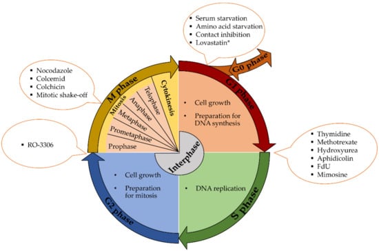
Cellular growth and the preparation of cells for division between two successive cell divisions is known as the cell cycle. In eukaryotic cells, it includes two basic parts—interphase and the M phase. Interphase, a part of the cell cycle when cells are duplicating the genetic information, expressing proteins and growing, is further divided into three separate phases—G1 (gap 1), S (synthetic) and G2 (gap 2). G1 and G2 are usually characterized by cell growth and high metabolic activity. The nuclear DNA is replicated during S phase. M phase is divided into six stages. The first five stages, prophase, prometaphase, metaphase, anaphase and telophase, are commonly known as mitosis. Mitosis involves nuclear division (daughter chromosomes are separated). The sixth stage of the M phase is known as cytokinesis and involves cytoplasmic division- (cell is divided into two daughter cells). (Figure 1) [,].

Figure 1.
Overview of the cell cycle phases and some synchronization methods. * The stage of G1 phase at which lovastatin exerts its effect is not clear.
The mammalian cell cycle is controlled by a subfamily of cyclin-dependent kinases (CDKs), the activity of which is modulated by several activators (cyclins) and inhibitors (Ink4, and Cip and Kip inhibitors) []. Progression through the cell cycle is controlled at distinct checkpoints: the G0/G1 checkpoint (restriction point), G1 checkpoint (or G1–S checkpoint), intra-S phase checkpoint, G2 checkpoint (or G2–M checkpoint) and mitosis-associated spindle assembly checkpoint [,,].
Cell cycle deregulation is a common feature of human cancer. Cancer cells frequently display unscheduled proliferation, genomic instability (with increased DNA mutations and chromosomal aberrations) and chromosomal instability (changes in chromosome number) []. This fact, together with the necessity to specifically address various aspects of cellular life during the cell cycle, has resulted in the development of many protocols providing cell populations enriched in cells at the specific stage of the cell cycle.
Basically, two different approaches to obtain such cell populations are used: (i) the treatment of asynchronous cell populations using special chemical agents, resulting in cell arrest at the specific phase of the cell cycle or (ii) mechanical isolation of cells at specific phase of the cell cycle. While the first group of methods suffers from the fact that the used treatments can result in unwanted effects on the cellular metabolism, the second group frequently does not provide a sufficient number of cells in the particular cell cycle phase or the synchrony of cell yield is low.
The first group includes methods based on the arrest of cells at the specific point in the G1 phase (e.g., by serum or amino acid starvation); methods based on the blockade of S phase (e.g., by thymidine or hydroxyurea); approaches based on the cell arrest in the M phase (e.g., by nocodazole) or at the G2/M border (e.g., RO-3306; Figure 1). The second group includes the isolation of mitotic cells mostly by mitotic shake off, the elutriation method or isolation of cells using flow cytometry and cell sorters.
Here we summarize the possibilities of cell synchronization at the G1/S boundary by substances impairing the deoxynucleotide metabolism, and consequently DNA replication, by aphidicolin—an inhibitor of DNA polymerase α, and by mimosine—a plant aminoacid exhibiting various effects on cell metabolism resulting in inhibition of DNA replication. A brief overview of other frequently-used methods for cell synchronization is included as well.
2. Metabolism of DNA Precursors
As the substances used for the synchronization of mammalian cells at the G1/S boundary frequently target the synthesis of deoxynucleotides, a short introduction of their metabolism in mammalian cells is given first. A complete overview of nucleotides’ metabolism is summarized, for example, in [].
The deoxynucleotides are de novo generated from ribonucleotides at the level of ribonucleotide diphosphates (adenosine diphosphate—ADP; guanosine 5′-diphosphate—GDP; cytidine 5′-diphosphate—CDP, and uridine 5′-diphosphate—UDP) by reduction at the 2’ position of the ribose subunit. This cytoplasmic reaction is catalyzed by the enzyme ribonucleotide reductase (RNR, Figure 2). Its appearance during evolution was a prerequisite for the transition from the “RNA world”, where RNA sufficed for both catalysis and information transfer, to today’s interplay among DNA, RNA, and proteins []. A general overview of the occurrence, catalytic function, regulation, and evolution of RNRs is reviewed, for example, in [].

Figure 2.
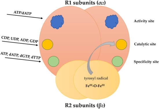
Schematic figure of the RNR heterotetramer. Each R1 subunit has two allosteric (activity and specificity sites) and one substrate binding site (catalytic site). The R2 subunits have a metal-oxygen center with a tyrosyl radical. This radical can be transferred to the catalytic site.
RNR is heterotetramer composed of two R1 subunits and two R2 subunits (Figure 2). During the S phase, the activity of RNR is greatly increased, while in the G1 phase its activity is very low [,]. In this respect, R2 subunit transcription, but not R1 subunit transcription, is repressed during G1 [,]. During mitosis, R2 subunits are degraded []. In resting cells (in G0 phase when cells are metabolically active but do not proliferate []), the R2 subunit is not transcribed []. It was found that the quiescent cells contain a second radical-providing small subunit, termed p53R2 with the same function as the homologous R2 []. Some data indicate that in the case of DNA repair, p53R2 is transcriptionally activated by p53 and translocates to the nucleus [,]. There, it can substitute for R2 forming a highly active RNR [,]. Besides its possible role in DNA repair, it was found that the p53R2 subunit has an essential role in mitochondrial DNA replication [,,,].
The enzyme activity is tightly regulated by allosteric regulation which prevents excessive concentration of each dNTP (deoxyribonucleotide triphosphate). R1 subunits contain an activity site, a specificity site and a catalytic site [,]. The activity site regulates the overall activity of the enzyme by binding of ATP (adenosine 5′-triphosphate; increase in the overall activity) and dATP (deoxyadenosine 5′-triphosphate; decrease in the overall enzyme activity (Figure 2) [,,]. The specificity site regulates the substrate specificity. This site binds dGTP (deoxyguanosine 5′-triphosphate), dTTP (deoxythymidine 5′-triphosphate), ATP and dATP []. This binding determines the substrate preference [,,,,]. Binding of ATP and dATP at the specificity site facilitates both CDP and UDP binding at the catalytic site. Binding of dTTP at the specificity site allows GDP binding at the catalytic site and dGTP binding at the specificity site facilitates ADP binding at the catalytic site []. Importantly, when dTTP is bound at the specificity site, it inhibits the reduction of both CDP and UDP []. After the ribonucleotide 5′ diphosphate reduction to deoxyribonucleotide 5′-diphosphate (Figure 3), the nucleoside diphosphate kinase catalyzes the transfer of the terminal phosphate groups from 5′-triphosphate to 5′-diphosphate nucleotides [].

Figure 3.
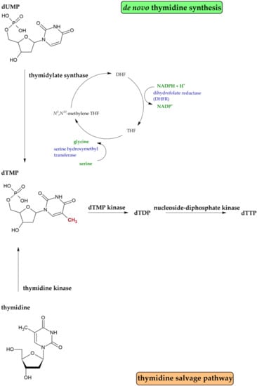
Simplified scheme of dNTP production.
dTMP (deoxythymidine 5′-monophosphate) is de novo synthesized by thymidylate synthase (TS) from dUMP (deoxyuridine 5′-monophosphate). dUMP is generated mainly by the enzyme dUTPase which hydrolyses dUTP (deoxyuridine 5′-triphosphate) to dUMP and pyrophosphate. This reaction provides the substrate for thymidylate synthase and concurrently eliminates dUTP from the DNA biosynthetic pathway []. TS catalyzes the reductive methylation of dUMP to dTMP using N5,N10-methylenetetrahydrofolate as the one-carbon methyl donor []. N5,N10-methylenetetrahydrofolate is oxidized during this reaction to dihydrofolate and has to be regenerated by dihydrofolate reductase (DHFR) and serine hydroxymethyltransferase (Figure 4) []. The second pathway of dTTP synthesis is a salvage pathway (Figure 4). In this case, thymidine is converted to dTMP by the enzyme thymidine kinase. The thymidine comes from intracellular nucleic acid degradation or from extracellular nucleosides circulating in the bloodstream [].

Figure 4.
Simplified scheme of dTTP production by de novo synthesis or the salvage pathway.
3. Targeting the Deoxynucleotide Metabolism and Its Use for Cell Synchronization
3.1. Thymidine
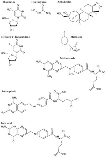
A high concentration of thymidine (Figure 5) is frequently used for cell synchronization at the G1/S boundary. After the addition to the culture medium, thymidine enters the cells and is rapidly converted to dTTP through a salvage pathway and its concentration in cells dramatically increases []. The mechanism of the thymidine action is based on the allosteric regulation of RNR enzyme when elevated dTTP concentration causes imbalance in the dNTP pool and inhibits reduction of CDP to dCDP by RNR [,,]. The thymidine block can be reversed either by the thymidine removal or by the addition of deoxycytidine [,]. Depletion of the nuclear dCTP pool after the increased dTTP concentration has been observed, for example, in Chinese hamster ovary (CHO) cells. Simultaneously, great increase in nuclear pools of dGTP and dATP was measured []. Similar data were also obtained in Molm-13 cells []. On the other hand, incubation of L929 mouse cells with 5 mM thymidine resulted only in an increase in the dTTP pool. The pools of dATP, dGTP and dCTP were all reduced []. This shows that the reaction of cells after thymidine treatment can vary substantially depending on the particular cell line.

Figure 5.
Formulae of the substances used for cell synchronization and of folic acid.
Typically, the thymidine concentrations used for cell synchronization are equal to, or above, 2 mM (see for example in [,]). The incubation time should be little longer than the sum of the lengths of G2, M and G1 phases. As cells in S phase could not transit this phase without the thymidine block removal, the synchronization by one thymidine block provides two populations of cells. One portion of cells is at G1/S boundary, the second one is trapped throughout the S phase. Therefore, a second block is usually performed after the release of cells from the first block. The time between release and the onset of the second block should somewhat exceed the length of the S phase. A typical protocol for HeLa cells can be found in [,].
3.2. Hydroxyurea
Hydroxyurea or hydroxycarbamide (Figure 5; HU) was first synthesized over a century ago in 1869 []. It is primarily used as an antineoplastic and antiviral agent []. HU inhibits RNR by directly reducing the diferric tyrosyl radical center in the smaller R2 subunit via a one-electron transfer from the drug []. HU thus inhibits production of dNTPs (Figure 3), and subsequently, also DNA synthesis. Because of the reversibility of its action, HU has commonly been used for cell synchronization. Its action is easily reversed by changing of the growth medium for drug-free medium. The treatment of cells by HU results in a decrease in purine pools in mammalian cells. Concerning the pyrimidine pools, conflicting data are available [,,,]. The complicated, often reciprocal, changes in individual dNTP pools occurring in HU-treated mammalian cells may be due to the compensatory activities of the deoxyribonucleotide salvage pathways in the higher eukaryotes [].
As HU treatment also results in trapping DNA synthesizing cells in the S phase, the HU treatment is typically combined with alternative synchronization protocols. One example is the protocol comprising isoleucine starvation followed by incubation with hydroxyurea []. In this case, cells are first incubated in a culture medium lacking isoleucine for a time corresponding to the sum of the G1, S, G2 and M phases of the particular cell line. According to Tobey and Crissman [], large quantities of cells may be reversibly arrested in early G1 by cultivation in an isoleucine-deficient medium. It was also shown that these cells do not enter a state of gross biochemical imbalance []. Then, the medium containing both isoleucine and hydroxyurea should be added, followed by the incubation of cells for a time period slightly exceeding the G1 phase length. The cells are released from the G1/S boundary after exchanging the medium for one without HU [].
Instead of isoleucine starvation, serum deprivation (starvation) can be used before HU treatment as well [,]. Although both methods based on isoleucine or serum starvation are efficient, they are not convenient for all cell lines and therefore, the preliminary tests are necessary.
3.3. Aminopterin and Methotrexate
Aminopterin and methotrexate are analogues of folic acid (Figure 5) []. They are potent inhibitors of dihydrofolate reductase [,]. Folic acid (vitamin B9) is not synthesized de novo by mammalian cells, therefore, it has to be obtained from food []. It is reduced by the action of dihydrofolate reductase either partially to the intermediate dihydrofolate (DHF) or completely to tetrahydrofolate (THF) []. THF is important for metabolism of thymidine, purines, glycine, methionine and choline []. Consequently, its lack results in the cessation of DNA replication. Methionine and choline are commonly present in the cell culture medium. To minimize the negative effects on processes other than DNA replication during cell synchronization by these two drugs, cell culture media also contain, besides aminopterin or methotrexate, hypoxanthine and glycine. This focuses the effect of THF depletion on the thymidine metabolism and consequently on the DNA synthesis []. In the case of methotrexate, this effect is further deepened by its inhibition of the thymidylate synthase []. The inhibitory effect of both antifolate drugs can be overcome either by the medium exchange for an antifolate-free medium or by the addition of thymidine []. The protocol for the synchronization of cells using aminopterin can be found, e.g., in the studies by Adams (1969) or Lindsay et al. (1970) [,], and the protocol for methotrexate-based synchronization is described, for example, in [,].
As antifolate drugs require the presence of additional substances in the growth media during synchronization and an additional synchronization step is required to obtain the highly synchronized cell population, their popularity as synchronization agents is very low. On the other hand, methotrexate is one of the most effective and extensively used drugs for treating many kinds of cancer or severe and resistant forms of autoimmune diseases [].
3.4. 5-Fluorodeoxyuridine
5-fluorodeoxyuridine (FdU; Figure 5) is an analogue of thymidine. It is transported into the cell where it is converted to FdUMP (fluorodeoxyuridine 5′-monophosphate) by the salvage pathway enzyme thymidine kinase []. A binary complex between 5-FdUMP and N5,N10-methylenetetrahydrofolate irreversibly inhibits thymidylate synthase, and thus blocks de novo synthesis of dTMP and results in the accumulation of dUMP []. FdU causes intracellular nucleotide pool imbalance with the decreased dTTP and increased dUTP levels and cessation of the DNA synthesis []. On the other hand, dUTP and FdUTP (fluorodeoxyuridine 5′-triphosphate) can be incorporated into DNA instead of dTTP, therefore, their incorporation results in base excision repair and excision of these nucleotides from the DNA [].
If cells are growing in a culture medium with FdU which is also supplemented with thymidine, they are able to synthesize dTMP using the thymidine kinase (through the salvage pathway) []. FdU-mediated inhibition of DNA synthesis can therefore be reversed by the addition of thymidine. The protocol of synchronization can be found in []. However, FdU and its derivative 5-fluorouracil has mainly been used in the treatment of various solid tumors [,,] and its use for cell synchronization is very uncommon.
3.5. Aphidicolin
Aphidicolin is a tetracyclic diterpenoid, obtained from Cephalosporium aphidicola (Figure 5) []. Aphidicolin inhibits the growth of eukaryotic cells by inhibiting the activity of DNA polymerase α without interfering with the activities of DNA polymerase β and γ. The effect of aphidicolin on DNA polymerase α is reversible []. Cell synchronization with aphidicolin is simple as aphidicolin-treated cells are released from the G1/S boundary by medium exchange []. On the other hand, similar to other protocols based on the DNA synthesis inhibitors, a large portion of cells is trapped in the S phase after aphidicolin treatment []. In this respect, aphidicolin treatment is usually combined with an additional synchronization step, e.g., with a subsequent second aphidicolin treatment after incubation of cells in an aphidicolin-free medium [], with the mitotic shake-off [] or with thymidine block [].
3.6. Mimosine
Mimosine [β-[N-(3-hydroxy-4-oxypyridyl)]-α-aminopropionic acid] (Figure 5) also blocks cells at the G1/S border. It seems that this block is mediated by several mechanisms. It has previously been suggested that mimosine can (i) alter deoxyribonucleotide metabolism by inhibition of ribonucleotide reductase [,]; (ii) inhibit initiation of DNA replication at replication origins []; (iii) attenuate serine hydroxymethyltransferase [] or (iv) enhance the levels of p27Kip1 []. The chelation of iron seems to be one of the main modes of action of mimosine for cell cycle arrest []. The other possible mechanisms of mimosine action are reviewed in []. Although mimosine’s effect on DNA replication is not completely clear, it is frequently used for cell synchronization on the G1/S boundary []. Mimosine action is also frequently combined with additional synchronization protocols to increase the percentage of cell synchrony at the G1/S border. Examples are the protocol combining thymidine block and mimosine treatment [] or the protocol based on nocodazole and mimosine treatment [].
4. Effects of Synchronization on Cellular Metabolism
Methods of cell synchronization based on targeting DNA replication are frequently used as they are relatively cheap and easy to perform; however, they exhibit several unwanted effects on the cell metabolism. These methods commonly result in trapping a relatively high proportion of cells in the S phase and this portion of cells encounters the consequences of replication stress as their replication forks are stalled. Stalled forks usually result in the formation of single stranded DNA (ssDNA) as replicative helicase continues to unwind the parental DNA []. The persistence of ssDNA, bound by replication protein A (RPA), and adjacent to the stalled newly replicated double-stranded DNA, generates a signal for activation of the replication stress response: a primer–template junction [,]. This structure serves as a signaling platform to recruit a number of replication-stress response proteins, including the protein kinase ataxia-telangiectasia mutated (ATM) and Rad3-related (ATR) [,,,,]. This response promotes fork stabilization and restart, while preventing progression through the cell cycle until DNA replication is completed. If stalled forks are not stabilized, or if they persist for an extended period, replication forks will collapse. This collapse can result in the formation of double-stranded DNA breaks [].
It has been reported that the exposure of cells to hydroxyurea, aphidicolin or thymidine at concentrations commonly used to synchronize cell populations led to the phosphorylation of histone H2AX on Ser139 (induction of γH2AX) through the activation of ATM and ATR protein kinase [,,]. DNA damage caused by hydroxyurea or aphidicolin treatment was also documented by Hammond and colleagues []. In addition, chromosomal aberrations were observed after the use of thymidine treatment []. Further, it was also shown that the synchronization using thymidine, mimosine or aphidicolin may lead to growth imbalance and can also induce imbalance in the expression of cell cycle regulatory proteins such as cyclins B1, A and E [].
Importantly, there are some cell cycle-dependent processes which are not inhibited or synchronized when DNA replication is arrested by hydroxyurea. Examples are centrosome replication [] and RRM2 transcription [].
Moreover, protocols based on the inhibition of deoxynucleotide synthesis inevitably result in imbalances in the nucleotide pools with various effects on cell metabolism. For example, in the case of FdU, impaired dTMP biosynthesis results in accelerated rates of genomic uracil incorporation [,] and DNA repair leading to the accumulation of DNA strand breaks [,]. In addition, it is supposed that FdUMP, phosphorylated by thymidylate kinase and nucleoside diphosphate kinase to its triphosphate form (FdUTP), can be incorporated into DNA and contributes to FdU-mediated toxicity []. Further, it is supposed that the incorporated FdUTP is recognized and excised by base excision repair machinery using the same mechanisms that remove genomic uracil []. Therefore, this method should not be used for studies focused on issues dealing with the metabolism of deoxynucleotides or base excision repair.
As the efficacy of particular protocols depends on the cell metabolism, chosen protocol should be experimentally verified and optimized for every cell line. In this respect, the overexpression of thymidylate synthase can result into resistance to FdU [,]. The overexpression of DHFR can contribute to the resistance of cells to methotrexate []. Moreover, mutation of CHO cells causing resistance to aphidicolin was described in [].
These data clearly show that, although the synchronization protocols based on the inhibition of DNA replication are easy to perform and can provide high amount of synchronized cells, they also have many negative effects on cell metabolism. These effects must be taken into account when planning the experiment.
5. Methods Overview
An overview of frequently used synchronization protocols, also involving those providing cells in cell cycle phases other than at G1/S border, is summarized in the Table 1. Irrespective of the protocol selection, optimization involving, e.g., dose and timing, should precede the experiments as too short incubation can result in insufficient cell synchronization while too long incubation can result in an increase in unwanted effects on cell metabolism.

Table 1.
Summarized overview of the commonly used synchronization approaches.
Author Contributions
Writing—original draft preparation, A.L. and K.K.; writing—review and editing, A.L and K.K. All authors have read and agreed to the published version of the manuscript.
Funding
This research was supported by the Technology Agency of the Czech Republic (grant number TN01000013); the Ministry of Education, Youth, and Sports of the Czech Republic (project EATRIS-CZ, grant number LM2018133); and the European Regional Development Fund (project ENOCH, grant number CZ.02.1.01/0.0/0.0/16_019/0000868).
Institutional Review Board Statement
Not applicable.
Informed Consent Statement
Not applicable.
Data Availability Statement
Not applicable.
Conflicts of Interest
The authors declare no conflict of interest. The funders had no role in the design of the study; in the collection, analyses, or interpretation of data; in the writing of the manuscript, or in the decision to publish the results.
References
- Cooper, G.M. The Eukaryotic Cell Cycle. In The Cell: A Molecular Approach, 2nd ed.; Sinauer Associates: Sunderland, MA, USA, 2000. [Google Scholar]
- Alberts, B.; Johnson, A.; Lewis, J.; Raff, M.; Roberts, K.; Walter, P. The Mechanics of Cell Division. In Molecular Biology of the Cell, 6th ed.; Garland Science: New York, NY, USA, 2002; pp. 1027–1062. [Google Scholar]
- Malumbres, M.; Barbacid, M. Cell Cycle, CDKs and Cancer: A Changing Paradigm. Nat. Rev. Cancer 2009, 9, 153–166. [Google Scholar] [CrossRef]
- Bower, J.J.; Vance, L.D.; Psioda, M.; Smith-Roe, S.L.; Simpson, D.A.; Ibrahim, J.G.; Hoadley, K.A.; Perou, C.M.; Kaufmann, W.K. Patterns of Cell Cycle Checkpoint Deregulation Associated with Intrinsic Molecular Subtypes of Human Breast Cancer Cells. NPJ Breast Cancer 2017, 3, 9. [Google Scholar] [CrossRef] [PubMed]
- Pardee, A.B. G1 Events and Regulation of Cell Proliferation. Science 1989, 246, 603–608. [Google Scholar] [CrossRef]
- Harashima, H.; Dissmeyer, N.; Schnittger, A. Cell Cycle Control Across the Eukaryotic Kingdom. Trends Cell Biol. 2013, 23, 345–356. [Google Scholar] [CrossRef] [PubMed]
- Lane, A.N.; Fan, T.W. Regulation of Mammalian Nucleotide Metabolism and Biosynthesis. Nucleic Acids Res. 2015, 43, 2466–2485. [Google Scholar] [CrossRef]
- Reichard, P. Ribonucleotide Reductases: The Evolution of Allosteric Regulation. Arch. Biochem. Biophys. 2002, 397, 149–155. [Google Scholar] [CrossRef]
- Jordan, A.; Reichard, P. Ribonucleotide Reductases. Annu. Rev. Biochem. 1998, 67, 71–98. [Google Scholar] [CrossRef]
- Nordlund, P.; Reichard, P. Ribonucleotide Reductases. Annu. Rev. Biochem. 2006, 75, 681–706. [Google Scholar] [CrossRef] [PubMed]
- DeGregori, J.; Kowalik, T.; Nevins, J.R. Cellular Targets for Activation by the E2F1 Transcription Factor Include DNA Synthesis- and G1/S-Regulatory Genes. Mol. Cell. Biol. 1995, 15, 4215–4224. [Google Scholar] [CrossRef]
- Chabes, A.L.; Pfleger, C.M.; Kirschner, M.W.; Thelander, L. Mouse Ribonucleotide Reductase R2 Protein: A New Target for Anaphase-Promoting Complex-Cdh1-Mediated Proteolysis. Proc. Natl. Acad. Sci. USA 2003, 100, 3925–3929. [Google Scholar] [CrossRef]
- Chabes, A.; Thelander, L. Controlled Protein Degradation Regulates Ribonucleotide Reductase Activity in Proliferating Mammalian Cells during the Normal Cell Cycle and in Response to DNA Damage and Replication Blocks. J. Biol. Chem. 2000, 275, 17747–17753. [Google Scholar] [CrossRef]
- Pontarin, G.; Ferraro, P.; Bee, L.; Reichard, P.; Bianchi, V. Mammalian Ribonucleotide Reductase Subunit p53R2 is Required for Mitochondrial DNA Replication and DNA Repair in Quiescent Cells. Proc. Natl. Acad. Sci. USA 2012, 109, 13302–13307. [Google Scholar] [CrossRef]
- Tanaka, H.; Arakawa, H.; Yamaguchi, T.; Shiraishi, K.; Fukuda, S.; Matsui, K.; Takei, Y.; Nakamura, Y. A Ribonucleotide Reductase Gene Involved in a p53-Dependent Cell-Cycle Checkpoint for DNA Damage. Nature 2000, 404, 42–49. [Google Scholar] [CrossRef] [PubMed]
- Guittet, O.; Hakansson, P.; Voevodskaya, N.; Fridd, S.; Graslund, A.; Arakawa, H.; Nakamura, Y.; Thelander, L. Mammalian p53R2 Protein Forms an Active Ribonucleotide Reductase In Vitro with the R1 Protein, Which is Expressed Both in Resting Cells in Response to DNA Damage and in Proliferating Cells. J. Biol. Chem. 2001, 276, 40647–40651. [Google Scholar] [CrossRef] [PubMed]
- Bourdon, A.; Minai, L.; Serre, V.; Jais, J.P.; Sarzi, E.; Aubert, S.; Chretien, D.; de Lonlay, P.; Paquis-Flucklinger, V.; Arakawa, H.; et al. Mutation of RRM2B, Encoding p53-Controlled Ribonucleotide Reductase (p53R2), Causes Severe Mitochondrial DNA Depletion. Nat. Genet. 2007, 39, 776–780. [Google Scholar] [CrossRef]
- Lim, A.Z.; McFarland, R.; Taylor, R.W.; Gorman, G.S. RRM2B Mitochondrial DNA Maintenance Defects. In GeneReviews ((R)); Adam, M.P., Ardinger, H.H., Pagon, R.A., Wallace, S.E., Bean, L.J.H., Mirzaa, G., Amemiya, A., Eds.; University of Washington: Seattle, WA, USA, 1993–2021. [Google Scholar]
- Thelander, L.; Reichard, P. Reduction of Ribonucleotides. Annu. Rev. Biochem. 1979, 48, 133–158. [Google Scholar] [CrossRef] [PubMed]
- Ahmad, M.F.; Dealwis, C.G. The Structural Basis for the Allosteric Regulation of Ribonucleotide Reductase. Prog. Mol. Biol. Transl. Sci. 2013, 117, 389–410. [Google Scholar] [CrossRef]
- Hofer, A.; Crona, M.; Logan, D.T.; Sjoberg, B.M. DNA Building Blocks: Keeping Control of Manufacture. Crit. Rev. Biochem. Mol. Biol. 2012, 47, 50–63. [Google Scholar] [CrossRef]
- Brown, N.C.; Canellakis, Z.N.; Lundin, B.; Reichard, P.; Thelander, L. Ribonucleoside Diphosphate Reductase. Purification of the Two Subunits, Proteins B1 and B2. Eur. J. Biochem. 1969, 9, 561–573. [Google Scholar] [CrossRef]
- Brown, N.C.; Reichard, P. Role of Effector Binding in Allosteric Control of Ribonucleoside Diphosphate Reductase. J. Mol. Biol. 1969, 46, 39–55. [Google Scholar] [CrossRef]
- Reichard, P.; Eliasson, R.; Ingemarson, R.; Thelander, L. Cross-Talk between the Allosteric Effector-Binding Sites in Mouse Ribonucleotide Reductase. J. Biol. Chem. 2000, 275, 33021–33026. [Google Scholar] [CrossRef]
- Xu, H.; Faber, C.; Uchiki, T.; Fairman, J.W.; Racca, J.; Dealwis, C. Structures of Eukaryotic Ribonucleotide Reductase I Provide Insights into dNTP Regulation. Proc. Natl. Acad. Sci. USA 2006, 103, 4022–4027. [Google Scholar] [CrossRef]
- Chimploy, K.; Mathews, C.K. Mouse Ribonucleotide Reductase Control: Influence of Substrate Binding upon Interactions with Allosteric Effectors. J. Biol. Chem. 2001, 276, 7093–7100. [Google Scholar] [CrossRef]
- Ray, N.B.; Mathews, C.K. Nucleoside Diphosphokinase: A Functional Link between Intermediary Metabolism and Nucleic Acid Synthesis. Curr. Top. Cell. Regul. 1992, 33, 343–357. [Google Scholar] [CrossRef]
- Nyiri, K.; Mertens, H.D.T.; Tihanyi, B.; Nagy, G.N.; Kohegyi, B.; Matejka, J.; Harris, M.J.; Szabo, J.E.; Papp-Kadar, V.; Nemeth-Pongracz, V.; et al. Structural Model of Human dUTPase in Complex with a Novel Proteinaceous Inhibitor. Sci. Rep. 2018, 8, 4326. [Google Scholar] [CrossRef]
- Rose, M.G.; Farrell, M.P.; Schmitz, J.C. Thymidylate Synthase: A Critical Target for Cancer Chemotherapy. Clin. Colorectal Cancer 2002, 1, 220–229. [Google Scholar] [CrossRef] [PubMed]
- Anderson, D.D.; Stover, P.J. SHMT1 and SHMT2 are Functionally Redundant in Nuclear de Novo Thymidylate Biosynthesis. PLoS ONE 2009, 4, e5839. [Google Scholar] [CrossRef] [PubMed]
- Okesli, A.; Khosla, C.; Bassik, M.C. Human Pyrimidine Nucleotide Biosynthesis as a Target for Antiviral Chemotherapy. Curr. Opin. Biotech. 2017, 48, 127–134. [Google Scholar] [CrossRef] [PubMed]
- Matsuda, S.; Kasahara, T. Simultaneous and Absolute Quantification of Nucleoside Triphosphates Using Liquid Chromatography-Triple Quadrupole Tandem Mass Spectrometry. Genes Environ. 2018, 40, 13. [Google Scholar] [CrossRef]
- Engstrom, J.U.; Kmiec, E.B. DNA Replication, Cell Cycle Progression and the Targeted Gene Repair Reaction. Cell Cycle 2008, 7, 1402–1414. [Google Scholar] [CrossRef]
- Chen, G.; Deng, X. Cell Synchronization by Double Thymidine Block. Bio Protoc. 2018, 8. [Google Scholar] [CrossRef]
- Bjursell, G.; Reichard, P. Effects of Thymidine on Deoxyribonucleoside Triphosphate Pools and Deoxyribonucleic Acid Synthesis in Chinese Hamster Ovary Cells. J. Biol. Chem. 1973, 248, 3904–3909. [Google Scholar] [CrossRef]
- Morris, N.R.; Fischer, G.A. Studies Concerning Inhibition of the Synthesis of Deoxycytidine by Phosphorylated Derivatives of Thymidine. Biochim. Biophys. Acta 1960, 42, 183–184. [Google Scholar] [CrossRef]
- Skoog, L.; Bjursell, G. Nuclear and Cytoplasmic Pools of Deoxyribonucleoside Triphosphates in Chinese Hamster Ovary Cells. J. Biol. Chem. 1974, 249, 6434–6438. [Google Scholar] [CrossRef]
- Adams, R.L.P.; Berryman, S.; Thomson, A. Deoxyribonucleoside Triphosphate Pools in Synchronized and Drug-Inhibited L929 Cells. Biochim. Biophys. Acta 1971, 240, 455–462. [Google Scholar] [CrossRef]
- Ligasova, A.; Raska, I.; Koberna, K. Organization of Human Replicon: Singles or Zipping Couples? J. Struct. Biol. 2009, 165, 204–213. [Google Scholar] [CrossRef]
- Ma, H.T.; Poon, R.Y. Synchronization of HeLa Cells. Methods Mol. Biol. 2011, 761, 151–161. [Google Scholar] [CrossRef]
- Dresler, W.F.C.; Stein, R. Ueber den Hydroxylharnstoff. Justus Liebigs Annalen der Chemie 1869, 150, 242–252. [Google Scholar] [CrossRef]
- Singh, A.; Xu, Y.J. The Cell Killing Mechanisms of Hydroxyurea. Genes 2016, 7, 99. [Google Scholar] [CrossRef]
- Skoog, L.; Nordenskjold, B. Effects of Hydroxyurea and 1-Beta-D-Arabinofuranosyl-Cytosine on Deoxyribonucleotide Pools in Mouse Embryo Cells. Eur. J. Biochem. 1971, 19, 81–89. [Google Scholar] [CrossRef]
- Tyrsted, G. Effect of Hydroxyurea and 5-Fluorodeoxy-Uridine on Deoxyribonucleoside Triphosphate Pools Early in Phytohemagglutinin-Stimulated Human-Lymphocytes. Biochem. Pharmacol. 1982, 31, 3107–3113. [Google Scholar] [CrossRef]
- Koc, A.; Wheeler, L.J.; Mathews, C.K.; Merrill, G.F. Hydroxyurea Arrests DNA Replication by a Mechanism that Preserves Basal dNTP Pools. J. Biol. Chem. 2004, 279, 223–230. [Google Scholar] [CrossRef]
- Tobey, R.A.; Crissman, H.A. Preparation of Large Quantities of Synchronized Mammalian Cells in Late G1 in the Pre-DNA Replicative Phase of the Cell Cycle. Exp. Cell Res. 1972, 75, 460–464. [Google Scholar] [CrossRef]
- Enger, M.D.; Tobey, R.A. Effects of Isoleucine Deficiency on Nucleic Acid and Protein Metabolism in Cultured Chinese Hamster Cells. Continued Ribonucleic Acid and Protein Synthesis in the Absence of Deoxyribonucleic Acid Synthesis. Biochemistry 1972, 11, 269–277. [Google Scholar] [CrossRef]
- Raska, I.; Koberna, K.; Jarnik, M.; Petrasovicova, V.; Bednar, J.; Raska, K., Jr.; Bravo, R. Ultrastructural Immunolocalization of Cyclin/PCNA in Synchronized 3T3 Cells. Exp. Cell. Res. 1989, 184, 81–89. [Google Scholar] [CrossRef]
- Walker, M.M.; Wanda, P.E. Immunochemical Detection of Cell Cycle Synchronization in a Human Erythroleukemia Cell Line, K562. J. Histochem. Cytochem. 1987, 35, 1143–1148. [Google Scholar] [CrossRef] [PubMed]
- Cronstein, B.N.; Aune, T.M. Methotrexate and its Mechanisms of Action in Inflammatory Arthritis. Nat. Rev. Rheumatol. 2020, 16, 145–154. [Google Scholar] [CrossRef]
- Raimondi, M.V.; Randazzo, O.; La Franca, M.; Barone, G.; Vignoni, E.; Rossi, D.; Collina, S. DHFR Inhibitors: Reading the Past for Discovering Novel Anticancer Agents. Molecules 2019, 24, 1140. [Google Scholar] [CrossRef]
- Avendano, C.; Menendez, J.C. Antimetabolites That Interfere with Nucleic Acid Biosynthesis. In Medicinal Chemistry of Anticancer Drugs, 2nd ed.; Avendano, C., Menendez, J.C., Eds.; Elsevier: Amsterdam, The Netherlands, 2015; pp. 23–79. [Google Scholar] [CrossRef]
- Liew, S.C. Folic Acid and Diseases—Supplement It or Not? Rev. Assoc. Med. Bras. 2016, 62, 90–100. [Google Scholar] [CrossRef] [PubMed]
- Cylwik, B.; Chrostek, L. Interactions between Alcohol and Folate. In Molecular Aspects of Alcohol and Nutrition; Patel, V.B., Ed.; Academic Press: San Diego, CA, USA, 2016; Chapter 13; pp. 157–169. [Google Scholar] [CrossRef]
- Ducker, G.S.; Rabinowitz, J.D. One-Carbon Metabolism in Health and Disease. Cell Metab. 2017, 25, 27–42. [Google Scholar] [CrossRef]
- Adams, R.L.P. Cell Synchronisation. In Laboratory Techniques in Biochemistry and Molecular Biology; Work, T.S., Burdon, R.H., Eds.; Elsevier: Amsterdam, The Netherlands, 1980; Volume 8, pp. 211–238. [Google Scholar]
- McBurney, M.W.; Whitmore, G.F. Mechanism of Growth Inhibition by Methotrexate. Cancer Res. 1975, 35, 586–590. [Google Scholar]
- Adams, R.L. The Effect of Endogenous Pools of Thymidylate on the Apparent Rate of DNA Synthesis. Exp. Cell Res. 1969, 56, 55–58. [Google Scholar] [CrossRef]
- Lindsay, J.G.; Berryman, S.; Adams, R.L. Characteristics of Deoxyribonucleic acid Polymerase Activity in Nuclear and Supernatant Fractions of Cultured Mouse Cells. Biochem. J. 1970, 119, 839–848. [Google Scholar] [CrossRef]
- Yunis, J.J. High Resolution of Human Chromosomes. Science 1976, 191, 1268–1270. [Google Scholar] [CrossRef]
- Rueckert, R.R.; Mueller, G.C. Studies on Unbalanced Growth in Tissue Culture. I. induction and Consequences of Thymidine Deficiency. Cancer Res. 1960, 20, 1584–1591. [Google Scholar] [PubMed]
- Kozminski, P.; Halik, P.K.; Chesori, R.; Gniazdowska, E. Overview of Dual-Acting Drug Methotrexate in Different Neurological Diseases, Autoimmune Pathologies and Cancers. Int. J. Mol. Sci. 2020, 21, 3483. [Google Scholar] [CrossRef]
- Rossana, C.; Gollakota Rao, L.; Johnson, L.F. Thymidylate Synthetase Overproduction in 5-Fluorodeoxyuridine-Resistant Mouse Fibroblasts. Mol. Cell. Biol. 1982, 2, 1118–1125. [Google Scholar] [CrossRef] [PubMed]
- Grogan, B.C.; Parker, J.B.; Guminski, A.F.; Stivers, J.T. Effect of the Thymidylate Synthase Inhibitors on dUTP and TTP Pool Levels and the Activities of DNA Repair Glycosylases on Uracil and 5-Fluorouracil in DNA. Biochemistry 2011, 50, 618–627. [Google Scholar] [CrossRef] [PubMed]
- Yan, Y.; Han, X.Z.; Qing, Y.L.; Condie, A.G.; Gorityala, S.; Yang, S.M.; Xu, Y.; Zhang, Y.W.; Gerson, S.L. Inhibition of Uracil DNA Glycosylase Sensitizes Cancer Cells to 5-Fluorodeoxyuridine through Replication Fork Collapse-Induced DNA Damage. Oncotarget 2016, 7, 59299–59313. [Google Scholar] [CrossRef]
- Webber, L.M.; Garson, O.M. Fluorodeoxyuridine Synchronization of Bone Marrow Cultures. Cancer Genet. Cytogenet. 1983, 8, 123–132. [Google Scholar] [CrossRef]
- Longley, D.B.; Harkin, D.P.; Johnston, P.G. 5-Fluorouracil: Mechanisms of Action and Clinical Strategies. Nat. Rev. Cancer 2003, 3, 330–338. [Google Scholar] [CrossRef] [PubMed]
- Malet-Martino, M.; Martino, R. Clinical Studies of Three Oral Prodrugs of 5-Fluorouracil (Capecitabine, UFT, S-1): A Review. Oncologist 2002, 7, 288–323. [Google Scholar] [CrossRef] [PubMed]
- Power, D.G.; Kemeny, N.E. The Role of Floxuridine in Metastatic Liver Disease. Mol. Cancer Ther. 2009, 8, 1015–1025. [Google Scholar] [CrossRef] [PubMed]
- Brundret, K.M.; Dalziel, W.; Hesp, B.; Jarvis, J.A.J.; Neidle, S. X-Ray Crystallographic Determination of the Structure of the Antibiotic Aphidicolin: A Tetracyclic Diterpenoid Containing a New Ring System. J. Chem. Soc. Chem. Commun. 1972, 18, 1027–1028. [Google Scholar] [CrossRef]
- Ikegami, S.; Taguchi, T.; Ohashi, M.; Oguro, M.; Nagano, H.; Mano, Y. Aphidicolin Prevents Mitotic Cell Division by Interfering with the Activity of DNA Polymerase-Alpha. Nature 1978, 275, 458–460. [Google Scholar] [CrossRef]
- Pedrali-Noy, G.; Spadari, S.; Miller-Faures, A.; Miller, A.O.; Kruppa, J.; Koch, G. Synchronization of HeLa Cell Cultures by Inhibition of DNA Polymerase Alpha with Aphidicolin. Nucleic Acids Res. 1980, 8, 377–387. [Google Scholar] [CrossRef]
- Matherly, L.H.; Schuetz, J.D.; Westin, E.; Goldman, I.D. A Method for the Synchronization of Cultured-Cells with Aphidicolin -Application to the Large-Scale Synchronization of L1210 Cells and the Study of the Cell-Cycle Regulation of Thymidylate Synthase and Dihydrofolate-Reductase. Anal. Biochem. 1989, 182, 338–345. [Google Scholar] [CrossRef]
- Fox, M.H.; Read, R.A.; Bedford, J.S. Comparison of Synchronized Chinese-Hamster Ovary Cells Obtained by Mitotic Shake-Off, Hydroxyurea, Aphidicolin, or Methotrexate. Cytometry 1987, 8, 315–320. [Google Scholar] [CrossRef]
- Kim, J.K.; Esteve, P.O.; Jacobsen, S.E.; Pradhan, S. UHRF1 Binds G9a and Participates in p21 Transcriptional Regulation in Mammalian Cells. Nucleic Acids Res. 2009, 37, 493–505. [Google Scholar] [CrossRef]
- Gilbert, D.M.; Neilson, A.; Miyazawa, H.; Depamphilis, M.L.; Burhans, W.C. Mimosine Arrests DNA-Synthesis at Replication Forks by Inhibiting Deoxyribonucleotide Metabolism. J. Biol. Chem. 1995, 270, 9597–9606. [Google Scholar] [CrossRef]
- Dai, Y.M.; Gold, B.; Vishwanatha, J.K.; Rhode, S.L. Mimosine Inhibits Viral-DNA Synthesis through Ribonucleotide Reductase. Virology 1994, 205, 210–216. [Google Scholar] [CrossRef]
- Mosca, P.J.; Dijkwel, P.A.; Hamlin, J.L. The Plant Amino-Acid Mimosine May Inhibit Initiation at Origins of Replication in Chinese-Hamster Cells. Mol. Cell Biol. 1992, 12, 4375–4383. [Google Scholar] [CrossRef] [PubMed][Green Version]
- Perry, C.; Sastry, R.; Nasrallah, I.M.; Stover, P.J. Mimosine Attenuates Serine Hydroxymethyltransferase Transcription by Chelating Zinc. Implications for Inhibition of DNA Replication. J. Biol. Chem. 2005, 280, 396–400. [Google Scholar] [CrossRef] [PubMed]
- Wang, G.; Miskimins, R.; Miskimins, W.K. Mimosine Arrests Cells in G1 by Enhancing the Levels of p27(Kip1). Exp. Cell Res. 2000, 254, 64–71. [Google Scholar] [CrossRef] [PubMed]
- Nguyen, B.C.; Tawata, S. The Chemistry and Biological Activities of Mimosine: A Review. Phytother. Res. 2016, 30, 1230–1242. [Google Scholar] [CrossRef] [PubMed]
- Galgano, P.J.; Schildkraut, C.L. G1/S Phase Synchronization Using Mimosine Arrest. CSH Protoc. 2006, 2006. [Google Scholar] [CrossRef] [PubMed]
- Park, S.Y.; Im, J.S.; Park, S.R.; Kim, S.E.; Wang, H.J.; Lee, J.K. Mimosine Arrests the Cell Cycle Prior to the Onset of DNA Replication by Preventing the Binding of Human Ctf4/and-1 to Chromatin via Hif-1 Alpha Activation in HeLa Cells. Cell Cycle 2012, 11, 761–766. [Google Scholar] [CrossRef]
- Zeman, M.K.; Cimprich, K.A. Causes and Consequences of Replication Stress. Nat. Cell Biol. 2014, 16, 2–9. [Google Scholar] [CrossRef]
- Byun, T.S.; Pacek, M.; Yee, M.C.; Walter, J.C.; Cimprich, K.A. Functional Uncoupling of MCM Helicase and DNA Polymerase Activities the ATR-Dependent Checkpoint. Gene Dev. 2005, 19, 1040–1052. [Google Scholar] [CrossRef]
- MacDougall, C.A.; Byun, T.S.; Van, C.; Yee, M.C.; Cimprich, K.A. The Structural Determinants of Checkpoint Activation. Gene Dev. 2007, 21, 898–903. [Google Scholar] [CrossRef]
- Marechal, A.; Zou, L. DNA Damage Sensing by the ATM and ATR Kinases. Cold Spring Harb. Perspect. Biol. 2013, 5, a012716. [Google Scholar] [CrossRef]
- Nam, E.A.; Cortez, D. AIR Signalling: More than Meeting at the Fork. Biochem. J. 2011, 436, 527–536. [Google Scholar] [CrossRef]
- Zou, L.; Elledge, S.J. Sensing DNA Damage through ATRIP Recognition of RPA-ssDNA Complexes. Science 2003, 300, 1542–1548. [Google Scholar] [CrossRef] [PubMed]
- Darzynkiewicz, Z.; Halicka, H.D.; Zhao, H.; Podhorecka, M. Cell Synchronization by Inhibitors of DNA Replication Induces Replication Stress and DNA Damage Response: Analysis by Flow Cytometry. Methods Mol. Biol. 2011, 761, 85–96. [Google Scholar] [CrossRef] [PubMed]
- Halicka, D.; Zhao, H.; Li, J.W.; Garcia, J.; Podhorecka, M.; Darzynkiewicz, Z. DNA Damage Response Resulting from Replication Stress Induced by Synchronization of Cells by Inhibitors of DNA Replication: Analysis by Flow Cytometry. Methods Mol. Biol. 2017, 1524, 107–119. [Google Scholar] [CrossRef] [PubMed]
- Kurose, A.; Tanaka, T.; Huang, X.; Traganos, F.; Darzynkiewicz, Z. Synchronization in the Cell Cycle by Inhibitors of DNA Replication Induces Histone H2AX Phosphorylation: An Indication of DNA Damage. Cell Prolif. 2006, 39, 231–240. [Google Scholar] [CrossRef]
- Hammond, E.M.; Green, S.L.; Giaccia, A.J. Comparison of Hypoxia-Induced Replication Arrest with Hydroxyurea and Aphidicolin-Induced Arrest. Mutat. Res. Mol. Mech. Mutagen. 2003, 532, 205–213. [Google Scholar] [CrossRef]
- Yang, S.J.; Hahn, G.M.; Bagshaw, M.A. Chromosome Aberrations Induced by Thymidine. Exp. Cell Res. 1966, 42, 130–135. [Google Scholar] [CrossRef]
- Gong, J.P.; Traganos, F.; Darzynkiewicz, Z. Growth Imbalance and Altered Expression of Cyclin-B1, Cyclin-a, Cyclin-E, and Cyclin-D3 in Molt-4 Cells Synchronized in the Cell-Cycle by Inhibitors of DNA-Replication. Cell Growth Differ. 1995, 6, 1485–1493. [Google Scholar]
- Kuriyama, R.; Terada, Y.; Lee, K.S.; Wang, C.L.C. Centrosome Replication in Hydroxyurea-Arrested CHO Cells Expressing GFP-Tagged Centrin2. J. Cell Sci. 2007, 120, 2444–2453. [Google Scholar] [CrossRef]
- MacFarlane, A.J.; Anderson, D.D.; Flodby, P.; Perry, C.A.; Allen, R.H.; Stabler, S.P.; Stover, P.J. Nuclear Localization of de Novo Thymidylate Biosynthesis Pathway Is Required to Prevent Uracil Accumulation in DNA. J. Biol. Chem. 2011, 286, 44015–44022. [Google Scholar] [CrossRef] [PubMed]
- MacFarlane, A.J.; Perry, C.A.; McEntee, M.F.; Lin, D.M.; Stover, P.J. Shmt1 Heterozygosity Impairs Folate-Dependent Thymidylate Synthesis Capacity and Modifies Risk of Apc(min)-Mediated Intestinal Cancer Risk. Cancer Res. 2011, 71, 2098–2107. [Google Scholar] [CrossRef] [PubMed]
- Schormann, N.; Ricciardi, R.; Chattopadhyay, D. Uracil-DNA Glycosylases-Structural and Functional Perspectives on An Essential Family of DNA Repair Enzymes. Protein Sci. 2014, 23, 1667–1685. [Google Scholar] [CrossRef] [PubMed]
- Schrader, C.E.; Guikema, J.E.J.; Wu, X.M.; Stavnezer, J. The roles of APE1, APE2, DNA Polymerase Beta and Mismatch Repair in Creating S Region DNA Breaks during Antibody Class Switch. Philos. Trans. R. Soc. B Biol. Sci. 2009, 364, 645–652. [Google Scholar] [CrossRef]
- Chon, J.; Stover, P.J.; Field, M.S. Targeting Nuclear Thymidylate Biosynthesis. Mol. Asp. Med. 2017, 53, 48–56. [Google Scholar] [CrossRef]
- Wyatt, M.D.; Wilson, D.M. Participation of DNA Repair in the Response to 5-Fluorouracil. Cell. Mol. Life Sci. 2009, 66, 788–799. [Google Scholar] [CrossRef]
- Berger, S.H.; Jenh, C.H.; Johnson, L.F.; Berger, F.G. Thymidylate Synthase Overproduction and Gene Amplification in Fluorodeoxyuridine-Resistant Human-Cells. Mol. Pharmacol. 1985, 28, 461–467. [Google Scholar]
- Banerjee, D.; Mayer-Kuckuk, P.; Capiaux, G.; Budak-Alpdogan, T.; Gorlick, R.; Bertino, J.R. Novel Aspects of Resistance to Drugs Targeted to Dihydrofolate Reductase and Thymidylate Synthase. BBA Acta Mol. Basis Dis. 2002, 1587, 164–173. [Google Scholar] [CrossRef]
- Feher, Z.; Mishra, N.C. Aphidicolin-Resistant Chinese-Hamster Ovary Cells Possess Altered DNA-Polymerases of the Alpha-Family. BBA Gene Struct. Expr. 1994, 1218, 35–47. [Google Scholar] [CrossRef]
- Zwanenburg, T.S.B. Standardized Shake-Off to Synchronize Cultured Cho Cells. Mutat. Res. 1983, 120, 151–159. [Google Scholar] [CrossRef]
- Liu, Y.; Nan, B.; Niu, J.; Kapler, G.M.; Gao, S. An Optimized and Versatile Counter-Flow Centrifugal Elutriation Workflow to Obtain Synchronized Eukaryotic Cells. Front. Cell Dev. Biol. 2021, 9, 664418. [Google Scholar] [CrossRef] [PubMed]
- Juan, G.; Hernando, E.; Cordon-Cardo, C. Separation of Live Cells in Different Phases of the Cell Cycle for Gene Expression Analysis. Cytometry 2002, 49, 170–175. [Google Scholar] [CrossRef] [PubMed]
- Vecsler, M.; Lazar, I.; Tzur, A. Using Standard Optical Flow Cytometry for Synchronizing Proliferating Cells in the G1 Phase. PLoS ONE 2013, 8, e83935. [Google Scholar] [CrossRef] [PubMed]
- De Brabander, M.J.; Van de Veire, R.M.; Aerts, F.E.; Borgers, M.; Janssen, P.A. The Effects of Methyl (5-(2-thienylcarbonyl)-1H-benzimidazol-2-yl) Carbamate, (R 17934; NSC 238159), a New Synthetic Antitumoral Drug Interfering with Microtubules, on Mammalian Cells Cultured In Vitro. Cancer Res. 1976, 36, 905–916. [Google Scholar]
- Salmon, E.D.; McKeel, M.; Hays, T. Rapid Rate of Tubulin Dissociation from Microtubules in the Mitotic Spindle In Vivo Measured by Blocking Polymerization with Colchicine. J. Cell Biol. 1984, 99, 1066–1075. [Google Scholar] [CrossRef] [PubMed]
- Blagosklonny, M.V. Mitotic Arrest and Cell-Fate Why and How Mitotic Inhibition of Transcription Drives Mutually Exclusive Events. Cell Cycle 2007, 6, 70–74. [Google Scholar] [CrossRef]
- Zieve, G.W.; Turnbull, D.; Mullins, J.M.; Mcintosh, J.R. Production of Large Numbers of Mitotic Mammalian-Cells by Use of the Reversible Microtubule Inhibitor Nocodazole-Nocodazole Accumulated Mitotic Cells. Exp. Cell Res. 1980, 126, 397–405. [Google Scholar] [CrossRef]
- Yao, Y.Z.; Zhang, Y.Y.; Liu, W.S.; Deng, X.M. Highly Efficient Synchronization of Sheep Skin Fibroblasts at G2/M Phase and Isolation of Sheep Y Chromosomes by Flow Cytometric Sorting. Sci. Rep. 2020, 10, 9933. [Google Scholar] [CrossRef]
- Romsdahl, M.M. Synchronization of Human Cell Lines with Colcemid. Exp. Cell Res. 1968, 50, 463–467. [Google Scholar] [CrossRef]
- Vassilev, L.T.; Tovar, C.; Chen, S.; Knezevic, D.; Zhao, X.; Sun, H.; Heimbrook, D.C.; Chen, L. Selective Small-Molecule Inhibitor Reveals Critical Mitotic Functions of Human CDK1. Proc. Natl. Acad. Sci. USA 2006, 103, 10660–10665. [Google Scholar] [CrossRef]
- Ma, H.T.; Tsang, Y.H.; Marxer, M.; Poon, R.Y. Cyclin A2-Cyclin-Dependent Kinase 2 Cooperates with the PLK1-SCFbeta-TrCP1-EMI1-Anaphase-Promoting Complex/Cyclosome Axis to Promote Genome Reduplication in the Absence of Mitosis. Mol. Cell Biol. 2009, 29, 6500–6514. [Google Scholar] [CrossRef] [PubMed]
- Vassilev, L.T. Cell Cycle Synchronization at the G2/M Phase Border by Reversible Inhibition of CDK1. Cell Cycle 2006, 5, 2555–2556. [Google Scholar] [CrossRef] [PubMed]
- Rao, S.; Lowe, M.; Herliczek, T.W.; Keyomarsi, K. Lovastatin Mediated G1 Arrest in Normal and Tumor Breast Cells is through Inhibition of CDK2 Activity and Redistribution of p21 and p27, Independent of p53. Oncogene 1998, 17, 2393–2402. [Google Scholar] [CrossRef] [PubMed]
- JavanMoghadam-Kamrani, S.; Keyomarsi, K. Synchronization of the Cell Cycle Using Lovastatin. Cell Cycle 2008, 7, 2434–2440. [Google Scholar] [CrossRef]
- Davis, P.K.; Ho, A.; Dowdy, S.F. Biological Methods for Cell-Cycle Synchronization of Mammalian Cells. Biotechniques 2001, 30, 1322–1326. [Google Scholar] [CrossRef]
- Kues, W.A.; Anger, M.; Carnwath, J.W.; Paul, D.; Motlik, J.; Niemann, H. Cell Cycle Synchronization of Porcine Fetal Fibroblasts: Effects of Serum Deprivation and Reversible Cell Cycle Inhibitors. Biol. Reprod. 2000, 62, 412–419. [Google Scholar] [CrossRef]
- Khammanit, R.; Chantakru, S.; Kitiyanant, Y.; Saikhun, J. Effect of Serum Starvation and Chemical Inhibitors on Cell Cycle Synchronization of Canine Dermal Fibroblasts. Theriogenology 2008, 70, 27–34. [Google Scholar] [CrossRef] [PubMed]
- Nilausen, K.; Green, H. Reversible Arrest of Growth in G1 of an Established Fibroblast Line (3T3). Exp. Cell Res. 1965, 40, 166–168. [Google Scholar] [CrossRef]
- Holley, R.W.; Kiernan, J.A. Contact Inhibition of Cell Division in 3t3 Cells. Proc. Natl. Acad. Sci. USA 1968, 60, 300–304. [Google Scholar] [CrossRef]
- Forsberg, F.; Mooser, R.; Arnold, M.; Hack, E.; Wyss, P. 3D Micro-Scale Deformations of Wood in Bending: Synchrotron Radiation μCT data Analyzed with Digital Volume Correlation. J. Struct. Biol. 2008, 164, 255–262. [Google Scholar] [CrossRef]
Publisher’s Note: MDPI stays neutral with regard to jurisdictional claims in published maps and institutional affiliations. |
© 2021 by the authors. Licensee MDPI, Basel, Switzerland. This article is an open access article distributed under the terms and conditions of the Creative Commons Attribution (CC BY) license (https://creativecommons.org/licenses/by/4.0/).