Review

Genetic Factors Affect the Survival and Behaviors of Selected Bacteria during Antimicrobial Blue Light Treatment

1
AgResearch Ltd., Hopkirk Research Institute, Cnr University Ave and Library Road, Massey University, Palmerston North 4442, New Zealand
2
New Zealand Food Safety Science and Research Centre, Massey University Manawatu (Turitea), Tennent Drive, Palmerston North 4474, New Zealand
*
Author to whom correspondence should be addressed.
Int. J. Mol. Sci. 2021, 22(19), 10452; https://doi.org/10.3390/ijms221910452
Submission received: 27 August 2021 / Revised: 22 September 2021 / Accepted: 24 September 2021 / Published: 28 September 2021
(This article belongs to the Section Molecular Microbiology)

Abstract

:
Antimicrobial resistance is a global, mounting and dynamic issue that poses an immediate threat to human, animal, and environmental health. Among the alternative antimicrobial treatments proposed to reduce the external use of antibiotics is electromagnetic radiation, such as blue light. The prevailing mechanistic model is that blue light can be absorbed by endogenous porphyrins within the bacterial cell, inducing the production of reactive oxygen species, which subsequently inflict oxidative damages upon different cellular components. Nevertheless, it is unclear whether other mechanisms are involved, particularly those that can affect the efficacy of antimicrobial blue light treatments. In this review, we summarize evidence of inherent factors that may confer protection to a selected group of bacteria against blue light-induced oxidative damages or modulate the physiological characteristics of the treated bacteria, such as virulence and motility. These include descriptions of three major photoreceptors in bacteria, chemoreceptors, SOS-dependent DNA repair and non-SOS protective mechanisms. Future directions are also provided to assist with research efforts to increase the efficacy of antimicrobial blue light and to minimize the development of blue light-tolerant phenotypes.

1. Introduction

According to the latest Global Antimicrobial Resistance and Use Surveillance System (GLASS) report by the World Health Organization, there were 3,106,602 laboratory-confirmed infections related to antimicrobial resistant pathogens in the year 2019, including reports on an increasing trend of resistance to last-resort antibiotics [1]. In particular, the report highlighted the roles of Escherichia coli resistant to third generation cephalosporins and methicillin-resistant Staphylococcus aureus (MRSA) in causing bloodstream infections. Inevitably, researchers have been prompted to find alternative strategies for inactivating these pathogens beyond the use of antibiotics. These strategies include the use of electromagnetic radiation, such as ultraviolet (UV) and visible light [2]. It is a well-established fact that UV, particularly UV-C (200–280 nm), possesses potent bactericidal properties through the induction of pyrimidine dimers upon its absorption by nucleic acids [3]. However, safety issues may arise from the use of UV as a bactericidal agent, for example, increased risk of skin cancer in humans related to constant exposure to low-intensity UV [4,5] or long-term injuries associated with accidental exposure to high-intensity UV [6].
In contrast, visible light is less harmful than UV [7]. Within the visible spectrum, blue light at the wavelength range of 400–450 nm has the highest bactericidal property associated with its absorbance by endogenous porphyrins, leading to cell death [2,8]. Porphyrins are intermediate species in the heme biosynthesis [9,10] and may be activated by blue light to produce reactive oxygen species (ROS), such as superoxide, hydrogen peroxide, hydroxyl radicals, and reactive singlet oxygen (1O2) [11]. These ROS can subsequently induce oxidative stress upon different bacterial cell constituents, especially genomic materials, cell membrane, and cell wall (Figure 1A). Prokaryotic genomes can be degraded by ROS during blue light treatments [12,13], particularly through the oxidation of guanine residues into 8-hydroxydeoxyguanosine [12]. Fatty acids within the bacterial cell membrane are susceptible to blue light-induced oxidation, commonly marked by the formation of malondialdehyde [14,15]. Breakages of bacterial cell wall have been reported in Gram-positive MRSA [16] and Gram-negative Acinetobacter baumannii [17], and the inactivation of E. coli lipopolysaccharide has also been observed in vitro [18].
Furthermore, the efficacy of antimicrobial blue light extends to non-planktonic bacteria. Fila et al. demonstrated blue light-mediated inhibition of the quorum sensing signaling systems in Pseudomonas aeruginosa, leading to the delayed formation of biofilm, and subsequently reduced pathogenicity in the animal model Caenorhabditis elegans [19]. Besides nucleic acids, proteins, and lipids [19,20,21], polysaccharide materials contained within the biofilm matrix could also be targeted by antimicrobial blue light, albeit only when an exogenous photosensitizer (i.e., photoactive compounds, such as porphyrins) is applied [22].
Antimicrobial blue light treatments have mostly been utilized in clinical and food settings [23,24]. It involves the exposure of bacteria in a given matrix to a blue light source (for example, light-emitting diodes) at a fixed light intensity (Watt/cm2) for a period of treatment time (second). The efficacy of antimicrobial blue light treatments is most commonly measured by the reduction in bacterial counts after exposure to a certain light dosage (Joule/cm2), defined as the multiplication product of light intensity and treatment time. Importantly, sensitivities to blue light vary across bacterial strains, i.e., light dosage required to inactivate different strains may vary considerably [25,26,27]. Thus, as with other antimicrobial treatments, antimicrobial blue light may be applied in an insufficient amount (i.e., sub-lethal), potentially resulting in the development of tolerance [28]. However, the underlying mechanism of this phenomenon remains elusive and this represents an important research gap that needs to be addressed to improve future designs of blue light-based antimicrobial treatments, particularly in deciding whether complementary treatments are necessary.
In this review, we summarize several pathways affecting bacterial responses to blue light, including when the blue light is applied at different growth temperatures, sub-lethal dosage or against varying forms of bacteria (planktonic, biofilm or spore). Beginning with the description of genes involved in the production of endogenous porphyrins, we subsequently discuss the roles of photoreceptors, chemoreceptors, and DNA repair-related genes in eliciting protective pathways against oxidative stress or in modulating bacterial physiological characteristics under blue light illumination (Figure 1B).

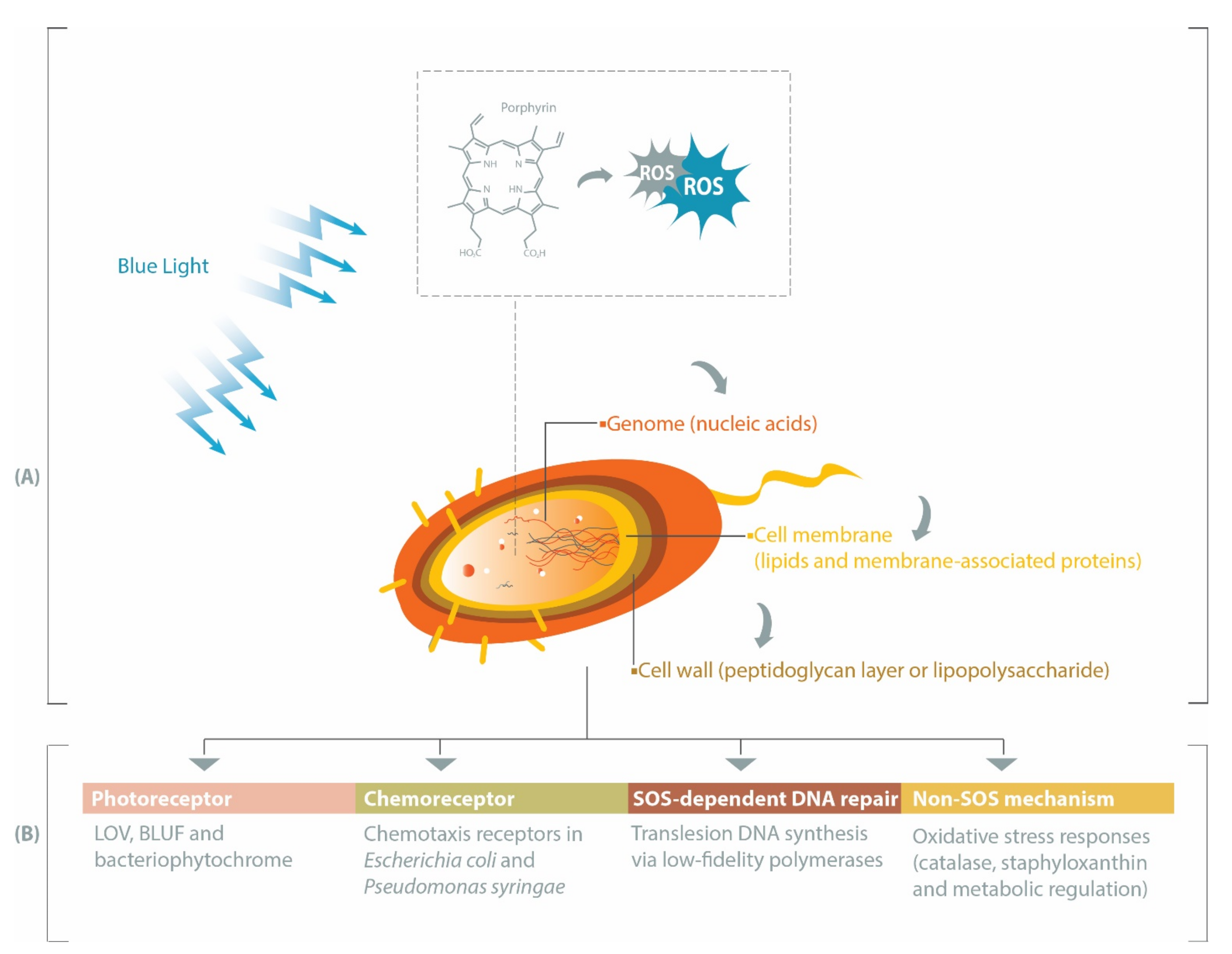
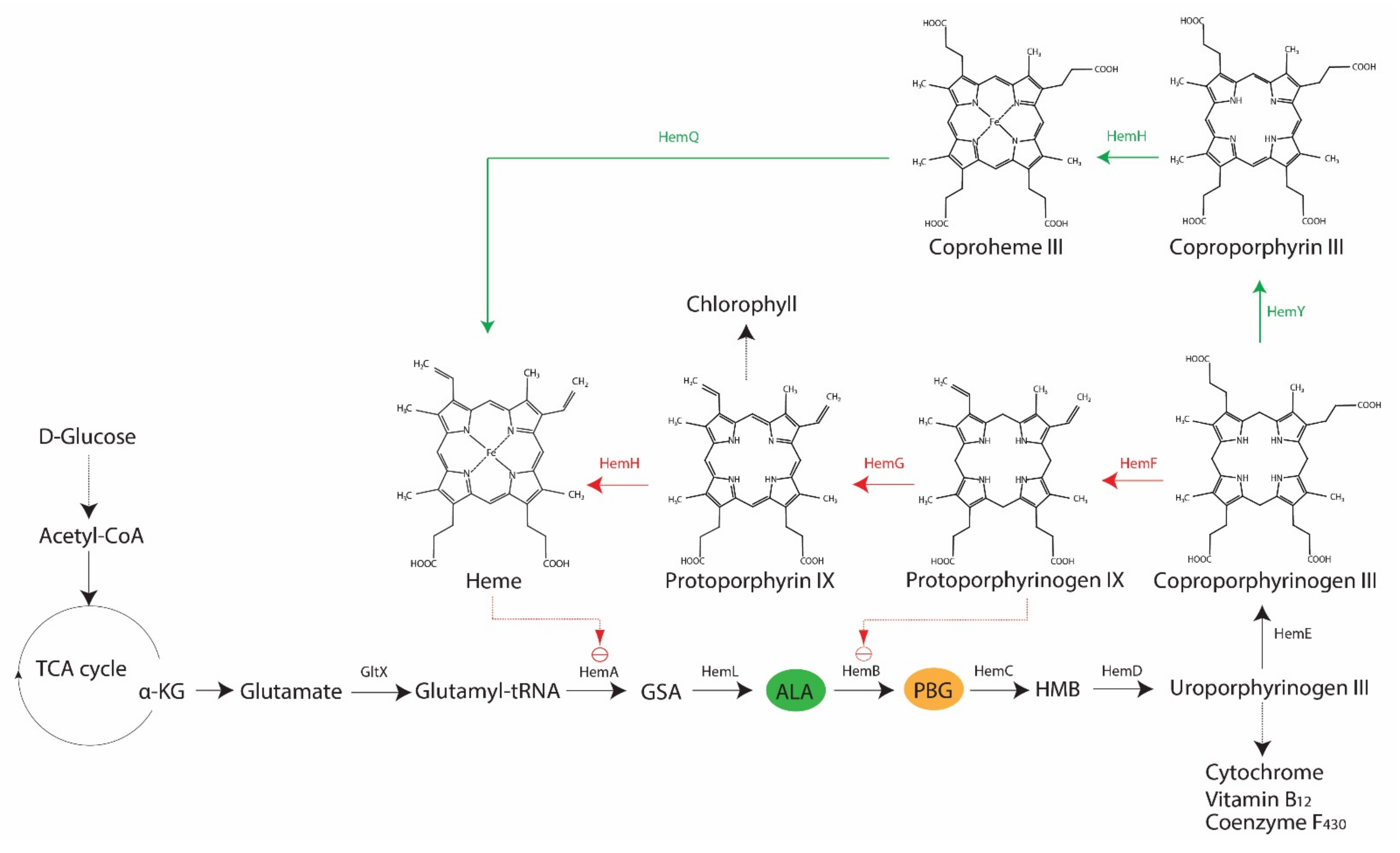
2. Porphyrin, Bacteria and Antimicrobial Blue Light

It is an established fact that various bacteria synthesize porphyrins within their cell, and that the type and levels of porphyrins may vary across species [23]. The two most relevant porphyrins are protoporphyrin IX and coproporphyrin (I and III), as their involvements in antimicrobial blue light treatments have been demonstrated in both Gram-positive and Gram-negative bacteria, such as S. aureus, P. aeruginosa, A. baumannii, and Helicobacter pylori [29,30,31,32]. However, there is also evidence of differing sensitivities to blue light between Gram-positive and Gram-negative bacteria, primarily due to the varying levels of coproporphyrin produced in the two bacterial types [29]. In bacteria, the core pathway of 5-carbon heme biosynthesis usually begins with a charged glutamyl-tRNA that undergoes a series of enzymatic conversion processes to form coproporphyrinogen III via the universal intermediate species δ-aminolevulinic acid (ALA), also known as 5-aminolevulinic acid (Figure 2). At this point, two possible branches emerge from the core pathway: (1) Gram-negative bacteria decarboxylate coproporphyrinogen III to protoporphyrinogen IX, oxidize protoporphyrinogen IX to protoporphyrin IX and then add metal to form protoheme; (2) Gram-positive bacteria oxidize coproporphyrinogen III to coproporphyrin, insert iron to make coproheme and oxidize coproheme to protoheme [9,10]. Alphaproteobacteria possess ALA synthase and may undergo heme biosynthesis via the non-canonical 4-carbon pathway, in which glutamyl-tRNA is replaced by a compound derived from the combination of succinyl CoA from the tricarboxylic acid cycle and glycine [9].
The rate-limiting compound in heme biosynthesis is ALA, which acts as the committed precursor that leads to the formation of the tetrapyrrole structure of porphyrins [9]. Expectedly, external addition of ALA to light treatments has been reported to increase the production of porphyrins in S. aureus, P. aeruginosa, and E. coli (strain K-12 and Ti05), and subsequently render these bacteria more susceptible to light-based antimicrobial treatments [34,35]. Downstream of ALA, various enzymes are involved in the conversion of one tetrapyrrole structure to another, leading to the formation of different photoactive porphyrins (Figure 2). In addition to their respective functions in the forward reaction of heme biosynthesis, some of these enzymes are involved in feedback regulations, as demonstrated in E. coli, where the overexpression of hemD and hemF resulted in the accumulation of ALA, whereas hemB, hemG and hemH had a negative effect on ALA production [33]. The inhibition of HemB by protoporphyrin IX also occurs in E. coli [33]. Furthermore, there are other regulatory mechanisms in heme biosynthesis, including hemA-related regulations in E. coli, Salmonella enterica subsp. enterica serovar Typhimurium, Bacillus subtilis, and P. aeruginosa or oxygen-dependent regulations of coproporphyrinogen oxidase genes (i.e., hemF or hemN) in Pseudomonas spp., E. coli, and B. subtilis [10]. These findings indicate the possibility of variations in porphyrin species present at any given point, which could also be influenced by growth conditions.
Porphyrin production has been suggested to depend on the bacterial growth stage. In Aggregatibacter actinomycetemcomitans and Pophyromonas gingivalis, the amounts of coproporphyrin (I and III) and protoporphyrin IX are highest in the first few days of growth, although variations have been observed across different types of porphyrins and bacterial strains [36]. Growth media could also impact porphyrin production, for example, P. gingivalis colonies cultured on a medium containing blood was reported to have increased production of protoporphyrin IX, relative to colonies grown on a medium lacking blood [36]. Consistently, there are evidences of strain-specific and growth phase-dependent inactivation of MRSA and E. coli by blue light, possibly related to porphyrin production [37,38]. In addition, a study revealed a positive correlation between ALA-mediated production of porphyrin species (protoporphyrin IX, coproporphyrin III, and uroporphyrin III) and elevated growth temperatures (up to 42 °C) in Propionibacterium acnes [39]. Nevertheless, there is still a need for further characterizations of endogenous porphyrins in other bacteria, along with assessments of their sensitivity to blue light, at different growth phases and temperatures. These data are particularly relevant for ensuring the appropriate design and application of blue light treatments, given that the relative absorption spectra of different porphyrin species vary. For example, protoporphyrin IX and uroporphyrin III absorb blue light at approximately 405–420 nm, whereas coproporphyrin III has the highest absorption at approximately 390 nm [40].

3. Heme Non-Producing Bacteria: Antimicrobial Blue Light or Photodynamic Therapy

Lactic acid bacteria (LAB), including pathogenic species belonging to the genera Streptococcus and Enterococcus, are not reliant upon heme for growth and thus unable to synthesize this cofactor, i.e., porphyrins are not prevalent in this group of bacteria [41]. Two studies have demonstrated that Enterococcus faecalis (formerly known as Streptococcus faecalis group D) and Enterococcus faecium devoid of porphyrins were insensitive to blue light-based antimicrobial treatments at 407–420 nm and 405 nm, respectively [29,42].
Intriguingly, others have reported on the inactivation of E. faecium by antimicrobial treatments with D90-values (light dosage required to obtain a 1-log inactivation) of 393 or 595 J/cm2 at 400 or 405 nm, respectively, which were considerably higher than other tested strains in both studies [25,43]. Insoluble fractions of the extracellular polymeric substance within Streptococcus mutans biofilms were also found to be susceptible to antimicrobial blue light (420 nm) [20]. However, the bactericidal mechanism of blue light against heme non-producing bacteria at these shorter wavelengths (400–420 nm) is yet unknown. In contrast, flavins are known to absorb blue light at longer wavelengths (450–460 nm), resulting in the inactivation of various bacterial species, including E. faecium, albeit with a lower efficacy than the conventional 405 nm antimicrobial blue light [43,44,45].
Alternative technology is also available in the form of antimicrobial photodynamic therapy (aPDT), which combines antimicrobial lights and photoactive compounds known as photosensitizers. Upon activation by light, photosensitizers at a ground state (lowest energy level) are converted into their excited singlet state (short-lived) or triplet state (long-lived), which, in the presence of oxygen, can undergo two types of energy transfer: (1) type I that produces toxic oxygen species, such as hydrogen peroxide (H2O2), superoxide or hydroxyl radicals; (2) type II that generates 1O2 [11]. Various photosensitizers and their derivatives are known to exhibit absorption within the ultraviolet/blue light spectrum [46], several of which have been applied in aPDT against various heme non-producing bacteria (Table 1). For a given aPDT, selection of a suitable photosensitizer and blue light combination, along with the optimization of photosensitizer concentration, is paramount to achieve maximum bactericidal efficacy [42,47,48,49,50].

4. Photoreceptors Facilitate Bacterial Responses to Blue Light

Light is ubiquitous in the environment and can be used as a regulatory signal by various organisms through photosensing. Photosensing is mainly mediated by six photoreceptors, namely rhodopsin, cryptochrome, BLUF (sensor of blue light using flavin adenine dinucleotide (FAD)), LOV (light, oxygen, and voltage), PYP (photoactive yellow protein), and phytochrome. In bacteria, phytochrome, BLUF, and LOV are the major photoreceptors [51,52]. Several bacteria may encode multiple photoreceptors, such as Methylbacterium spp. (phytochromes, LOV, and BLUF) or Pseudomonas spp. (phytochromes and LOV) [52]. As reviewed by Gomelsky and Hoff, these proteins are involved in controlling bacterial surface attachment, biofilm formation, and pathogenicity, among other functions [53]. Thus, future developments of blue light-based antimicrobial technologies could benefit from taking these photoreceptors into consideration, particularly their potential roles in conferring protection to bacteria against oxidative stress or in modulating the bacterial behaviors upon illumination by blue light at a sub-lethal dosage. In subsequent sections, we discuss the three major bacterial photoreceptors and their potential implications to blue light-based antimicrobial treatments.

4.1. LOV-Mediated Response to Blue Light

LOV domains have been found across species of archaea, fungi, bacteria, protists, and plants [54]. A phylogenetic analysis has revealed that 115 of 3254 sequenced bacterial genomes (3.5%) carry at least one gene encoding the LOV domain, predominantly occurring in those belonging to the class Alphaproteobacteria or the phylum Cyanobacteria [55]. Among Firmicutes, LOV-encoding genes seem to appear exclusively in Listeria spp. and Bacillus spp. In 439 and 84 sequenced genomes of Enterobacteriales and Vibrionales, respectively, no LOV homologs were found, possibly due to the fact that these bacteria utilize other photoreceptor protein families, such as BLUF [55].
These LOV domains belong to the Per-Arnt-Sim (PAS) domain superfamily that contains a β-scaffold with five antiparallel β-strands (Aβ, Bβ, Gβ, Hβ, and Iβ) connected by four α-helices (Cα, Dα, Eα, and Fα) [56,57]. This core structure is usually flanked with helices at either the N-terminal (N-cap) or the C-terminal (C-cap) and these play a role in downstream signaling cascades [58,59]. First discovered in the plant Arabidopsis thaliana [60], LOV domains were subsequently reported to bind flavin mononucleotide (FMN) in a stoichiometric manner at their amino terminal, and thus may serve as a light sensor [61]. During light excitation, a conserved cysteine residue in the LOV domain active site forms a covalent adduct with the C4a atom of a flavin isoalloxazine ring (preferentially FMN, but also FAD) [57,62]. This induces a range of conformational effects, including the unfolding of flanking helices, dimerization, and rotation of the LOV modules [56,63].
The architectures of LOV domains across bacterial species are diverse (Table 2), primarily associated with different effectors, which are defined as the nearest conserved domain to the sensors with respect to the primary structure [54,63,64]. In bacteria, histidine kinase, sulfate transporter and anti-sigma factor antagonist (STAS), and GGDEF are among the known LOV effectors [54]. A phylogenetic analysis of 496 bacterial LOV domains has revealed that the majority of these existed in association with a histidine kinase (48%) or a GGDEF/EAL domain (21%), or as standalone short LOVs (14%) [65]. Interestingly, short LOVs appear almost exclusively in Proteobacteria, with the exception of the actinobacterium Nakamurella multipartita (strain Y-104) [66].
Downstream effects of different LOVs vary with their respective effectors [63]. For example, histidine kinases are a part of the bacterial two-component system that have the ability of autophosphorylation upon activation, facilitating subsequent transfer of phosphoryl group to response regulators to induce downstream effects, such as virulence [67,68]. As its name suggests, STAS acts as a transcription regulator through its activity upon RNA polymerase-binding sigma factors and is essential for bacterial responses to environmental stimuli, including during host colonization [69,70]. GGDEF is a domain involved in the synthesis or hydrolysis of cyclic-bis (3→5′) dimeric guanosine monophosphate (c-di-GMP) or in the hydrolysis of cyclic adenosine monophosphate (c-AMP), indicating its role in prokaryotic signal transduction [71].
In the current literature, there are only a few studies that have assessed the role of LOV domains in bacterial survival against blue light. Here, we focus on the role of LOV photoreceptors during blue light inactivation of L. monocytogenes and B. subtilis, as these two bacteria are well-described. Additionally, discussions are also provided for blue light-dependent behaviors of several bacteria with short LOV or LOV-HK domains and a novel RNA-binding LOV protein.

4.1.1. Lmo0799 Photoreceptor and Transcription Factor Sig B (σB) in L. monocytogenes

Numerous environmental stress responses in L. monocytogenes are regulated by a supramolecular complex called stressosome. These stressosomes consist of multiple copies of RsbR (or its paralogs) and RsbS proteins arranged into an icosahedron, with both proteins containing a STAS domain at their C-terminus. In association with the core structure, another protein called RsbT acts as a kinase to activate the stressosome under stress—phosphorylation of RsbS at the S59 residue or RsbR at the T205 residue during prolonged stress [72]. Post-phosphorylation of RsbS, the RsbT detaches from the stressosome and binds to the phosphatase RsbU, which in turn dephosphorylates RsbV to release RsbW. Subsequently, RsbW (an anti-sigma) is released from the transcription factor SigB (σB) to allow downstream transcription of stress genes [72]. Indeed, σB is essential for mediating general stress responses in L. monocytogenes [73].
There are five RsbR homologs in Listeria spp., one of which is a protein called Lmo0799, which contains a LOV domain and acts as a photoreceptor [74]. Available evidences across various studies suggest that responses to blue light in L. monocytogenes is dependent on a functional Lmo0799, in association with σB [75,76,77,78]. In all of these studies, the motility of L. monocytogenes was reduced upon illumination by blue light, and this phenomenon has been attributed to the Lmo0799/σB-dependent transcription of an antisense RNA [76]. In one transcriptomic study, an antisense RNA transcript was found to negatively regulate several motility genes in L. monocytogenes upon activation by σB [79]. This notion is further consolidated by the finding of motility repression by blue light in wild-type, but not in ∆lmo0799 or ∆sigB strains [75].
O’Donoghue et al. explored the effects of blue light (460–470 nm) on the growth of L. monocytogenes (strains EGD-e and 10403S) as controlled by Lmo0799 and σB [77]. In the presence of sub-lethal blue light (i.e., lower light dosage), mutants that lacked σB exhibited a higher growth than the wild-type, suggesting an energetic cost of deploying σB-controlled stress response. However, when the light dosage was lethal, a survival advantage was observed in bacteria carrying σB [77]. This finding was corroborated by Dorey et al., who showed that only ten genes were affected by blue light in L. monocytogenes mutants lacking functional σB, as compared with 603 genes in the wild-type, confirming the importance of σB in blue light stress response [78]. Similar to the ∆sigB strains, L. monocytogenes mutants with deleted lmo0799 gene or missense mutation at the position 56 of the Lmo0799 protein (A56C mutation) were less affected by blue light at a sub-lethal dosage than the wild-type, as evidenced by the reduced motility repression and higher growth rate in these mutants than in the parental strains. Interestingly, when exposed to a lethal dosage of blue light, no difference in survival was observed between these mutants (∆lmo0799 or missense mutation) and the wild-type, which is indicative of other σB-activation pathways that bypass the canonical Lmo0799 light-mediated mechanism during severe stresses [77].
Furthermore, growth stage and temperature have also been reported to affect the survival of L. monocytogenes during blue light treatment, with bacterial cells in the stationary phase or cultured at 37 °C exhibiting less resistance against blue light than those in exponential phase or cultured at 30 °C, respectively [78]. These phenomena may be attributed to the varying levels of σB activation at different temperatures and growth stages, but not to the expression of Lmo0799 photoreceptor that remained constant [78]. In support of this finding, another study has also reported that there is a reduced retention of the FMN chromophore by the Lmo0799 photoreceptor at temperatures above 26 °C [80], possibly explaining the weaker light response of L. monocytogenes at higher temperatures.
The relationship between blue light and virulence factors in L. monocytogenes also needs to be considered (Table 3). In one study, virulence factor ActA was essential for the activation of σB and biofilm formation under blue light illumination, suggesting its role in resisting oxidative stress [76]. Another study reported a more concerning observation, in which blue light at 455 nm induced the transcription of virulence genes inlA and inlB in L. monocytogenes (strain EGD-e), subsequently leading to an increased infection rate in Caco-2 enterocyte-like human cells, particularly when an additional salt stress was present [75]. In support of these findings, Prli42 protein in L. monocytogenes has been shown to enhance virulence factors (ActA and listeriolysin O) under oxidative stress induced by bone-marrow-derived macrophages (BMDMs) or hydrogen peroxide, with the wild-type strain having a higher survival against these BMDMs than the Δprli42 mutants [74]. Intriguingly, while it is only 31 amino acids in length (miniprotein), Prli42 may interact with RsbR and promote tolerance to oxidative stress induced by hydrogen peroxide [74].

4.1.2. YtvA Photoreceptor and Transcription Factor σB in Bacillus subtilis

B. subtilis is a Gram positive, spore-forming bacterium, which has been used as a surrogate/model for understanding metabolism, resistance, and growth characteristics of other pathogenic spore formers. Similar to L. monocytogenes, B. subtilis has been reported to possess a signal integration hub acting as a stressosome. This stressosome is an icosahedral supramolecular complex consisting of RsbR (or its paralogs), RsbS and RsbT proteins, as determined by cryo-electron microscopy [87]. These proteins play various roles in signaling cascades involving Rsb proteins, including RsbT (kinase) and RsbU or RsbX (phosphatases), to activate σB [87,88]. An RsbR paralog, known as YtvA, is also present in B. subtilis—the first prokaryotic LOV protein to be identified—and it is capable of binding FMN and undergoing a photocycle [89]. Subsequent analyses have demonstrated the activation of YtvA by blue light through the cysteine 62 residue of its LOV domain, which induces a positive regulatory effect on the σB-mediated stress responses [90,91]—it is noteworthy that YtvA was overproduced in both studies. A more recent study has shown that YtvA is permanently incorporated into the B. subtilis stressosome, regardless of whether light is present or absent [92].
As mentioned above, the blue light-induced activity of YtvA and σB in B. subtilis has only been observed using overexpressed YtvA, except when additional stress (for example, salt stress) is present, in which case wild-type expression of YtvA is sufficient to elicit signaling cascades to activate σB [93]. In addition, this activity is also reliant on the interaction between YtvA and RsbRA in the stressosome, possibly through their respective STAS domains [94,95]. Interestingly, the removal of an RsbR paralog called RsbRB also increased the blue light-induced activation of σB in B. subtilis, even when YtvA was expressed at wild-type levels [94]. These findings demonstrate the multi-factorial consideration that needs to be evaluated during an antimicrobial blue light treatment of B. subtilis, particularly treatment conditions (i.e., whether other stresses are present) and possible presence of individual strains with varying stressosome compositions. Furthermore, a research gap exists on the possible role of stressosomes in the initiation of sporulation as a response to light treatment. σB in B. subtilis has been reported to be involved in spore and biofilm formation, which are two important characteristics that help in survival against adverse conditions [96].

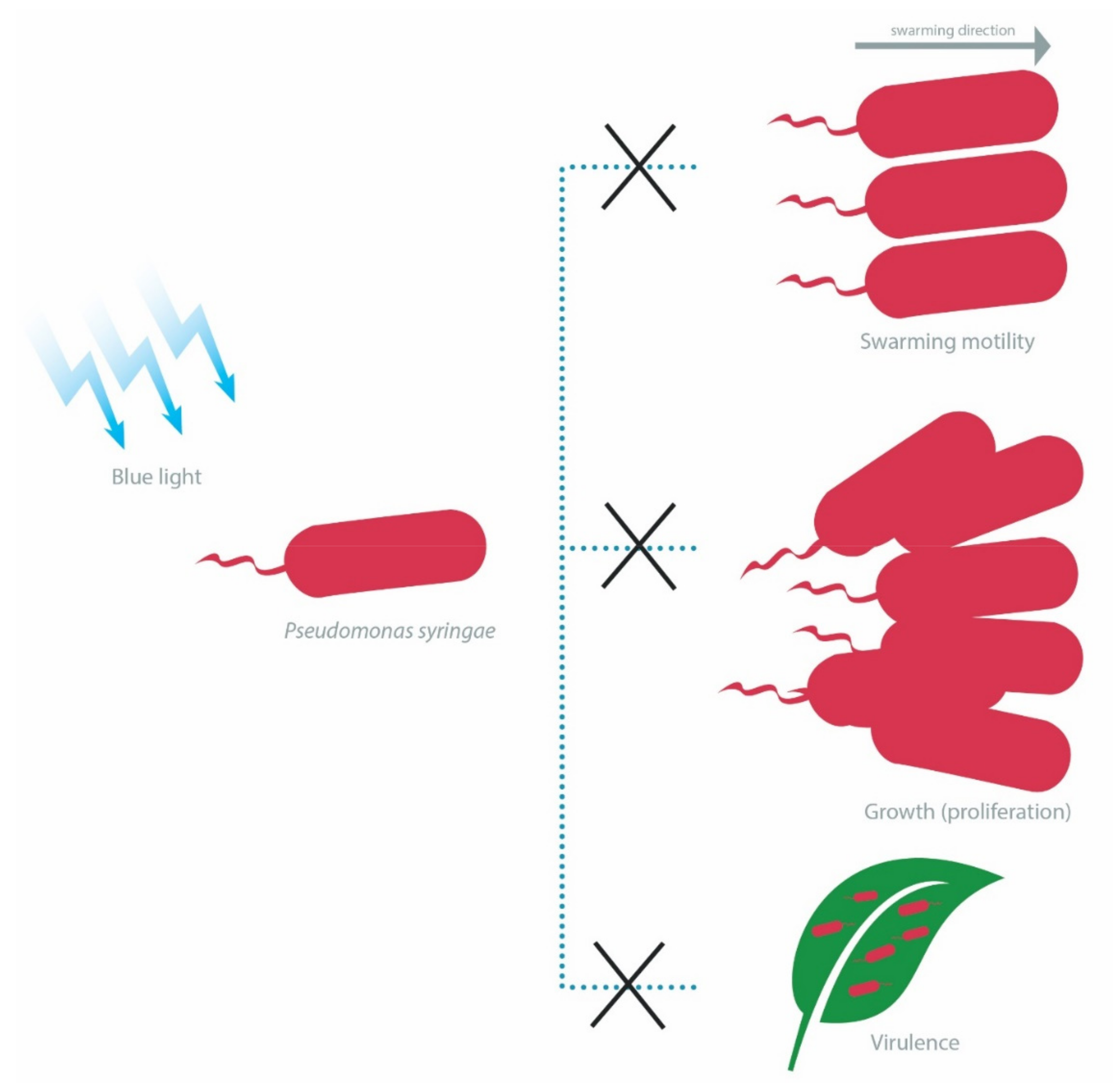
4.1.3. LOV-Dependent Differential Physiological Behaviors of Pseudomonas spp. and Rhodobacter sphaeroides Containing Short LOV or LOV-Histidine Kinase

LOV-containing proteins are present across the genera Pseudomonas, such as in Pseudomonas putida (for example, PpSB1-LOV and PpSB2-LOV) and Pseudomonas syringae (for example, PST-LOV) [97]. The LOV proteins in P. putida consist of short LOVs, which are equipped with auxiliary structures on each end—located outside of the core LOV domain—known as the N-cap and C-cap (Jα-helix) [66,98,99]. These elements are essential for maintaining the structural integrity of P. putida photoreceptors, as a study has shown that the short and truncated variants of PpSB1-LOV and PpSB2-LOV had severe structural issues, which subsequently led to the loss of chromophore-binding capacity [66]. Distinctively, P. syringae contain LOV-HK domain in association with a response regulator (RR) [100]. This LOV-HK-RR construct in PST-LOV is essential for light-dependent regulation, primarily through the change in the interaction between the LOV-HK domain and the RR domain. However, the histidine kinase activity has also been reported to be conserved in the absence of RR domains, indicating two distinct mechanisms involved in the activity of PST-LOV, i.e., those related to the activation of histidine kinase alone or via interactions with RR domains [100].
The short LOVs of P. putida (strain KT2440) have been reported to be positive regulators of blue light-dependent transcription of genes controlled by the P. putida light-inducible transcriptional regulators (PplRs), which belong to the light-induced transcriptional regulator (LitR) family of proteins. These genes include folE2, ufaM, and phrB that encode a GTP cyclohydrase, a methylase involved in furan ring-containing fatty acid synthesis and a deoxyribopyrimidine photolyase, respectively. Albeit, the growth rate of P. putida was not affected under white-light illumination, as evidenced by the constant expression of the essential sigma factor rpoD [97]. Conversely, the growth of P. syringae (strain DC3000) is adversely affected by the presence of PST-LOV in a blue light-dependent manner [101]. The authors further demonstrated the direct involvement of PST-LOV in inhibiting the expression of several sigma factors and also in reducing the proliferation of P. syringae in A. thaliana under blue light illumination at 400–500 nm (Figure 3) [101].
Besides growth, blue light also affects the swarming motility of P. syringae, albeit in a strain-dependent manner. A group of researchers reported that blue light at 470 nm inhibited the swarming motility of P. syringae (strain DC3000), with this phenomenon partially attributable to the presence of PST-LOV photoreceptors [102]. However, another study found that while blue light induced the reduction in swarming motility (relative to dark conditions) in another P. syringae strain (strain B728a), the presence of LOV photoreceptors instead positively regulated swarming motility under illumination and this could be attributed to the histidine kinase activity of the LOV-HK domain within the PST-LOV [103]. Furthermore, the virulence of wild-type P. syringae (strain DC3000) is attenuated in the presence of blue light, as compared with strains lacking the LOV photoreceptors (Figure 3) [101,102].
Similar to the PpSB2 photoreceptor in P. putida, the LOV domain in the photosynthetic bacteria R. sphaeroides is not associated with any effector and equipped with extensions at the N-terminus (short α-helix motif) and C-terminus (helix-turn-helix motif) [104,105]. This LOV domain exists as a dimer under dark condition and turns into a monomer upon blue light illumination, suggesting its role in sensing light [105,106]. As demonstrated in one study, the LOV photoreceptor in R. sphaeroides (strain 2.4.1) primarily acted as a repressor in blue light-dependent gene regulations, particularly genes related to carbon metabolism and chemotaxis [107]. Furthermore, the authors revealed the role of R. spaeroides LOV protein in photooxidative stress, including that caused by blue light (450 nm). In particular, the lack of LOV protein resulted in the increased expression of RSP_4257, which is an anti-sigma-factor antagonist (STAS), upon blue light illumination, although the relationship between LOV photoreceptor and survival ability of R. sphaeroides against blue light is yet to be firmly established [107].

4.2. BLUF-Mediated Response to Blue Light

BLUF domains were first discovered through their photosynthetic functions in the purple bacterium R. sphaeroides [108,109] and their involvement in the photoavoidance response in the algae Euglena gracilis [110]. A BLUF domain consists of a five-stranded β-sheet and two ferrodoxin-like α-strands aligned parallel to the β-sheet [58,111]. The isoalloxazine ring of a bound FAD molecule is sandwiched between the α-helices, with its adenosine side-chain protruding to the protein surface [56]. This interaction with FAD is facilitated by conserved tyrosine, glutamine and methionine (or tryptophan) residues in the BLUF domain [111,112]. Indeed, in two models predicting the light-mediated conformational changes of BLUF, tyrosine and glutamine residues are essential elements, alongside the FAD active site [56,58,111]. BLUF is found in at least 10% of the sequenced bacterial genomes and the majority of these proteins possibly function within a two-component system—a photosensing system in which the effector occurs in other molecules, such as DNA or protein—and they are often called short BLUF proteins [52]. An example of this is the BlrB photoreceptor in R. sphaeroides, which is possibly involved in signal transduction by interacting with effector proteins, such as those containing the EAL domain, through the exposed amino acid residues located at the mouth of its flavin-binding moiety [113]. A range of other BLUF architectures has also been predicted in prokaryotic cells (Table 4) [114]. In this review, we focus on two bacterial BLUF proteins, namely YcgF in E. coli and BlsA in A. baumannii, as they are well-described in terms of their role in blue light perception and the concomitant downstream effects on the lifestyle of these bacteria.

4.2.1. YcgF Photoreceptor in E. coli

Photosensing in E. coli is mediated by a photoreceptor known as YcgF. This protein contains a BLUF domain at its N-terminal and an EAL domain at the C-terminal. Interestingly, unlike other BLUF proteins, YcgF undergoes conformation change upon light illumination in a similar manner as LOV2 domain. Mediated by the Jα-helix at the C-terminal, this conformational change was initially thought to regulate the phosphodiesterase activity of EAL [115]. Unexpectedly, another study demonstrated that the EAL domain of YcgF lacked the phosphodiesterase activity to break down c-di-GMP [116], even when the missing conserved residues required for such activity were restored [117]. Instead, YcgF exhibited a regulatory role by antagonizing the activity of the YcgE protein, which is a repressor of eight genes, especially those present in the ycgZ-ymgABC operon—this operon is known to regulate the production of curli fimbriae and colanic acid. Upon illumination by blue light (470 nm), YcgF directly interacted with YcgE, resulting in the release of YcgE from its target operators, allowing for YmgA and YmgB to activate the Rcs phosphorelay pathway to induce the production of an exopolysaccharide component (colanic acid), promote acid resistance through the gene bdm and repress the expression of curli fimbriae [116]. This reaction is likely to occur through the blue light-induced conformation change in the EAL region of YcgF due to an altered hydrogen-bonding network around the FAD chromophore [118]. Furthermore, YcgF homologs have also been identified in Klebsiella pneumoniae and Enterobacter sp. 638, and a consistent pattern could be found in these bacterial species, in which a gene encoding a YcgF-like protein tends to occur next to a gene encoding a YcgE-related protein and a ycgz-ymgAB-like genetic unit [117].
Taken together, these findings indicate that YcgF, YcgE and the ycgz-ymgAB operon—or their homologs—constitute an important genetic pathway that controls blue light-dependent biofilm maturation and acid resistance in E. coli and potentially in other bacteria as well [116,117]. It is also noteworthy that the activity of YcgF upon YcgE is most prominent at low temperatures, particularly at 16 °C [116,118], albeit the downstream function of this pathway does not confer additional survival advantage against lethal blue light at this temperature [116].

4.2.2. BlsA Photoreceptor in A. baumannii

A. baumannii is a Gram-negative anaerobic (obligate) pathogen that has been associated with nosocomial infections [119]. Photosensing in A. baumannii occurs through the BlsA photoreceptor. The X-ray crystal structure of BlsA has recently been solved and it is consistent with the canonical BLUF protein structure (as described above). The conserved residues in the flavin-binding moiety at the N-terminal are tyrosine (Y7), glutamine (Q51), and methionine (M94). Upon illumination, the α-helices at the C-terminal undergo conformation changes, which modify the affinity of effector proteins to BlsA, including BfmR (biofilm response regulator), Fur (iron uptake regulator), AraC-like regulators (virulence), and OmpR (osmotic stress response regulator) [120]. This is another example of a short BLUF photoreceptor, in which the signal transduction occurs through protein-protein interactions.
Various studies have reported on the blue light-dependent regulation of motility, biofilm formation, and iron uptake in A. baumannii. One study demonstrated the inhibition of biofilm formation, pellicle formation, and motility in wild-type A. baumannii (strain ATCC 17978) by blue light (462 nm), whereas mutant strains lacking the functional blsA gene retained all three features [121]. Others unraveled the importance of the last fourteen residues at the C-terminal to the photodynamic properties of BlsA, as evidenced by the higher motility under blue light (469 nm) observed in strains with deleted (∆135–147) or substituted (K144E or K145E) residues than in the wild-type (A. baumannii ATCC 17978) [122]. The authors further suggested that Y7, Q51, and tryptophan (W92) residues of BlsA were also essential for the blue light perception of A. baumannii [122].
Metabolism of A. baumannii is also regulated through blue light. In iron deprived and dark conditions, BlsA binds to Fur, a repressor of acinetobactin, and promotes iron uptake. However, when activated by blue light (462 nm), BlsA detaches from Fur, which allows Fur to bind to its promoter, inhibiting iron uptake and growth [123]. In contrast, acetoin catabolism is induced by blue light (462 nm) through the interaction of activated BlsA with the repressor AcoN, allowing downstream expression of acetoin catabolic genes required for growth [124].
In all of these studies, blue light-mediated responses were only observed at ambient temperature (24 °C or lower), but not at elevated temperature (30 °C or above) [121,122,123,124]. This is expected as blue light photoactivity of BlsA is restricted to temperatures below 24 °C and that the expression of BlsA is mostly observed at 21–23 °C (especially in dark conditions), but negligible above 25 °C [125]. Furthermore, BlsA is unstable at high temperatures (above 30 °C) due to conformational changes that lead to the loss of FAD-binding capacity and protein aggregations, rendering the protein inoperative [125,126].
The virulence of A. baumannii is also affected by blue light, albeit the involvement of BlsA is yet to be determined. While blue light was found to increase the pathogenicity of A. baumannii in human keratinocyte cell line HaCaT at 37 °C, this phenomenon was independent of the BlsA photoreceptor [127]. In contrast, BlsA was reported to be involved in the killing of tup1 mutant Candida albicans at 24 °C, as the authors highlighted the difference between the wild-type and the ∆blsA A. baumannii strains [121].

4.3. Blue Light-Sensing Bacteriophytochrome and PAS-Containing Photoreceptor in P. aeruginosa

Unlike several other Pseudomonas species, such as P. syringae and P. putida, P. aeruginosa does not encode LOV proteins in their genome [97]. Instead, P. aeruginosa senses light through the bacteriophytochrome PaBphP that utilizes biliverdin IXα (produced by the bacterial heme oxygenases) as its chromophore. As with other phytochromes, PaBphP functions through photointerconversion between the red light-absorbing (Pr) and the far red light-absorbing (Pfr) forms [128]. One study has found that BphP was also responsive to blue light (430 nm)—other than to red light (630 nm) and far red light (730 nm)—and subsequently mediated light-dependent inhibition of biofilm and virulence genes in P. aeruginosa (strain PA14), in association with the two-component system KinB-AlgB [129]. The authors further elaborated that, upon illumination, BphP activated the alginate biosynthesis transcriptional regulatory protein (AlgB) via phosphorylation to promote the aforementioned inhibition of biofilm and virulence genes. In contrast, Kinase B (KinB) inactivated AlgB through its phosphatase activity, which resulted in the promotion of biofilm and virulence [129]. Two other studies have shown similar results: (1) blue light (462 nm) impaired the replication ability of P. aeruginosa (strains PAO1 or PAE4840) in HaCaT [127]; (2) virulence factors, such as pyocyanin, hemolysins, elastases, proteases, and lipases, in various P. aeruginosa strains were inhibited by blue light at 405 nm [130].
A recent study demonstrated that the possible involvement of a photoreceptor known as Rmca in the blue light-dependent inhibition of biofilm maturation in P. aeruginosa (strain PA14). The authors showed that blue light (422 nm) had a profound inhibitory effect on the synthesis of cyclic di-guanosine monophosphate (c-di-GMP), which was later found to be the result of phosphodiesterase activities of four proteins, particularly Rmca [131]. As previously demonstrated, Rmca possessed GGDEF-EAL and PAS domains, with the latter containing residues capable of binding a flavin cofactor [132]. Nevertheless, the role of Rmca in direct sensing of light and in the regulation of biofilm development in P. aeruginosa remains to be fully elucidated.

5. Role of Chemoreceptors in Responses to Blue Light

Five chemoreceptors (Aer, Tar, Tsr, Tap, and Trg) have also been reported to be involved in blue light-mediated tumbling (clockwise flagellar rotation) in E. coli. Wright et al. demonstrated that E. coli mutant strains expressing a single chemoreceptor (Aer or Tar) tumbled in response to blue light at 440 nm. A similar response was observed in strains expressing a combination of Aer and either Tar or Tsr, whereas the presence of Tsr alone failed to mediate any response to blue light [133]. More recently, Perlova et al. found that all five chemoreceptors responded to blue light (440 nm), with Aer, Tar, Tsr, and Trg initiating a tumbling response, whereas Tap-only strains exhibiting a running response under illumination. The authors also demonstrated that three functional proteins were required to produce blue light-mediated physiological changes, namely CheY (chemotactic signaling protein), CheB (methylesterase) and CheR (methyltransferase) [134]. As a well-established fact, chemotaxis in E. coli primarily relies on the histidine kinase CheA, which is coupled to the chemoreceptors through a linker protein (CheW). When the chemoreceptors are activated by a stimulus, CheA phosphorylates CheY, followed by the binding of the phosphorylated CheY to flagellar motors, causing a switch in the rotational direction of these motors—for example, counterclockwise to clockwise in a tumbling event [134,135]. CheB and CheR complete the signaling network by regulating the methylation of the chemoreceptors as a part of the sensory adaptation process (On or Off states) [134,135]. However, while Wright et al. and Perlova et al. have provided evidence of blue light-sensing capability of E. coli chemoreceptors, the precise mechanism is not fully elucidated. Aer contains a PAS domain that binds to FAD molecule, possibly explaining its response to blue light [136]. Nevertheless, the other chemoreceptors are not known to possess a chromophore-binding domain, and thus their blue light-related activities may be mediated by unknown blue light sensors elsewhere [133,134,135].
In contrast to E. coli, interlinked activities of blue light photoreceptors and chemoreceptors have been described in epiphytic P. syringae pv. tomato DC3000 (PsPto). Santamaría-Hernando et al. reported on the positive regulation of five PsPto methyl-accepting chemotaxis proteins (MCPs) by LOV and bacteriophytochrome (BphP1) photoreceptors under blue light illumination at 458 nm. More importantly, two of these MCPs (PSPTO_1008 and PSPTO_2526) are required for full virulence of PsPto [137], demonstrating the importance of blue light regulated pathways in the pathogenicity of plant pathogens.

6. Potential Development of Bacterial Tolerance to Blue Light via SOS-Dependent DNA Repair

In the event of oxidative stress, bacteria have various adaptive responses to enhance their survival. These adaptive responses could lead to the development of tolerance or resistance. To our knowledge of the current literature, no reports have identified full resistance of any bacterial species to antimicrobial blue light treatments. Nevertheless, one study has shown that five successive cycles of sub-lethal blue light (411 nm; 50 or 150 J/cm2 per cycle) resulted in the development of tolerance in S. aureus (strains USA300 JE2, community-associated methicillin-resistant; 1631, methicillin-sensitive; 2030, human-associated methicillin-resistant). More importantly, the authors found that this tolerance was stable after five successive culture transfers in the dark, and subsequently identified genetic alterations as the probable cause of the tolerance, particularly due to the expression of recA and umuC genes that triggered the SOS-dependent DNA repair [138]. Another study corroborated this result by demonstrating that sub-lethal exposures of S. agalactiae (strains ATCC 27956, 2306/07 or 2974/07) to blue/green light (515 nm; 10 cycles; up to 40 J/cm2 per cycle), in combination with a photosensitizer (rose bengal), altered the expression of six major oxidative stress response genes and led to the development of tolerance [139].
Mutations resulting from SOS-dependent pathway are best understood in E. coli, primarily through the translesion DNA synthesis (TLS), mediated by low-fidelity DNA polymerases, particularly pol V. Pol V consists of two UmuD homomers and one UmuC monomer (UmuD’2C) and is activated the RecA*, an assembly of recombinase A (RecA) protein associated with a single-stranded DNA. A RecA* nucleoprotein filament is known to activate pol V by transferring RecA-ATP complex from its DNA 3′-end to the UmuD’2C, obtaining the active pol V structure (UmuD’2C-RecA-ATP), also known as the pol V Mutasome (pol V Mut)—activation of pol V can only occur when RecA-ATP is transferred from the 3′-tip of RecA*, but not from the 5′-tip [140]. A region of the RecA protein, which encompasses amino acid residues 112, 113, and 117, has been identified as an important pol V activation surface [141]. During the induction of stress, the expression of genes in the SOS regulon is also initiated due to the release of the LexA repressor, as mediated by a RecA-dependent autocleavage.
There is evidence implicating SOS-dependent pathways in the development of resistant phenotypes in several bacterial species (Table 5). In addition, SOS responses involve varying mechanistic modes across bacterial species. For example, SOS-inducing stresses have been reported to trigger the development of superinfectious phage Pf4 (and subsequently small-colony variants) or an integron rearrangement leading to the production of β-lactamases in P. aeruginosa [142,143]. In S. Typhimurium, SOS-dependent pathway is used as a strategy to avoid lethal antibiotic treatments by inhibiting swarming motility through the disruption of equilibrium between RecA and CheW proteins, preventing the bacterial chemoreceptor polar cluster assembly [144,145].
Furthermore, there are variations in the SOS regulon across bacterial species. The canonical pol V (umuDC operon) and recA are prevalent in S. aureus, A. baumannii, S. Typhimurium and Vibrio spp [146,147,148,149,150]. In contrast, P. aeruginosa lacks the umuDC operon and instead possesses the LexA-repressed imuA/sulA-imuB-dnaE2 operon performing a similar function as the former [151]. Unlike other bacteria, A. baumannii does not have the LexA repressor, but instead possess an UmuD homolog, known as UmuDAb, which could undergo LexA-like cleavage in response to DNA damage and act as a RecA-dependent regulator of eight genes encoding seven error-prone DNA polymerases [152,153]. Taken together, these findings highlight the complexity of SOS-dependent stress responses existing in different bacteria, particularly their roles in promoting resistant bacterial phenotypes, and thus future studies are needed to explore their association with antimicrobial blue light-induced oxidative stresses.

7. Non-SOS Protective Mechanisms against Blue Light-Induced Oxidative Stress

Besides SOS-dependent pathways, several bacteria counteract blue light treatments by producing catalase, a ROS-scavenging enzyme. This enzyme is encoded by katA gene, which was found to be upregulated by 8-fold in S. aureus exposed to phototreatment at 462 nm and led to increased tolerance of the bacteria to hydrogen peroxide [156]. This is consistent with the findings of two other studies reporting increased tolerance to hydrogen peroxide in blue light-treated S. aureus [138,157]. Similarly, P. aeruginosa utilizes catalase to combat the photo-oxidative stress of blue light (464 nm), as evidenced by the higher susceptibility of mutants lacking katA to blue light than the wild-type [158]. Interestingly, one study demonstrated that blue light at 455 nm (225 J/cm2) was able to completely inactivate the enzymatic activity of a recombinant P. aeruginosa catalase A (KatA) in vitro, albeit the direct implication of the reduced KatA activity upon the blue light inactivation of P. aeruginosa remains unclear [159].
Furthermore, S. aureus produces staphyloxanthin, a membrane-bound carotenoid pigment with antioxidant properties. In a recent study, S. aureus possessing the crtM gene, which is involved in the production of staphyloxanthin, exhibited a tolerant trait against blue light at 405 nm [160]. Thus, the authors proposed a pre-treatment of the tolerant S. aureus with blue light at 460 nm—which did not exhibit direct bactericidal activities but was capable of promoting the degradation of staphyloxanthin—resulting in the increased efficacy of the subsequent treatment with 405-nm blue light. In support of this finding, another study showed that exposure to blue light at 460 nm degraded staphyloxanthin and rendered methicillin-resistant S. aureus susceptible to subsequent treatments with oxidative agents [161].
As described in one study, Cronobacter sakazakii responded to blue light by upregulating three genes encoding an oxidative stress-resistance chaperone (ESA_RS13255), an adhesin (ESA_RS09025), and a capsule biosynthesis protein (ESA_RS15435), with the former promoting higher antioxidant activities [15]. For B. subtilis spores, resistance to blue light (400 nm) depends on the presence of DNA protective α/β-type small, acid soluble proteins (SASP), DNA repair proteins (nucleotide or base excising repair proteins and spore photoproduct lyase), spore coat proteins and proteins for spore pigmentations—mutants lacking genes encoding these proteins were more susceptible to photoinactivation [162].
In V. cholerae, blue light-mediated oxidative stress response is controlled by an anti-sigma ChrR (represses SigmaE) and metalloregulatory-like (MerR) proteins, either together or separately. Both proteins control 222 genes that are responsible for a range of functions, including cellular protection, DNA repair, and carbon metabolism [163]. Intriguingly, while cryptochrome/photolyase proteins are the sole blue light photoreceptors in V. cholerae [164], these photoreceptors are not involved in the bacterial photo-oxidative responses, indicating possible involvement of pigments or unknown photoreceptors in the V. cholerae stress response to blue light [163]. That being said, Tardu et al. did not analyze specific DNA repair pathways in V. cholerae, for example, photoreactivation involving the photolyase protein, and thus this is a subject of future studies.

8. Concluding Remarks and Future Outlook

The complex biological pathways involved in bacterial responses to antimicrobial blue light present challenges to be overcome. Endogenous porphyrin-mediated oxidative stress remains the primary mechanistic model of antimicrobial blue light. However, given the diversity of porphyrin species occurring at different stages of the heme biosynthesis, and thus corresponding to different blue light absorption spectra, it is paramount for future studies to assess the porphyrin biosynthesis pathway as a function of culture conditions, such as bacterial growth stage, growth media, and temperature. Subsequently, a correlation between bacterial growth stages, porphyrin species present and survival rate against antimicrobial blue light can be established.
The lack of understanding of blue light-mediated inactivation of heme non-producing pathogenic bacteria is another research gap that needs addressing. These bacteria may import exogenous heme into the cell and assemble heme-containing proteins, such as cytochrome bd and catalase. A small set of heme-binding proteins has also been discovered in LAB, including alkyl hydroperoxide reductase C (AhpC) and HemW, which exhibit blue-light absorptions at 414 and 425 nm, respectively, when loaded with heme [41]. This could be explored further to devise a strategy to inactivate these bacteria in the presence of external heme. Furthermore, there are other uncharacterized endogenous photosensitizers that absorb blue light at longer wavelengths, especially at 470 nm, which may be targets for antimicrobial blue light treatments in the future [40].
Photoreceptors can confer protective effects through blue light-dependent activation of stress response pathways and promote virulence when the light dosage is sub-lethal. In addition, several bacteria encode more than one type of photoreceptor. This characteristic is particularly prevalent in plant pathogens, which could encode up to four types of photoreceptor, as demonstrated in Xanthomonas citri subsp. citri containing bacteriophytochrome, LOV, BLUF, and cryptochrome/photolyase [165]. Intriguingly, one study has demonstrated a blue light-dependent inhibitory activity of LOV photoreceptors against a bacteriophytochrome (BphP1), leading to the differentiated swarming motility of P. syringae (strain B728a) [103]. Similarly, the interaction of AppA (a BLUF-containing protein) and CryB (a cryptochrome) is also known to regulate photosynthetic genes of R. sphaeroides in a blue light-dependent manner [166]. Future studies may be aimed at revealing overlapping activities of different photoreceptors in other bacterial species, along with their downstream effects on the bacterial growth and/or physiological characteristics under blue light illumination.
Some bacteria are able to use chemoreceptors to modulate their motility or virulence in a blue light-dependent manner. While the underlying mechanism is not fully understood, there is evidence of synergistic interaction between chemoreceptors and photoreceptors in PsPto. In E. coli, the Aer chemoreceptor contains PAS domain, possibly indicating a capability of direct light sensing [136]. The next research step in this area may include screening of other chromophore-binding domains in known chemoreceptors present across different bacterial species, particularly pathogenic strains.
As with other antimicrobial treatments, antimicrobial blue light is prone to sub-lethal application, especially considering that bacteria possess protective mechanisms against oxidative stress, such as ROS-scavenging catalases. More concerningly, low-fidelity polymerases expressed during the SOS-dependent stress response could result in the emergence of tolerant phenotypes during blue light treatments, as demonstrated in S. aureus [138]. Thus, further investigations should be conducted to unravel the mechanisms and frequencies of blue light tolerance, or resistance, in other bacteria [28]. In addition, alternative strategies can be developed using compounds capable of inhibiting different components of the SOS pathway. In S. aureus, the inhibition of RecA by novobiocin, as mediated by a DNA gyrase subunit B (GyrB)-dependent mechanism, was reported to increase the susceptibility of the bacteria to photoinactivation by about 100-fold, including to blue light at 405 nm [13]. Similar effects were observed for a recombinant E. coli containing an α-helix peptide called 4E1—this peptide contains 20 amino acid residues that match the motif of RecX protein (a natural RecA inhibitor)—which was rendered more susceptible to UV than the wild-type [167]. There are other inhibitors targeting different SOS elements, such as LexA, RecBCD, pol IV, and single-strand DNA binding proteins [168]. Future studies can aim to explore the possible addition of these inhibitors into antimicrobial blue light treatments against different bacteria.
Taken together, the mechanisms presented here highlight several inherent characteristics that set one bacterial species apart from another. Consequently, blue light inactivation parameters must be validated within the context of specific bacterial strains. In addition to these endogenous factors, external treatment conditions, such as light intensity, light dosage, availability of nutrients, temperature, and presence of exogenous photosensitizers, are known to contribute to the varying blue light inactivation profiles in clinical and foodborne bacteria [23,24]. Thus, the frontier of research in this area needs to be extended to include systematic determination of both inherent and external factors affecting the efficacy of antimicrobial blue light against different bacterial strains, with the subsequent establishment of accessible databases for improved predictability of bacterial tolerances or resistances.

Author Contributions

G.B. conceived this review and acquired funding. J.H. conducted the literature investigation, wrote the original draft of this manuscript and edited the final version of this manuscript with the guidance of the other co-authors. S.W., A.S., A.G. and G.B. critically assessed the original draft and provided additional materials to be incorporated into the final version of this manuscript. All authors have read and agreed to the published version of the manuscript.

Funding

This work is funded through AgResearch Ltd. Strategic Science Investment Fund (SSIF) under the grant number PRJ0318704.

Acknowledgments

The authors wish to thank Eric Altermann and Delphine Rapp for their comments on the manuscript. Elisabeth Belianty is also acknowledged for providing assistance with the illustrations.

Conflicts of Interest

The authors declare no conflict of interest. The funders had no role in the design of the study; in the collection, analyses, or interpretation of data; in the writing of the manuscript, or in the decision to publish the results.

References

  1. World Health Organization. Global Antimicrobial Resistance and Use Surveillance System (GLASS) Report; World Health Organization: Geneva, Switzerland, 2021. [Google Scholar]
  2. Gwynne, P.J.; Gallagher, M.P. Light as a broad-spectrum antimicrobial. Front. Microbiol. 2018, 9, 119. [Google Scholar] [CrossRef]
  3. Yin, R.; Dai, T.; Avci, P.; Jorge, A.E.S.; De Melo, W.C.M.A.; Vecchio, D.; Huang, Y.Y.; Gupta, A.; Hamblin, M.R. Light based anti-infectives: Ultraviolet C irradiation, photodynamic therapy, blue light, and beyond. Curr. Opin. Pharmacol. 2013, 13, 731–762. [Google Scholar] [CrossRef] [Green Version]
  4. D’Orazio, J.; Jarrett, S.; Amaro-Ortiz, A.; Scott, T. UV Radiation and the Skin. Int. J. Mol. Sci. 2013, 14, 12222–12248. [Google Scholar] [CrossRef] [PubMed] [Green Version]
  5. Kütting, B.; Drexler, H. UV-induced skin cancer at workplace and evidence-based prevention. Int. Arch. Occup. Environ. Health 2010, 83, 843–854. [Google Scholar] [CrossRef]
  6. Zaffina, S.; Camisa, V.; Lembo, M.; Vinci, M.R.; Tucci, M.G.; Borra, M.; Napolitano, A.; Cannatã, V. Accidental exposure to UV radiation produced by germicidal lamp: Case report and risk assessment. Photochem. Photobiol. 2012, 88, 1001–1004. [Google Scholar] [CrossRef] [PubMed]
  7. Kleinpenning, M.M.; Smits, T.; Frunt, M.H.A.; van Erp, P.E.J.; van de Kerkhof, P.C.M.; Gerritsen, R.M.J.P. Clinical and histological effects of blue light on normal skin. Photodermatol. Photoimmunol. Photomed. 2010, 26, 16–21. [Google Scholar] [CrossRef] [PubMed]
  8. Angarano, V.; Smet, C.; Akkermans, S.; Watt, C.; Chieffi, A.; Van Impe, J.F.M. Visible light as an antimicrobial strategy for inactivation of Pseudomonas fluorescens and Staphylococcus epidermidis biofilms. Antibiotics 2020, 9, 171. [Google Scholar] [CrossRef] [PubMed] [Green Version]
  9. Dailey, H.A.; Dailey, T.A.; Gerdes, S.; Jahn, D.; Jahn, M.; O’Brian, M.R.; Warren, M.J. Prokaryotic Heme Biosynthesis: Multiple Pathways to a Common Essential Product. Microbiol. Mol. Biol. Rev. 2017, 81, e00048-16. [Google Scholar] [CrossRef] [Green Version]
  10. Choby, J.E.; Skaar, E.P. Heme Synthesis and Acquisition in Bacterial Pathogens. J. Mol. Biol. 2016, 428, 3408–3428. [Google Scholar] [CrossRef] [Green Version]
  11. Hu, X.; Huang, Y.Y.; Wang, Y.; Wang, X.; Hamblin, M.R. Antimicrobial photodynamic therapy to control clinically relevant biofilm infections. Front. Microbiol. 2018, 9, 1299. [Google Scholar] [CrossRef] [Green Version]
  12. Kim, M.J.; Bang, W.S.; Yuk, H.G. 405 ± 5 nm light emitting diode illumination causes photodynamic inactivation of Salmonella spp. on fresh-cut papaya without deterioration. Food Microbiol. 2017, 62, 124–132. [Google Scholar] [CrossRef]
  13. Grinholc, M.; Rodziewicz, A.; Forys, K.; Rapacka-Zdonczyk, A.; Kawiak, A.; Domachowska, A.; Golunski, G.; Wolz, C.; Mesak, L.; Becker, K.; et al. Fine-tuning recA expression in Staphylococcus aureus for antimicrobial photoinactivation: Importance of photo-induced DNA damage in the photoinactivation mechanism. Appl. Microbiol. Biotechnol. 2015, 99, 9161–9176. [Google Scholar] [CrossRef] [Green Version]
  14. Wu, J.; Chu, Z.; Ruan, Z.; Wang, X.; Dai, T.; Hu, X. Changes of Intracellular Porphyrin, Reactive Oxygen Species, and Fatty Acids Profiles During Inactivation of Methicillin-Resistant Staphylococcus aureus by Antimicrobial Blue Light. Front. Physiol. 2018, 9, 1658. [Google Scholar] [CrossRef]
  15. Chu, Z.; Hu, X.; Wang, X.; Wu, J.; Dai, T.; Wang, X. Inactivation of Cronobacter sakazakii by blue light illumination and the resulting oxidative damage to fatty acids. Can. J. Microbiol. 2019, 65, 922–929. [Google Scholar] [CrossRef]
  16. Dai, T.; Gupta, A.; Huang, Y.Y.; Sherwood, M.E.; Murray, C.K.; Vrahas, M.S.; Kielian, T.; Hamblin, M.R. Blue light eliminates community-acquired methicillin-resistant Staphylococcus aureus in infected mouse skin abrasions. Photomed. Laser Surg. 2013, 31, 531–538. [Google Scholar] [CrossRef] [PubMed] [Green Version]
  17. Zhang, Y.; Zhu, Y.; Gupta, A.; Huang, Y.; Murray, C.K.; Vrahas, M.S.; Sherwood, M.E.; Baer, D.G.; Hamblin, M.R.; Dai, T. Antimicrobial blue light therapy for multidrug-resistant Acinetobacter baumannii infection in a mouse burn model: Implications for prophylaxis and treatment of combat-related wound infections. J. Infect. Dis. 2014, 209, 1963–1971. [Google Scholar] [CrossRef] [PubMed] [Green Version]
  18. Giannelli, M.; Landini, G.; Materassi, F.; Chellini, F.; Antonelli, A.; Tani, A.; Nosi, D.; Zecchi-Orlandini, S.; Rossolini, G.M.; Bani, D. Effects of photodynamic laser and violet-blue led irradiation on Staphylococcus aureus biofilm and Escherichia coli lipopolysaccharide attached to moderately rough titanium surface: In vitro study. Lasers Med. Sci. 2017, 32, 857–864. [Google Scholar] [CrossRef] [PubMed]
  19. Fila, G.; Krychowiak, M.; Rychlowski, M.; Bielawski, K.P.; Grinholc, M. Antimicrobial blue light photoinactivation of Pseudomonas aeruginosa: Quorum sensing signaling molecules, biofilm formation and pathogenicity. J. Biophotonics 2018, 11, e201800079. [Google Scholar] [CrossRef] [PubMed] [Green Version]
  20. De Sousa, D.L.; Lima, R.A.; Zanin, I.C.; Klein, M.I.; Janal, M.N.; Duarte, S. Effect of twice-daily blue light treatment on matrix-rich biofilm development. PLoS ONE 2015, 10, e0131941. [Google Scholar] [CrossRef]
  21. Ferrer-Espada, R.; Wang, Y.; Goh, X.S.; Dai, T. Antimicrobial Blue Light Inactivation of Microbial Isolates in Biofilms. Lasers Surg. Med. 2020, 52, 472–478. [Google Scholar] [CrossRef]
  22. Beirão, S.; Fernandes, S.; Coelho, J.; Faustino, M.A.F.; Tomé, J.P.C.; Neves, M.G.P.M.S.; Tomé, A.C.; Almeida, A.; Cunha, A. Photodynamic inactivation of bacterial and yeast biofilms with a cationic porphyrin. Photochem. Photobiol. 2014, 90, 1387–1396. [Google Scholar] [CrossRef]
  23. Wang, Y.; Wang, Y.; Wang, Y.; Murray, C.K.; Hamblin, M.R.; Hooper, D.C.; Dai, T. Antimicrobial blue light inactivation of pathogenic microbes: State of the art. Drug Resist. Updates 2017, 33–35, 1–22. [Google Scholar] [CrossRef]
  24. Hyun, J.E.; Lee, S.Y. Blue light-emitting diodes as eco-friendly non-thermal technology in food preservation. Trends Food Sci. Technol. 2020, 105, 284–295. [Google Scholar] [CrossRef]
  25. Halstead, F.D.; Thwaite, J.E.; Burt, R.; Laws, T.R.; Raguse, M.; Moeller, R.; Webber, M.A.; Oppenheim, B.A. Antibacterial activity of blue light against nosocomial wound pathogens growing planktonically and as mature biofilms. Appl. Environ. Microbiol. 2016, 82, 4006–4016. [Google Scholar] [CrossRef] [Green Version]
  26. dos Anjos, C.; Sabino, C.P.; Bueris, V.; Fernandes, M.R.; Pogliani, F.C.; Lincopan, N.; Sellera, F.P. Antimicrobial blue light inactivation of international clones of multidrug-resistant Escherichia coli ST10, ST131 and ST648. Photodiagn. Photodyn. Ther. 2019, 27, 51–53. [Google Scholar] [CrossRef]
  27. Kim, M.J.; Adeline Ng, B.X.; Zwe, Y.H.; Yuk, H.G. Photodynamic inactivation of Salmonella enterica Enteritidis by 405 ± 5-nm light-emitting diode and its application to control salmonellosis on cooked chicken. Food Control 2017, 82, 305–315. [Google Scholar] [CrossRef]
  28. Rapacka-Zdonczyk, A.; Wozniak, A.; Nakonieczna, J.; Grinholc, M. Development of antimicrobial phototreatment tolerance: Why the methodology matters. Int. J. Mol. Sci. 2021, 22, 2224. [Google Scholar] [CrossRef]
  29. Nitzan, Y.; Salmon-Divon, M.; Shporen, E.; Malik, Z. ALA induced photodynamic effects on Gram positive and negative bacteria. Photochem. Photobiol. Sci. 2004, 3, 430–435. [Google Scholar] [CrossRef] [PubMed]
  30. Amin, R.M.; Bhayana, B.; Hamblin, M.R.; Dai, T. Antimicrobial blue light inactivation of Pseudomonas aeruginosa by photo-excitation of endogenous porphyrins: In vitro and in vivo studies. Lasers Surg. Med. 2016, 48, 562–568. [Google Scholar] [CrossRef] [PubMed] [Green Version]
  31. Wang, Y.; Wu, X.; Chen, J.; Amin, R.; Lu, M.; Bhayana, B.; Zhao, J.; Murray, C.K.; Hamblin, M.R.; Hooper, D.C.; et al. Antimicrobial Blue Light Inactivation of Gram-Negative Pathogens in Biofilms: In Vitro and in Vivo Studies. J. Infect. Dis. 2016, 213, 1380–1387. [Google Scholar] [CrossRef] [Green Version]
  32. Battisti, A.; Morici, P.; Ghetti, F.; Sgarbossa, A. Spectroscopic characterization and fluorescence imaging of Helicobacter pylori endogenous porphyrins. Biophys. Chem. 2017, 229, 19–24. [Google Scholar] [CrossRef]
  33. Zhang, J.; Kang, Z.; Chen, J.; Du, G. Optimization of the heme biosynthesis pathway for the production of 5-aminolevulinic acid in Escherichia coli. Sci. Rep. 2015, 5, 1–7. [Google Scholar] [CrossRef] [Green Version]
  34. Fotinos, N.; Convert, M.; Piffaretti, J.C.; Gurny, R.; Lange, N. Effects on gram-negative and gram-positive bacteria mediated by 5-aminolevulinic acid and 5-aminolevulinic acid derivatives. Antimicrob. Agents Chemother. 2008, 52, 1366–1373. [Google Scholar] [CrossRef] [Green Version]
  35. Morimoto, K.; Ozawa, T.; Awazu, K.; Ito, N.; Honda, N.; Matsumoto, S.; Tsuruta, D. Photodynamic therapy using systemic administration of 5-aminolevulinic acid and a 410-nm wavelength light-emitting diode for methicillin-resistant Staphylococcus aureus-infected ulcers in mice. PLoS ONE 2014, 9, e105173. [Google Scholar] [CrossRef]
  36. Fyrestam, J.; Bjurshammar, N.; Paulsson, E.; Mansouri, N.; Johannsen, A.; Östman, C. Influence of culture conditions on porphyrin production in Aggregatibacter actinomycetemcomitans and Porphyromonas gingivalis. Photodiagn. Photodyn. Ther. 2017, 17, 115–123. [Google Scholar] [CrossRef] [Green Version]
  37. Biener, G.; Masson-Meyers, D.S.; Bumah, V.V.; Hussey, G.; Stoneman, M.R.; Enwemeka, C.S.; Raicu, V. Blue/violet laser inactivates methicillin-resistant Staphylococcus aureus by altering its transmembrane potential. J. Photochem. Photobiol. B Biol. 2017, 170, 118–124. [Google Scholar] [CrossRef] [Green Version]
  38. Abana, C.M.; Brannon, J.R.; Ebbott, R.A.; Dunigan, T.L.; Guckes, K.R.; Fuseini, H.; Powers, J.; Rogers, B.R.; Hadjifrangiskou, M. Characterization of blue light irradiation effects on pathogenic and nonpathogenic Escherichia coli. Microbiologyopen 2017, 6, e00466. [Google Scholar] [CrossRef]
  39. Ramstad, S.; Le Anh-Vu, N.; Johnsson, A. The temperature dependence of porphyrin production in Propionibacterium acnes after incubation with 5-aminolevulinic acid (ALA) and its methyl ester (m-ALA). Photochem. Photobiol. Sci. 2006, 5, 66–72. [Google Scholar] [CrossRef]
  40. Hessling, M.; Spellerberg, B.; Hoenes, K. Photoinactivation of bacteria by endogenous photosensitizers and exposure to visible light of different wavelengths—A review on existing data. FEMS Microbiol. Lett. 2017, 364, fnw270. [Google Scholar] [CrossRef]
  41. Baureder, M.; Hederstedt, L. Heme Proteins in Lactic Acid Bacteria. Adv. Microb. Physiol. 2013, 62, 1–43. [Google Scholar] [CrossRef]
  42. Kang, S.M.; Jung, H.I.; Kim, B. Il Susceptibility of oral bacteria to antibacterial photodynamic therapy. J. Oral Microbiol. 2019, 11, 1644111. [Google Scholar] [CrossRef] [PubMed] [Green Version]
  43. Hoenes, K.; Bauer, R.; Meurle, T.; Spellerberg, B.; Hessling, M. Inactivation Effect of Violet and Blue Light on ESKAPE Pathogens and Closely Related Non-pathogenic Bacterial Species—A Promising Tool Against Antibiotic-Sensitive and Antibiotic-Resistant Microorganisms. Front. Microbiol. 2021, 11, 3429. [Google Scholar] [CrossRef] [PubMed]
  44. Cieplik, F.; Späth, A.; Leibl, C.; Gollmer, A.; Regensburger, J.; Tabenski, L.; Hiller, K.A.; Maisch, T.; Schmalz, G. Blue light kills Aggregatibacter actinomycetemcomitans due to its endogenous photosensitizers. Clin. Oral Investig. 2014, 18, 1763–1769. [Google Scholar] [CrossRef] [PubMed]
  45. Plavskii, V.Y.; Mikulich, A.V.; Tretyakova, A.I.; Leusenka, I.A.; Plavskaya, L.G.; Kazyuchits, O.A.; Dobysh, I.I.; Krasnenkova, T.P. Porphyrins and flavins as endogenous acceptors of optical radiation of blue spectral region determining photoinactivation of microbial cells. J. Photochem. Photobiol. B Biol. 2018, 183, 172–183. [Google Scholar] [CrossRef] [PubMed]
  46. Cieplik, F.; Deng, D.; Crielaard, W.; Buchalla, W.; Hellwig, E.; Al-Ahmad, A.; Maisch, T. Antimicrobial photodynamic therapy–what we know and what we don’t. Crit. Rev. Microbiol. 2018, 44, 571–589. [Google Scholar] [CrossRef] [Green Version]
  47. Bumah, V.V.; Cortez, P.M.; Morrow, B.N.; Rojas, P.; Bowman, C.R.; Masson-Meyers, D.S.; Enwemeka, C.S. Blue light absorbing pigment in Streptococcus agalactiae does not potentiate the antimicrobial effect of pulsed 450 nm light. J. Photochem. Photobiol. B Biol. 2021, 216, 112149. [Google Scholar] [CrossRef]
  48. Manoil, D.; Filieri, A.; Gameiro, C.; Lange, N.; Schrenzel, J.; Wataha, J.C.; Bouillaguet, S. Flow cytometric assessment of Streptococcus mutans viability after exposure to blue light-activated curcumin. Photodiagn. Photodyn. Ther. 2014, 11, 372–379. [Google Scholar] [CrossRef]
  49. Luke-Marshall, N.R.; Hansen, L.A.; Shafirstein, G.; Campagnari, A.A. Antimicrobial Photodynamic Therapy with Chlorin e6 Is Bactericidal against Biofilms of the Primary Human Otopathogens. mSphere 2020, 5. [Google Scholar] [CrossRef]
  50. Pileggi, G.; Wataha, J.C.; Girard, M.; Grad, I.; Schrenzel, J.; Lange, N.; Bouillaguet, S. Blue light-mediated inactivation of Enterococcus faecalis in vitro. Photodiagn. Photodyn. Ther. 2013, 10, 134–140. [Google Scholar] [CrossRef]
  51. van der Horst, M.A.; Key, J.; Hellingwerf, K.J. Photosensing in chemotrophic, non-phototrophic bacteria: Let there be light sensing too. Trends Microbiol. 2007, 15, 554–562. [Google Scholar] [CrossRef]
  52. Losi, A.; Gärtner, W. Bacterial bilin- and flavin-binding photoreceptors. Photochem. Photobiol. Sci. 2008, 7, 1168–1178. [Google Scholar] [CrossRef]
  53. Gomelsky, M.; Hoff, W.D. Light helps bacteria make important lifestyle decisions. Trends Microbiol. 2011, 19, 441–448. [Google Scholar] [CrossRef]
  54. Glantz, S.T.; Carpenter, E.J.; Melkonian, M.; Gardner, K.H.; Boyden, E.S.; Wong, G.K.S.; Chow, B.Y. Functional and topological diversity of LOV domain photoreceptors. Proc. Natl. Acad. Sci. USA 2016, 113, E1442–E1451. [Google Scholar] [CrossRef] [Green Version]
  55. Krauss, U.; Minh, B.Q.; Losi, A.; Gärtner, W.; Eggert, T.; Von Haeseler, A.; Jaeger, K.E. Distribution and phylogeny of light-oxygen-voltage-blue-light-signaling proteins in the three kingdoms of life. J. Bacteriol. 2009, 191, 7234–7242. [Google Scholar] [CrossRef] [Green Version]
  56. Conrad, K.S.; Manahan, C.C.; Crane, B.R. Photochemistry of flavoprotein light sensors. Nat. Chem. Biol. 2014, 10, 801–809. [Google Scholar] [CrossRef]
  57. Losi, A.; Gärtner, W. Old chromophores, new photoactivation paradigms, trendy applications: Flavins in blue light-sensing photoreceptors. Photochem. Photobiol. 2011, 87, 491–510. [Google Scholar] [CrossRef] [PubMed]
  58. Zoltowski, B.D.; Gardner, K.H. Tripping the light fantastic: Blue-light photoreceptors as examples of environmentally modulated protein-protein interactions. Biochemistry 2011, 50, 4–16. [Google Scholar] [CrossRef] [PubMed] [Green Version]
  59. Losi, A.; Gärtner, W. Solving Blue Light Riddles: New Lessons from Flavin-binding LOV Photoreceptors. Photochem. Photobiol. 2017, 93, 141–158. [Google Scholar] [CrossRef] [Green Version]
  60. Huala, E.; Oeller, P.W.; Liscum, E.; Han, I.S.; Larsen, E.; Briggs, W.R. Arabidopsis NPH1: A Protein Kinase with a Putative Redox-Sensing Domain. Science 1997, 278, 2120–2123. [Google Scholar] [CrossRef]
  61. Christie, J.M.; Salomon, M.; Nozue, K.; Wada, M.; Briggs, W.R. LOV (light, oxygen, or voltage) domains of the blue-light photoreceptor phototropin (nph1): Binding sites for the chromophore flavin mononucleotide. Proc. Natl. Acad. Sci. USA 1999, 96, 8779–8783. [Google Scholar] [CrossRef] [PubMed] [Green Version]
  62. Alexandre, M.T.A.; Domratcheva, T.; Bonetti, C.; Van Wilderen, L.J.G.W.; Van Grondelle, R.; Groot, M.L.; Hellingwerf, K.J.; Kennis, J.T.M. Primary reactions of the LOV2 domain of phototropin studied with ultrafast mid-infrared spectroscopy and quantum chemistry. Biophys. J. 2009, 97, 227–237. [Google Scholar] [CrossRef] [Green Version]
  63. Herrou, J.; Crosson, S. Function, structure and mechanism of bacterial photosensory LOV proteins. Nat. Rev. Microbiol. 2011, 9, 713–723. [Google Scholar] [CrossRef] [PubMed] [Green Version]
  64. Losi, A.; Mandalari, C.; Gärtner, W. The Evolution and Functional Role of Flavin-based Prokaryotic Photoreceptors. Photochem. Photobiol. 2015, 91, 1021–1031. [Google Scholar] [CrossRef] [Green Version]
  65. Mandalari, C.; Losi, A.; Gärtner, W. Distance-tree analysis, distribution and co-presence of bilin- and flavin-binding prokaryotic photoreceptors for visible light. Photochem. Photobiol. Sci. 2013, 12, 1144–1157. [Google Scholar] [CrossRef]
  66. Rani, R.; Jentzsch, K.; Lecher, J.; Hartmann, R.; Willbold, D.; Jaeger, K.E.; Krauss, U. Conservation of dark recovery kinetic parameters and structural features in the pseudomonadaceae “short” light, oxygen, voltage (lov) protein family: Implications for the design of lov-based optogenetic tools. Biochemistry 2013, 52, 4460–4473. [Google Scholar] [CrossRef]
  67. Dikiy, I.; Edupuganti, U.R.; Abzalimov, R.R.; Borbat, P.P.; Srivastava, M.; Freed, J.H.; Gardner, K.H. Insights into histidine kinase activation mechanisms from the monomeric blue light sensor EL346. Proc. Natl. Acad. Sci. USA 2019, 116, 4963–4972. [Google Scholar] [CrossRef] [Green Version]
  68. Sankhe, G.D.; Dixit, N.M.; Saini, D.K. Activation of Bacterial Histidine Kinases: Insights into the Kinetics of the cis Autophosphorylation Mechanism. mSphere 2018, 3, e00111-18. [Google Scholar] [CrossRef] [Green Version]
  69. Campbell, E.A.; Westblade, L.F.; Darst, S.A. Regulation of bacterial RNA polymerase σ factor activity: A structural perspective. Curr. Opin. Microbiol. 2008, 11, 121–127. [Google Scholar] [CrossRef] [Green Version]
  70. Moy, B.E.; Seshu, J. STAS Domain Only Proteins in Bacterial Gene Regulation. Front. Cell. Infect. Microbiol. 2021, 11, 564. [Google Scholar] [CrossRef]
  71. Ryjenkov, D.A.; Tarutina, M.; Moskvin, O.V.; Gomelsky, M. Cyclic diguanylate is a ubiquitous signaling molecule in bacteria: Insights into biochemistry of the GGDEF protein domain. J. Bacteriol. 2005, 187, 1792–1798. [Google Scholar] [CrossRef] [Green Version]
  72. Williams, A.H.; Redzej, A.; Rolhion, N.; Costa, T.R.D.; Rifflet, A.; Waksman, G.; Cossart, P. The cryo-electron microscopy supramolecular structure of the bacterial stressosome unveils its mechanism of activation. Nat. Commun. 2019, 10, 1–10. [Google Scholar] [CrossRef] [Green Version]
  73. Guerreiro, D.N.; Arcari, T.; O’Byrne, C.P. The σB-Mediated General Stress Response of Listeria monocytogenes: Life and Death Decision Making in a Pathogen. Front. Microbiol. 2020, 11, 1505. [Google Scholar] [CrossRef]
  74. Impens, F.; Rolhion, N.; Radoshevich, L.; Bécavin, C.; Duval, M.; Mellin, J.; Garciá Del Portillo, F.; Pucciarelli, M.G.; Williams, A.H.; Cossart, P. N-terminomics identifies Prli42 as a membrane miniprotein conserved in Firmicutes and critical for stressosome activation in Listeria monocytogenes. Nat. Microbiol. 2017, 2, 1–12. [Google Scholar] [CrossRef]
  75. Ondrusch, N.; Kreft, J. Blue and Red Light Modulates SigB-Dependent Gene Transcription, Swimming Motility and Invasiveness in Listeria monocytogenes. PLoS ONE 2011, 6, e16151. [Google Scholar] [CrossRef] [Green Version]
  76. Tiensuu, T.; Andersson, C.; Rydén, P.; Johansson, J. Cycles of light and dark co-ordinate reversible colony differentiation in Listeria monocytogenes. Mol. Microbiol. 2013, 87, 909–924. [Google Scholar] [CrossRef] [Green Version]
  77. O’Donoghue, B.; NicAogáin, K.; Bennett, C.; Conneely, A.; Tiensuu, T.; Johansson, J.; O’Byrne, C. Blue-light inhibition of Listeria monocytogenes growth is mediated by reactive oxygen species and is influenced by σB and the blue-light sensor Lmo0799. Appl. Environ. Microbiol. 2016, 82, 4017–4027. [Google Scholar] [CrossRef] [Green Version]
  78. Dorey, A.L.; Lee, B.H.; Rotter, B.; O’Byrne, C.P. Blue Light Sensing in Listeria monocytogenes Is Temperature-Dependent and the Transcriptional Response to It Is Predominantly SigB-Dependent. Front. Microbiol. 2019, 10, 2497. [Google Scholar] [CrossRef] [Green Version]
  79. Toledo-Arana, A.; Dussurget, O.; Nikitas, G.; Sesto, N.; Guet-Revillet, H.; Balestrino, D.; Loh, E.; Gripenland, J.; Tiensuu, T.; Vaitkevicius, K.; et al. The Listeria transcriptional landscape from saprophytism to virulence. Nature 2009, 459, 950–956. [Google Scholar] [CrossRef]
  80. Chan, R.H.; Lewis, J.W.; Bogomolni, R.A. Photocycle of the LOV-STAS protein from the pathogen Listeria monocytogenes. Photochem. Photobiol. 2013, 89, 361–369. [Google Scholar] [CrossRef]
  81. Yoshikawa, Y.; Ogawa, M.; Hain, T.; Yoshida, M.; Fukumatsu, M.; Kim, M.; Mimuro, H.; Nakagawa, I.; Yanagawa, T.; Ishii, T.; et al. Listeria monocytogenes ActA-mediated escape from autophagic recognition. Nat. Cell Biol. 2009, 11, 1233–1240. [Google Scholar] [CrossRef]
  82. Mitchell, G.; Ge, L.; Huang, Q.; Chen, C.; Kianian, S.; Roberts, M.F.; Schekman, R.; Portnoy, D.A. Avoidance of autophagy mediated by PlcA or ActA is required for listeria monocytogenes growth in macrophages. Infect. Immun. 2015, 83, 2175–2184. [Google Scholar] [CrossRef] [PubMed] [Green Version]
  83. Tamburro, M.; Sammarco, M.L.; Ammendolia, M.G.; Fanelli, I.; Minelli, F.; Ripabelli, G. Evaluation of transcription levels of inlA, inlB, hly, bsh and prfA genes in Listeria monocytogenes strains using quantitative reverse-transcription PCR and ability of invasion into human CaCo-2 cells. FEMS Microbiol. Lett. 2015, 362. [Google Scholar] [CrossRef] [PubMed] [Green Version]
  84. Phelps, C.C.; Vadia, S.; Arnett, E.; Tan, Y.; Zhang, X.; Pathak-Sharma, S.; Gavrilin, M.A.; Seveau, S. Relative roles of Listeriolysin O, InlA, and InlB in Listeria monocytogenes uptake by host cells. Infect. Immun. 2018, 86, e00555-18. [Google Scholar] [CrossRef] [PubMed] [Green Version]
  85. Kim, H.; Marquis, H.; Boor, K.J. σB contributes to Listeria monocytogenes invasion by controlling expression of inlA and inlB. Microbiology 2005, 151, 3215–3222. [Google Scholar] [CrossRef] [PubMed] [Green Version]
  86. Vadia, S.; Seveau, S. Fluxes of Ca2+ and K+ are required for the listeriolysin O-dependent internalization pathway of Listeria monocytogenes. Infect. Immun. 2014, 82, 1084–1091. [Google Scholar] [CrossRef] [PubMed] [Green Version]
  87. Marles-Wright, J.; Grant, T.; Delumeau, O.; van Duinen, G.; Firbank, S.J.; Lewis, P.J.; Murray, J.W.; Newman, J.A.; Quin, M.B.; Race, P.R.; et al. Molecular architecture of the “stressosome”, a signal integration and transduction hub. Science 2008, 322, 92–96. [Google Scholar] [CrossRef]
  88. Marles-Wright, J.; Lewis, R.J. The stressosome: Molecular architecture of a signalling hub. Biochem. Soc. Trans. 2010, 38, 928–933. [Google Scholar] [CrossRef] [Green Version]
  89. Losi, A.; Polverini, E.; Quest, B.; Gärtner, W. First evidence for phototropin-related blue-light receptors in prokaryotes. Biophys. J. 2002, 82, 2627–2634. [Google Scholar] [CrossRef]
  90. Ávila-Pérez, M.; Hellingwerf, K.J.; Kort, R. Blue light activates the σB-dependent stress response of Bacillus subtilis via YtvA. J. Bacteriol. 2006, 188, 6411–6414. [Google Scholar] [CrossRef] [Green Version]
  91. Gaidenko, T.A.; Kim, T.J.; Weigel, A.L.; Brody, M.S.; Price, C.W. The blue-light receptor YtvA acts in the environmental stress signaling pathway of Bacillus subtilis. J. Bacteriol. 2006, 188, 6387–6395. [Google Scholar] [CrossRef] [Green Version]
  92. Jurk, M.; Schramm, P.; Schmieder, P. The blue-light receptor YtvA from Bacillus subtilis is permanently incorporated into the stressosome independent of the illumination state. Biochem. Biophys. Res. Commun. 2013, 432, 499–503. [Google Scholar] [CrossRef]
  93. Suzuki, N.; Takaya, N.; Hoshino, T.; Nakamura, A. Enhancement of a σB-dependent stress response in Bacillus subtilis by ligh via YtvA photoreceptor. J. Gen. Appl. Microbiol. 2007, 53, 81–88. [Google Scholar] [CrossRef] [PubMed] [Green Version]
  94. van der Steen, J.B.; Ávila-Pérez, M.; Knippert, D.; Vreugdenhil, A.; van Alphen, P.; Hellingwerf, K.J. Differentiation of function among the RsbR paralogs in the general stress response of Bacillus subtilis with regard to light perception. J. Bacteriol. 2012, 194, 1708–1716. [Google Scholar] [CrossRef] [Green Version]
  95. Choi, S.; Nakasone, Y.; Hellingwerf, K.J.; Terazima, M. Photoreaction Dynamics of a Full-Length Protein YtvA and Intermolecular Interaction with RsbRA. Biochemistry 2020, 59, 4703–4710. [Google Scholar] [CrossRef] [PubMed]
  96. Rodriguez Ayala, F.; Bartolini, M.; Grau, R. The Stress-Responsive Alternative Sigma Factor SigB of Bacillus subtilis and Its Relatives: An Old Friend With New Functions. Front. Microbiol. 2020, 11, 1761. [Google Scholar] [CrossRef] [PubMed]
  97. Sumi, S.; Mutaguchi, N.; Ebuchi, T.; Tsuchida, H.; Yamamoto, T.; Suzuki, M.; Natsuka, C.; Shiratori-Takano, H.; Shintani, M.; Nojiri, H.; et al. Light response of Pseudomonas putida KT2440 mediated by class II LitR, a photosensor homolog. J. Bacteriol. 2020, 202, e00146-20. [Google Scholar] [CrossRef] [PubMed]
  98. Krauss, U.; Losi, A.; Gärtner, W.; Jaeger, K.E.; Eggert, T. Initial characterization of a blue-light sensing, phototropin-related protein from Pseudomonas putida: A paradigm for an extended LOV construct. Phys. Chem. Chem. Phys. 2005, 2804–2811. [Google Scholar] [CrossRef]
  99. Jentzsch, K.; Wirtz, A.; Circolone, F.; Drepper, T.; Losi, A.; Gärtner, W.; Jaeger, K.E.; Krauss, U. Mutual exchange of kinetic properties by extended mutagenesis in two short LOV domain proteins from Pseudomonas putida. Biochemistry 2009, 48, 10321–10333. [Google Scholar] [CrossRef] [PubMed]
  100. Cao, Z.; Buttani, V.; Losi, A.; Gärtner, W. A blue light inducible two-component signal transduction system in the plant pathogen Pseudomonas syringae pv. tomato. Biophys. J. 2008, 94, 897–905. [Google Scholar] [CrossRef] [Green Version]
  101. Moriconi, V.; Sellaro, R.; Ayub, N.; Soto, G.; Rugnone, M.; Shah, R.; Pathak, G.P.; Gärtner, W.; Casal, J.J. LOV-domain photoreceptor, encoded in a genomic island, attenuates the virulence of Pseudomonas syringae in light-exposed Arabidopsis leaves. Plant J. 2013, 76, 322–331. [Google Scholar] [CrossRef]
  102. Río-Álvarez, I.; Rodríguez-Herva, J.J.; Martínez, P.M.; González-Melendi, P.; García-Casado, G.; Rodríguez-Palenzuela, P.; López-Solanilla, E. Light regulates motility, attachment and virulence in the plant pathogen Pseudomonas syringae pv tomato DC3000. Environ. Microbiol. 2014, 16, 2072–2085. [Google Scholar] [CrossRef] [PubMed] [Green Version]
  103. Wu, L.; McGrane, R.S.; Beattie, G.A. Light regulation of swarming motility in Pseudomonas syringae integrates signaling pathways mediated by a bacteriophytochrome and a LOV protein. MBio 2013, 4, e00334-13. [Google Scholar] [CrossRef] [PubMed] [Green Version]
  104. Magerl, K.; Dick, B. Dimerization of LOV domains of: Rhodobacter sphaeroides (RsLOV) studied with FRET and stopped-flow experiments. Photochem. Photobiol. Sci. 2020, 19, 159–170. [Google Scholar] [CrossRef] [PubMed]
  105. Hendrischk, A.K.; Moldt, J.; Frühwirth, S.W.; Klug, G. Characterization of an unusual LOV domain protein in the α-proteobacterium Rhodobacter sphaeroides. Photochem. Photobiol. 2009, 85, 1254–1259. [Google Scholar] [CrossRef]
  106. Conrad, K.S.; Bilwes, A.M.; Crane, B.R. Light-induced subunit dissociation by a light-oxygen-voltage domain photoreceptor from Rhodobacter sphaeroides. Biochemistry 2013, 52, 378–391. [Google Scholar] [CrossRef] [Green Version]
  107. Metz, S.; Jäger, A.; Klug, G. Role of a short light, oxygen, voltage (LOV) domain protein in blue light-and singlet oxygen-dependent gene regulation in Rhodobacter sphaeroides. Microbiology 2012, 158, 368–379. [Google Scholar] [CrossRef]
  108. Braatsch, S.; Gomelsky, M.; Kuphal, S.; Klug, G. A single flavoprotein, AppA, integrates both redox and light signals in Rhodobacter sphaeroides. Mol. Microbiol. 2002, 45, 827–836. [Google Scholar] [CrossRef] [Green Version]
  109. Masuda, S.; Bauer, C.E. AppA is a blue light photoreceptor that antirepresses photosynthesis gene expression in Rhodobacter sphaeroides. Cell 2002, 110, 613–623. [Google Scholar] [CrossRef] [Green Version]
  110. Iseki, M.; Matsunaga, S.; Murakami, A.; Ohno, K.; Shiga, K.; Yoshida, K.; Sugai, M.; Takahashi, T.; Hori, T.; Watanabe, M. A blue-light-activated adenylyl cyclase mediates photoavoidance in Euglena gracilis. Nature 2002, 415, 1047–1051. [Google Scholar] [CrossRef]
  111. Masuda, S. Light detection and signal transduction in the BLUF photoreceptors. Plant Cell Physiol. 2013, 54, 171–179. [Google Scholar] [CrossRef] [Green Version]
  112. Park, S.Y.; Tame, J.R.H. Seeing the light with BLUF proteins. Biophys. Rev. 2017, 9, 169–176. [Google Scholar] [CrossRef] [PubMed] [Green Version]
  113. Jung, A.; Domratcheva, T.; Tarutina, M.; Wu, Q.; Ko, W.H.; Shoeman, R.L.; Gomelsky, M.; Gardner, K.H.; Schlichting, I. Structure of a bacterial BLUF photoreceptor: Insights into blue light-mediated signal transduction. Proc. Natl. Acad. Sci. USA 2005, 102, 12350–12355. [Google Scholar] [CrossRef] [PubMed] [Green Version]
  114. Kaushik, M.S.; Sharma, R.; Veetil, S.K.; Srivastava, S.K.; Kateriya, S. Modular diversity of the bluf proteins and their potential for the development of diverse optogenetic tools. Appl. Sci. 2019, 9, 3924. [Google Scholar] [CrossRef] [Green Version]
  115. Schroeder, C.; Werner, K.; Otten, H.; Krätzig, S.; Schwalbe, H.; Essen, L.O. Influence of a joining helix on the BLUF domain of the YcgF photoreceptor from Escherichia coli. ChemBioChem 2008, 9, 2463–2473. [Google Scholar] [CrossRef]
  116. Tschowri, N.; Busse, S.; Hengge, R. The BLUF-EAL protein YcgF acts as a direct anti-repressor in a blue-light response of Escherichia coli. Genes Dev. 2009, 23, 522–534. [Google Scholar] [CrossRef] [Green Version]
  117. Tschowri, N.; Lindenberg, S.; Hengge, R. Molecular function and potential evolution of the biofilm-modulating blue light-signalling pathway of Escherichia coli. Mol. Microbiol. 2012, 85, 893–906. [Google Scholar] [CrossRef] [Green Version]
  118. Nakasone, Y.; Ono, T.A.; Ishii, A.; Masuda, S.; Terazima, M. Temperature-sensitive reaction of a photosensor protein YcgF: Possibility of a role of temperature sensor. Biochemistry 2010, 49, 2288–2296. [Google Scholar] [CrossRef]
  119. Morris, F.C.; Dexter, C.; Kostoulias, X.; Uddin, M.I.; Peleg, A.Y. The Mechanisms of Disease Caused by Acinetobacter baumannii. Front. Microbiol. 2019, 10, 1601. [Google Scholar] [CrossRef] [Green Version]
  120. Chitrakar, I.; Iuliano, J.N.; He, Y.L.; Woroniecka, H.A.; Tolentino Collado, J.; Wint, J.M.; Walker, S.G.; Tonge, P.J.; French, J.B. Structural Basis for the Regulation of Biofilm Formation and Iron Uptake in A. baumannii by the Blue-Light-Using Photoreceptor, BlsA. ACS Infect. Dis. 2020, 6, 2592–2603. [Google Scholar] [CrossRef]
  121. Mussi, M.A.; Gaddy, J.A.; Cabruja, M.; Arivett, B.A.; Viale, A.M.; Rasia, R.; Actis, L.A. The opportunistic human pathogen Acinetobacter baumannii senses and responds to light. J. Bacteriol. 2010, 192, 6336–6345. [Google Scholar] [CrossRef] [Green Version]
  122. Wood, C.R.; Squire, M.S.; Finley, N.L.; Page, R.C.; Actis, L.A. Structural and functional analysis of the Acinetobacter baumannii BlsA photoreceptor and regulatory protein. PLoS ONE 2019, 14, e220918. [Google Scholar] [CrossRef] [Green Version]
  123. Tuttobene, M.R.; Cribb, P.; Mussi, M.A. BlsA integrates light and temperature signals into iron metabolism through fur in the human pathogen Acinetobacter baumannii. Sci. Rep. 2018, 8, 7728. [Google Scholar] [CrossRef]
  124. Tuttobene, M.R.; Fernández-García, L.; Blasco, L.; Cribb, P.; Ambroa, A.; Müller, G.L.; Fernández-Cuenca, F.; Bleriot, I.; Rodríguez, R.E.; Barbosa, B.G.V.; et al. Quorum and light signals modulate acetoin/Butanediol catabolism in Acinetobacter spp. Front. Microbiol. 2019, 10, 1376. [Google Scholar] [CrossRef]
  125. Golic, A.E.; Valle, L.; Jaime, P.C.; Álvarez, C.E.; Parodi, C.; Borsarelli, C.D.; Abatedaga, I.; Mussi, M.A. BlsA is a low to moderate temperature blue light photoreceptor in the human pathogen Acinetobacter baumannii. Front. Microbiol. 2019, 10, 1925. [Google Scholar] [CrossRef] [PubMed] [Green Version]
  126. Abatedaga, I.; Valle, L.; Golic, A.E.; Müller, G.L.; Cabruja, M.; Morán Vieyra, F.E.; Jaime, P.C.; Mussi, M.A.; Borsarelli, C.D. Integration of Temperature and Blue-Light Sensing in Acinetobacter baumannii Through the BlsA Sensor. Photochem. Photobiol. 2017, 93, 805–814. [Google Scholar] [CrossRef] [PubMed]
  127. Tuttobene, M.R.; Pérez, J.F.; Pavesi, E.S.; Mora, B.P.; Biancotti, D.; Cribb, P.; Altilio, M.; Müller, G.L.; Gramajo, H.; Tamagno, G.; et al. Light modulates important pathogenic determinants and virulence in ESKAPE pathogens Acinetobacter baumannii, Pseudomonas aeruginosa, and Staphylococcus aureus. J. Bacteriol. 2020, 203, e00566-20. [Google Scholar] [CrossRef]
  128. Tasler, R.; Moises, T.; Frankenberg-Dinkel, N. Biochemical and spectroscopic characterization of the bacterial phytochrome of Pseudomonas aeruginosa. FEBS J. 2005, 272, 1927–1936. [Google Scholar] [CrossRef] [PubMed]
  129. Mukherjee, S.; Jemielita, M.; Stergioula, V.; Tikhonov, M.; Bassler, B.L. Photosensing and quorum sensing are integrated to control Pseudomonas aeruginosa collective behaviors. PLoS Biol. 2019, 17, e3000579. [Google Scholar] [CrossRef] [Green Version]
  130. Fila, G.; Kawiak, A.; Grinholc, M.S. Blue light treatment of Pseudomonas aeruginosa: Strong bactericidal activity, synergism with antibiotics and inactivation of virulence factors. Virulence 2017, 8, 938–958. [Google Scholar] [CrossRef] [Green Version]
  131. Kahl, L.J.; Price-Whelan, A.; Dietrich, L.E.P. Light-mediated decreases in cyclic di-gmp levels inhibit structure formation in Pseudomonas aeruginosa biofilms. J. Bacteriol. 2020, 202, e00117-20. [Google Scholar] [CrossRef]
  132. Okegbe, C.; Fields, B.L.; Cole, S.J.; Beierschmitt, C.; Morgan, C.J.; Price-Whelan, A.; Stewart, R.C.; Lee, V.T.; Dietrich, L.E.P. Electron-shuttling antibiotics structure bacterial communities by modulating cellular levels of c-di-GMP. Proc. Natl. Acad. Sci. USA 2017, 114, E5236–E5245. [Google Scholar] [CrossRef] [Green Version]
  133. Wright, S.; Walia, B.; Parkinson, J.S.; Khan, S. Differential activation of Escherichia coli chemoreceptors by blue-light stimuli. J. Bacteriol. 2006, 188, 3962–3971. [Google Scholar] [CrossRef] [PubMed] [Green Version]
  134. Perlova, T.; Gruebele, M.; Chemla, Y.R. Blue light is a universal signal for Escherichia coli chemoreceptors. J. Bacteriol. 2019, 201. [Google Scholar] [CrossRef] [PubMed] [Green Version]
  135. Parkinson, J.S.; Hazelbauer, G.L.; Falke, J.J. Signaling and sensory adaptation in Escherichia coli chemoreceptors: 2015 update. Trends Microbiol. 2015, 23, 257–266. [Google Scholar] [CrossRef] [PubMed] [Green Version]
  136. Taylor, B.L. Aer on the inside looking out: Paradigm for a PAS-HAMP role in sensing oxygen, redox and energy. Mol. Microbiol. 2007, 65, 1415–1424. [Google Scholar] [CrossRef] [Green Version]
  137. Santamaría-Hernando, S.; Cerna-Vargas, J.P.; Martínez-García, P.M.; de Francisco-de Polanco, S.; Nebreda, S.; Rodríguez-Palenzuela, P.; Rodríguez-Herva, J.J.; López-Solanilla, E. Blue-light perception by epiphytic Pseudomonas syringae drives chemoreceptor expression, enabling efficient plant infection. Mol. Plant Pathol. 2020, 21, 1606–1619. [Google Scholar] [CrossRef] [PubMed]
  138. Rapacka-Zdonczyk, A.; Wozniak, A.; Pieranski, M.; Woziwodzka, A.; Bielawski, K.P.; Grinholc, M. Development of Staphylococcus aureus tolerance to antimicrobial photodynamic inactivation and antimicrobial blue light upon sub-lethal treatment. Sci. Rep. 2019, 9, 9423. [Google Scholar] [CrossRef] [Green Version]
  139. Pieranski, M.; Sitkiewicz, I.; Grinholc, M. Increased photoinactivation stress tolerance of Streptococcus agalactiae upon consecutive sublethal phototreatments. Free Radic. Biol. Med. 2020, 160, 657–669. [Google Scholar] [CrossRef]
  140. Jiang, Q.; Karata, K.; Woodgate, R.; Cox, M.M.; Goodman, M.F. The active form of DNA polymerase v is UmuD′ 2 C-RecA-ATP. Nature 2009, 460, 359–363. [Google Scholar] [CrossRef]
  141. Gruber, A.J.; Erdem, A.L.; Sabat, G.; Karata, K.; Jaszczur, M.M.; Vo, D.D.; Olsen, T.M.; Woodgate, R.; Goodman, M.F.; Cox, M.M. A RecA Protein Surface Required for Activation of DNA Polymerase V. PLoS Genet. 2015, 11, e1005066. [Google Scholar] [CrossRef] [Green Version]
  142. Hui, J.G.K.; Mai-Prochnow, A.; Kjelleberg, S.; McDougald, D.; Rice, S.A. Environmental cues and genes involved in establishment of the superinfective Pf4 phage of Pseudomonas aeruginosa. Front. Microbiol. 2014, 5, 654. [Google Scholar] [CrossRef] [Green Version]
  143. Hocquet, D.; Llanes, C.; Thouverez, M.; Kulasekara, H.D.; Bertrand, X.; Plésiat, P.; Mazel, D.; Miller, S.I. Evidence for induction of integron-based antibiotic resistance by the SOS response in a clinical setting. PLoS Pathog. 2012, 8, 1002778. [Google Scholar] [CrossRef] [Green Version]
  144. Irazoki, O.; Mayola, A.; Campoy, S.; Barbé, J. SOS system induction inhibits the assembly of chemoreceptor signaling clusters in Salmonella enterica. PLoS ONE 2016, 11, e0146685. [Google Scholar] [CrossRef] [PubMed] [Green Version]
  145. Mayola, A.; Irazoki, O.; Martínez, I.A.; Petrov, D.; Menolascina, F.; Stocker, R.; Reyes-Darias, J.A.; Krell, T.; Barbé, J.; Campoy, S. RecA protein plays a role in the chemotactic response and chemoreceptor clustering of Salmonella enterica. PLoS ONE 2014, 9, e105578. [Google Scholar] [CrossRef] [Green Version]
  146. Cirz, R.T.; Jones, M.B.; Gingles, N.A.; Minogue, T.D.; Jarrahi, B.; Peterson, S.N.; Romesberg, F.E. Complete and SOS-mediated response of Staphylococcus aureus to the antibiotic ciprofloxacin. J. Bacteriol. 2007, 189, 531–539. [Google Scholar] [CrossRef] [PubMed] [Green Version]
  147. Aranda, J.; Poza, M.; Shingu-Vázquez, M.; Cortés, P.; Boyce, J.D.; Adler, B.; Barbé, J.; Bou, G. Identification of a DNA-Damage-Inducible regulon in Acinetobacter baumannii. J. Bacteriol. 2013, 195, 5577–5582. [Google Scholar] [CrossRef] [PubMed] [Green Version]
  148. Mérida-Floriano, A.; Rowe, W.P.M.; Casadesús, J. Genome-Wide Identification and Expression Analysis of SOS Response Genes in Salmonella enterica serovar Typhimurium. Cells 2021, 10, 943. [Google Scholar] [CrossRef] [PubMed]
  149. Sanchez-Alberola, N.; Campoy, S.; Barbé, J.; Erill, I. Analysis of the SOS response of Vibrio and other bacteria with multiple chromosomes. BMC Genom. 2012, 13, 58. [Google Scholar] [CrossRef] [Green Version]
  150. Krin, E.; Pierlé, S.A.; Sismeiro, O.; Jagla, B.; Dillies, M.A.; Varet, H.; Irazoki, O.; Campoy, S.; Rouy, Z.; Cruveiller, S.; et al. Expansion of the SOS regulon of Vibrio cholerae through extensive transcriptome analysis and experimental validation. BMC Genom. 2018, 19, 373. [Google Scholar] [CrossRef]
  151. Cirz, R.T.; O’Neill, B.M.; Hammond, J.A.; Head, S.R.; Romesberg, F.E. Defining the Pseudomonas aeruginosa SOS response and its role in the global response to the antibiotic ciprofloxacin. J. Bacteriol. 2006, 188, 7101–7110. [Google Scholar] [CrossRef] [Green Version]
  152. Norton, M.D.; Spilkia, A.J.; Godoy, V.G. Antibiotic resistance acquired through a DNA damage-inducible response in Acinetobacter baumannii. J. Bacteriol. 2013, 195, 1335–1345. [Google Scholar] [CrossRef] [Green Version]
  153. Hare, J.M.; Adhikari, S.; Lambert, K.V.; Hare, A.E.; Grice, A.N. The Acinetobacter regulatory UmuDAb protein cleaves in response to DNA damage with chimeric LexA/UmuD characteristics. FEMS Microbiol. Lett. 2012, 334, 57–65. [Google Scholar] [CrossRef] [Green Version]
  154. Painter, K.L.; Strange, E.; Parkhill, J.; Bamford, K.B.; Armstrong-James, D.; Edwards, A.M. Staphylococcus aureus adapts to oxidative stress by producing H2O2-resistant small-colony variants via the SOS response. Infect. Immun. 2015, 83, 1830–1844. [Google Scholar] [CrossRef] [PubMed] [Green Version]
  155. Baharoglu, Z.; Mazel, D. Vibrio cholerae triggers SOS and mutagenesis in response to a wide range of antibiotics: A route towards multiresistance. Antimicrob. Agents Chemother. 2011, 55, 2438–2441. [Google Scholar] [CrossRef] [PubMed] [Green Version]
  156. Dosselli, R.; Millioni, R.; Puricelli, L.; Tessari, P.; Arrigoni, G.; Franchin, C.; Segalla, A.; Teardo, E.; Reddi, E. Molecular targets of antimicrobial photodynamic therapy identified by a proteomic approach. J. Proteom. 2012, 77, 329–343. [Google Scholar] [CrossRef] [PubMed]
  157. Tomb, R.M.; Maclean, M.; Coia, J.E.; MacGregor, S.J.; Anderson, J.G. Assessment of the potential for resistance to antimicrobial violet-blue light in Staphylococcus aureus. Antimicrob. Resist. Infect. Control 2017, 6, 1–13. [Google Scholar] [CrossRef] [PubMed] [Green Version]
  158. Orlandi, V.T.; Martegani, E.; Bolognese, F. Catalase A is involved in the response to photooxidative stress in Pseudomonas aeruginosa. Photodiagnosis Photodyn. Ther. 2018, 22, 233–240. [Google Scholar] [CrossRef] [PubMed]
  159. Martegani, E.; Bolognese, F.; Trivellin, N.; Orlandi, V.T. Effect of blue light at 410 and 455 nm on Pseudomonas aeruginosa biofilm. J. Photochem. Photobiol. B Biol. 2020, 204, 111790. [Google Scholar] [CrossRef] [PubMed]
  160. Leanse, L.G.; Goh, X.S.; Cheng, J.X.; Hooper, D.C.; Dai, T. Dual-wavelength photo-killing of methicillin-resistant Staphylococcus aureus. JCI Insight 2020, 5. [Google Scholar] [CrossRef] [PubMed]
  161. Dong, P.T.; Mohammad, H.; Hui, J.; Leanse, L.G.; Li, J.; Liang, L.; Dai, T.; Seleem, M.N.; Cheng, J.X. Photolysis of Staphyloxanthin in Methicillin-Resistant Staphylococcus aureus Potentiates Killing by Reactive Oxygen Species. Adv. Sci. 2019, 6, 1900030. [Google Scholar] [CrossRef]
  162. Djouiai, B.; Thwaite, J.E.; Laws, T.R.; Commichau, F.M.; Setlow, B.; Setlow, P.; Moeller, R. Role of DNA repair and protective components in Bacillus subtilis spore resistance to inactivation by 400-nm-wavelength blue light. Appl. Environ. Microbiol. 2018, 84. [Google Scholar] [CrossRef] [PubMed] [Green Version]
  163. Tardu, M.; Bulut, S.; Kavakli, I.H. MerR and ChrR mediate blue light induced photo-oxidative stress response at the transcriptional level in Vibrio cholerae. Sci. Rep. 2017, 7, 40817. [Google Scholar] [CrossRef] [PubMed]
  164. Worthington, E.N.; Kavakli, I.H.; Berrocal-Tito, G.; Bondo, B.E.; Sancar, A. Purification and characterization of three members of the photolyase/cryptochrome family blue-light photoreceptors from Vibrio cholerae. J. Biol. Chem. 2003, 278, 39143–39154. [Google Scholar] [CrossRef] [PubMed] [Green Version]
  165. Losi, A.; Gärtner, W. A light life together: Photosensing in the plant microbiota. Photochem. Photobiol. Sci. 2021, 20, 451–473. [Google Scholar] [CrossRef] [PubMed]
  166. Metz, S.; Haberzettl, K.; Frühwirth, S.; Teich, K.; Hasewinkel, C.; Klug, G. Interaction of two photoreceptors in the regulation of bacterial photosynthesis genes. Nucleic Acids Res. 2012, 40, 5901–5909. [Google Scholar] [CrossRef]
  167. Yakimov, A.; Pobegalov, G.; Bakhlanova, I.; Khodorkovskii, M.; Petukhov, M.; Baitin, D. Blocking the RecA activity and SOS-response in bacteria with a short α-helical peptide. Nucleic Acids Res. 2017, 45, 9788–9796. [Google Scholar] [CrossRef]
  168. Yakimov, A.; Bakhlanova, I.; Baitin, D. Targeting evolution of antibiotic resistance by SOS response inhibition. Comput. Struct. Biotechnol. J. 2021, 19, 777–783. [Google Scholar] [CrossRef] [PubMed]
Figure 1. Pathways involved during blue light-mediated antimicrobial treatment. (A) Widely accepted inactivation mechanism of antimicrobial blue light, in which endogenous porphyrins are activated to form reactive oxygen species (ROS) that subsequently inflict oxidative stress upon different parts of the bacterial cell. (B) Bacteria can respond to blue light through four other pathways, leading to differential physiological behaviors and survival rates, which are discussed in this review.
Ijms 22 10452 g001
Figure 2. Biosynthesis of heme via 5-carbon pathway in bacteria. In the core pathway, glutamyl-tRNA is converted to coproporphyrinogen III, after which two branches emerge: Gram-negative (red arrows) and Gram-positive (green arrows) bacteria. Dotted red arrows signify feedback-inhibition. This figure was recreated and modified from the work by Zhang et al. (2015) [33], which was published under Creative Commons Attribution 4.0 International License (https://creativecommons.org/licenses/by/4.0/, accessed on 28 July 2021). TCA: tricarboxylic acid; α-KG: α-ketoglutarate; GltX: glutamyl-tRNA synthetase; HemA: glutamyl-tRNA reductase; HemL: glutamate-1-semialdehyde aminotransferase; GSA: glutamate-1-semialdehyde; ALA: 5-aminolevulinic acid; HemB: 5-aminolevulinic acid dehydratase; PBG: porphobilinogen; HemC: porphobilinogen deaminase; HMB: hydromethylbilane; HemD: uroporphyrinogen III synthase; HemE: uroporphyrinogen carboxylase; HemF: coproporphyrinogen III oxidase; HemG: protoporphyrin oxidase; HemH: ferrochelatase; HemY: coproporphyrinogen III oxidase; HemQ: iron-coproporphyrin decarboxylase.
Ijms 22 10452 g002
Figure 3. Blue light inhibits swarming motility, growth and virulence of Pseudomonas syringae.
Ijms 22 10452 g003
Table 1. Selected in vitro studies on photodynamic treatment of heme non-producing bacterial species.
Bacterial SpeciesBlue LightPhotosensitizer aLight Dosage
(Joule/cm2) *, a
Bactericidal
Efficacy **, a
Reference
Streptococcus agalactiae450 nm (pulsed)CP III (0.08 mg/mL)
PP IX (0.08 mg/mL)
7.68.89 log CFU/mL
9.54 log CFU/mL
[47]
Streptococcus mutans360–550 nmcurcumin (2 µM)542 log cells ***[48]
Streptococcus pneumoniae (planktonic or biofilm)405 nmchlorin e6 (10 µM)12 or 90approximately 5 or 6.5 log CFU/mL[49]
Enterococcus faecalis
(planktonic or biofilm)
405–500 nmeosin Y (5 or 10 µM)
rose bengal (1 or 2 µM)
curcumin (5 or 1 µM)
1084.9 or 13.8 log CFU/mL
7.3 or 13.8 log CFU/mL
7.6 or 13.7 log CFU/mL
[50]
Enterococcus faecium405 nmcurcumin (1 µg/mL)
PP IX (0.1 µg/mL)
25.3significant drop in optical density (655 nm) at p < 0.001[42]
CP, coproporphyrin; PP, protoporphyrin; CFU, colony forming units. * Light dosage was calculated by multiplying light intensity (Watt/cm2) with treatment time (second). ** Bactericidal efficacy is defined as the reduction in the number of viable bacteria induced by antimicrobial blue light treatment. *** Number of bacterial cells was quantified by flow cytometry. a Photosensitizer concentrations, light dosages and bactericidal efficacies separated by the word “or” indicate differences between planktonic or biofilm cells.
Table 2. Selected prokaryotic proteins containing different LOV domains.
Bacterial Species
(Phylum/Class)
NCBI Accession Number of Parent ProteinLOV DomainNumber of Amino Acids
Listeria monocytogenes, Listeria innocua, Bacillus subtilis, Bacillus cereus (Firmicutes)WP_003738433.1 (LM), WP_003761135.1 (LI), WP_047183059.1 (BS) or AUZ27534.1 (BC)LOV+STAS253 (LM and LI) or 261 (BS and BC)
Acinetobacter baumannii 1294596 (ɣ-Proteobacteria)EXF56192.1GAF+PAS+LOV+GGDEAL+EAL855
Shewenella putrefaciens (ɣ-Proteobacteria)WP_025008327.1MASE+CHASE+PAS+LOV+GGDEF+EAL1216
Arthrospira maxima
(Cyanobacteria)
WP_006668677.1GAF+PAS+LOV+PAS+GAF+Kinase1184
Nakamurella multipartita
(Actinobacteria)
WP_015749472.1PAS+RR+LOV365
Brucella abortus
(α-Proteobacteria)
Q2YKK7.2LOV+PAS+HK489
LOV, light, oxygen, and voltage; STAS, sulfate transporter and anti-sigma factor antagonist; GAF, cGMP-specific phosphodiesterases, adenylate cyclases and FhlA; PAS, Per Arnt Sim; GGDEAL, diguanylate cyclase named after conserved amino acids; EAL, diguanylate phosphodiesterase named after conserved amino acids; MASE, membrane-associated sensor 1; CHASE, cyclases/histidine kinases associated sensory extracellular; RR, CheY-type response regulator; HK, histidine kinase.
Table 3. Virulence factors in Listeria monocytogenes that are affected, directly or indirectly, by blue light treatments.
Virulent FactorsDescriptionReference
ActAActA functions as a bacterial defense against autophagy and is controlled by the transcription factor PrfA. Inside the host cell cytoplasm, ActA recruits host cell cytoskeletal proteins to inhibit ubiquitin and p62, which renders L. monocytogenes unrecognizable during the autophagic process, allowing the bacteria to proliferate.[81,82]
InlA and InlBInvasins that are essential for internalization into the host cell. The efficacy of InlA and InlB varies across L. monocytogenes strains and types of host cell (particularly the expression of relevant receptors on different host cells). It is also known that InlA and InlB are regulated by σB, with all three components needed for an effective infection of L. monocytogenes.[83,84,85]
Listeriolysin OCholesterol-dependent cytolysin that primarily plays a critical role in breaking the membrane of phagosomes post-internalization, allowing L. monocytogenes to invade the cytosol. Pore-forming activity of listeriolysin O may also facilitate the internalization of L. monocytogenes at the early stage of infection in a calcium ion- and potassium ion-dependent manner.[84,86]
ActA, actin assembly-inducing protein; InlA, internalin A; InlB, internalin B.
Table 4. Selected prokaryotic proteins containing different BLUF domains. Data presented were obtained from Kaushik et al. (2019), which was published under Creative Commons Attribution 4.0 International License (https://creativecommons.org/licenses/by/4.0/, accessed on 28 July 2021.) [114].
Bacterial SpeciesNCBI Accession Number of Parent ProteinBLUF Domain
(Predicted Function)
Number of Amino Acids
Escherichia coliARH96915.1BLUF+EAL
(regulation of diguanylate cyclases and phosphodiesterase activity)
403
Rhodobacter sphaeroides ATCC 17025ABP71929.1BLUF+B12-binding
(enhancement of photosensing capability)
448
Leptonoma illini DSM 21528EHQ08139.1BLUF+CHD
(regulation of diguanylate cyclases)
309
Methylobacterium radiotoleransWP_012321331.1BLUF+PRK09039 superfamily
(regulation of phosphoribulokinase, uridine kinase and panthothenate kinase activity)
309
Hymenobacter sp. PAMC 26554AMR27912.1BLUF+REC
(regulation of chemotaxis)
292
Curtobacterium luteumWP_058726129.1BLUF+AcrR
(regulation of antibiotic resistance)
335
Thiocystis violascens DSM 198AFL74487.1BLUF+EAL+GGDEF
(regulation of c-di-GMP level)
597
Legionella steeleiWP_058511962.1BLUF+PAS
(regulation of cellular signaling)
402
BLUF, sensor of blue light using flavin adenine dinucleotide; EAL, diguanylate phosphodiesterase named after conserved amino acids; B12, vitamin B12; CHD, cyclase homology domain; PRK, phosphoribulosekinase; REC, cheY-homologous receiver domain; GGDEF, diguanylate cyclase named after conserved amino acids; c-di-GMP, cyclic di-guanosine monophosphate; PAS, Per-Arnst-Sim.
Table 5. Selected studies on sub-lethal SOS-inducing stresses resulting in differential bacterial phenotypes.
Bacterial SpeciesSOS ActivatorPhysiological ManifestationReference
Staphylococcus aerusH2O2formation of gentamicin-resistant and H2O2-tolerant SCVs due to enhanced catalase production mediated by pol V, RecA and RexAB proteins[154]
Acinetobacter baumanniiUV, MMS (alkylation), ciprofloxacin and dessicationincreased prevalence of rifampin-resistant mutants as mediated by RecA protein[152]
S. enterica subsp. enterica serovar TyphimuriumMMCdecreased swarming motility to avoid lethal MMC, as controlled by RecA protein, but not other proteins involved in SOS response[144]
Pseudomonas aeruginosaH2O2, nitric oxide and MMCformation of superinfective phage Pf4, resulting in the appearance of SCV within the bacterial biofilm[142]
Vibrio choleraeAntibiotics 1increased prevalence of rifampin-resistant mutants[155]
SCV, small-colony variants; UV, ultraviolet; MMS, methyl methanesulfonate; MMC, mitomycin C. 1 antibiotics used were ciprofloxacin, trimetophrim, ampicillin, aminoglycosides (spectinomycin, tobramycin or neomycin), chloramphenicol or tetracyclin.
Publisher’s Note: MDPI stays neutral with regard to jurisdictional claims in published maps and institutional affiliations.

