Article

Identification of FadT as a Novel Quorum Quenching Enzyme for the Degradation of Diffusible Signal Factor in Cupriavidus pinatubonensis Strain HN-2

1
Integrative Microbiology Research Centre, State Key Laboratory for Conservation and Utilization of Subtropical Agro-Bioresources, Guangdong Province Key Laboratory of Microbial Signals and Disease Control, South China Agricultural University, Guangzhou 510642, China
2
Guangdong Laboratory for Lingnan Modern Agriculture, Guangzhou 510642, China
*
Authors to whom correspondence should be addressed.
Both authors contributed equally to this work.
Int. J. Mol. Sci. 2021, 22(18), 9862; https://doi.org/10.3390/ijms22189862
Submission received: 12 July 2021 / Revised: 1 September 2021 / Accepted: 6 September 2021 / Published: 13 September 2021
(This article belongs to the Section Molecular Microbiology)

Abstract

:
Quorum sensing (QS) is a microbial cell–cell communication mechanism and plays an important role in bacterial infections. QS-mediated bacterial infections can be blocked through quorum quenching (QQ), which hampers signal accumulation, recognition, and communication. The pathogenicity of numerous bacteria, including Xanthomonas campestris pv. campestris (Xcc), is regulated by diffusible signal factor (DSF), a well-known fatty acid signaling molecule of QS. Cupriavidus pinatubonensis HN-2 could substantially attenuate the infection of XCC through QQ by degrading DSF. The QQ mechanism in strain HN-2, on the other hand, is yet to be known. To understand the molecular mechanism of QQ in strain HN-2, we used whole-genome sequencing and comparative genomics studies. We discovered that the fadT gene encodes acyl-CoA dehydrogenase as a novel QQ enzyme. The results of site-directed mutagenesis demonstrated the requirement of fadT gene for DSF degradation in strain HN-2. Purified FadT exhibited high enzymatic activity and outstanding stability over a broad pH and temperature range with maximal activity at pH 7.0 and 35 °C. No cofactors were required for FadT enzyme activity. The enzyme showed a strong ability to degrade DSF. Furthermore, the expression of fadT in Xcc results in a significant reduction in the pathogenicity in host plants, such as Chinese cabbage, radish, and pakchoi. Taken together, our results identified a novel DSF-degrading enzyme, FadT, in C. pinatubonensis HN-2, which suggests its potential use in the biological control of DSF-mediated pathogens.

1. Introduction

Black rot, caused by Xanthomonas campestris pv. campestris (Xcc), is a worldwide threat to plants. This disease mainly affects the aboveground parts of plants at any growth stage, resulting in a heavy loss of yield. All cruciferous vegetables, including pakchoi cabbage, broccoli, Chinese cabbage, radish, cauliflower, kale, asparagus, mustard, and turnip, are prone to suffering from black rot [1].
Quorum sensing (QS) is used by bacteria to interact with one another and coordinate their group activities. Thus, QS controls a variety of pathogen-mediated diseases in humans, animals, and plants [2]. QS systems are widely distributed in Xcc [3]. After infecting host plants, Xcc produces a series of extracellular enzymes that are important in bacterial pathogenesis. This process is linked to the signaling molecule diffusible signal factor (DSF), which was previously identified as cis-11-methyl-2-dodecenoic acid. Accumulated DSF results in the expression of tissue-macerating pathogenicity genes in Xcc [4,5]. DSF is a member of the QS signaling family, which is involved in the regulation of toxic substances produced by Gram-negative bacteria. Apart from being present in all Xanthomonas species, DSF is also found in Pseudomonas aeruginosa, Burkholderia sp., and marine bacteria. [6]. DSF family members’ signals are linked to bacterial toxicity, biofilm development, and pathogen antibiotic tolerance [7]. However, the main means of prevention and control of black rot are chemical pesticides and antibiotics. The excessive application of antibiotics and antimicrobials leads to drug resistance, which threatens nontarget organisms, including humans [8]. Therefore, for the consideration of human health and social security, it is becoming particularly important to find a late-model, effective, and safe prevention and control measure.
The discovery of quorum quenching (QQ) provides new avenues for the prevention and biocontrol of plant diseases. QQ disrupts the density-dependent attack of bacterial population by blocking the cell–cell communication. This novel strategy impairs the virulence of pathogens while not killing them, thus reducing the selective pressure on pathogens and delaying the evolution of QS-mediated drug resistance [9]. QQ has been engineered in plants to control pathogens and establish an active defense barrier [10]. The number of known organisms and enzymes that interfere with QS is growing, paving the way for new antimicrobial strategies to be developed [11].
In recent years, N-acyl homoserine lactone (AHL)-degrading bacteria and their degrading enzymes have undergone extensive studies, and their related catalytic mechanisms have been well characterized [12]. However, there is a scarcity of information about DSF-degrading bacteria and their corresponding enzymes. RpfB has been identified as an inactivation enzyme responsible for DSF degradation in Xcc and Xanthomonas oryzae pv. oryzae (Xoo) [13]. RpfB activates a variety of fatty acids into CoA esters in vitro, and furthermore, RpfB can take the place of the FadD of paradigm bacterial acyl-CoA ligase in the Escherichia coli β-oxidation pathway [14]. Thus far, the genome sequence and molecular mechanism of DSF-degrading strains are not clear, which limits the applications of these QQ strains.
In our previous study, Cupriavidus pinatubonensis strain HN-2, isolated from an agricultural field, exhibited superior DSF degradation activity and completely degraded 2 mM of DSF within 24 h. Moreover, strain HN-2 could reduce the black rot disease caused by Xcc. Application of the HN-2 strain as a biocontrol agent could substantially reduce the disease severity [15]. However, the QQ enzymes and related genes responsible for DSF degradation in strain HN-2 have not been explored. In this research, we identified a novel DSF-degrading enzyme FadT in C. pinatubonensis strain HN-2 through whole-genome sequencing (WGS) and comparative genomics. Our study further evaluated the biocontrol effects of recombinant strain E. coli BL21/pGEX fadT carrying FadT in Chinese cabbage (Brassica pekinensis (Lour.) Rupr.), radish (Raphanus sativus L.), and pakchoi (Brassica campestris L. ssp. chinensis Makino) host plants against DSF-mediated pathogens. Our findings revealed the molecular basis of highly efficient DSF-degrading C. pinatubonensis HN-2 and indicated useful genes and agents that show potential for biocontrol of black rot and other infectious diseases caused by DSF-dependent bacterial pathogens.

2. Materials and Methods

2.1. Strains, Plasmids, and Media

DSF-degrading strain C. pinatubonensis HN-2 was identified in our previous study [15]. Table 1 presents the strains and plasmids used. Escherichia coli strain DH5α, strain HN-2 with its derivatives, and Xcc strain XC1 with its derivatives were cultured in Luria–Bertani (LB) at 37, 30, and 28 °C, respectively. Antibiotics were used for cell cultures at the following concentrations: rifampicin, 50 µg/mL; ampicillin, 100 µg/mL; kanamycin, 50 µg/mL; gentamicin, 50 µg/mL; and tetracycline, 15 µg/mL. The mineral salt medium (MSM) used for the degradation of DSF in this study had the following composition: 2 g (NH4)2SO4, 0.2 g MgSO4·7H2O, 0.01 g CaCl2·2H2O, 0.001 g FeSO4·7H2O, 1.5 g Na2HPO4·12H2O, and 1.5 g KH2PO4 per liter of distilled water [16].

2.2. Whole-Genome Sequencing and Assembly

In this study, genomic data were used to gain a better understanding of the mechanisms underlying bacteria’s DSF-degrading activity. As a result, a whole-genome analysis was performed to identify the entire set of genes involved in DSF degradation. The C. pinatubonensis strain HN-2 was isolated from agricultural soil and deposited in Guangdong Microbial Culture Collection Center (GDMCC), China (collection number: GDMCC NO 60432) [15]. The procedure of extracting the HN-2 genome was as follows: HN-2 was first removed from the −80 °C freezer and activated on LB solid medium for two days at 20 °C. Then, the sodium dodecyl sulfate (SDS) method was used to collect the genomic DNA. Additionally, agarose gel electrophoresis was used to detect the extracted DNA, and Qubit was used to quantify it. The genome of HN-2 was sequenced using single-molecule real-time (SMRT) technology, and the sequencing was done by LC-Bio Technology Co., Ltd., Hangzhou, China. The low-quality reads were filtered using SMRT Link v5.0.1. (https://www.pacb.com/support/software-downloads/, accessed on 30 July 2020), and the reads were filtered to create a single configuration with no gaps [17,18]. The original underlying data were saved in an H5 file that included sequencing sequences, base quality values, and other details. The clean reads available for analysis were obtained by managing the quality of the original subordinate data and deleting low-quality sequences. The following information was obtained: clean reads, the total amount of data, quality value assignment, and long reads. SMRT Link V5.0.1 software was utilized to put together the reads, and Illumina short-read sequencing data were used to further polish the reads. (1) In the PacBio sequel platform, single-molecule real-time (SMRT) sequencing libraries were created with a 10 kb insert size using the SMRTbell Template kit (LC-Bio Technology Co., Ltd., Hangzhou, China), version 1.0. The whole procedure was as follows: Firstly, DNA was fragmented and concentrated. Secondly, damaged DNA ends were repaired, and the blunt ligation reaction was prepared. Then, the SMRTbell Templates were purified with 0.45XAMPure PB Beads. Moreover, the BluePippin System was used to select the size, and the DNA damage was repaired. Finally, a Qubit 2.0 Fluorometer (Thermo Scientific, Waltham, MA, USA) was used to assess the library quality, and Agilent 2100 (Agilent Technologies, Santa Clara, CA, USA) was used to detect the insert fragment size. (2) In the Illumina NovaSeq (Illumina, San Diego, CA, USA) platform, a total of 1 µg DNA from each sample was utilized as the input material for preparing the DNA sample. Sequencing libraries were created using NEBNext Ultra DNA Library Prep Kit for Illumina (NEB, Ipswich, MA, USA) according to the manufacturer’s instructions, and index codes were assigned to each sample’s sequences. In brief, sonication was used to fragment the DNA sample to 350 bp; the fragments were then A-tailed, end-polished, and ligated with a full-length adaptor for Illumina sequencing and further PCR amplification. Finally, PCR products were purified (AMPure XP system, Beckman Coulter, Beverly, CA, USA), and the size distribution of libraries was evaluated using an Agilent 2100 Bioanalyzer and quantified using real-time PCR. The wholeness of the genome assembly was evaluated using BUSCO v4.1.2 (https://busco.ezlab.org, accessed on 30 July 2020) and the Burkholderiales_odb10 benchmarking dataset.

2.3. Genome Component Prediction and Gene Function

The genome’s coding genes, repetitive sequences, dispersed repetitive sequences (DRs), sRNA, tRNA, rRNAs, gene islands, CRISPR, and prophages of C. pinatubonensis HN-2 were predicted. The steps were as follows: (1) GeneMarkS program (GeneMark, GA, Atlanta, USA) was used to analyze the related coding gene; (2) the RepeatMasker (Version open-4.0.5) (http://www.repeatmasker.org/, accessed on 30 July 2020) was used to predict the dispersed repetitive sequences (DRs), and Tandem Repeats Finder (TRF) Version 4.07b was used for analyzing tandem repeat sequences (TRs) [19,20]; (3) noncoding RNAs analysis, for example, transfer RNA (tRNA) gene prediction analyses, were conducted based on tRNAscan-SE analysis (Version 1.3.1), identification of ribosome RNA (rRNA) genes were done with rRNAmmer (Version 1.2) [21,22], and prediction of minute nuclear RNAs (snRNA) was done with the BLAST against the Rfam database [23,24]; (4) genomic islands were predicted with the IslandPath-DIOMB (Version 0.2) program (https://github.com/brinkmanlab/islandpath, accessed on 30 July 2020), transposons were predicted with transposon PSI using the homologous blast method [25,26], PHAST (Version 2.3) was used for predicting prophage (http://phast.wishartlab.com/, accessed on 30 July 2020), and the CRISPR Finder (Version 1.0) was used to identify clustered regularly interspaced short palindromic repeat sequences (CRISPR) [27,28].
The gene functions of HN-2 were predicted using the following databases: the Kyoto Encyclopedia of Genes and Genomes (KEGG) [29,30], Gene Ontology (GO) [31], Clusters of Orthologous Groups (COG) [32], Protein Families (Pfam), Swiss-Prot [33], the Transporter Classification Database (TCDB) [34], and the Non-Redundant Protein Database (NR) [35]. The above databases were subjected to a whole-genome BLAST search (E-value less than 1 × 10⁻⁵ minimal alignment length >40%), and the genes were annotated by comparing them to the genes in the above-mentioned databases. The Carbohydrate-Active Enzymes (CAZy) database was used to predict the carbohydrate-active enzymes [36].
Secretory proteins and type I–VII proteins released by pathogenic bacteria were predicted using SignalP [37]. Additionally, antiSMASH (version 2.0.2) was used to analyze the secondary metabolism gene clusters (http://antismash.secondarymetabolites.org, accessed on 30 July 2020) [38]. Furthermore, the Pathogen Host Interactions (PHI) database, the Antibiotic Resistance Genes Database (ARDB) [39], and the Virulence Factors of Pathogenic Bacteria (VFDB) database [40] were used for the analysis of drug resistance and pathogenicity [41]. Furthermore, the circular genome data visualization was graphed based on Circos software [42].

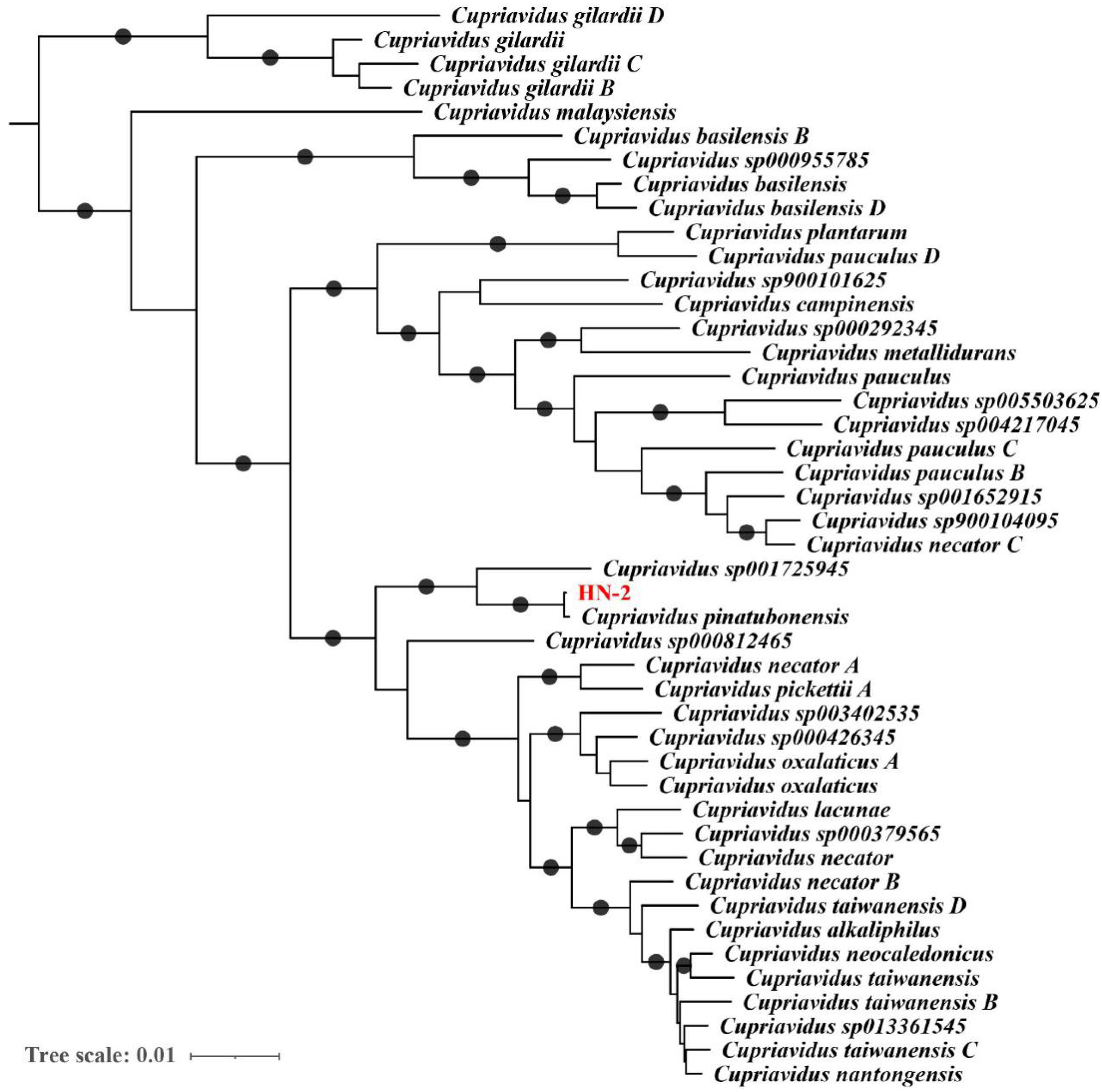
2.4. Genome-Based Taxonomic Classification Analysis

For a whole-genome-based taxonomic analysis, the genome sequence of Cupriavidus pinatubonensis HN-2 was analyzed by GTDB-Tk version 1.5.1 (database version release-202) [43], which is a toolkit used to classify prokaryotic genomes with the Genome Taxonomy Database [44,45,46,47,48,49,50]. The phylogenetic relationship between HN-2 and other bacteria was investigated by placing HN-2 in a bacterial reference tree based on 120 preselected marker genes.

2.5. Construction of an In-Frame Deletion Mutant and Complementation

To produce deletion mutants, C. pinatubonensis HN-2 wild-type was utilized as a parental strain. A 500 bp upstream and downstream fragments of the fadT gene were amplified by PCR and cloned into a HindIII-BamHI-linearized pK18mobsacB vector. Triparental mating with the helper strain HB101 (RK2013) on an LB plate at 30 °C for 10 h mobilized the recombinant plasmid into strain HN-2. Cells were selected with LB medium supplemented with gentamicin and chloramphenicol. Colonies were further selected on LB plates containing 10% sucrose. ∆fadT mutant was verified by PCR and DNA sequencing. Further, to investigate the inhibitory effect on DSF-mediated Xcc pathogenicity of the fadT in-frame ∆fadT deletion mutants, biocontrol efficacy against black rot in Chinese radish (Raphanus sativus L.) was performed. The bacterial suspensions were inoculated on radish root slices containing suspensions of black rot pathogen Xcc (6 × 10⁸ CFU·mL−1), suspensions of Xcc mixed with HN-2 wild-type (6 × 10⁸ CFU·mL−1), and suspensions of Xcc mixed with fadT in-frame ∆fadT deletion mutant (6 × 10⁸ CFU·mL−1). Radish root slices inoculated only with distilled water were used as the control. Three replicates of each treatment were set, and the experiments were carried out at least thrice. The radish root slices were incubated at 30 °C for 2 days in an incubator. Daily records of symptoms were taken after bacterial inoculation. The macerated area was measured to evaluate the disease severity [15,51].
For complementation of ∆fadT, the coding sequence of fadT and its indigenous promoter sequence were amplified by PCR and cloned into HindHIII/BamHI sites of pBBR1MCS-5, generating plasmid pBBR1MCS-5-fadT. pBBR1MCS5-fadT was mobilized into ∆fadT mutant by triparental conjugal mating as above. The transformants were selected on LB medium containing gentamicin and chloramphenicol. Correct transformants were confirmed by PCR and DNA sequencing. The primers used for cloning and detection are all listed in Table 2.

2.6. Purification of FadT

Enzyme purification was performed according to the methods described in previous reports [52,53] with modifications. Cell cultures (1 L) were separated via centrifugation at 8000× g for 10 min. The cell pellets were resuspended in 40 mL binding buffer (140 mM NaCl, 2.7 mM KCl, 10 mM Na2HPO4, 1.8 mM KH2PO4, pH 7.0) and subjected to 400 pulses of sonication (400 W, 3 s each with a 5 s interval) in an ice-water bath [54]. Following centrifugation (13,000× g) at 4 °C for 30 min, the supernatant was passed through a 0.22 μm filter and applied to a GSTrap (GE Healthcare, Marlborough, MA, USA) FF column of the liquid chromatography system, AKTAPure (GE Healthcare, Marlborough, MA, USA), and GST fusion proteins were eluted via a GSTrap FF column using 10 mM reduced glutathione according to standard affinity chromatography. GST fusion proteins were digested using PreScission protease (GE Healthcare, Marlborough, MA, USA) according to the manufacturer’s instructions.

2.7. Enzyme Activity Assay

FadT enzyme activity was investigated using a 0.5 mL reaction mixture containing 2 mM DSF, 100 mM sodium phosphate buffer (pH 7.0), and an appropriately diluted FadT preparation. After 30 min of incubation at 35 °C, the reaction was stopped by adding 100 μL of 1 M trichloroacetic acid [55]. Concentrations of DSF residues in the reaction mixture were extracted using ethyl acetate and detected via high-performance liquid chromatography (HPLC) (Waters 2690, Milford, MA, USA) according to the method described by Ye et al. [15]. Each treatment was performed in triplicate with inactivated enzyme as control. One unit of enzyme activity (U) was defined as the amount required to catalyze the formation of 1 μmol of product per minute [52,53].

2.8. Virulence Tests

The virulence of the Xanthomonas campestris pv. campestris strain on host plants was tested by the leaf-clipping method as previously described [56,57]. Healthy Chinese cabbage (Brassica pekinensis (Lour.) Rupr.), radish (Raphanus sativus L.), and pakchoi (Brassica campestris L. ssp. chinensis Makino) seeds were cultivated and acclimated in sterile soil under irregular irrigation for 30 days with no application of pesticides. These plants grew in flower pots in corridors and under a ceiling to block some of the rain. The day/night temperature, humidity, and photoperiod were all set to match the natural environment during the experiment. The experimental design included the following five treatments: (1) distilled water, which was used as the control; (2) Xcc at 6 × 10⁸ CFU·mL−1; (3) Xcc and ∆fadT at 6 × 10⁸ CFU·mL−1; (4) Xcc and ∆fadT (fadT) at 6 × 10⁸ CFU·mL−1; (5) Xcc (fadT) at 6 × 10⁸ CFU·mL−1. Three replicates of each treatment were used, and the experiments were carried out at least thrice. Daily records of symptoms were taken after bacterial inoculation [51,58]. The details of the tests are as follows: The strains used in this part were already cultured in 5 mL LB medium and shaken at 30 °C and 200 rpm for 12–16 h. Then, 100 μL of bacterial suspensions were inoculated on each plant. Next, Chinese cabbage and radish were harvested after 8 days, and pakchoi was harvested after 10 days. The macerated tissue was measured to evaluate the disease severity. The percentage of maceration was calculated by comparison with the preinoculation tissue [15,51].

2.9. GenBank Accession Number

The 16S rDNA gene sequences of C. pinatubonensis HN-2 were deposited into the NCBI nucleotide sequence database under the accession number MG561941.1. The whole-genome sequence of HN-2 and sequence of fadT were also deposited in the NCBI database with accession numbers CP080766-CP080769 and MZ734614, respectively. The genome annotation and all annotated protein sequences of HN-2 were deposited in Figshare (https://figshare.com/s/eee14ba9310748460cf8, accessed on 22 August 2021).

3. Results

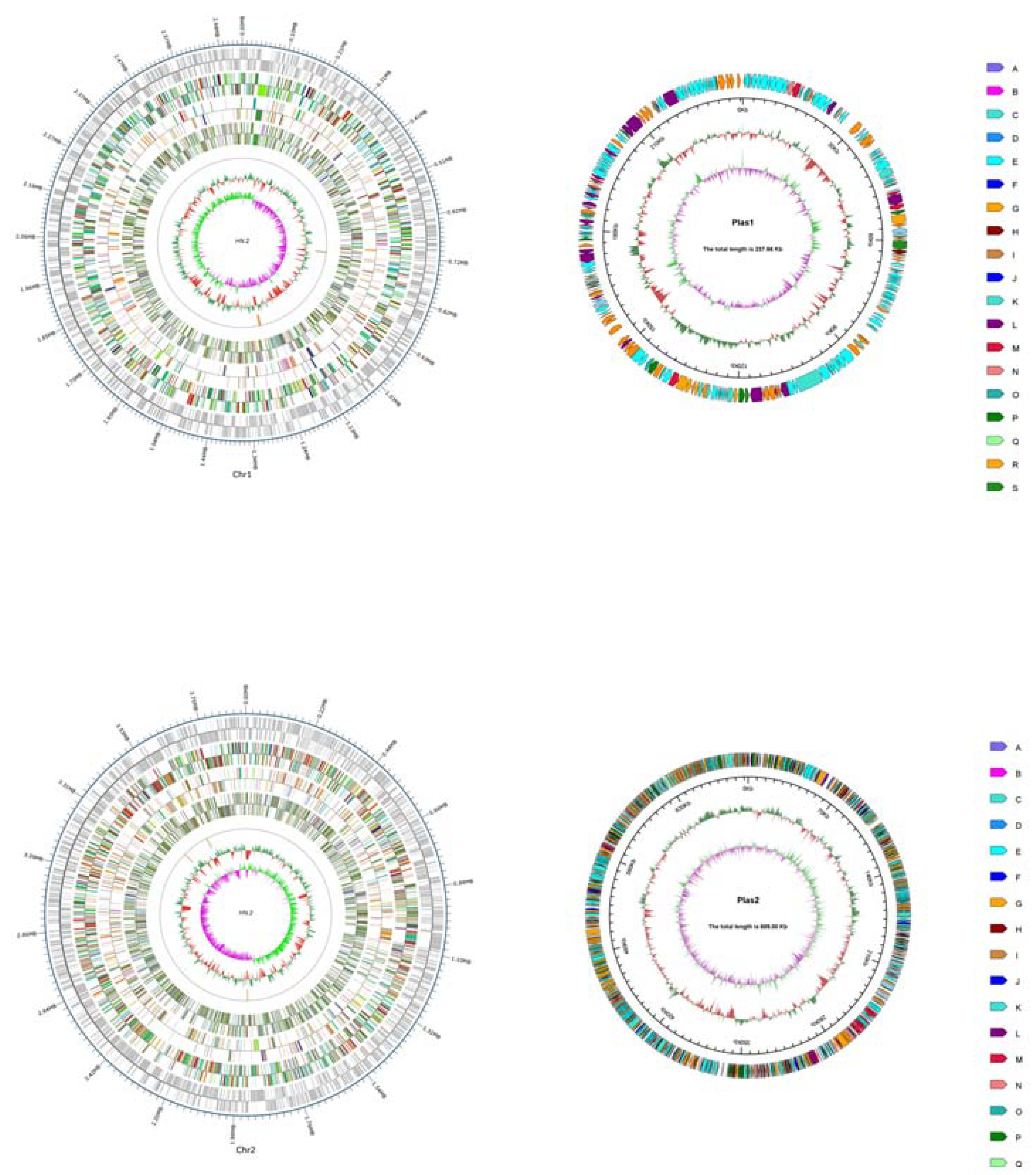
3.1. Sequencing and Analysis of the C. pinatubonensis HN-2 Genome

The whole genome of C. pinatubonensis HN-2 was sequenced by both Illumina short-read and Oxford Nanopore long-read sequencing technologies, generating 4.29 and 1.63 Gb of sequencing data, respectively. A hybrid de novo assembly using both types of sequencing data resulted in four circular replicons with a total size of 7.55 Mb and a GC-content of 64.29%. The C. pinatubonensis HN-2 genome assembly consists of two chromosomes (Chr1: 3.89 Mb; Chr2: 2.38 Mb) and two plasmids (Plas1: 689.0 kb; Plas2: 237.7 kb) (Figure 1). BUSCO assessment of the C. pinatubonensis HN-2 genome assembly showed that all the 688 Burkholderiales-wide single-copy orthologs were detected as complete genes, indicating that its quality was superior.
In total, 7101 protein-coding genes, 65 tRNA genes, 18 rRNA genes, and 1 small RNA gene were predicted in HN-2. The protein-coding genes have a total length of 6,556,812 bp, accounting for 86.86% of the genome size, and an average length of 923 bp (Table S1). Table S2 contains the findings of the functional annotation of protein-coding genes and the prediction of additional genomic characteristics. There were 14 gene islands with total and average lengths of 183,507 and 13,108 bp, respectively, and the statistical map of gene distribution in the gene island is shown in Figure S1. There were also four preprophages with total and average lengths of 269,803 and 67,450.8 bp, respectively.
There were 6747, 655, 5451, 4681, 2669, and 589 coding genes annotated by NR, KEGG, COG, GO, SwissProt, TCDB, PHI, and CAZy. From the GO database, 2544 complete CDSs (coding sequences) were obtained from the coding region prediction of the strain HN-2 genome, accounting for 89.23% of the total genome. There were 2512 genes involved in the metabolic process, representing the largest number of genes, followed by 2326 and 2247 genes involved in the cellular process and catalytic activity, respectively. Figure S2 presents the classification of HN-2 functional genes in the GO database. The COG database functionally classifies 3299 CDSs, accounting for 88.99% of the total number of coding genes, and Figure S3 presents the classification of HN-2 functional genes in the COG database. There were a total of 671 genes belonging to general predicted (R) functions that were not explicitly labeled to a specific group among these functional genes. Next, there were 623, 599, 513, and 499 genes with the functions of energy production and conversion (C), transcription (K), amino acid transport and metabolism (E), and lipid transport and metabolism (I), respectively. The above two databases provide a clearer view of the relevant functional properties of strain HN-2, in which genes with functions in metabolism and catalytic activity are relatively abundant, providing references for subsequent studies on genes that may be closely related to the function of strain HN-2 in DSF degradation. Based on the KEGG database, 223 genes were predicted as xenobiotic biodegradation and metabolism genes. According to the comparison of the database, the quantity and species of biodegradation metabolism pathways were abundant in the genome of strain HN-2, and this result was mutually confirmed with the results annotated by the GO and COG databases. Other KEGG pathway annotations are enumerated in Figure S4. The predicted genes were also annotated against the CAZy database, and the classification annotation results, statistical tables, and individual statistics plots are displayed in Figure S5.
Our previous 16S rRNA-based analysis classified strain HN-2 as a member of the Cupriavidus genus. To further determine the taxonomic assignment of strain HN-2, we analyzed its genome sequence using GTDB-Tk, which first placed strain HN2 into a prebuilt bacterial phylogeny based on a set of 120 proteins and then calculated average nucleotide identity (ANI) values between strain HN-2 and its close relatives in the tree. As a result, strain HN-2 was discovered to be the most closely related to C. pinatubonensis JMP134 (Figure 2), with an ANI value of 99.43%, much over the usually recognized prokaryotic species delimitation threshold of 95%. Therefore, we assigned strain HN-2 as a strain of C. pinatubonensis based on genome data analysis.

3.2. Identification and Cloning of the Gene Responsible for the DSF Degradation

DSF is an unsaturated fatty acid, and, therefore, the ability of the strain HN-2 to quench DSF-mediated QS probably depends on enzymes in the fatty acid degradation pathway. For identification of genes in charge of DSF degradation in strain HN-2, we selected eight genes for further investigation based on their functional annotations (domain structures of their protein products are shown in Figure S6); these genes encode four long-chain acyl-CoA synthetases (HN.2_GM000231, HN.2_GM003041, HN.2_GM005970, and HN.2_GM000743, which were annotated with the KO term K01897 and are also homologs of rpfB), three 3-hydroxyacyl-CoA dehydrogenases (HN.2_GM000404, HN.2_GM003236, and HN.2_GM004234, which were annotated with the KO term K07516), and one acyl-CoA dehydrogenase (HN.2_GM004237). To investigate whether these genes affected the degradation of DSF in strain HN-2, mutants with deletion of these genes were generated using strain HN-2 as the parental strain. Finally, the 0404 in-frame ∆0404 deletion mutant, the 3236 in-frame ∆3236 deletion mutant, the 4234 in-frame ∆4234 deletion mutant, the 4237 in-frame ∆4237 deletion mutant, the 0231 in-frame ∆0231 deletion mutant, the 3041 in-frame ∆3041 deletion mutant, the 5970 in-frame ∆5970 deletion mutant, and the 0743 in-frame ∆0743 deletion mutant were generated. After exploring their inhibitory effect on the pathogenicity of Xcc, GM004237 was initially identified as a DSF-degradation gene in strain HN-2 and named fadT. The other genes GM000231, GM003041, GM005970, GM000743, GM000404, GM003236, and GM004234 have no inhibitory effect on the pathogenicity of Xcc.
The biocontrol experiment showed that strain HN-2 could well decrease DSF-mediated Xcc pathogenicity, whereas the fadT in-frame ∆fadT deletion mutant had no inhibitory effect on DSF-mediated Xcc pathogenicity. Biocontrol efficacy against black rot in Chinese radish of the control (CK), wild-type (strain HN-2), and ∆fadT is shown in Figure 3. It is worth pointing out that the wild-type strain HN-2 exhibited superior DSF degradation activity and completely degraded 2 mM of DSF within 24 h. In addition, ∆fadY could not degrade DSF, and ∆fadT (fadT) regained the ability to degrade DSF, which further indicated that fadT played a key role in the degradation of DSF. The comparative study of DSF-degrading activity between the control (CK), wild-type strain HN-2, ∆fadT, and ∆fadT (fadT) cultured in MSM medium for 24 h is shown in Figure S7. These results identified fadT as the key gene affecting wild HN-2 in the alleviation of bacterial virulence on host plants, and it was thus identified as an important gene responsible for DSF degradation.

3.3. Codon Optimization, Expression, and Purification of FadT

In order to obtain a higher yield and a pure DSF-degrading enzyme, the gene fadT was codon optimized. After the optimized gene co-fadT was obtained, it was synthesized and ligated into the vector pGEX-6p-1. In addition, pGEX-6p-1-co-fadT was obtained and then transferred into E. coli BL21. Finally, the engineered strain E. coil BL21/pGEX fadT was acquired, and it was abundantly expressed to obtain the crude enzyme extract, followed by affinity purification. As shown in Figure S8, the SDS-PAGE staining showed that GST-FadT had a molecular weight of about 93 kDa as predicted in silico. The band of GST-FadT was single, indicating the enzymes were of high purity. The purified FadT protein exhibited high enzymatic activity and outstanding stability over a broad pH (6.0–8.0) and temperature (25–45 °C) range with maximal activity at pH 7.0 and 35 °C. No cofactors were required for FadT enzyme activity. Moreover, the FadT enzyme showed a strong ability to degrade DSF (Figure S9).

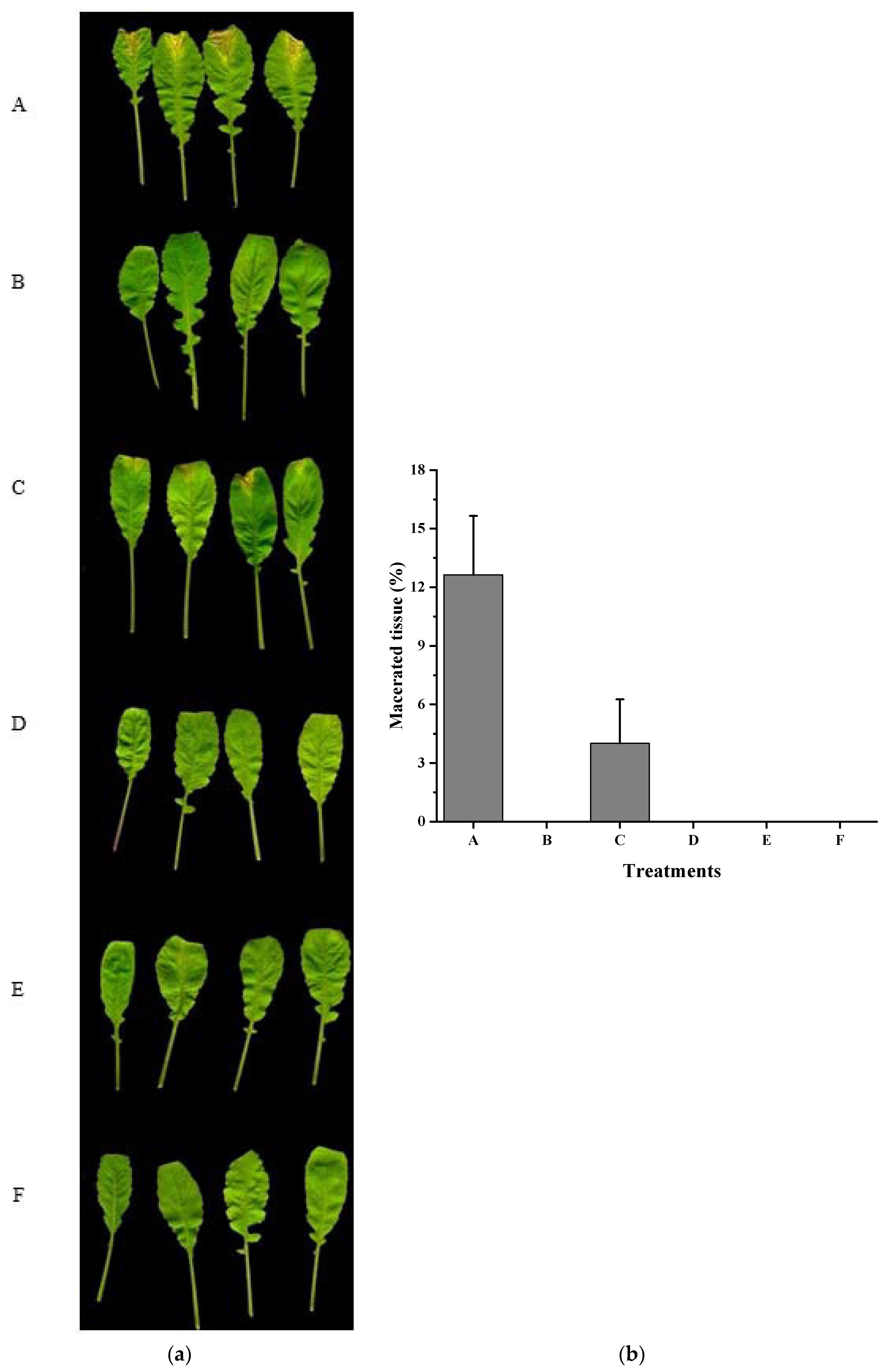
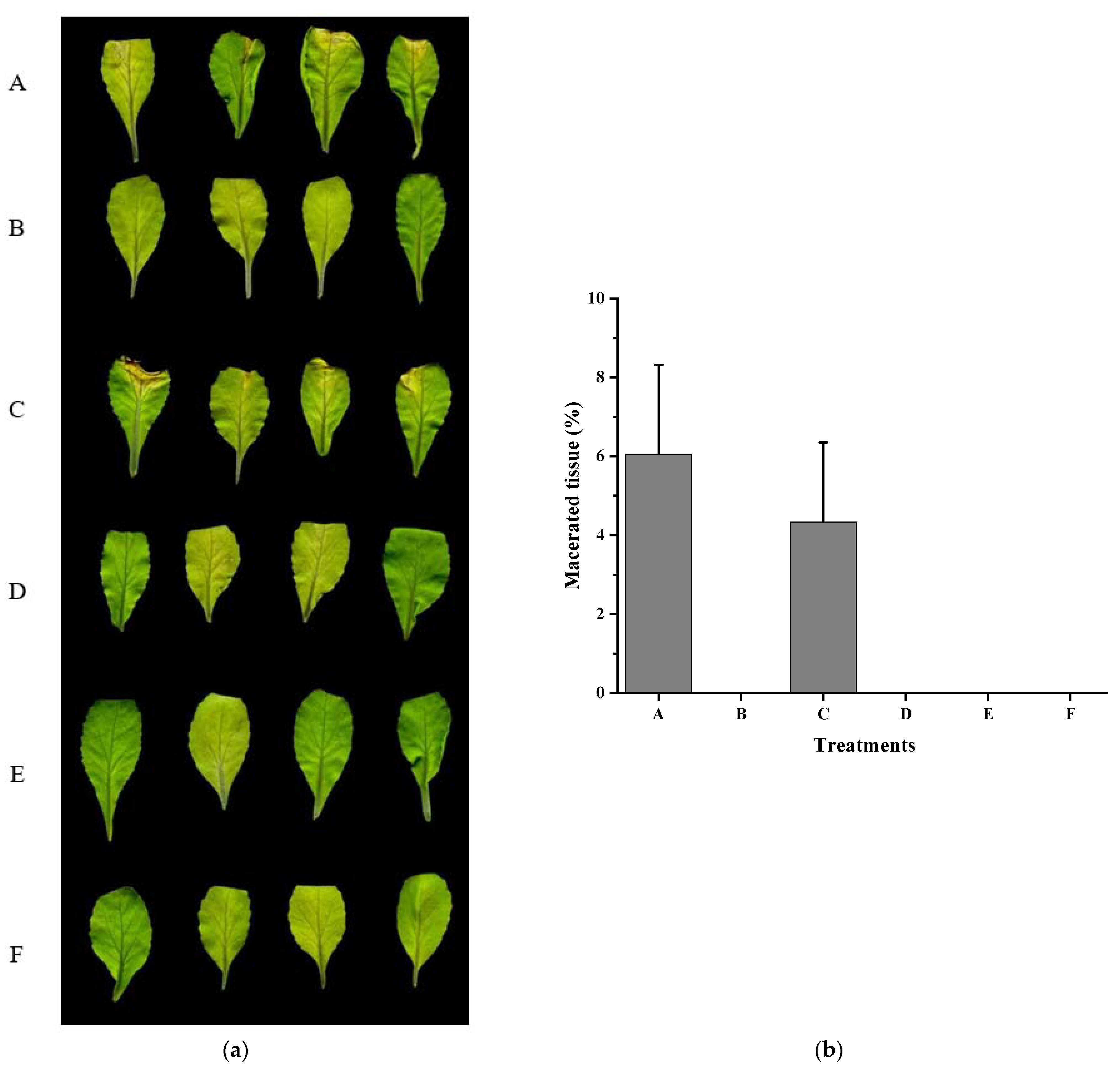
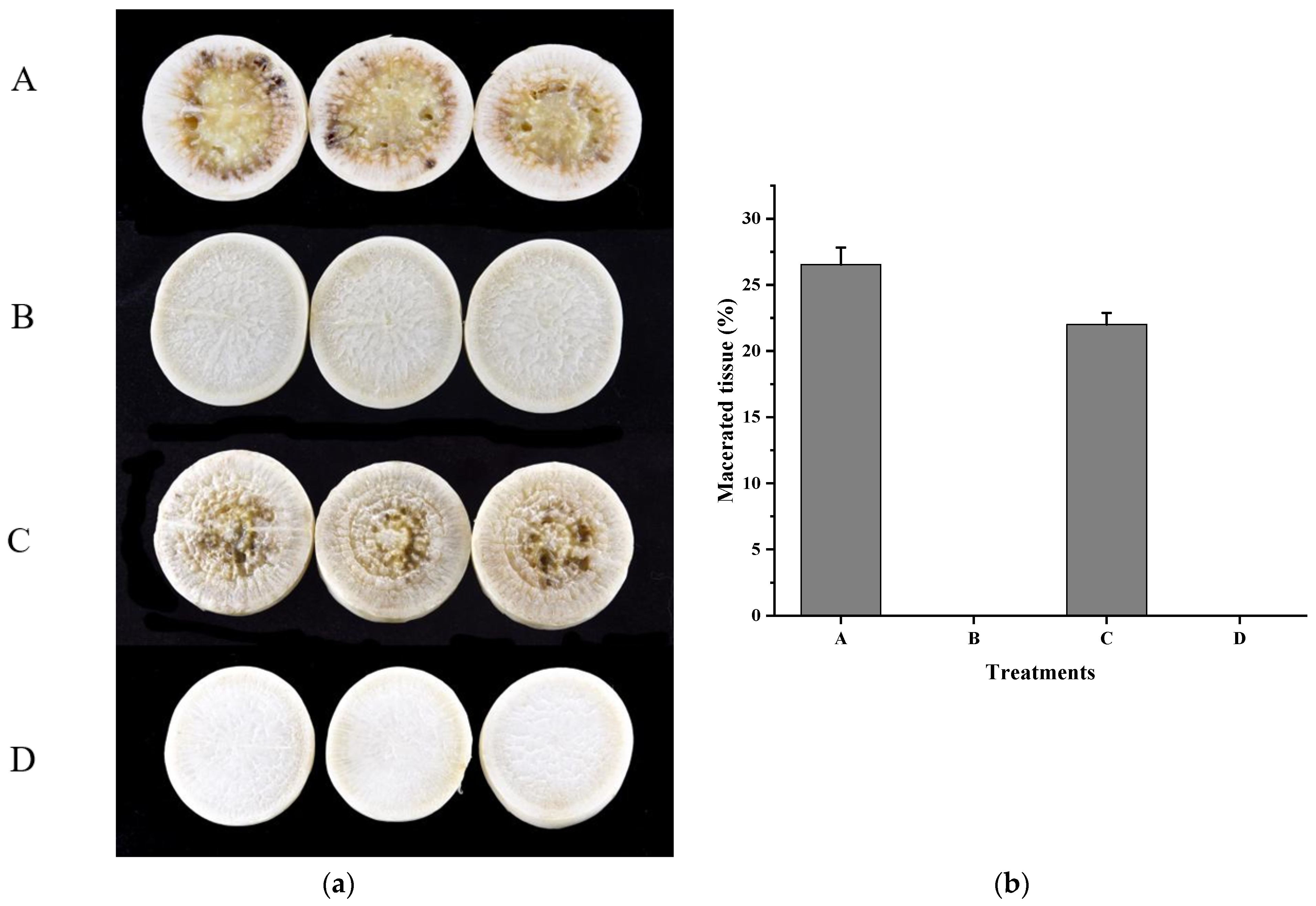
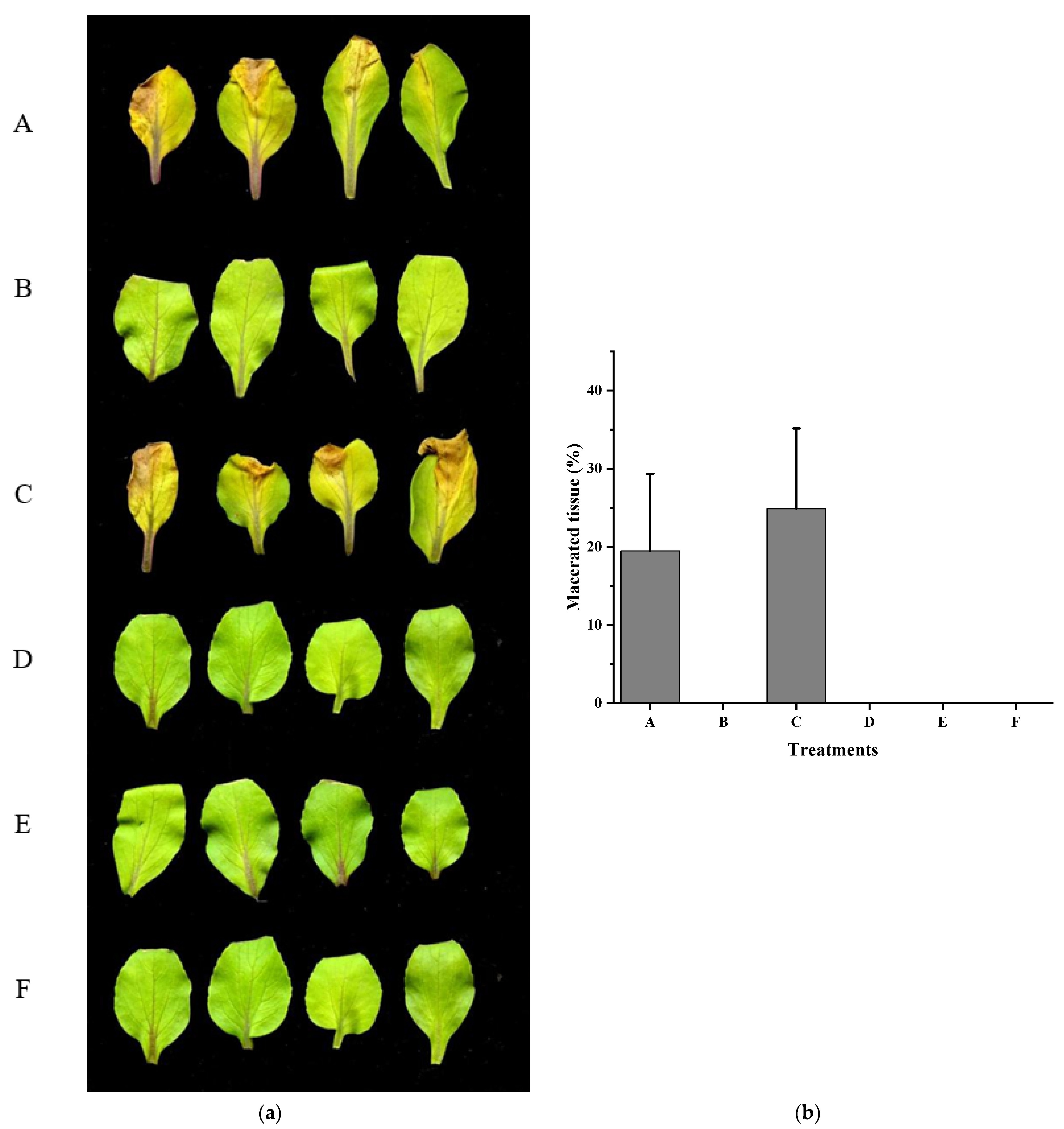
3.4. Xcc Expressing fadT Loses Virulence in Planta

To explore the function of fadT gene in the biocontrol of Xcc, we performed plant inoculation experiments. Radish (Raphanus sativus L.), Chinese cabbage (Brassica pekinensis (Lour.) Rupr.), and pakchoi (Brassica campestris L. ssp. chinensis Makino), commonly used in the test of biocontrol of black rot disease, were inoculated with Xcc together with wild-type or fadT-deletion strain HN-2. Each tested plant inoculated with Xcc alone presented severe black rot symptoms (Figure 4, Figure 5 and Figure 6). In contrast, inoculation of a mixture of Xcc with wild-type HN-2 almost completely diminished the plant disease symptoms, suggesting wild-type HN-2 counteracts the virulent effects of Xcc. To demonstrate the role of fadT gene in strain HN-2, Xcc was further mixed with fadT-deletion HN-2 in the plant inoculation experiment. As shown, strain HN-2 without fadT gene could not impair the pathogenicity of Xcc, while complementation of fadT gene in the fadT-deletion HN-2 restored its biocontrol ability. These results further demonstrated the essential role of fadT gene in the interaction of strain HN-2. Further, on the test plants, the virulence of Xcc expressing HN-2-derived fadT gene was also reduced. Taken together, the expression of fadT gene could effectively attenuate the black rot disease caused by Xcc in the host plants.

4. Discussion

Understanding the genomic diversity of bacterial strain and the relevant bioactive products are of importance for disease control [59,60]. In this study, whole-genome sequencing (WGS) combined with genome annotation was applied to uncover the mechanism of DSF degradation in Cupriavidus pinatubonensis strain HN-2. The genome sequence of strain HN-2 discovered in this study will be useful in the development of DSF-degrading bacteria or enzymes in the future. Further studies require a focus on genes related to the pathway that metabolizes DSF.
The emergence of antibiotic-resistant bacteria causes more difficulties in the treatment of bacterial infectious diseases. As for the control of diseases caused by bacteria, the use of QQ is a promising avenue to replace traditional antibiotics. In principle, QQ quenches the QS molecules of targeted bacteria, resulting in the shutdown of the expression of QS-controlled virulence genes [61,62,63,64]. As a result, a variety of bacteria that degrade DSF have been found and characterized, including Staphylococcus, Bacillus, Microbacterium, Paenibacillus, Cupriavidus, Acinetobacter, and Pseudomonas [15,65,66,67,68,69]. However, the genome sequence and molecular mechanisms of DSF-degrading strains are not clear, which limits the applications of these QQ strains. Since the discovery of the first AHL lactase AiiA isolated from Bacillus cereus [66] and the AHL amidase isolated from Variovorax paradoxus, an increasing amount of AHL-degrading enzymes have been well identified [70,71,72,73,74].
DSF has multiple effects on various cellular processes of bacteria. It has been reported that DSF can block protease secretion from attack-phase cells of Bdellovibrio bacteriovorus 109 J completely at the concentration of 50 mM and negatively regulate the membrane integrity at a concentration of 200 mM [75]. Additionally, DSF inhibits biofilm formation with no impacts on the growth of P. aeruginosa under static and continuous conditions [76]. DSF also regulates intercellular public goods (such as extracellular polymeric substances (EPS) and amino acids) [76]. DSF-based QS systems are classified into three major classes, the first from Xcc, the second from Burkholderia cenocepacia and Cronobacter turicensis, and the third from Pseudomonas aeruginosa [77,78,79,80]. However, limited DSF-degrading bacteria and their corresponding enzymes were identified. This is the first research that we are aware of that describes a novel DSF-degrading enzyme in C. pinatubonensis.
Our findings show that expressing the fadT gene in Xcc significantly reduced black rot symptoms in the tested host plants, indicating FadT as a novel DSF-degrading enzyme with significant efficacy in reducing Xcc pathogenicity. Our study highlights the potential use of FadT in the biocontrol of Xcc and, as a starting point, indicates the significance of the application of FadT in a broader range of DSF-dependent bacterial pathogens.

5. Conclusions

In summary, our study first reports the genome sequence and identifies a novel DSF-degrading enzyme, FadT, in C. pinatubonensis HN-2. Plant experiments showed that Xcc carrying a functioning FadT markedly suppressed its virulence, resulting in the elimination of the symptoms of black rot in the assayed host plants. Our study demonstrates for the first time that FadT is a powerful tool for the biocontrol of black rot caused by Xcc. These novel findings reveal the biochemical characteristics of a highly efficient DSF-degrading bacterial isolate and provide useful enzymes/genes with great potential for the control of infectious diseases caused by DSF-dependent bacterial pathogens.

Supplementary Materials

The following are available online at https://www.mdpi.com/article/10.3390/ijms22189862/s1. Figure S1. Statistical diagram of gene distribution in the Cupriavidus pinatubonensis HN-2 gene island. Figure S2. Classification of Cupriavidus pinatubonensis HN-2 functional genes in the GO database. Figure S3. Classification of Cupriavidus pinatubonensis HN-2 functional genes in the COG database. Figure S4. KEGG metabolic pathway classification diagram of Cupriavidus pinatubonensis HN-2. Figure S5. CAZy functional classification and corresponding gene number statistics of HN-2. AA: oxidoreductase; CBM: carbohydrate-binding module; CE: carbohydrate esterases; GH: glycoside hydrolase; GT: glycosyltransferase; PL: polysaccharide lyase; it was not found in the KEGG metabolic pathway of Cupriavidus pinatubonensis HN-2. Figure S6. InterProScan predicted domain structures of proteins encoded by the eight genes selected for deletion assay. (A) Four long-chain acyl-CoA synthetases encoded by HN.2_GM000231, HN.2_GM003041, HN.2_GM005970, and HN.2_GM000743. Two characteristic domains were predicted, namely IPR000873 “AMP-dependent synthetase/ligase” and IPR025110 “AMP-binding enzyme, C-terminal domain”. (B) Three 3-hydroxyacyl-CoA dehydrogenases encoded by HN.2_GM000404, HN.2_GM003236, and HN.2_GM004234. Two characteristic domains were predicted, namely IPR006176 “3-hydroxyacyl-CoA dehydrogenase, NAD binding” and IPR006108 “3-hydroxyacyl-CoA dehydrogenase, C-terminal”. (C) The acyl-CoA dehydrogenase encoded by HN.2_GM004237, which was named fadT in our study. Four characteristic domains were predicted, namely IPR020953 “Acyl-CoA dehydrogenase, N-terminal, bacteria”, IPR006091 “Acyl-CoA oxidase/dehydrogenase, central domain”, IPR009075 “Acyl-CoA dehydrogenase/oxidase C-terminal”, and IPR025878 “Acetyl-CoA dehydrogenase-like C-terminal domain”. Figure S7. SDS-PAGE analysis of purified FadT. M: Marker; 7: Purified FadT. Figure S8. Effects of temperature and pH on FadT activity. (A) Determination of optimal temperature; (B) determination of optimal pH. Figure S9. Diffusible signal factor (DSF)-degrading activity comparison of mutants and wild-type strain HN-2. CK: control; strain HN-2: wild-type; ∆fadT: fadT deletion mutant; ∆fadT (fadT): the complement of the fadT deletion mutant. Table S1. Genome characteristics of Cupriavidus pinatubonensis HN-2. Table S2. Prediction of noncoding RNA (ncRNA) in strain HN-2 genome.

Author Contributions

Conceptualization: S.C.; methodology: X.X., T.Y. and T.Z.; data analysis: X.X., T.Y., W.Z. and X.Z.; writing—original draft preparation: X.X. and T.Y.; writing—review and editing: X.X., T.Y., W.Z., T.Z., X.Z., W.D. and S.C.; supervision, funding acquisition, and project administration: X.Z., W.Z. and S.C. All authors have read and agreed to the published version of the manuscript.

Funding

This research was funded by the Key Realm R&D Program of Guangdong Province, China (2020B0202090001, 2018B020206001); the National Natural Science Foundation of China (31771341); the Natural Science Foundation of Guangdong Province, China (2021A1515010889); and the Guangdong Special Branch Plan for Young Talent with Scientific and Technological Innovation, China (2017TQ04N026).

Institutional Review Board Statement

Not applicable.

Informed Consent Statement

Not applicable.

Conflicts of Interest

The authors declare no conflict of interest. The funders had no role in the design of the study; in the collection, analyses, or interpretation of data; in the writing of the manuscript; or in the decision to publish the results.

References

  1. Gupta, M.; Vikram, A.; Bharat, N. Black rot—A devastating disease of crucifers: A review. Agric. Rev. 2013, 34, 269. [Google Scholar] [CrossRef]
  2. Basavaraju, M.; Sisnity, V.S.; Palaparthy, R.; Addanki, P.K. Quorum quenching: Signal jamming in dental plaque biofilms. J. Dent. Sci. 2016, 11, 349–352. [Google Scholar] [CrossRef] [PubMed] [Green Version]
  3. Turan, N.B.; Chormey, D.S.; Büyükpınar, Ç.; Engin, G.O.; Bakirdere, S. Quorum sensing: Little talks for an effective bacterial coordination. TrAC Trends Anal. Chem. 2017, 91, 1–11. [Google Scholar] [CrossRef]
  4. Wang, L.-H.; He, Y.; Gao, Y.; Wu, J.E.; Dong, Y.-H.; He, C.; Wang, S.X.; Weng, L.-X.; Xu, J.-L.; Tay, L.; et al. A bacterial cell-cell communication signal with cross-kingdom structural analogues. Mol. Microbiol. 2004, 51, 903–912. [Google Scholar] [CrossRef]
  5. Newman, K.L.; Chatterjee, S.; Ho, K.A.; Lindow, S.E. Virulence of Plant Pathogenic Bacteria Attenuated by Degradation of Fatty Acid Cell-to-Cell Signaling Factors. Mol. Plant-Microbe Interact. 2008, 21, 326–334. [Google Scholar] [CrossRef] [PubMed] [Green Version]
  6. Zhou, L.; Zhang, L.-H.; Cámara, M.; He, Y.-W. The DSF Family of Quorum Sensing Signals: Diversity, Biosynthesis, and Turnover. Trends Microbiol. 2017, 25, 293–303. [Google Scholar] [CrossRef]
  7. Ryan, R.P.; An, S.-Q.; Allan, J.H.; McCarthy, Y.; Dow, J.M. The DSF Family of Cell-Cell Signals: An Expanding Class of Bacterial Virulence Regulators. PLoS Pathog. 2015, 11, e1004986. [Google Scholar] [CrossRef]
  8. Ma, F.; Xu, S.; Tang, Z.; Li, Z.; Zhang, L. Use of antimicrobials in food animals and impact of transmission of antimicrobial resistance on humans. Biosaf. Health 2021, 3, 32–38. [Google Scholar] [CrossRef]
  9. Zhang, J.; Feng, T.; Wang, J.; Wang, Y.; Zhang, X.-H. The Mechanisms and Applications of Quorum Sensing (QS) and Quorum Quenching (QQ). J. Ocean Univ. China 2019, 18, 1427–1442. [Google Scholar] [CrossRef]
  10. Zhang, L.-H. Quorum quenching and proactive host defense. Trends Plant Sci. 2003, 8, 238–244. [Google Scholar] [CrossRef]
  11. Fetzner, S. Quorum quenching enzymes. J. Biotechnol. 2015, 201, 2–14. [Google Scholar] [CrossRef]
  12. Schuster, M.; Sexton, D.J.; Diggle, S.P.; Greenberg, E.P. Acyl-Homoserine Lactone Quorum Sensing: From Evolution to Application. Annu. Rev. Microbiol. 2013, 67, 43–63. [Google Scholar] [CrossRef]
  13. Zhou, L.; Wang, X.-Y.; Sun, S.; Yang, L.-C.; Jiang, B.-L.; He, Y.-W. Identification and characterization of naturally occurring DSF-family quorum sensing signal turnover system in the phytopathogen Xanthomonas. Environ. Microbiol. 2015, 17, 4646–4658. [Google Scholar] [CrossRef] [PubMed]
  14. Bi, H.; Yu, Y.; Dong, H.; Wang, H.; Cronan, J.E. Xanthomonas campestris RpfB is a fatty Acyl-CoA ligase required to counteract the thioesterase activity of the RpfF diffusible signal factor (DSF) synthase. Mol. Microbiol. 2014, 93, 262–275. [Google Scholar] [CrossRef] [PubMed] [Green Version]
  15. Ye, T.; Zhou, T.; Li, Q.; Xu, X.; Fan, X.; Zhang, L.; Chen, S. Cupriavidus sp. HN-2, a Novel Quorum Quenching Bacterial Isolate, is a Potent Biocontrol Agent Against Xanthomonas campestris pv. campestris. Microorganisms 2020, 8, 45. [Google Scholar] [CrossRef] [PubMed] [Green Version]
  16. Guo, Y.; Huang, Y.; Pang, S.; Zhou, T.; Lin, Z.; Yu, H.; Zhang, G.; Bhatt, P.; Chen, S. Novel Mechanism and Kinetics of Tetramethrin Degradation Using an Indigenous Gordonia cholesterolivorans A16. Int. J. Mol. Sci. 2021, 22, 9242. [Google Scholar] [CrossRef]
  17. Reiner, J.; Pisani, L.; Qiao, W.; Singh, R.; Yang, Y.; Shi, L.; Khan, W.A.; Sebra, R.; Cohen, N.; Babu, A.; et al. Cytogenomic identification and long-read single molecule real-time (SMRT) sequencing of a Bardet–Biedl Syndrome 9 (BBS9) deletion. npj Genom. Med. 2018, 3, 3. [Google Scholar] [CrossRef]
  18. Ardui, S.; Ameur, A.; Vermeesch, J.R.; Hestand, M.S. Single molecule real-time (SMRT) sequencing comes of age: Applications and utilities for medical diagnostics. Nucleic Acids Res. 2018, 46, 2159–2168. [Google Scholar] [CrossRef] [PubMed] [Green Version]
  19. Saha, S.; Bridges, S.; Magbanua, Z.V.; Peterson, D.G. Empirical comparison of ab initio repeat finding programs. Nucleic Acids Res. 2008, 36, 2284–2294. [Google Scholar] [CrossRef] [Green Version]
  20. Benson, G. Tandem repeats finder: A program to analyze DNA sequences. Nucleic Acids Res. 1999, 27, 573–580. [Google Scholar] [CrossRef] [PubMed] [Green Version]
  21. Lagesen, K.; Hallin, P.; Rødland, E.A.; Staerfeldt, H.-H.; Rognes, T.; Ussery, D. RNAmmer: Consistent and rapid annotation of ribosomal RNA genes. Nucleic Acids Res. 2007, 35, 3100–3108. [Google Scholar] [CrossRef]
  22. Lowe, T.M.; Eddy, S.R. tRNAscan-SE: A program for improved detection of transfer RNA genes in genomic sequence. Nucleic Acids Res. 1997, 25, 955–964. [Google Scholar] [CrossRef]
  23. Nawrocki, E.P.; Kolbe, D.L.; Eddy, S.R. Infernal 1.0: Inference of RNA alignments. Bioinformatics 2009, 25, 1335–1337. [Google Scholar] [CrossRef] [PubMed] [Green Version]
  24. Gardner, P.P.; Daub, J.; Tate, J.G.; Nawrocki, E.P.; Kolbe, D.L.; Lindgreen, S.; Wilkinson, A.C.; Finn, R.D.; Griffiths-Jones, S.; Eddy, S.R.; et al. Rfam: Updates to the RNA families database. Nucleic Acids Res. 2009, 37, D136–D140. [Google Scholar] [CrossRef] [PubMed] [Green Version]
  25. Eichinger, V.; Nussbaumer, T.; Platzer, A.; Jehl, M.E.; Arnold, R.; Rattei, T. EffectiveDB—Updates and novel features for a better annotation of bacterial secreted proteins and Type III, IV, VI secretion systems. Nucleic Acids Res. 2016, 44, D669–D674. [Google Scholar] [CrossRef]
  26. Hsiao, W.; Wan, I.; Jones, S.; Brinkman, F.S.L. IslandPath: Aiding detection of genomic islands in prokaryotes. Bioinformatics 2003, 19, 418–420. [Google Scholar] [CrossRef] [PubMed] [Green Version]
  27. Grissa, I.; Vergnaud, G.; Pourcel, C. CRISPRFinder: A web tool to identify clustered regularly interspaced short palindromic repeats. Nucleic Acids Res. 2007, 35, W52–W57. [Google Scholar] [CrossRef] [PubMed] [Green Version]
  28. Zhou, Y.; Liang, Y.; Lynch, K.H.; Dennis, J.; Wishart, D.S. PHAST: A Fast Phage Search Tool. Nucleic Acids Res. 2011, 39, W347–W352. [Google Scholar] [CrossRef]
  29. Kanehisa, M. From genomics to chemical genomics: New developments in KEGG. Nucleic Acids Res. 2006, 34, D354–D357. [Google Scholar] [CrossRef]
  30. Kanehisa, M.; Goto, S.; Kawashima, S.; Okuno, Y.; Hattori, M. The KEGG resource for deciphering the genome. Nucleic Acids Res. 2004, 32, D277–D280. [Google Scholar] [CrossRef] [Green Version]
  31. Ashburner, M.; Ball, C.A.; Blake, J.; Botstein, D.; Butler, H.; Cherry, J.M.; Davis, A.P.; Dolinski, K.; Dwight, S.S.; Eppig, J.T.; et al. Gene Ontology: Tool for the unification of biology. Nat. Genet. 2000, 25, 25–29. [Google Scholar] [CrossRef] [PubMed] [Green Version]
  32. Galperin, M.; Makarova, K.S.; Wolf, Y.; Koonin, E.V. Expanded microbial genome coverage and improved protein family annotation in the COG database. Nucleic Acids Res. 2015, 43, D261–D269. [Google Scholar] [CrossRef]
  33. Bairoch, A.; Apweiler, R. The SWISS-PROT protein sequence database and its supplement TrEMBL in 2000. Nucleic Acids Res. 2000, 28, 45–48. [Google Scholar] [CrossRef]
  34. Sayer, M.H., Jr.; Reddy, V.S.; Tamang, D.G.; Västermark, Å. The Transporter Classification Database. Nucleic Acids Res. 2014, 42, D251–D258. [Google Scholar] [CrossRef] [PubMed]
  35. Li, W.; Jaroszewski, L.; Godzik, A. Tolerating some redundancy significantly speeds up clustering of large protein databases. Bioinformatics 2002, 18, 77–82. [Google Scholar] [CrossRef] [PubMed]
  36. Cantarel, B.L.; Coutinho, P.M.; Rancurel, C.; Bernard, T.; Lombard, V.; Henrissat, B. The Carbohydrate-Active EnZymes database (CAZy): An expert resource for Glycogenomics. Nucleic Acids Res. 2009, 37, D233–D238. [Google Scholar] [CrossRef]
  37. Petersen, T.N.; Brunak, S.; von Heijne, G.; Nielsen, H. SignalP 4.0: Discriminating signal peptides from transmembrane regions. Nat. Methods 2011, 8, 785–786. [Google Scholar] [CrossRef]
  38. Medema, M.H.; Blin, K.; Cimermancic, P.; De Jager, V.; Zakrzewski, P.; Fischbach, M.A.; Weber, T.; Takano, E.; Breitling, R. antiSMASH: Rapid identification, annotation and analysis of secondary metabolite biosynthesis gene clusters in bacterial and fungal genome sequences. Nucleic Acids Res. 2011, 39, W339–W346. [Google Scholar] [CrossRef] [PubMed]
  39. Chen, L.; Xiong, Z.; Sun, L.; Yang, J.; Jin, Q. VFDB 2012 update: Toward the genetic diversity and molecular evolution of bacterial virulence factors. Nucleic Acids Res. 2012, 40, D641–D645. [Google Scholar] [CrossRef]
  40. Chen, G.; Gan, J.; Yang, C.; Zuo, Y.; Peng, J.; Li, M.; Huo, W.; Xie, Y.; Zhang, Y.; Wang, T.; et al. The SiaA/B/C/D signaling network regulates biofilm formation in Pseudomonas aeruginosa. EMBO J. 2020, 39, e105997. [Google Scholar] [CrossRef] [PubMed]
  41. Urban, M.; Pant, R.; Raghunath, A.; Irvine, A.G.; Pedro, H.; Hammond-Kosack, K. The Pathogen-Host Interactions database (PHI-base): Additions and future developments. Nucleic Acids Res. 2015, 43, D645–D655. [Google Scholar] [CrossRef] [Green Version]
  42. Krzywinski, M.; Schein, J.; Birol, I.; Connors, J.; Gascoyne, R.; Horsman, D.; Jones, S.; Marra, M.A. Circos: An information aesthetic for comparative genomics. Genome Res. 2009, 19, 1639–1645. [Google Scholar] [CrossRef] [Green Version]
  43. Chaumeil, P.-A.; Mussig, A.J.; Hugenholtz, P.; Parks, D.H. GTDB-Tk: A toolkit to classify genomes with the Genome Taxonomy Database. Bioinformatics 2019, 36, 1925–1927. [Google Scholar] [CrossRef]
  44. Parks, D.H.; Chuvochina, M.; Chaumeil, P.A.; Rinke, C.; Mussig, A.J.; Hugenholtz, P. A complete domain-to-species taxonomy for Bacteria and Archaea. Nat. Biotechnol. 2020, 38, 1079–1086. [Google Scholar] [CrossRef]
  45. Meier-Kolthoff, J.P.; Goeker, M. TYGS is an automated high-throughput platform for state-of-the-art genome-based taxonomy. Nat. Commun. 2019, 10, 2182. [Google Scholar] [CrossRef] [PubMed]
  46. Ondov, B.D.; Treangen, T.J.; Melsted, P.; Mallonee, A.B.; Bergman, N.H.; Koren, S.; Phillippy, A.M. Mash: Fast genome and metagenome distance estimation using MinHash. Genome Biol. 2016, 17, 1–14. [Google Scholar] [CrossRef] [PubMed] [Green Version]
  47. Meier-Kolthoff, J.P.; Auch, A.F.; Klenk, H.-P.; Göker, M. Genome sequence-based species delimitation with confidence intervals and improved distance functions. BMC Bioinform. 2013, 14, 60. [Google Scholar] [CrossRef] [PubMed] [Green Version]
  48. Lefort, V.; Desper, R.; Gascuel, O. FastME 2.0: A Comprehensive, Accurate, and Fast Distance-Based Phylogeny Inference Program. Mol. Biol. Evol. 2015, 32, 2798–2800. [Google Scholar] [CrossRef] [Green Version]
  49. Kreft, L.; Botzki, A.; Coppens, F.; Vandepoele, K.; Van Bel, M. PhyD3: A phylogenetic tree viewer with extended phyloXML support for functional genomics data visualization. Bioinformatics 2017, 33, 2946–2947. [Google Scholar] [CrossRef]
  50. Zhang, L.; Xu, J.; Birch, R.G. High affinity binding of albicidin phytotoxins by the AlbA protein from Klebsiella oxytoca. Microbiology 1998, 144, 555–559. [Google Scholar] [CrossRef] [Green Version]
  51. Ye, T.; Zhang, W.; Feng, Z.; Fan, X.; Xu, X.; Mishra, S.; Zhang, L.; Chen, S. Characterization of a Novel Quorum-Quenching Bacterial Strain, Burkholderia anthina HN-8, and Its Biocontrol Potential against Black Rot Disease Caused by Xanthomonas campestris pv. campestris. Microorganisms 2020, 8, 1485. [Google Scholar] [CrossRef]
  52. Gao, Y.; Chen, S.; Hu, M.; Hu, Q.; Luo, J.; Li, Y. Purification and Characterization of a Novel Chlorpyrifos Hydrolase from Cladosporium cladosporioides Hu-01. PLoS ONE 2012, 7, e38137. [Google Scholar] [CrossRef] [Green Version]
  53. Chen, S.; Lin, Q.; Xiao, Y.; Deng, Y.; Chang, C.; Zhong, G.; Hu, M.; Zhang, L.-H. Monooxygenase, a Novel Beta-Cypermethrin Degrading Enzyme from Streptomyces sp. PLoS ONE 2013, 8, e75450. [Google Scholar] [CrossRef] [Green Version]
  54. Bhatt, P.; Rene, E.R.; Kumar, A.J.; Zhang, W.; Chen, S. Binding interaction of allethrin with esterase: Bioremediation potential and mechanism. Bioresour. Technol. 2020, 315, 123845. [Google Scholar] [CrossRef]
  55. Bhatt, P.; Zhou, X.; Huang, Y.; Zhang, W.; Chen, S. Characterization of the role of esterases in the biodegradation of organophosphate, carbamate, and pyrethroid pesticides. J. Hazard. Mater. 2021, 411, 125026. [Google Scholar] [CrossRef] [PubMed]
  56. Ryan, R.; Fouhy, Y.; Lucey, J.F.; Jiang, B.-L.; He, Y.-Q.; Feng, J.-X.; Tang, J.-L.; Dow, J.M. Cyclic di-GMP signalling in the virulence and environmental adaptation of Xanthomonas campestris. Mol. Microbiol. 2007, 63, 429–442. [Google Scholar] [CrossRef] [PubMed]
  57. Zhang, W.; Fan, X.; Li, J.; Ye, T.; Mishra, S.; Zhang, L.; Chen, S. Exploration of the Quorum-Quenching Mechanism in Pseudomonas nitroreducens W-7 and Its Potential to Attenuate the Virulence of Dickeya zeae EC1. Front. Microbiol. 2021, 12, 694161. [Google Scholar] [CrossRef]
  58. Li, J.; Hu, M.; Xue, Y.; Chen, X.; Lu, G.; Zhang, L.; Zhou, J. Screening, Identification and Efficacy Evaluation of Antagonistic Bacteria for Biocontrol of Soft Rot Disease Caused by Dickeya zeae. Microorganisms 2020, 8, 697. [Google Scholar] [CrossRef]
  59. Lv, M.; Hu, M.; Li, P.; Jiang, Z.; Zhang, L.; Zhou, J. A two-component regulatory system VfmIH modulates multiple virulence traits in Dickeya zeae. Mol. Microbiol. 2019, 111, 1493–1509. [Google Scholar] [CrossRef]
  60. Li, S.; Tang, Y.; Fang, X.; Qiao, T.; Han, S.; Zhu, T. Whole-genome sequence of Arthrinium phaeospermum, a globally distributed pathogenic fungus. Genomics 2020, 112, 919–929. [Google Scholar] [CrossRef]
  61. Ye, T.; Zhou, T.; Xu, X.; Zhang, W.; Fan, X.; Mishra, S.; Zhang, L.; Zhou, X.; Chen, S. Whole-Genome Sequencing Analysis of Quorum Quenching Bacterial Strain Acinetobacter lactucae QL-1 Identifies the FadY Enzyme for Degradation of the Diffusible Signal Factor. Int. J. Mol. Sci. 2020, 21, 6729. [Google Scholar] [CrossRef] [PubMed]
  62. Chen, F.; Gao, Y.; Chen, X.; Yu, Z.; Li, X. Quorum Quenching Enzymes and Their Application in Degrading Signal Molecules to Block Quorum Sensing-Dependent Infection. Int. J. Mol. Sci. 2013, 14, 17477–17500. [Google Scholar] [CrossRef]
  63. Zhang, W.; Luo, Q.; Zhang, Y.; Fan, X.; Ye, T.; Mishra, S.; Bhatt, P.; Zhang, L.; Chen, S. Quorum Quenching in a Novel Acinetobacter sp. XN-10 Bacterial Strain against Pectobacterium carotovorum subsp. carotovorum. Microorganisms 2020, 8, 1100. [Google Scholar] [CrossRef]
  64. Caicedo, J.C.; Villamizar, S.; Ferro, M.I.T.; Kupper, K.C.; Ferro, J.A. Bacteria from the citrus phylloplane can disrupt cell–cell signalling in Xanthomonas citri and reduce citrus canker disease severity. Plant Pathol. 2016, 65, 5. [Google Scholar] [CrossRef]
  65. Ye, T.; Zhou, T.; Fan, X.; Bhatt, P.; Zhang, L.; Chen, S. Acinetobacter lactucae Strain QL-1, a Novel Quorum Quenching Candidate Against Bacterial Pathogen Xanthomonas campestris pv. campestris. Front. Microbiol. 2019, 10, 2867. [Google Scholar] [CrossRef]
  66. Dong, Y.H.; Xu, J.L.; Li, X.Z.; Zhang, L.H. AiiA, an enzyme that inactivates the acylhomoserine lactone quorum-sensing signal and attenuates the virulence of Erwinia carotovora. Proc. Natl. Acad. Sci. USA 2000, 97, 3526–3531. [Google Scholar] [CrossRef]
  67. An, S.-Q.; Potnis, N.; Dow, M.; Vorhölter, F.-J.; He, Y.-Q.; Becker, A.; Teper, D.; Li, Y.; Wang, N.; Bleris, L.; et al. Mechanistic insights into host adaptation, virulence and epidemiology of the phytopathogen Xanthomonas. FEMS Microbiol. Rev. 2020, 44, 1–32. [Google Scholar] [CrossRef] [Green Version]
  68. Li, W.; Hu, M.; Xue, Y.; Li, Z.; Zhang, Y.; Zheng, D.; Lu, G.; Wang, J.; Zhou, J. Five Fungal Pathogens Are Responsible for Bayberry Twig Blight and Fungicides Were Screened for Disease Control. Microorganisms 2020, 8, 689. [Google Scholar] [CrossRef] [PubMed]
  69. Wang, H.; Liao, L.; Chen, S.; Zhang, L.-H. A Quorum Quenching Bacterial Isolate Contains Multiple Substrate-Inducible Genes Conferring Degradation of Diffusible Signal Factor. Appl. Environ. Microbiol. 2020, 86. [Google Scholar] [CrossRef] [PubMed]
  70. Azimi, S.; Klementiev, A.D.; Whiteley, M.; Diggle, S.P. Bacterial Quorum Sensing During Infection. Annu. Rev. Microbiol. 2020, 74. [Google Scholar] [CrossRef]
  71. Ban, H.; Chai, X.; Lin, Y.; Zhou, Y.; Peng, D.; Zhou, Y.; Zou, Y.; Yu, Z.; Sun, M. Transgenic Amorphophallus konjac expressing synthesized acyl-homoserine lactonase (aiiA) gene exhibit enhanced resistance to soft rot disease. Plant Cell Rep. 2009, 28, 1847–1855. [Google Scholar] [CrossRef]
  72. Cho, H.S.; Park, S.Y.; Ryu, C.M.; Kim, J.F.; Kim, J.G.; Park, S.H. Interference of quorum sensing and virulence of the rice pathogen Burkholderia glumae by an engineered endophytic bacterium. FEMS Microbiol. Ecol. 2007, 60, 14–23. [Google Scholar] [CrossRef]
  73. Defoirdt, T.; Thanh, L.D.; Van Delsen, B.; De Schryver, P.; Sorgeloos, P.; Boon, N.; Bossier, P. N-acylhomoserine lactone-degrading Bacillus strains isolated from aquaculture animals. Aquaculture 2011, 311, 258–260. [Google Scholar] [CrossRef]
  74. Fan, X.; Ye, T.; Li, Q.; Bhatt, P.; Zhang, L.; Chen, S. Potential of a Quorum Quenching Bacteria Isolate Ochrobactrum intermedium D-2 Against Soft Rot Pathogen Pectobacterium carotovorum subsp. carotovorum. Front. Microbiol. 2020, 11, 898. [Google Scholar] [CrossRef] [PubMed]
  75. Dwidar, M.; Jang, H.; Sangwan, N.; Mun, W.; Im, H.; Yoon, S.; Choi, S.; Nam, D.; Mitchell, R.J. Diffusible signaling factor, a quorum-sensing molecule, interferes with and is toxic towards Bdellovibrio bacteriovorus 109J. Microb. Ecol. 2021, 81, 347–356. [Google Scholar] [CrossRef] [PubMed]
  76. Kim, H.; Cha, E.; Ham, S.; Park, J.; Nam, S.; Kwon, H.; Byun, Y.; Park, H. Linoleic acid inhibits Pseudomonas aeruginosa biofilm formation by activating diffusible signal factor-mediated quorum sensing. Biotechnol. Bioeng. 2021, 118, 82–93. [Google Scholar] [CrossRef] [PubMed]
  77. Deng, Y.; Wu, J.; Tao, F.; Zhang, L.-H. Listening to a New Language: DSF-Based Quorum Sensing in Gram-Negative Bacteria. Chem. Rev. 2011, 111, 160–173. [Google Scholar] [CrossRef] [PubMed]
  78. Papenfort, K.; Bassler, B.L. Quorum sensing signal-response systems in Gram-negative bacteria. Nat. Rev. Microbiol. 2016, 14, 576–588. [Google Scholar] [CrossRef]
  79. Deng, Y.; Liu, X.; Wu, J.; Lee, J.; Chen, S.; Cheng, Y.; Zhang, C.; Zhang, L.-H. The Host Plant Metabolite Glucose is the Precursor of Diffusible Signal Factor (DSF) Family Signals in Xanthomonas campestris. Appl. Environ. Microbiol. 2015, 81, 2861–2868. [Google Scholar] [CrossRef] [Green Version]
  80. Whiteley, M.; Diggle, S.P.; Greenberg, E. Progress in and promise of bacterial quorum sensing research. Nature 2017, 551, 313–320. [Google Scholar] [CrossRef]
Figure 1. Two chromosome and two plasmid diagrams of Cupriavidus pinatubonensis HN-2 (red: < mean; green: > mean; the higher the peak, the greater the difference from the mean); GC skew (GC skew = (G − C)/(G + C); inward pink: G > C, outward light green: G < C) could be fully demonstrated.
Ijms 22 09862 g001
Figure 2. Phylogenetic relationship between Cupriavidus pinatubonensis HN-2 and other Cupriavidus species. C. pinatubonensis HN-2 was placed in a bacterial reference tree using GTDB-tk based on 120 preselected marker genes. The subtree that includes HN-2 and all other Cupriavidus species is shown, and HN-2 is highlighted in red. Branches with support values ≥ 95% are indicated by black dots.
Ijms 22 09862 g002
Figure 3. Biocontrol effect of fadT gene deletion on Xcc pathogenicity. (a) Radishes (Raphanus sativus L.) were inoculated with Xcc alone (A), Xcc + HN-2 wild-type (B), Xcc +HN-2∆fadT (C), and distilled water (D). The photograph was taken 2 days after inoculation. (b) Maceration area rate in each treatment.
Ijms 22 09862 g003
Figure 4. Effect of fadT gene expression on Xcc pathogenicity. (a) Radishes (Raphanus sativus L.) were inoculated with Xcc (A), Xcc + HN-2 (B), Xcc + HN-2∆fadT (C), Xcc + HN-2∆fadT (fadT) (D), Xcc (fadT) (E), and distilled water (F). (b) Maceration area rate in each treatment. Gene fadT was cloned into shutter vector pBBR1-MCS5 and mobilized into strain HN-2 or Xcc. Photograph was taken 8 days after inoculation.
Ijms 22 09862 g004
Figure 5. Effect of fadT gene expression on Xcc pathogenicity. (a) Chinese cabbages (Brassica pekinensis (Lour.) Rupr.) were inoculated with Xcc (A), Xcc + HN-2 (B), Xcc + HN-2∆fadT (C), Xcc + HN-2∆fadT (fadT) (D), Xcc (fadT) (E), and distilled water (F). (b) Maceration area rate in each treatment. fadT gene was cloned into shutter vector pBBR1-MCS5 and mobilized into strain HN-2 or Xcc. Photograph was taken 8 days after inoculation.
Ijms 22 09862 g005
Figure 6. Effect of fadT gene expression on Xcc pathogenicity. (a) Pakchoi cabbages (Brassica campestris L. ssp. chinensis Makino) were inoculated with Xcc (A), Xcc + HN-2 wild-type (B), Xcc + HN-2∆fadT (C), Xcc + HN-2∆fadT (fadT) (D), Xcc (fadT) (E), and distilled water (F). (b) Maceration area rate in each treatment. fadT gene was cloned into shutter vector pBBR1-MCS5 and mobilized into strain HN-2 or Xcc. Photographs were taken 10 days after inoculation.
Ijms 22 09862 g006
Table 1. Strains and plasmids used in this study.
Strains or PlasmidsRelevant Genotype or PhenotypeSources
HN-2 strainsLaboratory storageThis study
fadTfadT deletion mutant of strain HN-2 with 4323-nt internal coding region deletedThis study
Xcc XC1Pathogenic bacteria responsible for black rotLab collection
Escherichia coli strains
DH5αspuE44lacU169(ϕ80lacZ∆M15) hsdR17λpir recA1 endA1 gyrA96 thi-1 relA1Lab collection
BL21FompThsdS (rBmB) dcm+ Tet r gal (DE3) endALab collection
pRK2013Tra+, Mob, ColE1-replicon, Kan r, Spe rLab collection
Plasmids
pBBR1-MCS5Broad-host-range expression vector; Gm rLab collection
pK18mobsacBBroad-host-range gene knockout vector, sacB, GmrLab collection
pBBR1-MCS5-fadTpBBR1-MCS5 containing fadT under control of PlacThis study
pK18-fadTpK18mobsacB containing flanking sequences of fadTThis study
pGEX-6p-1GST fusion protein expression vector, Amp rInvitrogen
pGEX-fadTpGEX-6p-1 containing fadTThis study
Note: Superscript “r” means “resistance”.
Table 2. Primers used in this study.
PrimersSequence (5′–3′)Purposes
DefadTupFGAGCTCGGTACCCGGGGATCCGGCCCGGTGTTCGCGGTGFor amplification of the 5′-region of fadT
DefadTupRATAGCGGCGCGGGGGTGTCTCCATGCGGGGC
DefadTdnFAGACACCCCCGCGCCGCTATCCGTGFor amplification of the 3′-region of fadT
DefadTdnRACGACGGCCAGTGCCAAGCTTTCGATGCCACGTTGATGATG
TsfadT-FCTTATGGATGTCCGGGGTCGFor identification of ∆fadT
TsfadT-RTTCATGACCTTGTCCCAGGC
cfadT-FGTCGACGGTATCGATAAGCTTAGTACGTCGTCGTGACGATGTGFor construction of pBBR1-MCS5-fadT
cfadT-RCGCTCTAGAACTAGTGGATCCGGTTCCCTGATGTTCCTCGAT
pfadT-FTTCCAGGGGCCCCTGGGATCC
ATGTCGCTGATCCTTTCCCG
For construction of pGEX-6P-1-fadT
pfadT-RCTCGAGTCGACCCGGGAATTC
TCAGAACCAGGCGTCCTGC
Publisher’s Note: MDPI stays neutral with regard to jurisdictional claims in published maps and institutional affiliations.

