Article

Rare Gain-of-Function KCND3 Variant Associated with Cerebellar Ataxia, Parkinsonism, Cognitive Dysfunction, and Brain Iron Accumulation

1
Department of Neurology, Taipei Veterans General Hospital, Taipei 11217, Taiwan
2
Department of Physiology, College of Medicine, National Taiwan University, Taipei 10051, Taiwan
3
Department of Neurology, School of Medicine, National Yang Ming Chiao Tung University, Taipei 11221, Taiwan
4
Brain Research Center, National Yang Ming Chiao Tung University, Taipei 11221, Taiwan
5
Department of Neurology, Perelman School of Medicine, University of Pennsylvania, Philadelphia, PA 19104, USA
6
Institute of Anatomy and Cell Biology, College of Medicine, National Yang Ming Chiao Tung University, Taipei 11221, Taiwan
7
Department of Neurology, Shuang Ho Hospital and Taipei Neuroscience Institute, Taipei Medical University, Taipei 11031, Taiwan
*
Authors to whom correspondence should be addressed.
Int. J. Mol. Sci. 2021, 22(15), 8247; https://doi.org/10.3390/ijms22158247
Submission received: 11 July 2021 / Revised: 27 July 2021 / Accepted: 28 July 2021 / Published: 31 July 2021
(This article belongs to the Special Issue Membrane Proteins in Human Health and Disease)

Abstract

:
Loss-of-function mutations in the KV4.3 channel-encoding KCND3 gene are linked to neurodegenerative cerebellar ataxia. Patients suffering from neurodegeneration associated with iron deposition may also present with cerebellar ataxia. The mechanism underlying brain iron accumulation remains unclear. Here, we aim to ascertain the potential pathogenic role of KCND3 variant in iron accumulation-related cerebellar ataxia. We presented a patient with slowly progressive cerebellar ataxia, parkinsonism, cognitive impairment, and iron accumulation in the basal ganglia and the cerebellum. Whole exome sequencing analyses identified in the patient a heterozygous KCND3 c.1256G>A (p.R419H) variant predicted to be disease-causing by multiple bioinformatic analyses. In vitro biochemical and immunofluorescence examinations revealed that, compared to the human KV4.3 wild-type channel, the p.R419H variant exhibited normal protein abundance and subcellular localization pattern. Electrophysiological investigation, however, demonstrated that the KV4.3 p.R419H variant was associated with a dominant increase in potassium current amplitudes, as well as notable changes in voltage-dependent gating properties leading to enhanced potassium window current. These observations indicate that, in direct contrast with the loss-of-function KCND3 mutations previously reported in cerebellar ataxia patients, we identified a rare gain-of-function KCND3 variant that may expand the clinical and molecular spectra of neurodegenerative cerebellar disorders associated with brain iron accumulation.

1. Introduction

In neurons, the voltage-gated potassium (K+) channel subunit KV4.3 is localized in the somatodendritic compartment and contributes to the generation of A-type K+ currents essential for regulating neuronal excitability and action potential firing [1,2]. The KV4.3 channel is also significantly expressed in the heart, where it plays a crucial role in mediating the transient outward K+ current that shapes the early repolarization phase of the cardiac action potential [3,4,5]. Perturbation of KV4.3 channel properties may therefore substantially affect neuronal and/or cardiac functions.
Loss-of-function mutations in the human KCND3 gene, which encodes the KV4.3 channel, have been associated with spinocerebellar ataxia type 19 and 22 (SCA19/22), a clinically heterogeneous group of neurodegenerative disorders characterized by variable degrees of cerebellar ataxia, parkinsonism, cognitive dysfunction, epilepsy, and extrapyramidal signs (MIM#607346) [6,7,8,9,10]. In contrast, gain-of-function mutations in the KCND3 gene are linked to cardiac arrhythmia, the Brugada syndrome [11]. Nonetheless, some of the cardiopathogenic gain-of-function KCND3 mutations may also be related to neurological disorders such as ataxia, cognitive dysfunction, and epilepsy [10,12,13,14].
In the brain, iron homeostasis is crucial for maintaining key physiological functions such as the synthesis of myelin and neurotransmitters [15]. In healthy aging and neurodegenerative diseases, excessive concentration of free iron leads to iron accumulation in brain regions including the basal ganglia and the cerebellum [15,16,17]. Patients suffering from neurodegeneration associated with iron deposition, which can be detected by magnetic resonance imaging (MRI), may develop movement disorders, cognitive decline, and cerebellar ataxia [15,18].
The detailed mechanisms underlying brain iron deposition remain unresolved, as only two of the 15 known causal genes for neurodegeneration with brain iron accumulation are directly linked to iron homeostasis (MIM#117700 and MIM#606159) [15,19]. Intriguingly, excessive iron accumulation has also been observed in patients suffering from neurodegenerative cerebellar ataxia disorders [20,21,22]. It remains unclear, however, whether ataxia-related KCND3 variants may be associated with brain iron accumulation. In this study, we report the identification of a rare KCND3 c.1256G>A (p.R419H) variant in a patient with cerebellar ataxia, parkinsonism, cognitive impairment, and brain iron accumulation. Further biochemical and electrophysiological analyses suggest that the KCND3 variant leads to a gain-of-function KV4.3 channel phenotype. Our findings expand the clinical and molecular spectra of neurodegenerative cerebellar ataxia associated with iron deposition in the brain.

2. Results

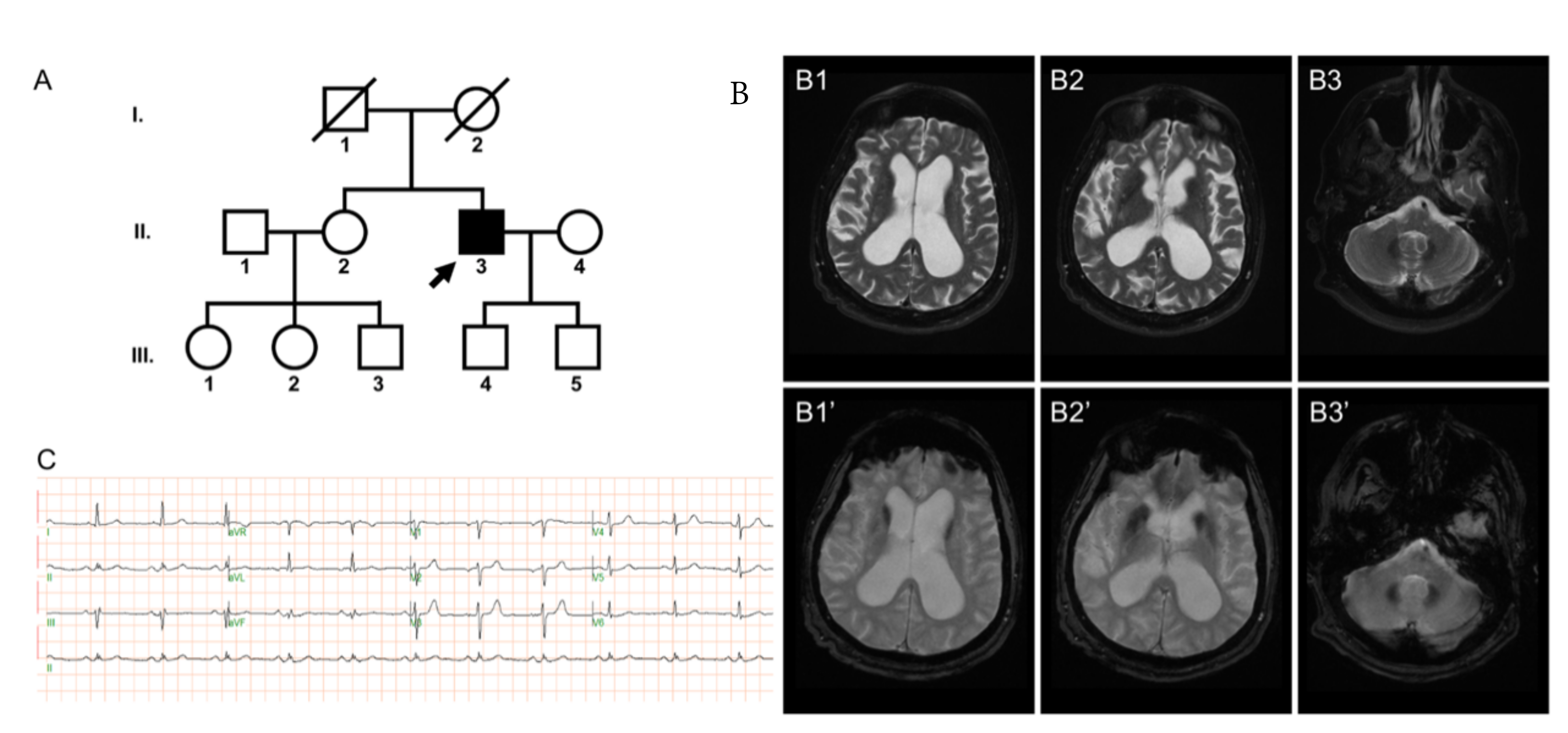
2.1. Case Presentation

The proband is a male with a past medical history of anxiety disorder who first presented to the University of Pennsylvania (UPenn) Movement Disorders Clinic at the age of 69 years with a chief complaint of gait dysfunction. He reported normal development with the exception of mild gait instability with occasional falls while playing sports as a child and throughout adulthood. In his mid-60s, the patient’s balance deteriorated with an increased frequency of falls, and he developed urinary urgency with incontinence, and cognitive changes characterized by poor recall, naming, personality changes, disinhibition, and inappropriate joking. The patient was born in a non-consanguineous pedigree with no reported family history of neurological diseases (Figure 1A). His parents are deceased due to non-neurological causes. No significant neurological deficit was noted for the patient’s sibling and offspring.
The neurological examination revealed cerebellar ataxia and mild parkinsonism, characterized by masked facial expression, perioral dyskinesias, mild intention tremor and mild dysmetria, bilateral limb ataxia with dysdiadochokinesia, lower greater than upper extremity bradykinesia, paratonia, upright rigid posture, wide-based gait with short stride length, and mild tandem gait impairment (Supplementary Video S1; Supplementary Figure S1). He achieved 25/30 points on the Montreal Cognitive Assessment. Neuropsychological evaluation at the age of 69 demonstrated average overall intellectual functioning and verbal skills, with high average perceptual reasoning abilities. Executive dysfunction was also evident with notable impairments in processing speed, complex sequencing, inhibition, novel problem-solving, and conceptual reasoning and perseveration, as well as set loss errors, impulsivity, confabulation, verbosity, tangentiality, conflation, poor organization/planning, and variable self-monitoring. Memory retention was intact.
Prior to presenting to the clinic, a presumptive diagnosis of normal pressure hydrocephalus prompted high-volume cerebrospinal fluid drainage (25 cc, opening pressure 15 cm-H2O) without improvement in gait or memory problems. Serological evaluation for ataxia was unrevealing. MRI of the brain on a 1.5 T scanner demonstrated two main features: (i) diffuse cerebral atrophy with ventriculomegaly and mild white matter disease; (ii) iron deposition in pallidal, caudate, and dentate nuclei (Figure 1B). A standard electrocardiogram (ECG) examination showed normal sinus rhythm (Figure 1C).
Gene panel screening revealed no detectable relevant sequence variant in 17 genes known to be linked to neurodegeneration with brain iron accumulation (ATP13A2, C19orf12, COASY, CP, DCAF17, FA2H, FTL, FUCA1, KIF1A, PANK2, PLA2G6, SCP2, SLC39A14, SQSTM1, TRIM32, VPS13A, EDR45). Subsequent clinical exome sequencing plus mitochondrial sequencing led to the identification of a heterozygous variant in exon 3 of KCND3 (c.1256G>A; p.R419H) (NM_004980.4).

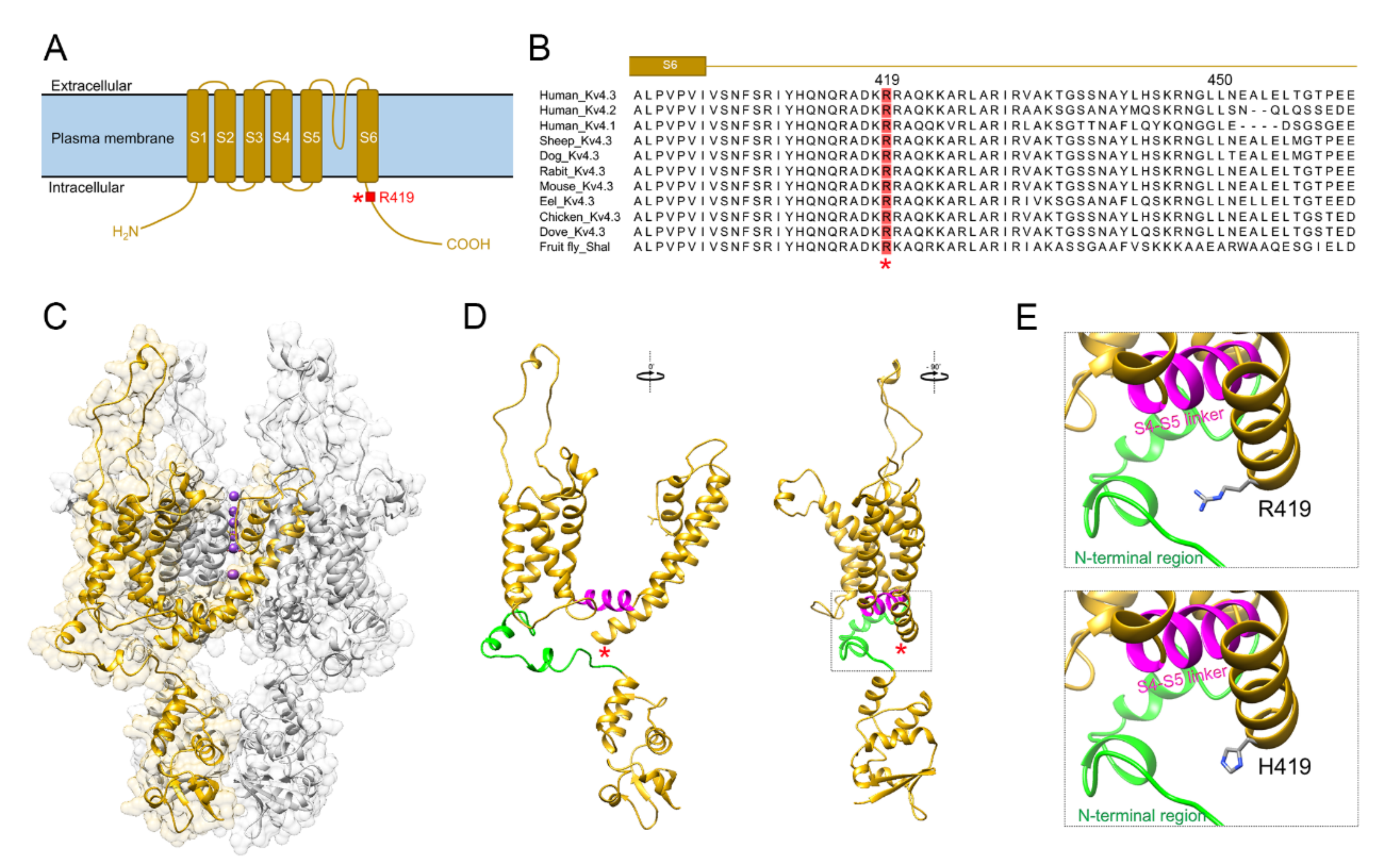
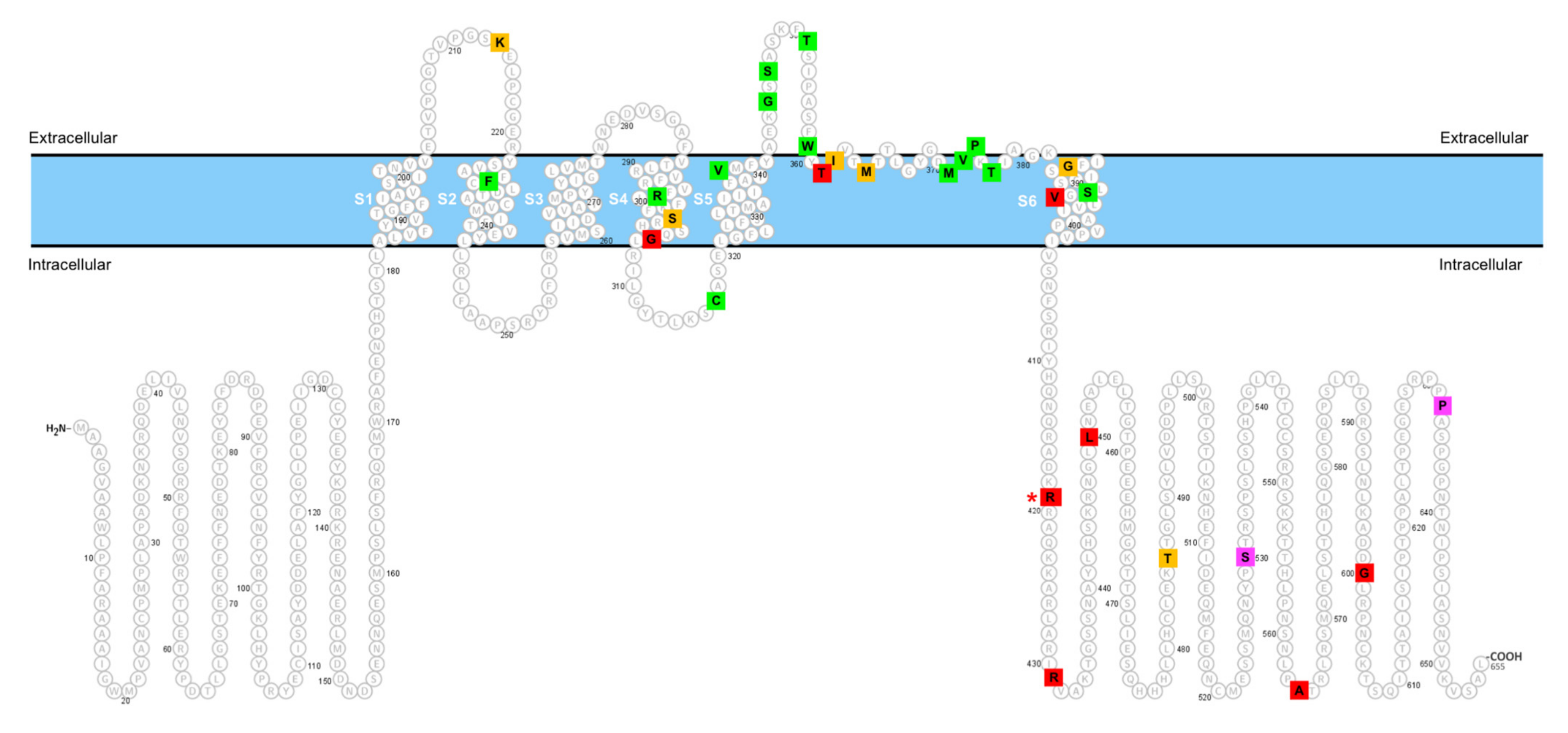
2.2. In Silico Pathogenicity

We employed population databases and bioinformatics analyses to evaluate the pathogenicity of the identified KCND3 variant (Table 1). In both the total and non-Finnish European population in the genome Aggregation Database (gnomAD), the estimated allele frequency of the c.1256G>A (p.R419H) variant is less than 0.0001, suggesting that this is a rare KCND3 variant.
The c.1256G>A (p.R419H) variant was predicted to be damaging/disease-causing based on several in silico prediction tools. The CADD program [23] estimated a Phred score of 28.2, suggesting the variant may be more deleterious than 99.85% of the other variants in the genome. Polyphen-2 predicted a probability score as high as 1, implying that the variant is probably damaging [24]. The programs SNPs&GO [25] and SIFT [26] predicted the variant is disease-related and deleterious, respectively. Moreover, MutationTaster [27] also revealed a high probability score close to 1, consistent with the idea that the rare KCND3 c.1256G>A (p.R419H) variant may be disease-associated.

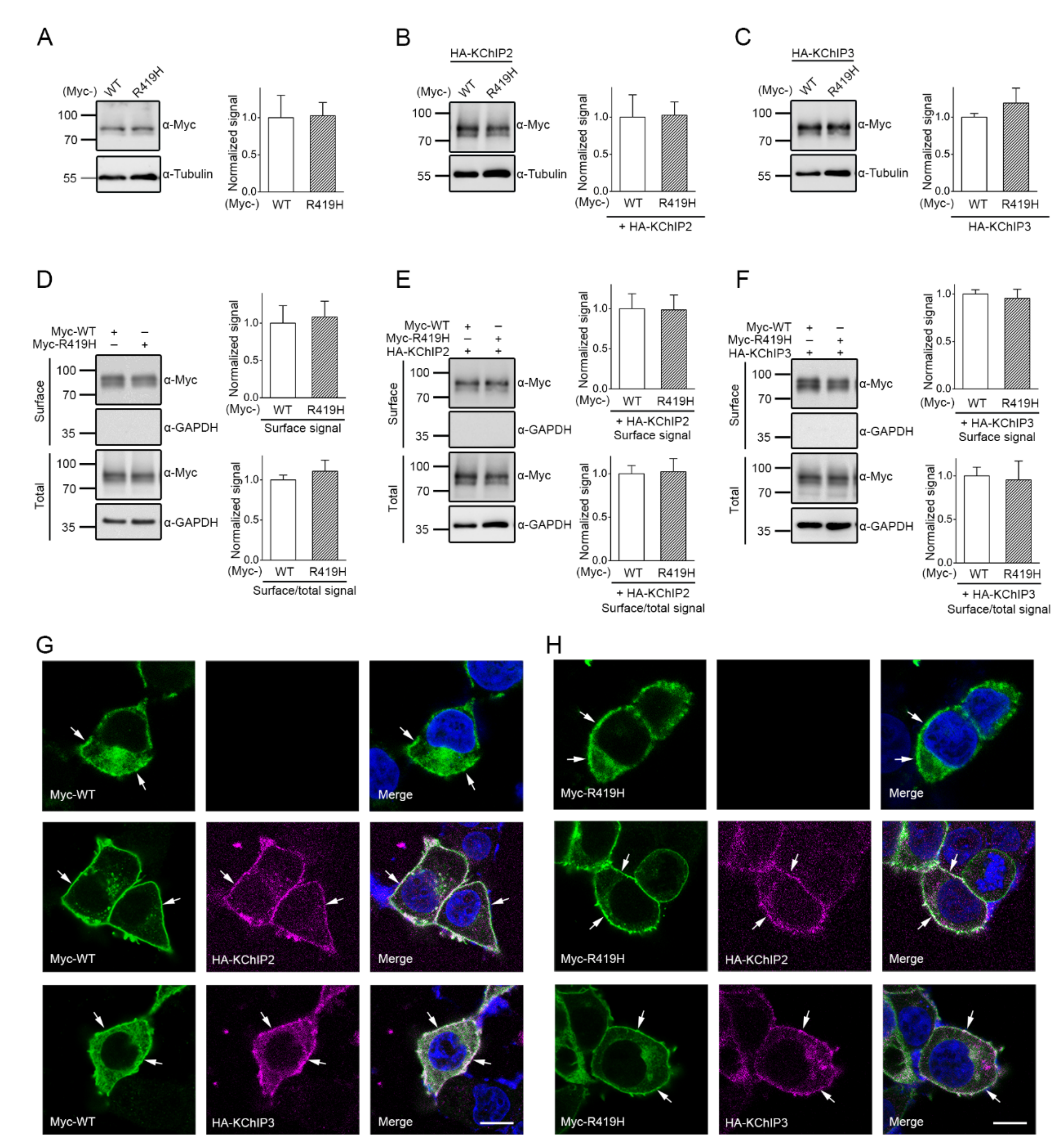
2.3. Lack of Effect of the p.R419H Variant on KV4.3 Protein Expression and Localization

We went on to investigate the potential effect of the p.R419H variant on the in vitro property of human KV4.3 channel. The KV4.3 subunit comprises an intracellular amino-terminal domain, six transmembrane segments (S1–S6) containing a K+-conducting pore loop in the S5–S6 linker region, and a cytoplasmic carboxyl-terminal domain (Figure 2A). The p.R419 residue is localized in the intracellular carboxyl-terminal region, close to the S6 transmembrane segment. p.R419 is a highly conserved residue across various KV4 channel protein orthologs from multiple animal species (Figure 2B), implying an evolutionary importance in the structure and function of KV4.3 channel. Substitution of the positively charged, aliphatic amino acid arginine at residue 419 with the imidazole-containing histidine results in a sizable reduction in the mean side-chain volume by about 17.3% (from 202 Å3 to 167 Å3) (Figure 2C–E).
To assess the pathophysiological significance of the p.R419H variant, we began by determining whether the p.R419H affects protein homeostasis of KV4.3 channels. KV4.3 wild-type (WT) and p.R419H proteins were individually expressed in HEK293T cells. As shown by the immunoblots depicted in Figure 3A–C, regardless of the absence or presence of the auxiliary K+ channel interacting protein 2 and 3 (KChIP2/KChIP3) subunits, no significant difference in the total protein level was observed between KV4.3 WT and the p.R419H variant. Moreover, surface biotinylation analyses demonstrated that KV4.3 WT and the p.R419H variant displayed comparable protein abundance at the plasma membrane in the absence/presence of the auxiliary KChIP2/KChIP3 subunits (Figure 3D–F). Consistent with these biochemical observations, immunofluorescence analyses also indicated that, whether the auxiliary KChIP2/KChIP3 subunits were present or not, the majority of KV4.3 WT and the p.R419H variant exhibited similar punctate staining pattern at the cell surface, consistent with the presence of effective plasma membrane-localization for both proteins (Figure 3G,H). Together, these results suggest that the p.R419H variant does not appear to detectably affect protein expression and subcellular localization of KV4.3 channels.

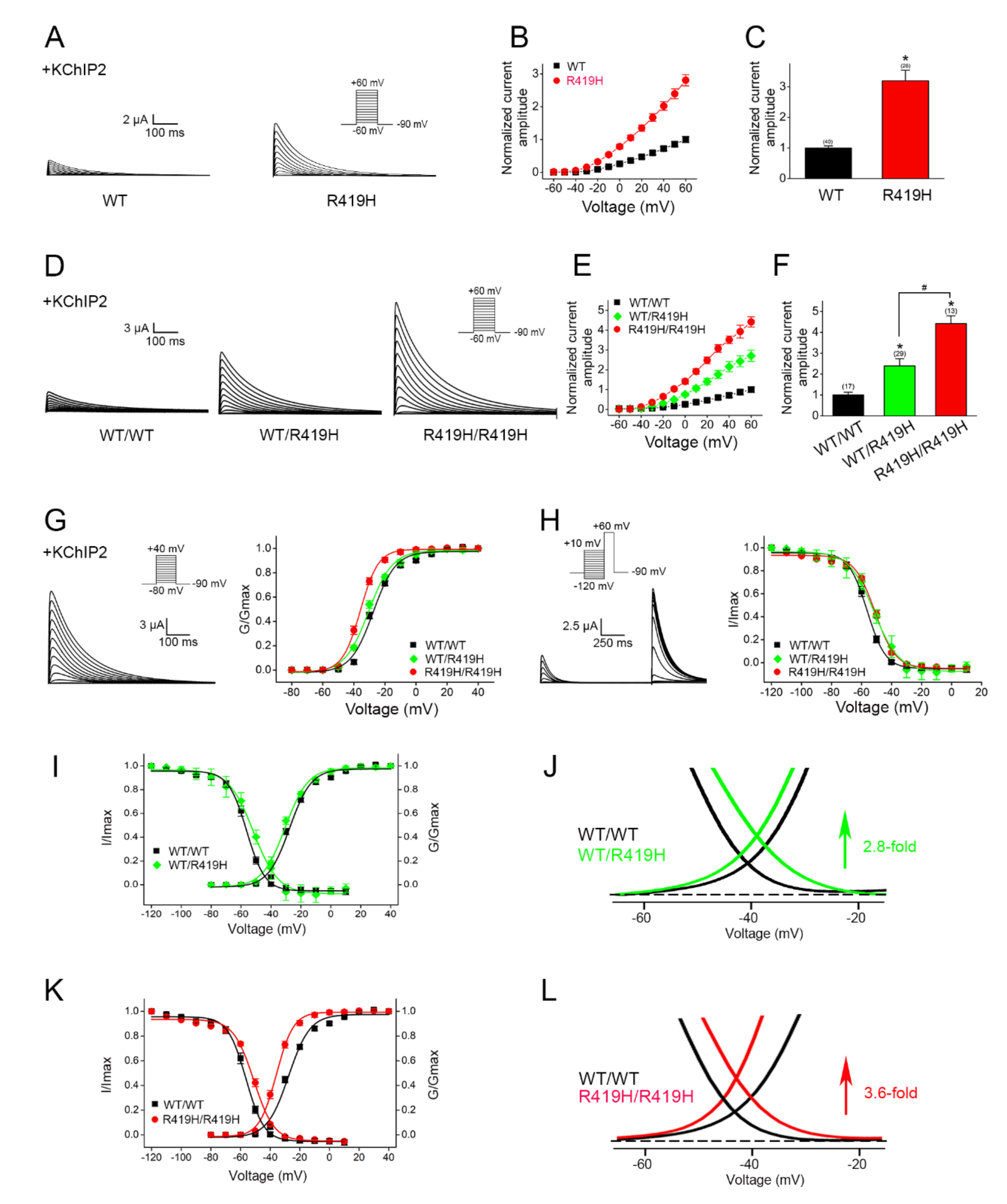
2.4. Dominant Gain-of-Function Effect of the p.R419H Variant on KV4.3 Channel Function

Next, we examined the impact of the p.R419H variant on KV4.3 channel function by performing electrophysiological analyses. Surprisingly, compared to its WT counterpart, KV4.3 channels harboring the p.R149H variant were associated with a more than three-fold increase in the K+ current level (Figure 4A–C), with no apparent change in the channel activation and inactivation kinetics. This result implies that the p.R419H variant may substantially promote KV4.3 channel function.
A functional voltage-gated K+ channel is formed by the assembly of four K+ channel protein subunits (tetramer). Given the fact that the proband carries the heterozygous c.1256G>A (p.R419H) variant in the KCND3 gene and that KV4.3 WT and the p.R419H variant appear to display comparable protein expression levels (Figure 3), it is likely that KV4.3 WT and p.R419H variant subunits may co-assemble and form heterotetrameric KV4.3 channels in native cells in the patient. We therefore asked whether the p.R419H variant may exert a dominant effect on the functional expression of its KV4.3 WT counterpart. To address this important issue, we co-expressed KV4.3 WT and WT (WT/WT homotetramer), WT and p.R419H (WT/R419H heterotetramer), or p.R419H and p.R419H (R419H/R419H homotetramer) in the same cell, followed by comparing their functional properties. As outlined in Figure 4D–F, the mean current amplitude of KV4.3 WT/R419H heterotetramers is more than two-fold lager than that of WT/WT homotetramers; moreover, the K+ current level of R419H/R419H homotetramers is substantially higher than that of WT/R419H heterotetramers. This R419H-dependent, progressive increase in K+ current level strongly argues that, in WT/R419H heterotetramers, the p.R419H variant is associated with a dominant effect on the functional expression of KV4.3 channels.
To further explore the potential mechanism underlying the observed enhanced current amplitude associated with the p.R419H variant, we analyzed the voltage-dependent gaiting property of KV4.3 channels comprising WT/WT homotetramers, WT/R419H heterotetramers, or R419H/R419H homotetramers. As clearly illustrated in Figure 4G,H and Table 2, a notable R419H-dependent modification effect was observed for both steady-state activation (G/Gmax) and inactivation (I/Imax) properties of KV4.3 channels. For example, compared to the WT/WT homotetramer control, the steady-state activation (G/Gmax) curve of the WT/R419H heterotetramer and the R419H/R419H homotetramer was left-shifted by about 2.6 and 8.2 mV, respectively (Figure 4G), implying that increasing the relative proportion of the p.R419H variant in WT/R419H heterotetramers may lead to progressively higher KV4.3 channel open probability at the resting membrane potential. Similarly, the R419H-dependent right-shift of the steady-state KV4.3 channel inactivation (I/Imax) curve (Figure 4H) suggests that increasing the relative proportion of the p.R419H variant may render WT/R419H heterotetramers less likely to be inactivated at the resting membrane potential. Together, these observations are consistent with the idea that, in WT/R419H heterotetramers, the p.R419H variant may exert a dominant effect on the voltage-dependent gating function of KV4.3 WT.
The superimposed region of steady-state activation and inactivation curves is known as the window current (Figure 4I–L), which correlates with the voltage range in which a significant fraction of inactivating ion channels may remain open with minimal inactivation, and therefore provides an estimate of the effective KV4.3 channel conductance under physiological conditions in neurons. In agreement with the foregoing R419H-dependent increase in K+ current amplitudes (Figure 4F), the WT/R419H heterotetramer and the R419H/R419H homotetramer displayed an enhancement of the size of the window current by about 2.8-fold and 3.6-fold, respectively (Figure 4J,L), indicating that the observed K+ current-potentiating effect can be in part attributed to the dominant gain-of-function voltage-dependent properties conferred by the p.R419H variant.

3. Discussion

Based on the standards and guidelines set forth by the American College of Medical Genetics (ACMG) [28], we employed multiple criteria to assess the clinical significance of the heterozygous KCND3 c.1256G>A (p.R419H) variant identified in a patient with slowly progressive cerebellar ataxia, parkinsonism, cognitive dysfunction, and brain ion accumulation. Several lines of evidence support the pathogenicity of this KCND3 variant: (i) this nonsynonymous variant in the KCND3 gene, whose missense variants are frequently linked to cerebellar ataxia, contributes to a low allele frequency (<0.0001) in the population database gnomAD (Table 1) (criterion PP2 in the ACMG guideline); (ii) prediction of deleterious or damaging effects by multiple in silico bioinformatics analyses (Table 1) (criterion PP3); and (iii) the patient’s presentation of specific neurological symptoms relevant to KCND3-mutation-related SCA19/22 (criterion PP4). In addition, we provided the direct in vitro functional evidence (Figure 4) showing that the p.R419H variant is associated with a dominant gain-of-function effect on the K+ current amplitude and voltage-dependent gating of its KV4.3 WT counterpart, a strong indication of the variant’s pathogenicity (criterion PS3). Taken together, we propose that we have identified a “likely pathogenic” gain-of-function KCND3 variant.
Table 3 outlines the genotype–phenotype relationship of known disease-associated KCND3 variants. Patients with KCND3-related neurological disorders are characterized by heterogeneous clinical presentations including cerebellar ataxia, cognitive dysfunction, and movement disorders such as parkinsonism [10]. KCND3-related ataxia is further known to be associated with a wide range of disease onset (from very early ages to later stages of life), as well as distinctly different clinical courses (including episodic, non-progressive, and slowly progressive). Consistent with these notions, in the current study the patient harboring the heterozygous KCND3 p.R419H variant presented with mild gait instability in his childhood and did not display complex neurological features until late adulthood. Moreover, the proband appears to be the only person in his family showing significant neurological disorders (Figure 1A), suggesting that the case is seemingly sporadic. Despite the fact that most KCND3-related disorders are autosomal dominant, de novo mutation and incomplete penentrance have been reported as well [6,9]. Therefore, the presence of four persons carrying the c.1256G>A (p.R419H) variant in gnomAD (Table 1) may additionally imply that this is a rare KCND3 variant with incomplete penetrance.
To date, nearly 30 KCND3 variants have been associated with neurological or cardiac disorders (Table 3). A majority of KCND3 variants linked to neurological (e.g., SCA19/22) and cardiac (e.g., Brugada syndrome) disorders display loss-of-function and gain-of-function phenotypes, respectively, even though some of the cardiopathogenic gain-of-function KCND3 mutations were later associated with neurological pathogenicty as well. The p.R419H variant identified in our ataxic patient exerts a dominant gain-of-function effect on functional K+ current level, which is probably attributed to a notable alteration of the steady-state voltage-dependent activation and inactivation of KV4.3 channels. Moreover, our ECG analysis reveals that the patient carrying the p.R419H variant does not appear to display detectable cardiac arrhythmia. Previously, another gain-of-function KCND3 variant, p.L450F, was originally linked to the Brugada syndrome; however, this variant was later identified in a patient with cerebellar gait ataxia but no significant heart problems [11,14]. As far as cerebellar ataxia is concerned, the aforementioned observations support the idea that functional homeostasis of KV4.3 plays an imperative role in the operation of cerebellar physiology, and that both loss- and gain-of-function phenotypes of KV4.3 variant channels may considerably perturb neuronal excitability in the cerebellar circuit and therefore contribute to the pathogenesis of ataxia. Consistent with these notions, both loss- and gain-of-function variants in the KCNC3 gene encoding another voltage-gated K+ channel (KV3.3) have been associated with spinocerebellar ataxia type 13 [47].
Figure 5 summarizes the topographic localization of currently known disease-associated KCND3 variants within the KV4.3 subunit. Interestingly, virtually all of the loss-of-function variants are located in the transmembrane region of the channel protein. In contrast, many of the gain-of-function variants, including the two ataxia-related gain-of-function variants p.R419H and p.L450F, are found in the cytoplasmic carboxyl-terminal region. It is unclear how the replacement of arginine with histidine at residue 419 may instigate such a significant gain-of-function effect on voltage-dependent gating of KV4.3 channel. The amino acid substitution at this evolutionary conserved KV4.3 residue is unlikely to dramatically affect the secondary protein structure of the proximal carboxyl-terminal region. Nonetheless, our in silico analyses suggest that p.R419 may be in close proximity with two intracellular domains, the S4-S5 linker and the amino-terminal region (Figure 2E), both essential for regulating voltage-dependent gating of K+ channels. Detailed experimental analyses will be required in the future to determine whether the histidine substitution may have a direct impact on the potential interaction between p.R419 and its structural microenvironment in KV4.3 channel.
As depicted in Figure 1B, brain MRI revealed significant iron deposition in bilateral caudate nuclei and lentiform nuclei of the basal ganglia, and in bilateral dentate nuclei of the cerebellum. It is an open question regarding the mechanistic link between KV4.3 current level and iron accumulation in neurons. Neuronal iron overload may result from enhanced postsynaptic iron uptake by the divalent metal transporter 1 (DMT1), and has been suggested to contribute to neurodegenerative diseases such as the Parkinson’s disease [48]. In addition, activation of K+ channels was shown to promote DMT1-mediated iron import into neuroblastoma cells [49]. We therefore speculate that the gain-of-function p.R419H variant may similarly potentiate DMT1-mediated iron uptake in specific regions in the brain. Since the iron-rich dentate nucleus serves as one of the largest deep cerebellar nuclei essential for the output signal from the cerebellum [50], dysregulation of iron homeostasis in bilateral dentate nuclei may lead to substantial anomaly of the cerebellar function. An analogous KV4.3 p.R419H-induced enhancement of iron uptake may also take place in bilateral lentiform and caudate nuclei of the basal ganglia, resulting in the parkinsonism observed in the patient. To the best of our knowledge, the current study provides the first evidence suggesting a potential association between KV4.3 gain-of-function and the susceptibility for brain iron accumulation. Taken as a whole, our findings highlight the wide variation in the phenotypical expression and pathophysiological outcome of disease-associated KCND3 variants.

4. Materials and Methods

4.1. Patient Evaluations and Ethics

This study was supported by the Neurogenetics Translational Center of Excellence, Department of Neurology, UPenn. The patient was evaluated by two neurologists with expertise in movement disorders and neurogenetics at UPenn. Molecular tests, neurocognitive evaluation, and neuroimaging studies were conducted as part of clinical care. Informed consent was provided by the patient.

4.2. Genetic Analyses

Genomic/mitochondrial DNA was extracted from white blood cells in the peripheral venous blood. Screening of a neurodegeneration with brain iron accumulation gene panel was implemented by Associated Regional and University Pathologists (ARUP) laboratories (Salt Lake City, UT, USA). Whole exome sequencing (WES), mitochondrial sequencing, and deletion testing were conducted by the XomeDxPlus test of GeneDx (BioReference Laboratories, Gaithersburg, MD, USA) to detect disease-relevant variant. The targeted exonic regions and flanking splice junctions of the genome were simultaneously sequenced with 100 bp paired-end reads by massively parallel sequencing on an Illumina HiSeq 2000 sequencing system (NextGen Healthcare, Irvine, CA, USA). A customized analysis tool (Xome Analyzer, GeneDx; BioReference Laboratories, Gaithersburg, MD, USA) was utilized to assemble and align the bi-directional sequence to reference genome sequences (GRCh37/UCSC hg19), as well as calling for sequence variants in the regions of interest throughout the genome. The potentially pathogenic variants originally identified were further confirmed by an appropriate method such as capillary sequencing. Sequence alterations were reported based on the Human Genome Variation Society (HGVS) nomenclature guideline. The identified variants were subject to further evaluation by searching in gnomAD and the 1000 Genome Browser (1000 Genomes).

4.3. Bioinformatics Tools

CADD [23], PolyPhen-2 [24], SNPs&GO [25], SIFT [26], and MutationTaster [27] were employed to assess the pathogenicity of the KCND3 variant. Amino acid sequences of KV4.3 homologs and orthologs from multiple species were aligned by using UniProt. UCSF Chimera [51] interfaced with Modeller [52] were applied to generate a KV4.3 homology model based on the crystal structure of the KV1.2 channel (PDB ID: 3LUT) [53].

4.4. cDNA Constructs

Amino-terminal Myc-tagged KV4.3 (Myc-KV4.3) was generated by subcloning human KV4.3 cDNA into the pcDNA3.1-Myc vector (Invitrogen, Carlsbad, CA, USA). The KV4.3 p.R419H variant was created by using the QuikChange Site-Directed Mutagenesis Kit (Stratagene, La Jolla, CA, USA). Amino-terminal HA-tagged KChIP2 and KChIP3 (HA-KChIP2 and HA-KChIP3) were created by subcloning human KChIP2 and KChIP3 cDNAs, respectively, into the pcDNA3-HA vector (Invitrogen, Carlsbad, CA, USA). All the constructs were verified by DNA sequencing. For in vitro transcription, appropriate restriction enzymes were applied to linearize cDNAs, from which capped cRNAs were transcribed using the Ambion mMessage mMachine T7 kit (Thermo Scientific, Waltham, MA, USA).

4.5. Cell Culture and Transfection

Human embryonic kidney (HEK) 293T cells were grown in Gibco Dulbecco’s modified Eagle’s medium (DMEM) (Thermo Scientific, Waltham, MA, USA) with 10% fetal bovine serum (Thermo Scientific, Waltham, MA, USA), 1 mM sodium pyruvate, 100 units/mL HyQ penicillin-streptomycin, and maintained at 37 °C in a humidified incubator with 95% air and 5% CO2. Cells were plated onto six- (for surface biotinylation) or 12-well plates (for total protein), or poly-D-lysine-coated coverslips in 24-well plates (for immunofluorescence) 24 h before transfection. Transient transfection was performed by the calcium phosphate method. Briefly, DNA/calcium phosphate precipitate was prepared by mixing one volume of DNA in 250 mM CaCl2 with an equal-volume 2X HEPES-buffered saline (HBS) [(in mM) 280 NaCl, 50 HEPES, 1.5 Na2HPO4, pH 7.0]. The calcium phosphate precipitate was allowed to form for 20 min in the dark at room temperature prior to being added to the cultures. The quantities of cDNA used for different experiments are as follows: 400 ng/well for immunofluorescence, 800 ng/well for total protein, and 1600 ng/well for surface biotinylation. The DNA/calcium phosphate precipitates were added drop-wise to cells, which were subject to 37 °C incubation for 3–4 h. To terminate the transfection, the mixture solution was replaced with fresh medium pre-warmed in the 37 °C incubator. The cells were returned to the 5% CO2 incubator at 37 °C until further processing. For co-expression experiments, cDNAs for individual KV4.3 and the auxiliary subunit were mixed in equimolar ratio.

4.6. Immunoblotting

Transfected cells were lysed in an ice-cold lysis buffer [(in mM) 150 NaCl, 5 EDTA, 50 Tris-HCl pH 7.6, 1% Triton X-100] containing a complete protease inhibitor cocktail (Roche Applied Science, Basel, Switzerland). After adding the Laemmli sample buffer to the lysates, samples were sonicated on ice (three times for five seconds each) and heated at 70 °C for 5 min for further process. Samples were then separated by 7.5% SDS-PAGE, electrophoretically transferred to nitrocellulose membranes, and detected using mouse anti-Myc (clone 9E10), or rabbit anti-α-tubulin (Bethyal Laboratories, Montgomery, TX, USA) antibodies. Blots were exposed to horseradish-peroxidase-conjugated goat anti-mouse IgG (Jackson ImmunoResearch, West Grove, PA, USA), or goat anti-rabbit IgG (Jackson ImmunoResearch, West Grove, PA, USA), and revealed by an enhanced chemiluminescence detection system (Thermo Scientific, Waltham, MA, USA). Acquisition of chemiluminescent signals from immunoblots was achieved by using the UVP AutoChemi image system (Ultra-Violet Products, Upland, CA, USA). Data shown are representative of at least 3 independent experiments. Densitometric scans of immunoblots were quantified with ImageJ (National Institute of Health, Bethesda, MD, USA).
For surface biotinylation analyses, transfected cells were incubated in 1 mg/mL sulfo-NHS-LC-biotin (Thermo Scientific, Waltham, MA, USA) in ice-cold phosphate-buffered saline (PBS) [(in mM) 136 NaCl, 2.5 KCl, 1.5 KH2PO4, 6.5 Na2HPO4, pH 7.4] with 0.9 mM CaCl2 and 0.5 mM MgCl2 at 4 °C for 1 h on orbital shaker. After the biotin reagents were removed, the cells were rinsed with glycine-containing PBS, followed by once in Tris-buffered saline [(in mM) 20 Tris-HCl, 150 NaCl, pH 7.4] to terminate biotinylation. Cells were solubilized in the lysis buffer. Cell lysates were incubated overnight at 4 °C with streptavidin-agarose beads (Thermo Scientific, Waltham, MA, USA). Beads were washed four times in the lysis buffer, followed by heating in Laemmli sample buffer to elute biotin-streptavidin complexes.

4.7. Immunofluorescence

HEK293T cells were seeded on poly-D-lysine-coated coverslips in 24-well culture dishes. Forty-eight hours after transfection, the coverslips containing HEK293T cells were fixed with 4% paraformaldehyde in PBS at room temperature for 20 min. Cells were permeabilized and blocked with a blocking buffer (5% normal goat serum in 20 mM phosphate buffer, pH 7.4, 0.1% (v/v) Triton X-100, and 450 mM NaCl) for 60 min at 4 °C. Appropriate dilutions of primary antibodies (1:200 for the mouse anti-Myc antibody; 1:200 for the rat anti-HA antibody) were appropriately applied in the blocking buffer overnight at 4 °C. Immunoreactivities were visualized with goat-anti-mouse antibodies conjugated to Alexa Fluor 488 (1:200; Invitrogen, Carlsbad, CA, USA), as well as goat-anti-rat antibodies conjugated to Alexa Fluor 633 (1:200; Invitrogen, Carlsbad, CA, USA) for 1 h at room temperature. Nuclei were labeled with DAPI. Finally, the coverslips were rinsed once in blocking buffer, twice in PBS, and twice in 0.1 M carbonate buffer, pH 9.2, before they were mounted on glass slides in a mounting medium (4% n-propyl gallate, 90% glycerol, 0.1 M carbonate, pH 9.2). A laser-scanning confocal microscope (Leica TCS SP8 STED; Wetzlar, Germany) was utilized to acquire fluorescence images.

4.8. Electrophysiological Analyses

Xenopus laevis oocytes (African Xenopus Facility, Knysna, South Africa) were used for functional studies. All animal procedures were in conformity with the animal protocol approved by the Institutional Animal Care and Use Committee of National Taiwan University. Frogs were anesthetized to dissect ovarian follicles, which were incubated in ND96 [(in mM): 96 NaCl, 2 KCl, 1.8 MgCl2, 1.8 CaCl2, and 5 HEPES, pH 7.2]. Stage V-VI oocytes were selected for cRNA injection, and the cRNAs for individual KV4.3 and KChIP2 were mixed in equimolar ratio. For analyzing KV4.3 WT/mutant heterotetramer, KV4.3 WT, mutant and KChIP2 were mixed in a molar ratio of 1:1:2. Injected oocytes were stored in ND96 at 16 °C for 2–3 days before being used for functional analyses. OC-725C oocyte clamp (Warner Instruments, Hamden, CT, USA) was used to record K+ currents through KV4.3 channels utilizing two-electrode voltage-clamp technology. The recording bath contained Ringer solution [(in mM): 3 KCl, 115 NaCl, 1.8 CaCl2, 10 HEPES, and 0.4 niflumic acid, pH 7.4 with methanesulfonic acid]. Borosilicate electrodes (0.1–1 MΩ) filled with 3 M KCl were used for voltage recording and current injection. Data were acquired and digitized via Digidata 1440A using pCLAMP 10.2 (Molecular Devices, San Jose, CA, USA). Oocytes were held at −90 mV and leak currents arising from passive membrane properties were subtracted by using the −P/4 method provided in the pCLAMP system. Data were obtained, normalized and analyzed as reported previously [54].
To generate peak K+ current–voltage curves for studying steady-state voltage-dependent activation of KV4.3, the voltage protocol comprised 500 ms test pulses stepped from −60 mV to +60 mV, in 10 mV increments. The relative conductance (G/Gmax) at a given test potential was calculated as G/Gmax = ∆IK/∆IK,+60, where ∆IK is current increment determined from peak current difference between adjacent test pulses, and ∆IK,+60 is the ∆IK at the test potential +60 mV. A steady-state voltage-dependent activation curve was then generated by fitting the G/Gmax–voltage (G-V) curve with a Boltzmann equation: G/Gmax = 1/{1 + exp[(V0.5a − V)/ka]}, where V0.5a is the half-activation voltage, and ka is the activation slope factor. To study the steady-state voltage-dependent inactivation of KV4.3, oocytes were subject to 1 s prepulses stepping from −120 to +10 mV with 10 mV increments, followed by a +60 mV test pulse for 500 ms. Normalized peak currents at +60 mV (I/Imax) were then plotted against corresponding prepulse potentials. A steady-state voltage-dependent inactivation curve was then generated by fitting a I/Imax voltage curve with a Boltzmann function: I/Imax = 1/{1 + exp[(V0.5iV)/ki]}, where V0.5i is the half-inactivation voltage, and ki is the inactivation slope factor. Window current analyses were determined from the apex and the area of the triangular overlap area between activation and inactivation curves.

4.9. Statistical Analyses

Statistical analyses were performed with Origin 7.0 (Microcal Software, Northampton, MA, USA). Numerical values were presented as mean ± SEM. The significance of the difference between two means was tested using Student’s t-test.

Supplementary Materials

The following are available online at https://www.mdpi.com/article/10.3390/ijms22158247/s1, Supplementary Video S1. The clinical presentation of the patient harboring KCND3 c.1256G>A (p.R419H) variant in this report. Neurological examination of the 69-year-old gentleman revealed cerebellar ataxia with mild parkinsonism, characterized by masked facial expression, perioral dyskinesias, mild intention tremor, bilateral dysrhythmokinesis, paratonia, lower greater than upper extremity bradykinesia, upright rigid posture, wide-based gait with short stride length, and mild tandem gait impairment. Supplementary Figure S1. Thumbnail image from Supplementary Video S1.

Author Contributions

Conceptualization, P.G.-A., B.-W.S., C.-Y.T. and C.-J.J.; investigation, T.F.T., T.M.B. and P.G.-A.; methodology, C.-T.H., S.-J.F., T.F.T. and T.M.B.; formal analysis, C.-T.H., S.-J.F., T.F.T. and T.M.B.; data curation, C.-T.H., T.F.T., S.-J.F., C.-Y.T. and C.-J.J.; writing—original draft preparation, C.-T.H., T.F.T. and S.-J.F.; writing—review and editing, C.-J.J. and C.-Y.T.; visualization, C.-T.H., T.F.T. and S.-J.F.; funding acquisition, T.F.T., C.-T.H., C.-J.J. and C.-Y.T. All authors have read and agreed to the published version of the manuscript.

Funding

This work was supported by research grants from Ministry of Science and Technology, Taiwan to C.-J.J. (108-2320-B-010-039-MY3), C.-Y.T. (108-2320-B-002-033-MY3), and C.-T.H. (110-2314-B-075-004-MY2), and from Ministry of Education, Taiwan, for The Brain Research Center of National Yang Ming Chiao Tung University on the Higher Education Sprout Project (109BRC-B404) to C.-J.J. T.F.T. is additionally supported by the NIH/NINDS (K23-NS11416-01A1).

Institutional Review Board Statement

All procedures involving animals were performed in conformity with the animal protocol approved by the Institutional Animal Care and Use Committee (IACUC), College of Medicine, National Yang Ming Chiao Tung University (protocol code 1080303; date of approval: 6 March 2019).

Informed Consent Statement

Patient consent was obtained.

Data Availability Statement

All data relevant to the study are included in the article or uploaded as Supplementary Information.

Acknowledgments

We are indebted to the patient who participated in this study.

Conflicts of Interest

The authors declare no conflict of interest.

Abbreviations

ACMGAmerican College of Medical Genetics
DMEMDulbecco’s modified Eagle’s medium
DMT1divalent metal transporter 1
ECG electrocardiogram
gnomADgenome Aggregation Database
HBS HEPES buffered saline
HEK human embryonic kidney
K+potassium
KChIP2/3 K+ channel interacting protein 2 and 3
PBS phosphate-buffered saline
SCA19/22spinocerebellar ataxia type 19 and 22
UPennUniversity of Pennsylvania
WES whole exome sequencing
WT wild-type

References

  1. Hoffman, D.A.; Magee, J.C.; Colbert, C.M.; Johnston, D. K+ channel regulation of signal propagation in dendrites of hippocampal pyramidal neurons. Nature 1997, 387, 869–875. [Google Scholar] [CrossRef]
  2. Nadal, M.S.; Amarillo, Y.; de Miera, E.V.-S.; Rudy, B. Evidence for the presence of a novel Kv4-mediated A-type K(+) channel-modifying factor. J. Physiol. 2001, 537, 801–809. [Google Scholar] [CrossRef] [PubMed]
  3. Wang, Z.; Feng, J.; Shi, H.; Pond, A.; Nerbonne, J.M.; Nattel, S. Potential Molecular Basis of Different Physiological Properties of the Transient Outward K + Current in Rabbit and Human Atrial Myocytes. Circ. Res. 1999, 84, 551–561. [Google Scholar] [CrossRef] [PubMed] [Green Version]
  4. Gaborit, N.; Le Bouter, S.; Szüts, V.; Varro, A.; Escande, D.; Nattel, S.; Demolombe, S. Regional and tissue specific transcript signatures of ion channel genes in the non-diseased human heart. J. Physiol. 2007, 582, 675–693. [Google Scholar] [CrossRef] [PubMed]
  5. Akar, F.G.; Wu, R.C.; Deschenes, I.; Armoundas, A.A.; Piacentino, V., III; Houser, S.R.; Tomaselli, G.F. Phenotypic differences in transient outward K+ current of human and canine ventricular myocytes: Insights into molecular composition of ventricular Ito. Am. J. Physiology. Heart Circ. Physiol. 2004, 286, H602–H609. [Google Scholar] [CrossRef] [PubMed] [Green Version]
  6. Lee, Y.-C.; Durr, A.; Majczenko, K.; Huang, Y.-H.; Liu, Y.-C.; Lien, C.-C.; Tsai, P.-C.; Ichikawa, Y.; Goto, J.; Monin, M.-L.; et al. Mutations inKCND3cause spinocerebellar ataxia type 22. Ann. Neurol. 2012, 72, 859–869. [Google Scholar] [CrossRef] [Green Version]
  7. Duarri, A.; Jezierska, J.; Fokkens, M.; Meijer, M.; Schelhaas, H.J.; den Dunnen, W.F.; van Dijk, F.; Verschuuren-Bemelmans, C.; Hageman, G.; van de Vlies, P.; et al. Mutations in potassium channel kcnd3 cause spinocerebellar ataxia type 19. Ann. Neurol. 2012, 72, 870–880. [Google Scholar] [CrossRef] [Green Version]
  8. Duarri, A.; Lin, M.C.; Fokkens, M.R.; Meijer, M.; Smeets, C.J.; Nibbeling, E.A.; Boddeke, E.; Sinke, R.J.; Kampinga, H.H.; Papazian, D.M.; et al. Spinocerebellar ataxia type 19/22 mutations alter heterocomplex Kv4.3 channel function and gating in a dominant manner. Cell. Mol. Life Sci. 2015, 72, 3387–3399. [Google Scholar] [CrossRef] [Green Version]
  9. Hsiao, C.T.; Fu, S.J.; Liu, Y.T.; Lu, Y.H.; Zhong, C.Y.; Tang, C.Y.; Soong, B.W.; Jeng, C.J. Novel SCA19/22-associated KCND3 mutations disrupt human K(V) 4.3 protein biosynthesis and channel gating. Hum. Mutat. 2019, 40, 2088–2107. [Google Scholar] [CrossRef] [PubMed]
  10. Pollini, L.; Galosi, S.; Tolve, M.; Caputi, C.; Carducci, C.; Angeloni, A.; Leuzzi, V. KCND3-Related Neurological Disorders: From Old to Emerging Clinical Phenotypes. Int. J. Mol. Sci. 2020, 21, 5802. [Google Scholar] [CrossRef]
  11. Giudicessi, J.R.; Ye, D.; Tester, D.J.; Crotti, L.; Mugione, A.; Nesterenko, V.V.; Albertson, R.M.; Antzelevitch, C.; Schwartz, P.J.; Ackerman, M.J. Transient outward current (Ito) gain-of-function mutations in the KCND3-encoded Kv4.3 potassium channel and Brugada syndrome. Heart Rhythm. 2011, 8, 1024–1032. [Google Scholar] [CrossRef] [PubMed] [Green Version]
  12. Nakajima, T.; Kawabata-Iwakawa, R.; Kaneko, Y.; Hamano, S.-I.; Sano, R.; Tamura, S.; Hasegawa, H.; Kobari, T.; Kominato, Y.; Nishiyama, M.; et al. Novel Cardiocerebral Channelopathy Associated with a KCND3 V392I Mutation. Int. Heart J. 2020, 61, 1049–1055. [Google Scholar] [CrossRef]
  13. Takayama, K.; Ohno, S.; Ding, W.-G.; Ashihara, T.; Fukumoto, D.; Wada, Y.; Makiyama, T.; Kise, H.; Hoshiai, M.; Matsuura, H.; et al. A de novo gain-of-function KCND3 mutation in early repolarization syndrome. Heart Rhythm. 2019, 16, 1698–1706. [Google Scholar] [CrossRef] [PubMed]
  14. Duarri, A.; Nibbeling, E.; Fokkens, M.R.; Meijer, M.; Boddeke, E.; Lagrange, E.; Stevanin, G.; Brice, A.; Durr, A.; Verbeek, D.S. The L450F [Corrected] mutation in KCND3 brings spinocerebellar ataxia and Brugada syndrome closer together. Neurogenetics 2013, 14, 257–258. [Google Scholar] [CrossRef]
  15. Ward, R.J.; Zucca, F.A.; Duyn, J.H.; Crichton, R.R.; Zecca, L. The role of iron in brain ageing and neurodegenerative disorders. Lancet Neurol. 2014, 13, 1045–1060. [Google Scholar] [CrossRef] [Green Version]
  16. Oakley, A.E.; Collingwood, J.; Dobson, J.; Love, G.; Perrott, H.R.; Edwardson, J.A.; Elstner, M.; Morris, C. Individual dopaminergic neurons show raised iron levels in Parkinson disease. Neurology 2007, 68, 1820–1825. [Google Scholar] [CrossRef]
  17. Tao, Y.; Wang, Y.; Rogers, J.T.; Wang, F. Perturbed Iron Distribution in Alzheimer’s Disease Serum, Cerebrospinal Fluid, and Selected Brain Regions: A Systematic Review and Meta-Analysis. J. Alzheimer’s Dis. 2014, 42, 679–690. [Google Scholar] [CrossRef]
  18. Kruer, M.; Boddaert, N.; Schneider, S.; Houlden, H.; Bhatia, K.; Gregory, A.; Anderson, J.; Rooney, W.; Hogarth, P.; Hayflick, S. Neuroimaging Features of Neurodegeneration with Brain Iron Accumulation. Am. J. Neuroradiol. 2011, 33, 407–414. [Google Scholar] [CrossRef] [Green Version]
  19. Levi, S.; Tiranti, V. Neurodegeneration with Brain Iron Accumulation Disorders: Valuable Models Aimed at Understanding the Pathogenesis of Iron Deposition. Pharmaceuticals 2019, 12, 27. [Google Scholar] [CrossRef] [PubMed] [Green Version]
  20. Claassen, J.; Gerding, W.M.; Kastrup, O.; Uslar, E.; Goericke, S.; Timmann, D. Excessive brain iron accumulation in spinocerebellar ataxia type 17. Neurology 2015, 84, 212–213. [Google Scholar] [CrossRef]
  21. Llorens, J.V.; Soriano, S.; Calap-Quintana, P.; Gonzalez-Cabo, P.; Moltó, M.D. The Role of Iron in Friedreich’s Ataxia: Insights From Studies in Human Tissues and Cellular and Animal Models. Front. Neurosci. 2019, 13, 75. [Google Scholar] [CrossRef] [Green Version]
  22. Bs, J.A.; Rogers, H.; Bs, A.H.; Snell, M.; Dill, M.; Judd, D.; Hagerman, P.; Martínez-Cerdeño, V. Iron accumulation and dysregulation in the putamen in fragile X-associated tremor/ataxia syndrome. Mov. Disord. 2017, 32, 585–591. [Google Scholar]
  23. Rentzsch, P.; Witten, D.; Cooper, G.M.; Shendure, J.; Kircher, M. CADD: Predicting the deleteriousness of variants throughout the human genome. Nucleic Acids Res. 2019, 47, D886–D894. [Google Scholar] [CrossRef]
  24. Adzhubei, I.A.; Schmidt, S.; Peshkin, L.; Ramensky, V.E.; Gerasimova, A.; Bork, P.; Kondrashov, A.S.; Sunyaev, S.R. A method and server for predicting damaging missense mutations. Nat. Methods 2010, 7, 248–249. [Google Scholar] [CrossRef] [Green Version]
  25. Capriotti, E.; Calabrese, R.; Fariselli, P.; Martelli, P.L.; Altman, R.B.; Casadio, R. WS-SNPs&GO: A web server for predicting the deleterious effect of human protein variants using functional annotation. BMC Genom. 2013, 14, S6. [Google Scholar]
  26. Kumar, P.; Henikoff, S.; Ng, P.C. Predicting the effects of coding non-synonymous variants on protein function using the SIFT algorithm. Nat. Protoc. 2009, 4, 1073–1081. [Google Scholar] [CrossRef]
  27. Schwarz, J.M.; Cooper, D.N.; Schuelke, M.; Seelow, D. MutationTaster2: Mutation prediction for the deep-sequencing age. Nat. Methods 2014, 11, 361–362. [Google Scholar] [CrossRef]
  28. Richards, S.; Aziz, N.; Bale, S.; Bick, D.; Das, S.; Gastier-Foster, J.; Grody, W.W.; Hegde, M.; Lyon, E.; Spector, E.; et al. Standards and guidelines for the interpretation of sequence variants: A joint consensus recommendation of the American College of Medical Genetics and Genomics and the Association for Molecular Pathology. Genet. Med. 2015, 17, 405–423. [Google Scholar] [CrossRef]
  29. Coutelier, M.; Hammer, M.B.; Stevanin, G.; Monin, M.-L.; Davoine, C.-S.; Mochel, F.; Labauge, P.; Ewenczyk, C.; Ding, J.; Gibbs, J.R.; et al. Efficacy of Exome-Targeted Capture Sequencing to Detect Mutations in Known Cerebellar Ataxia Genes. JAMA Neurol. 2018, 75, 591–599. [Google Scholar] [CrossRef]
  30. Huin, V.; Strubi-Vuillaume, I.; Dujardin, K.; Brion, M.; Delliaux, M.; Dellacherie, D.; Cuvellier, J.C.; Cuisset, J.M.; Riquet, A.; Moreau, C.; et al. Expanding the phenotype of SCA19/22: Parkinsonism, cognitive impairment and epilepsy. Parkinsonism Relat. Disord. 2017, 45, 85–89. [Google Scholar] [CrossRef]
  31. Chung, M.-Y.; Lu, Y.-C.; Cheng, N.-C.; Soong, B.-W. A novel autosomal dominant spinocerebellar ataxia (SCA22) linked to chromosome 1p21-q23. Brain 2003, 126, 1293–1299. [Google Scholar] [CrossRef] [Green Version]
  32. Smets, K.; Duarri, A.; Deconinck, T.; Ceulemans, B.; Van De Warrenburg, B.P.; Züchner, S.; Gonzalez, M.A.; Schüle, R.; Synofzik, M.; Van Der Aa, N.; et al. First de novo KCND3 mutation causes severe Kv4.3 channel dysfunction leading to early onset cerebellar ataxia, intellectual disability, oral apraxia and epilepsy. BMC Med. Genet. 2015, 16, 51. [Google Scholar] [CrossRef] [Green Version]
  33. Zanni, G.; Hsiao, C.T.; Fu, S.J.; Tang, C.Y.; Capuano, A.; Bosco, L.; Graziola, F.; Bellacchio, E.; Servidei, S.; Primiano, G.; et al. Novel KCND3 Variant Underlying Nonprogressive Congenital Ataxia or SCA19/22 Disrupt K(V)4.3 Protein Expression and K+ Currents with Variable Effects on Channel Properties. Int. J. Mol. Sci. 2021, 22, 4986. [Google Scholar] [CrossRef]
  34. Schelhaas, H.; Ippel, P.; Hageman, G.; Sinke, R.; Van Der Laan, E.; Beemer, F. Clinical and genetic analysis of a four-generation family with a distinct autosomal dominant cerebellar ataxia. J. Neurol. 2001, 248, 113–120. [Google Scholar] [CrossRef] [PubMed]
  35. Huang, Y.; Yang, J.; Xie, W.; Li, Q.; Zeng, Z.; Sui, H.; Shan, Z.; Huang, Z. A novel KCND3 mutation associated with early-onset lone atrial fibrillation. Oncotarget 2017, 8, 115503–115512. [Google Scholar] [CrossRef] [PubMed] [Green Version]
  36. Coutelier, M.; Coarelli, G.; Monin, M.-L.; Konop, J.; Davoine, C.-S.; Tesson, C.; Valter, R.; Anheim, M.; Béhin, A.; Castelnovo, G.; et al. A panel study on patients with dominant cerebellar ataxia highlights the frequency of channelopathies. Brain 2017, 140, 1579–1594. [Google Scholar] [CrossRef]
  37. Paucar, M.; Ågren, R.; Li, T.; Lissmats, S.; Bergendal, Å.; Weinberg, J.; Nilsson, D.; Savichetva, I.; Sahlholm, K.; Nilsson, J.; et al. V374A KCND3 Pathogenic Variant Associated With Paroxysmal Ataxia Exacerbations. Neurol. Genet. 2021, 7, e546. [Google Scholar] [CrossRef]
  38. Paucar, M.; Bergendal, Å.; Gustavsson, P.; Nordenskjöld, M.; Laffita-Mesa, J.; Savitcheva, I.; Svenningsson, P. Novel Features and Abnormal Pattern of Cerebral Glucose Metabolism in Spinocerebellar Ataxia 19. Cerebellum 2018, 17, 465–476. [Google Scholar] [CrossRef] [Green Version]
  39. Iqbal, Z.; Rydning, S.L.; Wedding, I.M.; Koht, J.; Pihlstrøm, L.; Rengmark, A.H.; Henriksen, S.P.; Tallaksen, C.M.; Toft, M. Targeted high throughput sequencing in hereditary ataxia and spastic paraplegia. PLoS ONE 2017, 12, e0174667. [Google Scholar]
  40. Kurihara, M.; Ishiura, H.; Sasaki, T.; Otsuka, J.; Hayashi, T.; Terao, Y.; Matsukawa, T.; Mitsui, J.; Kaneko, J.; Nishiyama, K.; et al. Novel De Novo KCND3 Mutation in a Japanese Patient with Intellectual Disability, Cerebellar Ataxia, Myoclonus, and Dystonia. Cerebellum 2017, 17, 237–242. [Google Scholar] [CrossRef]
  41. Wang, Y.; Koh, K.; Namekawa, M.; Takiyama, Y. Whole-exome sequencing reveals a missense mutation in theKCND3gene in a patient with SCA19/22. Neurol. Clin. Neurosci. 2015, 3, 197–199. [Google Scholar] [CrossRef]
  42. Giudicessi, J.R.; Ye, D.; Kritzberger, C.J.; Nesterenko, V.V.; Tester, D.J.; Antzelevitch, C.; Ackerman, M.J. Novel mutations in the KCND3-encoded Kv4.3 K+ channel associated with autopsy-negative sudden unexplained death. Hum. Mutat. 2012, 33, 989–997. [Google Scholar] [CrossRef] [PubMed] [Green Version]
  43. Wang, J.; Wen, Y.; Zhang, Q.; Yu, S.; Chen, Y.; Wu, X.; Zhang, Y.; Bao, X. Gene mutational analysis in a cohort of Chinese children with unexplained epilepsy: Identification of a new KCND3 phenotype and novel genes causing Dravet syndrome. Seizure 2019, 66, 26–30. [Google Scholar] [CrossRef] [Green Version]
  44. Li, X.; Li, Z.; Wang, D.W.W.; Wang, Y. A Novel Gain-of-Function KCND3 Variant Associated with Brugada Syndrome. Cardiology 2020, 145, 623–632. [Google Scholar] [CrossRef]
  45. Choi, K.-D.; Kim, J.-S.; Kim, H.-J.; Jung, I.; Jeong, S.-H.; Lee, S.-H.; Kim, D.U.; Kim, S.-H.; Choi, S.Y.; Shin, J.-H.; et al. Genetic Variants Associated with Episodic Ataxia in Korea. Sci. Rep. 2017, 7, 1–11. [Google Scholar] [CrossRef] [Green Version]
  46. Olesen, M.S.; Refsgaard, L.; Holst, A.G.; Larsen, A.P.; Grubb, S.; Haunsø, S.; Svendsen, J.H.; Olesen, S.-P.; Schmitt, N.; Calloe, K. A novel KCND3 gain-of-function mutation associated with early-onset of persistent lone atrial fibrillation. Cardiovasc. Res. 2013, 98, 488–495. [Google Scholar] [CrossRef]
  47. Minassian, N.A.; Lin, M.-C.A.; Papazian, D.M. Altered Kv3.3 channel gating in early-onset spinocerebellar ataxia type 13. J. Physiol. 2012, 590, 1599–1614. [Google Scholar] [CrossRef]
  48. Xu, H.; Jiang, H.; Xie, J. New Insights into the Crosstalk between NMDARs and Iron: Implications for Understanding Pathology of Neurological Diseases. Front. Mol. Neurosci. 2017, 10, 71. [Google Scholar] [CrossRef] [Green Version]
  49. Du, X.; Xu, H.; Shi, L.; Jiang, Z.; Song, N.; Jiang, H.; Xie, J. Activation of ATP-sensitive potassium channels enhances DMT1-mediated iron uptake in SK-N-SH cells in vitro. Sci. Rep. 2016, 6, 33674. [Google Scholar] [CrossRef]
  50. Bond, K.; Brinjikji, W.; Eckel, L.; Kallmes, D.; McDonald, R.; Carr, C. Dentate Update: Imaging Features of Entities That Affect the Dentate Nucleus. Am. J. Neuroradiol. 2017, 38, 1467–1474. [Google Scholar] [CrossRef] [Green Version]
  51. Pettersen, E.F.; Goddard, T.D.; Huang, C.C.; Couch, G.S.; Greenblatt, D.M.; Meng, E.C.; Ferrin, T.E. UCSF Chimera—A visualization system for exploratory research and analysis. J. Comput. Chem. 2004, 25, 1605–1612. [Google Scholar] [CrossRef] [Green Version]
  52. Webb, B.; Sali, A. Comparative Protein Structure Modeling Using MODELLER. Curr. Protoc. Bioinform. 2016, 54, 5.6.1–5.6.37. [Google Scholar] [CrossRef] [PubMed] [Green Version]
  53. Chen, X.; Wang, Q.; Ni, F.; Ma, J. Structure of the full-length Shaker potassium channel Kv1.2 by normal-mode-based X-ray crystallographic refinement. Proc. Natl. Acad. Sci. USA 2010, 107, 11352–11357. [Google Scholar] [CrossRef] [PubMed] [Green Version]
  54. Jeng, C.J.; Chen, Y.T.; Chen, Y.W.; Tang, C.Y. Dominant-negative effects of human P/Q-type Ca2+ channel mutations associated with episodic ataxia type 2. Am. J. Physiology. Cell Physiol. 2006, 290, C1209–C1220. [Google Scholar] [CrossRef] [Green Version]
Figure 1. Clinical features of the patient with the KCND3 c.1256G>A (p.R419H) variant. (A) Pedigree of the patient characterized in current study. In this pedigree, the heterozygous KCND3 variant was detected in the male individual II.3, who is now 70 years old. The patient’s father (I.1) passed away in his seventies due to heart failure, and his mother (I.2) in her eighties due to chronic obstructive pulmonary disease. He has a sister (II.2) who is five years older than him. His two sons (III.4 and III.5) are now in their thirties. Arrow denotes the index case who harbors the KCND3 c.1256G>A (p.R419H) variant. Filled symbol represents symptomatic member. Open symbols indicate unaffected individuals. Circles stand for females. Squares correspond to males. Diagonal lines refer to the deceased. (B) Neuroimages of the patient. The axial T2-weighted (B1B3) and the corresponding gradient-echo sequence (B1′B3′) images demonstrate hypointensity in bilateral caudate nuclei (B1,B1′) and lentiform nuclei (B2,B2′) of the basal ganglia, as well as in bilateral dentate nuclei of the cerebellum (B3,B3′). (C) Standard 12-lead ECG indicates normal sinus rhythm.
Ijms 22 08247 g001
Figure 2. Protein structure modeling of human KV4.3 WT and the p.R419H variant. (A) Schematic representation of the membrane topology of a single KV4.3 subunit, highlighting the cytoplasmic localization of the residue p.R419 in the proximal carboxyl-terminal region close to the S6 transmembrane segment. (B) Amino acid sequence alignment of human KV4.3 with various KV4-related proteins from multiple animal species. The highly conserved p.R419 residue is decorated in red and marked with an asterisk. (CE) Homology model of KV4.3 was based on the crystal structure of the voltage-dependent K+ channel KV1.2 (PBD ID: 3LUT) and generated using UCSF Chimera interfaced with Modeller. (C) Tetrameric structure of the KV4.3 channel. One KV4.3 monomer is displayed as ribbon in gold, with the other subunits in gray. The shadow refers to the surface representation of the homology model. Purple spheres denote K+ passing through the pore region. (D) (Left panel) The single KV4.3 subunit in gold in (C) is shown here. (Right panel) The same KV4.3 subunit is viewed with 90-degree rotation clockwise. The red asterisk highlights the location of the residue p.R419. The S4-S5 linker is shown as magenta helix, and a portion of the amino (N)-terminal region is colored in green. The area enclosed by dotted lines in the right panel is enlarged for inspection in (E). (E) Comparison of the side-chain structure of the p.R419 WT and the p.H419 variant with respect to the local microenvironment.
Ijms 22 08247 g002
Figure 3. Biochemical and immunofluorescence characterizations of human KV4.3 WT and the p.R419H variant. Myc-KV4.3 subunits were expressed in HEK293T cells in the absence or presence of auxiliary HA-KChIP2 and -KChIP3 subunits. (AC) (Left panels) Representative immunoblots. Proteins were detected with anti-Myc (α-Myc) and anti-tubulin (α-Tubulin) antibodies. Molecular weight markers (in kDa) are labeled to the left. (Right panels) Quantitative analyses indicating comparable protein level of the WT and the p.R419H KV4.3. Total KV4.3 protein density was standardized with cognate tubulin level, followed by normalization with respect to the WT (n = 4–6). (DF) Surface biotinylation analyses for Myc-KV4.3 subunits in the absence (D) or presence of auxiliary HA-KChIP2 (E) and -KChIP3 (F) subunits. Representative immunoblots and quantitative analyses for the surface signals are shown at the left and right panels, respectively (n = 4). The surface/total ratio corresponds to membrane trafficking efficiency. (G,H) Representative immunofluorescence images demonstrating similar subcellular localization pattern of KV4.3 WT and p.R419H (green) in the absence (top panels) or presence of KChIP2 (middle panels) and KChIP3 (bottom panels) (magenta). Cell nuclei were counterstained with DAPI (blue). Arrows denote membrane localization. Merged images are shown in the right panels. Scale bar: 10 μm.
Ijms 22 08247 g003
Figure 4. Characterization of the ion channel function of human KV4.3 WT and the p.R419H variant. KV4.3 was co-expressed with the auxiliary KChIP2 subunit in the 1:1 molar ratio in Xenopus oocytes. (AC) Compared to KV4.3 WT, the p.R419H variant is associated with a significantly higher functional expression level. (A) Representative K+ current traces recorded from homotetrameric KV4.3 WT and p.R419H channels. Current traces were induced by a voltage protocol comprising test potentials ranging from –60 mV to +60 mV in 10-mV steps. (B) Normalized peak current amplitudes were plotted against matching test pulse potentials. For each current trace induced by a test pulse potential, the peak current amplitude was measured, followed by normalization with respect to the corresponding mean peak current amplitude at +60 mV of KV4.3 WT. (C) Statistical comparison of the normalized peak current amplitude at +60 mV. Mean normalized peak current amplitude at +60 mV: WT, 1 ± 0.1; p.R419H, 3.2 ± 0.3. Asterisks denote significant difference from the WT control (p < 0.05). Numbers in parentheses refer to the amount of cells analyzed for each KV4.3 construct. (DF) The p.R419H variant exerts a dominant effect on the functional expression of KV4.3 channels. (D) Representative current traces recorded from KV4.3 WT/WT homotetramers, WT/R419H heterotetramers, and R419H/R419H homotetramers. (E) Normalized peak current-voltage plot. (F) Statistical comparison. Mean normalized peak current amplitude at +60 mV: WT/WT, 1 ± 0.1; WT/R419H, 2.4 ± 0.3; R419H/R419H, 4.4 ± 0.4. Asterisks denote significant difference from the WT/WT control (p < 0.05). The pound sign (#) indicates significant difference from the WT/R419H heterotetramer (p < 0.05). Numbers in parentheses refer to the amount of cells analyzed for each KV4.3 expression condition. (G,H) The p.R419H variant confers a substantial alteration in the voltage-dependent gating property of KV4.3 channels. (Left panels) Representative K+ current traces in response to the test pulse protocol for assessing voltage-dependent activation (G) and inactivation (H) of KV4.3 channels. (Right panels) Comparison of the steady-state voltage-dependent activation (G) and inactivation (H) curves of KV4.3 WT/WT homotetramers, WT/R419H heterotetramers, and R419H/R419H homotetramers. See Table 2 for detailed voltage-dependent gating parameters. (IL) The p.R419H variant confers a prominent increase in KV4.3 window current. (I,K) Combination of the steady-state activation (G) and inactivation (H) curves of the indicated KV4.3 homotetramers and heterotetramers. (J,L) The triangular area underneath the overlap of corresponding activation and inactivation curves defines the size of the window current. Both the WT/R419H heterotetramer and the R419H/R419H homotetramer are associated with a significant increase in the window current size of KV4.3 channels.
Ijms 22 08247 g004
Figure 5. Topographic representation of disease-associated KCND3 variants in the KV4.3 subunit. Green and red symbols represent KV4.3 variants with loss-of-function and gain-of-function phenotypes, respectively. Magenta symbols indicate KV4.3 variants with no significant alteration of functional properties. Yellow symbols refer to KV4.3 variants that have yet to be functionally characterized. The red asterisk denotes the p.R419H variant studied in the current report. See Table 3 for genotype–phenotype relationship.
Ijms 22 08247 g005
Table 1. Bioinformatics analyses of the KCND3 variant.
Variant at Chromosome Position: 1:112329579 C>T (GRCh37)
Substitution: c.1256G>A; p.R419H (Reference Sequence: NM_004980.4)
ToolsResultsInterpretation
gnomADVariant found in 4/251,408Allele frequency: 1.591 × 10⁻⁵
1000 GenomesNot identifiedAbsent in 1000 Genomes database
§ CADDPhred score: 28.2Top 0.15% most deleterious variant
PolyPhen-2Score: 1Probably damaging
†† SNPs&GOReliability Index: 6Disease-related
‡‡ SIFTScore: 0.01Deleterious (<0.05 deleterious)
§§ MutationTasterProb: 0.999999708078826Disease causing
Remarks: The KCND3 c.1256G>A (p.R419H) variant was found in 4 out of 251,408 sequenced alleles (total population), equivalent to an estimated allele frequency of 1.591 × 10⁻⁵. For non-Finnish European population, the estimated allele frequency is 3.519 × 10⁻⁵. The KCND3 variant was not found in 1000 genomes, in which a part of sequence data was not included in gnomAD. § The CADD Phred score is 28.2, suggesting that this KCND3 variant is among the top 0.15% most deleterious variants of the human genome. The polyphen-2 score (ranging from 0 to 1) for the KCND3 variant was calculated as 1.000, the highest score, suggestive of it probably being damaging. †† The reliability index (ranging from 0 to 10) for the KCND3 variant was 6, indicative of being disease-related with a high predicting confidence. ‡‡ The SIFT score for the KCND3 variant was predicted to be less than 0.05, signifying it being notably damaging. §§ This high probability score (close to 1) predicts that the KCND3 variant is likely disease causing.
Table 2. Comparison of the KV4.3 voltage-dependent gating parameters of the wild type (WT) and the p.R419H variant. Steady-state activation and inactivation curves were generated from the averages of 9–12 cells expressing the indicated KV4.3 constructs. Data were subject to fitting with the Boltzmann equation as described in the Materials and Methods section and depicted in Figure 4. V0.5a: half-activation voltage. ka: activation slope factor. V0.5i: half-inactivation voltage. ki: inactivation slope factor.
(+KChIP2)ActivationInactivation
V0.5a (mV)kaV0.5i (mV)ki
WT−27.67.2−56.36.1
WT/p.R419H−30.27.6−51.78.3
p.R419H−35.85.7−51.16.8
Table 3. The genotype–phenotype relationship for KCND3 variants associated with neurological or cardiac disorders. Notation for in vitro functional phenotype: GOF, gain of function; LOF, loss of function; NSFC, no significant function change; n.a., not available. See Figure 5 for topographic representation.
KCND3 VariantClinical FeatureRemarkIn Vitro FunctionReference
p.K214REpisodic gait disorder, vertigo, paraesthesia, pyramidal signs, abnormal ocular movement.An asymptomatic mother carried the variant, suggesting incomplete penetrance.n.a.[29]
p.F227 deletionSlowly progressive cerebellar ataxia, onset from teenage to middle age; oculomotor abnormalities, pyramidal signs parkinsonism, epilepsy, or cognitive impairment have been reported in some cases.Recurrently identified in pedigrees with autosomal dominant inheritance from multiple ethnic groups.LOF[6,30,31]
p.R293_F295 duplicationEarly onset cerebellar ataxia, intellectual disability, oral apraxia, and epilepsy.De novo mutation.LOF[32]
p.S301PEarly onset forms with neurodevelopmental disorder, epilepsy, parkinsonism-dystonia, and ataxia in adulthoodApparently de novo mutation.n.a.[10]
p.G306ACardiocerebral syndrome characterized by early repolarization syndrome in combination with refractory epilepsy, and intellectual disability.De novo mutation.GOF[13]
p.C317YCerebellar ataxia onset at teenage, developmental delay, intellectual disability, myoclonus, and dystonia.De novo mutation.LOF[9]
p.V338EAdult-onset cerebellar ataxia; cognitive dysfunction.Identified from an autosomal dominant inheritance pedigree.LOF[6,9]
p.G345VAdult-onset cerebellar ataxia; variable pyramidal signs and oculomotor abnormalities.Identified in autosomal dominant pedigrees from multiple ethnic groups. Incomplete penetrance was reported in a pedigree.LOF[6,33]
p.S347WAdult-onset slowly progressive cerebellar ataxia.Undetermined inheritance.LOF[33]
p.T352PMild cerebellar ataxia, cognitive impairment; variable degree of oculomotor disturbance, neuropathy, tremor, and myoclonus.Identified from a large pedigree with autosomal dominant cerebellar ataxia.LOF[7,34]
p.W359GCongenital nonprogressive ataxia; hypotonia.De novo mutation.LOF[33]
p.T361SEarly onset lone atrial fibrillation.One single case identified from a cohort with atrial fibrillation.GOF[35]
p.I362MCerebellar ataxia.Identified from a pedigree with autosomal dominant cerebellar ataxia.n.a.[36]
p.M365TCerebellar ataxia.One single case identified in an autosomal dominant cerebellar ataxia cohort study.n.a.[36]
p.M373LAdult-onset pure cerebellar ataxia.Two affected individuals from an autosomal dominant inheritance pedigree.LOF[7]
p.V374AProgressive cerebellar ataxia and bradyphrenia, cognitive impairment, paroxysmal ataxia exacerbations.Two affected individuals from an autosomal dominant inheritance pedigree.LOF[37]
p.P375STeenage- or adult-onset cerebellar ataxia; cognitive dysfunction, dystonia, and bradykinesia.A symptomatic mother–son pair from an autosomal dominant inheritance pedigree.LOF[9]
p.T377M
(i)
Adolescence or adult-onset cerebellar ataxia; cognitive impairment in some patients.
(ii)
Hereditary spastic paraplegia.
A recurrently reported mutation identified in multiple ethnic groups.
One single case identified from a cohort study of hereditary spastic paraplegia.
LOF[6,9,38,39]
p.G384SCerebellar ataxia, intellectual disability, dystonia, and myoclonus.De novo mutation.n.a.[40]
p.S390NTeenage- or adult-onset cerebellar ataxia; cognitive dysfunction in some patients.A recurrently reported mutation identified in multiple ethnic groups.LOF[7,41]
p.V392I
(i)
Sudden unexplained death syndrome
(ii)
Dravet syndrome.
(ii)
Cerebellar ataxia, intellectual disability, epilepsy, early repolarization syndrome and paroxysmal atrial fibrillation.
Identified in a case with autopsy-negative sudden unexplained death syndrome at first; one single case with Dravet syndrome was linked to the variant; a pair of siblings presented with cardiocerebral syndrome.GOF[12,42,43]
p.R419HSlowly progressive cerebellar ataxia, parkinsonism, and cognitive dysfunction.Identified in an apparently sporadic case.GOF(current study)
p.R431CEpisodic ataxia.One single case from a cohort study of episodic ataxia.n.a.[44,45]
p.R431HBrugada syndrome.Identified from a pedigree with Brugada syndrome.GOF[44]
p.L450F
(i)
Brugada syndrome
(ii)
Late onset cerebellar ataxia, pyramidal signs.
Identified in cases with Brugada syndrome at first; one case with autosomal dominant cerebellar ataxia was later reported.GOF[11,14,36]
p.T486A
(i)
Hereditary spastic paraplegia;
(ii)
Early-onset of persistent lone atrial fibrillation.
One single case identified from a cohort study of hereditary spastic paraplegia.
Two individuals observed in the cohort study of early-onset of persistent lone atrial fibrillation.
n.a.[39,46]
p.S530PAutopsy-negative sudden unexplained death syndrome.Identified from a cohort with sudden unexplained death syndrome. NSFC[42]
p.A564PEarly-onset of persistent lone atrial fibrillation.Identified from a cohort study. GOF.[46]
p.G600R
(p.G581R for the short isoform)
Brugada syndrome; autopsy-negative sudden unexplained death syndrome.Recurrently observed from patients with Brugada syndrome or sudden unexplained death syndrome.GOF[11,42]
p.P633S
(p.P614S for the short isoform)
Late onset cerebellar ataxia, decreased reflexes, and vibration sense.One single case from a cohort study of cerebellar ataxia. NSFC[14,36]
Publisher’s Note: MDPI stays neutral with regard to jurisdictional claims in published maps and institutional affiliations.

