Article

Gene Variant of Barrier to Autointegration Factor 2 (Banf2w) Is Concordant with Female Determination in Cichlids

1
Institute of Animal Science, Agricultural Research Organization, Rishon LeTsiyon 7528809, Israel
2
Robert H. Smith Faculty of Agriculture, Food and Environment, Hebrew University of Jerusalem, Rehovot 76100, Israel
3
Dor Research Station, Division of Fishery and Aquaculture, Hof HaCarmel 30820, Israel
*
Author to whom correspondence should be addressed.
Int. J. Mol. Sci. 2021, 22(13), 7073; https://doi.org/10.3390/ijms22137073
Submission received: 30 May 2021 / Revised: 23 June 2021 / Accepted: 27 June 2021 / Published: 30 June 2021
(This article belongs to the Special Issue Sex Determination Mechanisms and Disease)

Abstract

:
Oreochromis fishes exhibit variability of sex-determination (SD) genes whose characterization contributes to understanding of the sex differentiation network, and to effective tilapia farming, which requires all-male culture. However, O. niloticus (On) amh is the only master-key regulator (MKR) of SD that has been mapped (XY/XX SD-system on LG23). In O. aureus (Oa), LG3 controls a WZ/ZZ SD-system that has recently been delimited to 9.2 Mbp, with an embedded interval rich with female-specific variation, harboring two paics genes and banf2. Developing genetic markers within this interval and using a hybrid Oa stock that demonstrates no recombination repression in LG3, we mapped the critical SD region to 235 Kbp on the orthologous On physical map (p < 1.5 × 10⁻²⁶). DNA-seq assembly and peak-proportion analysis of variation based on Sanger chromatograms allowed the characterization of copy-number variation (CNV) of banf2. Oa males had three exons capable of encoding 90-amino-acid polypeptides, yet in Oa females, we found an extra copy with an 89-amino-acid polypeptide and three non-conservative amino acid substitutions, designated as banf2w. CNV analysis suggested the existence of two to five copies of banf2 in diploidic Cichlidae. Disrupting the Hardy–Weinberg equilibrium (p < 4.2 × 10⁻³), banf2w was concordant with female determination in Oa and in three cichlids with LG3 WZ/ZZ SD-systems (O. tanganicae, O. hornorum and Pelmatolapia mariae). Furthermore, exclusive RNA-seq expression in Oa females strengthened the candidacy of banf2w as the long-sought LG3 SD MKR. As banf genes mediate nuclear assembly, chromatin organization, gene expression and gonad development, banf2w may play a fundamental role inducing female nucleus formation that is essential for WZ/ZZ SD.

1. Introduction

A master key regulator (MKR) of sex determination (SD) is a gene capable of turning on an alternative gene-regulation program to that maintained at the default homogametic state (XX or ZZ) [1,2,3]. Most mammals have an XX/XY genetic SD system with the same MKR controlling it, i.e., sex-determining region Y (SRY) [4,5]. Similarly, a Z-linked dmrt1 is the common MKR utilized for the WZ/ZZ system of birds [6,7].
Fish, however, exhibit a remarkable variability of genetic and environmental mechanisms for SD [8] with distinct MKRs in different or even closely related species [9,10]. To date, fewer than a dozen SD MKRs have been reported, with occurrences of XX/XY exceeding WZ/ZZ systems fourfold [8]. According to the dominant gene hypothesis, the heterogametic sex chromosomes (Y or W) often differentiate from their X or Z counterparts by an extra gene copy, which is structurally different from its autosomal ortholog [8]. Alternatively, in a dosage-based hypothesis, two X- or Z-specific genes lead to female or male development, respectively, while a single X- or Z-specific gene is not sufficient to induce such development [11]. Despite the diversity found in fish, most MKRs are factors that have a known function in sexual differentiation and maturation, suggesting that the SD MKR role is performed by a limited number of factors that are part of a conserved cascade or network [3,12]. However, an immune-system-related SD MKR has been found in salmonids [13,14,15]. Nevertheless, extensive evidence still supports a canonical vertebrate gonadal differentiation pathway [8,16]. Thus, SD studies in fish can contribute to a holistic understanding of vertebrate gonadal differentiation mechanisms.
Production of all-male progeny is extremely advantageous for tilapia aquaculture, as it provides a fast and uniform growth rate [17]. Despite environmental and health concerns, currently, mono-sex production is induced by feeding fry with synthetic analogs of androgens [18]. Interspecific artificial fertilization between Oreochromis species with different types of SD mechanisms (WZ/ZZ and XX/XY) can yield all-male progeny as a result of the uniform gonosomal profile of the hybrid offspring. This has been reported for crosses of homogametic males (ZZ) of O. aureus (Oa) or O. urolepis hornorum (Oh) with homogametic females (XX) of O. niloticus (On) or O. mossambicus (Om) [19,20]. Thus, the investigation of the genetic SD mechanism of commercial Oreochromis species is highly important for the manipulation of fish breeding towards all-male progeny [21]. In addition to extensive study in the Oreochromis clade, SD has also been studied in the wider Cichlidae family, particularly from the African lakes, due to interest in diversification, and as a model for an adaptive radiation evolution process [22,23]. Apart from the MKR, sex can be affected by environment [24], or by additional genetic factors [25]. Oreochromis species hybrids are sexually active and are capable of mating amongst themselves and with individuals from the species of origin. Viability and reproducibility of hybrids, and the differences in their MKRs, provide a unique model for studying SD pathways and interactions between different MKRs [26]. Moreover, modulation of meiotic recombination patterns in SD regions from mating of closely related species has been observed and could aid in fine mapping and elucidation of MKRs of SD [27,28,29].
Some mono-factorial SD-systems have been observed in Oreochromis species and other cichlids in different independent studies [9,30]. These include XX/XY systems on linkage groups (LGs) 1 [31,32], 7 [23,33] and 23 [26,34,35,36] and WZ/ZZ systems on LGs 3 [31,37,38], 5 [23,39] and 7 [23,33,39]. Oa and Oh possess the same WZ/ZZ system on LG3, whereas two other species, On and Om, have different XX/XY systems on LGs 23 and 1, respectively [26]. Like Oa and Oh, two other cichlid species, Oreochromis tanganicae (Ot) and Pelmatolapia mariae (Pm) have WZ/ZZ systems on LG3 [31,37].
Currently, only in On has one of these MKRs, on LG23, been determined. The Y-chromosome (LG23Y) carries an extra copy of anti-Müllerian hormone (amh), which is different from the regular amh gene due to a 5 bp insertion and a 232 bp deletion in exons 6 and 7, respectively [26,34,36]. The WZ/ZZ system on LG3 was initially studied 40 years ago using sex-reversal and gynogenetic techniques and has attracted many studies since [31,37,38,40,41,42,43,44,45,46]. However, the effective MKR has not been found due to the size and complexity of LG3. LG3 is considered to be a mega-chromosome with a high proportion of repetitive elements, recombination repression and a limited feasibility for genetic mapping [41,43]. Indeed, sex chromosomes and sex-determining regions demonstrate a common recombination repression phenomenon during meiosis [47,48]. Comparing male and female linkage map and analyzing FISH staining of the synaptonemal complex, it has been shown that recombination is suppressed in the distal region of the long arms of the largest tilapia chromosome (LG3), which corresponds to the region of delayed pairing in meiosis [49,50]. These observations may be compatible with the two kinds of change affecting recombination rate of evolving sex chromosomes: a gradual reduction of crossover frequencies, due to the spread of genetic modifiers of recombination rates, and chromosome rearrangements such as inversions [51].
A recent study has reduced the SD critical region of Oa to 9.2 Mbp [42,43], and, most importantly, it has also identified an embedded critical region with a high female-specific variation density, which harbors only three genes: banf2, paics-1 and paics-2. The same study suggests paics as a candidate gene for the SD MKR based on chromosomal linkage and the sex-specific expression profile, although mammalian PAICS is known to encode the phosphoribosylaminoimidazole carboxylase that participates in the purine biosynthesis without sex-related functions [43].
In the current study, we focused on the genomic region on LG3 noted by Tao et al. for its high density of female-specific patterns of nucleotide variation [43]. To map the SD region, we used a hybrid Oa stock with a restored mono-factorial SD locus, which demonstrated a release of recombination repression in the SD region [29]. Based on a conserved trans-species gene variant, which is present exclusively in females, we suggest banf2w as a novel MKR for SD in Oa and three other cichlid fish that carry the WZ/ZZ SD system on LG3.

2. Results

2.1. Assembly of Sequence-Read Contigs for Marker Design and Analysis of Association in the Oa SD Region

Based on assembly of DNA-seq Oa reads of female and male Sequence-Read Archive (SRA) submissions (SRA accession numbers: ERX2240357 and ERX2240356, respectively) for the genes paics long (paics-1) and short (paics-2a) forms, foxm1l and banf2, we designed five genetic markers for studying association of these genes with SD in the Oa hybrid stock (Table 1, Figure 1). In banf2, examination of multiple SRA submissions (Table S1) revealed coding variation that was concordant with sex (Table S1). Using read-pair information, we assembled distinct haplotypes of the banf2 genes revealing four haplotypes of banf2 in an Oa female (SRA accession number: ERX2240357) and two haplotypes in an Oa male (SRA accession number: ERX2240356). These genes had a typical three-exon structure, including a non-coding first exon, and two coding exons capable of encoding for a 90-amino-acid (aa) polypeptide (Table 2). Two of the female haplotypes (nucleotide accession numbers: OU022051, OU022052) were similar to two male haplotypes (nucleotide accession numbers: OU022053, OU022055), capable of encoding identical 90 aa polypeptides; whereas two female-specific haplotypes had a significant variation that distinguished them from the male haplotypes. One of the female-specific haplotypes had a large deletion in an intronic region, but it did not differ at the protein level from the corresponding male form (nucleotide accession number: OU022054). However, the other haplotype had three aa alterations, including non-conservative aa substitutions, one in exon 2 and two in exon 3, followed by an aa deletion. Capable of encoding 89 aa, this variant may carry out different functions and was designated banf2w (nucleotide accession number: OU016046, Table 2, Figure 2).
We designed three additional genetic markers (Table 1): Banf2_del, based on the female-specific haplotype with the intron deletion, and Paics_short and Banf2w, which was assayed using high-resolution melt (HRM) analysis to detect the exon 3 aa alterations of banf2w (Figures S1 and S2). These markers were almost completely associated with sex in Oa hybrid stock (n = 96, a single mismatch, Table 3, Figure S1). However, markers in paics-1 (Paics_long) and in foxm1l (M1_like) showed a much weaker degree of association (Table 3, Table S2), thus mapping the critical SD region to 235 Kbp on the basis of the orthologs On genome (Figure 1). The association with SD of the three most significant markers, including Banf2w, fitted the WZ/ZZ SD model (Table 3). Firstly, no W/W individuals were found in a sample of 48 males and 48 females, which significantly deviated from the Hardy–Weinberg equilibrium for inheritance of an autosomal locus (p ≤ 4.2 × 10⁻³). Secondly, testing whether homozygotes for this locus are lethal, recessive lethal inheritance of this locus in females was rejected (Table 4).

2.2. The Gene Models of Banf2 Locus in Oa Hybrid Stock

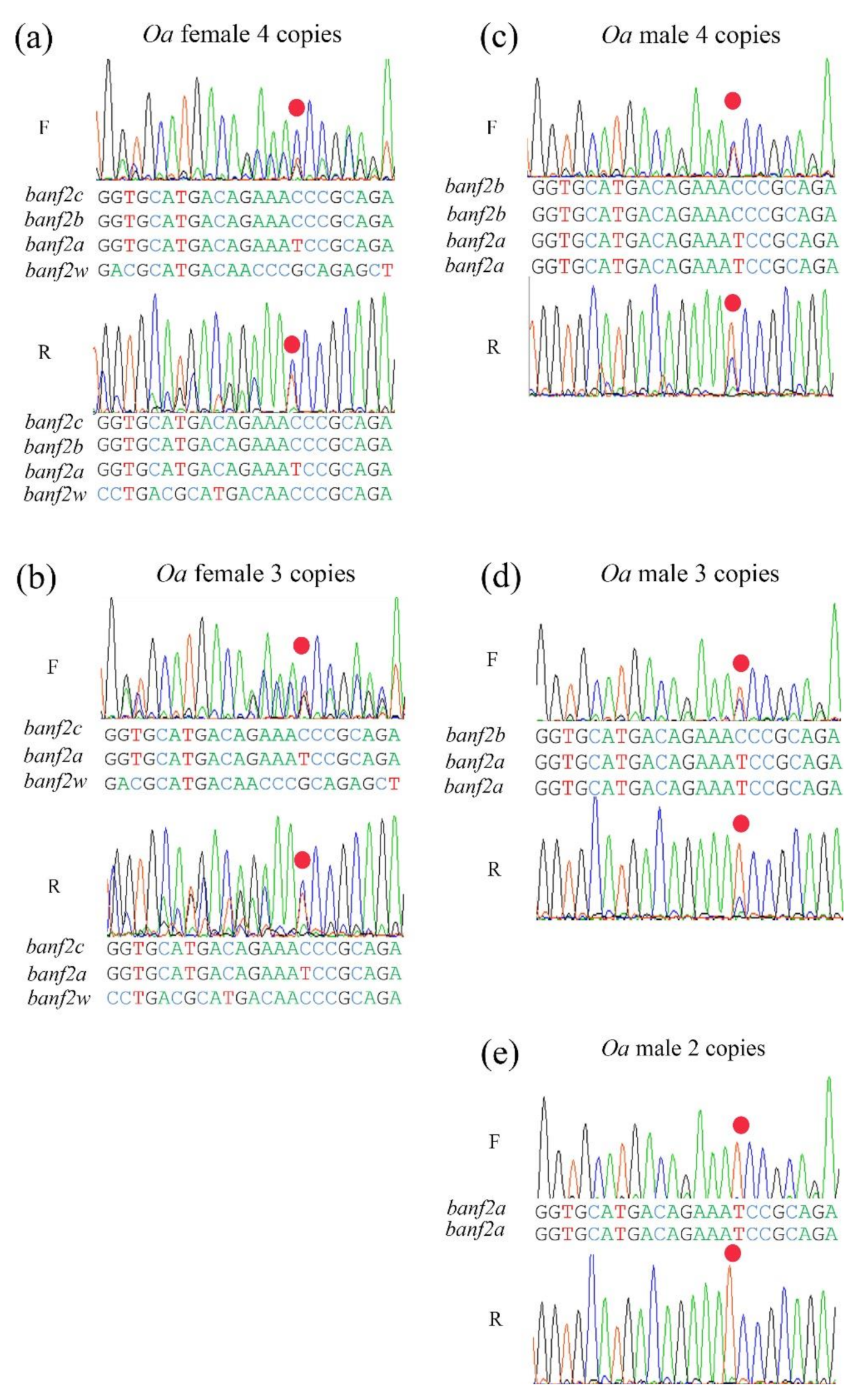
Sequence assemblies of two haplotypes of banf2 were female-specific, whereas two haplotypes were not, suggesting a model of four banf2 copies in Oa females and four in Oa males (Figure 3a,c). This was also supported by the association of the two female-specific haplotypes in the stock with femaleness (Table 3). In addition, sequence assemblies of the banf2 genes in an artificially produced Oa WW individual (SRA accession number: SRX7886422) revealed that it carried only two female-specific banf2 copies, as was to be expected from the proposed model. Indeed, peak-height analyses of Sanger chromatograms of some female individuals from the hybrid Oa stock also supported this model (Figure 4a). For these specimens, banf2w represented approximately 25% of the trace signal according to Tide software [52,53], suggesting a 3:1 ratio for banf2/ban2fw (Figure S3). According to this model, banf2w and banf2c are located on the W chromosome, while banf2a and banf2b are situated on the Z chromosome.
To further examine this model, the banf2 genes were also assembled using additional SRA submissions of male and female Oa (SRA accession numbers: SRX8298258, SRX7899544). The male (SRA accession number: SRX8298258) showed two haplotypes as in the sequence assembly of the first male (SRA accession number: ERX2240356). However, unlike the female sequence assembly (SRA accession number: ERX2240357), in this case (SRA accession number: SRX7899544), only three haplotypes were found. Moreover, sequence chromatograms of some of the specimens pointed to additional types of females with presumably three banf2 copies, in accordance with the decomposition analysis performed by Tide software (Figure 3b and Figure 4b and Figure S3) [52,53]. We were also able to distinguish male types in Sanger sequencing by an informative SNP within exon 3, which allowed deduction of the gene copy proportion by the peak signal ratio of the C/T nucleotides (Figure 4). Three types of males were found in Oa hybrid stock, with 2–4 copies of banf2 (Figure 3c–e). The gene models built based on these specimens suggested that CNV in hybrid Oa stock could be resolved by the presence or absence of a banf2 duplication on the Z chromosome (banf2b, Figure 3a,c,d).

2.3. The Gene Models of Banf2 Locus in Purebred Oa

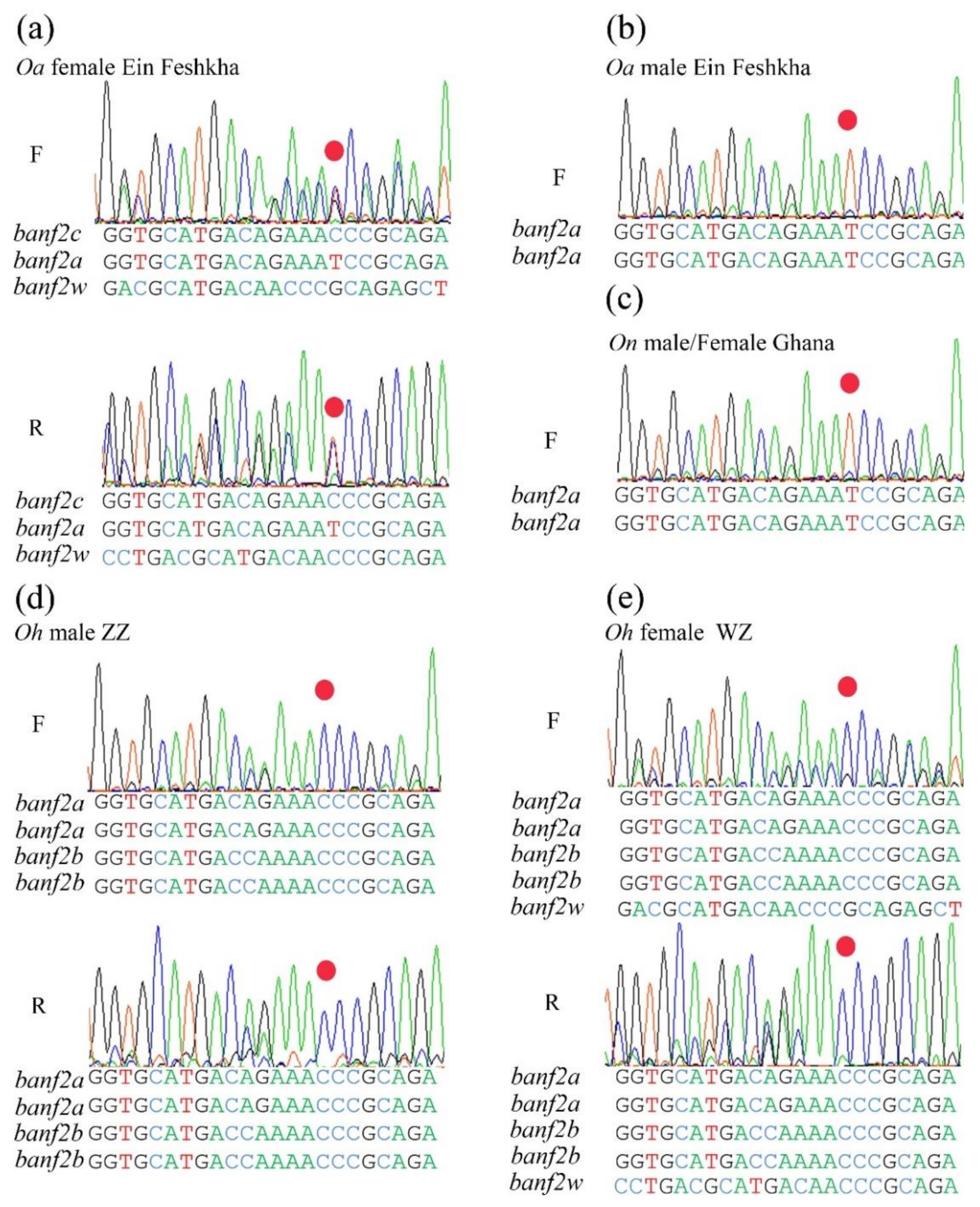
Assessment of gene copy proportions by sequence trace decomposition showed that the banf2w portion of the signal was stable (~33%, Figure 5a) for sequences of Oa females from the Jordan River and the Ein-Feshkha nature reserve, thus suggesting a model of 2:1 for banf2/banf2w in purebred Oa (gene configuration similar to Figure 3b). Accordingly, gene duplication probably occurred on the Z chromosome as a result of hybridization and therefore was not detected in purebred Oa. Male sequence chromatograms of purebred specimens also supported this assumption, attested to by the highly informative SNP within exon 3 (Figure 5b).

2.4. Banf2w Is Female-Specific in Cichlidae Species with WZ/ZZ SD System on LG3

The banf2 genes were assembled in two additional cichlid species Ot (SRA accession numbers: male: SRX6434465; female: SRX6434463) and Pm (SRA accession numbers: pooled males: SRX3638080, SRX3638081; pooled females: SRX3638084, SRX3638085), in which an orthologous SD locus segregates on LG3 [31,37]. Four haplotypes of banf2 were assembled in the Ot male, whereas in the Ot female an additional female-specific copy was found (banf2w, nucleotide accession number: OU016126). Comparison of Ot and Oa suggested a distinct model for the structure of the banf2 locus (Figure 3f): In the third exon of the Ot banf2w gene, aa alterations were similar to that of the Oa banf2w gene, although a different aa substitution was observed in exon 2.
It was difficult to assemble and resolve all banf2 haplotype alleles for Pm, as SRA submissions were either of pooled males or females, introducing the additional complexity of polymorphism. Still, the banf2w pattern of aa alterations in exon 3 was detected only in the female pool (Figure 2, nucleotide accession number: OU017683). The marker Banf2_del was not informative in Ot or Pm, as reflected in the sequence assembly results, since the intron deletion is absent in females from these species.
DNA-seq SRA submissions were not available for Oh. The genetic markers for banf2 (Banf2_del) and banf2w (Banf2_w) were tested on Oh specimens from two families comprising six males and six females from each. Banf2_del was not informative, yet the HRM marker based on the variation of exon 3 in banf2w (Table 1) showed full association with femaleness. Moreover, Sanger chromatograms revealed the same conserved aa substitutions in Oh banf2w as in Oa, Ot and Pm (Figure 2 and Figure 5). This analysis suggested that both Oh and Ot had similar gene models because banf2w’s signal was approximately 20% of the signal height (Figure 5), fitting a model of 4:1 for banf2/banf2w ratio (Figure 3f). The synonymous site divergence of banf2w reflected the expected cichlid phylogeny placing Pm distant from Oa (1.1% divergence) and closer to Ot and Oh (0.6 and 0.0% divergence, respectively).

2.5. Competitive Expression of Banf2w and Other Banf2 Genes in Oa Females

Banf2w and banf2 expression were compared between Oa SRA submissions of RNA-seq analyses of gonad transcriptomes of females and males (SRA accession numbers: SRX7906433 to SRX7906446, n = 14). Banf2w was exclusively present in female gonads in all developmental stages including 5, 30 and 180 days post-hatching (dph). It had a relatively higher expression value in females when compared to other banf2 genes (p ≤ 1 × 10⁻⁵) and reached its peak expression at 30 dph (Figure 6). Banf2 gene expression was generally higher in males (p ≤ 1.5 × 10⁻², ZZ), specifically at 5 (p ≤ 2.6 × 10⁻³) and 30 dph (p ≤ 2.1 × 10⁻³), when compared to females (Figure 6). It is noteworthy that expression of banf2 increased threefold in males between 5 and 30 dph with similar pattern of expression in both sexes peaking at 30 dph. Also of note is that the expression of banf2 decreased in males and converged to values that were similar to those of female expression at 180 dph; however, significance was not tested at this stage due to lack of replicates. The total banf2 FPKM value of gene expression (Z and W) was slightly higher in females at 5 dph (p ≤ 1.2 × 10⁻³) but was insignificantly enhanced at 30 dph (Figure 6). Interestingly the search for expression of banf2w in Ot adult ovary (SRA accession numbers: SRX6445742, SRX6445762, SRX6445765) did not detect any reads, whereas banf2 was abundant.

2.6. The Paics Gene Candidates for SD

At Oa LG3 SD locus, Tao et al. noted only paics-1 and paics-2 [43]. Based on the current genome build (assembly accession number: GCF_013358895) of a “ZZ” individual, two paics-1 genes were annotated as ade2-like genes (LOC116316740, LOC120434429). An additional member of the gene family has been placed between these genes (LOC116321116). In On, the LOC116321116 orthologous gene is mapped to LG23 as ppat and is adjacent to another paics gene, which is orthologous to Oa LG23 paics (Gene ID: 116331053). However, in the foxml1 gene, the genetic marker M1_like was associated with LG3 SD in our Oa stock (Table 3); it was mapped in this current Oa genome build to LG23. These observations suggest that in the current Oa genome build, LGs 3 and 23 might have been disordered; alternatively, the structure of LG3 in the hybrid stock substantially differs from that of the ZZ individual. Moreover, the ZZ-based Oa LG3 build is missing the paics-2a gene and thus differs from LG3 of Oa W genome. Based on assembly of DNA-seq reads from a WW individual (SRA accession number: SRX7886422), we partially inferred the paics-2a gene sequence (nucleotide accession OU234059). In exon 3, which was capable of encoding 65 aa, we encountered coding variation including six aa substitutions compared to the paics-1 paralogs (LOC116316740, LOC120434429), for which the third exon encoded identical polypeptides. A BLASTP search against the non-redundant GenBank dataset (NR) showed that paics-2a encoded a novel Paics unique to Oa females. To examine expression of paics-2a, we followed the procedure described for banf2w, testing 14 gonad transcriptomes using a 32 bp probe “CGAGTCTCAAGACCAGATCACAGCTGGGAACA” that represented the unique variation in exon3 of paics-2a. In ovaries, at five dph, the average expression was 1.2 FPKM, and it peaked at 180 dph reaching 71.03 FPKM. No expression was detected in testes. However, TBLASTN search against Ot and Pm SRA data indicated that paics-2a was specific to Oa and not conserved in females of these other species. We also failed to detect the 5’ paics-2a nucleotide sequence (Paics_short) in Ot and Pm. Thus, with no trans-species conservation paics-2a is a less likely candidate for an SD MKR.

3. Discussion

In this study, we describe banf2 genes of four cichlid species (Oa, Ot, Oh and Pm). These species have a known WZ/ZZ SD system, which has been mapped to genomic regions orthologous to the On LG3. For all four species, we identified a female-specific copy of banf2, designated banf2w. Conserved among the four species, a female-specific aa deletion and non-conservative aa substitutions in exon 3 of banf2w strengthened the candidacy of banf2w as a novel MKR of SD. Moreover, highlighting the uniqueness of this finding, a female-specific allele of banf2 with a large deletion in the second intron (banf2c, Figure 3) showed the same degree of association with sex in Oa as banf2w but was not observed in the other three species (Figure 3).
The region containing banf2 has previously been suggested as a candidate region for SD based on its high female-specific variation density in Oa [43]. This reported region includes only three genes: banf2, paics-1 and paics-2 [43]. As a follow up study, we characterized the proposed SD critical region harboring these three genes in an Oa hybrid stock, mapping it to 235 Kbp, based on the ortholog physical On genome (genome assembly O_niloticus_UMD_NMBU). Previously, paics has been proposed as a candidate MKR for SD in Oa based on differential expression between sexes. However, we did not find any conserved coding variation across different species for this gene to support its candidacy. Moreover, for the genetic marker designed in paics-1 (Paics_long), association with sex was weakened by multiple segregants. The marker in paics-2a (Paics_short) showed the same degree of association with sex as banf2w. However, without sex-related functions [43] or conservation across species, the paics-2a gene is unlikely to be the femaleness determiner. Hybrids were shown here to be a useful tool for characterization of the SD region. The actual mechanism of how hybridization of species with different SD systems induces meiotic recombination within the non-recombining blocks of SD regions is not clear. However, it is probably related to chromosomal sequence and structural differences between the mating species. MKRs on cichlid LGs 1, 5 and 7, are still unidentified. The discovery of amh as MKR for SD in On [54,55,56] has promoted the identification of the role of the orthologous amh genes in SD of distant fish species [57,58,59,60]. Similar implications for research are expected for banf2 genes.
Additional support for the candidacy of the banf gene family to affect SD is the observation that one of the four well-narrowed chromosomal regions of QTLs for spontaneous female-to-male sex reversal harbors banf1 in rainbow trout (Oncorhynchus mykiss) [61]. Therefore, it is important to sequence the banf1 genes in these males to locate genetic mutations that possibly underlie this spontaneous sex reversal.
Investigating the expression profile of banf2w, we found exclusive expression in Oa females, indicating that this gene was functional and had a sex-specific expression profile. In adult Ot female specimens, however, banf2w expression was not observed, which may be explained by a limited time window of expression of this gene in Ot. Similarly, a male-specific MKR for SD, fshry, has been reported for Mugil cephalus with no observed expression in males (40 dph to adulthood) [8,62]. Sry in mammalian males is constitutively expressed from early embryonic stages to adulthood in different tissues, implicating its role as a housekeeping gene [63]. An interesting open question for future research is why an MKR is constitutively expressed in some organisms and not in others.
In Oa females (30–180 dph), transcription of banf2w was higher than other banf2 genes. However, banf2 gene expression was low in comparison with male expression for all stages. These results were significant at 5 and 30 dph, while at 180 dph there were no replicates to allow significance testing. Taken together, the results indicate that banf2 expression observed in male larvae (1–30 dph and maybe older) was replaced in females by banf2w. Expression of banf2 in males and banf2w in females reaches a peak at 30 dph, which is similar to that of the SD amh in On males [36]. These peaks of expression, observed for different MKRs, coincide with the end of the time window in which hormonal and temperature masculinization can be induced [24].
As with most MKRs of SD, banf2w has a known function that associates it with SD. Mammalian banf2 is highly and almost exclusively expressed in testis [64] and is upregulated during spermatogenesis [65]. Banf2 is thought to be a regulator of Banf1 [64]. Interaction between Banf1 and Banf2 occurs through formation of homo- and hetero-dimers, which is especially important in testis [64]. Banf1 is a repressor of foxl2’s pro-apoptotic activity [66,67] and is involved with gonad development [64,68]. Thus, acting in chromatin assembly, the nuclear-lamina-associated Banf1 protein may indirectly impact granulosa cell differentiation and apoptosis [66,67]. Bound to emerin complexes in the nuclear envelope [69], Banf1 may repress foxl2 by mediating its relocation to the nuclear periphery [67]. Amh and foxl2 are central genes regulating testis and ovary development, respectively [2,70]. This is driven by their pro-apoptotic activity, which results in morphological changes of the bipotential gonad [71,72]. Thus, Banf2w may have a dominant effect by preventing the gene silencing function of Banf1. Shutdown of this function would promote foxl2′s apoptotic activity and consequently testis suppression and ovarian development. Overall, we postulate that banf2w has the potential for programing gene expression in cells, causing a switch to a femaleness state via modulation of the chromatin structure, thereby inducing female nucleus formation.
The suggested models of banf2 gene structures in Oa, Ot and Oh are based on short sequence-read assemblies, and analysis of peak ratios at polymorphic sites in Sanger chromatograms. Demonstrating that a combination of the two methods is a useful tool in characterizing the CNV of complex loci governing SD, we showed that banf2w is an extra copy of banf2 in females. Presumably, banf2w evolved by gene duplication and speciation. Indeed, On with an XX/XY SD system on LG23 did not carry a duplication of banf2. Similar structural variation for SD loci is frequent, indicating a typical gene formation pathway. In the closely related On species, an extra copy of amh in males governs sex (amhΔy); this is located <40 Kbp apart from the regular amh copy and is under the same promoter. However, in the present work, we did not determine the physical map of the Oa W chromosome containing banf2w and banf2c genes. Understanding these gene orientations and promoter positions, and comparing them with other similar cases, is important for deciphering mechanisms in which duplications evolve into specialized SD MKRs.
In the Oa hybrid stock, only partial association with sex was observed for the UNH168 marker that has been previously reported to be associated with sex in Oa and Oh [38,44]. However, the genetic markers Banf2_del, Paics_short and Banf2w showed almost complete association with sex in this stock. Through marker-assisted selection for these three SD loci, nearly all-male progeny in On × Oa crosses was obtained, thus demonstrating the mono-factorial SD on LG3. Only a single Oa male was found with a WZ SD genotype, without any male-determination alleles for the genetic markers for SD on LGs 1 and 23. This discrepancy may be explained by environmental sex reversal, involvement of other genetic factors or appearance of non-parental genotype in banf2 locus mediated by aberrant crossing-over [29] or unequal crossing over [73,74].
Both On female and male (Figure 5c), and Oa male (Figure 5b) gene models displayed a single copy of banf2 (banf2a). However, some Oa hybrid males were heterozygous or homozygous for an extra gene copy on the Z chromosome (banf2b). This additional copy produced sequence chromatograms similar to those of the banf2a copy, except for the informative T to C substitution (Figure 4). We did not find banf2b in our Egypt and Ghana On strains, yet it has been detected in GenBank (On Japanese and BYL78 strains, SRA accession numbers: SRX7899813, SRX726489). Hence, this gene variant may have originated from hybridization with an On strain. In contrast to Middle East and North Africa species Oa and On, two Oreochromis species of Central and South Africa, Ot and Oh, did have banf2b (Figure 3f,g). Thus, another possibility is that a contaminator of the Oa hybrid stock was either Oh or Om, both of which are common in aquaculture. It is important to note that the CNV in banf2 locus in the hybrid stock was not associated with SD and suggested that female sex is driven by the actual presence of banf2w and not by a banf2 dosage effect. Thus, unlike in medaka where simple duplication created a new female determiner in line with the dosage-based hypothesis [75], banf2w’s role in SD better fits the dominant gene hypothesis. This should be further explored by functional validation using advanced methods, such as CRISPR/Cas9, TALEN, or antisense RNA [36,60,76,77].

4. Materials and Methods

4.1. Fish

One-year-old females and males (48 of each) were randomly selected from an Oa hybrid stock of the Dor research station [29] and were used for analysis of genetic markers in the LG3 SD region for association with sex. Two additional male and female individuals from this stock have been previously sampled for Illumina HiSeq 2000 paired end sequencing (BioSample accession numbers: SAMEA104362031 and SAMEA104362030). Oh specimens from two families were received from a Fish Aquaculture Station in Brazil [78]. Purebred Oa specimens from local natural resources (Jordan River and Ein-Feshkha Reserve) and On specimens from different introduced strains (Ghana, Swansea and Canada) have been previously described [26].

4.2. DNA Extraction, PCR Amplification, Agarose Separation and Sanger Sequencing

A sample of the caudal fin (100–200 mg) preserved in ethanol was used for DNA extraction using a commercial kit (MasterPure DNA Purification, Madison, WI, USA). PCR was performed using relevant primers that were designed using Primer3 [79] (Table 1) and the Bio-X-ACT™ Long kit (Bioline Ltd., London, UK) according to the manufacturer’s instructions under the following conditions: 36 cycles for 30 s at 94 °C, 30 s at 59–63 °C and 30–50 s at 72 °C. Thereafter, the PCR products were examined for genotyping on the basis of size in a 1–2% agarose gel stained with ethidium bromide. Following excision from the gel, Sanger sequencing was conducted from both directions of the purified products (Montage Gel Extraction, Millipore, Bedford, MA, USA).

4.3. Marker Development and Search for Coding Polymorphism in O. aureus

Genetic markers were developed in the region on LG3 that contains paics and banf2 and has the highest density of sex-specific polymorphism in Oa [43]. For the sequence assembly of four genes (paics-1, paics-2, foxm1l and banf2) and identification of potential functional polymorphism, two previously published male and female Oa genomic-DNA libraries (SRA accession numbers: ERX2240356 and ERX2240357, respectively) were used (Figure 1, Table 1). Five developed markers (paics_long, paics short, M1-like, Banf2_del and Banf2w) with DNA length or SNP polymorphism were tested for association with sex by AGE or by a HRM analyses. Genotyping by HRM was conducted using a Real-Time PCR instrument (ECO, Illumina, California, SD, USA) and the qPCRBIO HRM Mix (PCR Biosystems, London, UK). In addition, UNH168, a microsatellite marker found to be highly associated with LG3 in several studies [38,44] was also examined. PCR for UNH168 used dye-labeled forward and unlabeled reverse primers (Table 1). The amplified products were separated on an ABI3130 DNA sequencer and sized using the GeneMapper software v. 4.0 (Applied Biosystems Ltd., Foster City, CA, USA) with GeneScan-500 LIZ size standard (Applied Biosystems) [80].

4.4. Copy Number Estimation with Sanger and Construction Genetic Models

Copy numbers were estimated taking into account coverage of next generation sequencing (NGS) reads, and copy number proportion was measured by Sanger chromatograms [52]. Proportions of copies were estimated using Tide software [52,53]. Each chromatogram analysis was performed using template DNA of at least six individuals with the same sex phenotype and genotypes. Such individuals yielded similar chromatograms of which a typical one was selected for further analysis and presentation.

4.5. Sequence Alignments

Protein sequences were aligned with ClustalW (http://clustalw.genome.jp, accessed on 29 June 2021), using default settings and Gonnet weight matrix. Nucleotide sequences were aligned using the same tool and the DNA option. The graphical image of the multiple alignment was made using BoxShade (https://www.ch.embnet.org/software/BOX_form.html, accessed on 29 June 2021). To calculate synonymohttps://embnet.vital-it.ch/software/BOX_form.htmlus (accessed on 29 June 2021) site divergence of banf2w, the banf2w nucleotide sequences were aligned and substitutions were counted within positions 2261–2438 (nucleotide accession number: OU016046). An indel was counted as a single event.

4.6. Expression Analysis

FPKM values were used for comparing expression values of different haplotypes. Fourteen RNA-seq libraries (SRA accession numbers: SRX7906433 to SRX7906446) were aligned against 32 bp probes of each haplotype, using the SRA Nucleotide BLAST [81] with parameters that force complete fit (32bp word). The numbers of aligned hits for each haplotype were then recorded for calculation of the FPKM.

4.7. Statistics

The JMP© statistical package (Pro 13, SAS Institute, Cary, NC, USA) was used for conducting Fisher’s exact chi-squared and Pearson chi-squared tests, which were applied for association study of genetic polymorphism and sex and for goodness of fit of inheritance models, respectively. In addition, one-way analysis of variance (ANOVA) was used for comparing differences in expression data of different developmental stages and haplotypes.

Supplementary Materials

The following are available online at https://www.mdpi.com/article/10.3390/ijms22137073/s1.

Author Contributions

Conceptualization, A.Y.C., A.S., M.R. and E.S.; methodology, A.Y.C., A.S. and E.S.; software, A.Y.C. and E.S.; formal analysis, A.Y.C., A.S. and E.S.; investigation, A.Y.C., A.S. and E.S.; resources, A.B.-P., A.N., S.I.L.-T. and H.S.; writing—original draft preparation, A.Y.C., A.S. and E.S.; supervision, A.B.-P., A.N., M.R. and E.S. All authors have read and agreed to the published version of the manuscript.

Funding

This research was partially funded by the Israeli Fish Breeders Association and by the Fisheries Department of the Israel Ministry of Agriculture and Rural Development.

Institutional Review Board Statement

The study was conducted according to the guidelines of and approved by the Institutional Ethics Committee of ARO (protocol code 739/17 IL, date of approval 6 December 2017).

Data Availability Statement

Data are contained within this article’s supplementary material. Nucleotide sequence data for Oa, Oh, Ot and Pm were deposited in ENA under project accession number PRJEB44866.

Conflicts of Interest

The authors declare no conflict of interest.

References

  1. Herpin, A.; Schartl, M. Vertebrate sex determination: Questioning the hierarchy. FEBS J. 2011, 278, 1001. [Google Scholar] [CrossRef] [PubMed]
  2. Capel, B. Vertebrate sex determination: Evolutionary plasticity of a fundamental switch. Nat. Rev. Genet. 2017, 18, 675–689. [Google Scholar] [CrossRef] [PubMed]
  3. Graves, J.A.M. How to evolve new vertebrate sex determining genes. Dev. Dyn. 2013, 242, 354–359. [Google Scholar] [CrossRef] [PubMed]
  4. Gubbay, J.; Collignon, J.; Koopman, P.; Capel, B.; Economou, A.; Münsterberg, A.; Vivian, N.; Goodfellow, P.; Lovell-Badge, R. A gene mapping to the sex-determining region of the mouse Y chromosome is a member of a novel family of embryonically expressed genes. Nature 1990, 346, 245–250. [Google Scholar] [CrossRef] [PubMed]
  5. Sinclair, A.H.; Berta, P.; Palmer, M.S.; Hawkins, J.R.; Griffiths, B.L.; Smith, M.J.; Foster, J.W.; Frischauf, A.-M.; Lovell-Badge, R.; Goodfellow, P.N. A gene from the human sex-determining region encodes a protein with homology to a conserved DNA-binding motif. Nature 1990, 346, 240–244. [Google Scholar] [CrossRef] [Green Version]
  6. Zhou, Q.; Zhang, J.; Bachtrog, D.; An, N.; Huang, Q.; Jarvis, E.D.; Gilbert, M.T.P.; Zhang, G. Complex evolutionary trajectories of sex chromosomes across bird taxa. Science 2014, 346, 1246338. [Google Scholar] [CrossRef] [Green Version]
  7. Smith, C.A.; Roeszler, K.N.; Ohnesorg, T.; Cummins, D.M.; Farlie, P.G.; Doran, T.J.; Sinclair, A.H. The avian Z-linked gene DMRT1 is required for male sex determination in the chicken. Nature 2009, 461, 267–271. [Google Scholar] [CrossRef]
  8. Curzon, A.Y.; Dor, L.; Shirak, A.; Rosenfeld, H.; Ashkenazi, I.M.; Ron, M.; Seroussi, E. A novel c.1759T>G variant in follicle-stimulating hormone-receptor gene is concordant with male determination in the flathead grey mullet (Mugil cephalus). G3 Genes Genomes Genet. 2020, 11, jkaa044. [Google Scholar] [CrossRef]
  9. Hammerman, I.S.; Avtalion, R.R. Sex determination in Sarotherodon (tilapia). Theor. Appl. Genet. 1979, 55, 177–187. [Google Scholar] [CrossRef]
  10. Wohlfarth, G.W.; Wedekind, H. The heredity of sex determination in tilapias. Aquaculture 1991, 92, 143–156. [Google Scholar] [CrossRef]
  11. Koopman, P. The genetics and biology of vertebrate sex determination. Cell 2001, 105, 843–847. [Google Scholar] [CrossRef] [Green Version]
  12. Graves, J.A.M.; Peichel, C.L. Are homologies in vertebrate sex determination due to shared ancestry or to limited options? Genome Biol. 2010, 11, 205. [Google Scholar] [CrossRef]
  13. Yano, A.; Guyomard, R.; Nicol, B.; Jouanno, E.; Quillet, E.; Klopp, C.; Cabau, C.; Bouchez, O.; Fostier, A.; Guiguen, Y. An immune-related gene evolved into the master sex-determining gene in rainbow trout, Oncorhynchus mykiss. Curr. Biol. 2012, 22, 1423–1428. [Google Scholar] [CrossRef] [Green Version]
  14. Yano, A.; Nicol, B.; Jouanno, E.; Quillet, E.; Fostier, A.; Guyomard, R.; Guiguen, Y. The sexually dimorphic on the Y-chromosome gene (sdY) is a conserved male-specific Y-chromosome sequence in many salmonids. Evol. Appl. 2013, 6, 486–496. [Google Scholar] [CrossRef]
  15. Herpin, A.; Schartl, M. Plasticity of gene-regulatory networks controlling sex determination: Of masters, slaves, usual suspects, newcomers, and usurpators. EMBO Rep. 2015, 16, 1260–1274. [Google Scholar] [CrossRef] [Green Version]
  16. Bertho, S.; Herpin, A.; Branthonne, A.; Jouanno, E.; Yano, A.; Nicol, B.; Muller, T.; Pannetier, M.; Pailhoux, E.; Miwa, M.; et al. The unusual rainbow trout sex determination gene hijacked the canonical vertebrate gonadal differentiation pathway. Proc. Natl. Acad. Sci. USA 2018, 115, 12781–12786. [Google Scholar] [CrossRef] [Green Version]
  17. Shirak, A.; Zak, T.; Dor, L.; Benet-Perlberg, A.; Weller, J.I.; Ron, M.; Seroussi, E. Quantitative trait loci on LGs 9 and 14 affect the reproductive interaction between two Oreochromis species, O. niloticus and O. aureus. Heredity 2018, 122, 341–353. [Google Scholar] [CrossRef] [Green Version]
  18. Megbowon, I.; Mojekwu, T.O. Tilapia sex reversal using methyl testosterone (MT) and its effect on fish, man and environment. Biotechnology 2014, 13, 213–216. [Google Scholar] [CrossRef] [Green Version]
  19. Pruginin, Y.; Rothbard, S.; Wohlfarth, G.; Halevy, A.; Moav, R.; Hulata, G. All-male broods of Tilapia nilotica × T. aurea hybrids. Aquaculture 1975, 6, 11–21. [Google Scholar] [CrossRef]
  20. Hickling, C.F. The Malacca tilapia hybrids. J. Genet. 1960, 57, 1–10. [Google Scholar] [CrossRef]
  21. Eknath, A.E.; Bentsen, H.B.; Gjerde, B.; Tayamen, M.M.; Abella, T.A.; Gjedrem, T.; Pullin, R.S.V. Approaches to national fish breeding programs: Pointers from tilapia pilot study. Naga ICLARM Q. 1991, 14, 10–12. Available online: http://aquaticcommons.org/id/eprint/9178 (accessed on 29 June 2021).
  22. Brawand, D.; Wagner, C.E.; Li, Y.I.; Malinsky, M.; Keller, I.; Fan, S.; Simakov, O.; Ng, A.Y.; Lim, Z.W.; Bezault, E.; et al. The genomic substrate for adaptive radiation in African cichlid fish. Nature 2015, 513, 375–381. [Google Scholar] [CrossRef] [PubMed] [Green Version]
  23. Ser, J.R.; Roberts, R.B.; Kocher, T.D. Multiple interacting loci control sex determination in lake Malawi Cichlid fishes. Evolution 2010, 64, 486–501. [Google Scholar] [CrossRef] [PubMed] [Green Version]
  24. Baroiller, J.F.; D’Cotta, H.; Bezault, E.; Wessels, S.; Hoerstgen-Schwark, G. Tilapia sex determination: Where temperature and genetics meet. Comp. Biochem. Physiol. A Mol. Integr. Physiol. 2009, 153, 30–38. [Google Scholar] [CrossRef] [PubMed]
  25. Mair, G.C.; Scott, A.G.; Penman, D.J.; Skibinski, D.O.F.; Beardmore, J.A. Sex determination in the genus Oreochromis—2. Sex reversal, hybridisation, gynogenesis and triploidy in O. aureus Steindachner. Theor. Appl. Genet. 1991, 82, 153–160. [Google Scholar] [CrossRef]
  26. Curzon, A.Y.; Shirak, A.; Dor, L.; Zak, T.; Perelberg, A.; Seroussi, E.; Ron, M. A duplication of the Anti-Müllerian hormone gene is associated with genetic sex determination of different Oreochromis niloticus strains. Heredity 2020, 125, 317–327. [Google Scholar] [CrossRef]
  27. Charlesworth, D.; Bergero, R.; Graham, C.; Gardner, J.; Yong, L. Locating the sex determining region of linkage group 12 of guppy (Poecilia reticulata). G3 Genes Genomes Genet. 2020, 10, 3639–3649. [Google Scholar] [CrossRef]
  28. Dor, L.; Shirak, A.; Kohn, Y.Y.; Gur, T.; Weller, J.I.; Zilberg, D.; Seroussi, E.; Ron, M. Mapping of the sex determining region on linkage group 12 of guppy (Poecilia reticulata). G3 Genes Genomes Genet. 2019, 9, 3867–3875. [Google Scholar] [CrossRef] [Green Version]
  29. Curzon, A.Y.; Shirak, A.; Zak, T.; Dor, L.; Benet-Perlberg, A.; Naor, A.; Low-Tanne, S.I.; Sharkawi, H.; Ron, M.; Seroussi, E. All-male production by marker-assisted selection for sex determining loci of admixed Oreochromis niloticus and Oreochromis aureus stocks. Anim. Genet. 2021, 52, 361–364. [Google Scholar] [CrossRef]
  30. Gammerdinger, W.J.; Kocher, T.D. Unusual diversity of sex chromosomes in african cichlid fishes. Genes 2018, 9, 480. [Google Scholar] [CrossRef] [Green Version]
  31. Gammerdinger, W.J.; Conte, M.A.; Sandkam, B.A.; Penman, D.J.; Kocher, T.D. Characterization of sex chromosomes in three deeply diverged species of Pseudocrenilabrinae (Teleostei: Cichlidae). Hydrobiologia 2019, 832, 397–408. [Google Scholar] [CrossRef]
  32. Gammerdinger, W.J.; Conte, M.A.; Baroiller, J.-F.; D’Cotta, H.; Kocher, T.D. Comparative analysis of a sex chromosome from the blackchin tilapia, Sarotherodon melanotheron. BMC Genom. 2016, 17, 808. [Google Scholar] [CrossRef] [Green Version]
  33. Böhne, A.; Weber, A.A.T.; Rajkov, J.; Rechsteiner, M.; Riss, A.; Egger, B.; Salzburger, W. Repeated evolution versus common ancestry: Sex chromosome evolution in the haplochromine cichlid Pseudocrenilabrus philander. Genome Biol. Evol. 2019, 11, 439–458. [Google Scholar] [CrossRef] [Green Version]
  34. Eshel, O.; Shirak, A.; Dor, L.; Band, M.; Zak, T.; Markovich-Gordon, M.; Chalifa-Caspi, V.; Feldmesser, E.; Weller, J.I.; Seroussi, E.; et al. Identification of male-specific amh duplication, sexually differentially expressed genes and microRNAs at early embryonic development of Nile tilapia (Oreochromis niloticus). BMC Genom. 2014, 15, 774. [Google Scholar] [CrossRef] [Green Version]
  35. Taslima, K.; Wehner, S.; Taggart, J.B.; de Verdal, H.; Benzie, J.A.H.; Bekaert, M.; McAndrew, B.J.; Penman, D.J. Sex determination in the GIFT strain of tilapia is controlled by a locus in linkage group 23. BMC Genet. 2020, 21, 49. [Google Scholar] [CrossRef]
  36. Li, M.; Sun, Y.; Zhao, J.; Shi, H.; Zeng, S.; Ye, K.; Jiang, D.; Zhou, L.; Sun, L.; Tao, W.; et al. A tandem duplicate of Anti-Müllerian hormone with a missense SNP on the Y chromosome is esential for male sex determination in Nile tilapia, Oreochromis niloticus. PLoS Genet. 2015, 11, e1005678. [Google Scholar] [CrossRef] [Green Version]
  37. Cnaani, A.; Kocher, T.D. Sex-linked markers and microsatellite locus duplication in the cichlid species Oreochromis tanganicae. Biol. Lett. 2008, 4, 700–703. [Google Scholar] [CrossRef] [Green Version]
  38. Zhu, H.; Liu, Z.; Lu, M.; Gaao, F.; Ke, X.; Ma, D.; Huang, Z.; Cao, J.; Wang, M. Screening and identification of a microsatellite marker associated with sex in Wami tilapia, Oreochromis urolepis hornorum. J. Genet. 2016, 95, 283–289. [Google Scholar] [CrossRef]
  39. Gammerdinger, W.J.; Conte, M.A.; Sandkam, B.A.; Ziegelbecker, A.; Koblmüller, S.; Kocher, T.D. Novel Sex Chromosomes in 3 Cichlid Fishes from Lake Tanganyika. J. Hered. 2018, 109, 489–500. [Google Scholar] [CrossRef]
  40. Conte, M.A.; Gammerdinger, W.J.; Bartie, K.L.; Penman, D.J.; Kocher, T.D. A high quality assembly of the Nile Tilapia (Oreochromis niloticus) genome reveals the structure of two sex determination regions. BMC Genom. 2017, 18, 341. [Google Scholar] [CrossRef]
  41. Conte, M.A.; Clark, F.E.; Roberts, R.B.; Xu, L.; Tao, W.; Zhou, Q.; Wang, D.; Kocher, T.D. Origin of a giant sex chromosome. Mol. Biol. Evol. 2020. [Google Scholar] [CrossRef]
  42. Wu, X.; Zhao, L.; Fan, Z.; Lu, B.; Chen, J.; Tan, D.; Jiang, D.; Tao, W.; Wang, D. Screening and characterization of sex-linked DNA markers and marker-assisted selection in blue tilapia (Oreochromis aureus). Aquaculture 2021, 530, 735934. [Google Scholar] [CrossRef]
  43. Tao, W.; Xu, L.; Zhao, L.; Zhu, Z.; Wu, X.; Min, Q.; Wang, D.; Zhou, Q. High-quality chromosome-level genomes of two tilapia species reveal their evolution of repeat sequences and sex chromosomes. Mol. Ecol. Resour. 2021, 21, 543–560. [Google Scholar] [CrossRef]
  44. Lee, B.-Y.; Hulata, G.; Kocher, T.D. Two unlinked loci controlling the sex of blue tilapia (Oreochromis aureus). Heredity 2004, 92, 543–549. [Google Scholar] [CrossRef] [Green Version]
  45. Jensen, G.L.; Shelton, W.L. Effects of estrogens on Tilapia aurea: Implications for production of monosex genetic male tilapia. Aquaculture 1979, 16, 233–242. [Google Scholar] [CrossRef]
  46. Avtalion, R.R.; Don, J. Sex-determining genes in tilapia: A model of genetic recombination emerging from sex ratio results of three generations of diploid gynogenetic Oreochromis aureus. J. Fish Biol. 1990, 37, 167–173. [Google Scholar] [CrossRef]
  47. Graves, J.A.M. Sex chromosome specialization and degeneration in mammals. Cell 2006, 124, 901–914. [Google Scholar] [CrossRef] [Green Version]
  48. Fraser, J.A.; Heitman, J. Chromosomal sex-determining regions in animals, plants and fungi. Curr. Opin. Genet. Dev. 2005, 15, 645–651. [Google Scholar] [CrossRef]
  49. Lee, B.Y.; Lee, W.J.; Streelman, J.T.; Carleton, K.L.; Howe, A.E.; Hulata, G.; Slettan, A.; Stern, J.E.; Terai, Y.; Kocher, T.D. A second-generation genetic linkage map of tilapia (Oreochromis spp.). Genetics 2005, 170, 237–244. [Google Scholar] [CrossRef] [Green Version]
  50. Ocalewicz, K.; Mota-Velasco, J.C.; Campos-Ramos, R.; Penman, D.J. FISH and DAPI staining of the synaptonemal complex of the Nile tilapia (Oreochromis niloticus) allow orientation of the unpaired region of bivalent 1 observed during early pachytene. Chromosom. Res. 2009, 17, 773–782. [Google Scholar] [CrossRef]
  51. Charlesworth, D.; Charlesworth, B.; Marais, G. Steps in the evolution of heteromorphic sex chromosomes. Heredity 2005, 95, 118–128. [Google Scholar] [CrossRef] [PubMed] [Green Version]
  52. Seroussi, E. Estimating copy-number proportions: The comeback of Sanger sequencing. Genes 2021, 12, 283. [Google Scholar] [CrossRef] [PubMed]
  53. Brinkman, E.K.; Chen, T.; Amendola, M.; Van Steensel, B. Easy quantitative assessment of genome editing by sequence trace decomposition. Nucleic Acids Res. 2014, 42, e168. [Google Scholar] [CrossRef] [PubMed]
  54. Shirak, A.; Seroussi, E.; Cnaani, A.; Howe, A.E.; Domokhovsky, R.; Zilberman, N.; Kocher, T.D.; Hulata, G.; Ron, M. Amh and dmrta2 genes map to tilapia (Oreochromis spp.) linkage group 23 within quantitative trait locus regions for sex determination. Genetics 2006, 174, 1573–1581. [Google Scholar] [CrossRef] [Green Version]
  55. Eshel, O.; Shirak, A.; Weller, J.I.; Hulata, G.; Ron, M. Linkage and physical mapping of sex region on LG23 of Nile tilapia (Oreochromis niloticus). G3 Genes Genomes Genet. 2012, 2, 35–42. [Google Scholar] [CrossRef] [Green Version]
  56. Eshel, O.; Shirak, A.; Weller, J.I.; Slossman, T.; Hulata, G.; Cnaani, A.; Ron, M. Fine-mapping of a locus on linkage group 23 for sex determination in Nile tilapia (Oreochromis niloticus). Anim. Genet. 2011, 42, 222–224. [Google Scholar] [CrossRef]
  57. Hattori, R.S.; Murai, Y.; Oura, M.; Masuda, S.; Majhi, S.K.; Sakamoto, T.; Fernandino, J.I.; Somoza, G.M.; Yokota, M.; Strussmann, C.A. A Y-linked anti-Mullerian hormone duplication takes over a critical role in sex determination. Proc. Natl. Acad. Sci. USA 2012, 109, 2955–2959. [Google Scholar] [CrossRef] [Green Version]
  58. Rondeau, E.B.; Laurie, C.V.; Johnson, S.C.; Koop, B.F. A PCR assay detects a male-specific duplicated copy of Anti-Müllerian hormone (amh) in the lingcod (Ophiodon elongatus). BMC Res. Notes 2016, 9, 230. [Google Scholar] [CrossRef] [Green Version]
  59. Bej, D.K.; Miyoshi, K.; Hattori, R.S.; Strüssmann, C.A.; Yamamoto, Y. A duplicated, truncated amh gene is involved in male sex determination in an old world Silverside. G3 Genes Genomes Genet. 2017, 7, 2489–2495. [Google Scholar] [CrossRef] [Green Version]
  60. Pan, Q.; Feron, R.; Yano, A.; Guyomard, R.; Jouanno, E.; Vigouroux, E.; Wen, M.; Busne, J.M.; Bobe, J.; Concordet, J.P.; et al. Identification of the master sex determining gene in Northern pike (Esox lucius) reveals restricted sex chromosome differentiation. PLoS Genet. 2019, 15, e1008013. [Google Scholar] [CrossRef] [Green Version]
  61. Fraslin, C.; Phocas, F.; Bestin, A.; Charles, M.; Bernard, M.; Krieg, F.; Dechamp, N.; Ciobotaru, C.; Hozé, C.; Petitprez, F.; et al. Genetic determinism of spontaneous masculinisation in XX female rainbow trout: New insights using medium throughput genotyping and whole-genome sequencing. Sci. Rep. 2020, 10, 1–13. [Google Scholar] [CrossRef]
  62. Ferraresso, S.; Bargelloni, L.; Babbucci, M.; Cannas, R.; Follesa, M.C.; Carugati, L.; Melis, R.; Cau, A.; Koutrakis, M.; Sapounidis, A.; et al. Fshr: A fish sex-determining locus shows variable incomplete penetrance across flathead grey mullet populations. iScience 2021, 24, 101886. [Google Scholar] [CrossRef]
  63. Turner, M.E.; Ely, D.; Prokop, J.; Milsted, A. Sry, more than testis determination? Am. J. Physiol. Integr. Comp. Physiol. 2011, 301, R561–R571. [Google Scholar] [CrossRef] [Green Version]
  64. Tifft, K.E.; Segura-Totten, M.; Lee, K.K.; Wilson, K.L. Barrier-to-autointegration factor-like (BAF-L): A proposed regulator of BAF. Exp. Cell Res. 2006, 312, 478–487. [Google Scholar] [CrossRef]
  65. Maratou, K.; Forster, T.; Costa, Y.; Taggart, M.; Speed, R.M.; Ireland, J.; Teague, P.; Roy, D.; Cooke, H.J. Expression profiling of the developing testis in wild-type and Dazl knockout mice. Mol. Reprod. Dev. 2004, 67, 26–54. [Google Scholar] [CrossRef]
  66. L’Hôte, D.; Georges, A.; Todeschini, A.L.; Kim, J.H.; Benayoun, B.A.; Bae, J.; Veitia, R.A. Discovery of novel protein partners of the transcription factor FOXL2 provides insights into its physiopathological roles. Hum. Mol. Genet. 2012, 21, 3264–3274. [Google Scholar] [CrossRef] [Green Version]
  67. Penrad-Mobayed, M.; Perrin, C.; Herman, L.; Todeschini, A.; Nigon, F.; Cosson, B.; Caburet, S.; Veitia, R.A. Conventional and unconventional interactions of the transcription factor FOXL2 uncovered by a proteome-wide analysis. FASEB J. 2020, 34, 571–587. [Google Scholar] [CrossRef] [Green Version]
  68. Margalit, A.; Segura-Totten, M.; Gruenbaum, Y.; Wilson, K.L. Barrier-to-autointegration factor is required to segregate and enclose chromosomes within the nuclear envelope and assemble the nuclear lamina. Proc. Natl. Acad. Sci. USA 2005, 102, 3290–3295. [Google Scholar] [CrossRef] [Green Version]
  69. Holaska, J.M.; Lee, K.K.; Kowalski, A.K.; Wilson, K.L. Transcriptional repressor germ cell-less (GCL) and barrier to autointegration factor (BAF) compete for binding to emerin in vitro. J. Biol. Chem. 2003, 278, 6969–6975. [Google Scholar] [CrossRef] [Green Version]
  70. Georges, A.; Auguste, A.; Bessière, L.; Vanet, A.; Todeschini, A.L.; Veitia, R.A. FOXL2: A central transcription factor of the ovary. J. Mol. Endocrinol. 2013, 52, 17–33. [Google Scholar] [CrossRef]
  71. Benayoun, B.A.; Dipietromaria, A.; Bazin, C.; Veitia, R.A. FOXL2: At the crossroads of female sex determination and ovarian function. Adv. Exp. Med. Biol. 2009, 665, 207–226. [Google Scholar] [CrossRef]
  72. Windley, S.P.; Wilhelm, D. Signaling pathways involved in mammalian sex determination and gonad development. Sex. Dev. 2016, 9, 297–315. [Google Scholar] [CrossRef]
  73. McMurray, C.T. Mechanisms of DNA expansion. Chromosoma 1995, 104, 2–13. [Google Scholar] [CrossRef]
  74. Katju, V.; Bergthorsson, U. Copy-number changes in evolution: Rates, fitness effects and adaptive significance. Front. Genet. 2013, 4, 273. [Google Scholar] [CrossRef] [Green Version]
  75. Nanda, I.; Kondo, M.; Hornung, U.; Asakawa, S.; Winkler, C.; Shimizu, A.; Shan, Z.; Haaf, T.; Shimizu, N.; Shima, A.; et al. A duplicated copy of DMRT1 in the sex-determining region of the Y chromosome of the medaka, Oryzias latipes. Proc. Natl. Acad. Sci. USA 2002, 99, 11778–11783. [Google Scholar] [CrossRef] [Green Version]
  76. Ventura, T.; Sagi, A. The insulin-like androgenic gland hormone in crustaceans: From a single gene silencing to a wide array of sexual manipulation-based biotechnologies. Biotechnol. Adv. 2012, 30, 1543–1550. [Google Scholar] [CrossRef]
  77. Bao, L.; Tian, C.; Liu, S.; Zhang, Y.; Elaswad, A.; Yuan, Z.; Khalil, K.; Sun, F.; Yang, Y.; Zhou, T.; et al. The Y chromosome sequence of the channel catfish suggests novel sex determination mechanisms in teleost fish. BMC Biol. 2019, 17, 6. [Google Scholar] [CrossRef] [Green Version]
  78. Shirak, A.; Dor, L.; Seroussi, E.; Ron, M.; Hulata, G.; Golani, D. DNA barcoding of fish species from the Mediterranean coast of Israel. Mediterr. Mar. Sci. 2016, 17, 459–466. [Google Scholar] [CrossRef] [Green Version]
  79. Untergasser, A.; Cutcutache, I.; Koressaar, T.; Ye, J.; Faircloth, B.C.; Remm, M.; Rozen, S.G. Primer3-new capabilities and interfaces. Nucleic Acids Res. 2012, 40, e115. [Google Scholar] [CrossRef] [Green Version]
  80. Dor, L.; Shirak, A.; Gorshkov, S.; Band, M.R.; Korol, A.; Ronin, Y.; Curzon, A.; Hulata, G.; Seroussi, E.; Ron, M. Construction of a microsatellites-based linkage map for the White Grouper (Epinephelus aeneus). G3 Genes Genomes Genet. 2014, 4, 1455–1464. [Google Scholar] [CrossRef] [Green Version]
  81. Pruitt, K.; Brown, G.; Tatusova, T.; Maglott, D. The Reference Sequence (RefSeq) database. In The NCBI Handbook; National Library of Medicine (US), National Center for Biotechnology Information: Bethesda, MD, USA, 2002; pp. 1–24. Available online: https://www.ncbi.nlm.nih.gov/books/NBK21091/ (accessed on 29 June 2021).
Figure 1. Position of five genetic markers in the SD region of Oreochromis aureus delineated on the O. niloticus genome map. Genetic markers (red arrows) were developed within paics-1 (long form), banf2 (LOC102076719), paics-2 (short form) and forkhead box protein M1-like (foxm1l). The SD region spanned 235 Kbp between the two external markers.
Ijms 22 07073 g001
Figure 2. An alignment of the predicted protein domains encoded by the third exons of banf2w (nucleotide accession number: OU016046) and banf2 (nucleotide accession number: OU022051) of Oreochromis aureus (Oa), O. tanganicae (Ot, nucleotide accession numbers: OU016126, OU016104), Pelmatolapia mariae (Pm, nucleotide accession numbers: OU017683, OU017684) and O. urolepis hornorum (Oh, nucleotide accession numbers: OU070172, OU070150) Dashes indicate gaps introduced by the alignment program. Identical amino acid residues in at least three of eight sequences are indicated by a black background. White boxes indicate non-conservative amino acid changes between the proteins.
Ijms 22 07073 g002
Figure 3. Suggested schematic models depicting the copy number variation of banf2 genes in the LG3 sex-determining loci of cichlid fish. Models include delineations for banf2 genes that are not sex-specific (blue boxes) and for the female determination genes (pink boxes). Gene models of banf2 for females (a,b) and males (ce) demonstrate variability within the Oreochromis aureus (Oa) hybrid stock and homogeneity of purebred Oa females (b) and males (e). The simplest gene model was observed for the banf2 locus of both sexes of O. niloticus (e). A five-copy model represents the gene arrangement predicted for both O. tanganicae and O. urolepis hornorum females (f), and a four-copy model that of males of these species (g). “Del” represents a large deletion in banf2c intron used as a genetic marker in Oa. Shown in circles, an informative C/T/G SNP in exon 3 of banf2 genes was used to resolve the models in a peak-ratio analysis of Sanger sequencing (also highlighted by red dots in Figure 4).
Ijms 22 07073 g003
Figure 4. Polymorphism in Oreochromis aureus (Oa) hybrid stock in Sanger chromatograms of the third exon of banf2 genes. Chromatograms a-e correspond to models a-e in Figure 4, respectively. Copy proportion analysis was based on both forward (F) and reverse (R) sequencing orientations. The nucleotide sequences below the chromatogram traces were interpreted using Tide software, which, by trace decomposition analysis, estimated that the ratio of banf2w was 25% of the signal intensity in some Oa females (a), and was 33% of this signal in others (b), suggesting four and three copies, respectively. The red dots mark an informative position disclosing multiple copies with either C, T or G peaks. For males, this informative site suggested a 2–4-copy model (ce). The presence of banf2a and 2b or 2c was reflected in the observed peak-height ratio between T and C. C was not observed in a homozygous state, and thus it represented a gene duplication that was in tandem with the copy carrying T.
Ijms 22 07073 g004
Figure 5. Polymorphism in Sanger chromatograms of third exon of banf2 genes of three purebred Oreochromis species. Typical traces are presented for female (a) and male (b) of Oreochromis aureus (Oa); common trace for female and male of O. niloticus (On) (c) and male (d) and female (e) of O. urolepis hornorum (Oh). Copy proportion analysis was based on both forward (F) and reverse (R) sequencing orientations. The nucleotide sequences below the chromatogram traces were interpreted using Tide software, which, by trace decomposition analysis, estimated that the banf2w was 33% of the signal intensity in Oa and was 20% in Oh. The red dots mark an informative position disclosing multiple copies in Oa females carrying either C, T or G.
Ijms 22 07073 g005
Figure 6. Expression in units of fragments per kilobase million (FPKM) of banf2w and other banf2 genes in Oreochromis aureus gonads of males (ZZ) and females (WZ) at three ages: 5, 30 and 90 days post-hatching (dph). The minimal threshold for expression detection was set to three reads.
Ijms 22 07073 g006
Table 1. Polymerase chain reaction (PCR) primers for amplicons used as genetic markers within genes at the critical region for sex determination on LG3.
MarkerPrimersAssayAccession No.
UNH168FFAM-TAAGAAGGTTAGAAAGAAAGTGPAGE 1G12320
RTATATAATAATTTCCTAAACGGC
Paics_longFGCGGGAGACTAGCTGCAATAAGE 2NC_031967
RTTGCAGCACATGGACAGTAG
Paics_shortFTCCTCACGTGGAAATCAATGAGEOU234059
RCCACCAGCTGGAAAATCTGT
Banf2_delFCTACTCTGGGGAGGGAGCTGAGEOU023198
RTGACTGTTTGCTCCACTGCT
Banf2wFCTCTGGCCTCCTCGGTCAHRM 3OU019478
RTGACTGTTTGCTCCACTGCT
M1_likeFGGCTAATATTTTGTTGTGTGTAGGGAGENC_031967
XM_025906134
RAGGAACAACTGCTCTTCAGGA
Banf2_EX3FCCAATCTTCTTGTTCCTGACCSanger
sequencing
OU019478
RGAGGTGCCTCTCAGGTAAAGG
1 Polyacrylamide-gel electrophoresis (PAGE) of fluorescent products. 2 Agarose-gel electrophoresis (AGE) with ethidium bromide staining. 3 High-resolution melt analysis.
Table 2. Genomic organization of the Oreochromis aureus banf2w gene.
Intron 1ExonIntron
No.Size (bp) Size (bp)
GTTAAAGTTCTA1119AAACAGgttcgtttgc1002
ttcttctcagGATGTC2127AGCAAGgtgcacaaaa668
gtgttttcagGCCTCT3231CTGTGAGGCCCCGCCCCCTTTACCT
GAGAGGCACCTCTCAGCTGATTAT
GTGCTGAGCATGAATAAA
ACATAAATAAAGTTTGTGTGCCGGAG
1 Intron and exon sequences are written in lowercase and uppercase letters, respectively. The first and last two bases of the introns are presented in bold type (gt and ag for donor and acceptor splice sites, respectively). The initiation and stop codons and the putative polyadenylation signals (ATG, TAG, AATAA) are shown in bold and underlined. Starting from the transcription initiation site, the genomic and transcript sizes of the banf2w gene were 2147 and 477 bp, respectively.
Table 3. Markers and their association with sex in Oreochromis aureus hybrid stock (n = 96).
MarkerPosition (Kbp)GenotypesFemales: Malesp-Value
UNH16864,515N/N 15:126 × 10−12
N/W22:2
N/Z8:29
Z/W13:0
Z/Z0:5
Paics_long
(Paics-1)
73,159F/F 227:143.9 × 10⁻³
F/S21:29
S/S0:5
Banf2_del73,177F/S 248:11.5 × 10⁻²⁶
S/S0:47
Banf2_w73,178W/Z48:11.5 × 10−26
Z/Z0:47
Paics_short
(Paics-2)
73,217P 348:11.5 × 10⁻²⁶
A0:47
M1_like73,394F/F 24:56.8 × 10⁻³
F/S25:10
S/S19:33
1 N—On originating alleles, W—Oa W allele, Z—Oa Z allele. 2 F—fast-migrating fragment, S—slow-migrating fragment. 3 Paics_short is a dominant marker, P—fragment is present, A—fragment is absent. The bold is the most significant probabilities.
Table 4. Goodness of fit of inheritance models with the observed distribution of Z/W based on banf2 genotypes in females of Oa stock (n = 48).
ZZWZWW
Observed Distribution
0480
Inheritance Models 1Expected Distribution2p
Autosomal locus 31224123.8 × 10−11
Lethal WW homozygote 4163206.4 × 10−5
Female determining W 504801
1 H0—there is no difference between observed and expected distributions based on the inheritance model with allele frequencies p (Z) = 0.5 and q (W) = 0.5. 2 Pearson chi-squared test. 3 Hardy–Weinberg equilibrium: p2, 2pq, q2. 4 Elimination of q2: p2/(1 − q2), 2pq/(1 − q2), 0. 5 Elimination of q2 and p2: 0, 1, 0.
Publisher’s Note: MDPI stays neutral with regard to jurisdictional claims in published maps and institutional affiliations.