Article

Genomic Aromatic Compound Degradation Potential of Novel Paraburkholderia Species: Paraburkholderia domus sp. nov., Paraburkholderia haematera sp. nov. and Paraburkholderia nemoris sp. nov.

1
Laboratory of Microbiology, Department of Biochemistry and Microbiology, Ghent University, K. L. Ledeganckstraat 35, B-9000 Ghent, Belgium
2
Center for Microbial Ecology and Technology, Department of Biotechnology, Faculty of Bioscience Engineering, Ghent University, B-9000 Ghent, Belgium
*
Author to whom correspondence should be addressed.
Int. J. Mol. Sci. 2021, 22(13), 7003; https://doi.org/10.3390/ijms22137003
Submission received: 11 May 2021 / Revised: 24 June 2021 / Accepted: 26 June 2021 / Published: 29 June 2021
(This article belongs to the Special Issue Microbial Comparative Genomics and Evolutionary Biology)

Abstract

:
We performed a taxonomic and comparative genomics analysis of 67 novel Paraburkholderia isolates from forest soil. Phylogenetic analysis of the recA gene revealed that these isolates formed a coherent lineage within the genus Paraburkholderia that also included Paraburkholderia aspalathi, Paraburkholderia madseniana, Paraburkholderia sediminicola, Paraburkholderia caffeinilytica, Paraburkholderia solitsugae and Paraburkholderia elongata and four unidentified soil isolates from earlier studies. A phylogenomic analysis, along with orthoANIu and digital DNA–DNA hybridization calculations revealed that they represented four different species including three novel species and P. aspalathi. Functional genome annotation of the strains revealed several pathways for aromatic compound degradation and the presence of mono- and dioxygenases involved in the degradation of the lignin-derived compounds ferulic acid and p-coumaric acid. This co-occurrence of multiple Paraburkholderia strains and species with the capacity to degrade aromatic compounds in pristine forest soil is likely caused by the abundant presence of aromatic compounds in decomposing plant litter and may highlight a diversity in micro-habitats or be indicative of synergistic relationships. We propose to classify the isolates representing novel species as Paraburkholderia domus with LMG 31832T (=CECT 30334) as the type strain, Paraburkholderia nemoris with LMG 31836T (=CECT 30335) as the type strain and Paraburkholderia haematera with LMG 31837T (=CECT 30336) as the type strain and provide an emended description of Paraburkholderia sediminicola Lim et al. 2008.

1. Introduction

The genera Burkholderia and Paraburkholderia are nearest neighbor taxa within the class Betaproteobacteria and can be distinguished by their phylogenomic position and a trend in slightly higher genomic G+C content in the former [1]. The genus Paraburkholderia comprises over 70 species that appear mostly restricted to environmental samples. They are isolated from different types of soil and, occasionally, aquatic environments, plant nodules and rhizosphere samples [2,3,4,5,6,7]; yet some Paraburkholderia species, including Paraburkholderia fungorum, Paraburkholderia tropica and Paraburkholderia ginsengisoli, have been isolated from a range of infections in humans and animals [8,9,10,11,12,13].
Paraburkholderia species are Gram-negative rods and have a G+C content ranging from 61.4 to 65.0 mol% and a genome size between 7 and 10 Mbp. They have a wide range of metabolic capabilities, which have partly been attributed to their large genomes. Several species have nitrogen fixing abilities [7,14,15], antifungal properties [16] or can degrade aromatic compounds [17,18]. Aromatic compounds, in particular polycyclic aromatic hydrocarbons (PAHs), are important environmental contaminants in air, soil and water sources with toxic and carcinogenic properties [19,20]. The use of aromatic compound degrading bacteria for bioremediation purposes has been widely studied and several Paraburkholderia species appeared particularly well equipped for aromatic compound bioremediation applications [21]. Moreover, a significant part of soil organic matter consists of natural aromatic compounds in the form of lignin and tannins, amongst others. The former is one of the main polymers in plant cell walls and the most abundant natural aromatic polymer [22,23]. Furthermore, the decomposition of plant litter, a process known as humification, results in the formation of humus which contains aromatic humic substances such as humic and fulvic acid [24,25,26]. Lignocellulose is one of the main parent materials for the formation of humic substances [27,28] which represent a major component of the carbon pool in soil [29]. Lignocellulosic biomass, consisting of lignin, cellulose and hemicellulose, is also used in second generation biofuel production. However, due to the recalcitrant nature of lignin there is a high need for novel degradation methods [22,30,31]. Not only are several Burkholderia and Paraburkholderia species able to degrade xenobiotic aromatics, several strains were also reported that can degrade lignin [23,32,33,34,35].
In the course of a long-term study of the cultivable diversity of a topsoil sample of the Aelmoeseneie forest (Belgium) we isolated multiple Paraburkholderia strains which were closely related to Paraburkholderia species with well-known aromatic compound degradation capacity. In the present study, we performed a polyphasic taxonomic and comparative genomics analysis of isolates from this topsoil sample to determine the taxonomic diversity and genomic aromatic compound degradation potential of the Paraburkholderia species within this ecosystem. We propose to classify them within three novel Paraburkholderia species, each with considerable genomic potential for aromatic compound degradation.

2. Results

2.1. Isolation of Paraburkholderia Isolates
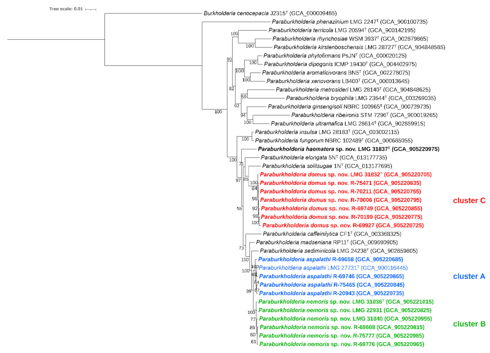
Sixty seven forest soil isolates were selected after matrix-assisted laser desorption/ionization time-of-flight mass spectrometry (MALDI-TOF MS) dereplication analyses performed in the present and previous studies [36] (Table 1). Of these isolates, 23 were picked from 1/100 diluted nutrient broth solidified with Gelzan CM (DNG), 24 from B. cepacia complex enrichment medium (BCEM), 17 from 1/10 diluted Pseudomonas cepacia azaleic acid tryptamine medium (PCAT) and 3 from modified Pikovskaya medium. Each of the 67 isolates represented a set of isolates with identical mass spectra which either remained unidentified or were identified as (Para)burkholderia species with low identification scores (1.70–1.99) when compared to the Bruker BDAL MSP-6309 database.
In the course of the present study, four Burkholderia isolates from previous studies [37,38,39,40] appeared to be closely related to isolates from the Aelmoeseneie forest soil sample (see section phylogeny). These included isolates from grassland soil, industrial coal tar-contaminated waste soil and succulent soil from the home environment of cystic fibrosis patients. An overview of all isolates is provided in Table 1.

2.2. Phylogeny

The recA gene of the 67 Aelmoeseneie forest soil isolates and the four additional soil isolates was sequenced for phylogenetic analysis. In the phylogenetic tree based on the recA gene (Figure 1), the 71 isolates represented three clusters and a single isolate, LMG 31837T, that occupied a distinct position. A total of 34 isolates clustered with P. aspalathi LMG 27731T as nearest neighbor type strain; three isolates clustered with P. solitsugae 1NT as nearest neighbor type strain and 33 isolates formed a distinct, well-separated cluster.
A subset of 18 representative isolates, based on their position in the recA gene based phylogenetic tree, was selected for whole genome sequence analysis. In addition, we also determined the whole genome sequence of P. sediminicola LMG 24238T. In a phylogenomic tree based on the analysis of 107 single-copy genes, the 18 isolates again represented a single line of descent together with the P. aspalathi, P. madseniana, P. sediminicola, P. caffeinilytica, P. solitsugae and P. elongata type strains (Figure 2). Seventeen isolates grouped in three clusters while isolate LMG 31837T again occupied a well-separated position. The isolates R-20943, R-69658, R-69746 and R-75465 grouped with P. aspalathi LMG 27731T in cluster A. Cluster B comprised LMG 31836T, LMG 22931, LMG 31840, R-69608, R-69776 and R-75777 and grouped with P. aspalathi LMG 27731T as nearest neighbor type strain. Cluster C comprised isolates LMG 31832T, R-69749, R-69927, R-70006, R-70199, R-70211 and R-75471 and grouped with P. solitsugae 1NT as nearest neighbor type strain.
The digital DNA-DNA hybridization (dDDH) and average nucleotide identity (orthoANIu) results generally confirmed the phylogenomic grouping as revealed in Figure 2 (Table 2 and Table 3). Cluster A isolates R-20943, R-69658, R-69746 and R-75465 yielded dDDH and orthoANIu values of at least 82.9% and 98.0%, respectively, towards P. aspalathi LMG 27731T, and maximum values of 57.4% and 94.2%, respectively, towards type strains of other Paraburkholderia species. The former dDDH and orthoANIu values were well above the species delineation thresholds of 70% dDDH [41] and 95–96% orthoANIu [42], and therefore cluster A isolates were assigned to P. aspalathi.
Cluster B isolates LMG 31836T, LMG 22931, LMG 31840, R-69608, R-69776 and R-75777 shared dDDH values between 89.2 and 98.0%, and orthoANIu values between 98.7 and 99.7%, again confirming they represented a single species. Average dDDH and orthoANIu values towards their closest neighbors were 62.9 ± 0.2% and 95.2 ± 0.1%, respectively, towards P. aspalathi LMG 27731T; 57.4 ± 0.2% and 94.2 ± 0.1%, respectively, towards P. madseniana RP11T; and 55.5 ± 0.1% and 93.9 ± 0.1%, respectively, towards P. sediminicola LMG 24238T. The degree of overall genomic relatedness between cluster B isolates and P. aspalathi LMG 27731T was therefore slightly below (dDDH) or near (OrthoANIu) the species delineation thresholds. Confidence intervals of the dDDH values ranged between 59.7 and 65.9% (Supplementary Table S1), and thus remained below the species delineation threshold. Average dDDH and orthoANIu values between cluster A isolates (i.e., other P. aspalathi isolates) and cluster B isolates were 63.5 ± 0.8% and 95.3 ± 0.1%, i.e., slightly below (dDDH) or near (OrthoANIu) the species delineation thresholds. Again, dDDH confidence intervals ranged between 59.9 and 68.1%, and thus remained below the species delineation threshold (dDDH).
Cluster C isolates LMG 31832T, R-69749, R-69927, R-70006, R-70199, R-70211 and R-75471 shared dDDH values between 95.1 and 98.5%, and orthoANIu values between 99.4 and 99.8%, again confirming they represented a single species. dDDH and orthoANIu values towards type strains of other Paraburkholderia species were 46.5 and 91.6%, respectively, or less, and clearly far below the species delineation thresholds. Likewise, dDDH and orthoANIu values towards cluster A and B strains were well below the species delineation thresholds.
Isolate LMG 31837T yielded dDDH and orthoANIu values of 44.8% and 91.2%, respectively, towards P. elongata 5NT, its nearest neighbor; 41.0% and 90.1%, respectively, towards P. madseniana RP11T; and 41.0% and 90.0%, respectively, towards P. solitsugae 1NT.
Finally, we extracted the 16S rRNA gene sequences from the draft genomes of the 18 strains. Analysis of these 16S rRNA sequences revealed P. sediminicola HU2-65WT as the nearest neighbor taxon for all 18 strains, with sequence similarities ranging between 98.9 and 100.0%. Cluster B isolate LMG 31836T exhibited sequence similarity values of 99.7% towards P. sediminicola HU2-65WT, 98.9% towards P. madseniana RP11T and P. aspalathi VG1CT, 98.7% towards P. insulsa PNG-AprilT and 98.6% towards P. caffeinilytica CF1T and P. kirstenboschensis Kb15T. Cluster C isolate LMG 31832T exhibited a sequence similarity of 98.9% towards P. sediminicola HU2-65WT and 98.5% towards P. metrosideri DNBP6-1T. Finally, isolate LMG 31837T yielded sequence similarity values of 99.2% towards P. sediminicola HU2-65WT, 98.8% towards P. caffeinilytica CF1T and 98.7% towards P. madseniana RP11T. These high similarity values confirmed that the 16S rRNA gene lacks resolution among closely related Paraburkholderia species.

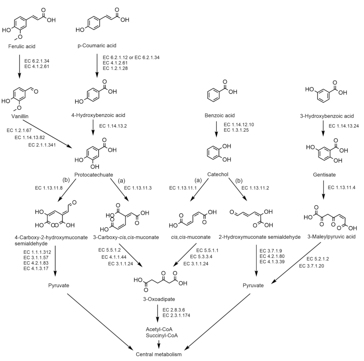
2.3. Genome Features

Genome statistics of the final genome assemblies of the 18 isolates and corresponding ENA accession numbers (PRJEB41851; P. sediminicola LMG 24238T PRJEB37806) are presented in Table 4.
Functional genome annotation of the 18 selected strains and their closest neighbors P. aspalathi LMG 27731T, P. sediminicola LMG 24238T, P. caffeinilytica CF1T, P. madseniana RP11T, P. solitsugae 1NT and P. elongata 5NT revealed and substantiated a considerable potential for the degradation of aromatic compounds. The genomes of strains LMG 31832T, LMG 31836T and LMG 31837T contained a large number of genes in the RAST SEED subsystem category for the metabolism of aromatics, i.e., 152, 150 and 134 genes, respectively. However, the largest number of genes in this category was present in the genomes of P. elongata 5NT (246 genes) and P. solitsugae 1NT (217 genes). Different pathways for the degradation of aromatic compounds were encoded in the draft genomes of the examined strains (Figure 3). Pathways for which the presence of coding sequences varied within and between species are presented in Table 5. The complete pathway for aerobic catabolism of benzoic acid to catechol (EC 1.14.12.10, EC 1.3.1.25) was encoded in the genomes of all examined strains and their closest relatives. The catechol intermediate is then degraded by a catechol 1,2-dioxygenase (EC 1.13.11.1), encoded by all strains, to cis,cis-muconate via the ortho-cleavage pathway (Figure 3). All enzymes required for further degradation of cis,cis-muconate to 3-oxoadipate are encoded in the genomes of all strains examined, except LMG 31836T and LMG 22931 (cluster B) and P. aspalathi R-20943, which lack a muconate cycloisomerase (EC 5.5.1.1) (Table 5). The enzymes required for the degradation of 3-oxoadipate (EC 2.8.3.6, EC 2.3.1.174) are encoded in the draft genomes of all examined strains. Alternatively, the catechol intermediate can be degraded to 2-hydroxymuconate semialdehyde via the meta-cleavage pathway. All cluster C strains encoded a catechol 2,3-dioxygenase (EC 1.13.11.2) for this pathway, except R-70211. LMG 31836T did not encode this enzyme; however, four other cluster B strains (LMG 31840, R-69608, R-69776, R-75777) did. The enzyme was not encoded by LMG 31837T. Close relatives P. elongata 5NT and P. aspalathi R-20943 also encoded a catechol 2,3-dioxygenase. Genes for complete further degradation of 2-hydroxymuconate semialdehyde were only present in one cluster C strain, R-69927. Cluster B strain R-69608 encoded a 2-oxopent-4-enoate hydratase (EC 4.2.1.80), which was also present in all cluster C strains but not in other cluster B strains. Cluster B strains LMG 22931 and LMG 31840 did not encode a 4-hydroxy-2-oxovalerate aldolase (EC 4.1.3.39), which was present in all cluster C strains, LMG 31837T and other cluster B strains. The genomes of all strains encoded a benzoate-CoA ligase (EC 6.2.1.25), a key enzyme for the conversion of benzoate to benzoyl-CoA. However, all strains lacked genes encoding essential enzymes for further anaerobic degradation of benzoyl-CoA. The isolates LMG 31832T, LMG 31836T and LMG 31837T did not encode a 3-hydroxybenzoate 6-monooxygenase (EC 1.14.13.24), nor did any other cluster B or C isolate. This enzyme is present in P. elongata 5NT and is a key enzyme for the degradation of 3-hydroxybenzoic acid to gentisate. None of the strains of the newly described species encoded a gentisate 1,2-dioxygenase (EC 1.13.11.4) for ring cleavage of the gentisate intermediate (Figure 3). Close relatives P. aspalathi R-20943, R-69746 and R- 75465, P. madseniana RP11T, P. solitsugae 1NT and P. elongata 5NT encoded a gentisate 1,2-dioxygenase. All strains encoded the essential enzymes for further degradation of 3-maleylpyruvic acid.
The ability to degrade aromatic compounds can be attributed to the presence of natural aromatic polymers in the soil in the form of, amongst others, lignin, tannins and humic substances. While the present literature has mainly focused on the capacity of Burkholderia and Paraburkholderia strains to degrade xenobiotic aromatic compounds, they have also been reported as bacteria that can degrade natural aromatic polymers such as lignin [23,32,33,34]. The presence of genes encoding DyP-type peroxidases, catalase-peroxidases and superoxide dismutases in the genomes of all strains suggests the ability to depolymerize lignin into oligomeric, dimeric and monomeric lignin-derived compounds (LDCs) [43]. The draft genomes of all strains encoded several enzymes involved in the degradation of monomeric LDCs such as ferulic acid and p-coumaric acid. Figure 3 shows the pathways through which these LDCs can be degraded. The enzymes required for the degradation of the LDCs and their degradation intermediates are presented in Table 5 (for coding sequences that varied within and between species). Ferulic acid is converted to vanillin via CoA ligation by feruloyl-CoA synthase (EC 6.2.1.34), which is then further degraded to protocatechuate. The complete pathway for conversion to vanillin is encoded in the draft genomes of all strains except P. caffeinilytica CF1T. However, none of the strains encoded the complete pathway for further degradation to protocatechuate. p-Coumaric acid is also converted by CoA ligation to 4-hydroxybenzoic acid and then further degraded to protocatechuate. All strains lacked the 4-coumarate-CoA ligase enzyme (EC 6.2.1.12) for CoA ligation, however, p-coumaric acid can also be converted by feruloyl-CoA synthase due to its broad substrate range [44]. The necessary enzymes for further degradation to 4-hydroxybenzoic acid and protocatechuate are present in the genomes of all strains, with the exception of a benzaldehyde dehydrogenase (EC 1.2.1.28) in all cluster C strains, including LMG 31832T. The protocatechuate intermediate is then further degraded to pyruvate via the meta-cleavage pathway or to acetyl-CoA and succinyl-CoA via the ortho-cleavage pathway. All examined strains encoded the complete ortho-cleavage pathway of protocatechuate via 3-oxoadipate (EC 1.13.11.3, EC 5.5.1.2, EC 4.1.1.44, EC 3.1.1.24, EC 2.8.3.6, EC 2.3.1.174). All strains, except P. caffeinilytica CF1T, encoded a protocatechuate 4,5-dioxygenase (EC 1.13.11.8) for ring cleavage via the meta-cleavage pathway. All cluster C strains, one cluster B strain (LMG 22931) and P. solitsugae 1NT encoded all necessary enzymes for further degradation of 4-carboxy-2-hydroxymuconate semialdehyde to pyruvate. Two cluster B strains (R-69776 and R-75777) lacked the presence of a 2-hydroxy-4-carboxymuconate 6-semialdehyde dehydrogenase (EC 1.1.1.312) but encoded all other enzymes for degradation to pyruvate.

2.4. Physiology

A phenotypic characterization of isolates LMG 31832T, LMG 31836T, LMG 31837T and their closest neighbors, P. aspalathi LMG 27731T, P. sediminicola LMG 24238T, P. caffeinilytica CF1T, P. madseniana RP11T, P. solitsugae 1NT and P. elongata 5NT was performed.
Cells of the examined strains were rod-shaped, non-motile and occurred alone or in pairs. Cells of strains LMG 31832T and LMG 31836T were approximately 2 µm long, cells of strain LMG 31837T were about 1 µm long. All cells were about 0.5 µm in diameter. After three days of incubation on NAB at 28 °C, colonies were circular with an entire edge and a smooth surface. They were flat or slightly raised, beige and opaque. They had a diameter of 1 mm.
After three days of incubation, all strains shared the following characteristics: growth on TSA and NAB at 20 and 28 °C, growth on MacConkey agar, on TSA supplemented with horse blood, growth at pH 6 and 7 and growth on Tween 20, 40, 60 and 80. The following characteristics were uniformly observed after seven days of incubation: growth on TSA and NAB at 15 °C and growth on Drigalski agar. When tested with the API 20NE microtest system, the following characteristics were present after 48 h of incubation: esculin hydrolysis and assimilation of glucose, mannose, mannitol, N-acetyl-glucosamine, potassium gluconate and malate. The following enzyme activities of the API ZYM microtest were present: butyrate esterase (C4) (weakly), caprylate esterase lipase (C8) (weakly), leucyl arylamidase and acid phosphatase.
The following characteristics were uniformly absent: growth on TSA and NAB at 4, 37, 40 and 45 °C, growth on cetrimide agar, growth in the presence of 2% to 10% of NaCl and at pH 4, 5, 8 and 9. No growth was observed on NAB, TSA and TSA supplemented with 10 mM KNO3 under anaerobic atmosphere. Hydrolysis of starch and casein and DNase activity were absent. The following characteristics were absent when examined with the API 20NE microtest system: indol production from L-tryptophane, fermentation of glucose, arginine dihydrolase and urease activity, gelatin liquefaction and assimilation of capric acid. The following characteristics were absent when examined with the API ZYM microtest system: myristate lipase (C14), valine arylamidase, cystin arylamidase, trypsin, chymotrypsin, alpha-galactosidase, beta-galactosidase, beta-glucuronidase, alpha-glucosidase, beta-glucosidase, N-acetyl-beta-glucosaminidase, alpha-mannosidase and alpha-fucosidase.
Strains LMG 31832T, LMG 31836T, LMG 31837T and their closest phylogenetic neighbors could be differentiated based on several characteristics (Table 6). The lack of beta-galactosidase activity distinguished strains LMG 31832T, LMG 31836T and LMG 31837T from P. aspalathi LMG 27731T and P. sediminicola LMG 24238T. Strains LMG 31832T and LMG 31836T were also differentiated from their closest neighbors by their ability to reduce nitrate and could be differentiated from each other based on assimilation of N-acetyl-glucosamine, trisodium citrate and phenylacetic acid. Strain LMG 31837T could be differentiated from its closest neighbor P. elongata 5NT based on hemolysis on horse blood agar and growth in 1% NaCl.

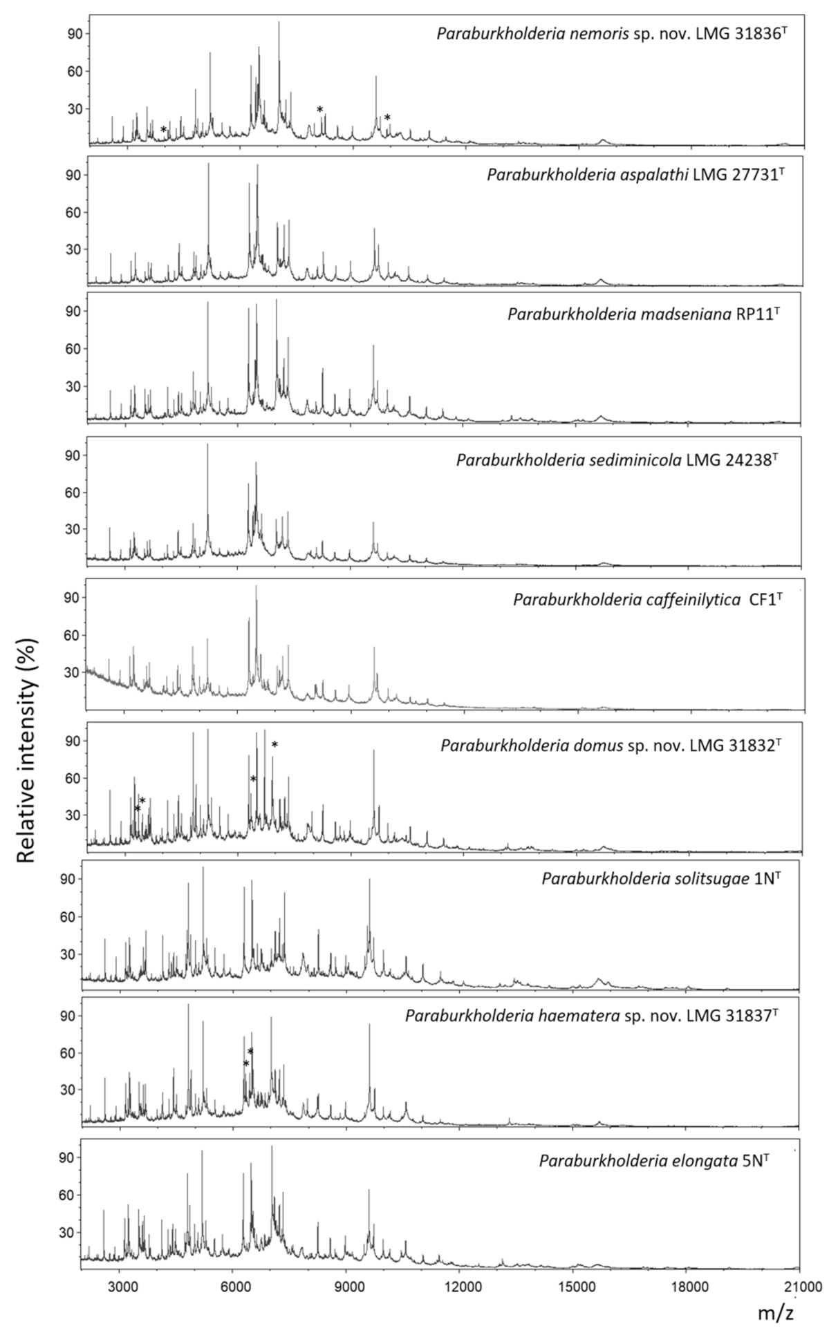
2.5. MALDI-TOF MS Analysis

All strains had highly similar MALDI-TOF MS profiles (Figure 4). Strain LMG 31836T showed two peaks at m/z 8152.8 ± 0.5 and 9892.3 ± 0.2 that were not present in the spectra of its closest neighbors P. aspalathi LMG 27731T, P. madseniana RP11T and P. sediminicola LMG 24238T. Strain LMG 31836T could also be differentiated from P. madseniana RP11T and P. sediminicola LMG 24238T by a peak at 4076.2 ± 0.4. Strain LMG 31832T could be differentiated from its closest neighbor P. solitsugae 1NT by four peaks at m/z 3350.4 ± 0.5, 3451.4 ± 0.7, 6336.8 ± 0.7 and 6905.4 ± 0.8. Moreover, P. solitsugae 1NT showed two peaks at m/z 6634.2 ± 1.8 and 7006.5 ± 2.2 that were not present in the spectra of strain LMG 31832T. Finally, LMG 31837T could be differentiated from its closest neighbor P. elongata 5NT by two peaks at m/z 6325.5 ± 1.0 and 6425.9 ± 0.7.

3. Discussion

In the frame of several efforts to maximally isolate the cultivable bacterial community of a topsoil sample of the Aelmoeseneie forest, more than 5000 isolates have been examined thus far ([36] and data from the present study). These isolates have all been dereplicated by MALDI-TOF MS using the SPeDE algorithm [45] to remove redundant isolates. The resulting SPeDE ‘references’ are strains with distinct MALDI-TOF mass spectra, as a proxy to isolates with a distinct genetic background [45] and included 67 isolates examined in the present study. RecA gene sequence analysis identified four additional isolates from earlier studies [37,38,39,40] that were closely related to the 67 forest soil isolates.
We used recA gene sequencing to subsequently select 18 isolates for comparative genomics analyses, in order to represent the observed diversity in recA gene sequences in a redundant manner (Figure 1). Their position in the recA and phylogenomic trees (Figure 1; Figure 2) and the dDDH and orthoANIu values (Table 2; Table 3) allowed to assign nine out of 71 isolates to P. aspalathi, 25 to the taxon represented by strain LMG 31836T, 36 to the taxon represented by strain LMG 31832T and a single isolate to the taxon represented by LMG 31837T. The divergence in recA gene sequences and the dDDH and OrthoANIu values among isolates for which draft genome sequences were generated demonstrated considerable genetic divergence within species for which multiple isolates were studied. Data from the present study therefore demonstrated that the 67 isolates from the Aelmoeseneie forest soil sample and four soil isolates from earlier studies [37,38,39,40] represented multiple clonal variants within four genospecies, one of which was identified as P. aspalathi, an organism from root nodules of a South African legume plant [5]. This included strain R-20943 (=Hg8) which was isolated from a coal tar-contaminated hillside soil in the USA [37].
A second group of 25 isolates, represented by strain LMG 31836T, appeared to be closely related to P. aspalathi LMG 27731T with average dDDH values of 62.9 ± 0.2% and average orthoANIu values of 95.2 ± 0.1% (Table 2; Table 3). The dDDH value was slightly below the species delineation threshold of 70% and was supported by confidence intervals ranging between 59.7% and 65.9%. In contrast, the orthoANIu value was at the species delineation threshold. Strain LMG 31836T could be differentiated from P. aspalathi LMG 27731T by several phenotypic characteristics (Table 6) and by its MALDI-TOF MS profile (Figure 4) which argued against a classification as P. aspalathi. Classifying these 25 isolates into a novel subspecies within P. aspalathi would not be appropriate either as subspecies typically share dDDH levels of 80% or higher [46]. Together, these data suggest that overall genomic relatedness values that are near the species delineation thresholds should be used in concert with other data to propose a classification that best reflects all data. We consider it most appropriate to classify strain LMG 31836T into a novel Paraburkholderia species. Isolates of this novel species have genomes with an average G+C content of 61.4 ± 0.1 mol% and which range in size between 8.64 and 9.28 Mbp (Table 4). This novel Paraburkholderia species and P. aspalathi cannot be distinguished through recA gene sequence analysis which corroborates the close genomic relatedness of both taxa (Figure 1). Strain LMG 22931 (=G47-12) was isolated from grassland soil in the Netherlands in 2003 and belongs to the same novel Paraburkholderia species [40].
A third group of 36 isolates, represented by strain LMG 31832T, formed a well separated novel species within this Paraburkholderia lineage with P. solitsugae 1NT as its nearest neighbor taxon (Figure 2) and could be distinguished from the type strains of its nearest neighbor taxa through various genomic and phenotypic analyses (Figure 1; Figure 2, Table 2, Table 3 and Table 6). Isolates of this novel species have genomes with an average G+C content of 61.3 ± 0.1 mol% and which range in size between 8.19 and 9.46 Mbp (Table 4). This novel species also includes strain LMG 19510 (=DSM 6431) which was isolated from industrial waste deposit soil in Germany [38] and strain R-23782 which was isolated from soil of a succulent plant in the home of a cystic fibrosis patient in Belgium [39].
Finally, strain LMG 31837T also represented a well-separated novel species within this Paraburkholderia lineage with P. elongata 5NT as its nearest neighbor taxon (Figure 2) and could be distinguished from the type strains of its nearest neighbor taxa through various genomic and phenotypic analyses (Figure 1; Figure 2, Table 2, Table 3 and Table 6). Its genome has a G+C content of 61.71 mol% and is 7.82 Mbp in size (Table 4). Remarkably, among the more than 5000 isolates that were examined in the course of several isolation campaigns, this was the only isolate that represented this novel species.
Like several of their nearest neighbor taxa and other Paraburkholderia species [2,18,21] these novel species have a metabolism that is highly adapted to the degradation of aromatic compounds. While the present literature has mainly focused on the capacity of Burkholderia and Paraburkholderia strains to degrade xenobiotic aromatic compounds, they have also been reported as bacteria that can degrade lignin [23,32,33,34] which is the most abundant natural aromatic polymer [23,47]. We hypothesize that the co-occurrence of multiple closely related Paraburkholderia strains and species with the capacity to degrade aromatic compounds in pristine forest soil is related to the presence of structurally complex, natural aromatic compounds such as lignin, tannins and humic substances generated through the decomposition of plant litter [24,25,26]. The degradation of such aromatic polymers, in particular lignin, has mostly been described for fungi [48]. Nonetheless, the genomes of all strains examined in the present study encoded enzymes involved in the depolymerization of lignin and the degradation of monomeric LDCs. A mutualistic relationship may exist between fungi and bacteria in soil, in which bacteria consume lignin degradation products generated by fungi [49,50]. Mutualistic relations between fungi and Burkholderia (now Paraburkholderia) in soils have indeed been described before [51]. The co-occurrence of multiple closely related Paraburkholderia strains and species in a single topsoil sample may highlight a diversity in micro-habitats with different species and strains occupying different micro-niches [52], or may be indicative of synergistic relationships, whereby strains encode different parts of degradation pathways. The latter is supported by the fact that the genomes of the examined species and strains did not encode the exact same pathways for aromatic compound degradation.
The observation that genetically distinct but closely related strains partition their environment at fine phylogenetic and phylogenomic scales is a finding that is not well understood. The existence and function of such microdiversity, i.e., the co-occurrence of closely related but ecologically and physiologically distinct taxonomic groups has been documented for about two decades and is an intrinsic property of many microorganisms [53]. Among others, microdiversity has been attributed to resource use and phosphate acquisition [52]. A deeper mining of the genomic and phenotypic potential of different strains of each of these four Paraburkholderia species will be required to improve our understanding of the functional role of this microdiverse Paraburkholderia community.

4. Materials and Methods

4.1. Isolation of Paraburkholderia Isolates

The Aelmoeseneie forest (Gontrode, Belgium: 50°58′ N 3°49′ E) is a temperate deciduous forest of 28 ha with mixed tree species (oak, beech, ash and others) and a lush undergrowth located in northwestern Belgium [54]. The southeastern part of the forest is considered ancient as it has been continuously forested since at least the oldest land-use map of 1775 [55,56]. One square meter of the top layer of soil (0–10 cm depth) was collected, sieved through a sterile brass sieve (aperture 1 cm), homogenized using a new concrete mixer that was decontaminated using sodium hypochlorite, aliquoted into 250 mL Nalgene© bottles and stored at −80 °C. In the frame of several studies, we performed large-scale campaigns to isolate the cultivable bacterial community of this soil sample.
For isolation experiments, 9 g of soil was mixed with 90 mL sodium pyrophosphate (0.05 M, pH 8) for 3 min. To maximize harvesting all cells, this suspension was centrifuged 4 times for 5 min. at 130× g, 2 times for 5 min. at 60× g, 2 times for 1 min. at 40× g and finally 2 times for 1 min. at 20× g. Between each centrifugation step, the supernatants were transferred and the pellet was resuspended in 45 mL sodium pyrophosphate and mixed for 3 min. Subsequently, all cell fractions were centrifuged for 15 min. at 8226× g, the supernatants were removed, and the cell pellets were resuspended in 5 mL saline (0.85% NaCl). Finally, all cell fractions were pooled and a 10-fold dilution series was prepared in saline and plated (i) onto 1/100 diluted nutrient broth (Oxoid, Basingstoke, UK) solidified with Gelzan CM (1 g L−1;Sigma-Aldrich, St.-Louis, MO, USA) (DNG) and supplemented with MgSO4·7H2O (1 g L−1) and cycloheximide (1 g L−1) and (ii) onto 1/10 diluted Pseudomonas cepacia azaleic acid tryptamine (PCAT) medium [57]. Isolates from DNG were picked after 4 weeks of incubation at 20 °C and transferred to 96-well microtiter plates containing 1/10 diluted nutrient broth (DNB; Oxoid, Basingstoke, UK). Isolates from 1/10 PCAT were picked after 2 weeks of incubation at 20 °C and transferred to 96-well microtiter plates containing tryptone soy broth (TSB; Oxoid, Basingstoke, UK). Isolates were subcultivated using the VIAFLO 96/384 liquid handler (Integra-Biosciences©, Zizers, Switzerland) until the third generation and cell suspensions were prepared for MALDI-TOF MS as described earlier [45].
Additionally, 9 g of soil sample was homogenized in 90 mL physiological saline for 3 min. A 10-fold serial dilution in saline was prepared and plated on B. cepacia complex enrichment medium (BCEM) [57] and on modified Pikovskaya medium for the isolation of phosphate solubilizers [58]. After 5 days of incubation at 20 °C isolates were picked and subcultivated until the third generation on phosphate buffered (0.45 g L−1 KH2PO4/2.39 g L−1 Na2HPO4·12H2O) nutrient agar (NAB; Oxoid, Basingstoke, UK) or modified Pikovskaya medium, respectively. Approximately 1 µL loop of biological material of the isolates picked from BCEM was harvested and transferred to sterile deepwell plates (Thermo Scientific, Roskilde, Denmark) containing phosphate buffered nutrient broth (BNB; Oxoid, Basingstoke, UK). After an incubation period of 6 days at 20 °C, cell suspensions for MALDI-TOF MS analysis were prepared as described earlier [45]. For the isolates picked from Pikovskaya medium, extracts were prepared for MALDI-TOF MS analysis as described previously [59].
MALDI-TOF MS spectra were generated for all isolates using a Bruker MicroflexTM LT/SH system (Bruker Daltonics, Bremen, Germany). Spectra were dereplicated with the SPeDE algorithm [45].

4.2. Phylogeny

The recA gene of 67 forest soil isolates and four additional soil isolates was sequenced for phylogenetic analysis. DNA was extracted by adding 1 µL loop of cell material to 20 µL of alkaline lysis buffer containing 0.25% (w/v) SDS and 0.05 M NaOH and heating to 95 °C for 15 min. After lysis, 180 µL of distilled water was added. The recA gene (663 bp) was amplified using a Taq DNA polymerase kit (Qiagen, Antwerp, Belgium) with forward primer 5′-AGGACGATTCATGGAAGAWAGC-3′ and reverse primer 5′- GACGCACYGAYGMRTAGAACTT-3′ [60,61]. The resulting products were purified using a NucleoFast 96 PCR clean-up kit (Macherey-Nagel, Eupen, Belgium). Purified products were sequenced by a commercial company (Eurofins, Ebersberg, Germany) using the above-described primers, a 3730xl Genetic Analyzer (Thermo Fisher Scientific, Roskilde, Denmark) and the Big Dye terminator v3.1 cycle sequencing kit (Thermo Fisher Scientific, Roskilde, Denmark). Sequences were assembled using BioNumerics version 7.6 (Applied Maths, Sint-Martens-Latem, Belgium). All recA gene sequences were compared with those of the type strains of all validly named Paraburkholderia species (data not shown). Sequences (633–1074 bp) were aligned based on amino acid sequences using Muscle [62] in MEGA X [63]. A phylogenetic tree of the 71 isolates and of the type strains of their nearest neighbor taxa, i.e., P. aspalathi LMG 27731T, P. madseniana RP11T, P. sediminicola LMG 24238T, P. caffeinilytica CF1T, P. solitsugae 1NT and P. elongata 5NT, was constructed using RAxML version 8.2.12 [64] with the GTRGAMMA substitution model and 1000 bootstrap analyses. All recA gene sequences were submitted into the European Nucleotide Archive (ENA) and accession numbers can be found in Figure 1.
Using their position in the recA gene based phylogenetic tree (Figure 1) a subset of 18 representative isolates was subsequently selected for whole genome sequence analysis to assess the genomic divergence within and between each of the clusters and the Paraburkholderia type strains. In addition, we determined the whole genome sequence of P. sediminicola LMG 24238T.
DNA was extracted using an automated Maxwell® DNA preparation instrument (Promega, Madison, WI, USA). DNA extracts were treated with RNAse (2 mg mL−1, 5 µL per 100 µL extract) and incubated at 37 °C for one hour. DNA quality was checked using 1% agarose gel electrophoresis and DNA quantification was performed using the QuantiFluor ONE dsDNA system and the Quantus fluorometer (Promega, Madison, WI, USA). In the first series of analyses, draft genomes of strains R-69608, R-69658, R-69746, R-69749, R-69776, R-69927, R-70006, R-70199, R-70211 and LMG 31837T were sequenced using the Illumina HiSeq4000 (PE150) platform at the Oxford Genomics Centre (Oxford, UK). In the second series of analyses, draft genomes of strains R-20943, R-75465, R-75777, R-75471, LMG 22931, LMG 31832T, LMG 31836T and LMG 24238T were sequenced using the Illumina NovaSeq6000 (PE150) platform. Finally, the draft genome of LMG 31840 was sequenced at the MiGS center (Pittsburgh, USA) using the Illumina NextSeq550 (PE150) platform. Quality reports were created with FastQC version 0.11.8. Prior to assembly, reads were trimmed (Phred score >Q30) and filtered (length >50 bp) with fastp 0.20.0 [65] with the correction option enabled. Assembly was performed with Shovill version 1.1.0, with SPAdes genome assembler 3.14.0 at its core and read error correction disabled. Contigs shorter than 500 bp were removed from the final assembly. Quality of the final assembly was verified with The Quality Assessment Tool for Genome Analysis (QUAST) which generates summary statistics such as the number of contigs, N50, L50 and the G+C content [66]. Raw reads were mapped against the assemblies using BWA-MEM [67]. Mapped reads were subjected to a quality control using Qualimap [68] to determine the coverage and error rate. Finally, the assemblies were checked for completeness and contamination using CheckM version 1.1.2 [69]. Annotation was performed using Prokka 1.14.5 [70]. The annotated genome assemblies were submitted into ENA and are publicly available under PRJEB41851 and PRJEB37806.
The genomes were submitted to the Type (Strain) Genome Server (TYGS) (accessed on 26-01-2021) [71] to identify their nearest phylogenomic neighbors and to calculate their degree of relatedness towards these nearest neighbor species. Digital DNA-DNA hybridization (dDDH) values and confidence intervals were calculated using the recommended settings of the GGDC 2.1. [41]. The average nucleotide identity (ANI) values were calculated using the OrthoANIu algorithm [72]. The whole-genome sequences of the 18 isolates and of the type strains of closely related Paraburkholderia species (downloaded from GenBank except for P. sediminicola LMG 24238T) were also used to construct a phylogenomic tree based on the analysis of 107 single-copy core genes using bcgTree [73] with Burkholderia cenocepacia J2315T as outgroup. Visualization and annotation of the tree was performed using iTOL [74].
For completeness, 16S rRNA gene sequences were extracted from the draft genomes of all 18 isolates using the BAsic Rapid Ribosomal RNA Predictor software (Barrnap) [75] and were submitted to the EZBiocloud identification service (accessed on 25-02-2020) [76]. The 16S rRNA gene sequences of the type strains LMG 31832T, LMG 31836T and LMG 31837T were submitted into ENA and are publicly available under HG995123, HG995124 and HG995125, respectively.

4.3. Functional Genome Annotation

The assembled genomes of the 18 selected isolates and of P. aspalathi LMG 27731T, P. sediminicola LMG 24238T, P. caffeinilytica CF1T, P. madseniana RP11T, P. solitsugae 1NT and P. elongata 5NT were submitted to RAST (accessed on 17-12-2020) [77] and eggNOG-mapper v2 (version 2.1.4) (accessed on 23-06-2021) [78,79] for functional genome annotation with a focus on aromatic compound degradation, a key characteristics of P. madseniana, P. solitsugae and P. elongata [2,18].

4.4. Physiology

A phenotypic characterization of isolates LMG 31832T, LMG 31836T, LMG 31837T and their closest neighbors, P. aspalathi LMG 27731T, P. sediminicola LMG 24238T, P. caffeinilytica CF1T, P. madseniana RP11T, P. solitsugae 1NT and P. elongata 5NT was performed.
Cell and colony morphology were assessed after three days of incubation on phosphate buffered (0.45 g/L KH2PO4/2.39 g/L Na2HPO4·12H2O) nutrient agar (NAB; Oxoid, Basingstoke, UK) at 28 °C. Motility was observed in young cultures by examining wet mounts in broth by phase-contrast microscopy. The assessment of catalase and oxidase activities and other tests were performed using conventional procedures [80]. Aerobic growth was tested at 28 °C on NAB, TSA, MacConkey agar (Oxoid, Basingstoke, UK), Drigalski agar (Biorad, Temse, Belgium) and Pseudomonas cetrimide agar (Oxoid, Basingstoke, UK). The temperature growth range was tested on NAB and TSA at 4, 15, 20, 28, 37, 40 and 45 °C. The effect of NaCl on growth was investigated in nutrient broth (Oxoid, Basingstoke, UK) with different concentrations of NaCl (0–10% with 1% intervals, w/v). The pH range for growth was evaluated in nutrient broth buffered at pH 4.0 to 9.0 at intervals of 1 pH unit using the following buffer systems: acetate buffer (4.0–5.0), phosphate buffer (pH 6.0–8.0) and Tris-HCl (pH 9.0). Anaerobic growth was tested on NAB, TSA and TSA supplemented with 10 mM KNO3. Growth tests were read after 1, 2, 3, 5 and 7 days of incubation. Hemolysis of horse blood was tested on TSA supplemented with 5% horse blood. DNase activity was tested using DNase agar (Sigma-Aldrich, St.-Louis, MO, USA). Starch hydrolysis was tested using NAB and TSA supplemented with 0.8% (w/v) soluble starch. Hydrolysis of casein was tested using Plate count agar (Oxoid, Basingstoke, UK) supplemented with 1.3% (w/v) dried skim milk (Oxoid, Basingstoke, UK). Hydrolysis of Tween 20, 40, 60 and 80 was tested as described by Sierra [81]. The activity of constitutive enzymes and other physiological properties were determined after growth on NAB for 2 days at 28 °C using the API 20NE and API ZYM microtest systems (BioMérieux, Marcy l’Etoile, France), according to the manufacturer’s instructions. Results were read after 24 h and 48 h, and after 4 h of incubation, respectively.

4.5. MALDI-TOF MS Analysis

MALDI-TOF MS profiles of strains LMG 31832T, LMG 31836T, LMG 31837T and their closest neighbors were obtained as described before [45]. The strains were cultivated on NAB at 28 °C for 2 days before cell extracts were prepared. Two technical and two biological replicates were obtained for each strain. The profiles were visualized using mMass 5.5.0 [82].

5. Conclusions

In conclusion, data from the present study confirmed that strains LMG 31832T, LMG 31836T and LMG 31837T represent three novel Paraburkholderia species, which can be distinguished from their nearest neighbor species using genomic (Table 2; Table 3, Figure 1; Figure 2), phenotypic (Table 6) and chemotaxonomic (Figure 4) characteristics.
We propose to classify the isolates examined in the present study into three novel Paraburkholderia species as Paraburkholderia domus sp. nov. with LMG 31832T (=CECT 30334) as the type strain, Paraburkholderia nemoris sp. nov. with LMG 31836T (=CECT 30335) as the type strain and Paraburkholderia haematera sp. nov. with LMG 31837T (=CECT 30336) as the type strain.

5.1. Description of Paraburkholderia domus sp. nov.

Paraburkholderia domus sp. nov. (do’mus. L. fem. gen. n. domus, from a home, household; because one of the earliest isolates of this species was collected from the home environment of a person with cystic fibrosis).
Cells are non-motile, rod-shaped, approximately 2 µm long and 0.5 µm wide. After incubation on NAB at 28 °C for three days, colonies have a diameter of 1 mm and are circular, opaque and beige. They are flat or slightly raised and have an entire edge and a smooth surface. Strain LMG 31832T optimally grows on NAB and TSA between 15 and 28 °C in aerobic conditions, and does not grow in anaerobic conditions on NAB, TSA or TSA supplemented with 10 mM KNO3. No growth at 4, 37, 40 and 45 °C. Growth is observed in the presence of 0% and 1% NaCl, but not 2% or more. Growth at pH 6.0 and 7.0, but not at 4.0, 5.0, 8.0 or 9.0. Growth on MacConkey and Drigalski agar, no growth on cetrimide agar. Growth on Tween 20, 40, 60 and 80 agar base and hydrolysis of all Tweens after 7 days of incubation. Hemolysis on horse blood agar but no DNase activity and hydrolysis of starch or casein. Oxidase and weak catalase activity is present.
When examined with the API 20NE microtest system, the following characteristics are present: nitrate reduction, esculin hydrolysis, assimilation of glucose, arabinose (weakly), mannose, mannitol, potassium gluconate, adipic acid and malate (weakly) but not indol production from L-tryptophane, fermentation of glucose, arginine dihydrolase, urease and beta-galactosidase (PNPG) activity, gelatin liquefaction and assimilation of N-acetyl-glucosamine, maltose, capric acid, trisodium citrate and phenylacetic acid.
When examined with the API ZYM microtest system, the following characteristics are present: alkaline phosphatase (weakly), butyrate esterase (C4) (weakly), caprylate esterase lipase (C8) (weakly), leucyl arylamidase, acid phosphatase and phosphoamidase but not myristate lipase (C14), valine arylamidase, cystin arylamidase, trypsin, chymotrypsin, alpha-galactosidase, beta-galactosidase, beta-glucuronidase, alpha-glucosidase, beta-glucosidase, N-acetyl-beta-glucosaminidase, alpha-mannosidase and alpha-fucosidase.
The type strain, LMG 31832T (=CECT 30334), was isolated in 2018 from forest soil. Other strains were isolated from soil of a succulent plant in the home environment of a person with cystic fibrosis and from industrial waste deposit soil (Table 1). The whole-genome sequence of strain LMG 31832T has a size of 8.75 Mbp. The G+C content is 61.32 mol%. The 16S rRNA gene, recA gene and whole-genome sequences of strain LMG 31832T are publicly available under accession numbers HG995123, HG995062 and CAJNAT000000000, respectively.

5.2. Description of Paraburkholderia nemoris sp. nov.

Paraburkholderia nemoris sp. nov. (ne.mo’ris. L. neut. gen. n. nemoris, from a forest; because most isolates of this species were obtained from a forest soil sample).
Cells are non-motile, rod-shaped, approximately 2 µm long and 0.5 µm wide. After incubation on NAB at 28 °C for three days, colonies have a diameter of 1 mm and are circular, opaque and beige. They are flat or slightly raised and have an entire edge and a smooth surface. Strain LMG 31836T grows optimally on NAB and TSA between 15 and 28 °C in aerobic conditions, but does not grow in anaerobic conditions on NAB, TSA or TSA supplemented with 10 mM KNO3. No growth at 4, 37, 40 and 45 °C. Growth is observed in the presence of 0 and 1% NaCl, but not 2% or more. Growth at pH 6.0 and 7.0, but not at 4.0, 5.0, 8.0 or 9.0. Growth on MacConkey agar, delayed growth on Drigalski agar and no growth on cetrimide agar. Growth on Tween 20, 40, 60 and 80 agar base and hydrolysis of all Tweens after 7 days of incubation. Hemolysis on horse blood agar but no DNase activity and hydrolysis of starch or casein. Oxidase and weak delayed catalase activity is present.
When examined with the API 20NE microtest system, the following characteristics are present: nitrate reduction, esculin hydrolysis, assimilation of glucose, arabinose (weakly), mannose, mannitol, N-acetyl-glucosamine, potassium gluconate, adipic acid, malate, trisodium citrate and phenylacetic acid, but not indol production from L-tryptophane, fermentation of glucose, activity of arginine dihydrolase, urease and beta-galactosidase (PNPG), gelatin liquefaction and assimilation of maltose and capric acid.
When examined with the API ZYM microtest system, the following characteristics are present: alkaline phosphatase (weakly), butyrate esterase (C4) (weakly), caprylate esterase lipase (C8) (weakly), leucyl arylamidase, acid phosphatase (weakly) and phosphoamidase (weakly) but not myristate lipase (C14), valine arylamidase, cystin arylamidase, trypsin, chymotrypsin, alpha-galactosidase, beta-galactosidase, beta-glucuronidase, alpha-glucosidase, beta-glucosidase, N-acetyl-beta-glucosaminidase, alpha-mannosidase and alpha-fucosidase.
The type strain, LMG 31836T (=CECT 30335), was isolated in 2018 from forest soil. The whole-genome sequence of strain LMG 31836T has a size of 9.27 Mbp. The G+C content is 61.59 mol%. The 16S rRNA gene, recA gene and whole-genome sequences of strain LMG 31836T are publicly available under accession numbers HG995124, HG995055 and CAJNBO000000000, respectively.

5.3. Description of Paraburkholderia haematera sp. nov.

Paraburkholderia haematera sp. nov. (hae.ma.te’ra. Gr. adj. haemateros, blood-thirsty; N.L. fem. adj. haematera, blood-thirsty; because of its hemolytic behavior on horse blood agar).
Cells are non-motile, rod-shaped, approximately 1 µm long and 0.5 µm wide. After incubation on NAB at 28 °C for three days, colonies have a diameter of 1 mm and are circular, opaque and beige. They are flat or slightly raised and have an entire edge and a smooth surface. Strain LMG 31837T optimally grows on NAB and TSA between 15 and 28 °C in aerobic conditions, but does not grow in anaerobic conditions on NAB, TSA or TSA supplemented with 10 mM KNO3. No growth at 4, 37, 40 and 45 °C. Growth in the presence of 0% and 1% NaCl, but not 2% or more. Growth at pH 6.0 and 7.0, but not at 4.0, 5.0, 8.0 or 9.0. Growth on MacConkey and Drigalski agar and no growth on cetrimide agar. Growth on Tween 20, 40, 60 and 80 agar base and hydrolysis of all Tweens after 7 days of incubation. Hemolysis on horse blood agar but no DNase activity and hydrolysis of starch or casein. Oxidase and weak catalase activity is present.
When examined with the API 20NE microtest system, the following characteristics are present: esculin hydrolysis, assimilation of glucose, arabinose (weakly), mannose, mannitol, N-acetyl-glucosamine, potassium gluconate, adipic acid (weakly), malate, trisodium citrate and phenylacetic acid but not nitrate reduction, indol production from L-tryptophane, fermentation of glucose, activity of arginine dihydrolase, urease and beta-galactosidase (PNPG), gelatin liquefaction and assimilation of maltose and capric acid.
When examined with the API ZYM microtest system, the following characteristics are present: alkaline phosphatase (weakly), butyrate esterase (C4) (weakly), caprylate esterase lipase (C8) (weakly), leucyl arylamidase, acid phosphatase and phosphoamidase (weakly) but not myristate lipase (C14), valine arylamidase, cystin arylamidase, trypsin, chymotrypsin, alpha-galactosidase, beta-galactosidase, beta-glucuronidase, alpha-glucosidase, beta-glucosidase, N-acetyl-beta-glucosaminidase, alpha-mannosidase and alpha-fucosidase.
The type strain is LMG 31837T (=CECT 30336) and was isolated in 2016 from forest soil. The whole-genome sequence of strain LMG 31837T has a size of 7.82 Mbp. The G+C content is 61.71 mol%. The 16S rRNA gene, recA gene and whole-genome sequences of strain LMG 31837T are publicly available under accession numbers HG995125, HG995035 and CAJNBK000000000, respectively.

5.4. Emended Description of Paraburkholderia sediminicola Lim et al. 2008

The description is as before [83] with the following addition. The G+C content of the type strain genome is 61.4 mol%, its approximate size is 8.64 Mbp. The whole-genome sequence of strain LMG 24238T is available under accession number CADIKC000000000.

Supplementary Materials

The following are available online at https://www.mdpi.com/article/10.3390/ijms22137003/s1.

Author Contributions

Conceptualization, S.V., N.B. and P.V.; methodology, S.V., A.D.W., C.P. and P.V.; software, S.V., A.D.W. and C.P.; formal analysis, S.V., E.D.R., C.D., A.D.W. and C.P.; data curation, S.V. and C.P.; writing—original draft preparation, S.V. and P.V.; writing—review and editing, E.D.R., C.D., A.D.W., C.P. and N.B.; visualization, S.V., A.D.W. and C.P.; supervision, P.V.; project administration, S.V., C.P., N.B. and P.V.; funding acquisition, N.B. and P.V. All authors have read and agreed to the published version of the manuscript.

Funding

This work was funded by a research grant from the Geconcerteerde Onderzoeksacties (GOA) of Ghent University (BOF15/GOA/006).

Institutional Review Board Statement

Not applicable.

Informed Consent Statement

Not applicable.

Data Availability Statement

Gene and genome sequences were deposited in the European Nucleotide Archive (ENA). The recA and 16S rRNA gene sequences, raw reads and annotated assemblies can be found via the BioProject accession numbers PRJEB41851 and PRJEB37806, and the accession numbers listed in Figure 1, Figure 2 and Table 4.

Acknowledgments

We thank Kris Verheyen for giving access to the Aelmoeseneie forest. We thank Margo Cnockaert for technical assistance. We thank the Oxford Genomics Centre at the Welcome Centre for Human Genetics (funded by Wellcome Trust grant reference 203141/Z/16/Z) for the generation and initial processing of the sequencing data. The research leading to results presented in this publication was carried out with infrastructure funded by EMBRC Belgium—FWO project GOH3817N.

Conflicts of Interest

The authors declare no conflict of interest.

References

  1. Vandamme, P.; Peeters, C.; De Smet, B.; Price, E.P.; Sarovich, D.S.; Henry, D.A.; Hird, T.J.; Zlosnik, J.E.A.; Mayo, M.; Warner, J.; et al. Comparative Genomics of Burkholderia singularis sp. nov., a Low GCC content, Free-Living bacterium that defies taxonomic dissection of the genus Burkholderia. Front. Microbiol. 2017, 8, 1679. [Google Scholar] [CrossRef] [PubMed] [Green Version]
  2. Wilhelm, R.C.; Murphy, S.J.L.; Feriancek, N.M.; Karasz, D.C.; Derito, C.M.; Newman, J.D.; Buckley, D.H. Paraburkholderia madseniana sp. nov., a phenolic acid-degrading bacterium isolated from acidic forest soil. Int. J. Syst. Evol. Microbiol. 2020, 70, 2137–2146. [Google Scholar] [CrossRef] [PubMed]
  3. Rusch, A.; Islam, S.; Savalia, P.; Amend, J.P. Burkholderia insulsa sp. nov., a facultatively chemolithotrophic bacterium isolated from an arsenic-rich shallow marine hydrothermal system. Int. J. Syst. Evol. Microbiol. 2015, 65, 189–194. [Google Scholar] [CrossRef] [PubMed]
  4. Xiao, S.Y.; Gao, Z.H.; Lin, Q.H.; Qiu, L.H. Paraburkholderia pallida sp. Nov. And paraburkholderia silviterrae sp. Nov., isolated from forest soil. Int. J. Syst. Evol. Microbiol. 2019, 69, 3777–3785. [Google Scholar] [CrossRef]
  5. Mavengere, N.R.; Ellis, A.G.; Le Roux, J.J. Burkholderia aspalathi sp. nov., isolated from root nodules of the South African legume Aspalathus abietina Thunb. Int. J. Syst. Evol. Microbiol. 2014, 64, 1906–1912. [Google Scholar] [CrossRef]
  6. Xiao, S.Y.; Gao, Z.H.; Yang, Z.; Bi, J.Y.; Qiu, L.H. Paraburkholderia telluris sp. nov., isolated from subtropical forest soil. Int. J. Syst. Evol. Microbiol. 2019, 69, 1274–1280. [Google Scholar] [CrossRef]
  7. Choi, G.M.; Im, W.T. Paraburkholderia azotifigens sp. nov., a nitrogen-fixing bacterium isolated from paddy soil. Int. J. Syst. Evol. Microbiol. 2018, 68, 310–316. [Google Scholar] [CrossRef]
  8. Nally, E.; Groah, S.L.; Pérez-Losada, M.; Caldovic, L.; Ljungberg, I.; Chandel, N.J.; Sprague, B.; Hsieh, M.H.; Pohl, H.G. Identification of Burkholderia fungorum in the urine of an individual with spinal cord injury and augmentation cystoplasty using 16S sequencing: Copathogen or innocent bystander? Spinal Cord Ser. Cases 2018, 4. [Google Scholar] [CrossRef] [Green Version]
  9. Loong, S.K.; Soh, Y.H.; Mahfodz, N.H.; Johari, J.; Abu Bakar, S. Synovial tissue infection with Burkholderia fungorum. Emerg. Infect. Dis. 2016, 22, 1834–1835. [Google Scholar] [CrossRef] [Green Version]
  10. Gerrits, G.P.; Klaassen, C.; Coenye, T.; Vandamme, P.; Meis, J.F. Burkholderia fungorum septicemia. Emerg. Infect. Dis. 2005, 11, 1115–1117. [Google Scholar] [CrossRef]
  11. Deris, Z.Z.; Van Rostenberghe, H.; Habsah, H.; Noraida, R.; Tan, G.C.; Chan, Y.Y.; Rosliza, A.R.; Ravichandran, M. First isolation of Burkholderia tropica from a neonatal patient successfully treated with imipenem. Int. J. Infect. Dis. 2010, 14, e73–e74. [Google Scholar] [CrossRef] [Green Version]
  12. Marks, L.R.; Dodd, H.; Russo, T.A.; Berenson, C.S. Burkholderia ginsengisoli bacteraemia: Emergence of a novel pathogen. BMJ Case Rep. 2016, 2016, bcr2015213584. [Google Scholar] [CrossRef] [Green Version]
  13. Depoorter, E.; Bull, M.J.; Peeters, C.; Coenye, T.; Vandamme, P.; Mahenthiralingam, E. Burkholderia: An update on taxonomy and biotechnological potential as antibiotic producers. Appl. Microbiol. Biotechnol. 2016, 100, 5215–5229. [Google Scholar] [CrossRef]
  14. Paulitsch, F.; Dall’Agnol, R.F.; Delamuta, J.R.M.; Ribeiro, R.A.; da Silva Batista, J.S.; Hungria, M. Paraburkholderia guartelaensis sp. nov., a nitrogen-fixing species isolated from nodules of Mimosa gymnas in an ecotone considered as a hotspot of biodiversity in Brazil. Arch. Microbiol. 2019, 201, 1435–1446. [Google Scholar] [CrossRef]
  15. Reis, V.M.; Estrada-de los Santos, P.; Tenorio-Salgado, S.; Vogel, J.; Stoffels, M.; Guyon, S.; Mavingui, P.; Baldani, V.L.D.; Schmid, M.; Baldani, J.I.; et al. Burkholderia tropica sp. nov., a novel nitrogen-fixing, plant-associated bacterium. Int. J. Syst. Evol. Microbiol. 2004, 54, 2155–2162. [Google Scholar] [CrossRef] [Green Version]
  16. Huo, Y.; Kang, J.P.; Kim, Y.J.; Yang, D.C. Paraburkholderia panacihumi sp. nov., an isolate from ginseng-cultivated soil, is antagonistic against root rot fungal pathogen. Arch. Microbiol. 2018, 200, 1151–1158. [Google Scholar] [CrossRef]
  17. Eberl, L.; Vandamme, P. Members of the genus Burkholderia: Good and bad guys. F1000Research 2016, 5. [Google Scholar] [CrossRef]
  18. Wilhelm, R.C.; Cyle, K.T.; Martinez, C.E.; Karasz, D.C.; Newman, J.D.; Buckley, D.H. Paraburkholderia solitsugae sp. nov. and paraburkholderia elongata sp. nov., phenolic acid-degrading bacteria isolated from forest soil and emended description of paraburkholderia madseniana. Int. J. Syst. Evol. Microbiol. 2020, 70, 5093–5105. [Google Scholar] [CrossRef]
  19. Nguyen, T.C.; Loganathan, P.; Nguyen, T.V.; Vigneswaran, S.; Kandasamy, J.; Slee, D.; Stevenson, G.; Naidu, R. Polycyclic aromatic hydrocarbons in road-deposited sediments, water sediments, and soils in Sydney, Australia: Comparisons of concentration distribution, sources and potential toxicity. Ecotoxicol. Environ. Saf. 2014, 104, 339–348. [Google Scholar] [CrossRef] [Green Version]
  20. Ghosal, D.; Ghosh, S.; Dutta, T.K.; Ahn, Y. Current state of knowledge in microbial degradation of polycyclic aromatic hydrocarbons (PAHs): A review. Front. Microbiol. 2016, 7, 1369. [Google Scholar] [CrossRef] [Green Version]
  21. Morya, R.; Salvachúa, D.; Thakur, I.S. Burkholderia: An Untapped but Promising Bacterial Genus for the Conversion of Aromatic Compounds. Trends Biotechnol. 2020, 38, 963–975. [Google Scholar] [CrossRef]
  22. Bugg, T.D.H.; Ahmad, M.; Hardiman, E.M.; Rahmanpour, R. Pathways for degradation of lignin in bacteria and fungi. Nat. Prod. Rep. 2011, 28, 1883–1896. [Google Scholar] [CrossRef]
  23. Morya, R.; Kumar, M.; Singh, S.S.; Thakur, I.S. Genomic analysis of Burkholderia sp. ISTR5 for biofunneling of lignin-derived compounds. Biotechnol. Biofuels 2019, 12, 277. [Google Scholar] [CrossRef]
  24. Kögel-Knabner, I. The macromolecular organic composition of Plant and microbial residues as inputs to soil organic matter. Soil Biol. Biochem. 2002, 34, 139–162. [Google Scholar] [CrossRef]
  25. Ikeya, K.; Hikage, T.; Arai, S.; Watanabe, A. Size distribution of condensed aromatic rings in various soil humic acids. Org. Geochem. 2011, 42, 55–61. [Google Scholar] [CrossRef]
  26. Cepáková, S.; Frouz, J. Changes in chemical composition of litter during decomposition: A review of published 13C NMR spectra. J. Soil Sci. Plant Nutr. 2015, 15, 805–815. [Google Scholar] [CrossRef]
  27. Steffen, K.T.; Hatakka, A.; Hofrichter, M. Degradation of humic acids by the litter-decomposing basidiomycete Collybia dryophila. Appl. Environ. Microbiol. 2002, 68, 3442–3448. [Google Scholar] [CrossRef] [Green Version]
  28. Valášková, V.; Šnajdr, J.; Bittner, B.; Cajthaml, T.; Merhautová, V.; Hofrichter, M.; Baldrian, P. Production of lignocellulose-degrading enzymes and degradation of leaf litter by saprotrophic basidiomycetes isolated from a Quercus petraea forest. Soil Biol. Biochem. 2007, 39, 2651–2660. [Google Scholar] [CrossRef]
  29. Tikhonov, V.V.; Yakushev, A.V.; Zavgorodnyaya, Y.A.; Byzov, B.A.; Demin, V.V. Effects of humic acids on the growth of bacteria. Eurasian Soil Sci. 2010, 43, 305–313. [Google Scholar] [CrossRef]
  30. Simmons, B.A.; Loqué, D.; Ralph, J. Advances in modifying lignin for enhanced biofuel production. Curr. Opin. Plant Biol. 2010, 13, 312–319. [Google Scholar] [CrossRef]
  31. Menon, V.; Rao, M. Trends in bioconversion of lignocellulose: Biofuels, platform chemicals & biorefinery concept. Prog. Energy Combust. Sci. 2012, 38, 522–550. [Google Scholar]
  32. Akita, H.; Kimura, Z.I.; Yusoff, M.Z.M.; Nakashima, N.; Hoshino, T. Isolation and characterization of Burkholderia sp. strain CCA53 exhibiting ligninolytic potential. Springerplus 2016, 5. [Google Scholar] [CrossRef] [PubMed] [Green Version]
  33. Yang, C.-X.; Wang, T.; Gao, L.-N.; Yin, H.-J.; Lü, X. Isolation, identification and characterization of lignin-degrading bacteria from Qinling, China. J. Appl. Microbiol. 2017, 123, 1447–1460. [Google Scholar] [CrossRef] [PubMed]
  34. Huang, X.-F.; Santhanam, N.; Badri, D.V.; Hunter, W.J.; Manter, D.K.; Decker, S.R.; Vivanco, J.M.; Reardon, K.F. Isolation and characterization of lignin-degrading bacteria from rainforest soils. Biotechnol. Bioeng. 2013, 110, 1616–1626. [Google Scholar] [CrossRef]
  35. Beckham, G.T.; Johnson, C.W.; Karp, E.M.; Salvachúa, D.; Vardon, D.R. Opportunities and challenges in biological lignin valorization. Curr. Opin. Biotechnol. 2016, 42, 40–53. [Google Scholar] [CrossRef] [Green Version]
  36. Qi, S.S.; Bogdanov, A.; Cnockaert, M.; Acar, T.; Ranty-Roby, S.; Coenye, T.; Vandamme, P.; König, G.M.; Crüsemann, M.; Carlier, A. Induction of antibiotic specialized metabolism by co-culturing in a collection of phyllosphere bacteria. Environ. Microbiol. 2021, 23, 2132–2151. [Google Scholar] [CrossRef]
  37. Wilson, M.S.; Herrick, J.B.; Jeon, C.O.; Hinman, D.E.; Madsen, E.L. Horizontal transfer of phnAc dioxygenase genes within one of two phenotypically and genotypically distinctive naphthalene-degrading guilds from adjacent soil environments. Appl. Environ. Microbiol. 2003, 69, 2172–2181. [Google Scholar] [CrossRef] [Green Version]
  38. Schindowski, A.; Wittich, R.-M.; Fortnagel, P. Catabolism of 3,5-dichlorosalicylate by Pseudomonas species strain JWS. FEMS Microbiol. Lett. 1991, 84, 63–69. [Google Scholar] [CrossRef]
  39. Vanlaere, E.; Coenye, T.; Samyn, E.; Plas, C.; Govan, J.; Baets, F.; Boeck, K.; Knoop, C.; Vandamme, P. A novel strategy for the isolation and identification of environmental Burkholderia cepacia complex bacteria. FEMS Microbiol. Lett. 2005, 249, 303–307. [Google Scholar] [CrossRef]
  40. Salles, J.F.; Samyn, E.; Vandamme, P.; Van Veen, J.A.; Van Elsas, J.D. Changes in agricultural management drive the diversity of Burkholderia species isolated from soil on PCAT medium. Soil Biol. Biochem. 2006, 38, 661–673. [Google Scholar] [CrossRef]
  41. Meier-Kolthoff, J.P.; Auch, A.F.; Klenk, H.P.; Göker, M. Genome sequence-based species delimitation with confidence intervals and improved distance functions. BMC Bioinform. 2013, 14, 60. [Google Scholar] [CrossRef] [Green Version]
  42. Chun, J.; Oren, A.; Ventosa, A.; Christensen, H.; Arahal, D.R.; da Costa, M.S.; Rooney, A.P.; Yi, H.; Xu, X.W.; De Meyer, S.; et al. Proposed minimal standards for the use of genome data for the taxonomy of prokaryotes. Int. J. Syst. Evol. Microbiol. 2018, 68, 461–466. [Google Scholar] [CrossRef]
  43. de Gonzalo, G.; Colpa, D.I.; Habib, M.H.M.; Fraaije, M.W. Bacterial enzymes involved in lignin degradation. J. Biotechnol. 2016, 236, 110–119. [Google Scholar] [CrossRef] [Green Version]
  44. Masai, E.; Harada, K.; Peng, X.; Kitayama, H.; Katayama, Y.; Fukuda, M. Cloning and characterization of the ferulic acid catabolic genes of Sphingomonas paucimobilis SYK-6. Appl. Environ. Microbiol. 2002, 68, 4416–4424. [Google Scholar] [CrossRef] [Green Version]
  45. Dumolin, C.; Aerts, M.; Verheyde, B.; Schellaert, S.; Vandamme, T.; Van Der Jeugt, F.; De Canck, E.; Cnockaert, M.; Wieme, A.D.; Cleenwerck, I.; et al. Introducing SPeDE: High-throughput dereplication and accurate determination of microbial diversity from matrix-assisted laser desorption–ionization time of flight mass spectrometry data. Msystems 2019, 4, 437–456. [Google Scholar] [CrossRef] [Green Version]
  46. Meier-Kolthoff, J.P.; Hahnke, R.L.; Petersen, J.; Scheuner, C.; Michael, V.; Fiebig, A.; Rohde, C.; Rohde, M.; Fartmann, B.; Goodwin, L.A.; et al. Complete genome sequence of DSM 30083T, the type strain (U5/41T) of Escherichia coli, and a proposal for delineating subspecies in microbial taxonomy. Stand. Genomic Sci. 2014, 9. [Google Scholar] [CrossRef] [Green Version]
  47. Brink, D.P.; Ravi, K.; Lidén, G.; Gorwa-Grauslund, M.F. Mapping the diversity of microbial lignin catabolism: Experiences from the eLignin database. Appl. Microbiol. Biotechnol. 2019, 103, 3979–4002. [Google Scholar] [CrossRef] [Green Version]
  48. Janusz, G.; Pawlik, A.; Sulej, J.; Świderska-Burek, U.; Jarosz-Wilkolazka, A.; Paszczyński, A. Lignin degradation: Microorganisms, enzymes involved, genomes analysis and evolution. FEMS Microbiol. Rev. 2017, 41, 941–962. [Google Scholar] [CrossRef] [Green Version]
  49. De Boer, W.; Folman, L.B.; Summerbell, R.C.; Boddy, L. Living in a fungal world: Impact of fungi on soil bacterial niche development. FEMS Microbiol. Rev. 2005, 29, 795–811. [Google Scholar] [CrossRef] [Green Version]
  50. Kamimura, N.; Takahashi, K.; Mori, K.; Araki, T.; Fujita, M.; Higuchi, Y.; Masai, E. Bacterial catabolism of lignin-derived aromatics: New findings in a recent decade: Update on bacterial lignin catabolism. Environ. Microbiol. Rep. 2017, 9, 679–705. [Google Scholar] [CrossRef] [Green Version]
  51. Stopnisek, N.; Zuhlke, D.; Carlier, A.; Barberan, A.; Fierer, N.; Becher, D.; Riedel, K.; Eberl, L.; Weisskopf, L. Molecular mechanisms underlying the close association between soil Burkholderia and fungi. ISME J. 2016, 10, 253–264. [Google Scholar] [CrossRef] [Green Version]
  52. Jaspers, E.; Overmann, J. Ecological significance of microdiversity: Identical 16S rRNA gene sequences can be found in bacteria with highly divergent genomes and ecophysiologies. Appl. Environ. Microbiol. 2004, 70, 4831–4839. [Google Scholar] [CrossRef] [Green Version]
  53. Larkin, A.A.; Martiny, A.C. Microdiversity shapes the traits, niche space, and biogeography of microbial taxa. Environ. Microbiol. Rep. 2017, 9, 55–70. [Google Scholar] [CrossRef]
  54. Walle, I.V.; Mussche, S.; Samson, R.; Lust, N.; Lemeur, R. The above- and belowground carbon pools of two mixed deciduous forest stands located in East-Flanders (Belgium). Ann. For. Sci. 2001, 58, 507–517. [Google Scholar] [CrossRef]
  55. Wang, B.; Blondeel, H.; Baeten, L.; Djukic, I.; De Lombaerde, E.; Verheyen, K. Direct and understorey-mediated indirect effects of human-induced environmental changes on litter decomposition in temperate forest. Soil Biol. Biochem. 2019, 138, 107579. [Google Scholar] [CrossRef]
  56. Vanhellemont, M.; Baeten, L.; Verheyen, K. Relating changes in understorey diversity to environmental drivers in an ancient forest in northern Belgium. Plant Ecol. Evol. 2014, 147, 22–32. [Google Scholar] [CrossRef] [Green Version]
  57. Peeters, C.; Depoorter, E.; Praet, J.; Vandamme, P. Extensive cultivation of soil and water samples yields various pathogens in patients with cystic fibrosis but not Burkholderia multivorans. J. Cyst. Fibros. 2016, 15, 769–775. [Google Scholar] [CrossRef] [Green Version]
  58. Gupta, R.; Singal, R.; Shankar, A.; Chander Kuhad, R.; Saxena, R.K. A modified plate assay for screening phosphate solubilizing microorganisms. J. Gen. Appl. Microbiol. 1994, 40, 255–260. [Google Scholar] [CrossRef]
  59. Wieme, A.D.; Spitaels, F.; Aerts, M.; De Bruyne, K.; Van Landschoot, A.; Vandamme, P. Identification of beer-spoilage bacteria using matrix-assisted laser desorption/ionization time-of-flight mass spectrometry. Int. J. Food Microbiol. 2014, 185, 41–50. [Google Scholar] [CrossRef]
  60. Spilker, T.; Baldwin, A.; Bumford, A.; Dowson, C.G.; Mahenthiralingam, E.; Lipuma, J.J. Expanded Multilocus Sequence Typing for Burkholderia Species. J. Clin. Microbiol. 2009, 47, 2607–2610. [Google Scholar] [CrossRef] [Green Version]
  61. Peeters, C.; Zlosnik, J.E.A.; Spilker, T.; Hird, T.J.; LiPuma, J.J.; Vandamme, P. Burkholderia pseudomultivorans sp. nov., a novel Burkholderia cepacia complex species from human respiratory samples and the rhizosphere. Syst. Appl. Microbiol. 2013, 36, 483–489. [Google Scholar] [CrossRef] [PubMed]
  62. Edgar, R.C. MUSCLE: A multiple sequence alignment method with reduced time and space complexity. BMC Bioinform. 2004, 5. [Google Scholar] [CrossRef] [PubMed] [Green Version]
  63. Kumar, S.; Stecher, G.; Li, M.; Knyaz, C.; Tamura, K. MEGA X: Molecular evolutionary genetics analysis across computing platforms. Mol. Biol. Evol. 2018, 35, 1547–1549. [Google Scholar] [CrossRef] [PubMed]
  64. Stamatakis, A. RAxML version 8: A tool for phylogenetic analysis and post-analysis of large phylogenies. Bioinformatics 2014, 30, 1312–1313. [Google Scholar] [CrossRef]
  65. Chen, S.; Zhou, Y.; Chen, Y.; Gu, J. Fastp, version 0.20.0, Fastp: An ultra-fast all-in-one FASTQ preprocessor. In Proceedings of the Bioinformatics; Oxford University Press: Oxford, UK, 2018; Volume 34, pp. i884–i890. [Google Scholar]
  66. Gurevich, A.; Saveliev, V.; Vyahhi, N.; Tesler, G. QUAST: Quality assessment tool for genome assemblies. Bioinformatics 2013, 29, 1072–1075. [Google Scholar] [CrossRef]
  67. Li, H. BWA-MEM, Aligning sequence reads, clone sequences and assembly contigs with BWA-MEM. arXiv 2013, arXiv:1303.3997. [Google Scholar]
  68. García-Alcalde, F.; Okonechnikov, K.; Carbonell, J.; Cruz, L.M.; Götz, S.; Tarazona, S.; Dopazo, J.; Meyer, T.F.; Conesa, A. Qualimap: Evaluating next-generation sequencing alignment data. Bioinformatics 2012, 28, 2678–2679. [Google Scholar] [CrossRef]
  69. Parks, D.H.; Imelfort, M.; Skennerton, C.T.; Hugenholtz, P.; Tyson, G.W. CheckM, version 1.1.2, CheckM: Assessing the quality of microbial genomes recovered from isolates, single cells, and metagenomes. Genome Res. 2015, 25, 1043–1055. [Google Scholar] [CrossRef] [Green Version]
  70. Seemann, T. Prokka, version 1.14.5, Prokka: Rapid prokaryotic genome annotation. Bioinformatics 2014, 30, 2068–2069. [Google Scholar] [CrossRef]
  71. Meier-Kolthoff, J.P.; Göker, M. TYGS is an automated high-throughput platform for state-of-the-art genome-based taxonomy. Nat. Commun. 2019, 10, 1–10. [Google Scholar] [CrossRef]
  72. Yoon, S.H.; Ha, S.; Lim, J.; Kwon, S.; Chun, J. A large-scale evaluation of algorithms to calculate average nucleotide identity. Antonie Van Leeuwenhoek 2017, 110, 1281–1286. [Google Scholar] [CrossRef]
  73. Ankenbrand, M.J.; Keller, A. BcgTree: Automatized phylogenetic tree building from bacterial core genomes. Genome 2016, 59, 783–791. [Google Scholar] [CrossRef]
  74. Letunic, I.; Bork, P. Interactive Tree of Life (iTOL) v4: Recent updates and new developments. Nucleic Acids Res. 2019, 47. [Google Scholar] [CrossRef] [Green Version]
  75. Seemann, T. barrnap 0.9: Rapid Ribosomal RNA Prediction. Available online: https://github.com/tseemann/barrnap (accessed on 18 December 2020).
  76. Yoon, S.H.; Ha, S.M.; Kwon, S.; Lim, J.; Kim, Y.; Seo, H.; Chun, J. Introducing EzBioCloud: A taxonomically united database of 16S rRNA gene sequences and whole-genome assemblies. Int. J. Syst. Evol. Microbiol. 2017, 67, 1613–1617. [Google Scholar] [CrossRef]
  77. Brettin, T.; Davis, J.J.; Disz, T.; Edwards, R.A.; Gerdes, S.; Olsen, G.J.; Olson, R.; Overbeek, R.; Parrello, B.; Pusch, G.D.; et al. RASTtk: A modular and extensible implementation of the RAST algorithm for building custom annotation pipelines and annotating batches of genomes. Sci. Rep. 2015, 5, 1–6. [Google Scholar] [CrossRef] [Green Version]
  78. Huerta-Cepas, J.; Forslund, K.; Coelho, L.P.; Szklarczyk, D.; Jensen, L.J.; Von Mering, C.; Bork, P. Fast genome-wide functional annotation through orthology assignment by eggNOG-mapper. Mol. Biol. Evol. 2017, 34, 2115–2122. [Google Scholar] [CrossRef] [Green Version]
  79. Huerta-Cepas, J.; Szklarczyk, D.; Heller, D.; Hernández-Plaza, A.; Forslund, S.K.; Cook, H.; Mende, D.R.; Letunic, I.; Rattei, T.; Jensen, L.J.; et al. EggNOG 5.0: A hierarchical, functionally and phylogenetically annotated orthology resource based on 5090 organisms and 2502 viruses. Nucleic Acids Res. 2019, 47, D309–D314. [Google Scholar] [CrossRef] [Green Version]
  80. MacFaddin, J. Biochemical Tests for Identification of Medical Bacteria, 3rd ed.; Williams Wilkins: Philadelphia, PA, USA, 2000. [Google Scholar]
  81. Sierra, G. A simple method for the detection of lipolytic activity of micro-organisms and some observations on the influence of the contact between cells and fatty substrates. Antonie Van Leeuwenhoek 1957, 23, 15–22. [Google Scholar] [CrossRef]
  82. Strohalm, M.; Kavan, D.; Novák, P.; Volný, M.; Havlíček, V. mMass 3: A cross-platform software environment for precise analysis of mass spectrometric data. Anal. Chem. 2010, 82, 4648–4651. [Google Scholar] [CrossRef]
  83. Lim, J.H.; Baek, S.-H.; Lee, S.-T. Burkholderia sediminicola sp. nov., isolated from freshwater sediment. Int. J. Syst. Evol. Microbiol. 2008, 58, 565–569. [Google Scholar] [CrossRef]
Figure 1. Phylogenetic tree based on recA gene sequences. Sequences (633–1074 bp) were aligned based on their amino acid sequences and phylogeny was inferred using the Maximum Likelihood method and GTRGAMMA substitution model in RAxML. The percentage of trees in which the associated taxa clustered together (1000 bootstrap replicates) is shown next to the branches. Burkholderia cepacia LMG 1222T was used as outgroup. The scale bar indicates the number of substitutions per site. Isolates in bold character type were selected for whole genome sequence analysis. Sequence accession numbers are shown in parentheses.
Ijms 22 07003 g001
Figure 2. Phylogenomic tree based on 107 single-copy core genes. BcgTree was used to extract the nucleotide sequence of 107 single-copy core genes and to construct their phylogeny by partitioned maximum-likelihood analysis. The percentage of replicate trees in which the associated taxa clustered together in the bootstrap test (1000 replicates) are shown next to the branches. Eighteen isolates of the present study, the type strains of their nearest phylogenetic neighbor species and a selection of other Paraburkholderia type strains were included. Burkholderia cenocepacia J2315T was used as outgroup. Strains reported in the present study are marked in bold character type. Bar, 0.01 changes per nucleotide position.
Ijms 22 07003 g002
Figure 3. Degradation pathways of benzoic acid, 3-hydroxybenzoic acid and the lignin derived compounds ferulic acid and p-coumaric acid. (a) Ortho-cleavage pathway; (b) meta-cleavage pathway. Chemical structures were drawn with ChemDraw 19.
Ijms 22 07003 g003
Figure 4. MALDI-TOF MS profiles of strains LMG 31832T, LMG 31836T, LMG 31837T and their closest phylogenetic neighbor type strains, ordered conform to genomic relatedness. Asterisks indicate peaks that allow differentiation of the strain from its closest neighbors.
Ijms 22 07003 g004
Table 1. Overview of the isolates studied. Isolates marked with an asterisk were selected for whole genome sequence analysis.
StrainSourceCountryYearIsolation MediumReference
Paraburkholderia aspalathi
R-20943 (=Hg8) *Coal tar-contaminated hillside soilUnited StatesUnknown/Wilson et al. [37]
R-69658 *Forest topsoilBelgium2016DNGThis study
R-69746 *Forest topsoilBelgium2016DNGThis study
R-69781Forest topsoilBelgium2016DNGThis study
R-69848Forest topsoilBelgium2016DNGThis study
R-69955Forest topsoilBelgium2016DNGThis study
R-70036Forest topsoilBelgium2016DNGThis study
R-75464Forest topsoilBelgium2018BCEMThis study
R-75465 *Forest topsoilBelgium2018BCEMThis study
Paraburkholderia nemoris sp. nov.
LMG 31836T *Forest topsoilBelgium2018BCEMThis study
LMG 22931 (=G47-12) *Grassland soilThe Netherlands2003/Salles et al. [40]
LMG 31840 *Forest topsoilBelgium2016DNGThis study
R-69608 *Forest topsoilBelgium2016DNGThis study
R-69616Forest topsoilBelgium2016DNGThis study
R-69643Forest topsoilBelgium2016DNGThis study
R-69776 *Forest topsoilBelgium2016DNGThis study
R-69861Forest topsoilBelgium2016DNGThis study
R-70025Forest topsoilBelgium2016DNGThis study
R-74083Forest topsoilBelgium2018PikovskayaThis study
R-74093Forest topsoilBelgium2018PikovskayaThis study
R-75460Forest topsoilBelgium2018BCEMThis study
R-75463Forest topsoilBelgium2018BCEMThis study
R-75466Forest topsoilBelgium2018BCEMThis study
R-75776Forest topsoilBelgium2017PCATThis study
R-75777 *Forest topsoilBelgium2017PCATThis study
R-75793Forest topsoilBelgium2017PCATThis study
R-75795Forest topsoilBelgium2017PCATThis study
R-75796Forest topsoilBelgium2017PCATThis study
R-75797Forest topsoilBelgium2017PCATThis study
R-75798Forest topsoilBelgium2017PCATThis study
R-75799Forest topsoilBelgium2017PCATThis study
R-75800Forest topsoilBelgium2017PCATThis study
R-75801Forest topsoilBelgium2017PCATThis study
R-75803Forest topsoilBelgium2017PCATThis study
Paraburkholderia domus sp. nov.
LMG 31832T *Forest topsoilBelgium2018BCEMThis study
LMG 19510 (=DSM 6431)Industrial waste deposit soilGermanyUnknown/Schindowski et al. [38]
R-23782Succulent soil (porch)Belgium2003/Vanlaere et al. [39]
R-69749 *Forest topsoilBelgium2016DNGThis study
R-69927 *Forest topsoilBelgium2016DNGThis study
R-69973Forest topsoilBelgium2016DNGThis study
R-69977Forest topsoilBelgium2016DNGThis study
R-69980Forest topsoilBelgium2016DNGThis study
R-69981Forest topsoilBelgium2016DNGThis study
R-70006 *Forest topsoilBelgium2016DNGThis study
R-70199 *Forest topsoilBelgium2016DNGThis study
R-70211 *Forest topsoilBelgium2016DNGThis study
R-75306Forest topsoilBelgium2018PikovskayaThis study
R-75471 *Forest topsoilBelgium2018BCEMThis study
R-75473Forest topsoilBelgium2018BCEMThis study
R-75474Forest topsoilBelgium2018BCEMThis study
R-75475Forest topsoilBelgium2018BCEMThis study
R-75476Forest topsoilBelgium2018BCEMThis study
R-75477Forest topsoilBelgium2018BCEMThis study
R-75478Forest topsoilBelgium2018BCEMThis study
R-75479Forest topsoilBelgium2018BCEMThis study
R-75480Forest topsoilBelgium2018BCEMThis study
R-75481Forest topsoilBelgium2018BCEMThis study
R-75482Forest topsoilBelgium2018BCEMThis study
R-75484Forest topsoilBelgium2018BCEMThis study
R-75485Forest topsoilBelgium2018BCEMThis study
R-75486Forest topsoilBelgium2018BCEMThis study
R-75487Forest topsoilBelgium2018BCEMThis study
R-75488Forest topsoilBelgium2018BCEMThis study
R-75589Forest topsoilBelgium2018BCEMThis study
R-75773Forest topsoilBelgium2017PCATThis study
R-75774Forest topsoilBelgium2017PCATThis study
R-75784Forest topsoilBelgium2017PCATThis study
R-75788Forest topsoilBelgium2017PCATThis study
R-75789Forest topsoilBelgium2017PCATThis study
R-75804Forest topsoilBelgium2017PCATThis study
Paraburkholderia haematera sp. nov.
LMG 31837T *Forest topsoilBelgium2016DNGThis study
BCEM, B. cepacia complex enrichment medium; DNG, 1/100 diluted nutrient broth solidified with Gelzan CM; PCAT, 1/10 diluted Pseudomonas cepacia azaleic acid tryptamine medium; Pikovskaya, modified Pikovskaya medium.
Table 2. Pairwise dDDH (%) values between strains of the present study and their closest phylogenetic relatives. Values of 70% or higher were highlighted in color according to their respective clusters (Figure 2); A, blue; B, green; C, red.
R-20943R-69658R-69746R-75465LMG 31836TLMG 22931LMG 31840R-69608R-69776R-75777LMG 31832TR-69749R-69927R-70006R-70199R-70211R-75471LMG 31837T
P. aspalathi LMG 27731T84.582.985.085.262.663.063.063.062.862.840.640.640.640.540.640.740.539.3
P. madseniana RP11T57.457.357.156.757.357.657.557.557.057.441.942.041.941.941.941.941.941.0
P. sediminicola LMG 24238T55.755.855.755.755.555.655.555.455.455.539.940.140.040.039.940.040.038.9
P. solitsugae 1NT41.441.441.341.341.241.441.441.441.241.446.346.346.446.446.446.446.541.0
P. elongata 5NT42.342.642.642.642.743.042.942.942.642.843.843.843.743.743.843.843.844.8
P. caffeinilytica CF1T43.243.243.343.444.044.043.843.943.743.939.839.839.739.739.839.839.839.1
R-20943/83.484.983.963.263.463.263.363.363.140.440.640.440.440.440.540.439.3
R-6965883.4/83.082.963.964.965.364.665.165.040.740.840.740.740.940.740.839.4
R-6974684.983.0/85.362.763.063.263.363.063.140.841.040.940.840.840.640.839.6
R-7546583.982.985.3/62.963.063.262.862.862.940.640.940.740.740.740.640.639.3
LMG 31836T63.263.962.762.9/90.489.290.689.590.140.741.040.640.640.740.740.639.5
LMG 2293163.464.963.063.090.4/89.489.689.790.140.941.340.840.940.940.840.939.7
LMG 3184063.265.363.263.289.289.4/91.491.291.641.243.741.141.141.240.841.139.7
R-6960863.364.663.362.890.689.691.4/91.991.641.243.541.141.141.240.941.139.8
R-6977663.365.163.062.889.589.791.291.9/98.041.143.941.141.141.140.741.139.7
R-7577763.165.063.162.990.190.191.691.698.0/40.943.540.940.940.940.840.939.8
LMG 31832T40.440.740.840.640.740.941.241.241.140.9/95.195.296.196.296.196.640.7
R-6974940.640.841.040.941.041.343.743.543.943.595.1/96.697.097.697.597.540.8
R-6992740.440.740.940.740.640.841.141.141.140.995.296.6/97.197.897.898.140.6
R-7000640.440.740.840.740.640.941.141.141.140.996.197.097.1/98.197.798.440.6
R-7019940.440.940.840.740.740.941.241.241.140.996.297.697.898.1/98.498.540.5
R-7021140.540.740.640.640.740.840.840.940.740.896.197.597.897.798.4/98.540.4
R-7547140.440.840.840.640.640.941.141.141.140.996.697.598.198.498.598.5/40.5
LMG 31837T39.339.439.639.339.539.739.739.839.739.840.740.840.640.640.540.440.5/
Table 3. Pairwise orthoANIu (%) values between strains of the present study and their closest phylogenetic relatives. Values of 96% or higher were highlighted in color according to their respective clusters (Figure 2); A, blue; B, green; C, red.
R-20943R-69658R-69746R-75465LMG 31836TLMG 22931LMG 31840R-69608R-69776R-75777LMG 31832TR-69749R-69927R-70006R-70199R-70211R-75471LMG 31837T
P. aspalathi LMG 27731T98.298.098.398.395.195.295.295.395.295.289.989.890.089.889.889.989.989.7
P. madseniana RP11T94.294.294.294.194.194.394.294.394.294.390.290.390.290.290.290.290.390.1
P. sediminicola LMG 24238T94.094.094.093.993.893.994.093.993.993.989.689.789.789.789.689.689.689.3
P. solitsugae 1NT90.190.090.090.090.190.290.190.390.190.391.591.591.591.691.591.691.690.0
P. elongata 5NT90.490.590.590.590.790.890.690.690.690.690.890.790.890.790.790.890.891.2
P. caffeinilytica CF1T90.790.890.790.890.991.090.990.990.991.089.689.689.689.589.689.589.689.4
R-20943/98.098.298.195.295.495.395.395.395.389.889.889.989.789.889.989.889.5
R-6965898.0/98.098.195.495.695.695.595.695.689.989.889.989.989.989.889.989.5
R-6974698.298.0/98.395.195.295.395.395.395.389.889.889.889.889.889.889.989.7
R-7546598.198.198.3/95.295.395.395.295.295.389.990.089.989.889.989.989.989.5
LMG 31836T95.295.495.195.2/98.998.798.998.798.889.989.989.789.989.789.989.889.6
LMG 2293195.495.695.295.398.9/98.798.898.798.889.990.190.090.090.189.989.989.7
LMG 3184095.395.695.395.398.798.7/99.098.999.090.190.990.090.090.189.990.189.7
R-6960895.395.595.395.298.998.899.0/99.099.090.090.890.090.090.089.990.189.8
R-6977695.395.695.395.298.798.798.999.0/99.790.090.990.089.990.090.090.189.6
R-7577795.395.695.395.398.898.899.099.099.7/89.990.889.989.989.989.990.089.8
LMG 31832T89.889.989.889.989.989.990.190.090.089.9/99.499.499.599.599.599.590.0
R-6974989.889.889.890.089.990.190.990.890.990.899.4/99.699.699.699.699.790.1
R-6992789.989.989.889.989.790.090.090.090.089.999.499.6/99.599.699.699.790.1
R-7000689.789.989.889.889.990.090.090.089.989.999.599.699.5/99.799.799.890.1
R-7019989.889.989.889.989.790.190.190.090.089.999.599.699.699.7/99.799.890.1
R-7021189.989.889.889.989.989.989.989.990.089.999.599.699.699.799.7/99.890.0
R-7547189.889.989.989.989.889.990.190.190.190.099.599.799.799.899.899.8/90.1
LMG 31837T89.589.589.789.589.689.789.789.889.689.890.090.190.190.190.190.090.1/
Table 4. Genome features and accession numbers of the final genome assemblies of P. sediminicola LMG 24238T and of the 18 isolates of the present study.
Genome Size (Mb)Mol %
G+C
No. of ContigsN50
(kb)
No. of CDSNo. of tRNA GenesNo. of rRNA GenesAccession Number
LMG 24238T8.6461.4040513.27690602CADIKC000000000
R-209438.7461.3439857.67831603CAJNBA000000000
R-696589.0561.4338070.98212602CAJNAU000000000
R-697469.6661.2552061.78837613CAJNBE000000000
R-754659.5061.2838792.48623603CAJNAX000000000
LMG 31836T9.2761.59128261.58438642CAJNBO000000000
LMG 229318.6461.4768263.07692623CAJNBC000000000
LMG 318408.8561.40152170.67822604CAJNBI000000000
R-696088.9161.40116252.57898613CAJNAW000000000
R-697769.2861.30113288.68167622CAJNBH000000000
R-757779.0861.3398320.98045613CAJNBN000000000
LMG 31832T8.7561.3299229.87830573CAJNAT000000000
R-697499.4661.14147210.78462572CAJNAY000000000
R-699278.7161.35133205.47784593CAJNAV000000000
R-700069.1661.19115208.58250592CAJNBD000000000
R-701999.0661.24124265.38195623CAJNBB000000000
R-702118.6361.2891323.07680592CAJNAS000000000
R-754718.1961.4283359.97292562CAJNAZ000000000
LMG 31837T7.8261.7176270.96967582CAJNBK000000000
Table 5. Overview of enzymes required for the degradation of benzoic acid, 3-hydroxybenzoic acid, lignin-derived compounds ferulic acid and p-coumaric acid and their degradation intermediates in the draft genomes of the 18 selected isolates and their closest relatives P. aspalathi LMG 27731T, P. madseniana RP11T, P. sediminicola LMG 24238T, P. solitsugae 1NT, P. elongata 5NT and P. caffeinilytica CF1T. +, coding sequence present; −, coding sequence absent.
LMG 31836T (cluster B)LMG 22931 (cluster B)LMG 31840 (cluster B)R-69608 (cluster B)R-69776 (cluster B)R-75777 (cluster B)LMG 31832T (cluster C)R-69749 (cluster C)R-69927 (cluster C)R-70006 (cluster C)R-70199 (cluster C)R-70211 (cluster C)R-75471 (cluster C)LMG 31837TP. aspalathi LMG 27731TP. aspalathi R-20943P. aspalathi R-69658P. aspalathi R-69746P. aspalathi R-75465P. madseniana RP11TP. sediminicola LMG 24238TP. solitsugae 1NTP. elongata 5NTP. caffeinilytica CF1T
Catechol (meta-cleavage pathway)
Catechol 2,3 dioxygenase (EC 1.13.11.2)++++++++++++
2-Hydroxymuconic semialdehyde hydrolase (EC 3.7.1.9)++
2-Oxopent-4-enoate hydratase (EC 4.2.1.80)++++++++++++
4-Hydroxy-2-oxovalerate aldolase (EC 4.1.3.39)++++++++++++++++++++
Catechol (ortho-cleavage pathway)
Catechol 1,2 dioxygenase (EC 1.13.11.1)++++++++++++++++++++++++
Muconate cycloisomerase (EC 5.5.1.1)+++++++++++++++++++++
Muconolactone isomerase (EC 5.3.3.4)++++++++++++++++++++++++
3-Oxoadipate enol-lactonase (EC 3.1.1.24)++++++++++++++++++++++++
Ferulic acid to vanillin
Feruloyl-CoA synthase (EC 6.2.1.34)+++++++++++++++++++++++
Feruloyl-CoA hydratase/lyase (EC 4.1.2.61)+++++++++++++++++++++++
Vanillin to protocatechuate
Vanillin dehydrogenase (EC 1.2.1.67)+++++++++++++++++++++
Vanillate monooxygenase (EC 1.14.13.82)+
Vanillate/3-O-methylgallate O-demethylase (EC 2.1.1.341)
p-Coumaric acid to protocatechuate
4-Coumarate-CoA ligase (EC 6.2.1.12)
Feruloyl-CoA hydratase/lyase (EC 4.1.2.61)+++++++++++++++++++++++
Benzaldehyde dehydrogenase (EC 1.2.1.28)+++++++++++++++++
4-Hydroxybenzoate 3-monooxygenase (EC 1.14.13.2)++++++++++++++++++++++++
Protocatechuate (meta-cleavage pathway)
Protocatechuate 4,5-dioxygenase (EC 1.13.11.8)+++++++++++++++++++++++
2-Hydroxy-4-carboxymuconate 6-semialdehyde dehydrogenase (EC 1.1.1.312)+++++++++
2-Pyrone-4,6-dicarboxylate hydrolase (EC 3.1.1.57)++++++++++++
4-Oxalomesaconate hydratase (EC 4.2.1.83)++++++++++++++++++++++++
4-Carboxy-4-hydroxy-2-oxoadipate aldolase (EC 4.1.3.17)+++++++++++++++++++++++
3-Hydroxybenzoic acid
3-Hydroxybenzoate 6-monooxygenase (EC 1.14.13.24)+
Gentisate 1,2-dioxygenase (EC 1.13.11.4)++++++
Maleylacetoacetate isomerase (EC 5.2.1.2)++++++++++++++++++++++++
3-Fumarylpyruvate hydrolase (EC 3.7.1.20)++++++++++++++++++++++++
Table 6. Differential phenotypic characteristics of strains LMG 31836T, LMG31832T, LMG 31837T and closely related type strains P. aspalathi LMG 27731T, P. madseniana RP11T, P. sediminicola LMG 24238T, P. solitsugae 1NT, P. elongata 5NT and P. caffeinilytica CF1T. All data are from the present study. +, positive reaction; w, weakly positive reaction; −, negative reaction; (d), delayed reaction. Results after 3 and 7 days are represented (3 days/7 days). Results of API 20NE and API ZYM microtests were read after 2 days and 4 h of incubation, respectively.
CharacteristicLMG 31836TLMG 31832TLMG 31837TP. aspalathi
LMG 27731T
P. madseniana
RP11T
P. sediminicola
LMG 24238T
P. solitsugae 1NTP. elongata 5NTP. caffeinilytica CF1T
Hemolysis on horse blood agarw/+w/+w/+−/+−/+−/+−/w−/−−/+
Growth in the presence of 1% NaCl+/++/++/++/++/+−/−−/−−/−−/+
Oxidase activity++++++w (d)w (d)+ (d)
Catalase activityw (d)ww+w++w (d)w
Lipase activity on:
Tween 20w/+w/+w/++/++/+−/++/++/+−/+
Tween 40+/+w/+w/+−/+−/+−/++/+−/−−/+
Tween 60+/+−/+−/+−/++/++/++/+w/w+/+
Tween 80w/++/++/++/++/+−/++/+w/w+/+
API 20NE:
Nitrate reduction++
Beta-galactosidase (PNPG)ww
Assimilation of (API 20NE):
Arabinosewww+w++++
N-acetyl-glucosamine++++++++
Maltosew
Adipic acid+ww+++++w
Trisodium citrate+++++w+
Phenylacetic acid++++++++
API ZYM:
Alkaline phosphatasewww+w+ww+
Phosphoamidasew+++w++++
Publisher’s Note: MDPI stays neutral with regard to jurisdictional claims in published maps and institutional affiliations.

