Review

From Genetics to Histomolecular Characterization: An Insight into Colorectal Carcinogenesis in Lynch Syndrome

by
Martina Lepore Signorile
1,
Vittoria Disciglio
1,
Gabriella Di Carlo
2,
Antonio Pisani
3,
Cristiano Simone
1,4,* and
Giuseppe Ingravallo
2,*
1
Medical Genetics, National Institute for Gastroenterology, IRCCS “S. de Bellis” Research Hospital, 70013 Castellana Grotte, Italy
2
Department of Emergency and Organ Transplantation, Section of Pathology, University of Bari Aldo Moro, 70124 Bari, Italy
3
Gastroenterology and Digestive Endoscopy Unit, National Institute for Gastroenterology, IRCCS “S. de Bellis” Research Hospital, 70013 Castellana Grotte, Italy
4
Medical Genetics, Department of Biomedical Sciences and Human Oncology (DIMO), University of Bari Aldo Moro, 70124 Bari, Italy
*
Authors to whom correspondence should be addressed.
Int. J. Mol. Sci. 2021, 22(13), 6767; https://doi.org/10.3390/ijms22136767
Submission received: 28 April 2021 / Revised: 18 June 2021 / Accepted: 21 June 2021 / Published: 23 June 2021
(This article belongs to the Special Issue Molecular Alterations in GastroIntestinal Pre-invasive Lesions)

Abstract

:
Lynch syndrome is a hereditary cancer-predisposing syndrome caused by germline defects in DNA mismatch repair (MMR) genes such as MLH1, MSH2, MSH6, and PMS2. Carriers of pathogenic mutations in these genes have an increased lifetime risk of developing colorectal cancer (CRC) and other malignancies. Despite intensive surveillance, Lynch patients typically develop CRC after 10 years of follow-up, regardless of the screening interval. Recently, three different molecular models of colorectal carcinogenesis were identified in Lynch patients based on when MMR deficiency is acquired. In the first pathway, adenoma formation occurs in an MMR-proficient background, and carcinogenesis is characterized by APC and/or KRAS mutation and IGF2, NEUROG1, CDK2A, and/or CRABP1 hypermethylation. In the second pathway, deficiency in the MMR pathway is an early event arising in macroscopically normal gut surface before adenoma formation. In the third pathway, which is associated with mutations in CTNNB1 and/or TP53, the adenoma step is skipped, with fast and invasive tumor growth occurring in an MMR-deficient context. Here, we describe the association between molecular and histological features in these three routes of colorectal carcinogenesis in Lynch patients. The findings summarized in this review may guide the use of individualized surveillance guidelines based on a patient’s carcinogenesis subtype.

1. Introduction

Lynch syndrome (LS) is a hereditary disorder with an autosomal dominant transmission that primarily predisposes to colorectal and endometrial cancer, but is also associated with other extra-colonic malignancies, such as stomach, small bowel, pancreatic, bladder, prostate, and biliary tract cancers [1]. Hereditary colorectal cancer (CRC) in LS patients accounts for 3–5% of all CRC cases in adults [2]. LS carriers are born with germline mutations in DNA mismatch repair (MMR) genes, such as MLH1, MSH2, MSH6, and PMS2, or, more rarely, deletions in the 3′ end of the EPCAM gene that lead to hypermethylation of MSH2 gene promoter. These mutations accelerate the inactivation of the second wild-type allele, accomplishing the classical Knudson’s two-hit hypothesis [3]. To date, heterozygous germline variants of other genes involved in the MMR pathway (MSH3, MLH3, and PMS1) have not been found, alone, to be causative of LS [4,5,6,7]. Inactivation of MMR genes leads to loss of MMR protein expression, which results in the accumulation of mutations in both coding and non-coding microsatellite regions (microsatellite instability; MSI) in tumor cells [8]. Indeed, microsatellite sequences are tracts of tandemly repeated DNA motifs ranging from one to six nucleotides in length, which are susceptible to accumulate mutations [9].
The selection of families for LS genetic testing is mainly based on personal and family cancer history using the Amsterdam criteria or the Bethesda guidelines [10,11]. Furthermore, in clinical practice, universal screening for LS based on MSI evaluation or MMR immunohistochemical (IHC) testing is recommended in order to identify patients who should be offered LS genetic testing [12]. MSI can be diagnosed by clinically useful tests: PCR or IHC analysis. Molecular testing is performed on DNA from fresh, frozen, or paraffin-embedded tumor tissue using a PCR-based assay. The highest specificity and sensitivity are reached using a panel of two or more polyA mononucleotide (BAT25, BAT26, NR-21, NR-22, NR-24, and NR-27) and three dinucleotide (D2S123, D5S346, and D17S250) markers. At least two of these different markers are needed to classify the tumor as MSI-high, whereas tumors with only one affected marker are considered MSI-low [9]. IHC is used to detect the expression of the four MMR proteins (MLH1, MSH2, MSH6, and PMS2), whose loss is highly concordant (>90%) with DNA-based assays. IHC positive staining is defined as unambiguous nuclear labeling in tumor cells, with staining intensity comparable to that of the internal control. A weak positive case is defined as an IHC stain that is visible as a nuclear label in tumor cells but whose intensity is lower than the internal control or only comparable to inert stromal cells. Loss of protein expression is defined as the complete absence of nuclear staining within tumor cells, with simultaneous positive labeling in non-neoplastic internal tissue [13].
Carriers of MMR pathogenic variants show an increased risk of developing a specific cancer type, and the relative cumulative risk depends on the underlying germline MMR defect. Several studies have been conducted with the aim of defining the cumulative risk of cancer in LS patients. The most recent National Comprehensive Cancer Network (NCCN) guidelines (Genetic/Familial High-Risk Assessment: Colorectal, Version: 1.2021; https://www.nccn.org/guidelines/category_2, accessed on 10 June 2021) for the management of familial CRC syndromes, including LS, report the cumulative cancer risk for specific DNA MMR gene mutations in LS carriers (Figure 1) [14,15,16,17,18,19,20,21,22,23,24,25,26,27,28,29].
MLH1 and MSH2 mutation carriers display a higher risk of cancer and an earlier age at presentation than MSH6 and PMS2 carriers. Endometrial cancer is the second most common cancer in women with LS, occurring in up to 54% of women with MLH1 and up to 57% of women with MSH2 or EPCAM mutations, while a lower risk (13–26%) is observed in PMS2 mutation carriers [14,15,18,19,21,22]. Women with LS also have a higher risk of developing ovarian cancer [14,15,17,23]. The cumulative risk for urinary tract cancer in LS patients varies, ranging from less than 1% to 28%, with greater risk among carriers of MSH2 mutations (2–28%) compared to carriers of MLH1 (0.2–5%) or MSH6 (0.7–5.5%) mutations [14,15,21,22,23,24,25,26]. Moreover, patients with LS are at higher risk for small bowel, stomach, hepatobiliary tract, prostate, and brain tumors [14,15,17,19,22,23,24,25,26,27,28,29]. Increased lifetime risk of pancreatic cancer has also been reported in LS patients [15], while the relative risk of breast cancer is not well established [15,17,22,23,25,26,28].
Currently, annual surveillance colonoscopy is recommended from the age of 25 years for MLH1 and MSH2 mutation carriers [18] and from the age of 30–35 for patients with deleterious mutations in MSH6 and PMS2 [30].
In the past, LS was often called hereditary nonpolyposis colorectal cancer (HNPCC) [3]. The term nonpolyposis CRC was meant to differentiate this condition, in which patients develop few early-onset adenomas (usually <10), from familial adenomatous polyposis (FAP), which is characterized by the presence of hundreds of adenomas. Moreover, accelerated adenoma-to-carcinoma progression has been reported in LS patients, with estimated polyp-to-cancer dwell times of 35 months compared with 10 to 15 years in sporadic cancer [31]. Colonoscopy is the most effective form of prevention in LS patients as it allows for the identification and removal of pre-invasive lesions and the diagnosis of early cancers in the absence of symptoms [18].
In fact, high-definition and quality colonoscopy enables gastroenterologists to recognize polypoid and non-polypoid lesions according to Paris classification and/or Kudo’s pit pattern classification. These procedures support the identification of characteristic alterations of the crypt morphology that are evocative of deep submucosal infiltration and thus help to assess whether radical surgical treatment will likely offer a better outcome than endoscopic removal. On the other side, the latter permits a preliminary histologic evaluation of the lesion to establish if polypectomy, mucosectomy, or submucosal dissection is indicated to obtain complete resection, without risk of nodal involvement [32,33].
Theoretically, a reduced incidence of CRC would be expected in patients undergoing more frequent colonoscopy, but emerging evidence supports increased CRC detection rates in post-colonoscopy LS patients. Indeed, in many cases, CRC becomes clinically manifest as an “interval” cancer, defined as a colon cancer that develops within 5 years of a complete colonoscopy and is therefore diagnosed between two screening colonoscopies [34]. Moreover, it is becoming evident that adenomas, the main precursors of CRC, can be missed during colonoscopy [2]. Indeed, in a meta-analysis of 15,000 tandem colonoscopies, miss rates were calculated to be 26% for adenomas and 9% for advanced adenomas, being particularly high for proximal advanced adenomas (14%), flat adenomas (34%), and in patients at high risk for CRC (33%) [35]. The precursor lesion of an LS-related CRC is an adenomatous polyp, which is often proximal and may frequently be non-polypoid rather than polypoid; besides, it frequently shows villous features, high-grade dysplasia, and a preponderance of tumor-infiltrating lymphocytes [2,30,36]. These types of CRC precursors may be difficult to recognize during colonoscopy. Due to the genetic predisposition of LS carriers, even small adenomas can be associated with accelerated progression along the adenoma–carcinoma sequence. Moreover, it is likely that in LS patients small adenomas do not remain dormant for many years as happens in the general population [37]. According to current evidence, surveillance approaches based on colonoscopy alone are still suboptimal in LS carriers, regardless of the screening interval [38]. Although no other options are currently available for effective non-invasive screening, novel modalities are emerging in order to optimize early detection of CRC in LS patients, including the use of next-generation sequencing (NGS) to complement colonoscopy [39].
Interestingly, CRC patients with LS show distinctive phenotypic hallmarks, such as preferential tumor localization in the right-sided colon, presence of multiple synchronous and metachronous CRCs, and poorly differentiated tumors [3]. To date, three different types of colorectal carcinogenesis have been characterized in LS patients based on the time of MMR deficiency onset. These patterns are characterized by different mutation spectra and histological features [40]. In the first pattern, tumors arise from polypoid lesions within an MMR-proficient background. However, most commonly, MMR deficiency is an early event in tumor formation and promotes the development of precursor lesions termed MMR-deficient crypt foci (MMR-DCF), which can progress either through a non-polypoid adenomatous phase (second type of carcinogenesis) or lead directly to invasive cancer (third type of carcinogenesis), which is why colonoscopy alone does not currently seem to be sufficient for early diagnosis [40] (Figure 2).

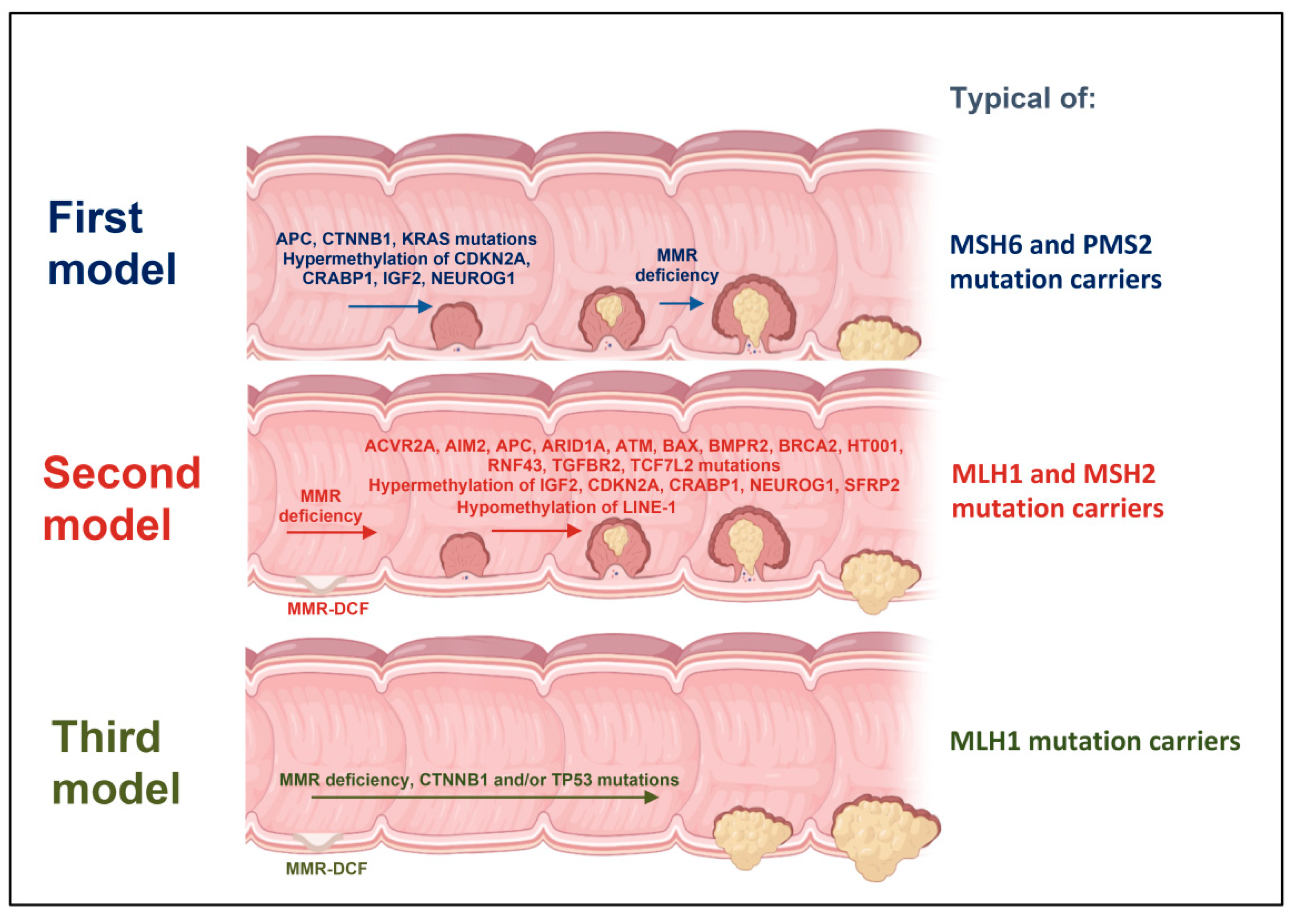
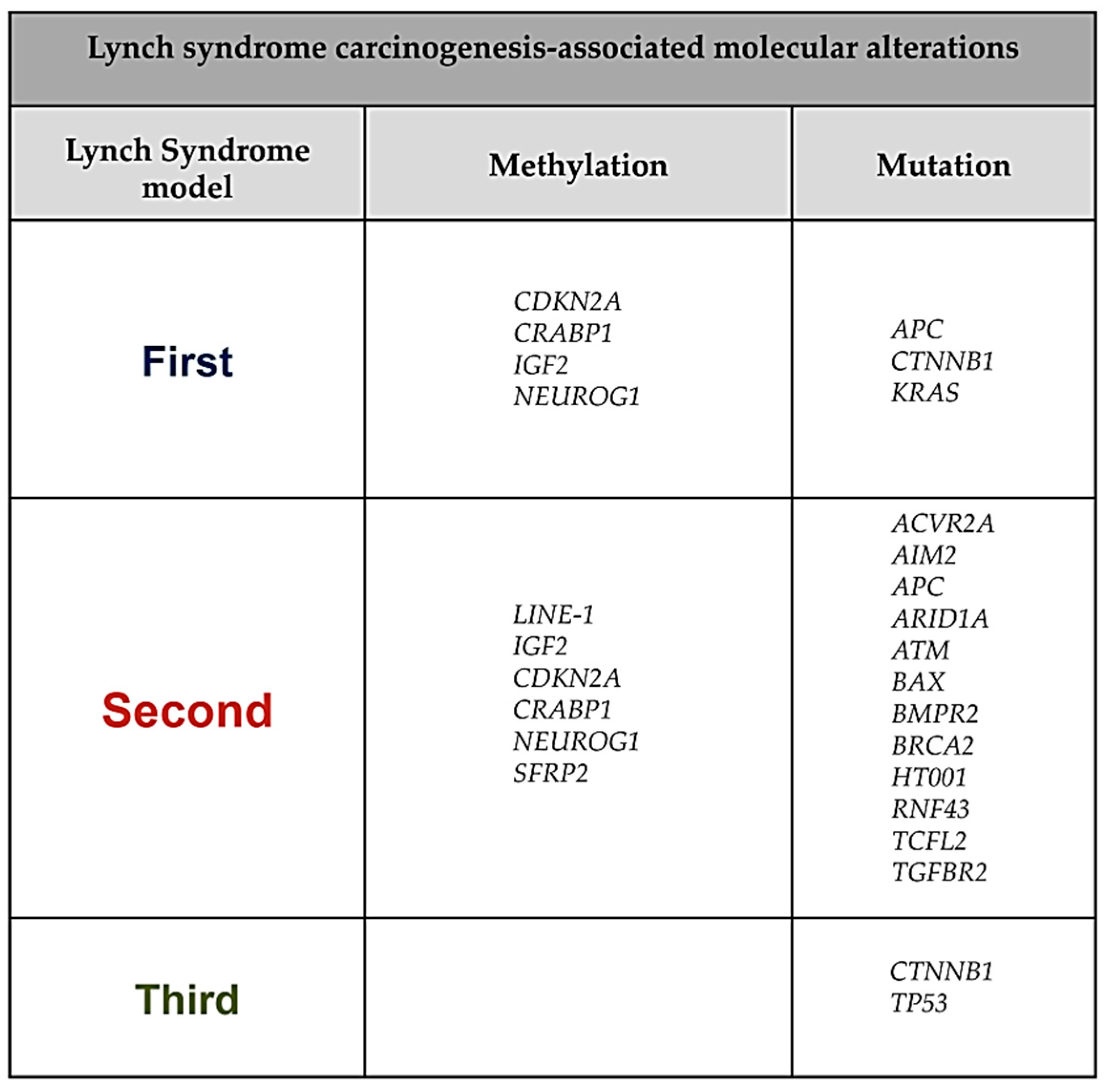
2. First Model of LS Colorectal Carcinogenesis: Adenoma Growth in an MMR-Proficient Background

Classical colorectal carcinogenesis follows the adenoma–carcinoma sequence, with MMR deficiency arising after adenoma development [41]. Indeed, for decades MMR deficiency was considered to be a secondary event in LS carcinogenesis. This idea was sustained by several studies noticing that adenomas retained the expression of MMR proteins [8]. However, it has recently been demonstrated that only up to 25% of all adenomas in LS patients are MMR proficient [42]. The existence of MMR-proficient adenomas supports the idea that tumor formation is possible even in the absence of the second hit required to inactivate the wild-type allele of an MMR gene. This model of carcinogenesis presumes the existence of other somatic events to initiate malignant transformation [8]. APC and KRAS mutations may represent the starting events and are believed to enhance tumorigenesis when MMR gene expression is still intact. Loss or inactivation of the APC gene on chromosome 5q drives the growth of small adenomas with a hypomethylated genome by promoting clonal expansion. Furthermore, almost all KRAS mutations appearing before MMR deficiency in LS patients are alterations involving specific sequences, such as KRAS G12V and KRAS A146T [8].
Epigenetic changes are defined as stable and hereditable alterations in gene expression and cell functions without changes in the original DNA sequence. Epigenetics plays a role in CRC in two different ways: on the one hand, the genome of the bulk tumor appears hypomethylated compared to normal colonic epithelia; on the other hand, there are particular regions, such as CpG islands, which are normally unmethylated, that appear hypermethylated. CpG islands are associated with the promoters of genes involved in several cell functions, including cell cycle control (CDKN2A), DNA repair (MLH1), and apoptosis (DAPK) [43]. Widespread DNA methylation at CpG sites in promoter regions [44] can be a putative “second hit” in LS carriers [45] and likely precedes MSI, representing an early event in tumor development. Indeed, aberrant CpG island methylation affecting several tumor suppressor genes leads to a CpG island methylator phenotype (CIMP) [45]. Moreover, Maki-Nevala and colleagues found higher methylation levels in four CIMP marker genes, namely NEUROG1, CDKN2A, IGF2, and CRABP1, in MMR-proficient adenomas compared to normal mucosa [45,46].
All these markers were identified hypermethylated also in these adenomas when MMR deficiency is acquired [8].
The NEUROG1 gene, located on chromosome 5 (5q23–q31), encodes for a transcription factor that binds to E-box elements. Methylation analysis of NEUROG1 in CRC tumors showed progressive hypermethylation associated with neoplastic development; indeed, increasing methylation levels were found from normal to tumor mucosa [47]. Currently, NEUROG1 is considered a potential diagnostic marker for early CRC since its methylation status can be detected in patient sera in a non-invasive way by population-wide screening for colorectal neoplasia, which is especially useful for people who refuse colonoscopy [48].
CDKN2A is an important tumor suppressor in CRC. Methylation of its promoter leads to CDKN2A gene silencing [49] and ultimately promotes uncontrolled cell proliferation [50]. Several studies evaluated the association between CDKN2A hypermethylation and shorter CRC patient survival, suggesting its role as an independent prognostic factor that might predict invasion and metastasis [51].
IGF2 encodes a protein that plays a major role in growth and development after birth. It is frequently altered in cancer and is involved in neoplastic proliferation [52]. LS patients with CRC show a higher amount of hypermethylated IGF2 in adenoma and adenocarcinoma tissue compared to normal colon mucosa [45]. Interestingly, IGF2 can be used as a prognostic marker since its methylation status can be screened by epigenetic blood testing in order to identify early in life LS carriers that are highly susceptible to developing CRC [53].
CRABP1 belongs to a family of fatty acid-binding proteins and is associated with a poor prognosis in several cancers. A recent study showed significantly elevated methylation levels of CRABP1 in MMR-proficient adenomas of LS patients [8].
From a histological point of view, the first model of LS carcinogenesis is characterized by the development of polypoid precursor lesions frequently found in the right side of the colon [36,54,55].
Riijcken and colleagues reported that adenomas in the right side of the colon are more prone to malignant conversion than left-sided adenomas [36] and have a short dwell time [55]. Consistently, Edelstein and colleagues estimated that adenoma dwell time in LS patients is considerably shorter than in patients with sporadic CRC [31]. Several studies reported a low incidence of serrated lesions in LS carriers, with the majority of adenomas appearing as conventional adenomas and/or hyperplastic polyps [56]. Moreover, most of them (about 80%) have a flat morphology, which is frequently missed during conventional endoscopic exams [2]. Other histological signs are the presence of differentiated mucinous cells and seal ring cells, a medullary growth model associated with a marked lymphocytic peritumoral inflammation that recalls the characteristics of the so-called ‘Crohn’s reaction’ [13].
In this type of colorectal carcinogenesis, the adenoma–carcinoma sequence is extremely accelerated [55,57], so that even colonoscopies performed annually may not be effective [58].
This first model of LS colorectal carcinogenesis is probably the most frequent pathway of tumor initiation in MSH6 and PMS2 mutation carriers. This finding is supported by the fact that low-grade adenomas in patients with MSH6 and PMS2 alterations are frequently microsatellite stable. Furthermore, Engel et al. reported a significant molecular signature in which MSH6 mutation carriers are associated with low frequency of CTNNB1 mutations and high frequency of APC mutations, suggesting that in these patients the onset of MMR deficiency only occurs after adenoma formation. Besides, MSH6 mutation carriers appear to be at low risk of cancer, probably because the isolated loss of MSH6 gene function does not completely abrogate MMR activity due to the overlapping functions of MSH3 [59]. Indeed, patients bearing alterations in MSH6 or PMS2 benefit more from current colonoscopy surveillance programs than MLH1 or MSH2 mutation carriers due to the different type of tumorigenesis involved [60] and have been reported to have a lower risk of developing CRC along with a later age at presentation [61]. Interestingly, the few MLH1 and MSH2 mutation carriers exhibiting the first pattern of LS carcinogenesis are susceptible to develop somatic mutations in CTNNB1 and APC genes, respectively, before the growth of adenomas [41].
Importantly, the first model of colorectal carcinogenesis in LS patients is characterized by the growth of adenomas as tumor precursor lesions in an MMR-proficient background, but Sekine and colleagues suggest that MMR deficiency occurs in adenomas before the progression to carcinomas, emphasizing the relevance of MMR impairment during LS carcinogenesis [62].

3. Second Model of LS Colorectal Carcinogenesis: MMR-DCF Leading to Adenoma Formation and Transition to Carcinoma

In recent years, the classic view of LS as an “accelerating” disease has been challenged, especially by the identification of MMR-DCF, which are colon crypts showing mostly normal histological features but already lacking MMR protein expression. This observation has suggested that MMR-deficient CRC in LS patients could also begin from such MMR-DCF [40]. Indeed, several studies have shown that about 75% of all adenomas in LS patients are MMR deficient [40,41].
From a histological point of view, MMR deficiency in LS patients can be detected heterogeneously in dysplastic crypts when it occurs in an already existing adenoma, while it can be observed consistently in adjacent and non-dysplastic MMR-DCF when it precedes adenoma formation [40]. This latter lesion type is unique to LS patients’ CRCs and has not been detected in sporadic CRC [61,63]. The occurrence of thousands of MMR-DCF, approximately 1 MMR-DCF per cm2 of mucosa, has been observed in phenotypically normal intestinal mucosa [61]. These lesions can be considered tumor precursors in LS patients and are difficult to detect by colonoscopy at a pre-invasive stage [41]. Importantly, most of these lesions do not seem to progress to malignancy, as suggested by the discrepancy between the large number of MMR-DCF and the small number of adenomas or carcinomas observed in LS carriers [61].Histologically, MMR-DCF are almost indistinguishable from normal colonic crypts. Slight differences are the nuclear enlargement of cells at the bottom of the crypt and features of neoplastic growth such as aberrant branching and typical crypt fission. Moreover, MMR-DCF appear as non-elevated lesions without widened luminal openings [61]. Despite these differences, MMR-DCF retain the potential to migrate and mature along the crypt–villus axis [42], and Ki-67 staining showed a physiological proliferation pattern similar to that observed in normal mucosa [61]. For this reason, MMR-DCF escape routine histological detection with methylene blue staining. Besides, morphological evaluation of MMR-DCF-adjacent mucosal areas did not reveal marks of altered immune infiltration [63]. This type of lesion is significantly associated with patients’ age at the time of tumor resection and with cancer location, being more frequent in patients with distal colorectal tumors [63]. This model of LS carcinogenesis is initiated by non-polypoid precursor lesions that can directly give rise to localized adenocarcinomas.
All tumors of LS patients displaying this type of colorectal carcinogenesis show MSI [64], which is characterized by instability in coding and non-coding short repeat microsatellite sequences caused by mutations in MMR genes [65]. These mutations can lead to reading frame shifts resulting in the inactivation of key tumor suppressor genes with growth-related functions [66]. Indeed, the above histological features are associated with molecular signatures typically identified upon mutations in major tumor suppressor genes, such as TGFBR2 [42]. TGFBR2 encodes for a type II TGF-β receptor that can activate the TGF-β pathway by specific ligand binding [67]. The TGF-β signaling pathway is involved in the inhibition of cell proliferation and the induction of apoptosis [68]. TGFBR2 gene sequence architecture is prone to replication errors because it contains several repeated DNA motifs [69], including ten adenosine residues that are frequently targeted by MMR gene inactivation [63]. Mutations in TGFBR2 were found in 80% of early colorectal adenomas with high microsatellite instability [70]. This finding suggests that TGFBR2 mutations occur early after bi-allelic inactivation of MMR genes [61], but other studies revealed that TGFBR2 mutations are also involved in neoplastic progression since this gene is frequently increased in typical advanced lesions such as poly-cryptic MMR-DCF [63]. Conversely, mono-cryptic MMR-DCF frequently show mutations in the microsatellite coding regions of HT001, AIM2, and BAX genes [63]. Interestingly, Pinheiro and colleagues detected the co-occurrence of TGFBR2, ACVR2A, and BMPR2 mutations, indicating that loss of one of these receptors is likely insufficient for complete TGF signaling disruption [71]. Furthermore, the molecular signature associated with the second model of LS colorectal carcinogenesis suggests the involvement of the WNT signaling pathway. Of note, low mutation frequency has been detected in core genes of this pathway, such as AXIN1, AXIN2, PTEN, and CTNNB1 [72]. Conversely, this signature is characterized by RNF43 mutations, mostly by frameshift affecting mononucleotide repeats [72]. RNF43 is a ubiquitin ligase of Frizzled cell surface receptors and acts as a negative regulator of the WNT pathway, which is frequently altered during CRC carcinogenesis [73]. Furthermore, despite the low frequency of APC mutations occurring after MMR deficiency onset, almost all of these mutations are insertions or deletions involving specific repeat sequences, such as an A5-repeat at codon 1455, an AG5-repeat at codon 1465, and an A6-repeat at codon 1554 [62]. Noteworthy, RNF43 and APC mutations, which both lead to WNT pathway activation and are usually mutually exclusive, often co-exist in LS patients, both in adenomas and adenocarcinomas [62]. The importance of the WNT pathway in LS carcinogenesis is also highlighted by the common transcriptional silencing of SFRP2, another WNT signaling antagonist. Indeed, hypermethylation of the SFRP2 promoter induces uncontrolled cell proliferation [45]. Moreover, several studies reported a high mutation frequency of TCF7L2, which encodes a component of the WNT signaling pathway and is associated with an increased risk of CRC. Indeed, up to 60% of LS patients carry mutations in TCF7L2 [71,74,75].
LS patients with the second type of colorectal carcinogenesis also display a high mutation rate in genes involved in DNA damage response signaling, such as ARID1A, ATM, and BRCA2, which play a role in homologous recombination and double-strand break repair [72].
Interestingly, this model of LS colorectal carcinogenesis is also associated with decreased methylation levels of LINE-1 (long interspersed nuclear elements) [63]. This epigenetic alteration may predispose cells to chromosomal rearrangements, resulting in increased mutation rates [76].
Moreover, MMR-deficient adenomas with a high grade of dysplasia revealed hypermethylation in four other genes, IGF2, CRABP1, NEUROG1, and CDKN2A [8].
The second model of LS colorectal carcinogenesis is characterized by the early acquisition of MMR deficiency, which precedes adenoma formation. This pathway is probably the most frequent type of carcinogenesis in MLH1 and MSH2 mutation carriers and does not show substantial differences in MMR-DCF frequency or with patient gender. Moreover, it has been reported that MSH6 and PMS2 mutation carriers can benefit more from colonoscopy surveillance since MMR-DCF are both less common and less likely to progress along the adenoma–carcinoma sequence [77].

4. Third Model of LS Colorectal Carcinogenesis: MMR-DCF Showing Direct Transition to Carcinoma

About 10% of LS-associated cancers are prone to skip the adenoma step of the classical adenoma–carcinoma sequence in the carcinogenesis process [58]. This quickly leads to an invasive phenotype presumably arising from MMR-DCF through somatic mutations in genes such as CTNNB1 and TP53 [41]. Since MMR-DCF can grow under an intact mucosal surface, these types of lesions frequently escape colonoscopic detection during recommended surveillance [40], directly evolving into manifest cancer without a macroscopically visible non-invasive precursor [42].
Indeed, unlike APC mutations, CTNNB1 mutations mostly occur in non-polypous CRCs lacking the adenomatous precursor stage [40]. CTNNB1 mutations are commonly observed in this type of colorectal carcinogenesis in LS patients and require at least one additional pre-existing alteration to exert their oncogenic effects and drive tumorigenesis in colonic mucosa [58]. The CTNNB1 mutations observed in LS cancer are frequently an amino acid substitution at codon 41 (c.121A > G) or other point mutations at codon 45, such as c.133T > C or c.134C > T [77]. In addition, CTNNB1 in-frame deletions have also been described, including c.133_135del or other deletions encompassing c.133 that result in the removal of the serine residue in position 45 (S45) [58]. S45 is a phosphorylation site for casein kinase-1 and is involved in the regulation of β-catenin stability; indeed, S45 mutations result in stronger activation of WNT/β-catenin signals and are frequently associated with malignant transformation [78].
In the two patterns of LS colorectal carcinogenesis described above, polypous regions are observed adjacent to the invasive margin of the tumors, while most cancers associated with the third type of LS colorectal carcinogenesis lack these histological features. Since MMR-DCF do not always evolve spontaneously into a neoplastic lesion, Ahadova and colleagues suggested that mutations in CTNNB1 and/or TP53 may be involved in the malignant conversion of these foci [58]. Despite TP53 mutations are believed to be uncommon in LS patients, Maki-Nevala and colleagues reported that up to 33% of LS adenomas showed mutations in the TP53 gene [8].
This third model of LS colorectal carcinogenesis is characterized by fast tumor growth with invasive features in an MMR-deficient context. This pattern is frequently associated with MLH1 mutations [41], while it is not observed in PMS2 mutation carriers, which may explain the reduced risk of CRC in these patients under surveillance programs [34].

5. Conclusions and Future Directions

Since LS patients do not seem to fully benefit from current surveillance strategies, researchers and clinicians are in the search for novel diagnostic approaches to prevent CRC development.
In order to optimize current surveillance programs for LS carriers, more sophisticated endoscopic techniques, such as chromoendoscopy, virtual chromoendoscopy as narrow-band imaging (NBI), could be implemented to improve the detection of adenomas compared to normal colonoscopy. These approaches would significantly benefit LS patients with the first and second models of colorectal carcinogenesis described above [34,79,80]. Colonic chromoendoscopy takes advantage of the topical application of stains such as indigo carmine, a deep-blue contrast dye, in order to improve mucosal contrast [81]. This technique allowed the detection of up to twice the number of adenomas compared to normal colonoscopy and identified a higher proportion of patients with at least one adenoma [82,83,84,85]. Importantly, no significant side effects were observed [82]. Other studies demonstrated that the use of colonoscopy with NBI also doubled the number of adenomas detected in LS patients [86].
In the future, artificial intelligence applied to colonoscopy (computer-aided diagnosis or CAD) could help to increase the adenoma detection rate through enhanced evaluation of superficial (epithelial and vascular) irregularities by using dedicated software to reduce missed adenomas [87].
LS patients could additionally benefit from novel screening modalities based on NGS, which are extremely informative in guiding surveillance [88]. Screening of distant media such as stool or blood could be incorporated in current surveillance programs of LS patients to detect genetic and epigenetic cancer-specific alterations. Indeed, the investigation of DNA markers methylation status characteristic of LS tumorigenesis could help to detect the presence of precursor lesions and CRC. Ballester and colleagues searched for novel markers to be used in a test that is already clinically available. These authors found that OPLAH alone and/or a combination of a few methylated DNA markers (ARHGEF4, LRRC4, ANTXR1, PITX1) can discriminate adenomas and CRC in LS patients [39]. All these DNA markers represent aberrantly hypermethylated sequences; indeed, the hypomethylation status is more difficult to recognize in distant media. Interestingly, aberrant hypermethylation of ALKBH5, a gene involved in DNA damage signaling, has been recognized as a unique sign in LS neoplasms compared to CRC sporadic neoplasms [39]. Ideally, a specific large panel should be created including these and other DNA markers identified in other studies, such as NEUROG1, CDKN2A, IGF2, and CRABP1. In the personalized medicine era, NGS-targeted gene panels comprising top-performing markers as cancer-specific mutations (TGFBR2, RNF43, ARID1A, ATM, BRCA2, CTNNB1, and others) may optimize early detection of CRC precursors and increase compliance in these high-risk patients (Figure 3).
Interestingly, the results gathered from these analyses can be used not only for diagnostic procedures but also for therapeutic approaches in LS patients. Indeed, ovarian, breast, pancreas, and prostate cancer patients with somatic BRCA mutations can be currently treated with poly(ADP-ribose) polymerase (PARP) inhibitors such as olaparib, rucaparib, niraparib, and talazoparib [89,90,91,92,93]. The increased frequency of somatic BRCA2 mutations documented in MMR-deficient LS carriers suggests the potential application of PARP inhibition therapy [72]. Moreover, growing evidence indicates that alteration of other major homologous recombination repair (HRR) proteins, such as ATM, may induce a response to PARP inhibitors in patients affected by multiple solid malignancies, such as prostate cancer and triple-negative breast cancer [94,95]. Indeed, various preclinical studies have shown that CRC cell lines carrying ATM mutations exhibit increased sensitivity to olaparib [96]. These data support the development of HRR inhibition therapy as a promising anticancer strategy also in LS patients.
Larger randomized trials are needed in order to apply these novel screening modalities in current clinical settings. These studies are necessary to provide sufficient statistical power to validate the effectiveness of these novel markers of colorectal carcinogenesis in LS patients.
Since no survival gain was observed in LS patients undergoing more frequent colonoscopies, the findings described above support the use of an individualized diagnostic approach based not only on the MMR mutation carried by the patient but also on the pattern of histological and molecular carcinogenesis. In this light, in addition to standard colonoscopy, LS patient screening programs should include sophisticated endoscopic techniques combined with non-invasive approaches based on NGS to analyze specific markers in distant media.

Author Contributions

Study conceptualization, G.I. and C.S., writing, review, and editing—original draft preparation, M.L.S., V.D., G.D.C., and A.P.; funding acquisition, C.S. All authors have read and agreed to the published version of the manuscript.

Funding

This research was funded by Cristiano Simone grant number the Italian Ministry of Health ‘Ricerca Corrente 2018–2020; 2019–2021’, PRIN-Research Projects of National Relevance (PRIN 2017, n. 2017WNKSLr-LS4) from the Italian MIUR and AIRC IG-23794 2020-2024.

Institutional Review Board Statement

Not applicable.

Informed Consent Statement

Not applicable.

Acknowledgments

We thank Francesco Paolo Jori for his helpful discussion during the preparation of the manuscript and editorial assistance.

Conflicts of Interest

The authors declare no conflict of interest.

References

  1. Bansidhar, B.J. Extracolonic Manifestations of Lynch Syndrome. Clin. Colon Rectal Surg. 2012, 25, 103–110. [Google Scholar] [CrossRef] [Green Version]
  2. Stoffel, E.M.; Turgeon, D.K.; Stockwell, D.H.; Zhao, L.; Normolle, D.P.; Tuck, M.K.; Bresalier, R.S.; Marcon, N.E.; Baron, J.A.; Ruffin, M.T.; et al. Missed Adenomas during Colonoscopic Surveillance in Individuals with Lynch Syndrome (Hereditary Nonpolyposis Colorectal Cancer). Cancer Prev. Res. 2008, 1, 470–475. [Google Scholar] [CrossRef] [PubMed] [Green Version]
  3. Duraturo, F.; Liccardo, R.; De Rosa, M.; Izzo, P. Genetics, Diagnosis and Treatment of Lynch Syndrome: Old Lessons and Current Challenges. Oncol. Lett. 2019, 17, 3048–3054. [Google Scholar] [CrossRef] [PubMed] [Green Version]
  4. Morak, M.; Käsbauer, S.; Kerscher, M.; Laner, A.; Nissen, A.M.; Benet-Pagès, A.; Schackert, H.K.; Keller, G.; Massdorf, T.; Holinski-Feder, E. Loss of MSH2 and MSH6 Due to Heterozygous Germline Defects in MSH3 and MSH6. Fam. Cancer 2017, 16, 491–500. [Google Scholar] [CrossRef] [PubMed]
  5. Liu, T.; Yan, H.; Kuismanen, S.; Percesepe, A.; Bisgaard, M.L.; Pedroni, M.; Benatti, P.; Kinzler, K.W.; Vogelstein, B.; Ponz de Leon, M.; et al. The Role of HPMS1 and HPMS2 in Predisposing to Colorectal Cancer. Cancer Res. 2001, 61, 7798–7802. [Google Scholar] [PubMed]
  6. Wu, Y.; Berends, M.J.; Sijmons, R.H.; Mensink, R.G.; Verlind, E.; Kooi, K.A.; van der Sluis, T.; Kempinga, C.; van der Zee, A.G.; Hollema, H.; et al. A Role for MLH3 in Hereditary Nonpolyposis Colorectal Cancer. Nat. Genet. 2001, 29, 137–138. [Google Scholar] [CrossRef] [PubMed]
  7. Ou, J.; Rasmussen, M.; Westers, H.; Andersen, S.D.; Jager, P.O.; Kooi, K.A.; Niessen, R.C.; Eggen, B.J.L.; Nielsen, F.C.; Kleibeuker, J.H.; et al. Biochemical Characterization of MLH3 Missense Mutations Does Not Reveal an Apparent Role of MLH3 in Lynch Syndrome. Genes. Chromosomes Cancer 2009, 48, 340–350. [Google Scholar] [CrossRef] [Green Version]
  8. Mäki-Nevala, S.; Valo, S.; Ristimäki, A.; Sarhadi, V.; Knuutila, S.; Nyström, M.; Renkonen-Sinisalo, L.; Lepistö, A.; Mecklin, J.-P.; Peltomäki, P. DNA Methylation Changes and Somatic Mutations as Tumorigenic Events in Lynch Syndrome-Associated Adenomas Retaining Mismatch Repair Protein Expression. EBioMedicine 2019, 39, 280–291. [Google Scholar] [CrossRef] [Green Version]
  9. Satorres, C.; García-Campos, M.; Bustamante-Balén, M. Molecular Features of the Serrated Pathway to Colorectal Cancer: Current Knowledge and Future Directions. Gut Liver 2021, 15, 31–43. [Google Scholar] [CrossRef]
  10. Vasen, H.F.; Watson, P.; Mecklin, J.P.; Lynch, H.T. New Clinical Criteria for Hereditary Nonpolyposis Colorectal Cancer (HNPCC, Lynch Syndrome) Proposed by the International Collaborative Group on HNPCC. Gastroenterology 1999, 116, 1453–1456. [Google Scholar] [CrossRef]
  11. Umar, A.; Boland, C.R.; Terdiman, J.P.; Syngal, S.; de la Chapelle, A.; Rüschoff, J.; Fishel, R.; Lindor, N.M.; Burgart, L.J.; Hamelin, R.; et al. Revised Bethesda Guidelines for Hereditary Nonpolyposis Colorectal Cancer (Lynch Syndrome) and Microsatellite Instability. J. Natl. Cancer Inst. 2004, 96, 261–268. [Google Scholar] [CrossRef]
  12. Cohen, S.A. Current Lynch Syndrome Tumor Screening Practices: A Survey of Genetic Counselors. J. Genet. Couns. 2014, 23, 38–47. [Google Scholar] [CrossRef]
  13. Remo, A.; Fassan, M.; Lanza, G. Immunohistochemical Evaluation of Mismatch Repair Proteins in Colorectal Carcinoma: The AIFEG/GIPAD Proposal. Pathologica 2016, 108, 104–109. [Google Scholar]
  14. Bonadona, V.; Bonaïti, B.; Olschwang, S.; Grandjouan, S.; Huiart, L.; Longy, M.; Guimbaud, R.; Buecher, B.; Bignon, Y.-J.; Caron, O.; et al. Cancer Risks Associated with Germline Mutations in MLH1, MSH2, and MSH6 Genes in Lynch Syndrome. JAMA 2011, 305, 2304–2310. [Google Scholar] [CrossRef] [PubMed]
  15. Møller, P.; Seppälä, T.T.; Bernstein, I.; Holinski-Feder, E.; Sala, P.; Gareth Evans, D.; Lindblom, A.; Macrae, F.; Blanco, I.; Sijmons, R.H.; et al. Cancer Risk and Survival in Path_MMR Carriers by Gene and Gender up to 75 Years of Age: A Report from the Prospective Lynch Syndrome Database. Gut 2018, 67, 1306–1316. [Google Scholar] [CrossRef] [PubMed] [Green Version]
  16. Ryan, N.A.J.; Morris, J.; Green, K.; Lalloo, F.; Woodward, E.R.; Hill, J.; Crosbie, E.J.; Evans, D.G. Association of Mismatch Repair Mutation With Age at Cancer Onset in Lynch Syndrome: Implications for Stratified Surveillance Strategies. JAMA Oncol. 2017, 3, 1702–1706. [Google Scholar] [CrossRef] [PubMed]
  17. Barrow, E.; Robinson, L.; Alduaij, W.; Shenton, A.; Clancy, T.; Lalloo, F.; Hill, J.; Evans, D.G. Cumulative Lifetime Incidence of Extracolonic Cancers in Lynch Syndrome: A Report of 121 Families with Proven Mutations. Clin. Genet. 2009, 75, 141–149. [Google Scholar] [CrossRef] [PubMed]
  18. Møller, P.; Seppälä, T.; Bernstein, I.; Holinski-Feder, E.; Sala, P.; Evans, D.G.; Lindblom, A.; Macrae, F.; Blanco, I.; Sijmons, R.; et al. Cancer Incidence and Survival in Lynch Syndrome Patients Receiving Colonoscopic and Gynaecological Surveillance: First Report from the Prospective Lynch Syndrome Database. Gut 2017, 66, 464–472. [Google Scholar] [CrossRef] [PubMed]
  19. Baglietto, L.; Lindor, N.M.; Dowty, J.G.; White, D.M.; Wagner, A.; Gomez Garcia, E.B.; Vriends, A.H.J.T.; Dutch Lynch Syndrome Study Group; Cartwright, N.R.; Barnetson, R.A.; et al. Risks of Lynch Syndrome Cancers for MSH6 Mutation Carriers. J. Natl. Cancer Inst. 2010, 102, 193–201. [Google Scholar] [CrossRef]
  20. Suerink, M.; Rodríguez-Girondo, M.; van der Klift, H.M.; Colas, C.; Brugieres, L.; Lavoine, N.; Jongmans, M.; Munar, G.C.; Evans, D.G.; Farrell, M.P.; et al. An Alternative Approach to Establishing Unbiased Colorectal Cancer Risk Estimation in Lynch Syndrome. Genet. Med. Off. J. Am. Coll. Med. Genet. 2019, 21, 2706–2712. [Google Scholar] [CrossRef]
  21. Senter, L.; Clendenning, M.; Sotamaa, K.; Hampel, H.; Green, J.; Potter, J.D.; Lindblom, A.; Lagerstedt, K.; Thibodeau, S.N.; Lindor, N.M.; et al. The Clinical Phenotype of Lynch Syndrome Due to Germ-Line PMS2 Mutations. Gastroenterology 2008, 135, 419–428. [Google Scholar] [CrossRef] [PubMed] [Green Version]
  22. Ten Broeke, S.W.; van der Klift, H.M.; Tops, C.M.J.; Aretz, S.; Bernstein, I.; Buchanan, D.D.; de la Chapelle, A.; Capella, G.; Clendenning, M.; Engel, C.; et al. Cancer Risks for PMS2-Associated Lynch Syndrome. J. Clin. Oncol. Off. J. Am. Soc. Clin. Oncol. 2018, 36, 2961–2968. [Google Scholar] [CrossRef] [PubMed] [Green Version]
  23. Engel, C.; Loeffler, M.; Steinke, V.; Rahner, N.; Holinski-Feder, E.; Dietmaier, W.; Schackert, H.K.; Goergens, H.; von Knebel Doeberitz, M.; Goecke, T.O.; et al. Risks of Less Common Cancers in Proven Mutation Carriers with Lynch Syndrome. J. Clin. Oncol. Off. J. Am. Soc. Clin. Oncol. 2012, 30, 4409–4415. [Google Scholar] [CrossRef] [PubMed]
  24. Dominguez-Valentin, M.; Joost, P.; Therkildsen, C.; Jonsson, M.; Rambech, E.; Nilbert, M. Frequent Mismatch-Repair Defects Link Prostate Cancer to Lynch Syndrome. BMC Urol. 2016, 16, 15. [Google Scholar] [CrossRef] [Green Version]
  25. Joost, P.; Therkildsen, C.; Dominguez-Valentin, M.; Jönsson, M.; Nilbert, M. Urinary Tract Cancer in Lynch Syndrome; Increased Risk in Carriers of MSH2 Mutations. Urology 2015, 86, 1212–1217. [Google Scholar] [CrossRef] [PubMed]
  26. Watson, P.; Vasen, H.F.A.; Mecklin, J.-P.; Bernstein, I.; Aarnio, M.; Järvinen, H.J.; Myrhøj, T.; Sunde, L.; Wijnen, J.T.; Lynch, H.T. The Risk of Extra-Colonic, Extra-Endometrial Cancer in the Lynch Syndrome. Int. J. Cancer 2008, 123, 444–449. [Google Scholar] [CrossRef] [Green Version]
  27. Capelle, L.G.; Van Grieken, N.C.T.; Lingsma, H.F.; Steyerberg, E.W.; Klokman, W.J.; Bruno, M.J.; Vasen, H.F.A.; Kuipers, E.J. Risk and Epidemiological Time Trends of Gastric Cancer in Lynch Syndrome Carriers in the Netherlands. Gastroenterology 2010, 138, 487–492. [Google Scholar] [CrossRef]
  28. Harkness, E.F.; Barrow, E.; Newton, K.; Green, K.; Clancy, T.; Lalloo, F.; Hill, J.; Evans, D.G. Lynch Syndrome Caused by MLH1 Mutations Is Associated with an Increased Risk of Breast Cancer: A Cohort Study. J. Med. Genet. 2015, 52, 553–556. [Google Scholar] [CrossRef]
  29. Haraldsdottir, S.; Rafnar, T.; Frankel, W.L.; Einarsdottir, S.; Sigurdsson, A.; Hampel, H.; Snaebjornsson, P.; Masson, G.; Weng, D.; Arngrimsson, R.; et al. Comprehensive Population-Wide Analysis of Lynch Syndrome in Iceland Reveals Founder Mutations in MSH6 and PMS2. Nat. Commun. 2017, 8, 14755. [Google Scholar] [CrossRef]
  30. Syngal, S.; Brand, R.E.; Church, J.M.; Giardiello, F.M.; Hampel, H.L.; Burt, R.W. American College of Gastroenterology ACG Clinical Guideline: Genetic Testing and Management of Hereditary Gastrointestinal Cancer Syndromes. Am. J. Gastroenterol. 2015, 110, 223–262, quiz 263. [Google Scholar] [CrossRef] [Green Version]
  31. Edelstein, D.L.; Axilbund, J.; Baxter, M.; Hylind, L.M.; Romans, K.; Griffin, C.A.; Cruz-Correa, M.; Giardiello, F.M. Rapid Development of Colorectal Neoplasia in Patients with Lynch Syndrome. Clin. Gastroenterol. Hepatol. Off. Clin. Pract. J. Am. Gastroenterol. Assoc. 2011, 9, 340–343. [Google Scholar] [CrossRef] [PubMed] [Green Version]
  32. Kudo, S.; Tamura, S.; Nakajima, T.; Yamano, H.; Kusaka, H.; Watanabe, H. Diagnosis of Colorectal Tumorous Lesions by Magnifying Endoscopy. Gastrointest. Endosc. 1996, 44, 8–14. [Google Scholar] [CrossRef]
  33. Lambert, R. The Paris Endoscopic Classification of Superficial Neoplastic Lesions: Esophagus, Stomach, and Colon. Gastrointest. Endosc. 2003, 58, S3–S43. [Google Scholar] [CrossRef]
  34. Ahadova, A.; Seppälä, T.T.; Engel, C.; Gallon, R.; Burn, J.; Holinski-Feder, E.; Steinke-Lange, V.; Möslein, G.; Nielsen, M.; ten Broeke, S.W.; et al. The “Unnatural” History of Colorectal Cancer in Lynch Syndrome: Lessons from Colonoscopy Surveillance. Int. J. Cancer 2021, 148, 800–811. [Google Scholar] [CrossRef] [PubMed]
  35. Zhao, S.; Wang, S.; Pan, P.; Xia, T.; Chang, X.; Yang, X.; Guo, L.; Meng, Q.; Yang, F.; Qian, W.; et al. Magnitude, Risk Factors, and Factors Associated With Adenoma Miss Rate of Tandem Colonoscopy: A Systematic Review and Meta-Analysis. Gastroenterology 2019, 156, 1661–1674.e11. [Google Scholar] [CrossRef] [PubMed]
  36. Rijcken, F.E.M.; Hollema, H.; Kleibeuker, J.H. Proximal Adenomas in Hereditary Non-Polyposis Colorectal Cancer Are Prone to Rapid Malignant Transformation. Gut 2002, 50, 382–386. [Google Scholar] [CrossRef] [PubMed]
  37. Walsh, M.D.; Buchanan, D.D.; Pearson, S.-A.; Clendenning, M.; Jenkins, M.A.; Win, A.K.; Walters, R.J.; Spring, K.J.; Nagler, B.; Pavluk, E.; et al. Immunohistochemical Testing of Conventional Adenomas for Loss of Expression of Mismatch Repair Proteins in Lynch Syndrome Mutation Carriers: A Case Series from the Australasian Site of the Colon Cancer Family Registry. Mod. Pathol. 2012, 25, 722–730. [Google Scholar] [CrossRef]
  38. Sánchez, A.; Roos, V.H.; Navarro, M.; Pineda, M.; Caballol, B.; Moreno, L.; Carballal, S.; Rodríguez-Alonso, L.; Ramon Y Cajal, T.; Llort, G.; et al. Quality of Colonoscopy Is Associated With Adenoma Detection and Postcolonoscopy Colorectal Cancer Prevention in Lynch Syndrome. Clin. Gastroenterol. Hepatol. Off. Clin. Pract. J. Am. Gastroenterol. Assoc. 2020. [Google Scholar] [CrossRef]
  39. Ballester, V.; Taylor, W.R.; Slettedahl, S.W.; Mahoney, D.W.; Yab, T.C.; Sinicrope, F.A.; Boland, C.R.; Lidgard, G.P.; Cruz-Correa, M.R.; Smyrk, T.C.; et al. Novel Methylated DNA Markers Accurately Discriminate Lynch Syndrome Associated Colorectal Neoplasia. Epigenomics 2020, 12, 2173–2187. [Google Scholar] [CrossRef]
  40. Ahadova, A.; Gallon, R.; Gebert, J.; Ballhausen, A.; Endris, V.; Kirchner, M.; Stenzinger, A.; Burn, J.; von Knebel Doeberitz, M.; Bläker, H.; et al. Three Molecular Pathways Model Colorectal Carcinogenesis in Lynch Syndrome. Int. J. Cancer 2018, 143, 139–150. [Google Scholar] [CrossRef] [Green Version]
  41. Engel, C.; Ahadova, A.; Seppälä, T.T.; Aretz, S.; Bigirwamungu-Bargeman, M.; Bläker, H.; Bucksch, K.; Büttner, R.; de Vos Tot Nederveen Cappel, W.T.; Endris, V.; et al. Associations of Pathogenic Variants in MLH1, MSH2, and MSH6 With Risk of Colorectal Adenomas and Tumors and With Somatic Mutations in Patients With Lynch Syndrome. Gastroenterology 2020, 158, 1326–1333. [Google Scholar] [CrossRef]
  42. Møller, P. The Prospective Lynch Syndrome Database Reports Enable Evidence-Based Personal Precision Health Care. Hered. Cancer Clin. Pract. 2020, 18, 6. [Google Scholar] [CrossRef] [PubMed]
  43. Momparler, R.L. Cancer Epigenetics. Oncogene 2003, 22, 6479–6483. [Google Scholar] [CrossRef] [PubMed] [Green Version]
  44. Toyota, M.; Ahuja, N.; Ohe-Toyota, M.; Herman, J.G.; Baylin, S.B.; Issa, J.P. CpG Island Methylator Phenotype in Colorectal Cancer. Proc. Natl. Acad. Sci. USA 1999, 96, 8681–8686. [Google Scholar] [CrossRef] [PubMed] [Green Version]
  45. Valo, S.; Kaur, S.; Ristimäki, A.; Renkonen-Sinisalo, L.; Järvinen, H.; Mecklin, J.-P.; Nyström, M.; Peltomäki, P. DNA Hypermethylation Appears Early and Shows Increased Frequency with Dysplasia in Lynch Syndrome-Associated Colorectal Adenomas and Carcinomas. Clin. Epigenet. 2015, 7, 71. [Google Scholar] [CrossRef] [Green Version]
  46. Flatin, B.T.B.; Vedeld, H.M.; Pinto, R.; Langerud, J.; Lind, G.E.; Lothe, R.A.; Sveen, A.; Jeanmougin, M. Multiregional Assessment of CIMP in Primary Colorectal Cancers: Phenotype Concordance but Marker Variability. Int. J. Cancer 2021, 148, 1652–1657. [Google Scholar] [CrossRef]
  47. Otero-Estévez, O.; Gallardo-Gomez, M.; de la Cadena, M.P.; Rodríguez-Berrocal, F.J.; Cubiella, J.; Ramirez, V.H.; García-Nimo, L.; Chiara, L.D. Value of Serum NEUROG1 Methylation for the Detection of Advanced Adenomas and Colorectal Cancer. Diagnostics 2020, 10, 437. [Google Scholar] [CrossRef]
  48. Herbst, A.; Rahmig, K.; Stieber, P.; Philipp, A.; Jung, A.; Ofner, A.; Crispin, A.; Neumann, J.; Lamerz, R.; Kolligs, F.T. Methylation of NEUROG1 in Serum Is a Sensitive Marker for the Detection of Early Colorectal Cancer. Am. J. Gastroenterol. 2011, 106, 1110–1118. [Google Scholar] [CrossRef]
  49. Tada, T.; Watanabe, T.; Kazama, S.; Kanazawa, T.; Hata, K.; Komuro, Y.; Nagawa, H. Reduced P16 Expression Correlates with Lymphatic Invasion in Colorectal Cancers. Hepatogastroenterology 2003, 50, 1756–1760. [Google Scholar]
  50. Samowitz, W.S.; Albertsen, H.; Herrick, J.; Levin, T.R.; Sweeney, C.; Murtaugh, M.A.; Wolff, R.K.; Slattery, M.L. Evaluation of a Large, Population-Based Sample Supports a CpG Island Methylator Phenotype in Colon Cancer. Gastroenterology 2005, 129, 837–845. [Google Scholar] [CrossRef]
  51. Xing, X.; Cai, W.; Shi, H.; Wang, Y.; Li, M.; Jiao, J.; Chen, M. The Prognostic Value of CDKN2A Hypermethylation in Colorectal Cancer: A Meta-Analysis. Br. J. Cancer 2013, 108, 2542–2548. [Google Scholar] [CrossRef] [PubMed]
  52. Kasprzak, A.; Adamek, A. Insulin-Like Growth Factor 2 (IGF2) Signaling in Colorectal Cancer-From Basic Research to Potential Clinical Applications. Int. J. Mol. Sci. 2019, 20, 4915. [Google Scholar] [CrossRef] [Green Version]
  53. Jirtle, R.L. IGF2 Loss of Imprinting: A Potential Heritable Risk Factor for Colorectal Cancer. Gastroenterology 2004, 126, 1190–1193. [Google Scholar] [CrossRef]
  54. De Jong, A.E.; Morreau, H.; Van Puijenbroek, M.; Eilers, P.H.; Wijnen, J.; Nagengast, F.M.; Griffioen, G.; Cats, A.; Menko, F.H.; Kleibeuker, J.H.; et al. The Role of Mismatch Repair Gene Defects in the Development of Adenomas in Patients with HNPCC. Gastroenterology 2004, 126, 42–48. [Google Scholar] [CrossRef]
  55. Rondagh, E.J.A.; Gulikers, S.; Gómez-García, E.B.; Vanlingen, Y.; Detisch, Y.; Winkens, B.; Vasen, H.F.A.; Masclee, A.A.M.; Sanduleanu, S. Nonpolypoid Colorectal Neoplasms: A Challenge in Endoscopic Surveillance of Patients with Lynch Syndrome. Endoscopy 2013, 45, 257–264. [Google Scholar] [CrossRef]
  56. Andersen, S.H.; Lykke, E.; Folker, M.B.; Bernstein, I.; Holck, S. Sessile Serrated Polyps of the Colorectum Are Rare in Patients with Lynch Syndrome and in Familial Colorectal Cancer Families. Fam. Cancer 2008, 7, 157–162. [Google Scholar] [CrossRef]
  57. Lanspa, S.J.; Jenkins, J.X.; Cavalieri, R.J.; Smyrk, T.C.; Watson, P.; Lynch, J.; Lynch, H.T. Surveillance in Lynch Syndrome: How Aggressive? Am. J. Gastroenterol. 1994, 89, 1978–1980. [Google Scholar]
  58. Ahadova, A.; von Knebel Doeberitz, M.; Bläker, H.; Kloor, M. CTNNB1-Mutant Colorectal Carcinomas with Immediate Invasive Growth: A Model of Interval Cancers in Lynch Syndrome. Fam. Cancer 2016, 15, 579–586. [Google Scholar] [CrossRef] [PubMed]
  59. Yurgelun, M.B.; Goel, A.; Hornick, J.L.; Sen, A.; Turgeon, D.K.; Ruffin, M.T.; Marcon, N.E.; Baron, J.A.; Bresalier, R.S.; Syngal, S.; et al. Microsatellite Instability and DNA Mismatch Repair Protein Deficiency in Lynch Syndrome Colorectal Polyps. Cancer Prev. Res. 2012, 5, 574–582. [Google Scholar] [CrossRef] [PubMed] [Green Version]
  60. Ten Broeke, S.W.; van Bavel, T.C.; Jansen, A.M.L.; Gómez-García, E.; Hes, F.J.; van Hest, L.P.; Letteboer, T.G.W.; Olderode-Berends, M.J.W.; Ruano, D.; Spruijt, L.; et al. Molecular Background of Colorectal Tumors From Patients With Lynch Syndrome Associated With Germline Variants in PMS2. Gastroenterology 2018, 155, 844–851. [Google Scholar] [CrossRef] [PubMed]
  61. Kloor, M.; Huth, C.; Voigt, A.Y.; Benner, A.; Schirmacher, P.; von Knebel Doeberitz, M.; Bläker, H. Prevalence of Mismatch Repair-Deficient Crypt Foci in Lynch Syndrome: A Pathological Study. Lancet Oncol. 2012, 13, 598–606. [Google Scholar] [CrossRef]
  62. Sekine, S.; Mori, T.; Ogawa, R.; Tanaka, M.; Yoshida, H.; Taniguchi, H.; Nakajima, T.; Sugano, K.; Yoshida, T.; Kato, M.; et al. Mismatch Repair Deficiency Commonly Precedes Adenoma Formation in Lynch Syndrome-Associated Colorectal Tumorigenesis. Mod. Pathol. 2017, 30, 1144–1151. [Google Scholar] [CrossRef] [PubMed]
  63. Staffa, L.; Echterdiek, F.; Nelius, N.; Benner, A.; Werft, W.; Lahrmann, B.; Grabe, N.; Schneider, M.; Tariverdian, M.; von Knebel Doeberitz, M.; et al. Mismatch Repair-Deficient Crypt Foci in Lynch Syndrome--Molecular Alterations and Association with Clinical Parameters. PLoS ONE 2015, 10, e0121980. [Google Scholar] [CrossRef] [PubMed] [Green Version]
  64. Aaltonen, L.A.; Salovaara, R.; Kristo, P.; Canzian, F.; Hemminki, A.; Peltomäki, P.; Chadwick, R.B.; Kääriäinen, H.; Eskelinen, M.; Järvinen, H.; et al. Incidence of Hereditary Nonpolyposis Colorectal Cancer and the Feasibility of Molecular Screening for the Disease. N. Engl. J. Med. 1998, 338, 1481–1487. [Google Scholar] [CrossRef]
  65. Perucho, M. Microsatellite Instability: The Mutator That Mutates the Other Mutator. Nat. Med. 1996, 2, 630–631. [Google Scholar] [CrossRef] [PubMed]
  66. Woerner, S.M.; Kloor, M.; von Knebel Doeberitz, M.; Gebert, J.F. Microsatellite Instability in the Development of DNA Mismatch Repair Deficient Tumors. Cancer Biomark. Sect. Dis. Markers 2006, 2, 69–86. [Google Scholar] [CrossRef] [PubMed]
  67. Massagué, J.; Blain, S.W.; Lo, R.S. TGFbeta Signaling in Growth Control, Cancer, and Heritable Disorders. Cell 2000, 103, 295–309. [Google Scholar] [CrossRef] [Green Version]
  68. Akhurst, R.J. TGF Beta Signaling in Health and Disease. Nat. Genet. 2004, 36, 790–792. [Google Scholar] [CrossRef]
  69. Xu, Y.; Pasche, B. TGF-Beta Signaling Alterations and Susceptibility to Colorectal Cancer. Hum. Mol. Genet. 2007, 16 Spec No 1, R14–R20. [Google Scholar] [CrossRef] [Green Version]
  70. Takayama, T.; Miyanishi, K.; Hayashi, T.; Sato, Y.; Niitsu, Y. Colorectal Cancer: Genetics of Development and Metastasis. J. Gastroenterol. 2006, 41, 185–192. [Google Scholar] [CrossRef]
  71. Pinheiro, M.; Pinto, C.; Peixoto, A.; Veiga, I.; Lopes, P.; Henrique, R.; Baldaia, H.; Carneiro, F.; Seruca, R.; Tomlinson, I.; et al. Target Gene Mutational Pattern in Lynch Syndrome Colorectal Carcinomas According to Tumour Location and Germline Mutation. Br. J. Cancer 2015, 113, 686–692. [Google Scholar] [CrossRef] [PubMed]
  72. Wang, J.; Li, R.; He, Y.; Yi, Y.; Wu, H.; Liang, Z. Next-Generation Sequencing Reveals Heterogeneous Genetic Alterations in Key Signaling Pathways of Mismatch Repair Deficient Colorectal Carcinomas. Mod. Pathol. 2020, 33, 2591–2601. [Google Scholar] [CrossRef]
  73. Fennell, L.J.; Clendenning, M.; McKeone, D.M.; Jamieson, S.H.; Balachandran, S.; Borowsky, J.; Liu, J.; Kawamata, F.; Bond, C.E.; Rosty, C.; et al. RNF43 Is Mutated Less Frequently in Lynch Syndrome Compared with Sporadic Microsatellite Unstable Colorectal Cancers. Fam. Cancer 2018, 17, 63–69. [Google Scholar] [CrossRef]
  74. Cancer Genome Atlas Network. Comprehensive Molecular Characterization of Human Colon and Rectal Cancer. Nature 2012, 487, 330–337. [Google Scholar] [CrossRef] [PubMed] [Green Version]
  75. Seshagiri, S.; Stawiski, E.W.; Durinck, S.; Modrusan, Z.; Storm, E.E.; Conboy, C.B.; Chaudhuri, S.; Guan, Y.; Janakiraman, V.; Jaiswal, B.S.; et al. Recurrent R-Spondin Fusions in Colon Cancer. Nature 2012, 488, 660–664. [Google Scholar] [CrossRef]
  76. Estécio, M.R.H.; Gharibyan, V.; Shen, L.; Ibrahim, A.E.K.; Doshi, K.; He, R.; Jelinek, J.; Yang, A.S.; Yan, P.S.; Huang, T.H.-M.; et al. LINE-1 Hypomethylation in Cancer Is Highly Variable and Inversely Correlated with Microsatellite Instability. PLoS ONE 2007, 2, e399. [Google Scholar] [CrossRef] [PubMed]
  77. Mecklin, J.-P.; Aarnio, M.; Läärä, E.; Kairaluoma, M.V.; Pylvänäinen, K.; Peltomäki, P.; Aaltonen, L.A.; Järvinen, H.J. Development of Colorectal Tumors in Colonoscopic Surveillance in Lynch Syndrome. Gastroenterology 2007, 133, 1093–1098. [Google Scholar] [CrossRef] [PubMed]
  78. Gao, C.; Wang, Y.; Broaddus, R.; Sun, L.; Xue, F.; Zhang, W. Exon 3 Mutations of CTNNB1 Drive Tumorigenesis: A Review. Oncotarget 2017, 9, 5492–5508. [Google Scholar] [CrossRef] [PubMed] [Green Version]
  79. Engel, C.; Vasen, H.F.; Seppälä, T.; Aretz, S.; Bigirwamungu-Bargeman, M.; de Boer, S.Y.; Bucksch, K.; Büttner, R.; Holinski-Feder, E.; Holzapfel, S.; et al. No Difference in Colorectal Cancer Incidence or Stage at Detection by Colonoscopy Among 3 Countries With Different Lynch Syndrome Surveillance Policies. Gastroenterology 2018, 155, 1400–1409.e2. [Google Scholar] [CrossRef]
  80. Bisschops, R.; Tejpar, S.; Willekens, H.; De Hertogh, G.; Van Cutsem, E. Virtual Chromoendoscopy (I-SCAN) Detects More Polyps in Patients with Lynch Syndrome: A Randomized Controlled Crossover Trial. Endoscopy 2017, 49, 342–350. [Google Scholar] [CrossRef]
  81. ASGE Technology Committee; Wong Kee Song, L.M.; Adler, D.G.; Chand, B.; Conway, J.D.; Croffie, J.M.B.; Disario, J.A.; Mishkin, D.S.; Shah, R.J.; Somogyi, L.; et al. Chromoendoscopy. Gastrointest. Endosc. 2007, 66, 639–649. [Google Scholar] [CrossRef] [PubMed]
  82. Rahmi, G.; Lecomte, T.; Malka, D.; Maniere, T.; Le Rhun, M.; Guimbaud, R.; Lapalus, M.-G.; Le Sidaner, A.; Moussata, D.; Caron, O.; et al. Impact of Chromoscopy on Adenoma Detection in Patients with Lynch Syndrome: A Prospective, Multicenter, Blinded, Tandem Colonoscopy Study. Am. J. Gastroenterol. 2015, 110, 288–298. [Google Scholar] [CrossRef] [PubMed]
  83. Lecomte, T.; Cellier, C.; Meatchi, T.; Barbier, J.P.; Cugnenc, P.H.; Jian, R.; Laurent-Puig, P.; Landi, B. Chromoendoscopic Colonoscopy for Detecting Preneoplastic Lesions in Hereditary Nonpolyposis Colorectal Cancer Syndrome. Clin. Gastroenterol. Hepatol. Off. Clin. Pract. J. Am. Gastroenterol. Assoc. 2005, 3, 897–902. [Google Scholar] [CrossRef]
  84. Hurlstone, D.P.; Karajeh, M.; Cross, S.S.; McAlindon, M.E.; Brown, S.; Hunter, M.D.; Sanders, D.S. The Role of High-Magnification-Chromoscopic Colonoscopy in Hereditary Nonpolyposis Colorectal Cancer Screening: A Prospective “Back-to-Back” Endoscopic Study. Am. J. Gastroenterol. 2005, 100, 2167–2173. [Google Scholar] [CrossRef] [PubMed]
  85. Hüneburg, R.; Lammert, F.; Rabe, C.; Rahner, N.; Kahl, P.; Büttner, R.; Propping, P.; Sauerbruch, T.; Lamberti, C. Chromocolonoscopy Detects More Adenomas than White Light Colonoscopy or Narrow Band Imaging Colonoscopy in Hereditary Nonpolyposis Colorectal Cancer Screening. Endoscopy 2009, 41, 316–322. [Google Scholar] [CrossRef]
  86. East, J.E.; Suzuki, N.; Stavrinidis, M.; Guenther, T.; Thomas, H.J.W.; Saunders, B.P. Narrow Band Imaging for Colonoscopic Surveillance in Hereditary Non-Polyposis Colorectal Cancer. Gut 2008, 57, 65–70. [Google Scholar] [CrossRef]
  87. Kudo, S.-E.; Mori, Y.; Misawa, M.; Takeda, K.; Kudo, T.; Itoh, H.; Oda, M.; Mori, K. Artificial Intelligence and Colonoscopy: Current Status and Future Perspectives. Dig. Endosc. Off. J. Jpn. Gastroenterol. Endosc. Soc. 2019, 31, 363–371. [Google Scholar] [CrossRef] [Green Version]
  88. Jiang, A.C.; Buckingham, L.; Barbanera, W.; Korang, A.Y.; Bishesari, F.; Melson, J. LINE-1 Is Preferentially Hypomethylated within Adenomatous Polyps in the Presence of Synchronous Colorectal Cancer. Clin. Epigenet. 2017, 9, 25. [Google Scholar] [CrossRef] [Green Version]
  89. Kamel, D.; Gray, C.; Walia, J.S.; Kumar, V. PARP Inhibitor Drugs in the Treatment of Breast, Ovarian, Prostate and Pancreatic Cancers: An Update of Clinical Trials. Curr. Drug Targets 2018, 19, 21–37. [Google Scholar] [CrossRef]
  90. Gupta, M.; Iyer, R.; Fountzilas, C. Poly(ADP-Ribose) Polymerase Inhibitors in Pancreatic Cancer: A New Treatment Paradigms and Future Implications. Cancers 2019, 11, 1980. [Google Scholar] [CrossRef] [PubMed] [Green Version]
  91. Zhu, H.; Wei, M.; Xu, J.; Hua, J.; Liang, C.; Meng, Q.; Zhang, Y.; Liu, J.; Zhang, B.; Yu, X.; et al. PARP Inhibitors in Pancreatic Cancer: Molecular Mechanisms and Clinical Applications. Mol. Cancer 2020, 19, 49. [Google Scholar] [CrossRef]
  92. Buchtel, K.M.; Vogel Postula, K.J.; Weiss, S.; Williams, C.; Pineda, M.; Weissman, S.M. FDA Approval of PARP Inhibitors and the Impact on Genetic Counseling and Genetic Testing Practices. J. Genet. Couns. 2018, 27, 131–139. [Google Scholar] [CrossRef]
  93. Sanese, P.; Fasano, C.; Buscemi, G.; Bottino, C.; Corbetta, S.; Fabini, E.; Silvestri, V.; Valentini, V.; Disciglio, V.; Forte, G.; et al. Targeting SMYD3 to Sensitize Homologous Recombination-Proficient Tumors to PARP-Mediated Synthetic Lethality. iScience 2020, 23, 101604. [Google Scholar] [CrossRef] [PubMed]
  94. Rimar, K.J.; Tran, P.T.; Matulewicz, R.S.; Hussain, M.; Meeks, J.J. The Emerging Role of Homologous Recombination Repair and PARP Inhibitors in Genitourinary Malignancies. Cancer 2017, 123, 1912–1924. [Google Scholar] [CrossRef] [PubMed]
  95. Geenen, J.J.J.; Linn, S.C.; Beijnen, J.H.; Schellens, J.H.M. PARP Inhibitors in the Treatment of Triple-Negative Breast Cancer. Clin. Pharmacokinet. 2018, 57, 427–437. [Google Scholar] [CrossRef] [PubMed]
  96. Wang, C.; Jette, N.; Moussienko, D.; Bebb, D.G.; Lees-Miller, S.P. ATM-Deficient Colorectal Cancer Cells Are Sensitive to the PARP Inhibitor Olaparib. Transl. Oncol. 2017, 10, 190–196. [Google Scholar] [CrossRef]
Figure 1. Cumulative risk of developing cancer in patients with LS according to the type of mismatch repair gene mutation by the age of 80. Dark and light blue bars indicate the lower and upper cumulative risk, respectively, for each cancer type. The red numbers above each bar represent the estimated mean age at presentation for each cancer type. Asterisks (*) and double asterisks (**) indicate that limited or no data are available for the mean age at presentation and the cumulative cancer risk, respectively.
Ijms 22 06767 g001
Figure 2. Three different models of colorectal carcinogenesis in Lynch syndrome patients.
Ijms 22 06767 g002
Figure 3. List of potential molecular markers that could be analyzed non-invasively for methylation or mutational status by testing distant media (stool or blood).
Ijms 22 06767 g003
Publisher’s Note: MDPI stays neutral with regard to jurisdictional claims in published maps and institutional affiliations.

