Review

Regulation of Bone Cell Differentiation and Activation by Microbe-Associated Molecular Patterns

1
Department of Oral Microbiology and Immunology, and Dental Research Institute, School of Dentistry, Seoul National University, Seoul 08826, Korea
2
Department of Agricultural Biotechnology, and Research Institute of Agriculture and Life Sciences, Seoul National University, Seoul 08826, Korea
*
Authors to whom correspondence should be addressed.
These authors contributed equally to this article.
Int. J. Mol. Sci. 2021, 22(11), 5805; https://doi.org/10.3390/ijms22115805
Submission received: 23 April 2021 / Revised: 25 May 2021 / Accepted: 26 May 2021 / Published: 28 May 2021
(This article belongs to the Special Issue Osteoclastogenesis and Osteogenesis)

Abstract

:
Gut microbiota has emerged as an important regulator of bone homeostasis. In particular, the modulation of innate immunity and bone homeostasis is mediated through the interaction between microbe-associated molecular patterns (MAMPs) and the host pattern recognition receptors including Toll-like receptors and nucleotide-binding oligomerization domains. Pathogenic bacteria such as Porphyromonas gingivalis and Staphylococcus aureus tend to induce bone destruction and cause various inflammatory bone diseases including periodontal diseases, osteomyelitis, and septic arthritis. On the other hand, probiotic bacteria such as Lactobacillus and Bifidobacterium species can prevent bone loss. In addition, bacterial metabolites and various secretory molecules such as short chain fatty acids and cyclic nucleotides can also affect bone homeostasis. This review focuses on the regulation of osteoclast and osteoblast by MAMPs including cell wall components and secretory microbial molecules under in vitro and in vivo conditions. MAMPs could be used as potential molecular targets for treating bone-related diseases such as osteoporosis and periodontal diseases.

1. Introduction

The bone remodeling process is regulated by representative bone cells known as osteoclasts and osteoblasts [1]. The balance between bone-resorbing osteoclasts and bone-forming osteoblasts is essential for maintaining bone homeostasis [2]. However, imbalance between bone resorption and formation could lead to bone diseases [3]. Excessive osteoclast activity causes various bone diseases including osteoporosis, septic arthritis, osteomyelitis, and alveolar bone loss in periodontal diseases [4,5,6]. Especially, bacterial infections can directly affect bone homeostasis by increasing osteoclast differentiation and activation and/or decreasing osteoblast differentiation and activation [7]. For example, Streptococcus pyogenes, Staphylococcus aureus, and Neisseria gonorrhoeae are commonly found in patients with septic arthritis, resulting in cartilage and bone destruction within the joint [8]. Staphylococcus species such as S. aureus and Staphylococcus epidermidis are etiological agents of osteomyelitis [5]. Major oral pathogens, including Porphyromonas gingivalis and Fusobacterium nucleatum, are associated with periodontal diseases, manifesting alveolar bone loss [9]. However, unlike those pathogens, probiotics which are microorganisms that offer health benefits to the hosts are known to increase mineral density and volume of the bone [10]. For instance, Lactobacillus reuteri and Lactobacillus rhamnosus GG upregulate bone volume of mice [11,12]. In addition, other probiotics such as Lactobacillus gasseri and Lactobacillus brevis reduce bone loss and inflammation in mouse periodontitis model [13,14].
Bacteria have unique structural components called microbe-associated molecular patterns (MAMPs) including lipopolysaccharide (LPS), lipoteichoic acid (LTA), lipoprotein (LPP), and peptidoglycan (PGN) [15]. The recognition of MAMPs by pattern recognition receptors (PRRs) is crucial for inducing host immune responses [15]. In addition, secretory microbial molecules including short chain fatty acid (SCFA), extracellular vesicle (EV), extracellular polysaccharide, and cyclic dinucleotide (CDN) also modulate bone cells [16,17,18]. Therefore, for a clear understanding of the regulation of bone metabolism by bacteria, it is essential to understand the effects of MAMPs and secretory microbial molecules on bone cells and their regulatory mechanism. Based on those understanding, we could better prevent bacteria-mediated inflammatory bone diseases and formulate therapeutic strategies by using bacteria-derived substances.

2. Microbe-Associated Molecular Patterns

MAMPs are structural or secretory molecules that are highly conserved in most microbes [19]. Well-known MAMPs are bacterial polysaccharides (LPS and LTA), surface proteins (LPP and adhesin), PGNs, and secretory molecules (SCFA, EV, extracellular polysaccharide, and CDN) [20]. These MAMPs can be sensed by various host PRRs, such as Toll-like receptors (TLRs) and nucleotide-binding oligomerization domain (NOD)-like receptors (NLRs), or G-protein coupled receptors (GPCRs) [21,22]. Indeed, there are many host PRRs classified according to their location, function, and ligand specificity [23]. There are typically four types of PRRs: TLRs, NLRs, C-type lectin receptors, and RIG-1 like receptors [21]. Among these, TLRs localized at plasma membrane or in endosomes and NLRs localized in cytoplasm are the major PRRs in recognizing bacterial MAMPs [21]. For instance, TLR4 senses LPS, and TLR2 senses LPP and LTA [24]. On the other hand, NOD1 and NOD2 recognize bacterial PGNs through their distinct structural moieties, d-glutamyl-meso-diaminopimelic acid (iE-DAP) and muramyl dipeptide (MDP), respectively [25]. Based on their displayed patterns, each host receptor responds to its specific bacterial ligand, subsequently producing anti- or pro-inflammatory cytokines and chemokines to counteract against invading microbes [26]. It has been reported that pathogens or probiotics and their MAMPs could also affect osteoimmunological responses (Table 1) [27]. Therefore, we will focus on MAMPs and their effects on bone homeostasis in this section.

2.1. Bacterial Polysaccharides

2.1.1. Lipopolysaccharide

LPS, also known as endotoxin, is a characteristic cell wall component of Gram-negative bacteria. It is composed of a hydrophobic lipid A, a hydrophilic core polysaccharide, and a hydrophilic O antigen-specific side polysaccharide chain (Figure 1) [58]. Lipid A, an anchoring part of LPS on bacterial outer membrane, plays a crucial role in inducing host immune responses [59]. O antigen is a sequential sugar molecule, which varies among bacterial species [59]. Bacteria could escape host immune responses like phagocytosis by using O antigen [59]. When Gram-negative bacteria infect the host, LPS is recognized by TLR4 in cooperation with other various host molecules, such as cluster of differentiation (CD) 14, LPS-binding protein, and myeloid differentiation (MD)-2 [60,61,62]. LPS-mediated TLR4 signaling is transferred through Toll/interleukin-1 receptor (TIR) domain-containing adaptor protein/myeloid differentiation factor 88 (MyD88) or TIR domain-containing adaptor inducing interferon (IFN)-β (TRIF)/TRIF-related adaptor molecule [63,64]. These downstream signals activate nuclear factor-κB (NF-κB) or IFN regulatory factor 3 (IRF3), prompting the production of pro-inflammatory cytokines such as interleukin (IL)-1β, IL-6, tumor necrosis factor (TNF)-α, nitric oxide, or type I IFNs [63,65,66].
To date, many reports have demonstrated that LPS induces bone loss at various sites in vivo [28,29,30,67]. For instance, Chen et al. showed that LPS reduces the number of trabecular bone and bone mineral density in mice [28]. Rid et al. reported that injection of LPS into gingival sulcus on rats triggers periodontal and alveolar bone damage by the induction of pro-inflammatory cytokines [29]. In addition, some reports demonstrated that LPS causes local bone resorption on murine calvaria [30,67]. Overall, numerous research indicates that LPS induces bone loss under various physiological conditions [28,29,30,67].
Unlike in vivo, LPS plays dual roles in osteoclastogenesis depending on the timing of LPS treatment during osteoclast differentiation in vitro [31,32]. LPS inhibits osteoclast differentiation when treated on the mouse bone marrow-derived macrophages also known as pre-osteoclasts, but it triggers osteoclastogenesis when treated on receptor activator of NF-κB (RANK) ligand (RANKL)-pretreated macrophages which are committed osteoclasts [32]. Mouse bone marrow-derived monocytes treated with macrophage-colony stimulating factor (M-CSF) and RANKL are differentiated into osteoclasts, while monocytes treated with M-CSF and LPS are not differentiated into osteoclasts, indicating that LPS cannot be substituted for RANKL [31]. In addition, LPS prevents RANKL-induced differentiation of mouse macrophages into mature osteoclasts [32]. In contrast, committed osteoclasts from mouse show different patterns from macrophages [32]. Retreatment with M-CSF and LPS to committed osteoclasts in the absence of RANKL leads to vigorous mature osteoclast differentiation, indicating that LPS is a potent osteoclastogenic factor in committed osteoclasts [32]. In consideration of bone loss effects by LPS in mouse or rat [28], we suggest that committed osteoclasts, rather than pre-osteoclasts, are more suitable to represent in vivo situation in bone.
Meanwhile, diminished osteoclastogenesis by TLR ligands seems to be a common phenomenon that occurs in macrophages [68]. When TLR ligands are treated, macrophages from mouse may preferentially cause to execute the host defense strategy rather than inducing osteoclast differentiation [68]. In fact, macrophages fail to differentiate into osteoclast and induce the pro-inflammatory cytokines when stimulated by TLR ligands including LPS, PGN, poly(I:C), CpG DNA, and LTA [31,32,68,69].
On the other hand, several studies demonstrated that osteoblast differentiation is suppressed by LPS through the downregulation of Runx2, osterix, and activating transcription factor (ATF) 4 expression [33,34,35]. In addition, LPS-stimulated osteoblasts induce osteoclastogenesis by producing pro-inflammatory mediators, such as IL-1, IL-6, prostaglandin E2 (PGE2), and RANKL, which are well-known osteoclast-activating factors [70,71,72,73]. In conclusion, LPS is a potent bone resorbing MAMP that upregulates osteoclastogenesis and downregulates osteoblastogenesis.

2.1.2. Lipoteichoic Acid

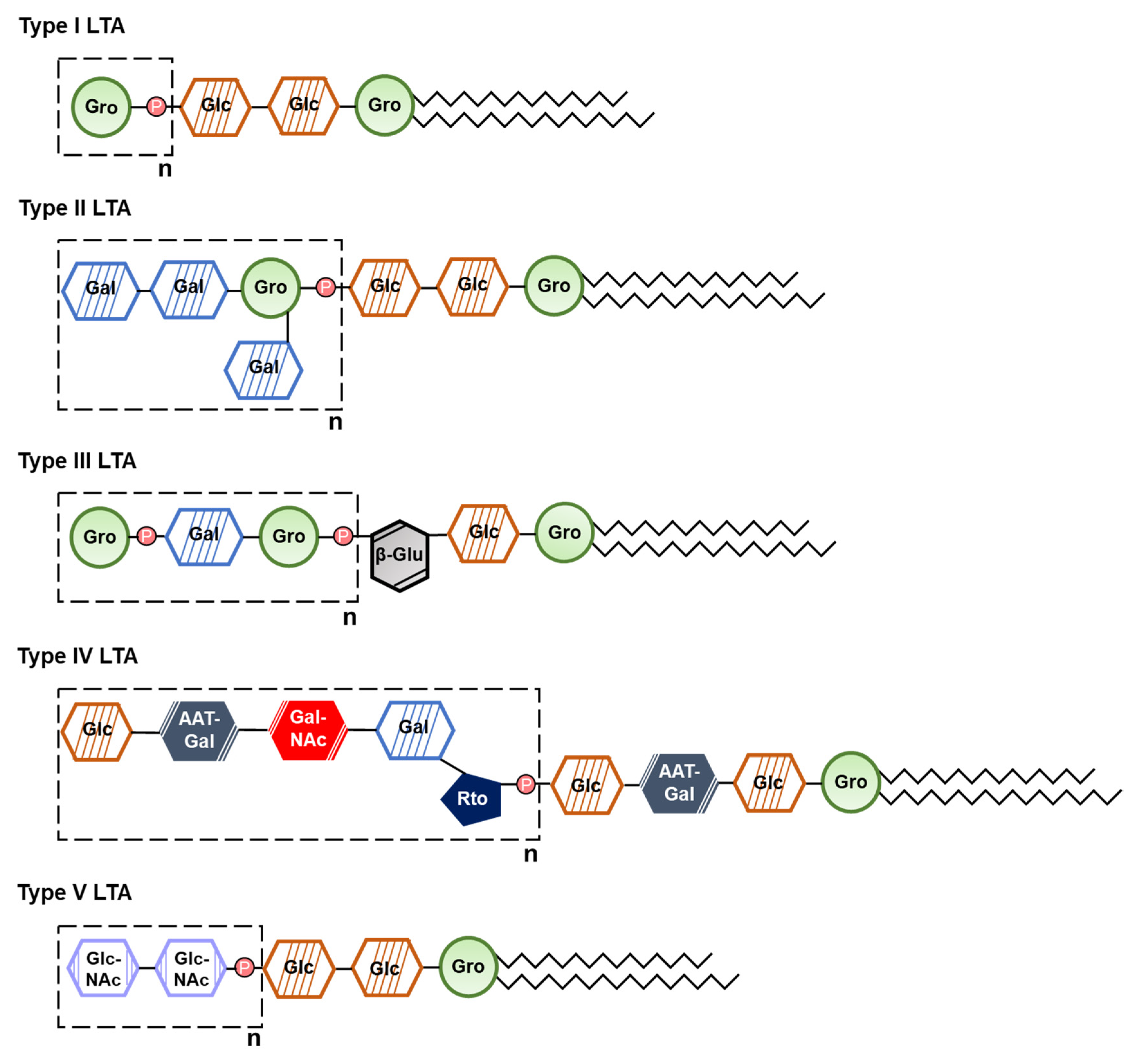
LTA is one of the important virulence factors of Gram-positive bacteria, which consists of alditol phosphate-containing polymer and lipid anchor [74]. Based on its chemical structure, LTA is classified into five types (type I to V), and each bacterium has a distinct characteristic LTA structure (Figure 2) [74,75]. Bacterial LTA has association with various inflammatory diseases such as skin infection and sepsis [75]. LTA specifically attaches to the host cells through TLR2 and CD14, leading to the recruitment of MyD88 and TNF receptor associated factor (TRAF) 6 [76]. It sequentially induces mitogen-activated protein kinase (MAPK) and NF-κB activation [76]. Consequently, the downstream cascade induces innate immune responses, such as the production of nitric oxide and TNF-α [77,78].
In addition, LTA attenuates osteoclast differentiation from mouse bone marrow-derived macrophages [36,37,38]. LTA from Enterococcus faecalis inhibits differentiation of macrophages into mature osteoclasts. Macrophages seem to retain phagocytic activity against bacterial infection [36]. Recently, Wang et al. reported that these inhibitory effects occur through the transcription factor, recombination signal binding protein (RBP)-Jκ [37]. Not only E. faecalis LTA but also staphylococcal LTA inhibits osteoclastogenesis and bone resorption through TLR2 pathway [38].
LTA is also responsible for modulating osteoblast differentiation and bone formation. For instance, mesenchymal stem cells stimulated by LTA from S. aureus upregulate the expression of various osteogenic markers, such as Runx2, alkaline phosphatase (ALP), type I collagen, and calcium deposition through enhanced autophagy [39]. Additionally, LTA from S. aureus promotes the synthesis of bone bridge, ossification, and healing of femoral fractures induced by medial parapatellar arthrotomy [40]. It is likely that such phenomenon was induced by enhancing osteoblast differentiation and inhibiting osteoclast activation [40]. Although LTA appears to be a potential treatment for bone diseases, further studies are needed because LTA causes differential immune responses depending on the source of bacteria.

2.2. Surface Proteins

2.2.1. Lipoprotein

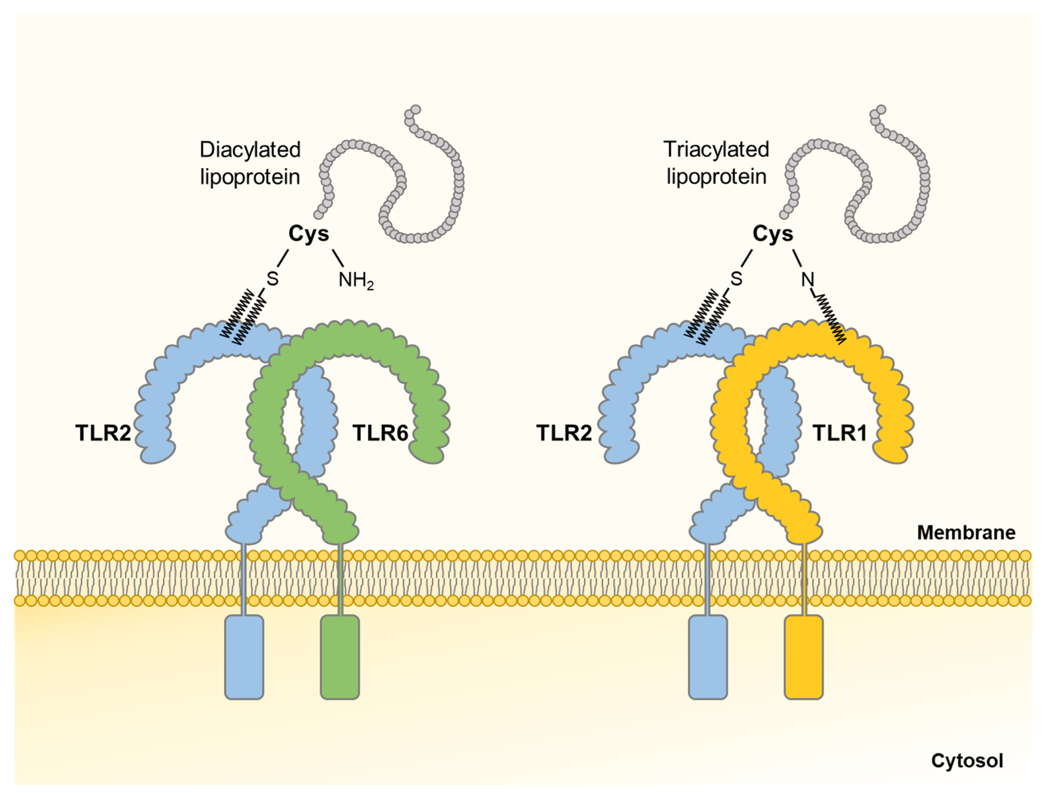
Bacterial LPPs, which are anchored to the cell membrane by N-terminally-linked fatty acids, are one of the major virulence factors causing potent immuno-stimulatory effects [79]. The structure of LPP consists of a protein with a lipid moiety [80]. The protein is in charge of physiological functions, while the lipid moiety anchors LPPs in the bacterial cell membranes and induces bacteria-specific immune responses via TLR2 and additional receptors [80,81]. Based on the number of lipid moieties, LPPs are classified into diacylated or triacylated forms, which are mainly expressed on Gram-positive or Gram-negative bacteria, respectively [82]. Diacylated LPPs contain S-diacylated cysteine residues and triacylated LPPs have N-acyl-S-diacylated cysteine residues [82]. Meanwhile, TLR2 forms a heterodimer with TLR1 or TLR6, recognizing LPPs. Diacylated LPPs are sensed by TLR2/TLR6, while triacylated LPPs are recognized by TLR1/TLR2 (Figure 3) [83,84]. The recognition of LPPs through TLR1/TLR2 or TLR2/TLR6 heterodimer mediates MyD88-mediated signaling transduction and subsequently activates NF-κB, enabling the production of pro-inflammatory cytokines and chemokines [85].
In bone homeostasis, bacterial LPP is known as a potent bone-destructing factor. Kim et al. demonstrated that committed osteoclasts treated with wild-type S. aureus enhances osteoclast differentiation, whereas LPP-deficient S. aureus loses such effect [41]. Furthermore, synthetic lipopeptides, Pam2CSK4 and Pam3CSK4, which mimic bacterial LPP induce osteoclastogenesis by activating TLR2/MyD88 pathway and secreting pro-inflammatory cytokines such as IL-6 and TNF-α [41,42]. In addition, Pam2CSK4 and Pam3CSK4 upregulate RANKL production while downregulating osteoprotegerin (OPG) by stimulating osteoblasts [41].
LPPs also induce bone loss in vivo. Pam2CSK4 and Pam3CSK4 destruct calvarial bone in the mouse implanted with a collagen sheet [41]. In addition, intraperitoneal administration of Pam2CSK4 or Pam3CSK4 significantly decreases the femur bone density of mice [41,43]. Souza et al. also reported that Pam2CSK4 promotes periodontal destruction in mice by inducing gingival inflammation and alveolar bone resorption [43]. Consequently, bacterial LPPs induce differentiation of mouse committed osteoclasts into mature osteoclasts in vitro and bone resorption in vivo via TLR2/MyD88 pathway [41,42,43].

2.2.2. Adhesin

Bacteria possess various macromolecules on their cell surface allowing adhesion and/or interaction with the host [86]. Therefore, these surface molecules play vital roles in bacterial pathogenesis and host immune responses. There are a number of carbohydrates and protein adhesins in both Gram-positive and Gram-negative bacteria [87]. Protein adhesins are further classified into fimbrial and non-fimbrial associated structures [87]. Among them, fimbriae are the most representative bacterial surface adhesins which stick out from the surface and play a major role in host cell invasion [88].
Porphyromonas fimbriae are known as a potent osteoclastogenesis factor. It was reported that fimbriae of Porphyromonas gulae and P. gingivalis induce osteoclast differentiation and cytokine production such as IL-1β, IL-6, and TNF-α in bone marrow-derived macrophages [44,45]. In addition, P. gingivalis fimbriae trigger bone resorption by utilizing tyrosine kinases [46,47]. P. gingivalis fimbriae affect osteoclast differentiation but not osteoblast differentiation [48]. Overall, bacterial fimbriae can induce bone resorption predominantly by inducing osteoclastogenesis in mice [44,45,46,47,48]. However, little is known about the effects of other adhesins on bone and bone-related cells. Therefore, it is essential to demonstrate how various bacterial adhesins affect bone remodeling in the days to come.

2.3. Peptidoglycan

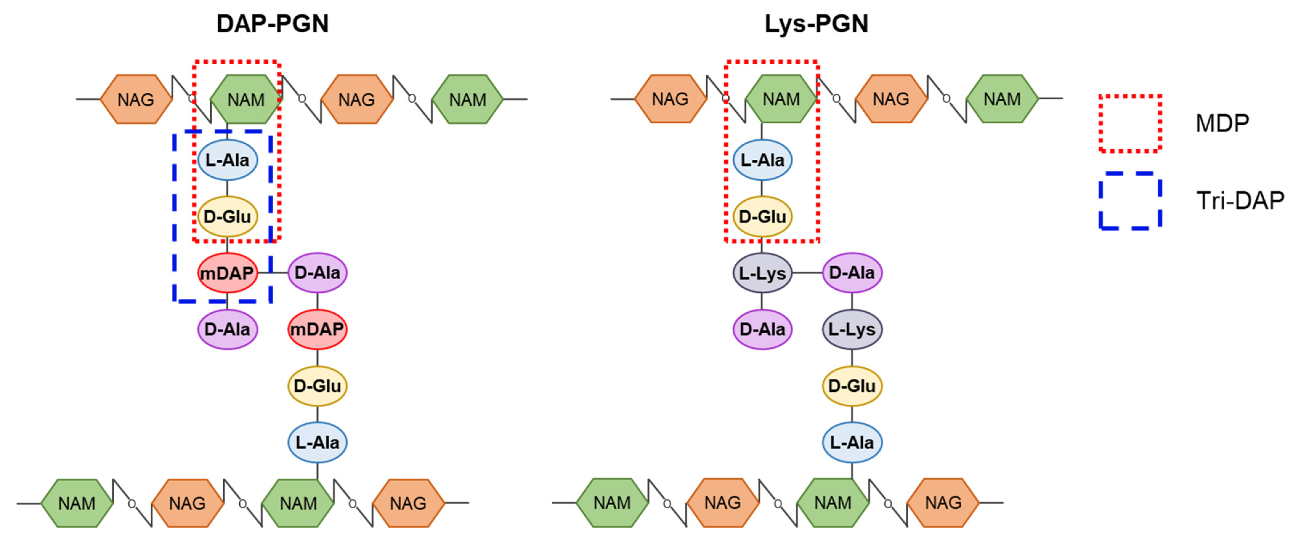
PGN is a highly conserved bacterial cell wall component. It is made up of polymers composed of N-acetylglucosamines (NAGs) and N-acetylmuramic acids (NAMs). Each NAM has a short peptide chain that is involved in forming a cross-linked peptide bridge between polymers [89]. MDP (NAM-l-Ala-d-Glu) is a minimal essential structural motif of PGNs in both Gram-positive and Gram-negative bacteria (Figure 4) [89]. Gram-positive bacteria possess lysine-type PGNs, while Gram-negative bacteria have DAP-type PGNs [90].
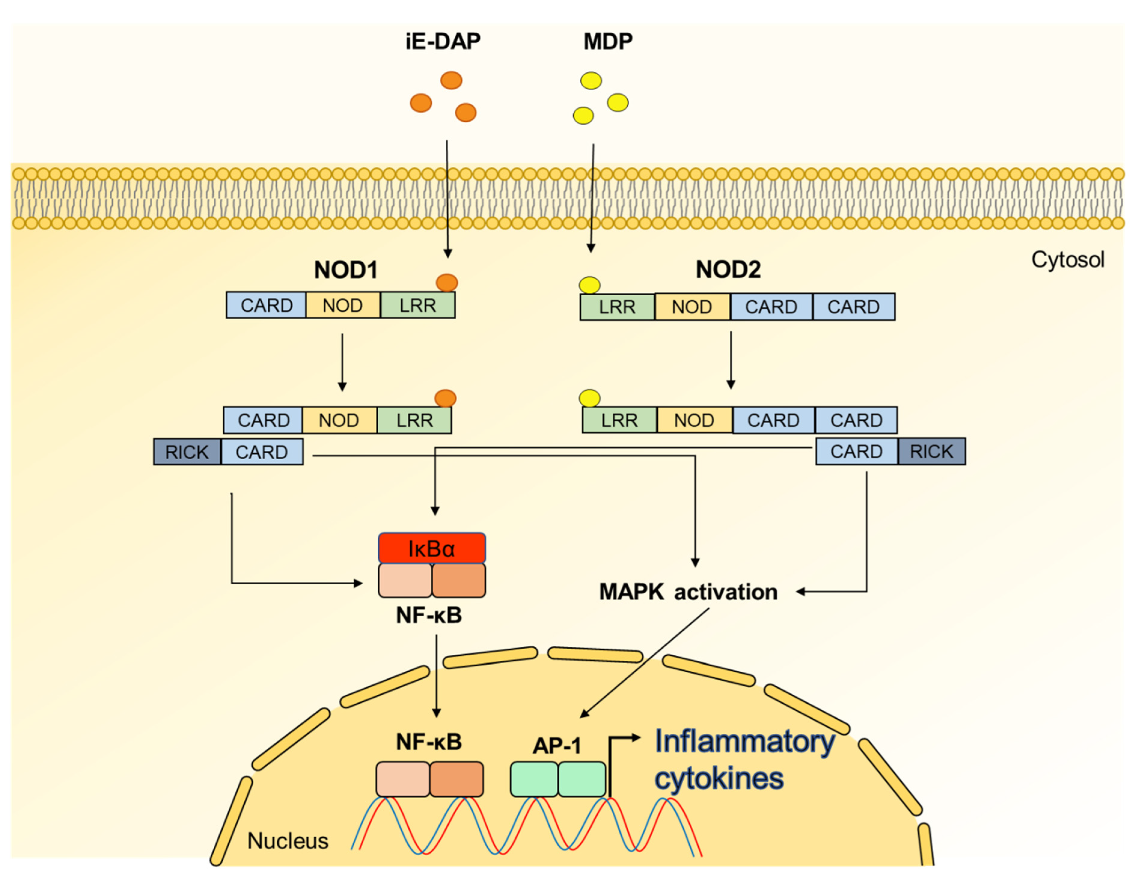
NODs, which are present in the host cytoplasm, are responsible for sensing PGN motifs [91]. NODs are composed of caspase recruitment domain (CARD) at N-terminal, an NOD at intermediate site, and a leucine-rich repeat domain at C-terminal [91]. Among the NODs, NOD1 recognizes iE-DAP of Gram-negative bacterial PGNs, whereas NOD2 senses MDP moieties of ubiquitous bacterial PGNs [91]. Once iE-DAP and MDP are recognized by NOD1 and NOD2, respectively, both NODs induce CARD-CARD interaction and then form the complex with the adaptor molecules, receptor-interacting protein-like interacting caspase-like apoptosis regulatory protein kinase (RICK), leading to NF-κB and MAPK activation for triggering inflammatory responses (Figure 5) [91,92,93].
Accumulating reports suggested that PGN plays a bi-functional role in bone metabolism. Kishimoto et al. reported that PGN and LPS synergistically induce bone resorption and osteoclastogenesis [50]. They showed that S. aureus PGN or Escherichia coli PGN accelerates osteoclast formation and bone resorption only when they are co-stimulated with LPS. However, when treated independently, only S. aureus PGN, but not E. coli PGN, induces alveolar bone resorption [50]. Similarly, Ozaki et al. demonstrated that S. aureus PGN and E. coli LPS exacerbate alveolar bone loss and induce osteoclastogenesis from committed osteoclasts through the upregulation of TNF-α, IL-10, and IL-17 [51]. It was reported that MDP together with LPS, not MDP alone, could enhance osteoclastogenesis and bone loss through upregulation of RANKL and TLR4 expression [52]. PGN of Actinomyces naeslundii also induces osteoclastogenesis and alveolar bone resorption by triggering pro-inflammatory cytokines, such as IL-1β, IL-6, and TNF-α [53]. The importance of NOD1 stimulation has been suggested because stimulation of NOD1 induces alveolar bone loss and periodontitis [54]. In addition, Chaves et al. described that osteoclast formation is increased in NOD1 knockout mice, suggesting that NOD1 affects the upregulation of osteoclastogenesis [54]. Collectively, PGN-induced osteoclastogenesis and bone resorption occur through NOD1-related downstream signals and production of pro-inflammatory cytokines.
Unlike the NOD1 signaling, NOD2 signals could induce bone formation. Park et al. reported that MDP could enhance bone mineral density by upregulation of bone formation [57]. Osteoblasts treated with MDP augment Runx2 expression, which is a major transcriptional factor of osteoblast differentiation. In addition, MDP indirectly reduces osteoclastogenesis through the downregulation of RANKL/OPG ratio from osteoblasts. Furthermore, pre- or post-treatment of MDP alleviates RANKL-induced osteoporosis via NOD2 signaling [57]. Because MDP increases NOD2 expression level and other NOD2 ligands also induce bone formation similarly to the action of MDP, NOD2 agonists like MDP could be a novel therapeutic agent of osteoporosis [57]. Lactobacillus fermentum, which activates NOD2 signaling, attenuates bone resorption and decreases the number of osteoclasts [54]. Likewise, intraperitoneal injection of L. plantarum PGN in mice enhances bone density of femurs [57]. Collectively, NOD2-stimulating bacterial PGNs can ameliorate bone health by increasing bone formation and diminishing bone resorption.

2.4. Secretory Microbial Molecules

2.4.1. Short Chain Fatty Acid

SCFAs, which consist of fewer than six carbons, are metabolites mainly produced by commensal bacteria through fermentation of dietary fibers [94]. Acetate, propionate, and butyrate are the most predominant form of SCFAs in the gastrointestinal tract and have a molar ratio varying from 40:40:20 to 75:15:10 depending on the diet [95,96]. There are two major pathways for modulation of host cells by SCFAs. One major pathway utilized by the host is through GPCRs. GPCRs are seven transmembrane receptors of host’s signaling molecules or MAMPs to induce intracellular signaling pathways [97]. Among GPCRs, SCFAs can bind and activate GPCR 40, 41, and 43, which are designated as free fatty acid receptor (FFAR) 1, 3, and 2, respectively, or GPCR 109a [98]. Conserved two arginine residues at transmembrane helixes 5 and 7 are important for the recognition of SCFAs by FFAR 1, 2, and 3 [99]. In addition, GPCRs are commonly expressed in bone metabolism-involved cells, including adipocytes, neutrophils, macrophages, osteoclasts, and osteoblasts [100,101,102,103]. Another route of host cell modulation by SCFAs is the inhibition of histone deacetylases (HDACs). HDAC plays an important role in regulating gene expression by epigenetic modification of chromosome structure [104]. Among SCFAs, butyrate inhibits HDAC activity, leading to decreased production of MAMP-induced nitric oxide or pro-inflammatory cytokines [105]. Consequently, SCFAs have various effects on the host’s health through the activation of GPCR or HDAC inhibition, such as modulating intestinal homeostasis, enhancing the production of antimicrobial peptides, providing anti-inflammatory immune responses, and augmenting mucosal vaccine properties [105,106]. SCFAs also affect bone metabolism by the regulation of osteoclasts and osteoblasts via GPCR or HDAC inhibition [107,108,109].
Iwami et al. reported that sodium butyrate potently attenuates the formation of tartrate-resistant acid phosphatase (TRAP)-positive multinucleated cells, which are cultured from bone marrow cells [16]. Besides, FFAR1 knockout mice show less bone density than wild-type mice. In addition, activation of FFAR1 suppresses the mRNA expression of osteoclast-specific genes, such as TRAP, matrix metalloproteinase-9 (MMP-9), and cathepsin K, and RANKL-induced osteoclastogenesis from macrophages via inhibiting RANKL-induced NF-κB signaling pathway [110]. Similarly, increased alveolar bone loss is observed in FFAR2 knockout mice. In fact, activation of FFAR2 by SCFAs or its agonists decreases the RANKL-induced osteoclast differentiation from macrophages and prevents alveolar bone loss [111]. On the other hand, mRNA expression levels of FFAR3 or GPCR 109a are downregulated during RANKL-induced osteoclast differentiation from macrophages. Moreover, inhibitory effect of SCFAs on osteoclastogenesis treated with RANKL is not observed if SCFAs were administered to committed osteoclasts [107]. Interestingly, two HDAC inhibitors, trichostatin A and sodium butyrate, inhibit the RANKL-induced osteoclast differentiation from macrophages through downregulation of osteoclast-specific gene expression, such as RANK and cathepsin K [108]. Because HDAC inhibition dampens osteoclast differentiation [103], inhibition of HDAC by SCFAs may be involved in the suppression of osteoclastogenesis [108]. Collectively, SCFAs inhibit the osteoclastogenesis from macrophages via HDAC inhibition and/or FFAR1 or 2 activation but not from committed osteoclasts [107,108,110,111].
SCFAs also influence osteoblast proliferation and differentiation in both animals and humans. Sodium butyrate increases the ALP activity of MC3T3-E1 murine osteoblastic cell line [16]. In contrast, a high concentration of sodium butyrate inhibits the differentiation and mineralization of ROS17/2.8 rat osteoblastic cell line via suppression of osteoblast-specific factors, such as Runx2, osterix, and Dlx5 [112]. Moreover, a low concentration of butyrate induces histone H3 acetylation with concurring expression of ALP, osteonectin, and OPG in MG-63 human osteoblastic cell line [109]. In fact, treatment of sodium butyrate at 16 mM attenuates osteoblast proliferation by suppression of cell cycle in vitro [113]. Notably, they have the potential to treat destructive bone diseases caused by postmenopausal or inflammatory conditions in animal models [114]. Thus, the optimal concentration of SCFAs could be used as therapeutic agents for treating bone diseases.

2.4.2. Extracellular Vesicle

EVs could be released from archaea, eukaryote, and bacteria [115]. EVs are usually classified into three types (exosomes, microvesicles, and apoptotic bodies) according to their biogenesis [116]. Among them, the diameter of bacterial EVs is roughly 20~500 nanometers, and these spherical membrane-enveloped particles are secreted from parental bacteria into the extracellular environment [117]. Bacterial EVs carry diverse cargos such as membrane-bound proteins, LPPs, polysaccharides, enzymes, toxins, metabolites, and nucleic acids [118]. Bacteria can utilize EVs for horizontal gene transfer [119]. Various host cells recognize the content of EVs via diverse PRRs such as TLR, NOD, and retinoic acid-inducible gene, potentially leading to inflammatory conditions, or in some cases, immune tolerogenic conditions [120].
It has been reported that EVs derived from Filifactor alocis inhibit the differentiation of bone-derived mesenchymal stromal cells in vitro [17]. In addition, F. alocis EVs potently downregulate osteogenic factors, such as Runx2, osterix, ALP, osteocalcin (OCN), and type I collagen, thereby attenuating mineralization. Notably, F. alocis EVs activate TLR2 but not TLR4, and the inhibitory effect of F. alocis EVs on osteogenic differentiation is fully dependent on TLR2 signaling pathway which mediates the activation of MAPK and NF-κB [17]. Unfortunately, the role of bacterial EVs on osteoclast differentiation and function has been poorly investigated. However, emerging evidence indicates that bacterial EVs might directly or indirectly influence osteoclast differentiation. Bacterial EVs activate the monocyte-derived dendritic cells to induce pro-inflammatory cytokines, such as IL-1β, IL-6, or TNF-α, which can trigger the osteoclast differentiation from committed osteoclasts [121,122]. Moreover, bacterial EVs regulate the expression of RANKL and OPG, which are modulators of osteoclastogenesis, through TLR2 in mesenchymal stromal cells [17]. Further studies are needed to understand the role of bacterial EVs on bone metabolism.

2.4.3. Extracellular Polysaccharide

Many bacteria produce extracellular polysaccharides which are classified into two types: exopolysaccharides (EPS) and capsular polysaccharides (CPS) [123]. EPS are defined as released polysaccharides around the bacterial cell surface, becoming an integral component of biofilm [124]. On the other hand, CPS are covalently bonded to the bacterial cell surface [125]. Bacteria exploit the extracellular polysaccharide as a barrier to protect themselves against harsh environments [126].
Mounting evidence suggested that extracellular polysaccharides of bacteria have bi-functional effects on bone metabolism. For example, EPS purified from Bifidobacterium longum (EPS-624) inhibit osteoclast differentiation from mouse bone marrow-derived macrophages by activating TLR2 signaling pathway [127]. In addition, EPS-624 increase the differentiation of osteoblasts from human bone marrow-derived mesenchymal stromal cells [127]. Thus, authors suggested the potential therapeutic use of EPS-624 against destructive bone diseases [127]. Moreover, EPS isolated from Vibrio diabolicus, which are hyaluronic acid-like EPS, potently enhance bone healing without abnormal bone growth in vivo [128]. Indeed, lying osteoblasts on trabecular bone surfaces and increasing osteocytes inclusion are observed in the bone treated with EPS [128]. In contrast, oversulfated EPS produced by Alteromonas infernus (OS-EPS) inhibit the proliferation and mineralization activity of osteoblasts in vitro [129]. Also, OS-EPS decrease the RANKL-induced osteoclast differentiation from CD14+ human monocytes while increasing the collagenolytic activity of osteoclasts by using cathepsin K [129]. Notably, OS-EPS cause trabecular bone loss through enhanced osteoclastogenesis [129].
Similar to EPS, CPS also affect the activation and differentiation of bone cells, including osteoclasts and osteoblasts. CPS from Aggregatibacter actinomycetemcomitans Y4 (Aa-CPS) enhance the formation of osteoclasts and promote the bone resorptive activity through the induction of IL-1α in vitro [130]. Aa-CPS also activate osteoclasts by upregulating PGE2, which has a positive effect on osteoclast formation in vitro [131]. These reports indicate that Aa-CPS-induced PGE2 and IL-1α are involved in inflammatory bone diseases such as periodontitis by promoting osteoclastogenesis. In addition, Aa-CPS show anti-proliferative activity by causing Fas-mediated apoptotic cell death in MC3T3-E1 murine osteoblastic cell line in vitro [132]. Moreover, immunization of CPS from P. gingivalis exhibits immunoglobulin responses and protects P. gingivalis-induced oral bone loss in vivo, suggesting that CPS are one of the responsible molecules for bone diseases [133]. In conclusion, because extracellular polysaccharides have controversial effects on the bone metabolism, further studies are needed.

2.4.4. Cyclic Dinucleotide

CDNs were originally identified in 1987 as bacterial second messengers that regulate cellulose synthesis [134]. Bacterial CDNs are classified as cyclic diadenylate monophosphate (c-di-AMP), cyclic diguanylate monophosphate (c-di-GMP), and 3′,3′-cyclic guanosine monophosphate-adenosine monophosphate (3′3′-cGAMP) [135]. CDN contains two nucleotide monophosphates that are linked to each other by phosphodiester bonds to form a cyclic structure [135]. CDNs are important for the maintenance of bacterial life cycle including survival, colonization, and biofilm formation [136]. For instance, CDNs trigger extracellular matrix production, subsequently forming biofilm in bacteria [137]. Furthermore, CDNs released from bacteria can be recognized by host cells and activate various host immune responses [138].
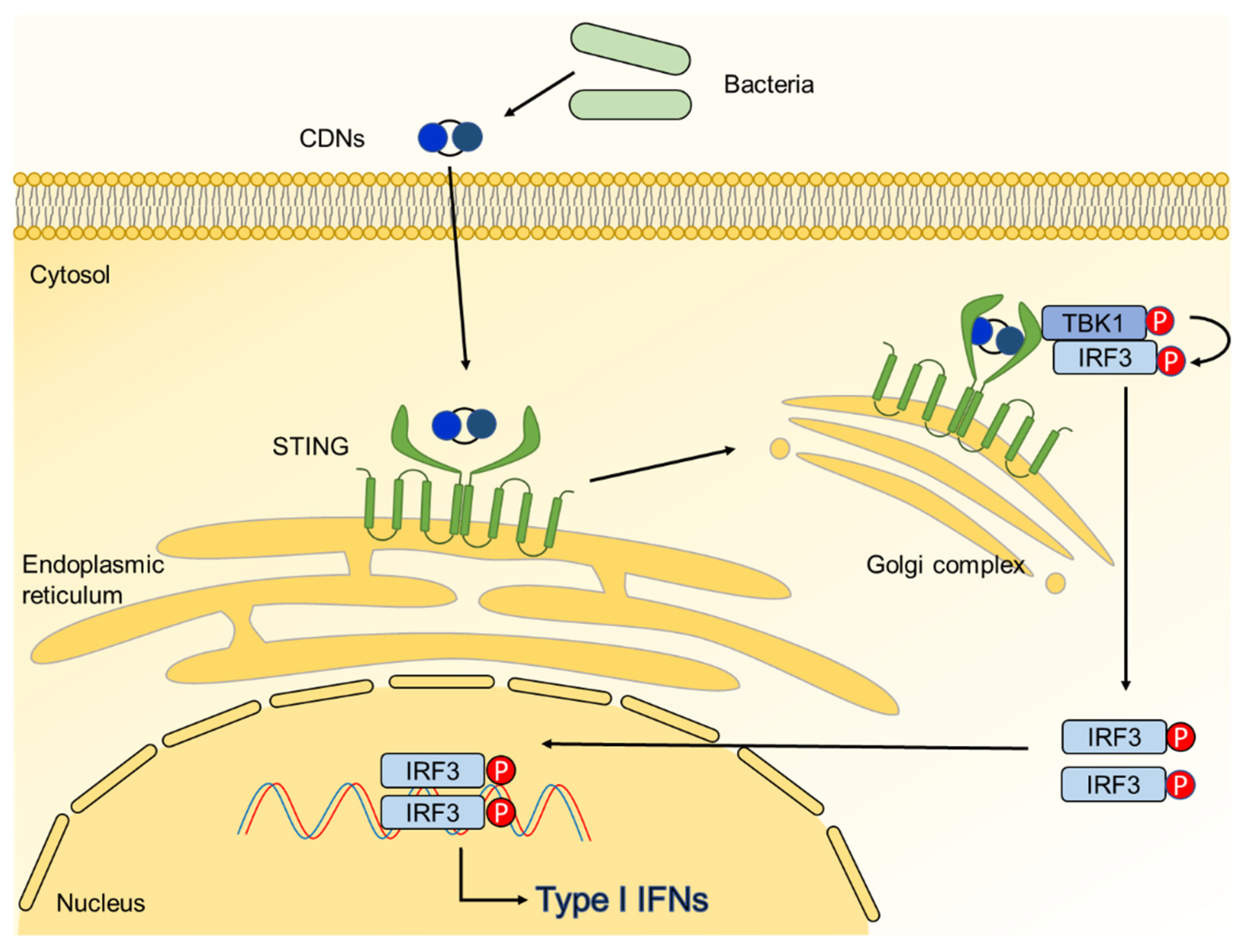
Stimulator of IFN genes (STING), also known as transmembrane 173, has 4 transmembrane regions and is located at the endoplasmic reticulum of host cells [139]. STING directly recognizes the cytosolic CDNs, leading to secretion of type I IFNs (Figure 6) [140]. When CDNs bind to STING, STING recruits TRAF-associated NF-κB activator-binding kinase 1 (TBK1) to the C-terminal tail [141]. Recruited TBK1 phosphorylates IRF3, forming a homodimer [142]. Phosphorylated IRF3 homodimer enters through the nucleus pore, inducing the gene expression of IFN-β [142]. Several studies investigated that STING is involved in bone metabolism. Overexpression of STING in RAW 264.7 cell inhibits RANKL-induced osteoclast differentiation and expression of osteoclast-specific genes, such as TRAP, cathepsin K, and MMP-9 [143]. On the other hand, lack of STING suppresses bone accrual via inhibition of pro-osteogenic gene expression [144].
Interestingly, CDNs can also influence bone metabolism. CDNs inhibit RANKL-induced osteoclast differentiation from mouse bone marrow-derived macrophages through STING-dependent signaling pathway [18]. Authors demonstrated that CDNs potently trigger STING-TBK1-IRF3 cascade and induce the mRNA expression of IFN-β during osteoclast differentiation from macrophages. IFN-β in turn acts as a negative regulator of osteoclast differentiation by activating Janus kinase (Jak)-signal transducer and activator of transcription (STAT) signaling [145], which is the major pathway responsible for the inhibition of osteoclastogenesis. In contrast, because ubiquitin-mediated degradation of Jak in committed osteoclasts, CDN-induced IFN-β cannot activate the Jak-STAT signaling pathway during differentiation of committed osteoclasts from mouse. Notably, CDNs prevent the RANKL-induced bone destruction in collagen sheet implanted mouse model [18]. Recently, it has been reported that STING interacts with TRAF6, which is a TLR signaling mediator, in human keratinocytes and monocytes [146]. Thus, further studies are needed to understand cooperative effects of MAMPs and CDNs on bone metabolism. In conclusion, targeting STING using CDNs serves as a novel therapeutic strategy for bone disorder treatment due to its heavy regulatory association with bone metabolism.

3. Therapeutics

Microbes influence bone metabolism by constant interaction with host using their various MAMPs (Table 2) [7]. In infectious condition, MAMPs often trigger immoderate osteoclastogenesis or inhibit osteoblast differentiation through the activation of immune responses, causing bone diseases such as osteomyelitis, osteoporosis, and periodontitis [7]. Antibiotics are commonly used to treat MAMP-induced bone diseases in bacterial infection [147]. Nevertheless, the emergence of antibiotic-resistant bacteria and remaining MAMPs after treatment pose significant challenge for complete clearance [148]. Therefore, further studies are needed to understand the role of MAMPs in bone diseases and to control the immune responses induced by MAMPs.
On the other hand, several studies investigated that some MAMPs, especially derived from probiotics, decrease bone resorption or enhance bone formation by controlling the differentiation of osteoclasts or osteoblasts, respectively, in both in vitro and in vivo studies [18,57,114]. Many therapeutic drugs, such as bisphosphonates, monoclonal antibodies, or hormone preparations, are traditionally developed to treat bone diseases by inhibiting bone resorption or inducing bone formation [149,150,151]. However, conventional drugs show unexpected side effects, such as nausea or osteonecrosis of jaw [151,152,153]. Therefore, we suggest that probiotic-derived MAMPs could alternatively be used in place of conventional therapies. To evaluate their therapeutic use, we have discussed below how to treat MAMP-induced bone diseases and how to exploit MAMPs in bone health.

3.1. Treatment of Microbe-Associated Molecular Patterns-Induced Bone Diseases

In general, most MAMPs are potent inducers of pro-inflammatory cytokines, such as IL-1, IL-6, or TNF-α, via the recognition by PRRs on animal and human cells, including epithelial cells, endothelial cells, and immune cells [154]. These MAMP-induced pro-inflammatory cytokines positively influence the differentiation of animal and human committed osteoclasts into mature osteoclasts and the activity of osteoclasts, leading to bone loss [155]. Thus, targeting pro-inflammatory cytokines or their receptors can become one of the therapeutic strategies for MAMP-induced bone diseases. For example, through the use of blocking antibodies that antagonize TNF-α and IL-1 receptors, significant reduction of inflammation and osteoclastogenesis were observed in experiment using mouse committed osteoclasts in vitro [41]. Antibody specific to IL-6 or IL-6 receptor directly inhibits osteoclast differentiation from mouse committed osteoclasts in in vitro experiment and restores bone erosion in TNF-α-transgenic mice [41,156]. Furthermore, many preclinical and clinical studies reported that TLR inhibitors or blocking antibodies alleviate inflammatory diseases [157]. Since MAMPs directly promote osteoclast differentiation from mouse committed osteoclasts through the activation of PRR signaling pathway in both in vitro and in vivo, regulation of MAMPs using inhibitors or antibodies may also be effective to control MAMP-induced osteoclastogenesis [41,158].
In the case of osteoblasts, targeting and inhibiting some potent MAMPs involved in diminishing osteoblasts could be a useful way to alleviate bone diseases [159]. For instance, hindering the action of LPS, which is a potent osteoblast inhibitor, might be valuable to prevent LPS-induced bone loss in bacterial infections [159,160,161,162]. There are several ways to inhibit the action of LPS. Jung et al. demonstrated that TLR4 decoy receptor inhibits LPS-induced NF-κB activation in human lymphatic microvascular endothelial cells in vitro and prevents Gram-negative bacterial sepsis in LPS-induced sepsis mouse model [159]. Indeed, anti-TLR4-antibody effectively treated stroke in vivo, hinting the therapeutic potency of anti-TLR4-antibody [160]. Therefore, blocking TLR4 by its specific antibody or decoy receptor might be helpful in LPS-induced osteoporosis patients. Another way to prevent LPS from binding osteoblasts is direct neutralization of the LPS. There are several peptides that can bind and inhibit LPS-induced inflammation. Antitoxin peptide Pep 19-2.5, which is designed to bind to LPS, reduced TNF-α expression and inflammation in several in vitro and in vivo models [161]. In addition, polymyxin B, which neutralizes LPS, shut down NF-κB signaling pathway in vitro [162]. As mentioned, LPS-neutralizing peptides could inhibit downstream signaling pathway of LPS and, therefore, are expected to be useful in preventing LPS-induced osteoblast reduction. Furthermore, LPPs or adhesins, which negatively affect osteoblast differentiation, could also be prevented by blocking osteoblast recognition receptors or neutralizing the MAMPs by antibodies or neutralizing peptides.

3.2. Probiotics as Therapeutic Agent for Bone Health

Recent decades, numerous studies are implemented to understand the role of commensal microbiota on digestive, endocrine, nervous, and immune system in the host [163]. Notably, emerging evidence indicates that commensal microbiota could regulate bone metabolism by controlling immune function and enhancing barrier function (Table 3) [11,164].
For instance, supplementation of probiotics, microorganisms that offer health benefit to the host, prevents bone loss in postmenopausal ovariectomized mice [11,164]. In addition, L. reuteri ATCC 6475 upregulates bone volume/tissue volume (BV/TV), trabecular number (Tb.N), and trabecular thickness (Tb.Th) in normal mice, postmenopausal ovariectomized female mice, and type I diabetic bone loss male mice [12,165,166]. L. rhamnosus GG increases BV/TV and OCN level but decreases RANKL, TNF-α, and IL-17 mRNA expression in ovariectomized osteoporotic mice [11]. Lactobacillus paracasei and L. plantarum enhance BV/TV, Tb.N, and cortical bone in osteoporotic mice [168]. L. casei prevents wear-debris induced osteolysis [169]. Furthermore, L. rhamnosus GG, L. gasseri SBT2055, or L. brevis CD2 downregulates alveolar bone loss and inflammation in periodontitis animal model [13,14,167]. These reports suggest that probiotics are enough to induce bone formation and reduce bone resorption in the host, presenting their possibility as novel bone healing agents. Moreover, the underlying action mechanisms of probiotics on bone metabolism are being elucidated. A well-known mechanism is that probiotic-derived MAMPs, such as cell wall components and secretory molecules, interact with bone cells, resulting in the regulation of bone metabolism.
As mentioned previously, MDP has positive effects on bone metabolism. MDP potently augments osteoblast differentiation and induces bone formation through the activation of NOD2 signaling pathway [57]. MDP has both preventive and therapeutic effects against RANKL-induced osteoporosis mouse model [57]. Other NOD2 ligands, including M-TriLYS, L18-MDP, and murabutide, also promote osteoblast differentiation in vitro and upregulate bone volume when injected in normal mice [57]. Furthermore, MDP indirectly inhibits osteoclastogenesis with attenuated RANKL/OPG ratio [57]. Notably, increased RANKL/OPG ratio can be observed in postmenopausal women with low bone mineral density [170], suggesting that MDP can effectively be used to treat patients with abnormal RANKL/OPG ratio. Collectively, MDP and other NOD2 agonists including NOD2-stimulating PGNs are promising therapeutic agents for the prevention or treatment of bone diseases by controlling the differentiation of both osteoclasts and osteoblasts.
Probiotic-derived secretory molecules, such as SCFAs and CDNs, also influence bone metabolism. SCFAs directly decrease the formation and function of mouse osteoclasts by HDAC inhibition and change the metabolic condition of mouse bone marrow cells in vitro [114]. In addition, adequate dose of SCFAs upregulates ALP activity of MC3T3-E1 mouse osteoblastic cell line in vitro [16]. Low dose of SCFAs increases osteoblast differentiation by HDAC inhibition in vitro using MG-63 human osteoblastic cell line [109]. Moreover, SCFAs indirectly increase osteoblast differentiation in wild-type mice compared to T-cell receptor knockout mice in which butyrate-activated T cells release the osteoblast differentiation factor, Wnt10b [171,172]. Furthermore, SCFAs systemically increase bone density in postmenopausal ovariectomized or collagen-induced inflammatory arthritis mouse model [114]. Like SCFAs, CDNs which are secretory bacterial second messengers also decrease osteoclast differentiation from mouse macrophages in vitro via STING-mediated IFN-β signaling pathway. In addition, CDNs potently prevent calvarial bone loss in collagen sheet implanted mouse model [18]. It is likely that bacterial secretory molecules, such as SCFAs and CDNs, can be developed as therapeutic agents for the treatment of bone diseases by inhibiting excessive osteoclastogenesis and promoting the activity of osteoblasts. Further studies are necessary to fully understand the regulatory effects of probiotic-derived molecules on osteoclast or osteoblast differentiation and activation.

4. Conclusions

In this review, we discussed the properties of MAMPs on bone metabolism in both in vitro and in vivo conditions. The MAMPs, including the cell wall components and secretory molecules, can directly or indirectly modulate the differentiation and activation of osteoclasts and osteoblasts. Thus, the MAMPs could be promising molecular targets for bacteria-induced bone diseases such as osteoporosis and periodontal diseases. Furthermore, a use of beneficial MAMPs on bone metabolism might be a novel therapeutic strategy for prevention or treatment of bone disorders.

Author Contributions

Conceptualization, S.H.H. and O.-J.P.; writing—original draft preparation, Y.K., C.P., J.L., D.H.P., S.J. and O.-J.P.; writing—review and editing, S.H.H., C.Y. and O.-J.P.; supervision, S.H.H.; project administration, S.H.H.; funding acquisition, S.H.H. and O.-J.P. All authors have read and agreed to the published version of the manuscript.

Funding

This work was supported by grants from the National Research Foundation of Korea, which is funded by the Korean government (NRF-2019R1A2C2007041, NRF-2018R1A5A2024418, and NRF-2019R1I1A1A01060952).

Institutional Review Board Statement

Not applicable.

Informed Consent Statement

Not applicable.

Data Availability Statement

Not applicable.

Conflicts of Interest

The authors declare no conflict of interest.

References

  1. Kular, J.; Tickner, J.; Chim, S.M.; Xu, J. An overview of the regulation of bone remodelling at the cellular level. Clin. Biochem. 2012, 45, 863–873. [Google Scholar] [CrossRef]
  2. Robling, A.G.; Castillo, A.B.; Turner, C.H. Biomechanical and molecular regulation of bone remodeling. Annu. Rev. Biomed. Eng. 2006, 8, 455–498. [Google Scholar] [CrossRef] [PubMed]
  3. Feng, X.; McDonald, J.M. Disorders of bone remodeling. Annu. Rev. Pathol. 2011, 6, 121–145. [Google Scholar] [CrossRef] [PubMed] [Green Version]
  4. Krauss, J.L.; Roper, P.M.; Ballard, A.; Shih, C.C.; Fitzpatrick, J.A.J.; Cassat, J.E.; Ng, P.Y.; Pavlos, N.J.; Veis, D.J. Staphylococcus aureus Infects Osteoclasts and Replicates Intracellularly. mBio 2019, 10. [Google Scholar] [CrossRef] [PubMed] [Green Version]
  5. Wright, J.A.; Nair, S.P. Interaction of staphylococci with bone. Int. J. Med. Microbiol. 2010, 300, 193–204. [Google Scholar] [CrossRef] [PubMed] [Green Version]
  6. Martin, T.R.; Mathison, J.C.; Tobias, P.S.; Leturcq, D.J.; Moriarty, A.M.; Maunder, R.J.; Ulevitch, R.J. Lipopolysaccharide binding protein enhances the responsiveness of alveolar macrophages to bacterial lipopolysaccharide. Implications for cytokine production in normal and injured lungs. J. Clin. Investig. 1992, 90, 2209–2219. [Google Scholar] [CrossRef] [Green Version]
  7. Charles, J.F.; Nakamura, M.C. Bone and the innate immune system. Curr. Osteoporos. Rep. 2014, 12, 1–8. [Google Scholar] [CrossRef] [PubMed] [Green Version]
  8. Sakurai, A.; Okahashi, N.; Nakagawa, I.; Kawabata, S.; Amano, A.; Ooshima, T.; Hamada, S. Streptococcus pyogenes infection induces septic arthritis with increased production of the receptor activator of the NF-kappaB ligand. Infect. Immun. 2003, 71, 6019–6026. [Google Scholar] [CrossRef] [Green Version]
  9. Binder Gallimidi, A.; Fischman, S.; Revach, B.; Bulvik, R.; Maliutina, A.; Rubinstein, A.M.; Nussbaum, G.; Elkin, M. Periodontal pathogens Porphyromonas gingivalis and Fusobacterium nucleatum promote tumor progression in an oral-specific chemical carcinogenesis model. Oncotarget 2015, 6, 22613–22623. [Google Scholar] [CrossRef] [Green Version]
  10. Parvaneh, K.; Jamaluddin, R.; Karimi, G.; Erfani, R. Effect of probiotics supplementation on bone mineral content and bone mass density. Sci. World J. 2014, 2014, 595962. [Google Scholar] [CrossRef] [Green Version]
  11. Li, J.Y.; Chassaing, B.; Tyagi, A.M.; Vaccaro, C.; Luo, T.; Adams, J.; Darby, T.M.; Weitzmann, M.N.; Mulle, J.G.; Gewirtz, A.T.; et al. Sex steroid deficiency-associated bone loss is microbiota dependent and prevented by probiotics. J. Clin. Investig. 2016, 126, 2049–2063. [Google Scholar] [CrossRef] [Green Version]
  12. McCabe, L.R.; Irwin, R.; Schaefer, L.; Britton, R.A. Probiotic use decreases intestinal inflammation and increases bone density in healthy male but not female mice. J. Cell. Physiol. 2013, 228, 1793–1798. [Google Scholar] [CrossRef] [Green Version]
  13. Kobayashi, R.; Kobayashi, T.; Sakai, F.; Hosoya, T.; Yamamoto, M.; Kurita-Ochiai, T. Oral administration of Lactobacillus gasseri SBT2055 is effective in preventing Porphyromonas gingivalis-accelerated periodontal disease. Sci. Rep. 2017, 7, 545. [Google Scholar] [CrossRef] [PubMed]
  14. Maekawa, T.; Hajishengallis, G. Topical treatment with probiotic Lactobacillus brevis CD2 inhibits experimental periodontal inflammation and bone loss. J. Periodontal Res. 2014, 49, 785–791. [Google Scholar] [CrossRef] [Green Version]
  15. Chu, H.; Mazmanian, S.K. Innate immune recognition of the microbiota promotes host-microbial symbiosis. Nat. Immunol. 2013, 14, 668–675. [Google Scholar] [CrossRef] [PubMed]
  16. Iwami, K.; Moriyama, T. Effects of short chain fatty acid, sodium butyrate, on osteoblastic cells and osteoclastic cells. Int. J. Biochem. 1993, 25, 1631–1635. [Google Scholar] [CrossRef]
  17. Song, M.K.; Kim, H.Y.; Choi, B.K.; Kim, H.H. Filifactor alocis-derived extracellular vesicles inhibit osteogenesis through TLR2 signaling. Mol. Oral Microbiol. 2020, 35, 202–210. [Google Scholar] [CrossRef]
  18. Kwon, Y.; Park, O.J.; Kim, J.; Cho, J.H.; Yun, C.H.; Han, S.H. Cyclic Dinucleotides Inhibit Osteoclast Differentiation Through STING-Mediated Interferon-beta Signaling. J. Bone Miner. Res. 2019, 34, 1366–1375. [Google Scholar] [CrossRef]
  19. Boller, T.; Felix, G. A renaissance of elicitors: Perception of microbe-associated molecular patterns and danger signals by pattern-recognition receptors. Annu. Rev. Plant Biol. 2009, 60, 379–406. [Google Scholar] [CrossRef]
  20. Choi, H.W.; Klessig, D.F. DAMPs, MAMPs, and NAMPs in plant innate immunity. BMC Plant Biol. 2016, 16, 232. [Google Scholar] [CrossRef] [Green Version]
  21. Akira, S.; Uematsu, S.; Takeuchi, O. Pathogen recognition and innate immunity. Cell 2006, 124, 783–801. [Google Scholar] [CrossRef] [Green Version]
  22. Sun, M.; Wu, W.; Liu, Z.; Cong, Y. Microbiota metabolite short chain fatty acids, GPCR, and inflammatory bowel diseases. J. Gastroenterol. 2017, 52, 1–8. [Google Scholar] [CrossRef] [PubMed]
  23. Brubaker, S.W.; Bonham, K.S.; Zanoni, I.; Kagan, J.C. Innate immune pattern recognition: A cell biological perspective. Annu. Rev. Immunol. 2015, 33, 257–290. [Google Scholar] [CrossRef] [Green Version]
  24. Kawai, T.; Akira, S. TLR signaling. Semin. Immunol. 2007, 19, 24–32. [Google Scholar] [CrossRef] [PubMed]
  25. Platnich, J.M.; Muruve, D.A. NOD-like receptors and inflammasomes: A review of their canonical and non-canonical signaling pathways. Arch. Biochem. Biophys. 2019, 670, 4–14. [Google Scholar] [CrossRef]
  26. Chatterjee, S.; Jungraithmayr, W.; Bagchi, D. Immunity and Inflammation in Health and Disease: Emerging Roles of Nutraceuticals and Functional Foods in Immune Support; Academic Press: London, UK, 2018; pp. 175–187. [Google Scholar]
  27. D’Amelio, P.; Sassi, F. Gut Microbiota, Immune System, and Bone. Calcif. Tissue Int. 2018, 102, 415–425. [Google Scholar] [CrossRef]
  28. Chen, M.F.; Chang, C.H.; Hu, C.C.; Wu, Y.Y.; Chang, Y.; Ueng, S.W.N. Periprosthetic Joint Infection Caused by Gram-Positive Versus Gram-Negative Bacteria: Lipopolysaccharide, but not Lipoteichoic Acid, Exerts Adverse Osteoclast-Mediated Effects on the Bone. J. Clin. Med. 2019, 8, 1289. [Google Scholar] [CrossRef] [Green Version]
  29. Ridwan, R.D.; Sidarningsih, T.K.; Salim, S. Effect of lipopolysaccharide derived from surabaya isolates of Actinobacillus actinomycetemcomitans on alveolar bone destruction. Vet. World 2018, 11, 161–166. [Google Scholar] [CrossRef] [Green Version]
  30. Nishihara, T.; Ishihara, Y.; Koseki, T.; Boutsi, E.A.; Senpuku, H.; Hanada, N. Membrane-associated interleukin-1 on macrophages stimulated with Actinobacillus actinomycetemcomitans lipopolysaccharide induces osteoclastic bone resorption in vivo. Cytobios 1995, 81, 229–237. [Google Scholar] [PubMed]
  31. Zou, W.; Bar-Shavit, Z. Dual modulation of osteoclast differentiation by lipopolysaccharide. J. Bone Miner. Res. 2002, 17, 1211–1218. [Google Scholar] [CrossRef]
  32. Liu, J.; Wang, S.; Zhang, P.; Said-Al-Naief, N.; Michalek, S.M.; Feng, X. Molecular mechanism of the bifunctional role of lipopolysaccharide in osteoclastogenesis. J. Biol. Chem. 2009, 284, 12512–12523. [Google Scholar] [CrossRef] [Green Version]
  33. Kadono, H.; Kido, J.; Kataoka, M.; Yamauchi, N.; Nagata, T. Inhibition of osteoblastic cell differentiation by lipopolysaccharide extract from Porphyromonas gingivalis. Infect. Immun. 1999, 67, 2841–2846. [Google Scholar] [CrossRef] [Green Version]
  34. Tomomatsu, N.; Aoki, K.; Alles, N.; Soysa, N.S.; Hussain, A.; Nakachi, H.; Kita, S.; Shimokawa, H.; Ohya, K.; Amagasa, T. LPS-induced inhibition of osteogenesis is TNF-alpha dependent in a murine tooth extraction model. J. Bone Miner. Res. 2009, 24, 1770–1781. [Google Scholar] [CrossRef]
  35. Bandow, K.; Maeda, A.; Kakimoto, K.; Kusuyama, J.; Shamoto, M.; Ohnishi, T.; Matsuguchi, T. Molecular mechanisms of the inhibitory effect of lipopolysaccharide (LPS) on osteoblast differentiation. Biochem. Biophys. Res. Commun. 2010, 402, 755–761. [Google Scholar] [CrossRef] [PubMed]
  36. Yang, J.; Park, O.J.; Kim, J.; Baik, J.E.; Yun, C.H.; Han, S.H. Lipoteichoic Acid of Enterococcus faecalis Inhibits the Differentiation of Macrophages into Osteoclasts. J. Endod. 2016, 42, 570–574. [Google Scholar] [CrossRef] [PubMed]
  37. Wang, S.; Heng, B.C.; Qiu, S.; Deng, J.; Shun Pan Cheung, G.; Jin, L.; Zhao, B.; Zhang, C. Lipoteichoic acid of Enterococcus faecalis inhibits osteoclastogenesis via transcription factor RBP-J. Innate Immun. 2019, 25, 13–21. [Google Scholar] [CrossRef]
  38. Yang, J.; Ryu, Y.H.; Yun, C.H.; Han, S.H. Impaired osteoclastogenesis by staphylococcal lipoteichoic acid through Toll-like receptor 2 with partial involvement of MyD88. J. Leukoc. Biol. 2009, 86, 823–831. [Google Scholar] [CrossRef] [PubMed]
  39. Liu, X.; Wang, Y.; Cao, Z.; Dou, C.; Bai, Y.; Liu, C.; Dong, S.; Fei, J. Staphylococcal lipoteichoic acid promotes osteogenic differentiation of mouse mesenchymal stem cells by increasing autophagic activity. Biochem. Biophys. Res. Commun. 2017, 485, 421–426. [Google Scholar] [CrossRef]
  40. Hu, C.C.; Chang, C.H.; Hsiao, Y.M.; Chang, Y.; Wu, Y.Y.; Ueng, S.W.N.; Chen, M.F. Lipoteichoic Acid Accelerates Bone Healing by Enhancing Osteoblast Differentiation and Inhibiting Osteoclast Activation in a Mouse Model of Femoral Defects. Int. J. Mol. Sci. 2020, 21, 5550. [Google Scholar] [CrossRef] [PubMed]
  41. Kim, J.; Yang, J.; Park, O.J.; Kang, S.S.; Kim, W.S.; Kurokawa, K.; Yun, C.H.; Kim, H.H.; Lee, B.L.; Han, S.H. Lipoproteins are an important bacterial component responsible for bone destruction through the induction of osteoclast differentiation and activation. J. Bone Miner. Res. 2013, 28, 2381–2391. [Google Scholar] [CrossRef]
  42. Sato, N.; Takahashi, N.; Suda, K.; Nakamura, M.; Yamaki, M.; Ninomiya, T.; Kobayashi, Y.; Takada, H.; Shibata, K.; Yamamoto, M.; et al. MyD88 but not TRIF is essential for osteoclastogenesis induced by lipopolysaccharide, diacyl lipopeptide, and IL-1alpha. J. Exp. Med. 2004, 200, 601–611. [Google Scholar] [CrossRef] [PubMed] [Green Version]
  43. Souza, J.A.C.; Magalhaes, F.A.C.; Oliveira, G.; RS, D.E.M.; Zuanon, J.A.; Souza, P.P.C. Pam2CSK4 (TLR2 agonist) induces periodontal destruction in mice. Braz. Oral Res. 2020, 34, e012. [Google Scholar] [CrossRef] [PubMed] [Green Version]
  44. Sasaki, H.; Watanabe, K.; Toyama, T.; Koyata, Y.; Hamada, N. Porphyromonas gulae 41-kDa fimbriae induced osteoclast differentiation and cytokine production. J. Vet. Med. Sci. 2015, 77, 265–271. [Google Scholar] [CrossRef] [Green Version]
  45. Hiramine, H.; Watanabe, K.; Hamada, N.; Umemoto, T. Porphyromonas gingivalis 67-kDa fimbriae induced cytokine production and osteoclast differentiation utilizing TLR2. FEMS Microbiol. Lett. 2003, 229, 49–55. [Google Scholar] [CrossRef] [Green Version]
  46. Kawata, Y.; Hanazawa, S.; Amano, S.; Murakami, Y.; Matsumoto, T.; Nishida, K.; Kitano, S. Porphyromonas gingivalis fimbriae stimulate bone resorption in vitro. Infect. Immun. 1994, 62, 3012–3016. [Google Scholar] [CrossRef] [PubMed] [Green Version]
  47. Hanazawa, S.; Kawata, Y.; Murakami, Y.; Naganuma, K.; Amano, S.; Miyata, Y.; Kitano, S. Porphyromonas gingivalis fimbria-stimulated bone resorption in vitro is inhibited by a tyrosine kinase inhibitor. Infect. Immun. 1995, 63, 2374–2377. [Google Scholar] [CrossRef] [Green Version]
  48. Zhang, W.; Ju, J.; Rigney, T.; Tribble, G.D. Fimbriae of Porphyromonas gingivalis are important for initial invasion of osteoblasts, but not for inhibition of their differentiation and mineralization. J. Periodontol. 2011, 82, 909–916. [Google Scholar] [CrossRef]
  49. Zhang, W.; Ju, J.; Rigney, T.; Tribble, G. Integrin alpha5beta1-fimbriae binding and actin rearrangement are essential for Porphyromonas gingivalis invasion of osteoblasts and subsequent activation of the JNK pathway. BMC Microbiol. 2013, 13, 5. [Google Scholar] [CrossRef] [Green Version]
  50. Kishimoto, T.; Kaneko, T.; Ukai, T.; Yokoyama, M.; Ayon Haro, R.; Yoshinaga, Y.; Yoshimura, A.; Hara, Y. Peptidoglycan and lipopolysaccharide synergistically enhance bone resorption and osteoclastogenesis. J. Periodontal Res. 2012, 47, 446–454. [Google Scholar] [CrossRef]
  51. Ozaki, Y.; Kishimoto, T.; Yamashita, Y.; Kaneko, T.; Higuchi, K.; Mae, M.; Oohira, M.; Mohammad, A.I.; Yanagiguchi, K.; Yoshimura, A. Expression of osteoclastogenic and anti-osteoclastogenic cytokines differs in mouse gingiva injected with lipopolysaccharide, peptidoglycan, or both. Arch. Oral Biol. 2021, 122, 104990. [Google Scholar] [CrossRef]
  52. Ishida, M.; Kitaura, H.; Kimura, K.; Sugisawa, H.; Aonuma, T.; Takada, H.; Takano-Yamamoto, T. Muramyl dipeptide enhances lipopolysaccharide-induced osteoclast formation and bone resorption through increased RANKL expression in stromal cells. J. Immunol. Res. 2015, 2015, 132765. [Google Scholar] [CrossRef]
  53. Sato, T.; Watanabe, K.; Kumada, H.; Toyama, T.; Tani-Ishii, N.; Hamada, N. Peptidoglycan of Actinomyces naeslundii induces inflammatory cytokine production and stimulates osteoclastogenesis in alveolar bone resorption. Arch. Oral Biol. 2012, 57, 1522–1528. [Google Scholar] [CrossRef] [PubMed]
  54. Chaves de Souza, J.A.; Frasnelli, S.C.; Curylofo-Zotti, F.A.; Avila-Campos, M.J.; Spolidorio, L.C.; Zamboni, D.S.; Graves, D.T.; Rossa, C., Jr. NOD1 in the modulation of host-microbe interactions and inflammatory bone resorption in the periodontal disease model. Immunology 2016, 149, 374–385. [Google Scholar] [CrossRef] [Green Version]
  55. Kitaura, H.; Ishida, M.; Kimura, K.; Sugisawa, H.; Kishikawa, A.; Shima, K.; Ogawa, S.; Qi, J.; Shen, W.R. Role of Muramyl Dipeptide in Lipopolysaccharide-Mediated Biological Activity and Osteoclast Activity. Anal. Cell. Pathol. 2018, 2018, 8047610. [Google Scholar] [CrossRef] [PubMed] [Green Version]
  56. Jiao, Y.; Darzi, Y.; Tawaratsumida, K.; Marchesan, J.T.; Hasegawa, M.; Moon, H.; Chen, G.Y.; Nunez, G.; Giannobile, W.V.; Raes, J.; et al. Induction of bone loss by pathobiont-mediated Nod1 signaling in the oral cavity. Cell Host Microbe 2013, 13, 595–601. [Google Scholar] [CrossRef] [PubMed] [Green Version]
  57. Park, O.J.; Kim, J.; Yang, J.; Yun, C.H.; Han, S.H. Muramyl Dipeptide, a Shared Structural Motif of Peptidoglycans, Is a Novel Inducer of Bone Formation through Induction of Runx2. J. Bone Miner. Res. 2017, 32, 1455–1468. [Google Scholar] [CrossRef] [Green Version]
  58. Rietschel, E.T.; Kirikae, T.; Schade, F.U.; Mamat, U.; Schmidt, G.; Loppnow, H.; Ulmer, A.J.; Zahringer, U.; Seydel, U.; Di Padova, F.; et al. Bacterial endotoxin: Molecular relationships of structure to activity and function. FASEB J. 1994, 8, 217–225. [Google Scholar] [CrossRef]
  59. Kabanov, D.S.; Prokhorenko, I.R. Structural analysis of lipopolysaccharides from Gram-negative bacteria. Biochemistry 2010, 75, 383–404. [Google Scholar] [CrossRef]
  60. Chow, J.C.; Young, D.W.; Golenbock, D.T.; Christ, W.J.; Gusovsky, F. Toll-like receptor-4 mediates lipopolysaccharide-induced signal transduction. J. Biol. Chem. 1999, 274, 10689–10692. [Google Scholar] [CrossRef] [Green Version]
  61. Shimazu, R.; Akashi, S.; Ogata, H.; Nagai, Y.; Fukudome, K.; Miyake, K.; Kimoto, M. MD-2, a molecule that confers lipopolysaccharide responsiveness on Toll-like receptor 4. J. Exp. Med. 1999, 189, 1777–1782. [Google Scholar] [CrossRef]
  62. Su, G.L.; Klein, R.D.; Aminlari, A.; Zhang, H.Y.; Steinstraesser, L.; Alarcon, W.H.; Remick, D.G.; Wang, S.C. Kupffer cell activation by lipopolysaccharide in rats: Role for lipopolysaccharide binding protein and toll-like receptor 4. Hepatology 2000, 31, 932–936. [Google Scholar] [CrossRef] [PubMed]
  63. Plociennikowska, A.; Hromada-Judycka, A.; Borzecka, K.; Kwiatkowska, K. Co-operation of TLR4 and raft proteins in LPS-induced pro-inflammatory signaling. Cell. Mol. Life Sci. 2015, 72, 557–581. [Google Scholar] [CrossRef] [Green Version]
  64. Fitzgerald, K.A.; Palsson-McDermott, E.M.; Bowie, A.G.; Jefferies, C.A.; Mansell, A.S.; Brady, G.; Brint, E.; Dunne, A.; Gray, P.; Harte, M.T.; et al. Mal (MyD88-adapter-like) is required for Toll-like receptor-4 signal transduction. Nature 2001, 413, 78–83. [Google Scholar] [CrossRef] [PubMed]
  65. Zhang, F.X.; Kirschning, C.J.; Mancinelli, R.; Xu, X.P.; Jin, Y.; Faure, E.; Mantovani, A.; Rothe, M.; Muzio, M.; Arditi, M. Bacterial lipopolysaccharide activates nuclear factor-kappaB through interleukin-1 signaling mediators in cultured human dermal endothelial cells and mononuclear phagocytes. J. Biol. Chem. 1999, 274, 7611–7614. [Google Scholar] [CrossRef] [PubMed] [Green Version]
  66. Kim, B.H.; Jung, S.H.; Jung, S. Beet root (Beta vulgaris) protects lipopolysaccharide and alcohol-induced liver damage in rat. Toxicol. Res. 2020, 36, 275–282. [Google Scholar] [CrossRef] [PubMed]
  67. Nishihara, T.; Ohsaki, Y.; Ueda, N.; Saito, N.; Mundy, G.R. Mouse interleukin-1 receptor antagonist induced by Actinobacillus actinomycetemcomitans lipopolysaccharide blocks the effects of interleukin-1 on bone resorption and osteoclast-like cell formation. Infect. Immun. 1994, 62, 390–397. [Google Scholar] [CrossRef] [Green Version]
  68. Takami, M.; Kim, N.; Rho, J.; Choi, Y. Stimulation by toll-like receptors inhibits osteoclast differentiation. J. Immunol. 2002, 169, 1516–1523. [Google Scholar] [CrossRef] [PubMed] [Green Version]
  69. Park, O.J.; Yang, J.; Kim, J.; Yun, C.H.; Han, S.H. Enterococcus faecalis attenuates the differentiation of macrophages into osteoclasts. J. Endod. 2015, 41, 658–662. [Google Scholar] [CrossRef]
  70. Ishimi, Y.; Miyaura, C.; Jin, C.H.; Akatsu, T.; Abe, E.; Nakamura, Y.; Yamaguchi, A.; Yoshiki, S.; Matsuda, T.; Hirano, T.; et al. IL-6 is produced by osteoblasts and induces bone resorption. J. Immunol. 1990, 145, 3297–3303. [Google Scholar]
  71. Keeting, P.E.; Rifas, L.; Harris, S.A.; Colvard, D.S.; Spelsberg, T.C.; Peck, W.A.; Riggs, B.L. Evidence for interleukin-1 beta production by cultured normal human osteoblast-like cells. J. Bone Miner. Res. 1991, 6, 827–833. [Google Scholar] [CrossRef]
  72. Sakuma, Y.; Tanaka, K.; Suda, M.; Komatsu, Y.; Yasoda, A.; Miura, M.; Ozasa, A.; Narumiya, S.; Sugimoto, Y.; Ichikawa, A.; et al. Impaired bone resorption by lipopolysaccharide in vivo in mice deficient in the prostaglandin E receptor EP4 subtype. Infect. Immun. 2000, 68, 6819–6825. [Google Scholar] [CrossRef] [Green Version]
  73. Kikuchi, T.; Matsuguchi, T.; Tsuboi, N.; Mitani, A.; Tanaka, S.; Matsuoka, M.; Yamamoto, G.; Hishikawa, T.; Noguchi, T.; Yoshikai, Y. Gene expression of osteoclast differentiation factor is induced by lipopolysaccharide in mouse osteoblasts via Toll-like receptors. J. Immunol. 2001, 166, 3574–3579. [Google Scholar] [CrossRef] [PubMed] [Green Version]
  74. Percy, M.G.; Grundling, A. Lipoteichoic acid synthesis and function in gram-positive bacteria. Annu. Rev. Microbiol. 2014, 68, 81–100. [Google Scholar] [CrossRef] [PubMed] [Green Version]
  75. Kang, S.S.; Sim, J.R.; Yun, C.H.; Han, S.H. Lipoteichoic acids as a major virulence factor causing inflammatory responses via Toll-like receptor 2. Arch. Pharm. Res. 2016, 39, 1519–1529. [Google Scholar] [CrossRef] [PubMed]
  76. Chien, H.F.; Yeh, K.Y.; Jiang-Shieh, Y.F.; Wei, I.H.; Chang, C.Y.; Chang, M.L.; Wu, C.H. Signal transduction pathways of nitric oxide release in primary microglial culture challenged with gram-positive bacterial constituent, lipoteichoic acid. Neuroscience 2005, 133, 423–436. [Google Scholar] [CrossRef]
  77. Kang, S.S.; Ryu, Y.H.; Baik, J.E.; Yun, C.H.; Lee, K.; Chung, D.K.; Han, S.H. Lipoteichoic acid from Lactobacillus plantarum induces nitric oxide production in the presence of interferon-gamma in murine macrophages. Mol. Immunol. 2011, 48, 2170–2177. [Google Scholar] [CrossRef]
  78. Hong, S.W.; Baik, J.E.; Kang, S.S.; Yun, C.H.; Seo, D.G.; Han, S.H. Lipoteichoic acid of Streptococcus mutans interacts with Toll-like receptor 2 through the lipid moiety for induction of inflammatory mediators in murine macrophages. Mol. Immunol. 2014, 57, 284–291. [Google Scholar] [CrossRef]
  79. Hashimoto, M.; Tawaratsumida, K.; Kariya, H.; Kiyohara, A.; Suda, Y.; Krikae, F.; Kirikae, T.; Gotz, F. Not lipoteichoic acid but lipoproteins appear to be the dominant immunobiologically active compounds in Staphylococcus aureus. J. Immunol. 2006, 177, 3162–3169. [Google Scholar] [CrossRef] [PubMed] [Green Version]
  80. Nguyen, M.T.; Uebele, J.; Kumari, N.; Nakayama, H.; Peter, L.; Ticha, O.; Woischnig, A.K.; Schmaler, M.; Khanna, N.; Dohmae, N.; et al. Lipid moieties on lipoproteins of commensal and non-commensal staphylococci induce differential immune responses. Nat. Commun. 2017, 8, 2246. [Google Scholar] [CrossRef] [Green Version]
  81. Akira, S. Mammalian Toll-like receptors. Curr. Opin. Immunol. 2003, 15, 5–11. [Google Scholar] [CrossRef]
  82. Kurokawa, K.; Lee, H.; Roh, K.B.; Asanuma, M.; Kim, Y.S.; Nakayama, H.; Shiratsuchi, A.; Choi, Y.; Takeuchi, O.; Kang, H.J.; et al. The Triacylated ATP Binding Cluster Transporter Substrate-binding Lipoprotein of Staphylococcus aureus Functions as a Native Ligand for Toll-like Receptor 2. J. Biol. Chem. 2009, 284, 8406–8411. [Google Scholar] [CrossRef] [PubMed] [Green Version]
  83. Jin, M.S.; Kim, S.E.; Heo, J.Y.; Lee, M.E.; Kim, H.M.; Paik, S.G.; Lee, H.; Lee, J.O. Crystal structure of the TLR1-TLR2 heterodimer induced by binding of a tri-acylated lipopeptide. Cell 2007, 130, 1071–1082. [Google Scholar] [CrossRef] [PubMed] [Green Version]
  84. Kang, J.Y.; Nan, X.; Jin, M.S.; Youn, S.J.; Ryu, Y.H.; Mah, S.; Han, S.H.; Lee, H.; Paik, S.G.; Lee, J.O. Recognition of lipopeptide patterns by Toll-like receptor 2-Toll-like receptor 6 heterodimer. Immunity 2009, 31, 873–884. [Google Scholar] [CrossRef] [PubMed] [Green Version]
  85. Verstak, B.; Nagpal, K.; Bottomley, S.P.; Golenbock, D.T.; Hertzog, P.J.; Mansell, A. MyD88 adapter-like (Mal)/TIRAP interaction with TRAF6 is critical for TLR2- and TLR4-mediated NF-kappaB proinflammatory responses. J. Biol. Chem. 2009, 284, 24192–24203. [Google Scholar] [CrossRef] [PubMed] [Green Version]
  86. Vinod, V.; Vijayrajratnam, S.; Vasudevan, A.K.; Biswas, R. The cell surface adhesins of Mycobacterium tuberculosis. Microbiol. Res. 2020, 232, 126392. [Google Scholar] [CrossRef] [PubMed]
  87. Solanki, V.; Tiwari, M.; Tiwari, V. Host-bacteria interaction and adhesin study for development of therapeutics. Int. J. Biol. Macromol. 2018, 112, 54–64. [Google Scholar] [CrossRef]
  88. Pizarro-Cerda, J.; Cossart, P. Bacterial adhesion and entry into host cells. Cell 2006, 124, 715–727. [Google Scholar] [CrossRef] [PubMed] [Green Version]
  89. Vollmer, W.; Blanot, D.; de Pedro, M.A. Peptidoglycan structure and architecture. FEMS Microbiol. Rev. 2008, 32, 149–167. [Google Scholar] [CrossRef] [Green Version]
  90. Swaminathan, C.P.; Brown, P.H.; Roychowdhury, A.; Wang, Q.; Guan, R.; Silverman, N.; Goldman, W.E.; Boons, G.J.; Mariuzza, R.A. Dual strategies for peptidoglycan discrimination by peptidoglycan recognition proteins (PGRPs). Proc. Natl. Acad. Sci. USA 2006, 103, 684–689. [Google Scholar] [CrossRef] [Green Version]
  91. Jeon, D.I.; Park, S.R.; Ahn, M.Y.; Ahn, S.G.; Park, J.H.; Yoon, J.H. NOD1 and NOD2 stimulation triggers innate immune responses of human periodontal ligament cells. Int. J. Mol. Med. 2012, 29, 699–703. [Google Scholar] [CrossRef] [Green Version]
  92. Inohara, N.; Koseki, T.; Lin, J.; del Peso, L.; Lucas, P.C.; Chen, F.F.; Ogura, Y.; Nunez, G. An induced proximity model for NF-kappa B activation in the Nod1/RICK and RIP signaling pathways. J. Biol. Chem. 2000, 275, 27823–27831. [Google Scholar] [CrossRef] [Green Version]
  93. Girardin, S.E.; Tournebize, R.; Mavris, M.; Page, A.L.; Li, X.; Stark, G.R.; Bertin, J.; DiStefano, P.S.; Yaniv, M.; Sansonetti, P.J.; et al. CARD4/Nod1 mediates NF-kappaB and JNK activation by invasive Shigella flexneri. EMBO Rep. 2001, 2, 736–742. [Google Scholar] [CrossRef] [PubMed] [Green Version]
  94. Takahashi, N. Oral Microbiome Metabolism: From “Who Are They?” to “What Are They Doing?”. J. Dent. Res. 2015, 94, 1628–1637. [Google Scholar] [CrossRef] [PubMed]
  95. Cummings, J.H. Short chain fatty acids in the human colon. Gut 1981, 22, 763–779. [Google Scholar] [CrossRef] [Green Version]
  96. Bergman, E.N. Energy contributions of volatile fatty acids from the gastrointestinal tract in various species. Physiol. Rev. 1990, 70, 567–590. [Google Scholar] [CrossRef] [Green Version]
  97. Wootten, D.; Christopoulos, A.; Marti-Solano, M.; Babu, M.M.; Sexton, P.M. Mechanisms of signalling and biased agonism in G protein-coupled receptors. Nat. Rev. Mol. Cell Biol. 2018, 19, 638–653. [Google Scholar] [CrossRef]
  98. Priyadarshini, M.; Kotlo, K.U.; Dudeja, P.K.; Layden, B.T. Role of Short Chain Fatty Acid Receptors in Intestinal Physiology and Pathophysiology. Compr. Physiol. 2018, 8, 1091–1115. [Google Scholar] [CrossRef]
  99. Stoddart, L.A.; Smith, N.J.; Jenkins, L.; Brown, A.J.; Milligan, G. Conserved polar residues in transmembrane domains V, VI, and VII of free fatty acid receptor 2 and free fatty acid receptor 3 are required for the binding and function of short chain fatty acids. J. Biol. Chem. 2008, 283, 32913–32924. [Google Scholar] [CrossRef] [PubMed] [Green Version]
  100. Hong, Y.H.; Nishimura, Y.; Hishikawa, D.; Tsuzuki, H.; Miyahara, H.; Gotoh, C.; Choi, K.C.; Feng, D.D.; Chen, C.; Lee, H.G.; et al. Acetate and propionate short chain fatty acids stimulate adipogenesis via GPCR43. Endocrinology 2005, 146, 5092–5099. [Google Scholar] [CrossRef] [Green Version]
  101. Futosi, K.; Fodor, S.; Mocsai, A. Neutrophil cell surface receptors and their intracellular signal transduction pathways. Int. Immunopharmacol. 2013, 17, 638–650. [Google Scholar] [CrossRef] [Green Version]
  102. Wang, X.; Iyer, A.; Lyons, A.B.; Korner, H.; Wei, W. Emerging Roles for G-protein Coupled Receptors in Development and Activation of Macrophages. Front. Immunol. 2019, 10, 2031. [Google Scholar] [CrossRef] [Green Version]
  103. Luo, J.; Sun, P.; Siwko, S.; Liu, M.; Xiao, J. The role of GPCRs in bone diseases and dysfunctions. Bone Res. 2019, 7, 19. [Google Scholar] [CrossRef] [Green Version]
  104. Yi, S.J.; Lee, H.; Lee, J.; Lee, K.; Kim, J.; Kim, Y.; Park, J.I.; Kim, K. Bone Remodeling: Histone Modifications as Fate Determinants of Bone Cell Differentiation. Int. J. Mol. Sci. 2019, 20, 3147. [Google Scholar] [CrossRef] [PubMed] [Green Version]
  105. Park, J.W.; Kim, H.Y.; Kim, M.G.; Jeong, S.; Yun, C.H.; Han, S.H. Short-chain Fatty Acids Inhibit Staphylococcal Lipoprotein-induced Nitric Oxide Production in Murine Macrophages. Immune Netw. 2019, 19, e9. [Google Scholar] [CrossRef]
  106. Sim, J.R.; Kang, S.S.; Lee, D.; Yun, C.H.; Han, S.H. Killed Whole-Cell Oral Cholera Vaccine Induces CCL20 Secretion by Human Intestinal Epithelial Cells in the Presence of the Short-Chain Fatty Acid, Butyrate. Front. Immunol. 2018, 9, 55. [Google Scholar] [CrossRef] [Green Version]
  107. Yan, J.; Takakura, A.; Zandi-Nejad, K.; Charles, J.F. Mechanisms of gut microbiota-mediated bone remodeling. Gut Microbes 2018, 9, 84–92. [Google Scholar] [CrossRef] [PubMed]
  108. Rahman, M.M.; Kukita, A.; Kukita, T.; Shobuike, T.; Nakamura, T.; Kohashi, O. Two histone deacetylase inhibitors, trichostatin A and sodium butyrate, suppress differentiation into osteoclasts but not into macrophages. Blood 2003, 101, 3451–3459. [Google Scholar] [CrossRef]
  109. Chang, M.C.; Chen, Y.J.; Lian, Y.C.; Chang, B.E.; Huang, C.C.; Huang, W.L.; Pan, Y.H.; Jeng, J.H. Butyrate Stimulates Histone H3 Acetylation, 8-Isoprostane Production, RANKL Expression, and Regulated Osteoprotegerin Expression/Secretion in MG-63 Osteoblastic Cells. Int. J. Mol. Sci. 2018, 19, 4071. [Google Scholar] [CrossRef] [Green Version]
  110. Wauquier, F.; Philippe, C.; Leotoing, L.; Mercier, S.; Davicco, M.J.; Lebecque, P.; Guicheux, J.; Pilet, P.; Miot-Noirault, E.; Poitout, V.; et al. The free fatty acid receptor G protein-coupled receptor 40 (GPR40) protects from bone loss through inhibition of osteoclast differentiation. J. Biol. Chem. 2013, 288, 6542–6551. [Google Scholar] [CrossRef] [PubMed] [Green Version]
  111. Montalvany-Antonucci, C.C.; Duffles, L.F.; de Arruda, J.A.A.; Zicker, M.C.; de Oliveira, S.; Macari, S.; Garlet, G.P.; Madeira, M.F.M.; Fukada, S.Y.; Andrade, I., Jr.; et al. Short-chain fatty acids and FFAR2 as suppressors of bone resorption. Bone 2019, 125, 112–121. [Google Scholar] [CrossRef]
  112. Morozumi, A. High concentration of sodium butyrate suppresses osteoblastic differentiation and mineralized nodule formation in ROS17/2.8 cells. J. Oral Sci. 2011, 53, 509–516. [Google Scholar] [CrossRef] [Green Version]
  113. Chang, M.C.; Tsai, Y.L.; Liou, E.J.; Tang, C.M.; Wang, T.M.; Liu, H.C.; Liao, M.W.; Yeung, S.Y.; Chan, C.P.; Jeng, J.H. Effect of Butyrate on Collagen Expression, Cell Viability, Cell Cycle Progression and Related Proteins Expression of MG-63 Osteoblastic Cells. PLoS ONE 2016, 11, e0165438. [Google Scholar] [CrossRef] [Green Version]
  114. Lucas, S.; Omata, Y.; Hofmann, J.; Bottcher, M.; Iljazovic, A.; Sarter, K.; Albrecht, O.; Schulz, O.; Krishnacoumar, B.; Kronke, G.; et al. Short-chain fatty acids regulate systemic bone mass and protect from pathological bone loss. Nat. Commun. 2018, 9, 55. [Google Scholar] [CrossRef] [PubMed] [Green Version]
  115. Kalluri, R.; LeBleu, V.S. The biology, function, and biomedical applications of exosomes. Science 2020, 367. [Google Scholar] [CrossRef] [PubMed]
  116. Doyle, L.M.; Wang, M.Z. Overview of Extracellular Vesicles, Their Origin, Composition, Purpose, and Methods for Exosome Isolation and Analysis. Cells 2019, 8, 727. [Google Scholar] [CrossRef] [Green Version]
  117. Toyofuku, M.; Nomura, N.; Eberl, L. Types and origins of bacterial membrane vesicles. Nat. Rev. Microbiol. 2019, 17, 13–24. [Google Scholar] [CrossRef] [PubMed]
  118. Lee, E.Y.; Bang, J.Y.; Park, G.W.; Choi, D.S.; Kang, J.S.; Kim, H.J.; Park, K.S.; Lee, J.O.; Kim, Y.K.; Kwon, K.H.; et al. Global proteomic profiling of native outer membrane vesicles derived from Escherichia coli. Proteomics 2007, 7, 3143–3153. [Google Scholar] [CrossRef] [PubMed]
  119. Klieve, A.V.; Yokoyama, M.T.; Forster, R.J.; Ouwerkerk, D.; Bain, P.A.; Mawhinney, E.L. Naturally occurring DNA transfer system associated with membrane vesicles in cellulolytic Ruminococcus spp. of ruminal origin. Appl. Environ. Microbiol. 2005, 71, 4248–4253. [Google Scholar] [CrossRef] [Green Version]
  120. Macia, L.; Nanan, R.; Hosseini-Beheshti, E.; Grau, G.E. Host- and Microbiota-Derived Extracellular Vesicles, Immune Function, and Disease Development. Int. J. Mol. Sci. 2019, 21, 107. [Google Scholar] [CrossRef] [Green Version]
  121. Codemo, M.; Muschiol, S.; Iovino, F.; Nannapaneni, P.; Plant, L.; Wai, S.N.; Henriques-Normark, B. Immunomodulatory Effects of Pneumococcal Extracellular Vesicles on Cellular and Humoral Host Defenses. mBio 2018, 9. [Google Scholar] [CrossRef] [Green Version]
  122. Zupan, J.; Jeras, M.; Marc, J. Osteoimmunology and the influence of pro-inflammatory cytokines on osteoclasts. Biochem. Med. 2013, 23, 43–63. [Google Scholar] [CrossRef] [PubMed]
  123. Schmid, J.; Sieber, V.; Rehm, B. Bacterial exopolysaccharides: Biosynthesis pathways and engineering strategies. Front. Microbiol. 2015, 6, 496. [Google Scholar] [CrossRef] [PubMed] [Green Version]
  124. Nwodo, U.U.; Green, E.; Okoh, A.I. Bacterial exopolysaccharides: Functionality and prospects. Int. J. Mol. Sci. 2012, 13, 14002–14015. [Google Scholar] [CrossRef] [Green Version]
  125. Taylor, C.M.; Roberts, I.S. Capsular polysaccharides and their role in virulence. Contrib. Microbiol. 2005, 12, 55–66. [Google Scholar] [CrossRef] [PubMed]
  126. Kumar, A.S.; Mody, K.; Jha, B. Bacterial exopolysaccharides—A perception. J. Basic Microbiol. 2007, 47, 103–117. [Google Scholar] [CrossRef]
  127. Wallimann, A.; Hildebrand, M.; Groeger, D.; Stanic, B.; Akdis, C.A.; Zeiter, S.; Richards, R.G.; Moriarty, T.F.; O’Mahony, L.; Thompson, K. An Exopolysaccharide Produced by Bifidobacterium longum 35624(R) Inhibits Osteoclast Formation via a TLR2-Dependent Mechanism. Calcif. Tissue Int. 2021, 108, 654–666. [Google Scholar] [CrossRef] [PubMed]
  128. Zanchetta, P.; Lagarde, N.; Guezennec, J. A new bone-healing material: A hyaluronic acid-like bacterial exopolysaccharide. Calcif. Tissue Int. 2003, 72, 74–79. [Google Scholar] [CrossRef]
  129. Velasco, C.R.; Baud’huin, M.; Sinquin, C.; Maillasson, M.; Heymann, D.; Colliec-Jouault, S.; Padrines, M. Effects of a sulfated exopolysaccharide produced by Altermonas infernus on bone biology. Glycobiology 2011, 21, 781–795. [Google Scholar] [CrossRef] [Green Version]
  130. Nishihara, T.; Ueda, N.; Amano, K.; Ishihara, Y.; Hayakawa, H.; Kuroyanagi, T.; Ohsaki, Y.; Nagata, K.; Noguchi, T. Actinobacillus actinomycetemcomitans Y4 capsular-polysaccharide-like polysaccharide promotes osteoclast-like cell formation by interleukin-1 alpha production in mouse marrow cultures. Infect. Immun. 1995, 63, 1893–1898. [Google Scholar] [CrossRef] [Green Version]
  131. Ueda, N.; Nishihara, T.; Ishihara, Y.; Amano, K.; Kuroyanagi, T.; Noguchi, T. Role of prostaglandin in the formation of osteoclasts induced by capsular-like polysaccharide antigen of Actinobacillus actinomycetemcomitans strain Y4. Oral Microbiol. Immunol. 1995, 10, 69–75. [Google Scholar] [CrossRef]
  132. Yamamoto, S.; Mogi, M.; Kinpara, K.; Ishihara, Y.; Ueda, N.; Amano, K.; Nishihara, T.; Noguchi, T.; Togari, A. Anti-proliferative capsular-like polysaccharide antigen from Actinobacillus actinomycetemcomitans induces apoptotic cell death in mouse osteoblastic MC3T3-E1 cells. J. Dent. Res. 1999, 78, 1230–1237. [Google Scholar] [CrossRef]
  133. Gonzalez, D.; Tzianabos, A.O.; Genco, C.A.; Gibson, F.C., 3rd. Immunization with Porphyromonas gingivalis capsular polysaccharide prevents P. gingivalis-elicited oral bone loss in a murine model. Infect. Immun. 2003, 71, 2283–2287. [Google Scholar] [CrossRef] [Green Version]
  134. Ross, P.; Weinhouse, H.; Aloni, Y.; Michaeli, D.; Weinberger-Ohana, P.; Mayer, R.; Braun, S.; de Vroom, E.; van der Marel, G.A.; van Boom, J.H.; et al. Regulation of cellulose synthesis in Acetobacter xylinum by cyclic diguanylic acid. Nature 1987, 325, 279–281. [Google Scholar] [CrossRef]
  135. Danilchanka, O.; Mekalanos, J.J. Cyclic dinucleotides and the innate immune response. Cell 2013, 154, 962–970. [Google Scholar] [CrossRef] [Green Version]
  136. Corrigan, R.M.; Grundling, A. Cyclic di-AMP: Another second messenger enters the fray. Nat. Rev. Microbiol. 2013, 11, 513–524. [Google Scholar] [CrossRef]
  137. Gjermansen, M.; Ragas, P.; Tolker-Nielsen, T. Proteins with GGDEF and EAL domains regulate Pseudomonas putida biofilm formation and dispersal. FEMS Microbiol. Lett. 2006, 265, 215–224. [Google Scholar] [CrossRef] [PubMed] [Green Version]
  138. Gries, C.M.; Bruger, E.L.; Moormeier, D.E.; Scherr, T.D.; Waters, C.M.; Kielian, T. Cyclic di-AMP Released from Staphylococcus aureus Biofilm Induces a Macrophage Type I Interferon Response. Infect. Immun. 2016, 84, 3564–3574. [Google Scholar] [CrossRef] [PubMed] [Green Version]
  139. Ishikawa, H.; Barber, G.N. STING is an endoplasmic reticulum adaptor that facilitates innate immune signalling. Nature 2008, 455, 674–678. [Google Scholar] [CrossRef]
  140. Barker, J.R.; Koestler, B.J.; Carpenter, V.K.; Burdette, D.L.; Waters, C.M.; Vance, R.E.; Valdivia, R.H. STING-dependent recognition of cyclic di-AMP mediates type I interferon responses during Chlamydia trachomatis infection. mBio 2013, 4, e00018-13. [Google Scholar] [CrossRef] [PubMed] [Green Version]
  141. Zhang, C.; Shang, G.; Gui, X.; Zhang, X.; Bai, X.C.; Chen, Z.J. Structural basis of STING binding with and phosphorylation by TBK1. Nature 2019, 567, 394–398. [Google Scholar] [CrossRef] [PubMed]
  142. Tanaka, Y.; Chen, Z.J. STING specifies IRF3 phosphorylation by TBK1 in the cytosolic DNA signaling pathway. Sci. Signal. 2012, 5, ra20. [Google Scholar] [CrossRef] [Green Version]
  143. Choe, C.H.; Park, I.S.; Park, J.; Yu, K.Y.; Jang, H.; Kim, J.; Jang, Y.S. Transmembrane protein 173 inhibits RANKL-induced osteoclast differentiation. FEBS Lett. 2015, 589, 836–841. [Google Scholar] [CrossRef] [PubMed] [Green Version]
  144. Baum, R.; Sharma, S.; Organ, J.M.; Jakobs, C.; Hornung, V.; Burr, D.B.; Marshak-Rothstein, A.; Fitzgerald, K.A.; Gravallese, E.M. STING Contributes to Abnormal Bone Formation Induced by Deficiency of DNase II in Mice. Arthritis Rheumatol. 2017, 69, 460–471. [Google Scholar] [CrossRef] [PubMed] [Green Version]
  145. Takayanagi, H.; Kim, S.; Matsuo, K.; Suzuki, H.; Suzuki, T.; Sato, K.; Yokochi, T.; Oda, H.; Nakamura, K.; Ida, N.; et al. RANKL maintains bone homeostasis through c-Fos-dependent induction of interferon-beta. Nature 2002, 416, 744–749. [Google Scholar] [CrossRef] [PubMed]
  146. Dunphy, G.; Flannery, S.M.; Almine, J.F.; Connolly, D.J.; Paulus, C.; Jonsson, K.L.; Jakobsen, M.R.; Nevels, M.M.; Bowie, A.G.; Unterholzner, L. Non-canonical Activation of the DNA Sensing Adaptor STING by ATM and IFI16 Mediates NF-kappaB Signaling after Nuclear DNA Damage. Mol. Cell 2018, 71, 745–760. [Google Scholar] [CrossRef] [Green Version]
  147. Cortes-Penfield, N.W.; Kulkarni, P.A. The History of Antibiotic Treatment of Osteomyelitis. Open Forum Infect. Dis. 2019, 6, ofz181. [Google Scholar] [CrossRef]
  148. Handel, A.; Margolis, E.; Levin, B.R. Exploring the role of the immune response in preventing antibiotic resistance. J. Theor. Biol. 2009, 256, 655–662. [Google Scholar] [CrossRef] [Green Version]
  149. Drake, M.T.; Clarke, B.L.; Khosla, S. Bisphosphonates: Mechanism of action and role in clinical practice. Mayo Clin. Proc. 2008, 83, 1032–1045. [Google Scholar] [CrossRef] [Green Version]
  150. Hanley, D.A.; Adachi, J.D.; Bell, A.; Brown, V. Denosumab: Mechanism of action and clinical outcomes. Int. J. Clin. Pract. 2012, 66, 1139–1146. [Google Scholar] [CrossRef] [Green Version]
  151. Nikitovic, D.; Kavasi, R.M.; Berdiaki, A.; Papachristou, D.J.; Tsiaoussis, J.; Spandidos, D.A.; Tsatsakis, A.M.; Tzanakakis, G.N. Parathyroid hormone/parathyroid hormone-related peptide regulate osteosarcoma cell functions: Focus on the extracellular matrix (Review). Oncol. Rep. 2016, 36, 1787–1792. [Google Scholar] [CrossRef] [Green Version]
  152. Kyrgidis, A.; Toulis, K.A. Denosumab-related osteonecrosis of the jaws. Osteoporos. Int. 2011, 22, 369–370. [Google Scholar] [CrossRef]
  153. Woo, T.; Adachi, J.D. Role of bisphosphonates and calcitonin in the prevention and treatment of osteoporosis. Best Pract. Res. Clin. Rheumatol. 2001, 15, 469–481. [Google Scholar] [CrossRef]
  154. Park, O.J.; Kwon, Y.; Park, C.; So, Y.J.; Park, T.H.; Jeong, S.; Im, J.; Yun, C.H.; Han, S.H. Streptococcus gordonii: Pathogenesis and Host Response to Its Cell Wall Components. Microorganisms 2020, 8, 1852. [Google Scholar] [CrossRef] [PubMed]
  155. Alves, C.H.; Farrell, E.; Vis, M.; Colin, E.M.; Lubberts, E. Animal Models of Bone Loss in Inflammatory Arthritis: From Cytokines in the Bench to Novel Treatments for Bone Loss in the Bedside-a Comprehensive Review. Clin. Rev. Allergy Immunol. 2016, 51, 27–47. [Google Scholar] [CrossRef] [PubMed] [Green Version]
  156. Axmann, R.; Bohm, C.; Kronke, G.; Zwerina, J.; Smolen, J.; Schett, G. Inhibition of interleukin-6 receptor directly blocks osteoclast formation in vitro and in vivo. Arthritis Rheum. 2009, 60, 2747–2756. [Google Scholar] [CrossRef]
  157. Gao, W.; Xiong, Y.; Li, Q.; Yang, H. Inhibition of Toll-Like Receptor Signaling as a Promising Therapy for Inflammatory Diseases: A Journey from Molecular to Nano Therapeutics. Front. Physiol. 2017, 8, 508. [Google Scholar] [CrossRef] [PubMed]
  158. Souza, P.P.C.; Lerner, U.H. Finding a Toll on the Route: The Fate of Osteoclast Progenitors After Toll-Like Receptor Activation. Front. Immunol. 2019, 10, 1663. [Google Scholar] [CrossRef] [Green Version]
  159. Jung, K.; Lee, J.E.; Kim, H.Z.; Kim, H.M.; Park, B.S.; Hwang, S.I.; Lee, J.O.; Kim, S.C.; Koh, G.Y. Toll-like receptor 4 decoy, TOY, attenuates gram-negative bacterial sepsis. PLoS ONE 2009, 4, e7403. [Google Scholar] [CrossRef] [PubMed]
  160. Andresen, L.; Theodorou, K.; Grunewald, S.; Czech-Zechmeister, B.; Konnecke, B.; Luhder, F.; Trendelenburg, G. Evaluation of the Therapeutic Potential of Anti-TLR4-Antibody MTS510 in Experimental Stroke and Significance of Different Routes of Application. PLoS ONE 2016, 11, e0148428. [Google Scholar] [CrossRef]
  161. Heinbockel, L.; Weindl, G.; Martinez-de-Tejada, G.; Correa, W.; Sanchez-Gomez, S.; Barcena-Varela, S.; Goldmann, T.; Garidel, P.; Gutsmann, T.; Brandenburg, K. Inhibition of Lipopolysaccharide- and Lipoprotein-Induced Inflammation by Antitoxin Peptide Pep19-2.5. Front. Immunol. 2018, 9, 1704. [Google Scholar] [CrossRef] [Green Version]
  162. Tsuzuki, H.; Tani, T.; Ueyama, H.; Kodama, M. Lipopolysaccharide: Neutralization by polymyxin B shuts down the signaling pathway of nuclear factor kappaB in peripheral blood mononuclear cells, even during activation. J. Surg. Res. 2001, 100, 127–134. [Google Scholar] [CrossRef]
  163. Fan, Y.; Pedersen, O. Gut microbiota in human metabolic health and disease. Nat. Rev. Microbiol. 2021, 19, 55–71. [Google Scholar] [CrossRef]
  164. Schepper, J.D.; Collins, F.L.; Rios-Arce, N.D.; Raehtz, S.; Schaefer, L.; Gardinier, J.D.; Britton, R.A.; Parameswaran, N.; McCabe, L.R. Probiotic Lactobacillus reuteri Prevents Postantibiotic Bone Loss by Reducing Intestinal Dysbiosis and Preventing Barrier Disruption. J. Bone Miner. Res. 2019, 34, 681–698. [Google Scholar] [CrossRef]
  165. Britton, R.A.; Irwin, R.; Quach, D.; Schaefer, L.; Zhang, J.; Lee, T.; Parameswaran, N.; McCabe, L.R. Probiotic L. reuteri treatment prevents bone loss in a menopausal ovariectomized mouse model. J. Cell. Physiol. 2014, 229, 1822–1830. [Google Scholar] [CrossRef] [Green Version]
  166. Zhang, J.; Motyl, K.J.; Irwin, R.; MacDougald, O.A.; Britton, R.A.; McCabe, L.R. Loss of Bone and Wnt10b Expression in Male Type 1 Diabetic Mice Is Blocked by the Probiotic Lactobacillus reuteri. Endocrinology 2015, 156, 3169–3182. [Google Scholar] [CrossRef] [Green Version]
  167. Gatej, S.M.; Marino, V.; Bright, R.; Fitzsimmons, T.R.; Gully, N.; Zilm, P.; Gibson, R.J.; Edwards, S.; Bartold, P.M. Probiotic Lactobacillus rhamnosus GG prevents alveolar bone loss in a mouse model of experimental periodontitis. J. Clin. Periodontol. 2018, 45, 204–212. [Google Scholar] [CrossRef] [PubMed]
  168. Chiang, S.S.; Pan, T.M. Antiosteoporotic effects of Lactobacillus-fermented soy skim milk on bone mineral density and the microstructure of femoral bone in ovariectomized mice. J. Agric. Food Chem. 2011, 59, 7734–7742. [Google Scholar] [CrossRef]
  169. Wang, Z.; Xue, K.; Bai, M.; Deng, Z.; Gan, J.; Zhou, G.; Qian, H.; Bao, N.; Zhao, J. Probiotics protect mice from CoCrMo particles-induced osteolysis. Int. J. Nanomed. 2017, 12, 5387–5397. [Google Scholar] [CrossRef] [PubMed] [Green Version]
  170. Azizieh, F.Y.; Shehab, D.; Jarallah, K.A.; Gupta, R.; Raghupathy, R. Circulatory Levels of RANKL, OPG, and Oxidative Stress Markers in Postmenopausal Women With Normal or Low Bone Mineral Density. Biomark. Insights 2019, 14, 1177271919843825. [Google Scholar] [CrossRef]
  171. Zaiss, M.M.; Jones, R.M.; Schett, G.; Pacifici, R. The gut-bone axis: How bacterial metabolites bridge the distance. J. Clin. Investig. 2019, 129, 3018–3028. [Google Scholar] [CrossRef] [PubMed] [Green Version]
  172. Kato, M.; Patel, M.S.; Levasseur, R.; Lobov, I.; Chang, B.H.; Glass, D.A., 2nd; Hartmann, C.; Li, L.; Hwang, T.H.; Brayton, C.F.; et al. Cbfa1-independent decrease in osteoblast proliferation, osteopenia, and persistent embryonic eye vascularization in mice deficient in Lrp5, a Wnt coreceptor. J. Cell Biol. 2002, 157, 303–314. [Google Scholar] [CrossRef] [PubMed]
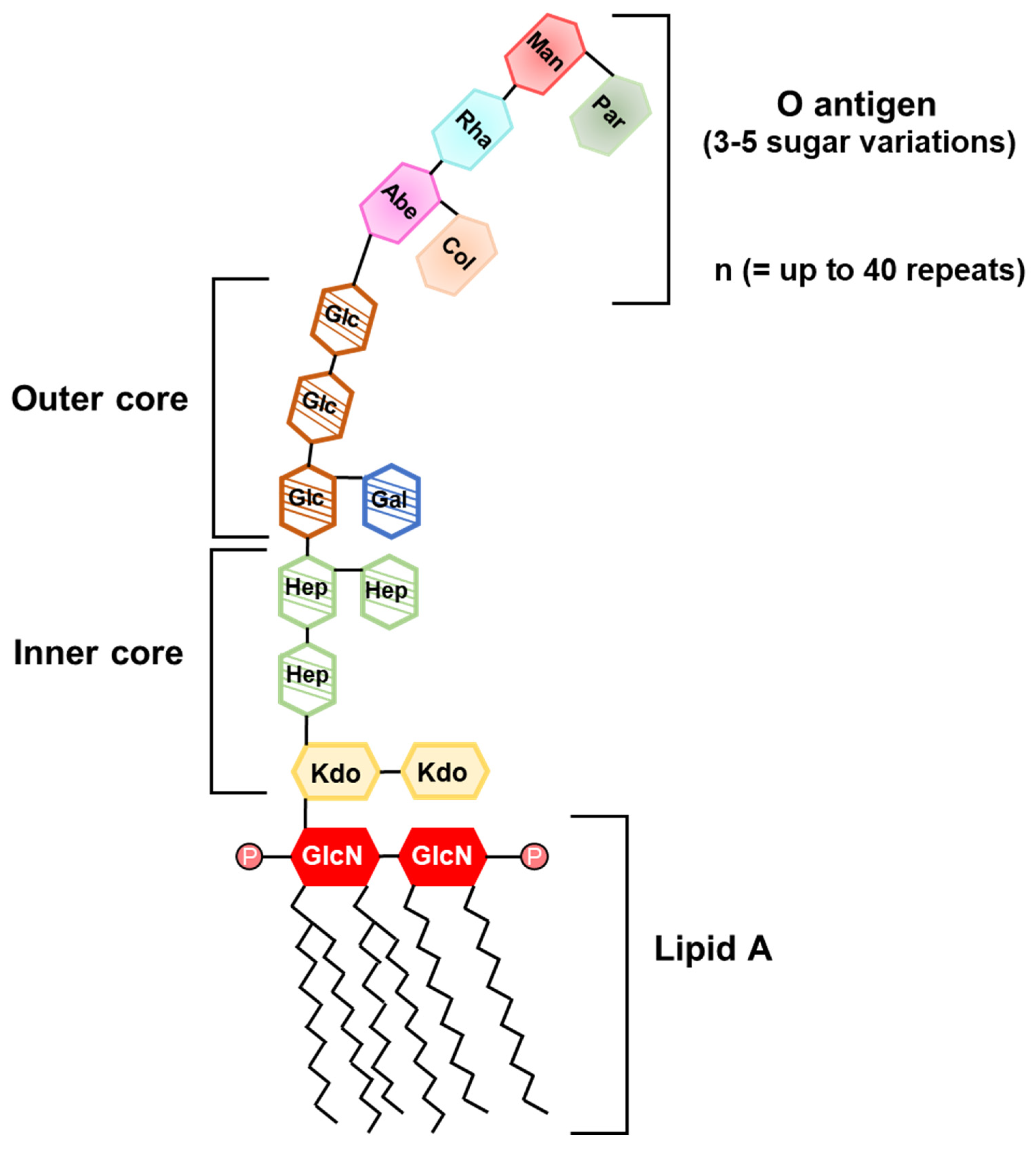
Figure 1. Illustration of lipopolysaccharide (LPS) structure. LPS is a potent immuno-stimulatory molecule of Gram-negative bacteria. It is composed of O antigen, outer or inner core polysaccharide, and lipid A. O antigen consists of repeating sugar molecules (n can be up to 40 repeats) and outer or inner core is a continuous polysaccharide chain. Composition and length of the O antigen and core polysaccharide are varied among bacterial strains. Lipid A consists of two phosphorylated glucosamines and acyl chains. The number of acyl chains and branched points in lipid A vary among bacterial species. Man, Mannose; Par, Paratose; Rha, Rhamnose; Abe, Abequose; Col, Colitose; Glc, Glucose; Gal, Galactose; Hep, Heptose; Kdo, 3-deoxy-d-manno-2-octulosonic acid; GlcN, Glucosamine; P, phosphate.
Ijms 22 05805 g001
Figure 2. Illustration of lipoteichoic acid (LTA) structure. LTA is a Gram-positive bacterial cell wall component which is responsible for stimulating immune responses of hosts. There are five types of LTA which varies among bacterial species. Gro, Glycerol; Glc, Glucose; Gal, Galactose; β-Glu, β-Glucan; AAT-Gal, 2-acetamido-4-amino-2,4,6-trideoxy-d-galactose; Gal-NAc, N-acetylgalactosamine; Rto, Ribitol; Glc-NAc, N-acetylglucosamine.
Ijms 22 05805 g002
Figure 3. Structures of Toll-like receptor (TLR) 2 heterodimers and lipoproteins (LPPs). TLRs consist of extracellular leucine-rich repeat, transmembrane helix, and intracellular Toll/interleukin-1 receptor domain. Bacterial LPPs bind to the extracellular domains of TLR. Especially, TLR2/TLR6 heterodimers recognize diacylated LPPs and TLR1/TLR2 heterodimers sense triacylated LPPs.
Ijms 22 05805 g003
Figure 4. Illustration of peptidoglycan (PGN) structure. PGN is composed of two amino sugars, N-acetylmuramic acid (NAM) and N-acetylglucosamine (NAG), and amino acids. NAM and NAG are connected by β-1,4-glycosidic linkage. The peptide chain of three to five amino acids attaches to NAM. Lysine-type PGN and diaminopimelic acid (DAP)-type PGN contain a lysine and a meso-DAP at the third position of the peptide stem and predominantly found in Gram-positive and Gram-negative bacteria, respectively.
Ijms 22 05805 g004
Figure 5. Signaling pathway of nucleotide-binding oligomerization domain (NOD) 1 and NOD2. d-glutamyl-meso-diaminopimelic acid (iE-DAP) of Gram-negative bacteria and muramyl dipeptide (MDP) of both Gram-positive and Gram-negative bacteria are recognized by NOD1 and NOD2, respectively. After activation of NODs, RICK is recruited through CARD-CARD interactions, leading to the activation of NF-κB and MAPK pathways.
Ijms 22 05805 g005
Figure 6. Signaling pathway of stimulator of interferon genes (STING). Bacterial cyclic dinucleotides (CDNs) are recognized by STING localized at endoplasmic reticulum. When STING is activated by CDNs, it is translocated from endoplasmic reticulum to the Golgi complex. This translocation triggers the STING-TBK1-IRF3 signaling cascade to increase expression of type I interferons.
Ijms 22 05805 g006
Table 1. Effects of cell wall components on bone metabolism.
MAMPsReceptorEffectsReferences
LipopolysaccharideTLR4Inducing bone loss
Inhibiting osteoclastogenesis on macrophages
Facilitating osteoclast differentiation on committed osteoclasts
Downregulating osteoblast differentiation
[28,29,30,31,32,33,34,35]
Lipoteichoic acidTLR2Healing femoral fractures in mice
Attenuating osteoclast differentiation and activating phagocytosis
Upregulating osteogenic markers and osteoblastogenesis
[36,37,38,39,40]
LipoproteinTLR2Promoting bone resorption
Upregulating osteoclast differentiation
Stimulating osteoblasts to elevate RANKL/OPG ratio
[41,42,43]
FimbriaTLR4Inducing osteoclastogenesis and bone resorption[44,45,46,47,48,49]
PeptidoglycanNOD1Enhancing osteoclastogenesis and bone resorption
Triggering osteoclast differentiation synergistically with LPS
[50,51,52,53,54,55,56]
NOD2Upregulation of bone density
Facilitating osteoblast differentiation
Diminishing osteoclastogenesis by reducing RANKL/OPG ratio
[54,57]
Table 2. Effects of secretory microbial molecules on bone metabolism.
MAMPsMechanismEffects on Bone MetabolismReferences
Short chain fatty acidsActivation of GPCRs
Inhibition of histone deacetylases
Inhibited osteoclast differentiation and function
Upregulated osteogenic factors in low dose
Attenuated osteoblast differentiation and mineralization
Prevented bone loss in various mouse models
[109,111,112,114]
Extracellular vesiclesActivation of TLR2
Induction of pro-inflammatory cytokines
Downregulated osteoblast differentiation and activity
Regulated RANKL and OPG expression in mesenchymal cells
[17]
Extracellular polysaccharidesActivation of TLR2Inhibited osteoclast differentiation from macrophages, but some EPS increased collagenolytic activity of osteoclasts
Enhanced osteoblast differentiation, but oral pathogen-derived CPS decreased proliferation of osteoblasts
[127,128,129,132]
Cyclic dinucleotidesInduction of STING-mediated IFN-βInhibited differentiation of macropahges into mature osteoclasts
Alleviated RANKL-induced bone destruction
[18]
Table 3. Benefits of probiotics on bone health.
ProbioticsBone EffectsAnimal ModelReferences
IncreaseDecrease
L. reuteri ATCC 6475BV/TV, Tb.N, Tb.Th, OCN Normal[12]
BV/TV, Tb.N, Tb.ThRANKL, TRAP5Ovariectomy[165]
BV/TV, OCN, Wnt10b Diabetic osteoporosis[166]
L. rhamnosus GGBV/TV, OCNRANKL, TNF-α, IL-17Ovariectomy[11]
Bone loss, InflammationPeriodontitis[167]
L. paracasei
and L. plantarum
BV/TV, Tb.N, Cortical bone Ovariectomy[168]
L. casei OsteolysisCalvarial resorption[169]
L. gasseri SBT2055 Bone loss, InflammationPeriodontitis[13]
L. brevis CD2 Bone loss, InflammationPeriodontitis[14]
Publisher’s Note: MDPI stays neutral with regard to jurisdictional claims in published maps and institutional affiliations.